
Abdominal Pain—Beyond Colonic Lipoma Intussusception

 , , , , ,
Abstract
1. Introduction
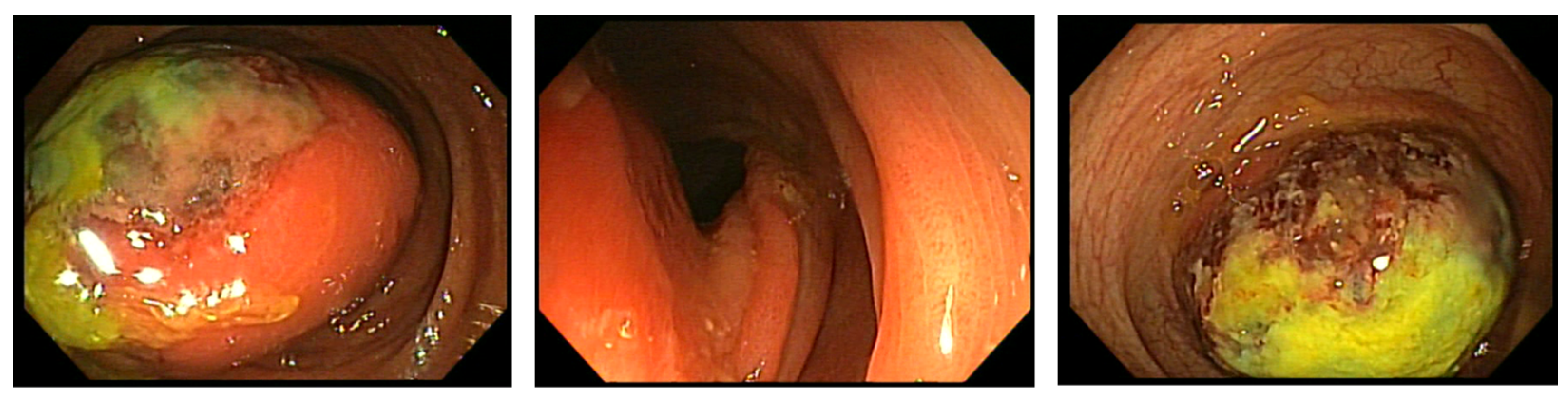
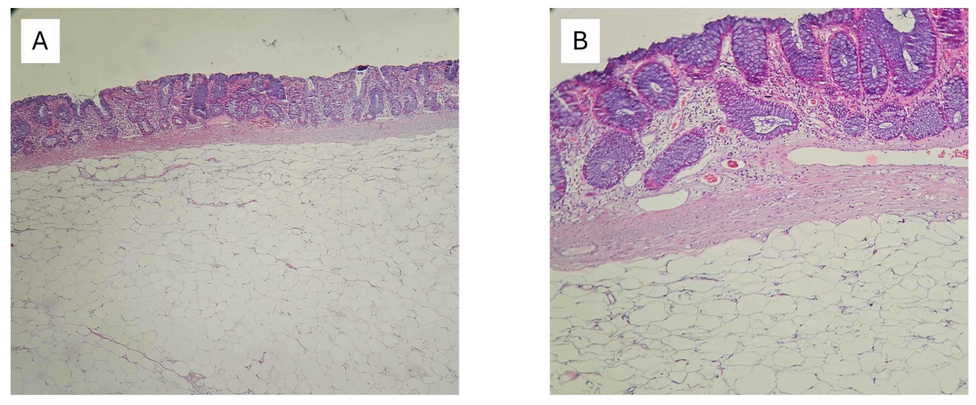
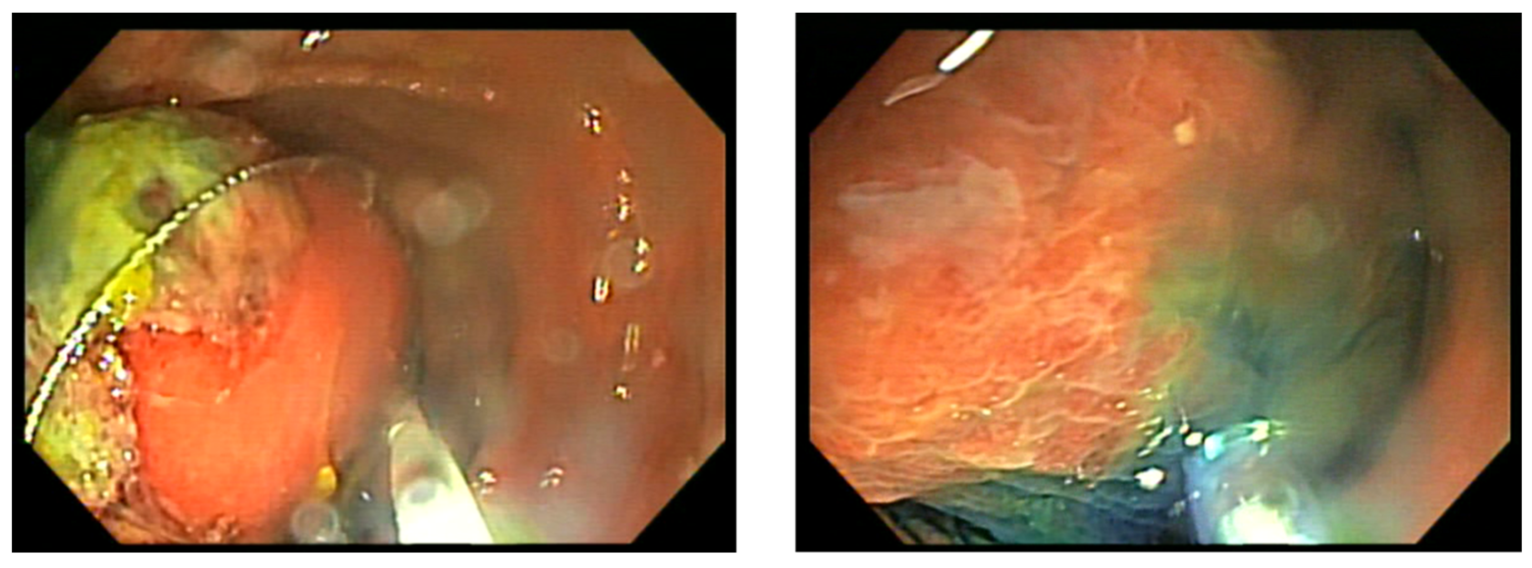
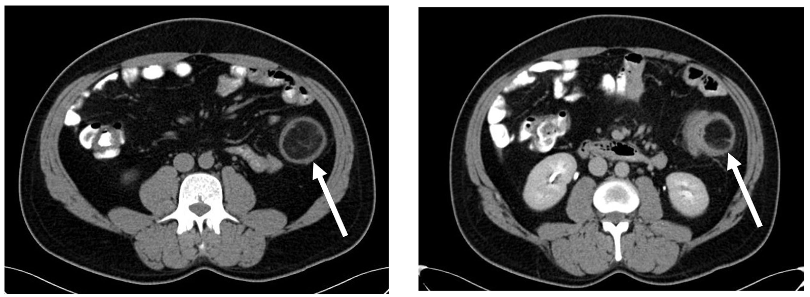
2. Case Report
3. Discussion
4. Conclusions
Author Contributions
Funding
Institutional Review Board Statement
Informed Consent Statement
Data Availability Statement
Conflicts of Interest
References
- Ryan, J.; Martin, J.E.; Pollock, D.J. Fatty tumours of the large intestine: A clinicopathological review of 13 cases. Br. J. Surg. 2005, 76, 793–796. [Google Scholar] [CrossRef]
- Zhang, H.; Cong, J.C.; Chen, C.S.; Qiao, L.; Liu, E.Q. Submucous colon lipoma: A case report and review of the literature. World J. Gastroenterol. WJG 2005, 11, 3167. [Google Scholar] [CrossRef]
- Nallamothu, G.; Adler, D.G. Large colonic lipomas. Gastroenterol. Hepatol. 2011, 7, 490–492. [Google Scholar]
- Gould, D.J.; Anne Morrison, C.; Liscum, K.R.; Silberfein, E.J. A lipoma of the transverse colon causing intermittent obstruction: A rare cause for surgical intervention. Gastroenterol. Hepatol. 2011, 7, 487–490. [Google Scholar]
- Farfour, A.N.; AbuOmar, N.A.; Alsohaibani, F.I. Large lipoma of the ascending colon: A case report and review of literature. J. Surg. Case Rep. 2020, 2020, rjaa354. [Google Scholar] [CrossRef]
- Rogers, S.O.; Lee, M.C.; Ashley, S.W. Giant colonic lipoma as lead point for intermittent colo-colonic intussusception. Surgery 2002, 131, 687–688. [Google Scholar] [CrossRef]
- Paškauskas, S.; Latkauskas, T.; Valeikaite, G.; Paršeliunas, A.; Švagždys, S.; Saladžinskas, Ž.; Tamelis, A.; Pavalkis, D. Colonic intussusception caused by colonic lipoma: A case report. Medicina 2010, 46, 477. [Google Scholar] [CrossRef] [PubMed]
- Miloudi, N.; Hefaiedh, R.; Khalfallah, M. Giant lipoma of the transverse colon causing colo-colonic intussusceptions. J. Visc. Surg. 2012, 149, 421–422. [Google Scholar] [CrossRef] [PubMed]
- Zhou, X.C.; Hu, K.Q.; Jiang, Y. A 4-cm lipoma of the transverse colon causing colonic intussusception: A case report and literature review. Oncol. Lett. 2014, 8, 1090–1092. [Google Scholar] [CrossRef] [PubMed]
- Vishnu, J.; Kapil, N.; Pe, V.; Patnaik, P. Giant colonic lipoma remains a surgeon’s domain: A report of two cases. Cureus 2023, 15, e43488. [Google Scholar] [CrossRef]
- Sapalidis, K.; Laskou, S.; Kosmidis, C.; Passos, I.; Mantalobas, S.; Michalopoulos, N.; Alexandrou, V.; Koimtzis, G.; Koulouris, C.; Katsaounis, A.; et al. Symptomatic colonic lipomas: Report of two cases and a review of the literature. SAGE Open Med. Case Rep. 2019, 7, 2050313X19830477. [Google Scholar] [CrossRef]
- Bronswijk, M.; Vandenbroucke, A.M.; Bossuyt, P. Endoscopic treatment of large symptomatic colon lipomas: A systematic review of efficacy and safety. United Eur. Gastroenterol. J. 2020, 8, 1147–1154. [Google Scholar] [CrossRef]
- An, H.H.; Duong, T.T.; Van Truong, N.; Van Quoc, L.; Son, V.N.; Thang, N.V.P.; Van Thach, N.; Cuong, D.D.; Duc, N.M. A large lipoma of the descending colon: A rare case report. Radiol. Case Rep. 2021, 16, 3396–3399. [Google Scholar] [CrossRef]
- Târcoveanu, E.; Chifan, M.; Veisa, E.; Epure, O.; Florea, N. Colonic lipoma. Chirurgia 2000, 95, 353–357. [Google Scholar]
- Ullah, S.; Ahmed, H.; Jehangir, E. Giant colonic lipoma presenting with intermittent intestinal obstruction. J. Coll. Physicians Surg. Pak. 2012, 22, 792–793. [Google Scholar]
- Crocetti, D.; Sapienza, P.; Sterpetti, A.V.; Paliotta, A.; Gori, A.D.; Pedullà, G.; Toma, G.D. Surgery for Symptomatic Colon Lipoma: A Systematic Review of the Literature. Anticancer Res. 2014, 34, 6271–6276. [Google Scholar]
- Popa, B.; Ilie, M.; Sandru, V.; Hortopan, M.; Beuran, M.; Constantinescu, G. Endoscopic resection of a giant colonic lipoma causing severe anemia. J. Gastrointestin. Liver Dis. 2015, 24, 142. [Google Scholar] [CrossRef] [PubMed]
- Yano, Y.; Yamaguchi, T.; Imai, M.; Toyama, K.; Toguchi, K.; Hirabayashi, K. A Case of Giant Lipoma of the Transverse Colon with Intussusception Treated by Laparoscopic Surgery: A Review of 36 Cases Reported in the Japanese Literature. Nihon Gekakei Rengo Gakkaishi J. Jpn. Coll. Surg. 2019, 44, 753–760. [Google Scholar] [CrossRef]
- Ozen, O.; Guler, Y.; Yuksel, Y. Giant colonic lipoma causing intussusception: CT scan and clinical findings. Pan Afr. Med. J. 2019, 32, 27. [Google Scholar] [PubMed]
- Sagar, J.; Kumar, V.; Shah, D.K. Meckel’s diverticulum: A systematic review. J. R. Soc. Med. 2006, 99, 501–505. [Google Scholar] [CrossRef] [PubMed]
- Hansen, C.C.; Søreide, K. Systematic review of epidemiology, presentation, and management of Meckel’s diverticulum in the 21st century. Medicine 2018, 97, e12154. [Google Scholar] [CrossRef]
- Zani, A.; Eaton, S.; Rees, C.M.; Pierro, A. Incidentally detected Meckel diverticulum: To resect or not to resect? Ann. Surg. 2008, 247, 276–281. [Google Scholar] [CrossRef] [PubMed]
- UpToDate. Meckel’s Diverticulum. Available online: https://www.uptodate.com/contents/meckels-diverticulum (accessed on 30 December 2023).
- Park, J.J.; Wolff, B.G.; Tollefson, M.K.; Walsh, E.E.; Larson, D.R. Meckel diverticulum: The Mayo Clinic experience with 1476 patients (1950–2002). Ann. Surg. 2005, 241, 529–533. [Google Scholar] [CrossRef]
- Dumper, J.; Mackenzie, S.; Mitchell, P.; Sutherland, F.; Quan, M.L.; Mew, D. Complications of Meckel’s diverticula in adults. Can. J. Surg. 2006, 49, 353–357. [Google Scholar]
- Groebli, Y.; Bertin, D.; Morel, P. Meckel’s diverticulum in adults: Retrospective analysis of 119 cases and historical review. Eur. J. Surg. 2001, 167, 518–524. [Google Scholar] [PubMed]
- Huang, C.C.; Lai, M.W.; Hwang, F.M.; Yeh, Y.C.; Chen, S.Y.; Kong, M.S.; Lai, J.Y.; Chen, J.C.; Ming, Y.C. Diverse presentations in pediatric Meckel’s diverticulum: A review of 100 cases. Pediatr. Neonatol. 2014, 55, 369–375. [Google Scholar] [CrossRef]
- Mackey, W.C.; Dineen, P. A fifty year experience with Meckel’s diverticulum. Surg. Gynecol. Obstet. 1983, 156, 56–64. [Google Scholar] [PubMed]
- Zulfikaroglu, B.; Ozalp, N.; Zulfikaroglu, E.; Ozmen, M.M.; Tez, M.; Koc, M. Is incidental Meckel’s diverticulum resected safely? N. Z. Med. J. 2008, 121, 39–44. [Google Scholar]
- Robijn, J.; Sebrechts, E.; Miserez, M. Management of incidentally found Meckel’s diverticulum a new approach: Resection based on a Risk Score. Acta Chir. Belg. 2006, 106, 467–470. [Google Scholar] [CrossRef]
- Mukai, M.; Takamatsu, H.; Noguchi, H.; Fukushige, T.; Tahara, H.; Kaji, T. Does the external appearance of a Meckel’s diverticulum assist in choice of the laparoscopic procedure? Pediatr. Surg. Int. 2002, 18, 231–233. [Google Scholar] [CrossRef]
- Varcoe, R.L.; Wong, S.W.; Taylor, C.F.; Newstead, G.L. Diverticulectomy is inadequate treatment for short Meckel’s diverticulum with heterotopic mucosa. ANZ J. Surg. 2004, 74, 869–872. [Google Scholar] [CrossRef] [PubMed]
- Blouhos, K.; Boulas, K.A.; Tsalis, K.; Barettas, N.; Paraskeva, A.; Kariotis, I.; Keskinis, C.; Hatzigeorgiadis, A. Meckel’s diverticulum in adults: Surgical concerns. Front. Surg. 2018, 5, 55. [Google Scholar] [CrossRef] [PubMed]


Disclaimer/Publisher’s Note: The statements, opinions and data contained in all publications are solely those of the individual author(s) and contributor(s) and not of MDPI and/or the editor(s). MDPI and/or the editor(s) disclaim responsibility for any injury to people or property resulting from any ideas, methods, instructions or products referred to in the content. |
© 2024 by the authors. Licensee MDPI, Basel, Switzerland. This article is an open access article distributed under the terms and conditions of the Creative Commons Attribution (CC BY) license (https://creativecommons.org/licenses/by/4.0/).
Share and Cite
Dascalu, R.-I.; Constantinescu, G.; Scafa, A.; Plotogea, O.-M.; Sandru, V.; Rinja, E.M.; Jichitu, A.; Ilie, M. Abdominal Pain—Beyond Colonic Lipoma Intussusception. Gastroenterol. Insights 2024, 15, 213-220. https://doi.org/10.3390/gastroent15010015
Dascalu R-I, Constantinescu G, Scafa A, Plotogea O-M, Sandru V, Rinja EM, Jichitu A, Ilie M. Abdominal Pain—Beyond Colonic Lipoma Intussusception. Gastroenterology Insights. 2024; 15(1):213-220. https://doi.org/10.3390/gastroent15010015
Chicago/Turabian StyleDascalu, Raluca-Ioana, Gabriel Constantinescu, Alexandru Scafa, Oana-Mihaela Plotogea, Vasile Sandru, Ecaterina Mihaela Rinja, Alexandra Jichitu, and Madalina Ilie. 2024. "Abdominal Pain—Beyond Colonic Lipoma Intussusception" Gastroenterology Insights 15, no. 1: 213-220. https://doi.org/10.3390/gastroent15010015
APA StyleDascalu, R.-I., Constantinescu, G., Scafa, A., Plotogea, O.-M., Sandru, V., Rinja, E. M., Jichitu, A., & Ilie, M. (2024). Abdominal Pain—Beyond Colonic Lipoma Intussusception. Gastroenterology Insights, 15(1), 213-220. https://doi.org/10.3390/gastroent15010015