Abstract
The aim of this study was to investigate the predictive role of preoperative pancreatic density and muscular mass, assessed via CT imaging, in patients undergoing duodenocephalopancreasectomy, specifically in relation to the occurrence of postoperative pancreatic fistula (POPF). A retrospective analysis was conducted on a cohort of 57 consecutive patients who had been diagnosed with cephalo-pancreatic disease and had undergone duodenocephalopancreasectomy in the last five years. The most prevalent pathologies observed were ductal adenocarcinoma (29.2%), biliary adenocarcinoma (12.9%), and duodenal and papillary adenocarcinoma (13.9%). We collected information about age, sex, histopathological findings, type of surgery, presence or absence of pancreatic fistula, pancreatic density on preoperative CT images, and muscular area, calculated at the level of the L3 vertebra using “3D Slicer” software. Our data show that 28% of patients developed a pancreatic fistula, with an average attenuation of pancreatic density of 27 HU, which was lower than that observed in the non-fistula group (33.31 HU). However, statistical analysis did not reveal a significant association between low pancreatic density and fistula development. Therefore, our findings do not establish a significant association between pancreatic fistula and pancreatic density, aligning with the existing literature on the subject.
1. Introduction
Duodenocephalopancreasectomy (DCP) is a very complex surgical procedure carried out for neoplasms of the pancreatic head, uncinate process, ampulla of Vater, duodenum, and distal common bile duct. It is also recommended in some types of chronic pancreatitis and in severe pancreatic trauma [1].
Postoperative complications associated with DCP include intra-abdominal abscess formation, surgical wound infection, postoperative hemorrhage, delayed gastric emptying, and pancreatic fistula formation [1]. Albeit less common, acute pancreatitis can also occur as a late complication of the surgery. For example, a case in which a patient developed inflammation and edema of the pancreatic stump, leading to hospitalization for cardio-respiratory failure, was reported. In this particular case, an additional necrosectomy procedure was deemed necessary, and utmost caution was exercised to prevent any harm to the anastomotic site [2].
According to the guidelines issued by the International Study Group for Pancreatic Surgery (ISGPS) in 2005, postoperative pancreatic fistula (POPF) is defined as an abnormal communication between the pancreatic epithelium and another epithelium, resulting in the transfer of fluid containingpancreatic enzymes [3]. In the updated version released in 2016, the ISGPS categorizes pancreatic fistulas into three stages of severity, as follows. (1) POPF A: Asymptomatic extravasation of pancreatic juice. This occurrence is not considered a real fistula and has no evident clinical impact or relapse in the postoperative period. (2) POPF B: This requires administration of antibiotics and prolonged insertion of surgical drains at the operative site for an extended period (usually 21 days). It may also become necessary to use more invasive procedures to drain evolving collections; and (3) POPF C: This indicates organ failure that requires surgical reintervention to recreate the pancreatic anastomosis or complete pancreatectomy.
The development of a pancreatic fistula is attributed to dehiscence of the pancreatic anastomosis, which is caused by several factors that are currently under study. Understanding these mechanisms is crucial for early identification and correction to prevent subsequent complications. This event, especially in its severe forms, is often associated with postoperative death, with a frequency of up to 50% [4]. Therefore, current studies focus on early identification of the causes of the anastomotic dehiscence to prevent its occurrence.
A study by Deng et al. revealed the presence of pancreatic fistula in 529 patients (28.5%) one month after DCP. Among these cases, 79 belonged to group A (14.9%), 60 to group B (11.3%), and 12 to group C (2.3%) [4]. Clinically, the most severe forms present with pain, abdominal distension, changes in bowel habit, fever, and elevated inflammatory markers.
The diagnosis of POPF is mainly based on clinical presentation and biochemical analysis of drained fluid obtained on the third day after surgery. A positive result indicates amylase levels exceeding three times those found in the blood. Although diagnostic imaging is not essential for a correct diagnosis, it can be very useful in identifying and evaluating the extent of developing collections, as well as confirming proper placement of surgical drains [5].
Multiple studies have proposed that reduced muscle mass, specifically sarcopenia, is another risk factor associated with worse outcomes, including longer hospitalization, functional limitations, and deteriorated quality of life (QoL) [6,7,8]. In 2018, the European Working Group on Sarcopenia in Older People redefined sarcopenia as a progressive and generalized skeletal muscle disorder associated with a higher probability of adverse effects, such as falls, fractures, physical disability, and death. In general, sarcopenia is considered severe when it causes a decline in the physical performance of an individual [9]. Finally, other studies have shown that pancreatic density, assessed by histological investigations, is a significant risk factor for the development of pancreatic fistulas following pancreatectomy [4,10,11].
The aim of this study was to evaluate the effectiveness of CT in measuring the density of the pancreatic parenchyma in patients undergoing DCP. In particular, we sought to identify areas in the glandular tissue replaced by adipose tissue prior to surgery, as these regions are more prone to postoperative complications, such as pancreatic fistulas. Lastly, within the same cohort of patients and using the same imaging technique, we investigated the correlation between sarcopenia and the development of pancreatic fistulas.
2. Materials and Methods
Between October 2016 and May 2021, a total of 61 patients who had been diagnosed with pancreatic or biliary malignancies underwent DCP surgery at our institution. To assess the risk of developing a pancreatic or biliary fistula, we conducted a retrospective study that systematically evaluated the preoperative CT imaging of these patients. Our study included patients that underwent DCP with or without pancreatico-enteric anastomosis for malignant and benign pathology, including ductal adenocarcinoma, biliary adenocarcinoma, duodenal or papillary adenocarcinoma, neuroendocrine tumors, chronic pancreatitis, high-grade adenoma, mixed carcinoma, acinar adenocarcinoma, and mucinous papillary intraductal neoplasms. These pathologies were not equally distributed, with ductal adenocarcinoma being the most frequent (29.2%), followed by papillary adenocarcinoma (13.9%) and biliary adenocarcinoma (12.9%) (Table 1).

Table 1.
Frequency of different histological types and their respective incidences of postoperative pancreatic fistula (POPF) cases.
Patients that had undergone total pancreatectomy which required biliary and digestive but not pancreatic anastomosis, as well as patients undergoing distal pancreatectomy, were excluded from the study. Four patients were further excluded due to the unavailability of relevant preoperative imaging records.
All images were acquired in the preoperative phase, either at the Department of Radiology or the Emergency Room, using a 256-, 64-, or 32-slice CT scanner. Subsequently, two consultants examined the images with the aim of evaluating both pancreatic density and muscle mass.
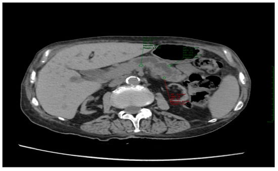
To calculate pancreatic density, two separate regions of interest (ROIs) were measured using a circular graphic tool with a radius ranging from 2 to 5 mm. The measurements were made on the pre-contrast study at the level of the planned resection site (identified on the postoperative CT) (Figure 1).

Figure 1.
The axial Computed Tomography static image depicts the pancreatic body, with green-highlighted regions of interest (ROIs) placed within the parenchyma for assessing pancreatic density. The ROIs exclude the dilated pancreatic duct to ensure accurate evaluation.
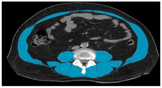
To assess muscle mass, we used the “3D Slicer” software to extract a static image from the preoperative unenhanced CT, specifically at the level of the L3 inferior endplate, excluding any inter-muscle fat striae [12]. The software facilitated the selection and highlighting of tissues according to their density (Figure 2).

Figure 2.
Axial CT image at the level of the L3 inferior endplate. The muscle bulk was highlighted using a 3D Slicer software, excluding any inter-muscle fat striae.
For the identification of muscular tissues, a density range was set between −30 and +150 HU. Following this, the software processed the total area encompassing the highlighted tissues.
Discrete quantitative data are represented by absolute and relative frequency values, while quantitative continuous data are summarized using average values.
To analyze the predictive values (e.g., age, gender, pancreatic density, and muscular area), the chi-square test was employed. Statistical significance was determined at a p-value < 0.050.
3. Results
During the study period, we collected data from 61 patients subjected to DCP surgery. Among them, four patients were excluded due to the lack of a preoperative CT for the measurement of pancreatic density. Therefore, the analysis was conducted on a total of 57 patients (35 males and 22 females) (Table 2).

Table 2.
Gender distribution, age, and frequency of pancreatic fistula formation in the study population.
The study revealed an overall incidence of postoperative pancreatic fistula of 28.07% (16/57). Pancreatic density measurements in the study cohort ranged from −17 HU to +51 HU, with an average value of 32.07 HU (Table 3). The muscular area showed values between a minimum of 38.73 and a maximum of 217 , with an average value of 95.29 (Table 4).

Table 3.
Average values and minimum and maximum values of pancreatic density (HU) in the group of patients with or without fistula.

Table 4.
Average values and minimum and maximum values of muscular area (cm2) in the group of patients with or without fistula.
Furthermore, when analyzing the 16 patients that developed POPF, the following findings were documented: (i) the median age was 72.5 years; (ii) there were 11 males and 5 females; (iii) the average pancreatic density value was 27.08 HU, ranging from −17 HU to +51 HU; and (iv) the average muscular area was 98.11 cm2, with a range of 61.90 cm2 to 154.50 cm2.
Conversely, for the group of patients who did not develop postoperative POPF, the following observations were made: (a) a median age of 70.1 years, ranging from 49 to 84 years; (b) an average value of pancreatic density of 33.31 HU, ranging from +10 HU to +51 HU; and (c) an average value of muscular area of 94.2 cm2, with a range of 38.73 cm2 to 217 cm2.
Statistical univariate analysis using the chi-square test did not reveal any statistical significance for the analyzed variables (i.e., age, sex, pancreatic density, muscular area).
4. Discussion
DCP has long been associated with a very high morbidity and peri/postoperative mortality rates. The majority of patients with pancreatic cancer die within the first year of diagnosis, particularly those with tumors affecting the body and the tail [13].
Both DCP and total pancreatectomy are considered radical surgery procedures, even though the latter is now rarely performed. Both procedures require an adequate nutritional and hydration state of the patient, as well as careful management of any concurrent coagulopathy.
DCP can be performed using either an open surgery technique or, more commonly, a laparoscopic approach, which was first described by Cuschieri et al. [14]. Following this, Nigri et al. [15] conducted a meta-analysis comparing the main postoperative complications of both procedures. The study revealed that laparoscopy was associated with a reduced incidence of complications, faster resumption of oral feeding, and shorter postoperative hospital stay.
There are two variants of DCP. The classic approach, known as the Whipple technique, consists of a complete resection of the gastric antrum, including the pylorus. By contrast, the “Longmire-Traverso” variant, which preserves the pylorus with a resection margin located 2–3 cm distal to it, has been shown to substantially improve patients’ QoL after surgery [16].
Following resection, in order to restore gastro-intestinal transit as well as exocrine hepatic and pancreatic functions, anastomoses are usually performed at the level of the pancreas, biliary tract, and stomach. Although there are several variations of the pancreatico-jejunal anastomosis, each aiming to enhance its efficacy, no single approach has demonstrated clear superiority over the others [17]. Regardless of the approach, it is important, after surgery, to place surgical drains within the surgical bed to facilitate the detection of potential anastomotic dehiscence by analyzing the drained fluid for elevated amylase levels.
Another crucial aspect that needs to be carefully taken into account to adequately plan for the intraoperative phase of DCP is the identification of the risk factors associated with postoperative complications. Therefore, the aim of this study was to investigate the potential role of pancreatic density and muscle mass as risk factors for the development of POPF.
The main risk factors for POPF can be classified into three groups: preoperative, intraoperative, and postoperative [18]:
- Preoperative risk factors include the following:
- i.
- Obesity (BMI ≥ 30): obese patients undergoing minimally invasive pancreatic surgery (MIDP) have shown higher rates of intraoperative hemorrhage and longer operative times compared to normal-weight patients [19];
- ii.
- Anemia: the risk of developing MIDP is significantly higher in patients with anemia (Hb 12 < mg/dL) compared to those with normal Hb (31.0 % vs. 15.9 %) [20];
- iii.
- Biliary or ascitic fluid infections: these infections, especially when caused by Klebsiella or commensal anaerobes, have an unfavorable impact on the development of POPF [21,22,23].
- Among intraoperative risk factors, pancreatic stiffness is considered the best predictor of POPF. Technically, it is an intraoperative assessment of pancreatic consistency made by the surgeon during the operation [11]. Since it relies on the surgeon’s experience, pancreatic stiffness is considered a subjective parameter [4]. An increase in pancreatic consistency can occur due to fibrosis or pathological conditions that lead to chronic inflammation and the replacement of glandular tissue with fibrosis [4]. Conversely, a decrease in pancreatic consistency, resulting from progressive fatty infiltration, is a natural process associated with senile involution. This decrease correlates with the body mass index (BMI) and can also be linked to metabolic disorders such as abdominal obesity, insulin resistance, type 2 diabetes mellitus, dyslipidemia, hypertension, and metabolic syndrome. Similarly, autoimmune pathologies, viral infections, drug treatments, and toxic substances (including alcohol) may all contribute to the replacement of pancreatic tissue with fat [24,25].
An additional intraoperative risk factor is represented by the type of anastomosis. For example, the “modified Blumgart anastomosis”, a variant to traditional anastomoses which involves a “tension free” suture between the jejunum and the pancreatic stump combined with a ductulo-mucosal suture, has been shown to reduce the risk of fistula occurrence compared to traditional anastomosis in patients undergoing DCP surgery (25.4% vs. 10.5%) [26].
Finally, a further intraoperative risk factor is represented by the hypoperfusion of the surgical site. Indeed, several studies have shown that intraoperative hemorrhage is correlated with an increased risk of anastomotic dehiscence and subsequent fistula formation [27,28].
- 3.
- Among the postoperative factors, fatty infiltration of the pancreas is the primary risk factor for developing a pancreatic fistula. However, this risk factor can only be identified after surgery through histological analysis of the surgical specimen. There are several signs related to fatty infiltration, including the following:
- (a)
- Soft consistency of the pancreas: this is subjectively estimated by the surgeon during the surgical procedure [10,11,29];
- (b)
- CT value: this represents the average CT density of the pancreas before surgery [11]. This parameter directly assesses the extent of fatty infiltration in the pancreas. Its significance lies in its ability to predict the occurrence of POPF prior to surgery, and it is a non-invasive method [4].
- (c)
- The diameter of main pancreatic duct (MPD): a recent study has shown that a cut-off < 4 mm is associated with a higher risk of developing post-DCP fistula [30].
In addition to risk factors, there are also positive prognostic factors that can be identified. One intraoperative factor that correlates with a lower risk of fistula development is the placement of an intraoperative pancreatic drain, which helps improve biliary drainage in the postoperative phase. In this regard, a meta-analysis has demonstrated a statistically significant reduction in POPF when a pancreatic duct stent is inserted compared to cases where it is not used [31]. Therefore, it is advisable to consider its introduction in all patients undergoing this procedure.
Certain tumor histotypes can also be considered positive prognostic factors for fistula development, in particular pancreatic ductal adenocarcinoma (PDAC). Despite PDAC having an unfavorable overall prognosis, the presence of fibrous tumor cells within the parenchymal background actually hinders fistula formation [32].
In addition to the aforementioned factors, recent studies have highlighted the benefits of chemotherapy and neoadjuvant chemoradiotherapy in the treatment of PDAC. These treatments have shown significant improvements in overall survival and a reduction in the rate of postoperative pancreatic fistula formation compared to immediate surgery alone [33,34,35], which has been ascribed to the beneficial effects on the parenchyma caused by radio-induced fibrosis. In contrast, tumors of the distal bile duct (DBDC) are associated with a higher incidence of soft pancreatic parenchyma [36]. Similarly, Vater’s ampullary carcinomas (VACs) are linked to an increased risk of developing postoperative pancreatic fistula, probably as a consequence of jaundice or elevated preoperative serum bilirubin levels that may induce an increase in serum proinflammatory cytokines and endotoxin [37,38].
To obtain a preoperative and non-invasive assessment of the pancreatic consistency, the average CT pancreatic density (CT value) associated with pancreatic texture is deemed a reliable factor [4]. Specifically, a significant correlation between the creation of the fistula and a CT value below 40 UH has been reported [11]. The reason behind this association is that adipose infiltration predisposes patients to weaker sutures, increasing the risk of dehiscence, whereas glandular and fibrotic tissues contribute to more stable sutures.
In our study, which included a cohort of 61 patients, 16 developed a postoperative fistula, with an incidence of 28.07%. Among this group, the pancreatic density had a reduced average value of 27.08 HU in comparison with the group that did not develop this complication (average value of 33.31 HU). This is in good agreement with Deng et al. [4], where a CT value below 30 HU was identified as a predictive factor for POPF. Although our study reveals a clear disparity in pancreatic tissue attenuation between the two groups, this difference did not reach statistical significance (p > 0.05), presumably due to the small sample size of the study.
The second arm of our study focused on the association between sarcopenia—evaluated as muscular area on CT—and the development of postoperative complications.
Muscle mass undergoes changes throughout an individual’s life, peaking during youth, stabilizing in adulthood, and declining with age. Genetic factors and lifestyle can promote the loss of muscle mass and the progression toward functional deficit and disability. However, engaging in regular physical activity and maintaining a healthy diet can help slow down this process.
Sarcopenia can be classified into two types: primary and secondary. While primary sarcopenia is an age-related condition that occurs in the absence of other evident causes, secondary sarcopenia is associated with additional factors other than age, such as systemic diseases, lifestyle choices, immobility, inadequate nutrient intake, anorexia, and malabsorption.
There are several methods for estimating muscle mass. CT can provide at least two of these:
- Total abdominal muscle area (TAMA): This method calculates the cross-sectional area of the abdominal and paraspinal muscles at the level of the inferior endplate of L3. It is a reliable indicator of overall muscle mass [39,40], and its results can be used to predict prognosis [41,42].
- Total psoas area (TPA): This consists of measuring the psoas muscle area bilaterally at the level of L3, along with the muscles of the middle third of the leg. However, it is considered less accurate in assessing overall muscle mass when compared to other methods [43].
There is no consensus on a specific cut-off value of TAMA that indicates sarcopenia. In a study by Martin et al. [44], sarcopenia was diagnosed in patients with a skeletal mass index (SMI) < 43 cm2/m2 (in males with a BMI < 25 kg/m2) and with an SMI < 53 cm2/m2 (in males with a BMI ≥ 25 kg/m2). For females, the cut-off value is an SMI < 41 cm2/m2 [6].
Sarcopenia is known to be associated with a worse recovery from stressful events, such as chemotherapy, in cancer patients, or major surgical procedures. Likewise, DCP is linked to a slow recovery process because of inefficient tissue repair, which can lead to several complications, including POPF [4].
Our study did not find a statistically significant association between sarcopenia and development of postoperative complications. The average muscular tissue area in the group that developed a fistula was 98.11 cm2, while the group without a fistula had an average value of 94.20 cm2. These findings contradict the current literature, which generally associates sarcopenia with an increased incidence of fistula formation.
Similar to our results, studies by Yang et al. [11] and Deng et al. [4] did not show any significant correlation. However, Pecorelli’s group demonstrated that in a cohort of 202 patients with PADC who underwent pancreatic resection, the preoperative assessment of muscle mass played a role as an independent predictor of fistula formation [45].
5. Conclusions
Even though our data do not point to a significant association between pancreatic fistula formation and pancreatic density as an independent predictor, they are in line with the existing literature.
The major limitation of our study lies in its retrospective nature and consequently the small sample size. In addition, the exclusion of patients undergoing other types of pancreatic resection surgery, such as total pancreatectomy and left spleen pancreatectomy, further limited the size of our cohort. To address these limitations, we propose continuing prospectively enrolling patients who meet the aforementioned inclusion criteria. This will allow us to obtain a larger sample size and a more accurate statistical meta-analysis. Another approach to overcoming this limitation is to include patients from other hospitals who meet the inclusion criteria required in our study. This would not only increase the sample size but also provide a multicentric approach. Lastly, obtaining anamnestic and laboratory data during the enrolment process, in addition to preoperative imaging, would provide a more comprehensive clinical picture of the patient, which would help highlight any correlations between the data obtained and the risk of developing a pancreatic fistula, thus identifying predisposing factors for the onset of this complication in DCP. In this regard, a previous study analyzing the correlation between markers of local pancreatic inflammation, such as serum lipase, and measurable systemic response, such as IL-6, with the risk of developing postoperative pancreatic fistula (POPF) found that patients with elevated levels of both markers had a higher rate of fistula development compared to those within normal limits [46]. This suggests that the use of these biomarkers as predictors of risk could aid in the early detection of POPF and prompt initiation of further treatment to prevent potentially fatal complications.
Author Contributions
Conceptualization, methodology, investigation, writing, original draft and visualization: N.C. and C.G.; Conceptualization, methodology, investigation, writing, review and editing and supervision: C.B.; Conceptualization, methodology, investigation, writing and original draft: T.B.; Writing—review and editing: P.B. and A.A.-O.; Writing—review and editing and supervision: M.S.; Resources and supervision: R.R.; Resources, writing—review and editing, and supervision: A.C. All authors have read and agreed to the published version of the manuscript.
Funding
This research received no external funding.
Institutional Review Board Statement
The study did not require ethical approval.
Informed Consent Statement
Informed consent was obtained from all subjects involved in the study.
Data Availability Statement
Not applicable.
Conflicts of Interest
The authors declare no conflict of interest.
References
- D’Cruz, J.R.; Misra, S.; Shamsudeen, S. Pancreaticoduodenectomy. In StatPearls; StatPearls Publishing: St. Petersburg, FL, USA, 2022; pp. 7–11. [Google Scholar]
- Ashitomi, Y.; Sugawara, S.; Takahashi, R.; Ashino, K.; Watanabe, T.; Hachiya, O.; Kimura, W. Severe acute pancreatitis 5 years after pancreaticoduodenectomy: A case report. Int. J. Surg. Case Rep. 2019, 61, 99–102. [Google Scholar] [CrossRef] [PubMed]
- Pulvirenti, A.; Ramera, M.; Bassi, C. Modifications in the International Study Group for Pancreatic Surgery (ISGPS) definition of postoperative pancreatic fistula. Transl. Gastroenterol. Hepatol. 2017, 2, 107. [Google Scholar] [CrossRef]
- Deng, Y.; Zhao, B.; Yang, M.; Li, C.; Zhang, L. Association Between the Incidence of Pancreatic Fistula After Pancreaticoduodenectomy and the Degree of Pancreatic Fibrosis. J. Gastrointest. Surg. 2018, 22, 438–443. [Google Scholar] [CrossRef] [PubMed]
- Bassi, C.; Dervenis, C.; Butturini, G.; Fingerhut, A.; Yeo, C.; Izbicki, J.; Neoptolemos, J.; Sarr, M.; Traverso, W.; Buchler, M.; et al. Postoperative pancreatic fistula: An international study group (ISGPF) definition. Surgery 2005, 138, 8–13. [Google Scholar] [CrossRef] [PubMed]
- Nishida, Y.; Kato, Y.; Kudo, M.; Aizawa, H.; Okubo, S.; Takahashi, D.; Nakayama, Y.; Kitaguchi, K.; Gotohda, N.; Takahashi, S.; et al. Preoperative Sarcopenia Strongly Influences the Risk of Postoperative Pancreatic Fistula Formation After Pancreaticoduodenectomy. J. Gastrointest. Surg. 2016, 20, 1586–1594. [Google Scholar] [CrossRef] [PubMed]
- Ibrahim, K.; May, C.; Patel, H.P.; Baxter, M.; Sayer, A.A.; Roberts, H. A feasibility study of implementing grip strength measurement into routine hospital practice (GRImP): Study protocol. Pilot Feasibility Study 2016, 2, 27. [Google Scholar] [CrossRef]
- Leong, D.P.; Teo, K.K.; Rangarajan, S.; Lopez-Jaramillo, P.; Avezum, A.; Orlandini, A.; Seron, P.; Ahmed, S.H.; Rosengren, A.; Kelishadi, R.; et al. Prognostic value of grip strength: Findings from the Prospective Urban Rural Epidemiology (PURE) study. Lancet 2015, 386, 266–273. [Google Scholar] [CrossRef]
- Cruz-Jentoft, A.J.; Bahat, G.; Bauer, J.; Boirie, Y.; Bruyère, O.; Cederholm, T.; Cooper, C.; Landi, F.; Rolland, Y.; Sayer, A.A.; et al. Sarcopenia: Revised European consensus on definition and diagnosis. Age Ageing 2019, 48, 16–31. [Google Scholar] [CrossRef]
- Jin, J.; Xiong, G.; Li, J.; Guo, X.; Wang, M.; Li, Z.; Zhu, F.; Qin, R. Predictive factors of postoperative pancreatic fistula after laparoscopic pancreatoduodenectomy. Ann. Transl. Med. 2021, 9, 41. [Google Scholar] [CrossRef]
- Yang, M.W.; Deng, Y.; Huang, T.; Zhang, L.D. Clinical study on the relationship between pancreatic fistula and the degree of pancreatic fibrosis after pancreatic and duodenal resection. Zhonghua Wai Ke Za Zhi 2017, 55, 373–377. [Google Scholar]
- Linder, N.; Schaudinn, A.; Langenhan, K.; Krenzien, F.; Hau, H.M.; Benzing, C.; Atanasov, G.; Schmelzle, M.; Kahn, T.; Busse, H.; et al. Power of computed-tomography-defined sarcopenia for prediction of morbidity after pancreaticoduodenectomy. BMC Med. Imaging 2019, 19, 32. [Google Scholar] [CrossRef]
- Dionigi, R. Chirurgia—Basi Teoriche e Chirurgia Generale, 6th ed.; Edra: Pisa, Italy, 2017; pp. 1727–1731. [Google Scholar]
- Cuschieri, A.; Jakimowicz, J.J.; van Spreeuwel, J. Laparoscopic distal 70% pancreatectomy and splenectomy for chronic pancreatitis. Ann. Surg. 1996, 223, 280–285. [Google Scholar] [CrossRef] [PubMed]
- Fancellu, A.; Petrucciani, N.; Porcu, A.; Deiana, G.; Sanna, V.; Ninniri, C.; Perra, T.; Celoria, V.; Nigri, G. The Impact on Survival and Morbidity of Portal-Mesenteric Resection During Pancreaticoduodenectomy for Pancreatic Head Adenocarcinoma: A Systematic Review and Meta-Analysis of Comparative Studies. Cancers 2020, 2, 1976. [Google Scholar] [CrossRef] [PubMed]
- Basilico, V.; Griffa, B.; Clerici, D.; Giacci, F.; Capriata, G. Tecnica di Traverso-Longmire e qualità di vita nei pazienti operati per neoplasie periampollari. Contributo clinico [Traverso-Longmire technique and quality of life in patients operated on for periampullary neoplasms. Clinical contribution]. Minerva Chir. 1998, 53, 973–978. [Google Scholar] [PubMed]
- Berger, A.C.; Howard, T.J.; Kennedy, E.P.; Sauter, P.K.; Bower-Cherry, M.; Dutkevitch, S.; Hyslop, T.; Schmidt, C.M.; Rosato, E.L.; Lavu, H.; et al. Does type of pancreaticojejunostomy after pancreaticoduodenectomy decrease rate of pancreatic fistula? A randomized, prospective, dual-institution trial. J. Am. Coll. Surg. 2009, 208, 738–749. [Google Scholar] [CrossRef]
- Søreide, K.; Healey, A.J.; Mole, D.J.; Parks, R.W. Pre-, peri- and post-operative factors for the development of pancreatic fistula after pancreatic surgery. HPB 2019, 21, 1621–1631. [Google Scholar] [CrossRef] [PubMed]
- van der Heijde, N.; Balduzzi, A.; Alseidi, A.; Dokmak, S.; Polanco, P.M.; Sandford, D.; Shrikhande, S.V.; Vollmer, C.; Wang, S.E.; Besselink, M.G.; et al. The role of older age and obesity in minimally invasive and open pancreatic surgery: A systematic review and meta-analysis. Pancreatology 2020, 20, 1234–1242. [Google Scholar] [CrossRef]
- Xu, J.Y.; Tian, X.D.; Yang, Y.M.; Song, J.H.; Wei, J.M. Preoperative Anemia Is a Predictor of Worse Postoperative Outcomes Following Open Pancreatoduodenectomy: A Propensity Score-Based Analysis. Front. Med. 2022, 9, 818805. [Google Scholar] [CrossRef]
- Nagakawa, Y.; Matsudo, T.; Hijikata, Y.; Kikuchi, S.; Bunso, K.; Suzuki, Y.; Kasuya, K.; Tsuchida, A. Bacterial contamination in ascitic fluid is associated with the development of clinically relevant pancreatic fistula after pancreatoduodenectomy. Pancreas 2013, 42, 701–706. [Google Scholar] [CrossRef]
- Sugiura, T.; Mizuno, T.; Okamura, Y.; Ito, T.; Yamamoto, Y.; Kawamura, I.; Kurai, H.; Uesaka, K. Impact of bacterial contamination of the abdominal cavity during pancreaticoduodenectomy on surgical-site infection. Br. J. Surg. 2015, 102, 1561–1566. [Google Scholar] [CrossRef]
- Rogers, M.B.; Aveson, V.; Firek, B.; Yeh, A.; Brooks, B.; Brower-Sinning, R.; Steve, J.; Banfield, J.F.; Zureikat, A.; Hogg, M.; et al. Disturbances of the Perioperative Microbiome Across Multiple Body Sites in Patients Undergoing Pancreaticoduodenectomy. Pancreas 2017, 46, 260–267. [Google Scholar] [CrossRef]
- Takahashi, M.; Hori, M.; Ishigamori, R.; Mutoh, M.; Imai, T.; Nakagama, H. Fatty pancreas: A possible risk factor for pancreatic cancer in animals and humans. Cancer Sci. 2018, 109, 3013–3023. [Google Scholar] [CrossRef]
- Barbier, L.; Mège, D.; Reyre, A.; Moutardier, V.M.; Ewald, J.A.; Delpero, J.R. Predict pancreatic fistula after pancreaticoduodenectomy: Ratio body thickness/main duct. ANZ J. Surg. 2018, 88, E451–E455. [Google Scholar] [CrossRef]
- He, Y.G.; Yang, X.M.; Peng, X.H.; Li, J.; Huang, W.; Jian, G.C.; Wu, J.; Tang, Y.C.; Wang, L.; Huang, X.B. Association of a Modified Blumgart Anastomosis With the Incidence of Pancreatic Fistula and Operation Time After Laparoscopic Pancreatoduodenectomy: A Cohort Study. Front. Surg. 2022, 9, 931109. [Google Scholar] [CrossRef]
- Del Chiaro, M.; Rangelova, E.; Ansorge, C.; Blomberg, J.; Segersvärd, R. Impact of body mass index for patients undergoing pancreaticoduodenectomy. World J. Gastrointest. Pathophysiol. 2013, 4, 37–42. [Google Scholar] [CrossRef] [PubMed]
- Callery, M.P.; Pratt, W.B.; Kent, T.S.; Chaikof, E.L.; Vollmer, C.M., Jr. A prospectively validated clinical risk score accurately predicts pancreatic fistula after pancreatoduodenectomy. J. Am. Coll. Surg. 2013, 216, 1–14. [Google Scholar] [CrossRef] [PubMed]
- Mathur, A.; Pitt, H.A.; Marine, M.; Saxena, R.; Schmidt, C.M.; Howard, T.J.; Nakeeb, A.; Zyromski, N.J.; Lillemoe, K.D. Fatty pancreas: A factor in postoperative pancreatic fistula. Ann. Surg. 2007, 246, 1058–1064. [Google Scholar] [CrossRef] [PubMed]
- Chen, J.S.; Liu, G.; Li, T.R.; Chen, J.Y.; Xu, Q.M.; Guo, Y.Z.; Li, M.; Yang, L. Pancreatic fistula after pancreaticoduodenectomy: Risk factors and preventive strategies. J. Cancer Res. Ther. 2019, 15, 857–863. [Google Scholar] [PubMed]
- Xiong, J.J.; Altaf, K.; Mukherjee, R.; Huang, W.; Hu, W.M. Systematic review and meta-analysis of outcomes after intraoperative pancreatic duct stent placement during pancreaticoduodenectomy. Br. J. Surg. 2012, 99, 1050–1061. [Google Scholar] [CrossRef]
- Erkan, M.; Hausmann, S.; Michalski, C.W.; Schlitter, A.M.; Fingerle, A.A.; Dobritz, M.; Friess, H.; Kleeff, J. How fibrosis influences imaging and surgical decisions in pancreatic cancer. Front. Physiol. 2012, 3, 389. [Google Scholar] [CrossRef]
- Van Dongen, J.C.; Wismans, L.V.; Suurmeijer, J.A.; Besselink, M.G.; de Wilde, R.F.; Koerkamp, B.G.; van Eijck, C.H. The effect of preoperative chemotherapy and chemoradiotherapy on pancreatic fistula and other surgical complications after pancreatic resection: A systematic review and meta-analysis of comparative studies. HPB 2021, 23, 1321–1331. [Google Scholar] [CrossRef]
- Cloyd, J.M.; Heh, V.; Pawlik, T.M.; Ejaz, A.; Dillhoff, M.; Tsung, A.; Williams, T.; Abushahin, L.; Bridges, J.F.; Santry, H. Neoadjuvant Therapy for Resectable and Borderline Resectable Pancreatic Cancer: A Meta-Analysis of Randomized Controlled Trials. J. Clin. Med. 2020, 9, 1129. [Google Scholar] [CrossRef] [PubMed]
- Van Dongen, J.C.; Suker, M.; Versteijne, E.; Bonsing, B.A.; Mieog, J.S.D.; de Vos-Geelen, J.; Van Der Harst, E.; Patijn, G.A.; De Hingh, I.H.; Festen, S.; et al. Surgical Complications in a Multicenter Randomized Trial Comparing Preoperative Chemoradiotherapy and Immediate Surgery in Patients With Resectable and Borderline Resectable Pancreatic Cancer (PREOPANC Trial). Ann. Surg. 2022, 275, 979–984. [Google Scholar] [CrossRef] [PubMed]
- Andrianello, S.; Paiella, S.; Allegrini, V.; Ramera, M.; Pulvirenti, A.; Malleo, G.; Salvia, R.; Bassi, C. Pancreaticoduodenectomy for distal cholangiocarcinoma: Surgical results, prognostic factors, and long-term follow-up. Langenbecks Arch. Surg. 2015, 400, 623–628. [Google Scholar] [CrossRef] [PubMed]
- Yang, Y.; Fu, X.; Zhu, S.; Cai, Z.; Qiu, Y.; Mao, L. Vater’s ampullary carcinoma increases the risk of clinically relevant postoperative pancreatic fistula after pancreaticoduodenectomy: A retrospective and propensity score-matched analysis. BMC Gastroenterol. 2022, 22, 51. [Google Scholar] [CrossRef] [PubMed]
- Ljungdahl, M.; Osterberg, J.; Ransjö, U.; Engstrand, L.; Haglund, U. Inflammatory response in patients with malignant obstructive jaundice. Scand. J. Gastroenterol. 2007, 42, 94–102. [Google Scholar] [CrossRef]
- Mourtzakis, M.; Prado, C.M.; Lieffers, J.R.; Reiman, T.; McCargar, L.J.; Baracos, V.E. A practical and precise approach to quantification of body composition in cancer patients using computed tomography images acquired during routine care. Appl. Physiol. Nutr. Metab. 2008, 33, 997–1006. [Google Scholar] [CrossRef]
- Fearon, K.; Strasser, F.; Anker, S.D.; Bosaeus, I.; Bruera, E.; Fainsinger, R.L.; Jatoi, A.; Loprinzi, C.; MacDonald, N.; Mantovani, G.; et al. Definition and classification of cancer cachexia: An international consensus. Lancet Oncol. 2011, 12, 489–495. [Google Scholar] [CrossRef]
- Kim, E.Y.; Kim, Y.S.; Park, I.; Ahn, H.K.; Cho, E.K.; Jeong, Y.M. Prognostic Significance of CT-Determined Sarcopenia in Patients with Small-Cell Lung Cancer. J. Thorac. Oncol. 2015, 10, 1795–1799. [Google Scholar] [CrossRef]
- Baracos, V.; Kazemi-Bajestani, S.M. Clinical outcomes related to muscle mass in humans with cancer and catabolic illnesses. Int. J. Biochem. Cell Biol. 2013, 45, 2302–2308. [Google Scholar] [CrossRef]
- Park, J.; Gil, J.R.; Shin, Y.; Won, S.E.; Huh, J.; You, M.W.; Park, H.J.; Sung, Y.S.; Kim, K.W. Reliable and robust method for abdominal muscle mass quantification using CT/MRI: An explorative study in healthy subjects. PLoS ONE 2019, 14, e0222042. [Google Scholar] [CrossRef] [PubMed]
- Martin, L.; Birdsell, L.; Macdonald, N.; Reiman, T.; Clandinin, M.T.; McCargar, L.J.; Murphy, R.; Ghosh, S.; Sawyer, M.B.; Baracos, V.E. Cancer cachexia in the age of obesity: Skeletal muscle depletion is a powerful prognostic factor, independent of body mass index. J. Clin. Oncol. 2013, 31, 1539–1547. [Google Scholar] [CrossRef] [PubMed]
- Pecorelli, N.; Carrara, G.; De Cobelli, F.; Cristel, G.; Damascelli, A.; Balzano, G.; Beretta, L.; Braga, M. Effect of sarcopenia and visceral obesity on mortality and pancreatic fistula following pancreatic cancer surgery. Br. J. Surg. 2016, 103, 434–442. [Google Scholar] [CrossRef] [PubMed]
- Gasteiger, S.; Primavesi, F.; Göbel, G.; Braunwarth, E.; Cardini, B.; Maglione, M.; Sopper, S.; Öfner, D.; Stättner, S. Early Post-Operative Pancreatitis and Systemic Inflammatory Response Assessed by Serum Lipase and IL-6 Predict Pancreatic Fistula. World J. Surg. 2020, 44, 4236–4244. [Google Scholar] [CrossRef]
Disclaimer/Publisher’s Note: The statements, opinions and data contained in all publications are solely those of the individual author(s) and contributor(s) and not of MDPI and/or the editor(s). MDPI and/or the editor(s) disclaim responsibility for any injury to people or property resulting from any ideas, methods, instructions or products referred to in the content. |
© 2023 by the authors. Licensee MDPI, Basel, Switzerland. This article is an open access article distributed under the terms and conditions of the Creative Commons Attribution (CC BY) license (https://creativecommons.org/licenses/by/4.0/).