Review

Troubleshooting Difficult Bile Duct Access: Advanced ERCP Cannulation Techniques, Percutaneous Biliary Drainage, or EUS-Guided Rendezvous Technique?

by
Ting-Ting Chan
1,2,
Marcus C. H. Chew
1 and
Raymond S. Y. Tang
1,2,*
1
Department of Medicine & Therapeutics, Prince of Wales Hospital, The Chinese University of Hong Kong, Hong Kong, China
2
Institute of Digestive Disease, The Chinese University of Hong Kong, Hong Kong, China
*
Author to whom correspondence should be addressed.
Gastroenterol. Insights 2021, 12(4), 405-422; https://doi.org/10.3390/gastroent12040039
Submission received: 27 August 2021 / Revised: 3 October 2021 / Accepted: 20 October 2021 / Published: 27 October 2021
(This article belongs to the Section Pancreas)

Abstract

:
Despite experienced hands and availability of various well-designed catheters and wires, selective bile duct cannulation may still fail in 10–20% of cases during endoscopic retrograde cholangiopancreatography (ERCP). In case standard ERCP cannulation technique fails, salvage options include advanced ERCP cannulation techniques such as double-guidewire technique (DGW) with or without pancreatic stenting and precut papillotomy, percutaneous biliary drainage (PBD), and endoscopic ultrasound-guided Rendezvous (EUS-RV) ERCP. If the pancreatic duct is inadvertently entered during cannulation attempts, DGW technique is a reasonable next step, which can be followed by pancreatic stenting to reduce risks of post-ERCP pancreatitis (PEP). Studies suggest that early precut papillotomy is not associated with a higher risk of PEP, while needle-knife fistulotomy is the preferred method. For patients with critical clinical condition who may not be fit for endoscopy, surgically altered anatomy in which endoscopic biliary drainage is not feasible, and non-communicating multisegmental biliary obstruction, PBD has a unique role to provide successful biliary drainage efficiently in this particular population. As endoscopic ultrasound (EUS)-guided biliary drainage techniques advance, EUS-RV ERCP has been increasingly employed to guide bile duct access and cannulation with satisfactory clinical outcomes and is especially valuable for benign pathology at centres where expertise is available. Endoscopists should become familiar with each technique’s advantages and limitations before deciding the most appropriate treatment that is tailored to patient’s anatomy and clinical needs.

1. Introduction

In modern clinical practice, endoscopic retrograde cholangiopancreatography (ERCP) is considered as the first-line treatment modality for cholangitis and biliary obstruction in patients without contraindications for endoscopy. Selective bile duct cannulation is an essential skill to achieve biliary drainage in which up to 10–20% of cases encountered may fail even under experienced endoscopists’ hands [1,2]. While variations exist in the definition of difficult biliary cannulation in published studies on this topic, the European Society of Gastrointestinal Endoscopy (ESGE) has proposed the following criteria for difficult biliary cannulation: unsuccessful cannulation after more than five contacts with the papilla, more than 5 min spent after visualization of the papilla for cannulation attempts, or more than one unintended pancreatic duct cannulation or opacification [3]. Success rate of selective bile duct cannulation can be influenced by both operator (endoscopist’s experience) and patient (anatomy) factors. Conditions such as intradiverticular or floppy papilla, small inconspicuous ampullary orifice, long and narrow distal segment of the bile duct, difficult bile duct axis, tumor invasion to major papilla, surgically altered anatomy, or unstable duodenoscope position could cause endoscopists difficulty in achieving bile duct access. (Figure 1A–C).
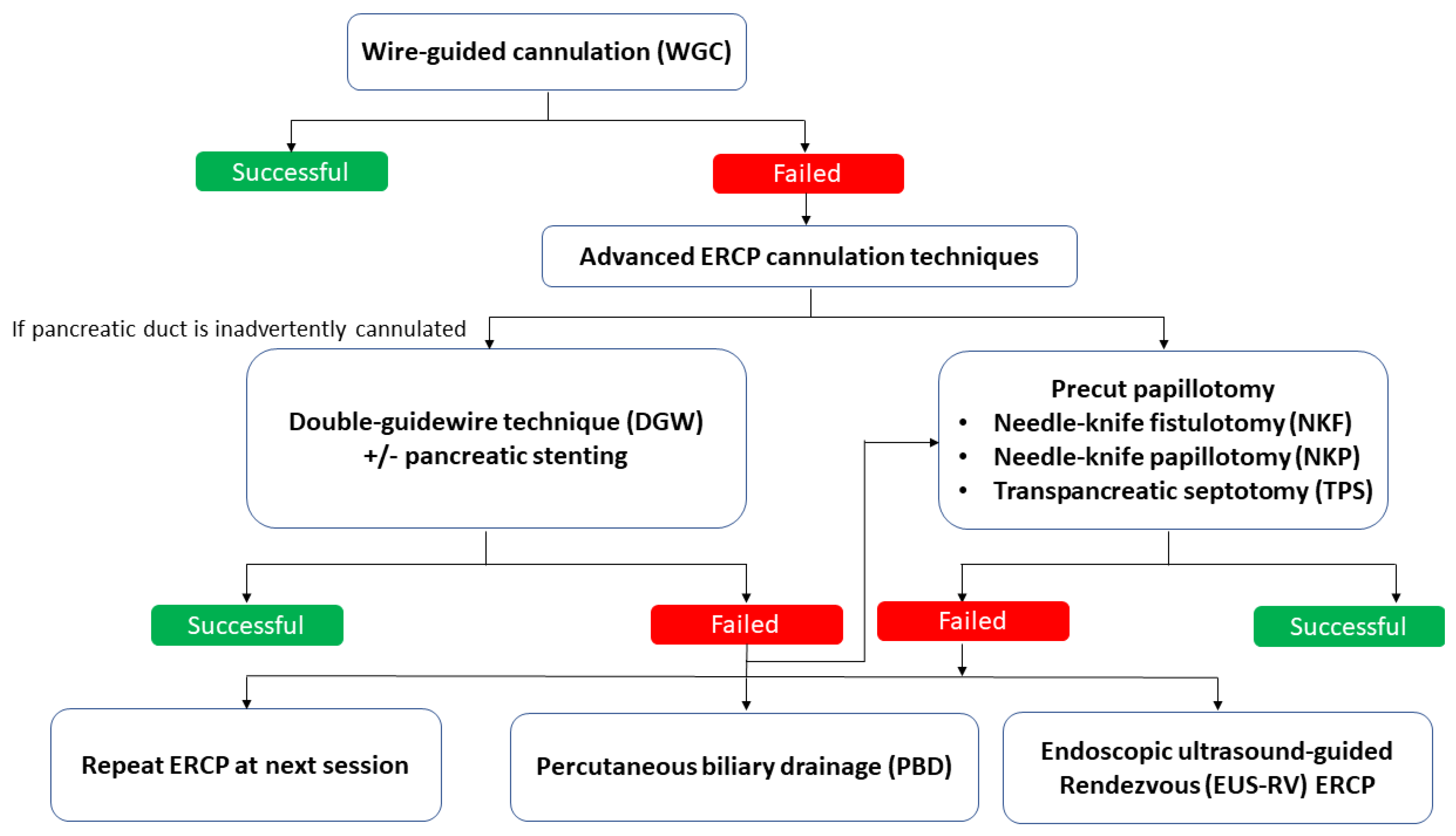
Wire-guided cannulation (WGC) with either sphincterotome or catheters has been adopted by major society and consensus guidelines as the preferred method of standard ERCP cannulation technique [3,4]. In case this fails, advanced ERCP cannulation techniques such as double-guidewire (DGW) technique with or without pancreatic stenting and precut papillotomy can be applied. If cannulation still cannot be achieved by advanced ERCP cannulation techniques, salvage options including percutaneous biliary drainage (PBD) and endoscopic ultrasound-guided Rendezvous (EUS-RV) ERCP can be considered (Figure 2). Last but not least, repeating ERCP in another session within 4–7 days or referral to expert centres [5,6,7,8] are always reasonable decisions. In this review, the techniques, clinical efficacy, and potential pitfalls of advanced ERCP techniques, percutaneous biliary drainage, and EUS-RV ERCP will be each discussed (Table 1).

2. Advanced ERCP Cannulation Techniques

2.1. Double-Guidewire (DGW) Technique with or without Pancreatic Stenting

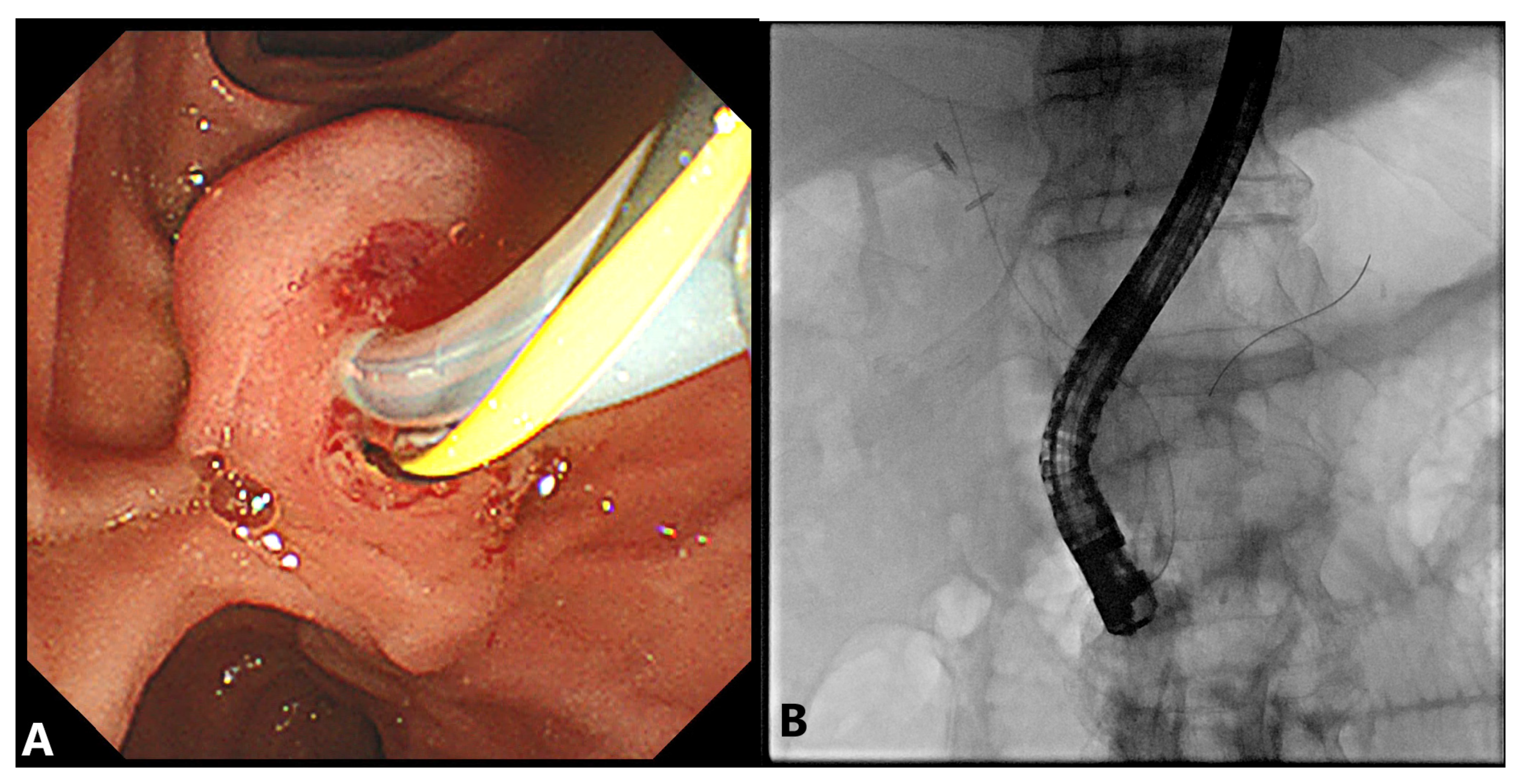
Inadvertent pancreatic duct (PD) cannulation may occur while attempting for selective biliary access. This may happen particularly when the bile duct axis is inconspicuous or the common channel is short. With the guidewire already in the main PD, it can be used to the advantage of the endoscopists, as it helps to straighten the common channel and provides a clearer roadmap of the bile duct trajectory. One could reinsert the sphincterotome preloaded by another guidewire alongside while keeping the existing PD wire in place, targeting the 10–11 o’clock position of the ampullary orifice where the bile duct is usually running along. After successful selective bile duct cannulation, the PD wire can then be removed [9,10,11] (Figure 3). The pancreatic wire works by providing both an endoscopic and fluoroscopic roadmap to guide the biliary axis, blocking the entry to the main PD and deflecting the new guidewire into the bile duct [12]. It also opens up a stenotic papillary orifice, stabilizes the papilla’s position, and straightens the common channel to facilitate bile duct access [13]. A Japanese pilot trial randomized 53 patients who experienced difficult bile duct cannulation after 10 min to either preinsertion of a guidewire into the PD or persistence trial with a conventional catheter. The pancreatic wire group was shown to have a significantly higher rate of success (93% vs. 58%, p < 0.05) than the persistence group. No episodes of clinical pancreatitis or other complications were reported in either arm, although serum amylase levels were significantly higher in the pancreatic guidewire group (551 ± 736 IU/l vs. 105 ± 50.5 IU/l) [14]. Several studies have also provided solid support for the DGW technique with an average success rate of 70% to 90% [15,16]. The reported technical success rate was not significantly different among the groups with or without pancreatic stenting but did reveal a lower chance of requiring needle-knife precut papillotomy to further assist cannulation in the DGW group without pancreatic stenting (66.7% vs. 86.2%, p = 0.003) [17].

2.1.1. Advantages

Performance of the DGW technique is relatively straightforward as it does not require any cutting-edge gadgets except one extra guidewire for PD access. It is also relatively easier to master, even for trainees. In a small-scale prospective study looking at the learning curve of the DGW technique performed by trainees, only 15 procedures of learning were needed to achieve competency, which was defined as 80% success rate in subsequent bile duct cannulation [18]. The PD wire placement allows insertion of a pancreatic stent and effectively minimizes risks of post-ERCP pancreatitis (PEP), especially after repeated trials of cannulation in challenging cases [19,20,21]. A multicentre prospective randomized controlled trial has shown that a prophylactic stent insertion after inadvertent PD cannulation significantly reduced the rate of PEP (odds ratio 0.43, 95%CI 0.19–0.98, p = 0.04), with a number needed to treat to be around 8 [22].

2.1.2. Disadvantages

Nevertheless, PD cannulation can be sometimes opportunistic. In cases of a diminutive or tortuous PD, the guidewire may be difficult to advance deeply even by experienced endoscopists. There are also risks of perforation to side-branches of PD when the wire is searching its way through [23]. A loop-tip wire technique has been proposed for initial PD cannulation to avoid mechanical trauma [24]. PEP is an important concern with DGW technique, which carries a significantly increased risk over other endoscopic techniques (risk ratio 1.98, 95% CI 1.14–3.42), as reported in a systematic review [25]. The rate of PEP in DGW ranges from 10% to 17% if performed without pancreatic stenting [15,17,26]. Robust data are available to support placement of pancreatic stents to reduce such risks after PD manipulation during the double-wire technique [15,20,27,28,29].

2.2. Precut Papillotomy

Precut papillotomy techniques mainly include needle-knife fistulotomy (NKF), needle-knife papillotomy (NKP), and transpancreatic septotomy (TPS). NKF starts cutting from 3–5 mm above the papilla where the biliary axis is aligned back to the orifice [30], while NKP involves cutting of the major papilla from orifice towards the 11 o’clock position [31], (Figure 4A,B). In TPS, the pancreatic duct is first cannulated by sphincterotome, followed by cutting of the septum between bile and pancreatic ducts [32]. The initial technical success rates for NKF, NKP, and TPS are comparable at 75–100%, 73–84%, and 95–100%, respectively [4,30,33,34,35]. On pooled analysis from a systemic review and meta-analysis, NKF significantly lowered the risk of PEP (odds ratio 0.27, 95%CI 0.09–0.82; p = 0.02) by an absolute risk reduction of 5% [36]. As such, the European Society of Gastrointestinal Endoscopy recommends needle-knife fistulotomy as the preferred techniques [3]. A recent prospective cohort advocated that the choice of NKF or NKP should depend on the morphology of major duodenal papillary fold. In general, higher efficiency and equal safety was observed when NKF was used for swollen or normal papillae and NKP for small or flat ones [37]. On the other hand, TPS seemed to facilitate a higher cannulation rate than the DGW technique (84.6% vs. 69.7%, p = 0.01) without increasing adverse events if the guidewire was already in the pancreatic duct, as shown in a latest randomized controlled trial [38].
Compared to multiple cannulation attempts, early precut papillotomy yielded a success rate of 80–90% to selective biliary cannulation with an even lower reported risk of PEP in several meta-analyses [36,39,40,41]. Early precut papillotomy refers to introduction of needle-knife techniques when the endoscopist fails selective bile duct cannulation by standard techniques within 10 min or there are more than five pancreatic cannulations. A prospective study showed that early precut improved the bile duct cannulation rate from 80% to 96.5%, irrespective of whether NKP or NKF was used [42]. The overall complication rate did not differ when compared to persistent cannulation attempts, including cholangitis, bleeding, and perforation on top of PEP (6.2% vs. 6.9%, odds ratio 0.85, 95%CI 0.51–1.41) [40]. Questions have been raised concerning whether incidence of PEP shall be attributed to repeated and prolonged cannulation attempts rather than precut papillotomy itself. A recent randomized controlled trial reported that primary precut indeed resulted in less risk of both PEP and asymptomatic hyperamylasaemia than precutting after two failed attempts by standard technique [43]. This suggests that recurrent acts of cannulation, rather than precut papillotomy alone, may have a bigger effect on PEP [44].
If inadvertent pancreatic cannulation is repeatedly obtained, a 3Fr or 5Fr pancreatic duct stent can be placed first before performing precut papillotomy. Precutting over a pancreatic duct stent provides a better guide to the pancreaticobiliary axis for the needle-knife movement while effectively reducing the incidence of PEP at the same time. This modified technique performed significantly better than the conventional method, both in terms of cannulation success (96.9% vs. 86.1%, p = 0.0189) and adverse event rates (7.1% vs. 33%, p < 0.001) [45]. It is recommended that the pancreatic stent should be left in situ for at least 12–24 hours to reduce the risk of PEP [19]. In a single-centre, randomized, prospective study, patients with pancreatic stents left behind had a significantly lower rate of PEP (4.3% vs. 21.3%, p = 0.027) in contrast to those who had the stent removed post-procedurally [46].

2.2.1. Advantages

The papillary mucosal fold and sphincter can be dissected during precut papillotomy, allowing better exposure of the biliary orifice and axis for subsequent cannulation. This is particularly useful for bulging papilla with long longitudinal segments. The risk of PEP is potentially lower if early precut is adopted, especially with the NKF technique, as discussed.

2.2.2. Disadvantages

Despite the advantages of early precut, the overall complication rate of precut papillotomy is about 6–7%. Perforation, bleeding, cholangitis, and post-ERCP pancreatitis are commonly quoted. In a prospective multicentre study with over 2000 patients, precut was an independent risk factor for postprocedural duodenal perforation [47]. For patients who are on anticoagulation or double-antiplatelet drugs, precut papillotomy is not the best immediate option unless the antithrombotic drugs had been adequately suspended before endoscopy [48,49]. Precut papillotomy may not be preferred in patients with friable ampullary tumours or papilla hidden in a diverticulum. Finally, precut papillotomy also demands precise and stable hand movements by skilful operators with a considerable learning curve. A study following 200 consecutive patients undergoing precut sphincterotomy by a single endoscopist found that the immediate bleeding rates during the first 100 procedures were significantly higher than the subsequent ones (28% vs. 7%, p < 0.05) [50]. The ESGE recommends that precut papillotomy should be done by endoscopists who achieve selective bile duct cannulation in more than 80% of cases by standard techniques [3].

3. Percutaneous Biliary Drainage (PBD)

Percutaneous biliary drainage (PBD) has been the traditional rescue treatment to alleviate biliary obstruction if bile duct cannulation by ERCP fails despite advanced techniques. This includes percutaneous transhepatic cholecystostomy (PTC) and percutaneous transhepatic biliary drainage (PTBD). Transabdominal ultrasound is first performed to visualize either the gallbladder or the intrahepatic bile ducts. A percutaneous puncture under ultrasound and fluoroscopy guidance by an 18-gauge needle is then made after local anaesthesia. Once bile return is confirmed, a 0.035-inch hydrophilic soft-tip wire, or a 0.018-inch wire if a 21-gauge needle is used for smaller duct size, is passed through the needle to the biliary system. The needle is then exchanged over the guidewire, followed by a 7Fr or 8Fr catheter insertion over the wire into the gallbladder or targeted bile ducts with or without preceding tract dilatation. By connecting the percutaneous catheter to an external collection bag, biliary drainage can be achieved [51]. Subsequently, one could consider reattempting ERCP by passing a guidewire through the external catheter to the biliary system, negotiating out of the papilla. The duodenoscope is then inserted and directed to grasp the distal end of the guidewire at the duodenum. The bile duct can then be assessed by sphincterotome cannulation over or alongside the percutaneous wire. This combined interventional radiology–endoscopy procedure was first reported in 1984 as an “antegrade catheterization” [52], which was renamed as one of the Rendezvous techniques later (PBD-RV ERCP) [53,54].
The reported technical success rate of PBD-RV ERCP was around 80–90%, holding a special role in helping patients with surgically altered anatomy [55,56,57]. Compared with solely conventional percutaneous drainage, the rate of clinical complications was significantly lower if PBD-RV ERCP was followed (16.6% vs. 26.4%; p = 0.037) [57]. Data support the completion of this two-step procedure in one single session for fewer external catheter-related and septic complications, as well as an obviously shorter hospital stay [58,59].

3.1. Advantages

PTC and PTBD serve as an immediate and quick relief of biliary obstruction, particularly when the patients are too unstable for further endoscopy. They act as a temporary salvage drainage method to stabilize critically ill cases, such as patients in septic shock or in respiratory distress, bridging patients for future endoscopic therapy. PTBD is particularly useful in cases with surgically altered anatomy and non-communicating multisegmental biliary obstruction for which successful endoscopic drainage by enteroscopy-assisted ERCP or EUS-guided biliary drainage may not always be achieved [4,60,61].

3.2. Disadvantages

The most remarkable pitfall of PBD is catheter-related complications, such as tube leakage, blockage, dislocation, and dislodgement, which can be up to 15–30%, necessitating reintervention, hospital admission, and nursing care [62]. Temporary external drainage also results in possibility of fluid and electrolyte loss. In general, PTC and PTBD carry low risk of procedural-related complications (<5%). These include flare-up of biliary sepsis (2.5%); haemorrhage (2.5%); other infective adverse events such as abscess, peritonitis, or cholecystitis (1.2–2.9%); liver biloma (0.5%); and pleural injury (0.5%) [57,63]. Although most postprocedural bleeding is self-limiting, severe episodes can occur. A Japanese nationwide audit found that the rate of major haemorrhage following PBD requiring transfusion or arterial embolization was 2.3% [64]. When intervention to the distal bile duct, for example, internalization of the external drain or PBD-RV ERCP is performed, the chance of postprocedural pancreatitis is approximately 4–6% [65]. PBD-RV ERCP is also technically demanding, as it requires coordination from both experienced interventional radiologists and endoscopists. If the percutaneous tract is lost during guidewire manipulation, there will be considerable risks of bile leak and peritonitis. As a result, PBD-RV ERCP is sometimes preferred to be proceeded as a two-step approach, usually around 2–4 weeks later, when the percutaneous tract becomes more established with less ductal edema and after acute biliary sepsis settled. Before proceeding to PBD, care of any active bleeding diathesis or anticoagulant use should be taken. Any significant amount of concomitant ascites should be drained beforehand to facilitate the procedure.

4. EUS-Guided Rendezvous (EUS-RV) Technique

As interventional endoscopic ultrasound (EUS) procedures evolve with more dedicated accessories developed in the last decade, EUS-guided biliary drainage (EUS-BD) has been increasingly applied in patients with benign or malignant pancreatobiliary pathologies. It can be classified into transpapillary technique such as EUS-guided Rendezvous (EUS-RV) and EUS-antegrade approach (EUS-AG), and transmural biliary drainage including choledochoduodenostomy (CDS) and hepaticogastrostomy (HGS) [66]. In general, transmural drainage has been more commonly applied in patients with malignant biliary obstructions with good outcome [67,68,69], whereas the EUS-RV technique is usually the preferred option for difficult biliary access in benign biliary pathology over percutaneous biliary drainage when the major papilla is accessible. (Figure 5) In this review, only EUS-RV was discussed so as to focus on methods to perform transpapillary cannulation and retrograde drainage.
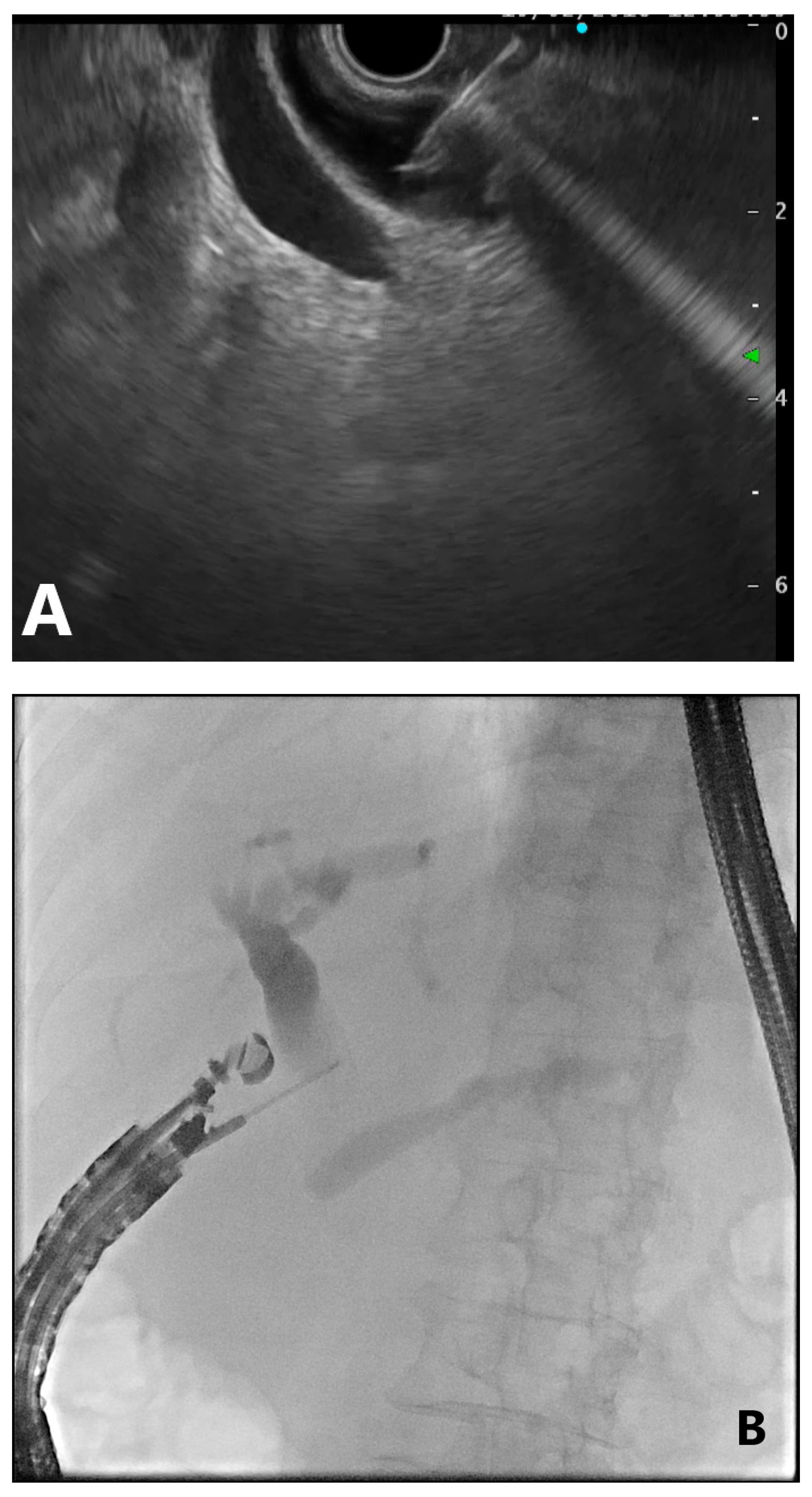
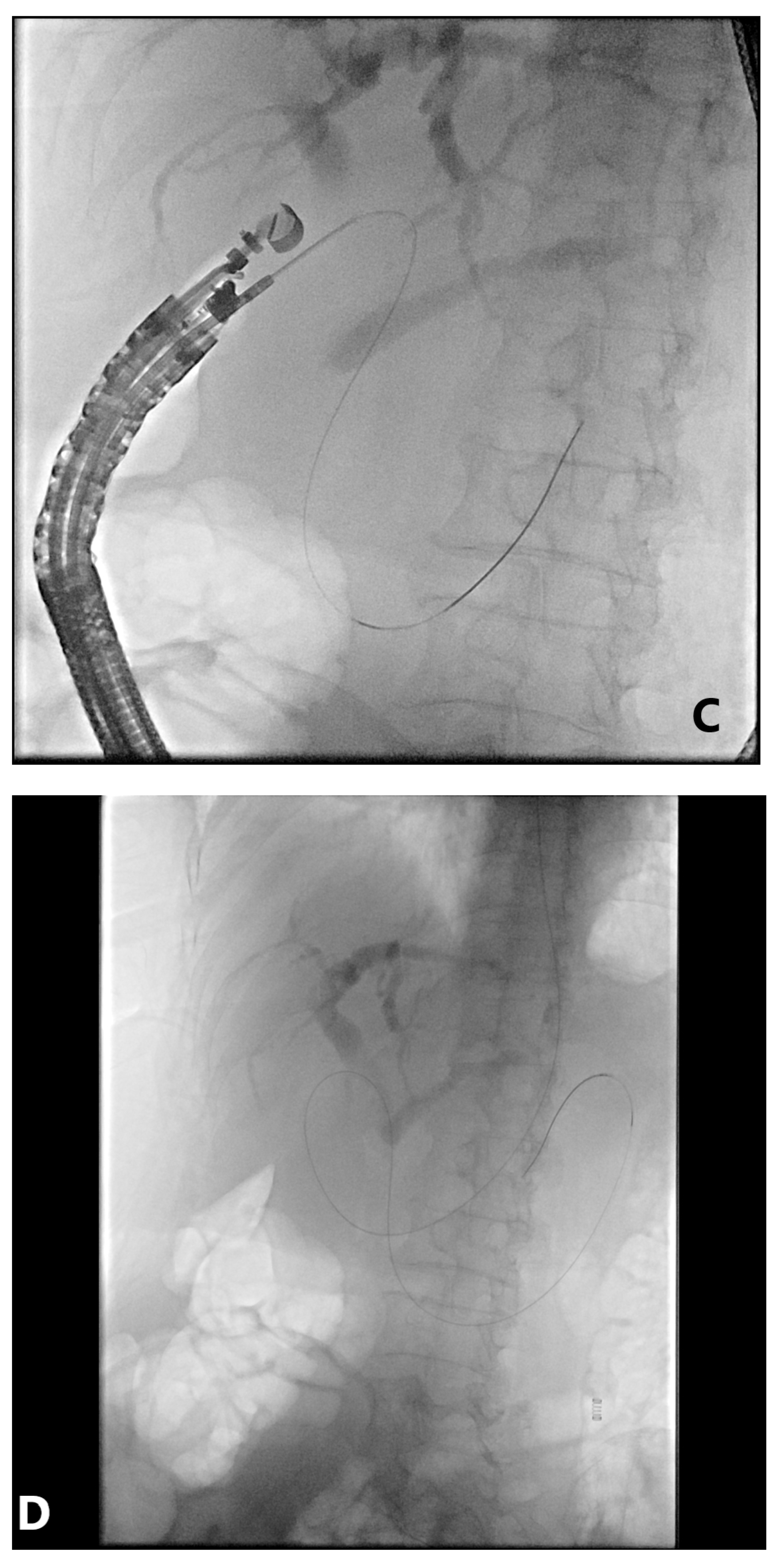
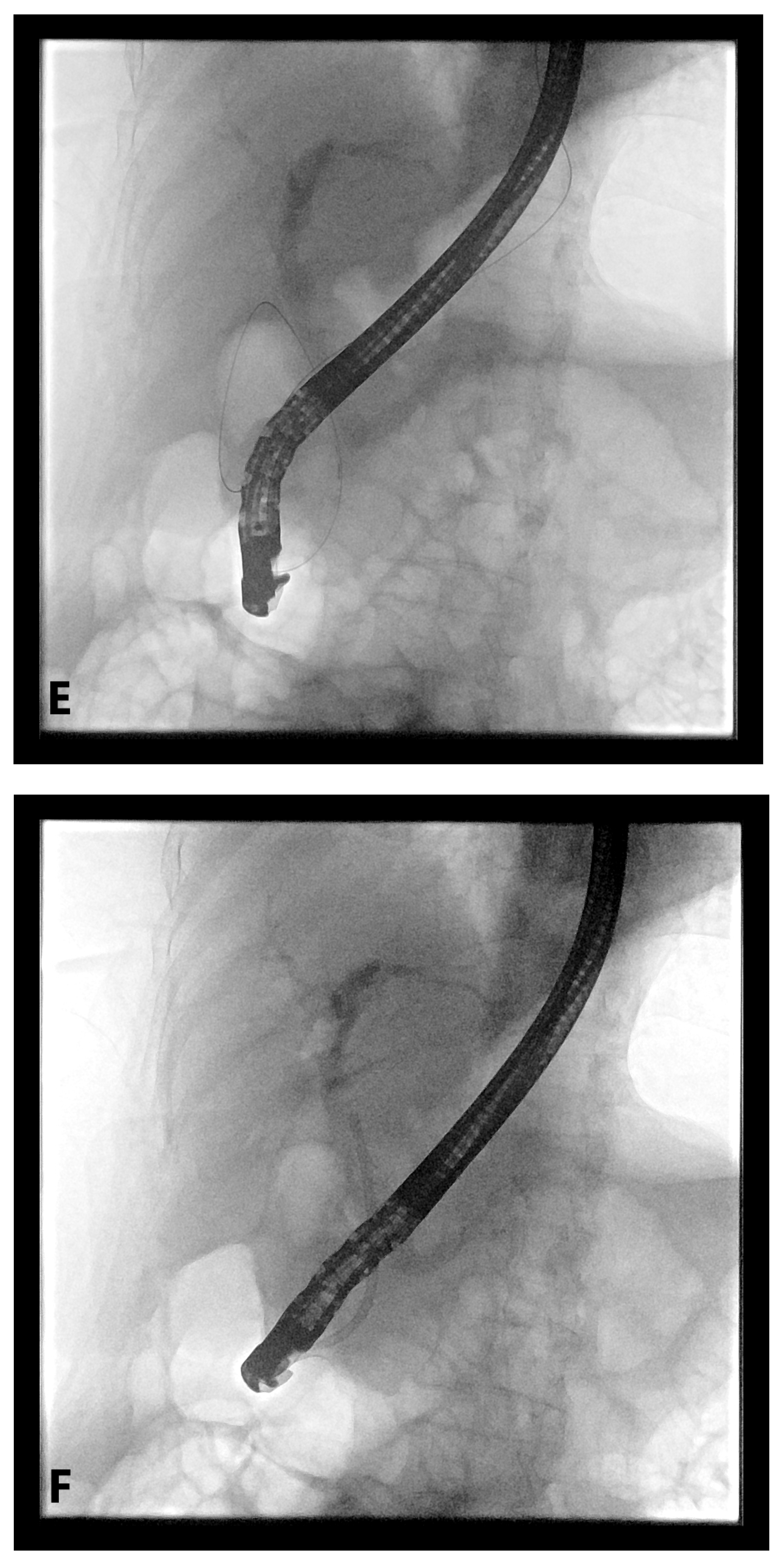
EUS-RV ERCP begins with an EUS-guided puncture to the dilated biliary system by a 19-gauge needle, followed by contrast injection to obtain the cholangiogram. While either a long (450 cm) 0.035-inch or 0.025-inch guidewire can be inserted into a 19G needle, a 0.025-inch guidewire would be preferred for wire manipulation through the bile duct towards and out of the major papilla with a theoretically lower risk of wire coating shearing. The needle and echoendoscope are then exchanged over the wire with reintroduction of the duodenoscope to complete the ERCP. Similar to the PBD-RV ERCP technique, deep bile duct cannulation followed by standard endoscopic biliary intervention can be achieved by either grasping the Rendezvous wire into the accessory channel of duodenoscope, or cannulating alongside the Rendezvous wire with a separate guidewire [70,71] (Figure 6A–F).
Since the first report of EUS-RV ERCP by Giovannini, M. et al. back in 2001 [72], a wide variety of techniques have been reported in case series, such as using short (260 cm) hydrophilic wires [73], “hitch-and-ride” catheters [74], smaller 22-gauge needles and 0.018-inch guidewires [75], and the latest steerable access catheters [76]. While early case series published before 2010 reported a lower technical success rate ranging from 50–80%, this has increased over time to 73–100% in studies published after 2010 [71,77,78,79]. A meta-analysis of 12 studies focusing on the technical and clinical outcomes of EUS-RV after failed biliary cannulation in ERCP published this year reported a pooled technical success rate of 86.1% and a pooled clinical success rate of 80.8% at a dispense of an adverse event rates of around 14% [80].
The dilated biliary tree can be accessed by EUS either from the stomach through the liver to the intrahepatic ducts (transhepatic, TH) or at the proximal duodenum directly to the common bile duct (extrahepatic, EH) with a long scope or a short scope position [71]. A multicentre prospective study showed that the EH approach had a higher rate of technical success (100% vs. 66.7%) than the TH route [81]. The EH route also appeared significantly safer, with more favourable clinical outcomes in patients with distal common bile duct (CBD) obstruction, in terms of lower incidence of post-procedural pain (5.5% vs. 44.1%; p = 0.017), bile leak (0% vs. 11.7%; p = 0.228), and pneumoperitoneum (0% vs. 11.7%; p = 0.228). The procedural time was also shorter (25.7 min vs. 34.4 min; p = 0.0004), with fewer days of hospital stay in the EH group (0.17 days vs. 2.52 days; p = 0.015) [82]. In summary, the choice of EH or TH approach genuinely depends on the site of biliary obstruction, underlying pathology, and scope stability. For example, in cases of hilar obstruction when the CBD is not significantly dilated, TH route targeting the left intrahepatic duct would be preferred due to the presence of a hilar stricture and relative non-dilated CBD.

4.1. Advantages

When compared to PBD-RV ERCP, which would mostly require a second session to complete after percutaneous drainage performed, EUS-RV has the advantage to be completed in the same session when cannulation during the initial ERCP is unsuccessful. Indeed, it has been shown to be superior to precut papillotomy for single-session biliary access in two retrospective cohorts (95–98% vs. 75–90%) [83,84]. Compared to percutaneous external drainage, it had a higher success rate, markedly fewer adverse events, and significantly lower cost due to a less frequent need for reinterventions [85,86]. The EUS-RV technique also facilitated a shorter post-procedural hospital stay and quicker discharge in patients with malignant distal biliary obstruction [87]. It allows better quality of life as patients can be exempted from external drainage bags and risks of PTBD dislodgement. Although there have been no randomized controlled trials directly comparing EUS-RV and PBD so far, EUS-RV should be proposed before PBD with regards to the above advantages when expertise is available.

4.2. Disadvantages

Despite the high technical success rates reported in the two retrospective comparative studies with precut, EUS-RV is technically demanding, requiring competence in both interventional EUS and ERCP skills. These include but are not limited to accurate needle puncture to the target bile duct, wire manipulation towards and exiting major papilla without shearing, and grabbing of the RV wire or cannulation alongside the RV wire without wire loss. Endoscopists also need to consider conversion to EUS-guided transmural drainage as a rescue if the wire cannot be manipulated out of the papilla due to tight malignant or benign biliary strictures. If guidewire passage through the papilla fails in EUS-RV, potential rescue options for patients with malignant biliary obstruction include EUS-guided transmural biliary drainage (e.g., CDS, HGS) or PTBD. In patients with benign biliary obstruction but failed EUS-RV, PTBD is generally offered. Yet the feasibility of EUS-guided HGS using a dedicated plastic stent for biliary drainage has also been described [88]. The right biliary ductal system is generally a technically challenging target for EUS access, but cases of successful right intrahepatic ductal access from the duodenum have been reported. The overall procedural-related complication rate ranges 10–15%, including bleeding (4%), pancreatitis (1.6%), pneumoperitoneum (3%), bile leak (4%), peritonitis (1.3%), and flare-up of sepsis (2.4%). [75,79,89,90,91].

5. Conclusions

In conclusion, advanced ERCP cannulation techniques, percutaneous biliary drainage, and EUS-guided Rendezvous techniques are available rescue methods for difficult bile duct access when standard ERCP cannulation fails. Becoming familiar with each technique’s advantages and limitations would help the endoscopists to make the most appropriate treatment decisions that are tailored to the patient’s anatomy and clinical needs.

Author Contributions

Conceptualization, T.-T.C. and R.S.Y.T.; methodology, T.-T.C. and R.S.Y.T.; resources, R.S.Y.T.; writing—original draft preparation, T.-T.C.; writing—review and editing, R.S.Y.T. and M.C.H.C.; visualization, M.C.H.C.; supervision, R.S.Y.T. All authors have read and agreed to the published version of the manuscript.

Funding

This research received no external funding.

Institutional Review Board Statement

Not applicable.

Informed Consent Statement

Not applicable.

Conflicts of Interest

The authors declare no conflict of interest.

Abbreviations

ERCPendoscopic retrograde cholangiopancreatography
ESGEEuropean Society of Gastrointestinal Endoscopy
WGCwire-guided cannulation
DGWdouble-guidewire
PBDpercutaneous biliary drainage
EUS-RVendoscopic ultrasound-guided Rendezvous
PDpancreatic duct
PEPpost-ERCP pancreatitis
NKFneedle-knife fistulotomy
NKPneedle-knife papillotomy
TPStranspancreatic septotomy
PTCpercutaneous transhepatic cholecystostomy
PTBDpercutaneous transhepatic biliary drainage
EUSendoscopic ultrasound
EUS-BDEUS-guided biliary drainage
EUS-AGEUS-antegrade approach
CDScholedochoduodenostomy
HGShepaticogastrostomy
THtranshepatic
EHextrahepatic

References

  1. Varadarajulu, S.; Kilgore, M.L.; Wilcox, C.M.; Eloubeidi, M.A. Relationship among hospital ERCP volume, length of stay, and technical outcomes. Gastrointest. Endosc. 2006, 64, 338–347. [Google Scholar] [CrossRef] [PubMed]
  2. Bailey, A.A.; Bourke, M.J.; Williams, S.J.; Walsh, P.R.; Murray, M.A.; Lee, E.Y.; Kwan, V.; Lynch, P.M. A prospective randomized trial of cannulation technique in ERCP: Effects on technical success and post-ERCP pancreatitis. Endoscopy 2008, 40, 296–301. [Google Scholar] [CrossRef] [PubMed]
  3. Testoni, P.A.; Mariani, A.; Aabakken, L.; Arvanitakis, M.; Bories, E.; Costamagna, G.; Devière, J.; Dinis-Ribeiro, M.; Dumonceau, J.M.; Giovannini, M.; et al. Papillary cannulation and sphincterotomy techniques at ERCP: European Society of Gastrointestinal Endoscopy (ESGE) Clinical Guideline. Endoscopy 2016, 48, 657–683. [Google Scholar] [CrossRef]
  4. Liao, W.-C.; Angsuwatcharakon, P.; Isayama, H.; Dhir, V.; Devereaux, B.; Khor, C.J.L.; Ponnudurai, R.; Lakhtakia, S.; Lee, D.-K.; Ratanachu-ek, T.; et al. International consensus recommendations for difficult biliary access. Gastrointest. Endosc. 2017, 85, 295–304. [Google Scholar] [CrossRef] [Green Version]
  5. Swan, M.P.; Bourke, M.J.; Williams, S.J.; Alexander, S.; Moss, A.; Hope, R.; Ruppin, D. Failed biliary cannulation: Clinical and technical outcomes after tertiary referral endoscopic retrograde cholangiopancreatography. World J. Gastroenterol. 2011, 17, 4993–4998. [Google Scholar] [CrossRef]
  6. Pavlides, M.; Barnabas, A.; Fernandopulle, N.; Bailey, A.A.; Collier, J.; Phillips-Hughes, J.; Ellis, A.; Chapman, R.; Braden, B. Repeat endoscopic retrograde cholangiopancreaticography after failed initial precut sphincterotomy for biliary cannulation. World J. Gastroenterol. 2014, 20, 13153–13158. [Google Scholar] [CrossRef] [PubMed]
  7. Donnellan, F.; Enns, R.; Kim, E.; Lam, E.; Amar, J.; Telford, J.; Byrne, M.F. Outcome of repeat ERCP after initial failed use of a needle knife for biliary access. Dig. Dis. Sci. 2012, 57, 1069–1071. [Google Scholar] [CrossRef] [PubMed]
  8. Kevans, D.; Zeb, F.; Donnellan, F.; Courtney, G.; Aftab, A.R. Failed biliary access following needle knife fistulotomy: Is repeat interval ERCP worthwhile? Scand. J. Gastroenterol. 2010, 45, 1238–1241. [Google Scholar] [CrossRef]
  9. Freeman, M.L.; Guda, N.M. ERCP cannulation: A review of reported techniques. Gastrointest. Endosc. 2005, 61, 112–125. [Google Scholar] [CrossRef]
  10. Gyökeres, T.; Duhl, J.; Varsányi, M.; Schwab, R.; Burai, M.; Pap, Á. Double guide wire placement for endoscopic pancreaticobiliary procedures. Endoscopy 2003, 35, 95–96. [Google Scholar] [CrossRef] [Green Version]
  11. Draganov, P.; Devonshire, D.A.; Cunningham, J.T. A new technique to assist in difficult bile duct cannulation at the time of endoscopic retrograde cholangiopancreatography. JSLS 2005, 9, 218–221. [Google Scholar]
  12. Fry, L.C.; Barriga, J.A.; Linder, J.D.; Mönkemüller, K.E. Placement of a biliary catheter in the pancreatic duct to aid common bile duct cannulation. Endoscopy 2003, 35, 97. [Google Scholar] [CrossRef] [Green Version]
  13. Gotoh, Y.; Tamada, K.; Tomiyama, T.; Wada, S.; Ohashi, A.; Satoh, Y.; Higashizawa, T.; Miyata, T.; Ido, K.; Sugano, K. A new method for deep cannulation of the bile duct by straightening the pancreatic duct. Gastrointest. Endosc. 2001, 53, 820–822. [Google Scholar] [CrossRef] [PubMed]
  14. Maeda, S.; Hayashi, H.; Hosokawa, O.; Dohden, K.; Hattori, M.; Morita, M.; Kidani, E.; Ibe, N.; Tatsumi, S. Prospective randomized pilot trial of selective biliary cannulation using pancreatic guide-wire placement. Endoscopy 2003, 35, 721–724. [Google Scholar] [CrossRef] [PubMed]
  15. Ito, K.; Fujita, N.; Noda, Y.; Kobayashi, G.; Obana, T.; Horaguchi, J.; Takasawa, O.; Koshita, S.; Kanno, Y. Pancreatic guidewire placement for achieving selective biliary cannulation during endoscopic retrograde cholangio-pancreatography. World J. Gastroenterol. 2008, 14, 5595–5600, discussion 5599. [Google Scholar] [CrossRef] [PubMed]
  16. Huang, C.; Kung, J.; Liu, Y.; Tse, A.; Datta, A.; Singh, I.; Eysselein, V.E.; Reicher, S. Use of double wire-guided technique and transpancreatic papillary septotomy in difficult ERCP: 4-year experience. Endosc. Int. Open 2016, 4, E1107–E1110. [Google Scholar] [CrossRef] [Green Version]
  17. Yang, M.J.; Hwang, J.C.; Yoo, B.M.; Kim, J.H.; Ryu, H.-K.; Kim, S.S.; Kang, J.K.; Kim, M.K. Wire-guided cannulation over a pancreatic stent versus double guidewire technique in patients with difficult biliary cannulation. BMC Gastroenterol. 2015, 15, 150. [Google Scholar] [CrossRef] [Green Version]
  18. Wang, X.; Ren, G.; Xi, Y.; Luo, H.; Liang, S.; Wang, B.; Tao, Q.; Luo, B.; Qin, Q.; Farrell, J.J.; et al. Learning curve of double-guidewire technique by trainees during hands-on endoscopic retrograde cholangiopancreatography training. J. Gastroenterol. Hepatol. 2020, 35, 2176–2183. [Google Scholar] [CrossRef]
  19. Dumonceau, J.M.; Kapral, C.; Aabakken, L.; Papanikolaou, I.S.; Tringali, A.; Vanbiervliet, G.; Beyna, T.; Dinis-Ribeiro, M.; Hritz, I.; Mariani, A.; et al. ERCP-related adverse events: European Society of Gastrointestinal Endoscopy (ESGE) guideline. Endoscopy 2020, 52, 127–149. [Google Scholar] [CrossRef] [Green Version]
  20. Kawaguchi, Y.; Ogawa, M.; Omata, F.; Ito, H.; Shimosegawa, T.; Mine, T. Randomized controlled trial of pancreatic stenting to prevent pancreatitis after endoscopic retrograde cholangiopancreatography. World J. Gastroenterol. 2012, 18, 1635–1641. [Google Scholar] [CrossRef]
  21. Hakuta, R.; Hamada, T.; Nakai, Y.; Isayama, H.; Kogure, H.; Takahara, N.; Mizuno, S.; Yagioka, H.; Togawa, O.; Matsubara, S.; et al. Early pancreatic stent placement in wire-guided biliary cannulation: A multicenter retrospective study. J. Gastroenterol. Hepatol. 2019, 34, 1116–1122. [Google Scholar] [CrossRef]
  22. Phillip, V.; Pukitis, A.; Epstein, A.; Hapfelmeier, A.; Haf, D.; Schwab, M.; Demir, I.E.; Rosendahl, J.; Hoffmeister, A.; Schmid, R.M.; et al. Pancreatic stenting to prevent post-ERCP pancreatitis: A randomized multicenter trial. Endosc. Int. Open 2019, 7, E860–E868. [Google Scholar] [CrossRef] [PubMed] [Green Version]
  23. Hawes, R.H.; Devière, J. How I cannulate the bile duct. Endoscopy 2018, 50, 75–77. [Google Scholar]
  24. Masci, E.; Mangiavillano, B.; Luigiano, C.; Bizzotto, A.; Limido, E.; Cantù, P.; Manes, G.; Viaggi, P.; Spinzi, G.; Radaelli, F.; et al. Comparison between loop-tip guidewire-assisted and conventional endoscopic cannulation in high risk patients. Endosc. Int. Open 2015, 3, E464–E470. [Google Scholar] [CrossRef] [Green Version]
  25. Tse, F.; Yuan, Y.; Moayyedi, P.; Leontiadis, G.I.; Barkun, A.N. Double-guidewire technique in difficult biliary cannulation for the prevention of post-ERCP pancreatitis: A systematic review and meta-analysis. Endoscopy 2017, 49, 15–26. [Google Scholar] [CrossRef]
  26. Herreros de Tejada, A.; Calleja, J.L.; Díaz, G.; Pertejo, V.; Espinel, J.; Cacho, G.; Jiménez, J.; Millán, I.; García, F.; Abreu, L. Double-guidewire technique for difficult bile duct cannulation: A multicenter randomized, controlled trial. Gastrointest. Endosc. 2009, 70, 700–709. [Google Scholar] [CrossRef] [PubMed]
  27. Freeman, M.L.; Overby, C.; Qi, D. Pancreatic stent insertion: Consequences of failure and results of a modified technique to maximize success. Gastrointest. Endosc. 2004, 59, 8–14. [Google Scholar] [CrossRef]
  28. Ito, K.; Fujita, N.; Noda, Y.; Kobayashi, G.; Obana, T.; Horaguchi, J.; Takasawa, O.; Koshita, S.; Kanno, Y.; Ogawa, T. Can pancreatic duct stenting prevent post-ERCP pancreatitis in patients who undergo pancreatic duct guidewire placement for achieving selective biliary cannulation? A prospective randomized controlled trial. J. Gastroenterol. 2010, 45, 1183–1191. [Google Scholar] [CrossRef]
  29. Ito, K.; Horaguchi, J.; Fujita, N.; Noda, Y.; Kobayashi, G.; Koshita, S.; Kanno, Y.; Ogawa, T.; Masu, K.; Hashimoto, S. Clinical usefulness of double-guidewire technique for difficult biliary cannulation in endoscopic retrograde cholangiopancreatography. Dig. Endosc. 2014, 26, 442–449. [Google Scholar] [CrossRef]
  30. Mavrogiannis, C.; Liatsos, C.; Romanos, A.; Petoumenos, C.; Nakos, A.; Karvountzis, G. Needle-knife fistulotomy versus needle-knife precut papillotomy for the treatment of common bile duct stones. Gastrointest. Endosc. 1999, 50, 334–339. [Google Scholar] [CrossRef]
  31. Rollhauser, C.; Johnson, M.; Al-Kawas, F.H. Needle-knife papillotomy: A helpful and safe adjunct to endoscopic retrograde cholangiopancreatography in a selected population. Endoscopy 1998, 30, 691–696. [Google Scholar] [CrossRef]
  32. Goff, J.S. Long-term experience with the transpancreatic sphincter pre-cut approach to biliary sphincterotomy. Gastrointest. Endosc. 1999, 50, 642–645. [Google Scholar] [CrossRef]
  33. Horiuchi, A.; Nakayama, Y.; Kajiyama, M.; Tanaka, N. Effect of precut sphincterotomy on biliary cannulation based on the characteristics of the major duodenal papilla. Clin. Gastroenterol. Hepatol. 2007, 5, 1113–1118. [Google Scholar] [CrossRef]
  34. Zang, J.; Zhang, C.; Gao, J. Guidewire-assisted transpancreatic sphincterotomy for difficult biliary cannulation: A prospective randomized controlled trial. Surg. Laparosc. Endosc. Percutan. Tech. 2014, 24, 429–433. [Google Scholar] [CrossRef] [PubMed]
  35. Katsinelos, P.; Gkagkalis, S.; Chatzimavroudis, G.; Beltsis, A.; Terzoudis, S.; Zavos, C.; Gatopoulou, A.; Lazaraki, G.; Vasiliadis, T.; Kountouras, J. Comparison of three types of precut technique to achieve common bile duct cannulation: A retrospective analysis of 274 cases. Dig. Dis. Sci. 2012, 57, 3286–3292. [Google Scholar] [CrossRef] [PubMed]
  36. Choudhary, A.; Winn, J.; Siddique, S.; Arif, M.; Arif, Z.; Hammoud, G.M.; Puli, S.R.; Ibdah, J.A.; Bechtold, M.L. Effect of precut sphincterotomy on post-endoscopic retrograde cholangiopancreatography pancreatitis: A systematic review and meta-analysis. World J. Gastroenterol. 2014, 20, 4093–4101. [Google Scholar] [CrossRef]
  37. Zhang, Q.-S.; Xu, J.-H.; Dong, Z.-Q.; Gao, P.; Shen, Y.-C. Success and safety of needle knife papillotomy and fistulotomy based on papillary anatomy: A prospective controlled trial. Dig. Dis. Sci. 2021. online ahead of print. [Google Scholar] [CrossRef]
  38. Kylänpää, L.; Koskensalo, V.; Saarela, A.; Ejstrud, P.; Udd, M.; Lindström, O.; Rainio, M.; Tenca, A.; Halttunen, J.; Qvigstad, G.; et al. Transpancreatic biliary sphincterotomy versus double guidewire in difficult biliary cannulation: A randomized controlled trial. Endoscopy 2021, 53, 1011–1019. [Google Scholar]
  39. Cennamo, V.; Fuccio, L.; Zagari, R.M.; Eusebi, L.H.; Ceroni, L.; Laterza, L.; Fabbri, C.; Bazzoli, F. Can early precut implementation reduce endoscopic retrograde cholangiopancreatography-related complication risk? Meta-analysis of randomized controlled trials. Endoscopy 2010, 42, 381–388. [Google Scholar] [CrossRef]
  40. Navaneethan, U.; Konjeti, R.; Venkatesh, P.G.; Sanaka, M.R.; Parsi, M.A. Early precut sphincterotomy and the risk of endoscopic retrograde cholangiopancreatography related complications: An updated meta-analysis. World J. Gastrointest. Endosc. 2014, 6, 200–208. [Google Scholar] [CrossRef]
  41. Gong, B.; Hao, L.; Bie, L.; Sun, B.; Wang, M. Does precut technique improve selective bile duct cannulation or increase post-ERCP pancreatitis rate? A meta-analysis of randomized controlled trials. Surg. Endosc. 2010, 24, 2670–2680. [Google Scholar] [CrossRef]
  42. Kaffes, A.J.; Sriram, P.V.; Rao, G.V.; Santosh, D.; Reddy, D.N. Early institution of pre-cutting for difficult biliary cannulation: A prospective study comparing conventional vs. a modified technique. Gastrointest. Endosc. 2005, 62, 669–674. [Google Scholar] [CrossRef] [PubMed]
  43. Maharshi, S.; Sharma, S.S. Early precut versus primary precut sphincterotomy to reduce post-ERCP pancreatitis: Randomized controlled trial (with videos). Gastrointest. Endosc. 2021, 93, 586–593. [Google Scholar] [CrossRef]
  44. Davee, T.; Garcia, J.A.; Baron, T.H. Precut sphincterotomy for selective biliary duct cannulation during endoscopic retrograde cholangiopancreatography. Ann. Gastroenterol. 2012, 25, 291–302. [Google Scholar] [PubMed]
  45. Kubota, K.; Sato, T.; Kato, S.; Watanabe, S.; Hosono, K.; Kobayashi, N.; Hisatomi, K.; Matsuhashi, N.; Nakajima, A. Needle-knife precut papillotomy with a small incision over a pancreatic stent improves the success rate and reduces the complication rate in difficult biliary cannulations. J. Hepatobiliary Pancreat. Sci. 2013, 20, 382–388. [Google Scholar] [CrossRef]
  46. Cha, S.W.; Leung, W.D.; Lehman, G.A.; Watkins, J.L.; McHenry, L.; Fogel, E.L.; Sherman, S. Does leaving a main pancreatic duct stent in place reduce the incidence of precut biliary sphincterotomy-associated pancreatitis? A randomized, prospective study. Gastrointest. Endosc. 2013, 77, 209–216. [Google Scholar] [CrossRef]
  47. Loperfido, S.; Angelini, G.; Benedetti, G.; Chilovi, F.; Costan, F.; De Berardinis, F.; De Bernardin, M.; Ederle, A.; Fina, P.; Fratton, A. Major early complications from diagnostic and therapeutic ERCP: A prospective multicenter study. Gastrointest. Endosc. 1998, 48, 1–10. [Google Scholar] [CrossRef]
  48. Veitch, A.M.; Vanbiervliet, G.; Gershlick, A.H.; Boustiere, C.; Baglin, T.P.; Smith, L.A.; Radaelli, F.; Knight, E.; Gralnek, I.M.; Hassan, C.; et al. Endoscopy in patients on antiplatelet or anticoagulant therapy, including direct oral anticoagulants: British Society of Gastroenterology (BSG) and European Society of Gastrointestinal Endoscopy (ESGE) guidelines. Gut 2016, 65, 374–389. [Google Scholar] [CrossRef]
  49. Acosta, R.D.; Abraham, N.S.; Chandrasekhara, V.; Chathadi, K.V.; Early, D.S.; Eloubeidi, M.A.; Evans, J.A.; Faulx, A.L.; Fisher, D.A.; Fonkalsrud, L.; et al. The management of antithrombotic agents for patients undergoing GI endoscopy. Gastrointest. Endosc. 2016, 83, 3–16. [Google Scholar] [CrossRef] [PubMed]
  50. Akaraviputh, T.; Lohsiriwat, V.; Swangsri, J.; Methasate, A.; Leelakusolvong, S.; Lertakayamanee, N. The learning curve for safety and success of precut sphincterotomy for therapeutic ERCP: A single endoscopist’s experience. Endoscopy 2008, 40, 513–516. [Google Scholar] [CrossRef]
  51. Madhusudhan, K.S.; Gamanagatti, S.; Srivastava, D.N.; Gupta, A.K. Radiological interventions in malignant biliary obstruction. World J. Radiol. 2016, 8, 518–529. [Google Scholar] [CrossRef] [Green Version]
  52. Long, W.B.; Schwarz, W.; Ring, E.J. Endoscopic sphincterotomy assisted by catheterization antegrade. Gastrointest. Endosc. 1984, 30, 36–39. [Google Scholar] [CrossRef]
  53. Rieber, A.; Brambs, H.J.; Friedl, P. Therapy of bile duct occlusion. Transhepatic/endoscopic “Rendezvous procedure” and percutaneous transhepatic stent placement. Fortschr. Medizin 1990, 108, 565–567. [Google Scholar]
  54. Calvo, M.M.; Bujanda, L.; Heras, I.; Cabriada, J.L.; Bernal, A.; Orive, V.; Miguelez, J. The rendezvous technique for the treatment of choledocholithiasis. Gastrointest. Endosc. 2001, 54, 511–513. [Google Scholar] [CrossRef] [PubMed]
  55. Yang, M.J.; Kim, J.H.; Hwang, J.C.; Yoo, B.M.; Kim, S.S.; Lim, S.G.; Won, J.H. Usefulness of combined percutaneous-endoscopic rendezvous techniques after failed therapeutic endoscopic retrograde cholangiography in the era of endoscopic ultrasound guided rendezvous. Medicine 2017, 96, e8991. [Google Scholar] [CrossRef] [PubMed]
  56. Okuno, M.; Iwashita, T.; Yasuda, I.; Mabuchi, M.; Uemura, S.; Nakashima, M.; Doi, S.; Adachi, S.; Mukai, T.; Moriwaki, H. Percutaneous transgallbladder rendezvous for enteroscopic management of choledocholithiasis in patients with surgically altered anatomy. Scand. J. Gastroenterol. 2013, 48, 974–978. [Google Scholar] [CrossRef]
  57. Bokemeyer, A.; Müller, F.; Niesert, H.; Brückner, M.; Bettenworth, D.; Nowacki, T.; Beyna, T.; Ullerich, H.; Lenze, F. Percutaneous-transhepatic-endoscopic rendezvous procedures are effective and safe in patients with refractory bile duct obstruction. United Eur. Gastroenterol. J. 2019, 7, 397–404. [Google Scholar] [CrossRef]
  58. Wayman, J.; Mansfield, J.C.; Matthewson, K.; Richardson, D.L.; Griffin, S.M. Combined percutaneous and endoscopic procedures for bile duct obstruction: Simultaneous and delayed techniques compared. Hepatogastroenterology 2003, 50, 915–918. [Google Scholar]
  59. Tomizawa, Y.; Di Giorgio, J.; Santos, E.; McCluskey, K.M.; Gelrud, A. Combined interventional radiology followed by endoscopic therapy as a single procedure for patients with failed initial endoscopic biliary access. Dig. Dis. Sci. 2014, 59, 451–458. [Google Scholar] [CrossRef]
  60. Ko, G.Y.; Sung, K.B.; Yoon, H.K.; Kim, K.R.; Gwon, D.I.; Lee, S.G. Percutaneous transhepatic treatment of hepaticojejunal anastomotic biliary strictures after living donor liver transplantation. Liver Transpl. 2008, 14, 1323–1332. [Google Scholar] [CrossRef]
  61. Nakai, Y.; Kogure, H.; Yamada, A.; Isayama, H.; Koike, K. Endoscopic management of bile duct stones in patients with surgically altered anatomy. Dig. Endosc. 2018, 30, 67–74. [Google Scholar] [CrossRef] [Green Version]
  62. Born, P.; Rösch, T.; Triptrap, A.; Frimberger, E.; Allescher, H.D.; Ott, R.; Weigert, N.; Lorenz, R.; Classen, M. Long-term results of percutaneous transhepatic biliary drainage for benign and malignant bile duct strictures. Scand. J. Gastroenterol. 1998, 33, 544–549. [Google Scholar]
  63. Saad, W.E.A.; Wallace, M.J.; Wojak, J.C.; Kundu, S.; Cardella, J.F. Quality improvement guidelines for percutaneous transhepatic cholangiography, biliary drainage, and percutaneous cholecystostomy. J. Vasc. Interv. Radiol. 2010, 21, 789–795. [Google Scholar] [CrossRef] [PubMed]
  64. Hamada, T.; Yasunaga, H.; Nakai, Y.; Isayama, H.; Horiguchi, H.; Fushimi, K.; Koike, K. Severe bleeding after percutaneous transhepatic drainage of the biliary system: Effect of antithrombotic agents—Analysis of 34,606 cases from a Japanese Nationwide Administrative Database. Radiology 2015, 274, 605–613. [Google Scholar] [CrossRef]
  65. Al-Bahrani, A.Z.; Holt, A.; Hamade, A.M.; Abid, G.H.; Laasch, H.U.; O’Shea, S.J.; Lee, S.H.; Ammori, B.J. Acute pancreatitis: An under-recognized risk of percutaneous transhepatic distal biliary intervention. HPB 2006, 8, 446–450. [Google Scholar] [CrossRef] [Green Version]
  66. Iwashita, T.; Doi, S.; Yasuda, I. Endoscopic ultrasound-guided biliary drainage: A review. Clin. J. Gastroenterol. 2014, 7, 94–102. [Google Scholar] [CrossRef] [PubMed] [Green Version]
  67. Lyu, Y.; Li, T.; Cheng, Y.; Wang, B.; Cao, Y.; Wang, Y. Endoscopic ultrasound-guided vs ERCP-guided biliary drainage for malignant biliary obstruction: A up-to-date meta-analysis and systematic review. Dig. Liver Dis. 2021, 53, 1247–1253. [Google Scholar] [CrossRef]
  68. Miller, C.S.; Barkun, A.N.; Martel, M.; Chen, Y.I. Endoscopic ultrasound-guided biliary drainage for distal malignant obstruction: A systematic review and meta-analysis of randomized trials. Endosc. Int. Open 2019, 7, E1563–E1573. [Google Scholar] [CrossRef] [PubMed] [Green Version]
  69. Han, S.Y.; Kim, S.-O.; So, H.; Shin, E.; Kim, D.U.; Park, D.H. EUS-guided biliary drainage versus ERCP for first-line palliation of malignant distal biliary obstruction: A systematic review and meta-analysis. Sci. Rep. 2019, 9, 16551. [Google Scholar] [CrossRef] [PubMed]
  70. Itoi, T.; Dhir, V. EUS-guided biliary rendezvous: Slow, hesitant, baby steps forward. Gastrointest. Endosc. 2016, 83, 401–403. [Google Scholar] [CrossRef] [Green Version]
  71. Tsuchiya, T.; Itoi, T.; Sofuni, A.; Tonozuka, R.; Mukai, S. Endoscopic ultrasonography-guided rendezvous technique. Dig. Endosc. 2016, 28, 96–101. [Google Scholar] [CrossRef] [Green Version]
  72. Giovannini, M.; Moutardier, V.; Pesenti, C.; Bories, E.; Lelong, B.; Delpero, J.R. Endoscopic ultrasound-guided bilioduodenal anastomosis: A new technique for biliary drainage. Endoscopy 2001, 33, 898–900. [Google Scholar] [CrossRef]
  73. Dhir, V.; Kwek, B.E.; Bhandari, S.; Bapat, M.; Maydeo, A. EUS-guided biliary rendezvous using a short hydrophilic guidewire. J. Interv. Gastroenterol. 2011, 1, 153–159. [Google Scholar] [CrossRef] [PubMed] [Green Version]
  74. Nakai, Y.; Isayama, H.; Matsubara, S.; Kogure, H.; Mizuno, S.; Hamada, T.; Takahara, N.; Nakamura, T.; Sato, T.; Takeda, T.; et al. A novel “hitch-and-ride” deep biliary cannulation method during rendezvous endoscopic ultrasound-guided ERCP technique. Endoscopy 2017, 49, 983–988. [Google Scholar] [CrossRef]
  75. Martínez, B.; Martínez, J.; Casellas, J.A.; Aparicio, J.R. Endoscopic ultrasound-guided rendezvous in benign biliary or pancreatic disorders with a 22-gauge needle and a 0.018-inch guidewire. Endosc. Int. Open 2019, 7, E1038–E1043. [Google Scholar]
  76. Marrache, M.K.; Al-Sabban, A.; Itani, M.; Farha, J.; Fayad, L.; Khashab, M.A.; Kumbhari, V. Endoscopic ultrasound-guided rendezvous ERCP using a steerable access device. Endoscopy 2020, 52, E355–E356. [Google Scholar] [CrossRef] [PubMed] [Green Version]
  77. Mallery, S.; Matlock, J.; Freeman, M.L. EUS-guided rendezvous drainage of obstructed biliary and pancreatic ducts: Report of 6 cases. Gastrointest. Endosc. 2004, 59, 100–107. [Google Scholar] [CrossRef]
  78. Kim, Y.S.; Gupta, K.; Mallery, S.; Li, R.; Kinney, T.; Freeman, M.L. Endoscopic ultrasound rendezvous for bile duct access using a transduodenal approach: Cumulative experience at a single center. A case series. Endoscopy 2010, 42, 496–502. [Google Scholar] [CrossRef] [PubMed]
  79. Iwashita, T.; Lee, J.G.; Shinoura, S.; Nakai, Y.; Park, D.H.; Muthusamy, V.R.; Chang, K.J. Endoscopic ultrasound-guided rendezvous for biliary access after failed cannulation. Endoscopy 2012, 44, 60–65. [Google Scholar] [CrossRef]
  80. Klair, J.S.; Zafar, Y.; Ashat, M.; Bomman, S.; Murali, A.R.; Jayaraj, M.; Law, J.; Larsen, M.; Singh, D.P.; Rustagi, T.; et al. Effectiveness and safety of EUS rendezvous after failed biliary cannulation with ERCP: A Systematic review and proportion meta-analysis. J. Clin. Gastroenterol. 2021. online ahead of print. [Google Scholar] [CrossRef]
  81. Iwashita, T.; Yasuda, I.; Mukai, T.; Iwata, K.; Ando, N.; Doi, S.; Nakashima, M.; Uemura, S.; Mabuchi, M.; Shimizu, M. EUS-guided rendezvous for difficult biliary cannulation using a standardized algorithm: A multicenter prospective pilot study (with videos). Gastrointest. Endosc. 2016, 83, 394–400. [Google Scholar] [CrossRef] [PubMed]
  82. Dhir, V.; Bhandari, S.; Bapat, M.; Joshi, N.; Vivekanandarajah, S.; Maydeo, A. Comparison of transhepatic and extrahepatic routes for EUS-guided rendezvous procedure for distal CBD obstruction. United Eur. Gastroenterol. J. 2013, 1, 103–108. [Google Scholar] [CrossRef] [Green Version]
  83. Dhir, V.; Bhandari, S.; Bapat, M.; Maydeo, A. Comparison of EUS-guided rendezvous and precut papillotomy techniques for biliary access (with videos). Gastrointest. Endosc. 2012, 75, 354–359. [Google Scholar] [CrossRef] [PubMed]
  84. Lee, A.; Aditi, A.; Bhat, Y.M.; Binmoeller, K.F.; Hamerski, C.; Sendino, O.; Kane, S.; Cello, J.P.; Day, L.W.; Mohamadnejad, M.; et al. Endoscopic ultrasound-guided biliary access versus precut papillotomy in patients with failed biliary cannulation: A retrospective study. Endoscopy 2017, 49, 146–153. [Google Scholar] [CrossRef]
  85. Khashab, M.A.; Valeshabad, A.K.; Afghani, E.; Singh, V.K.; Kumbhari, V.; Messallam, A.; Saxena, P.; El Zein, M.; Lennon, A.M.; Canto, M.I.; et al. A comparative evaluation of EUS-guided biliary drainage and percutaneous drainage in patients with distal malignant biliary obstruction and failed ERCP. Dig. Dis. Sci. 2015, 60, 557–565. [Google Scholar] [CrossRef]
  86. Sharaiha, R.Z.; Khan, M.A.; Kamal, F.; Tyberg, A.; Tombazzi, C.R.; Ali, B.; Tombazzi, C.; Kahaleh, M. Efficacy and safety of EUS-guided biliary drainage in comparison with percutaneous biliary drainage when ERCP fails: A systematic review and meta-analysis. Gastrointest. Endosc. 2017, 85, 904–914. [Google Scholar] [CrossRef]
  87. Bill, J.G.; Darcy, M.; Fujii-Lau, L.L.; Mullady, D.K.; Gaddam, S.; Murad, F.M.; Early, D.S.; Edmundowicz, S.A.; Kushnir, V.M. A comparison between endoscopic ultrasound-guided rendezvous and percutaneous biliary drainage after failed ERCP for malignant distal biliary obstruction. Endosc. Int. Open 2016, 4, E980–E985. [Google Scholar] [CrossRef] [PubMed] [Green Version]
  88. Matsunami, Y.; Itoi, T.; Sofuni, A.; Tsuchiya, T.; Ishii, K.; Tanaka, R.; Tonozuka, R.; Honjo, M.; Mukai, S.; Nagai, K.; et al. EUS-guided hepaticoenterostomy with using a dedicated plastic stent for the benign pancreaticobiliary diseases: A single-center study of a large case series. Endosc. Ultrasound 2021, 10, 294–304. [Google Scholar] [PubMed]
  89. Shiomi, H.; Yamao, K.; Hoki, N.; Hisa, T.; Ogura, T.; Minaga, K.; Masuda, A.; Matsumoto, K.; Kato, H.; Kamada, H.; et al. Endoscopic ultrasound-guided rendezvous technique for failed biliary cannulation in benign and resectable malignant biliary disorders. Dig. Dis. Sci. 2018, 63, 787–796. [Google Scholar] [CrossRef] [PubMed]
  90. Isayama, H.; Nakai, Y.; Kawakubo, K.; Kawakami, H.; Itoi, T.; Yamamoto, N.; Kogure, H.; Koike, K. The endoscopic ultrasonography-guided rendezvous technique for biliary cannulation: A technical review. J. Hepatobiliary Pancreat. Sci. 2013, 20, 413–420. [Google Scholar] [CrossRef]
  91. Wang, K.; Zhu, J.; Xing, L.; Wang, Y.; Jin, Z.; Li, Z. Assessment of efficacy and safety of EUS-guided biliary drainage: A systematic review. Gastrointest. Endosc. 2016, 83, 1218–1227. [Google Scholar] [CrossRef] [PubMed]
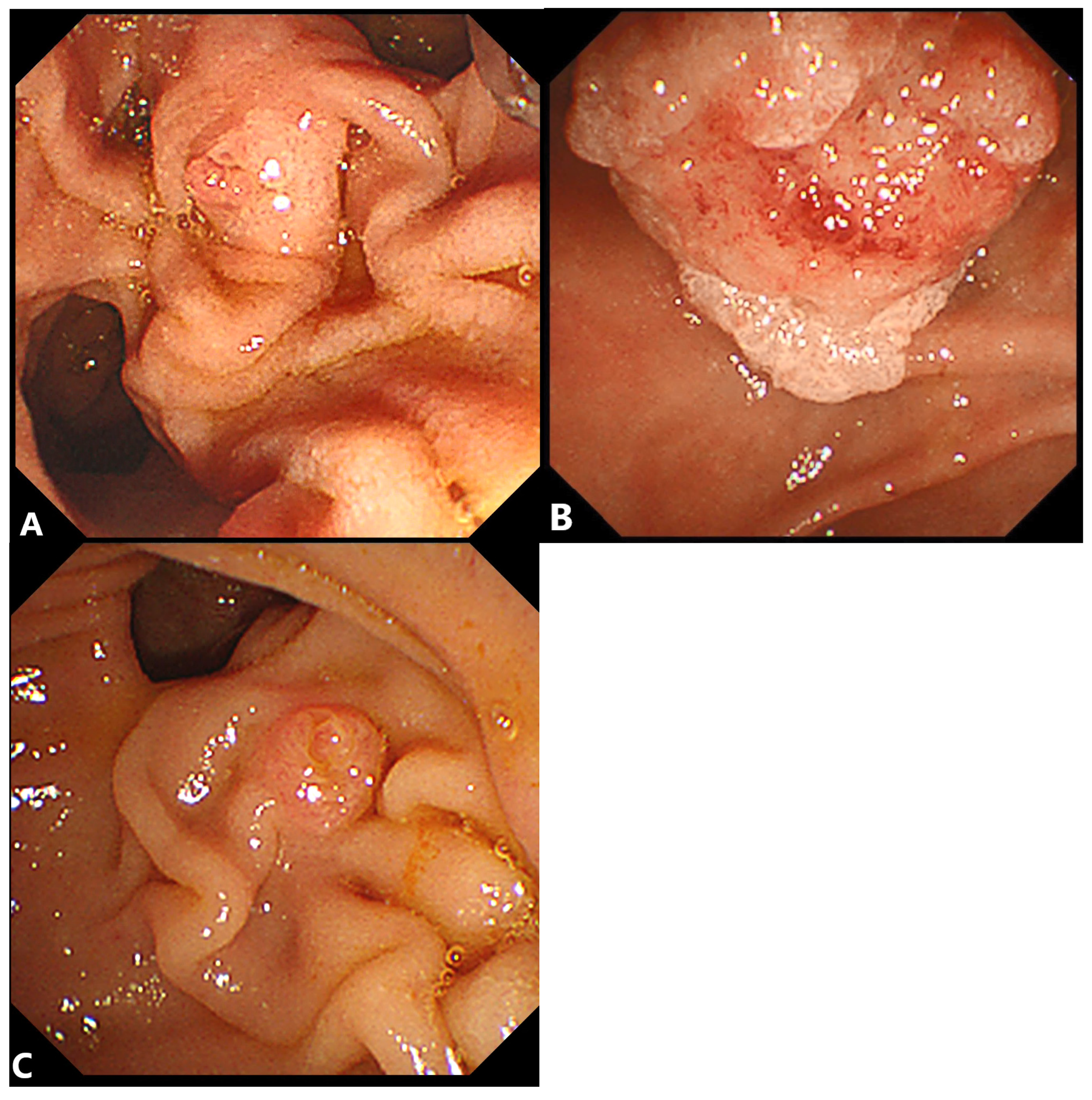
Figure 1. Examples of anatomical variants of major papilla that are predisposed to difficult biliary cannulation. (A) Floppy papilla. (B) Ampullary adenoma. (C) Presence of a periampullary diverticulum with difficult bile duct axis.
Gastroent 12 00039 g001
Figure 2. Salvage methods for difficult bile duct access.
Gastroent 12 00039 g002
Figure 3. Bile duct cannulation by double-wire technique. (A) (left) Luminal view: The yellow-black wire represents the pancreatic duct wire with the sphincterotome cannulating along the bile duct axis. (B) (right) Fluoroscopic view: Orientation of bile duct and pancreatic duct wires.
Gastroent 12 00039 g003
Figure 4. Precut papillotomy by needle-knife papillotomy. (A) Precut by needle-knife starting from the orifice on a bulging major papilla. (B) Common channel and bile duct axis exposed after precut.
Gastroent 12 00039 g004
Figure 5. Classification of EUS-guided biliary drainage.
Gastroent 12 00039 g005
Figure 6. EUS-guided Rendezvous technique. (A) EUS-guided transduodenal puncture of the extrahepatic bile duct. (B) Contrast injection through the EUS needle to obtain cholangiogram. (C) Wire negotiating out of the major papilla. (D) Configuration of guidewire after exchange of EUS needle and echoendoscope. (E) Successful bile duct cannulation by sphincterotome by grasping the Rendezvous wire into the accessory channel of duodenoscope. (F) Plastic stent inserted to common bile duct for drainage.
Gastroent 12 00039 g006aGastroent 12 00039 g006bGastroent 12 00039 g006c
Table 1. Summary on advantages and disadvantages of each salvage method for difficult bile duct access.
TechniquesAdvantagesDisadvantages
Double-guidewire (DGW) technique with or without pancreatic stenting
Readily available and straightforward
Relatively easier to master with a short learning curve
Facilitates subsequent pancreatic stent insertion to reduce risk of post-ERCP pancreatitis (PEP)
🗴
Potentially challenging if diminutive or tortuous pancreatic duct (PD)
🗴
Significant risk of PEP if without pancreatic duct stenting
Precut papillotomy
Exposes the papillary orifice and bile duct axis better
Early precut was not found to be associated with a higher risk of PEP
🗴
Risks of perforation, bleeding, cholangitis, and PEP
🗴
Requires adequate preprocedural suspension of antiplatelets or anticoagulants
🗴
Limited by anatomical factors such as ampullary tumours or papilla hidden in a diverticulum
🗴
Requires skilful operators with a considerable learning curve
Percutaneous biliary drainage (PBD)
Quick procedure
Helpful in stabilizing critically ill cases who may not be fit for endoscopy
Good for cases with surgically altered anatomy and non-communicating multisegmental biliary obstruction
🗴
Catheter-related complications necessitating reintervention, hospital admission, and nursing care
🗴
Fluid and electrolyte loss from external drainage
🗴
Risk of flare-up of biliary sepsis, haemorrhage from vascular injury, peritonitis, liver biloma, and pleural injury
🗴
Requires correction of bleeding diathesis and adequate preprocedural suspension of antiplatelets or anticoagulants
🗴
Ascites should be drained beforehand
EUS-guided Rendezvous (EUS-RV) ERCP
Can be performed at the same session when initial ERCP cannulation is unsuccessful
Higher successful rate than precut papillotomy
Fewer adverse events and less need for reintervention
Shorter postprocedural hospital stay
Exempts patients from external drainage
🗴
Technically demanding procedure requiring operators to be familiar with both ERCP and interventional EUS techniques
🗴
May require conversion to EUS-guided transmural drainage as rescue in some patients with malignant biliary obstruction
🗴
Risks of bleeding, pancreatitis, pneumoperitoneum, bile leak, peritonitis, and flare-up of sepsis
Publisher’s Note: MDPI stays neutral with regard to jurisdictional claims in published maps and institutional affiliations.