Case Report

Malignant Transformation of Heterotopic Pancreatic Tissue in a Patient with BRCA2 Mutation

by
Danson Xue Wei Yeo
1,
Nicholette Goh
1,*,
Khoon Leong Chuah
2,
Sanghvi Kaushal Amitbhai
1,
Aung Myint Oo
1,
Abdul Kareem Saleem Ahmed
1 and
Koura Aaryan Nath
1
1
Department of General Surgery, Tan Tock Seng Hospital, Singapore 308433, Singapore
2
Department of Pathology, Tan Tock Seng Hospital, Singapore 308433, Singapore
*
Author to whom correspondence should be addressed.
Gastroenterol. Insights 2021, 12(1), 10-16; https://doi.org/10.3390/gastroent12010002
Submission received: 26 October 2020 / Revised: 22 December 2020 / Accepted: 27 December 2020 / Published: 19 January 2021
(This article belongs to the Section Pancreas)

Abstract

:
Background: Malignant transformation of heterotopic pancreatic tissue is a rare entity with only several case reports published in the scientific literature. Adjuvant chemotherapy following oncological resection for lesions with nodal metastasis has not been well described and there are no guidelines available to guide the management of these patients. Case Presentation: We present a case of gastric heterotopic pancreatic carcinoma with nodal metastasis in a young patient with breast cancer gene (BRCA) 2 mutation. He had undergone a laparoscopic wedge resection for a gastric lesion initially thought to be a gastrointestinal stroma tumor. Given the involvement of the wedge resection margins, the patient underwent a distal gastrectomy with oncological lymph nodal clearance. One out of the 33 harvested lymph nodes harboured micrometastasis while the main gastrectomy specimen did not have any residual malignancy. Following the histological diagnosis, he received an adjuvant chemotherapy regime akin to that prescribed for locally advanced pancreatic adenocarcinoma with good response. This is, to our knowledge, also the first such case report in a patient with BRCA2 mutation. Conclusions: Pre-operative diagnostic confirmation is challenging and endoscopic procedures pose significant false negatives. Reports of nodal metastasis following oncological resection are limited and there are no guidelines regarding adjuvant therapies. We would recommend a chemotherapy regimen similar to that for primary locally advanced pancreatic carcinoma in patients found to have nodal metastasis.

1. Introduction

Heterotopic pancreatic tissue can be found in various locations of the gastrointestinal tract and malignant transformation of this tissue is exceedingly rare with only a few documented cases in scientific literature. Following oncological resection of these lesions, adjuvant therapy for those with a positive nodal status has not been well described and guidelines with regards to management of these patients are lacking. The authors present a young male patient with gastric heterotopic pancreatic carcinoma with nodal metastasis who was subsequently detected to have the breast cancer gene (BRCA) 2 mutation. He had good response to adjuvant chemotherapy following oncological resection. This is to our knowledge, the first such case report in a patient with BRCA2 mutation.

2. Case Report

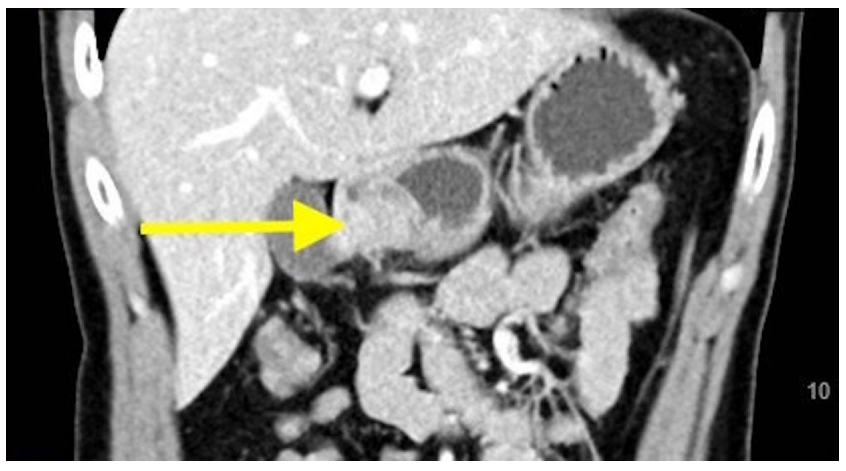
We present a case of a 33-year-old Chinese male initially presenting with dyspepsia for which an oesophagoduodenoscopy was performed. This revealed a submucosal bulge with central umbilication at the antrum (Figure 1), and a subsequent computed tomographic (CT) scan of the abdomen and pelvis showed a 2.3-cm enhancing nodule over the anterior aspect of the pylorus suggestive of a gastrointestinal tract stromal tumour (GIST) (Figure 2 and Figure 3).
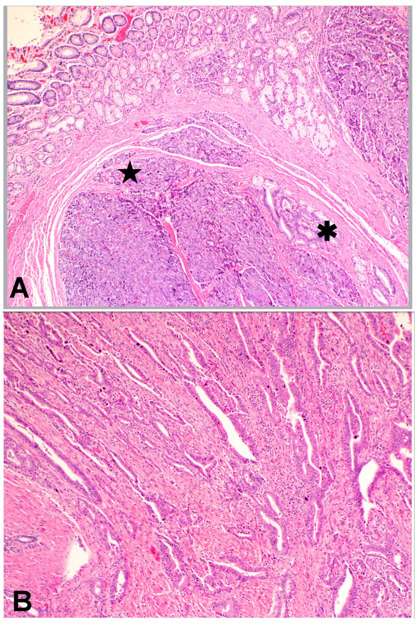
The patient underwent laparoscopic wedge resection of the tumour. Histopathological assessment (Figure 4) demonstrated the presence of heterotopic pancreatic tissue displaying cellular proliferation resembling acinar cells and ducts of the pancreas. An adjacent invasive adenocarcinoma comprising malignant glands was present and demonstrated areas of central necrosis and perineural invasion. The tumour extended to the radial and deep margins. On immunohistochemistry, the tumour showed positive staining for CK7 and focal staining for CDX2 was noted. The stains for synaptophysin, chromogranin, TTF-1, CK20 and Gata3 were negative. ki-67 proliferative index was in the region of 75%. The findings were consistent with a pancreatic ductal adenocarcinoma arising from heterotopic pancreas within the stomach.
A CT scan of the thorax after the first surgery confirmed no distant metastasis. Given the involvement of the wedge resection margins, the patient underwent a distal gastrectomy with oncological lymph nodal clearance. One out of the 33 harvested lymph nodes harboured micrometastasis while the main gastrectomy specimen did not have any residual malignancy. Postoperatively, the patient underwent adjuvant chemotherapy with oxaliplatin, irinotecan, folinic acid and fluorouracil (FOLFOXIRI). The chemotherapy regime was guided by the standard of care for pancreatic cancer after discussion at our multidisciplinary tumour board meeting. A CT scan of the thorax, abdomen and pelvis one year after the surgery did not demonstrate any evidence of local recurrence or metastasis. Tumour markers were not assessed prior to surgery, however cancer antigen 19-9 (CA 19-9) and carcinoembryonic antigen (CEA) levels have remained within the normal range after the surgery.
Following surgery, a discussion regarding hereditary cancer syndromes was held with the patient and he was offered genetic testing consisting of the following genes: APC, ATM, BMPR1A, BRCA1, BRCA2, CDKN2A (p14ARF), CDKN2A (p16INK4a), EPCAM, MEN1, MLH1, MSH2, MSH3, MSH6, NF1, PALB2, PMS2, SMAD4, STK11, TP53, TSC1, TSC2, VHL. This returned as positive for a pathogenic variant in the BRCA2 gene, whereby c.7007 + 1G > C (splice donor) was reported. Of note, he does not have a significant family history of malignancy and genetic screening was also offered to his first-degree family members. The patient will continue to be closely followed up for other BRCA2-related malignancies like breast and prostatic cancer.

3. Discussion

This rare entity of heterotopic pancreatic tissue was first described in 1727 where it was identified by Schultz as an ileal diverticulum and thereafter proven histologically by Klob in 1859 [1]. It has since been found in various locations of the gastrointestinal tract where the most common locations include the stomach, followed by the duodenum, jejunum (accounting for more than 70% of cases) [2,3] and more uncommonly the esophagus, ileum, Meckel diverticulum and biliary tree [4,5]. In the stomach, it has a predilection for the antrum and prepyloric region [6]. Albeit likely under-reported due to its presentation as an incidental finding in most cases, the reported incidence in autopsy studies and upper abdominal surgeries ranges from 0.5% to 13.7% [7]. A further rarity would be the presence of malignant transformation within the heterotopic pancreatic tissue. The incidence of malignant transformation of heterotopic pancreatic tissue is less than 2% [8].
The diagnostic criteria proposed by Guillou et al. to confirm that the malignancy indeed arises from heterotopic pancreatic tissue can be summarized into three main points. Firstly, the tumour must be found within, or close to the aberrant pancreatic tissue. Secondly, identification of a transitional area between pancreatic structures and carcinoma. Lastly, there has to be evidence of pancreatic acini and/or ductal structures seen within the non-neoplastic heterotopic pancreatic tissue [9].
Although the diagnosis of ectopic pancreatic tissue is often incidental, the majority of patients with malignancy are symptomatic at presentation with abdominal pain being the predominant complaint in more than half [10]. These ectopic pancreatic tumours are most commonly found in the stomach and a subepithelial tumour-like appearance is most frequently observed. With histological analysis, adenocarcinoma is the commonest subtype. It is interesting to note that in patients with ectopic pancreatic adenocarcinoma, the rate of having an elevated serum tumour marker is lower than that compared to primary pancreatic cancer [10,11].
It remains a challenge to obtain pre-operative confirmation of the diagnosis with conventional imaging like endoscopic ultrasound or computed tomographic scans as they often reveal a nonspecific mass. Common differentials include gastrointestinal tract stromal tumours (GIST), carcinoid tumours and gastric cancers. More than half of ectopic pancreatic tissue is located within the submucosal layer, while less than a fifth are located within the submucosa and muscularis propria [12]. Given that the commonest location for these tumours is the gastric antrum, endoscopic ultrasound should be a consideration. Ectopic pancreatic tissue is identified based not only on its location within the gastrointestinal wall, but also described to be a poorly defined, low echoic mass with an area of internal spotty high echo inside with a spindle-shaped, thickened muscularis propria neighbouring the lesion [12,13].
However, in majority of the cases described in literature, surgical resection was performed following the initial diagnosis of other tumours like GIST or gastric cancers and the final histology of ectopic pancreatic cancer was confirmed based on the resected surgical specimen [10]. Endo et al. described the diagnosis of malignancy based on pre-operative biopsy and have advocated for the use of endoscopic biopsy such as endoscopic ultrasonography-guided, fine-needle aspiration (EUS-FNA) and endoscopic mucosal resection with a transparent plastic cap-fitted panendoscope (EMR-C) followed by a biopsy [10]. Nonetheless, there appears to be a significant false negative rate in the biopsy of submucosal lesions—up to 60% in a series by Matsuki et al. [14].
Besides breast and ovarian cancer, germline mutations in BRCA2 significantly increases the risk of pancreatic cancer with a relative risk of 3.51 [15]. While familial pancreatic cancers account for 5–10% of pancreatic cancers [16], Goggins et al. [17] found that the incidence of germline BRCA2 mutations in apparently sporadic pancreatic cancer may be at least as high (7.3%) as in breast or ovarian cancer, even in patients without family history, suggestive of an inherited predisposition to cancer. There are reports of earlier onset of pancreatic cancer is cases with BRCA2 mutations, with a mean age of 53–62 years [18,19], however 5 year survivals are no different from that of pancreatic cancer patients without genetic mutations. Pancreatic cancer may result from the germline mutations in various cancer susceptibility genes such as BRCA1, BRCA2, TP53, PALB2, p16/CDKN2A, SMAD4, STK11, ataxia-telangiectasia-mutated (AM) gene and mismatch repair (MMR) genes [16].
To date, there are no clear guidelines for the management of malignant ectopic pancreas. Authors have recommended local excision for lesions proven to be benign and extensive resection with oncological lymph nodal clearance for malignant lesions [20]. As such, pre-operative confirmation of the neoplastic process would be beneficial. There are limited reports of adjuvant therapy following oncological resection of these tumours. Hickman et al. describe that the course of ectopic pancreatic carcinomas within the stomach appear to behave like primary gastric cancers and suggest that they be managed in a similar fashion [21], while other patients underwent adjuvant chemotherapy similar to that for pancreatic cancer [22]. There is no available literature on the efficacy of chemotherapy for adenocarcinoma arising from a heterotopic pancreas [23]. In our patient, the adjuvant chemotherapy regime was akin to that prescribed for locally advanced pancreatic adenocarcinoma, for which in the recent PRODIGE 4/ACCORD 11 randomized trials, a four-drug regimen called FOLFIRINOX demonstrated to prolong overall survival compared to conventional gemcitabine monotherapy [24]. Given his BRCA2 status, the patient will continue to be closely followed-up for other associated malignancies like prostatic cancer and his family members have also been offered genetic screening. In terms of follow-up for the malignant ectopic pancreas, the surveillance with regular cross-sectional imaging would not differ from that of locally advanced pancreatic adenocarcinoma.

4. Conclusions

Adenocarcinoma arising from heterotopic pancreatic tissue as a result of a BRCA2 mutation is exceedingly rare. Pre-operative diagnostic confirmation is challenging and endoscopic procedures pose significant false negatives. Reports of nodal metastasis following oncological resection are limited and there are no guidelines regarding adjuvant therapies. We would recommend a chemotherapy regimen similar to that for primary locally advanced pancreatic carcinoma in patients found to have nodal metastasis. When atypical cancers are found in young patients, genetic counselling should be strongly considered after discussion with the patient.

Author Contributions

Conceptualization, data curation, literature review, writing of original draft, review and editing, D.X.W.Y. and N.G. Pathohistological analysis, review and editing, K.L.C. Review and editing S.K.A., A.M.O. and A.K.S.A. Main attending consultant of patient, supervision, K.A.N. All authors have read and agreed to the published version of the manuscript.

Funding

This research received no external funding.

Institutional Review Board Statement

Not applicable.

Informed Consent Statement

Consent to publish the personal and clinical details (including figures) has been obtained from the patient.

Data Availability Statement

Not applicable.

Conflicts of Interest

The authors declare no conflict of interest.

References

  1. Hunt, V.C. Meckel’s diverticulum containing abberant pancreas. Arch. Surg. 1934, 28, 425–439. [Google Scholar] [CrossRef]
  2. Goodarzi, M.; Rashid, A.; Maru, D. Invasive ductal adenocarcinoma arising from pancreatic heterotopia in rectum: Case report and review of literature. Hum. Pathol. 2010, 41, 1809–1813. [Google Scholar] [CrossRef] [PubMed]
  3. Stock, C.; Keutgen, X.M.; Pisapia, D.; Crawford, C.; Zarnegar, R. Heterotopic pancreatic neoplasm presenting as an obstructing mass at the fourth portion of the duodenum. JOP 2011, 12, 241–243. [Google Scholar] [PubMed]
  4. Ulrych, J.; Fryba, V.; Skalova, H.; Krska, Z.; Krechler, T.; Zogala, D. Premalignant and malignant lesions of the heterotopic pancreas in the esophagus: A case report and review of the literature. J. Gastrointest. Liver Dis. 2015, 24, 235–239. [Google Scholar] [CrossRef] [PubMed]
  5. Kung, J.W.; Brown, A.; Kruskal, J.B.; Goldsmith, J.D.; Pedrosa, I. Heterotopic pancreas: Typical and atypical imaging findings. Clin. Radiol. 2010, 65, 403–407. [Google Scholar] [CrossRef]
  6. Song, D.E.; Kwon, Y.; Kim, K.R.; Oh, S.T.; Kim, J.S. Adenocarcinoma arising in gastric heterotopic pancreas: A case report. J. Korean Med. Sci. 2004, 19, 145–148. [Google Scholar] [CrossRef] [PubMed] [Green Version]
  7. Herold, G.; Kraft, K. Adenocarcinoma arising from ectopic gastric pancreas: Two case reports with a review of the literature. Z. Gastroenterol. 1995, 33, 260–264. [Google Scholar]
  8. Yamaoka, Y.; Yamaguchi, T.; Kinugasa, Y.; Shiomi, A.; Kagawa, H.; Yamakawa, Y.; Numata, M.; Sugimoto, S.; Imai, K.; Hotta, K.; et al. Adenocarcinoma arising from jejunal ectopic pancreas mimicking peritoneal metastasis from colon cancer: A case report and literature review. Surg. Case Rep. 2015, 1, 114. [Google Scholar] [CrossRef] [Green Version]
  9. Guillou, L.; Nordback, P.; Gerber, C.; Schneider, R.P. Ductal adenocarcinoma arising in a heterotopic pancreas situated in a hiatal hernia. Arch. Pathol. Lab. Med. 1994, 118, 568–571. [Google Scholar]
  10. Endo, S.; Saito, R.; Ochi, D.; Yamada, T.; Hirose, M.; Hiroshima, Y.; Yamamoto, Y.; Ueno, T.; Hasegawa, N.; Moriwaki, T.; et al. Effectiveness of an endoscopic biopsy procedure using EUS-FNA and EMR-C for diagnosing adenocarcinoma arising from ectopic pancreas: Two case reports and a literature review. Intern. Med. 2014, 53, 1055–1062. [Google Scholar] [CrossRef] [Green Version]
  11. Safi, F.; Schlosser, W.; Falkenreck, S.; Beger, H.G. Prognostic value of CA 19-9 serum course in pancreatic cancer. Hepatogastroenterology 1998, 45, 253–259. [Google Scholar] [PubMed]
  12. Yasuda, K.; Nakajima, M.; Yoshida, S.; Kiyota, K.; Kawai, K. The diagnosis of submucosal tumors of the stomach by endoscopic ultrasonography. Gastrointest. Endosc. 1989, 35, 10–15. [Google Scholar] [CrossRef]
  13. Matsushita, M.; Hajiro, K.; Okazaki, K.; Takakuwa, H. Gastric aberrant pancreas: EUS analysis in comparison with the histology. Gastrointest. Endosc. 1999, 49, 493–497. [Google Scholar] [CrossRef]
  14. Matsuki, M.; Gouda, Y.; Ando, T.; Matsuoka, H.; Morita, T.; Uchida, N.; Kuriyama, S. Adenocarcinoma arising from aberrant pancreas in the stomach. J. Gastroenterol. 2005, 40, 652–656. [Google Scholar] [CrossRef] [PubMed]
  15. The Breast Cancer Linkage Consortium. Cancer risks in BRCA2 mutation carriers. J. Natl. Cancer Inst. 1999, 91, 1310–1316. [Google Scholar] [CrossRef] [PubMed]
  16. Iqbal, J.; Ragone, A.; Lubinski, J.; Lynch, H.T.; Moller, P.; Ghadirian, P.; Foulkes, W.D.; Armel, S.; Eisen, A.; Neuhausen, S.L.; et al. The incidence of pancreatic cancer in BRCA1 and BRCA2 mutation carriers. Br. J. Cancer 2012, 107, 2005–2009. [Google Scholar] [CrossRef]
  17. Goggins, M.; Schutte, M.; Lu, J.; Moskaluk, C.A.; Weinstein, C.L.; Petersen, G.M.; Yeo, C.J.; Jackson, C.E.; Lynch, H.T.; Hruban, R.H.; et al. Germline BRCA2 gene mutations in patients with apparently sporadic pancreatic carcinomas. Cancer Res. 1996, 56, 5360–5364. [Google Scholar] [PubMed]
  18. Phelan, C.M.; Lancaster, J.M.; Tonin, P.; Gumbs, C.; Cochran, C.; Carter, R.; Ghadirian, P.; Perret, C.; Moslehi, R.; Dion, F.; et al. Mutation analysis of the BRCA2 gene in 49 site-specific breast cancer families. Nat. Genet. 1996, 13, 120–122. [Google Scholar] [CrossRef]
  19. Couch, F.J.; Johnson, M.R.; Rabe, K.G.; Brune, K.; de Andrade, M.; Goggins, M.; Rothenmund, H.; Gallinger, S.; Klein, A.; Petersen, G.M.; et al. The prevalence of BRCA2 mutations in familial pancreatic cancer. Cancer Epidemiol. Biomark. Prev. 2007, 16, 342–346. [Google Scholar] [CrossRef] [Green Version]
  20. Jeng, K.S.; Yang, K.C.; Kuo, S.H. Malignant degeneration of heterotopic pancreas. Gastrointest. Endosc. 1991, 37, 196–198. [Google Scholar] [CrossRef]
  21. Hickman, D.M.; Frey, C.F.; Carson, J.W. Adenocarcinoma arising in gastric heterotopic pancreas. West. J. Med. 1981, 135, 57–62. [Google Scholar] [PubMed]
  22. Kaneko, T.; Ohara, M.; Okamura, K.; Fujiwara-Kuroda, A.; Miyasaka, D.; Yamabuki, T.; Takahashi, R.; Komuro, K.; Suzuoki, M.; Iwashiro, N.; et al. Adenocarcinoma arising from an ectopic pancreas in the duodenum: A case report. Surg. Case Rep. 2019, 5, 126. [Google Scholar] [CrossRef] [PubMed] [Green Version]
  23. Fukino, N.; Oida, T.; Mimatsu, K.; Kuboi, Y.; Kida, K. Adenocarcinoma arising from heterotopic pancreas at the third portion of the duodenum. World J. Gastroenterol. 2015, 21, 4082–4088. [Google Scholar] [CrossRef] [PubMed]
  24. Conroy, T.; Desseigne, F.; Ychou, M.; Bouche, O.; Guimbaud, R.; Becouarn, Y.; Adenis, A.; Raoul, J.L.; Gourgou-Bourgade, S.; de la Fouchardiere, C.; et al. FOLFIRINOX versus gemcitabine for metastatic pancreatic cancer. N. Engl. J. Med. 2011, 364, 1817–1825. [Google Scholar] [CrossRef] [PubMed] [Green Version]
Figure 1. Endoscopic photograph of submucosal bulge with central umbilication.
Gastroent 12 00002 g001
Figure 2. Computed tomographic scan image (transverse cuts) demonstrating the gastric pylorus tumour.
Gastroent 12 00002 g002
Figure 3. Computed tomographic scan image (coronal cuts) demonstrating the gastric pylorus tumour.
Gastroent 12 00002 g003
Figure 4. (A). Low-power view revealing the presence of heterotopic pancreatic tissue with acinar cells (star) and pancreatic ducts (asterisk) in the submucosa of the stomach. (H&E ×40); (B). Medium-power view showing malignant glands invading the muscularis layer of the stomach with accompanying desmoplasia (H&E, ×100).
Gastroent 12 00002 g004
Publisher’s Note: MDPI stays neutral with regard to jurisdictional claims in published maps and institutional affiliations.