Brief Report

Susceptibility Weighted Imaging in Migraines with and Without Aura: A Case–Control Study

1
Department of Neurology, Inselspital, University Hospital Bern, University of Bern, 3010 Bern, Switzerland
2
Institute of Diagnostic and Interventional Neuroradiology, Bern University Hospital, 3010 Bern, Switzerland
3
CTU Bern, University of Bern, 3012 Bern, Switzerland
4
Centre for Migraine and Headache, Bellevue Medical Group, 8001 Zürich, Switzerland
*
Author to whom correspondence should be addressed.
These authors contributed equally to this work.
Neurol. Int. 2025, 17(7), 104; https://doi.org/10.3390/neurolint17070104
Submission received: 18 May 2025 / Revised: 24 June 2025 / Accepted: 27 June 2025 / Published: 8 July 2025
(This article belongs to the Section Pain Research)

Abstract

Background: The asymmetry of cortical veins in susceptibility weighted imaging (SWI) in MRI might be a biomarker for migraine auras and cortical spreading depression (CSD). The aim of this study was to assess in humans if SWI asymmetry can be found in patients who have migraine attacks without auras. Methods: We included patients (n = 100 per group) from the emergency room setting when they (i) presented with an acute neurological deficit or headache; (ii) had a discharge diagnosis of a migraine aura, a migraine without an aura, or neither (controls without stroke or epilepsy); and (iii) had a brain MRI with SWI in the acute setting. Results: In the migraines with auras group, SWI asymmetry was found in 26% (95% CI 18–35) compared to patients with migraines without auras (3%, [95% CI 1–8], p < 0.001) and controls 7% [95% CI 3–14], p < 0.001). There was no difference between patients with migraines without auras and controls (p = 0.19). Conclusions: The distinct SWI changes in migraines with and without auras suggest that CSD might not be involved in the pathophysiology of migraines without auras.

1. Introduction

There is still uncertainty about whether cortical spreading depression (CSD) causes migraine headaches, particularly in patients with migraines without auras [1,2]. While several experimental studies demonstrated the activation of peripheral and/or central nociceptive trigeminal pathways by CSD [3,4,5,6,7,8,9], others have failed to confirm this association [10,11]. The hypothesis that CSD causes migraine headaches is challenged by clinical observations, such as different responses to prophylactic treatments of migraine headaches compared to auras, the lack of a locked temporal relationship between migraine auras and migraine headaches, and the occurrence of migraine auras without headaches [1,12,13,14,15].
Susceptibility weighted imaging (SWI) in magnetic resonance imaging (MRI) depicts cerebral veins according to their oxyhemoglobin concentration [16]. In 1944, Leão demonstrated the dilation of pial arteries and veins in rabbits, as well as an increase in oxyhemoglobin in pial veins following a wave of CSD [17]. Asymmetric abnormalities on SWI, i.e., dilated veins, can be found during or shortly after migraine auras and possibly reflect recent CSD [18].
The aim of this study was to assess in humans if SWI asymmetry can be identified in patients who have migraine attacks without auras to test the hypothesis that migraines without auras might also be caused by CSD, which would then be considered ‘silent auras’. For this, we searched for traces of CSD using SWI in emergency room patients who had spontaneous acute migraine attacks without auras.

2. Methods

This is a retrospective case–control study that was approved by the local ethics committee (Bern cantonal ethics committee, 2020-00115, approval date 3 February 2020). The preliminary results were presented as a poster at the European Academy of Neurology in 2023. Patients were recruited as part of a previous study [19]. Included patients had to provide written informed consent, as required by the local regulations. The written general consent allows patient-related data from the routine clinical care to be used in anonymized form for research purposes. In brief, patients with migraine with aura and controls without migraine and aura (n = 100 per group, consecutive for each group) were included when they (i) presented at our emergency department with an acute neurological deficit, (ii) had a brain MRI either when symptoms were still present or within eight hours after cessation of symptoms, and (iii) had a discharge diagnosis of migraine with aura or diagnoses other than migraine, acute ischemic stroke, and epileptic seizures (controls). Patients with alternative explanations for SWI changes (e.g., brain tumor and vascular malformation) were excluded. The cut-off of eight hours has been chosen based on previous findings demonstrating persistent dilated cerebral veins within this timeframe in patients with migraine aura [18]. Patients with migraine without aura (n = 100) were included when (i) they had a brain MRI performed during the migraine headache and (ii) had a discharge diagnosis of migraine without aura. Due to the exploratory nature of this analysis and lack of data on frequency of SWI changes in migraine without aura, no prior power calculation was performed.
Clinical information was taken from the discharge letters. For this study, health-related data assessed within the scope of clinical routine examinations and decision making were used. Diagnoses have been made by experienced neurologists in the emergency department who have been trained to identify migraine with and without aura according to the International Classification of Headache Disorders [20]. No imputation for missing data has been performed. AS and CJS had access to all clinical and radiological data.
The SWI changes were assessed by two neuroradiologists (TK and VP) blinded to the diagnosis. An SWI change was defined as an asymmetry right versus left involving at least two veins (Figure 1). The MRI scanners used had a field strength of 1.5 or 3 Tesla. The SWI slice thickness varied from 1.2 mm to 2.0 mm. SWI images were first visually screened for unilateral prominence of cortical veins by the rating of neuroradiologists. When ≤2 veins appeared prominent, as rated by the expert neuroradiologist, we applied the approach described in the study by Slavova et al. [21]. The maximal diameter of the most prominent vein was measured on the source SWI image and divided by the diameter of its contralateral homolog as originally applied for the definition of index vein. A ratio ≥ 2.0 was considered significant. When >2 veins appeared prominent after visual analysis, the venous asymmetry was graded qualitatively (mild/moderate/marked) by the two blinded neuroradiologists (VP and TK) in the predefined regions as described in previous studies [18,19]. Discrepancies were resolved by consensus.
For the statistical analysis, STATA (StataCorp LCC 1985–2019, College Station, TX, USA) has been used. The categorical data is presented in counts and confidence intervals according to Wilson’s method. Continuous variables are presents as means with standard deviation or medians with interquartile ranges, as appropriate. For comparisons between categorical variables, Chi-square or Fischer’s exact test was used, as appropriate. For comparisons between non-normally distributed continuous variables, Mann–Whitney or Kruskal–Wallis test was performed, as appropriate. For multiple comparisons, Bonferroni correction has been performed to correct for Type I error.
Upon reasonable request, study data will be made available. All methods used in this study were carried out in accordance with the local regulations.

3. Results

There were more women in the migraines with and without auras group compared to controls (68 females with migraine with aura, 86 in migraine without aura, and 55 in controls; PBonferroni < 0.001), and patients with migraines with and without auras were younger than controls (39 ± 15 years old in migraine with aura, 36 ± 11 in migraine without aura, and 61 ± 13 in controls; statistics, PKruskal-Wallis < 0.001). In the migraines with auras, migraines without auras, and controls groups, the MRI was performed after a median of 4 (IQR 2.7–5.9), 72 (IQR 26–216) and 4.1 h (IQR 2.9–6.8) after the onset of symptoms. For co-morbidities see Table 1.
Cohen’s kappa for the SWI assessment was 0.85. In the migraines with auras group, SWI asymmetry was found in 26% (95%CI 18–35), significantly more often than in patients with migraines without auras (3%, 95%CI 1–8, p < 0.001) and controls (7%, 95%CI 3–14, p < 0.001). There was no difference between patients with migraines without auras and controls (p = 0.19). The clinical information of the patients with migraines without auras and controls who had asymmetric SWI is shown in Table 2. In nine patients with a migraine without an aura, MRI was performed within eight hours after the symptom onset. None of these had SWI asymmetry.
In patients with a migraine with an aura, only a speech disturbance was associated with the SWI asymmetry (Table 3).
To assess the impact of the time after the symptoms had ceased on the SWI asymmetry, the frequency of the SWI asymmetry was separately assessed in patients with ongoing and ceased focal neurological symptoms (i.e., in migraine with aura and controls only). In patients with migraines with auras and ongoing symptoms, 29% (8/35) had SWI venous asymmetry compared to 25% (16/65) whose symptoms had ceased prior to the MRI (p = 0.84). In controls, the proportions were 6% (5/77) and 8% (2/23), respectively (p = 0.65). There was no association between the time from the symptom onset to the MRI and SWI changes (Pwhole population = 0.41, Pmigraine aura = 0.24, Pmigraine without aura = 0.47, Pcontrols = 0.76).

4. Discussion

The main finding of our study is the higher frequency of the SWI asymmetry in patients with migraines with auras compared to migraines without auras. Assuming that the SWI changes reflect the CSD, our results suggest that the CSD is not involved in the pathophysiology of migraines without auras [10,11,12,13,14,15,22].
This large-scale study in patients with spontaneous attacks complements a smaller case series using perfusion studies that were not able to show any perfusion changes, i.e., another potential imaging marker suggestive of migraine auras, in migraines without auras in humans [22,23]. In contrast, previous reports demonstrated symmetric occipital or global perfusion changes in the headache phase of migraines without auras [24,25,26], which cannot be compared directly with our findings of asymmetric SWI alterations. Typically, CSD is a unilateral event [27] and should not result in global or bilateral perfusion changes. In our opinion, therefore, both, the presence of symmetric perfusion abnormalities and the lack of asymmetric SWI alterations are consistent with the absence of CSD in migraines without an aura.
We found more SWI asymmetry in controls (7%) than in patients with migraines without an aura (3%), although this was not significant (p = 0.19). Four of the seven controls with SWI changes had transient global amnesia (TGA). Given the association between TGA and migraines with auras [28], the SWI changes in patients with TGA might have been mediated by mechanisms similar to those involved in migraine auras. Similarly, two of the seven control patients with SWI asymmetry had benign positional vertigo, a condition also associated with the diagnosis of migraines, although the relevance of auras was not reported [29].
In contrast to ‘silent auras’ causing attacks of migraines without auras, diencephalic and brainstem structures might be more important as concluded from functional neuroimaging studies about the premonitory phase of triggered and spontaneous migraine attacks [30,31,32]. Our data aligns with a recent case study indicating that the occurrence of headaches in both migraines with and without auras is probably not connected to CSD [32].
One limitation of this study is the long time-lapse between the headache onset and MRI (median 72 h) in migraines without auras compared to the time-lapse between the aura onset and MRI (median 4 h) in the migraines with auras group. There might have been a selection bias of patients with headaches of longer durations, given the clinical suspicion of a secondary headache which requires an MRI examination. However, several points argue against this confounding our results: (i) None of the migraine without aura patients who had the MRI within 8 h after headache onset had SWI asymmetry. This would be extremely uncommon if CSD occurred in patients with migraines without auras, since SWI-alterations persist after the CSD for at least 8 h [18]. (ii) Furthermore, the lack of an association between the time of the symptom onset and SWI changes suggests that the SWI changes are longer lasting, making them a reliable finding in assessing migraine auras. (iii) Furthermore, the frequency of SWI changes in migraine with aura and controls increased only marginally and non-significantly after comparing patients with ongoing symptoms to those with symptoms that had ceased during the MRI. This finding, however, needs confirmation—ideally through longitudinal studies involving repeat MRIs at different time points—since previous reports found an association between venous changes assessed by MRI and the time from the symptom onset in migraines with auras [23]. Due to the heterogeneity of daily and migraine abortive medication and a lack of precise documentation of the time point of the medication intake (e.g., of triptans), we did not assess the possible influence of treatments on our findings. Another limitation of our study is its retrospective design and lack of a systematic assessment of clinical data (e.g., of baseline characteristics such as obesity). Due to the inclusion of patients treated in the emergency department, MRI scanners with different field strengths were utilized. Additionally, there was a variation in the slice thickness of the SWI. The main strength is the large number of subjects investigated.
In conclusion, asymmetric SWI changes in migraines without auras occur significantly less frequently than in migraines with auras. Assuming a canonical succession of the aura to the headache, the lack of SWI changes in patients with migraines without auras argues against ‘silent’ auras causing migraines without auras.

Author Contributions

Conceptualization A.S., R.W., and C.J.S.; Data curation A.S., T.K., D.M., N.S., V.P., and M.B.; Formal analysis A.S., T.K., M.B., and C.J.S.; Investigation A.S., T.K., D.M., and C.J.S.; Methodology, A.S., T.K., N.S., R.W., and C.J.S.; Validation A.S., S.J., U.F., F.R., and C.J.S.; Writing—original draft A.S., and C.J.S.; Writing—review and editing A.S., T.K., D.M., N.S., V.P., S.J., M.B., U.F., F.R., R.W., and C.J.S. All authors have read and agreed to the published version of the manuscript.

Funding

No funding was received for this manuscript.

Institutional Review Board Statement

The study was conducted in accordance with the Bern cantonal ethics committee (2020-00115).

Informed Consent Statement

For the publication of Figure 1, written consent to publish MRI images was obtained.

Data Availability Statement

Upon reasonable request to the corresponding author, data used for this research will be made available.

Conflicts of Interest

D.M., T.K., NS, V.P., M.B., F.R., and R.W.: none. A.S. has received research support from the Swiss Heart Foundation. S.J. has received research support from the Swiss Heart Foundation and Swiss National Science Foundation. U.F. receives research support from the Swiss National Science Foundation (32003B_197009), the Swiss Heart Foundation, and Medtronic. C.J.S.: received research support from the Deutsche Migräne und Kopfschmerzgesellschaft (www.dmkg.de), Eye on Vision Foundation, Swiss Heart Foundation, and Baasch-Medicus Foundation.

References

  1. Goadsby, P.J. Migraine, aura, and cortical spreading depression: Why are we still talking about it? Ann. Neurol. 2001, 49, 4–6. [Google Scholar] [CrossRef] [PubMed]
  2. Bolay, H.; Vuralli, D.; Goadsby, P.J. Aura and Head pain: Relationship and gaps in the translational models. J. Headache Pain 2019, 20, 94. [Google Scholar] [CrossRef] [PubMed]
  3. Olesen, J.; Burstein, R.; Ashina, M.; Tfelt-Hansen, P. Origin of pain in migraine: Evidence for peripheral sensitisation. Lancet Neurol. 2009, 8, 679–690. [Google Scholar] [CrossRef]
  4. Shatillo, A.; Koroleva, K.; Giniatullina, R.; Naumenko, N.; Slastnikova, A.; Aliev, R.; Bart, G.; Atalay, M.; Gu, C.; Khazipov, R.; et al. Cortical spreading depression induces oxidative stress in the trigeminal nociceptive system. Neuroscience 2013, 253, 341–349. [Google Scholar] [CrossRef]
  5. Zhang, X.; Levy, D.; Noseda, R.; Kainz, V.; Jakubowski, M.; Burstein, R. Activation of meningeal nociceptors by cortical spreading depression: Implications for migraine with aura. J. Neurosci. 2010, 30, 8807–8814, Erratum in: J. Neurosci. 2010, 30, 10259. [Google Scholar] [CrossRef] [PubMed]
  6. Zhang, X.; Levy, D.; Kainz, V.; Noseda, R.; Jakubowski, M.; Burstein, R. Activation of central trigeminovascular neurons by cortical spreading depression. Ann. Neurol. 2011, 69, 855–865. [Google Scholar] [CrossRef] [PubMed]
  7. Harriott, A.M.; Chung, D.Y.; Uner, A.; Bozdayi, R.O.; Morais, A.; Takizawa, T.; Qin, T.; Ayata, C. Optogenetic Spreading Depression Elicits Trigeminal Pain and Anxiety Behavior. Ann. Neurol. 2021, 89, 99–110. [Google Scholar] [CrossRef]
  8. Bogdanov, V.B.; Bogdanova, O.V.; Lombard, A.; Chauvel, V.; Multon, S.; Kot, L.I.; Makarchuk, M.Y.; Schoenen, J. Cortical spreading depression decreases Fos expression in rat periaqueductal gray matter. Neurosci. Lett. 2015, 585, 138–143. [Google Scholar] [CrossRef]
  9. Zhao, J.; Levy, D. Modulation of intracranial meningeal nociceptor activity by cortical spreading depression: A reassessment. J. Neurophysiol. 2015, 113, 2778–2785. [Google Scholar] [CrossRef]
  10. Ebersberger, A.; Schaible, H.G.; Averbeck, B.; Richter, F. Is there a correlation between spreading depression, neurogenic inflammation, and nociception that might cause migraine headache? Ann. Neurol. 2001, 49, 7–13. [Google Scholar] [CrossRef]
  11. Fioravanti, B.; Kasasbeh, A.; Edelmayer, R.; Skinner, D.P., Jr.; Hartings, J.A.; Burklund, R.D.; De Felice, M.; French, E.D.; O Dussor, G.; Dodick, D.W.; et al. Evaluation of cutaneous allodynia following induction of cortical spreading depression in freely moving rats. Cephalalgia 2011, 31, 1090–1100. [Google Scholar] [CrossRef] [PubMed]
  12. Wolthausen, J.; Sternberg, S.; Gerloff, C.; May, A. Are cortical spreading depression and headache in migraine causally linked? Cephalalgia 2009, 29, 244–249. [Google Scholar] [CrossRef] [PubMed]
  13. Bogdanov, V.B.; Multon, S.; Chauvel, V.; Bogdanova, O.V.; Prodanov, D.; Makarchuk, M.Y.; Schoenen, J. Migraine preventive drugs differentially affect cortical spreading depression in rat. Neurobiol. Dis. 2011, 41, 430–435. [Google Scholar] [CrossRef] [PubMed]
  14. Alemdar, M.; Akman, Ö.; Selekler, H.; Komsuoğlu, S.; Ateş, N. Does metoprolol inhibit the cortical spreading depression? Acute effects of systematic metropol on CSD in rats. Cephalalgia 2007, 27, 1010–1013. [Google Scholar] [CrossRef]
  15. Silberstein, S.; Schoenen, J.; Göbel, H.; Diener, H.; Elkind, A.; Klapper, J.; Howard, R. Tonabersat, a gap-junction modulator: Efficacy and safety in two randomized, placebo-controlled, dose-ranging studies of acute migraine. Cephalalgia 2009, 29, 17–27. [Google Scholar] [CrossRef]
  16. Haacke, E.M.; Xu, Y.; Cheng, Y.C.; Reichenbach, J.R. Susceptibility weighted imaging (SWI). Magn. Reson. Med. 2004, 52, 612–618. [Google Scholar] [CrossRef]
  17. Leao, A.A.P.; Morison, R.S. Propagation of spreading cortical depression. J. Neurophysiol. 1945, 8, 33–45. [Google Scholar] [CrossRef]
  18. Kellner-Weldon, F.; Lehmann, V.F.; Breiding, P.S.; Grunder, L.; Muri, R.; Pastore-Wapp, M.; Bigi, S.; Wiest, R.; El-Koussy, M.; Slavova, N. Findings in susceptibility weighted imaging in pediatric patients with migraine with aura. Eur. J. Paediatr. Neurol. 2020, 28, 221–227. [Google Scholar] [CrossRef]
  19. Scutelnic, A.; Petroulia, V.; Schraml, L.; Jung, S.; Branca, M.; Beyeler, M.; Fischer, U.; Wiest, R.; Slavova, N.; Schankin, C.J. The “Index Vein” as a sign for migraine aura in the emergency setting. Cephalalgia 2023, 43, 3331024221132010. [Google Scholar] [CrossRef]
  20. Headache Classification Committee of the International Headache Society (IHS). The International Classification of Headache Disorders, 3rd Edition. Cephalalgia 2018, 38, 1–211. [Google Scholar] [CrossRef]
  21. Slavova, N.; Denier, N.; El-Koussy, M.; Wiest, R.; Kellner-Weldon, F.; Fischer, U.; Schankin, C.J. The index vein pointing to the origin of the migraine aura symptom: A case series. Neurology 2020, 94, e2577–e2580. [Google Scholar] [CrossRef] [PubMed]
  22. Ferrari, M.D.; Haan, J.; Blokland, J.K.; Arndt, J.W.; Minnee, P.; Zwinderman, A.H.; Pauwels, E.K.; Saxena, P.R. Cerebral blood flow during migraine attacks without aura and effect of sumatriptan. Arch. Neurol. 1995, 52, 135–139. [Google Scholar] [CrossRef] [PubMed]
  23. Sanchez del Rio, M.; Bakker, D.; Wu, O.; Agosti, R.; Mitsikostas, D.D.; Østergaard, L.; Wells, W.A.; Rosen, B.R.; Sorensen, G.; Moskowitz, M.A.; et al. Perfusion weighted imaging during migraine: Spontaneous visual aura and headache. Cephalagia 1999, 19, 701–707. [Google Scholar] [CrossRef]
  24. Woods, R.P.; Iacoboni, M.; Mazziotta, J.C. Brief report: Bilateral spreading cerebral hypoperfusion during spontaneous migraine headache. N. Engl. J. Med. 1994, 331, 1689–1692. [Google Scholar] [CrossRef]
  25. Denuelle, M.; Fabre, N.; Payoux, P.; Chollet, F.; Geraud, G. Posterior cerebral hypoperfusion in migraine without aura. Cephalalgia 2008, 28, 856–862. [Google Scholar] [CrossRef]
  26. Bednarczyk, E.M.; Remler, B.; Weikart, C.; Nelson, A.D.; Reed, R.C. Global cerebral blood flow, blood volume, and oxygen metabolism in patients with migraine headache. Neurology 1998, 50, 1736–1740. [Google Scholar] [CrossRef]
  27. Lauritzen, M. Pathophysiology of the migraine aura. The spreading depression theory. Brain 1994, 117, 199–210. [Google Scholar] [CrossRef]
  28. Larner, A.J. Transient global amnesia: Model, mechanism, hypothesis. Cortex 2022, 149, 137–147. [Google Scholar] [CrossRef] [PubMed]
  29. Kim, S.K.; Hong, S.M.; Park, I.S.; Choi, H.G. Association Between Migraine and Benign Paroxysmal Positional Vertigo Among Adults in South Korea. JAMA Otolaryngol. Head Neck Surg. 2019, 145, 307–312. [Google Scholar] [CrossRef]
  30. Schulte, L.H.; Allers, A.; May, A. Hypothalamus as a mediator of chronic migraine: Evidence from high-resolution fMRI. Neurology 2017, 88, 2011–2016. [Google Scholar] [CrossRef]
  31. Maniyar, F.H.; Sprenger, T.; Monteith, T.; Schankin, C.; Goadsby, P.J. Brain activations in the premonitory phase of nitroglycerin-triggered migraine attacks. Brain 2014, 137, 232–241. [Google Scholar] [CrossRef] [PubMed]
  32. Mehnert, J.; Fischer-Schulte, L.; May, A. Aura phenomena do not initiate migraine attacks-Findings from neuroimaging. Headache 2013, 63, 1040–1044. [Google Scholar] [CrossRef] [PubMed]
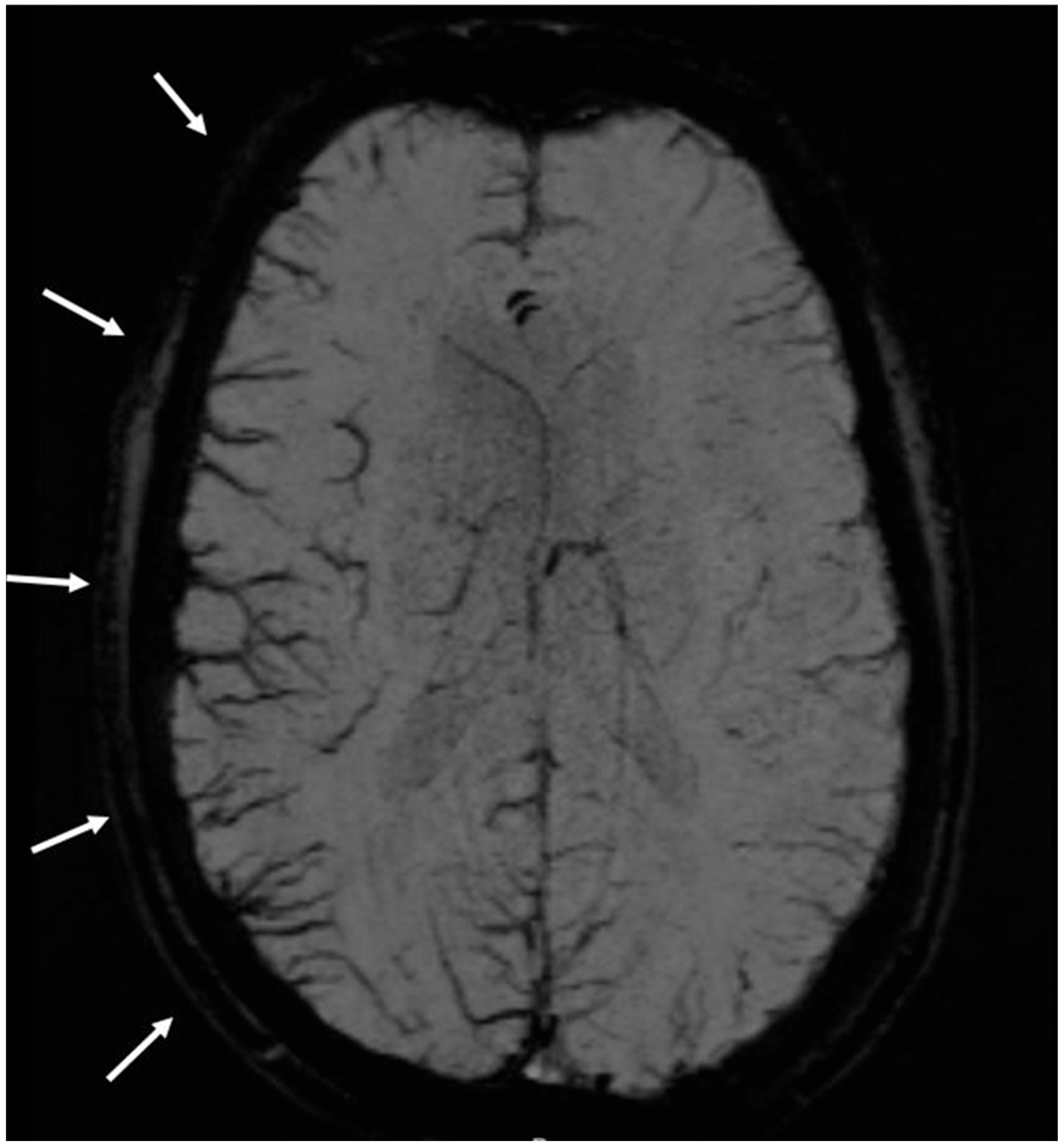
Figure 1. MRI susceptibility-weighted imaging (SWI), MinIP (minimum intensity projection) of a 32-year-old patient presenting to the emergency department with sensory disturbances in the left extremities and recent-onset headaches. The image reveals pseudodilated cerebral veins over the right hemisphere, crossing vascular territories. Discharge diagnosis was migraine with aura.
Neurolint 17 00104 g001
Table 1. Demographics, co-morbidities and vascular risk factors in the migraine with aura (MA), migraine without aura (MO) and controls (C).
Co-Morbidities and Vascular Risk FactorsMigraine with AuraMigraine Without AuraControlsp Value (Bonferroni)Post-Hoc Test
p Value MA vs. MO p Value MA vs. C p Value MO vs. C
age39+/−1536+/−1161+/−13< 0.0010.1<0.001<0.001
female sex688655< 0.0010.090.059<0.001
arterial hypertension15633< 0.0010.030.003<0.001
obesity *13310.620.621
atrial fibrillation2050.180.490.240.059
diabetes mellitus64810.520.570.23
cigarette smoking179140.720.930.550.39
family history of stroke/myocardial infarction012110.491
chronic alcohol abuse ††41410.3610.36
Illicit drugs2121111
depression21180.120.010.0520.46
chronic kidney failure0130.5110.240.62
sleep apnea20410.490.40.12
dyslipidemia9320<0.0010.070.02<0.001
* defined as body mass index of > 25 kg/m2, Mann Whitney, Chi2 or Fisher’s exact test, †† defined as >30 drinks monthly. MA = migraine with aura, MO = migraine without aura, C = controls.
Table 2. Characteristics of the controls and patients with migraine without aura with SWI changes.
NoSexAgeDiagnosis at DischargeCo-MorbiditiesVascular Risk Factors
1male49benign positional vertigohistory of migraine without aura, fungal sinusitis ethmoidalisarterial hypertension
2female71transient global amnesiarestless-legs syndromenone
3male59transient global amnesianonearterial hypertension
4female56benign positional vertigohypothyreosis, osteoarthritisarterial hypertension, smoking, dyslipidemia
5female35idiopathic facial nerve palsynonearterial hypertension
6female74transient global amnesiaIgG (1&3) antibody deficiency syndrome none
7male65transient global amnesianonenone
8female42migraine without auranonedepression
9male44migraine without auranonenone
10male52migraine without auranonearterial hypertension
Table 3. Symptoms in patients with migraine with aura stratified for the presence of SWI changes.
SymptomsSWI Changes Present (N = 26)SWI Changes Absent (N = 74)p Value
Visual symptoms n (%)16 (62)49 (66)0.66
Sensory symptoms n (%)16 (62)37 (50)0.31
Motor symptoms n (%)4 (15)14 (19)0.68
Speech disturbance n (%)17 (65)24 (32)0.003
Coordination problems n (%)3 (12)1 (1)0.053
Vertigo n (%)3 (12)11 (15)0.67
Confusion n (%)01 (1)1
Disclaimer/Publisher’s Note: The statements, opinions and data contained in all publications are solely those of the individual author(s) and contributor(s) and not of MDPI and/or the editor(s). MDPI and/or the editor(s) disclaim responsibility for any injury to people or property resulting from any ideas, methods, instructions or products referred to in the content.