Review

Nanoscale Porphyrin-Based Metal–Organic Frameworks for Enhanced Radiotherapy–Radiodynamic Therapy: A Comprehensive Review

1
The People’s Hospital of Danyang, Affiliated Danyang Hospital of Nantong University, Danyang 212300, China
2
School of Pharmacy, Nantong University, Nantong 226001, China
*
Authors to whom correspondence should be addressed.
Pharmaceutics 2025, 17(7), 883; https://doi.org/10.3390/pharmaceutics17070883
Submission received: 8 May 2025 / Revised: 26 June 2025 / Accepted: 1 July 2025 / Published: 4 July 2025
(This article belongs to the Special Issue Advanced Nanotechnology for Combination Therapy and Diagnosis)

Abstract

The phototherapeutic applications of porphyrin-based nanoscale metal–organic frameworks (nMOFs) are limited by the poor penetration of conventional excitation light sources into biological tissues. Radiodynamic therapy (RDT), which directly excites photosensitizers using X-rays, can overcome the issue of tissue penetration. However, RDT faces the problems of low energy conversion efficiency, requiring a relatively high radiation dose, and the potential to cause damage to normal tissues. Researchers have found that by using some metals with high atomic numbers (high Z) as X-ray scintillators and coordinating them with porphyrin photosensitizers to form MOF materials, the excellent antitumor effect of radiotherapy (RT) and RDT can be achieved under low-dose X-ray irradiation, which can not only effectively avoid the penetration limitations of light excitation methods but also eliminate the defect issues associated with directly using X-rays to excite photosensitizers. This review summarizes the relevant research work in recent years, in which researchers have used metal ions with high Z, such as Hf4+, Th4+, Ta5+, and Bi3+, in coordination with carboxyl porphyrins to form MOF materials for combined RT and RDT toward various cancer cells. This review compares the therapeutic effects and advantages of using different high-Z metals and introduces the application of the heavy atom effect. Furthermore, it explores the introduction of a chemodynamic therapy (CDT) mechanism through iron coordination at the porphyrin center, along with optimization strategies such as oxygen delivery using hemoglobin to enhance the efficacy of these MOFs as radiosensitizers. This review also summarizes the potential of these materials in preclinical applications and highlights the current challenges they face. It is expected that the summary and prospects outlined in this review can further promote preclinical biomedical research into and the development of porphyrin-based nMOFs.

Graphical Abstract

1. Introduction

The structural diversity and facile tunability of MOFs have enabled their widespread investigation across various fields, including gas adsorption, chemical sensing, catalysis, and drug delivery [1,2,3]. Porphyrin-based nMOF materials can effectively prevent the aggregation of hydrophobic porphyrin molecules, making them highly promising as carrier materials for biomedical applications of porphyrin photosensitizers [4,5]. Early research primarily focused on using visible light in the range of 400–680 nm to excite porphyrin photosensitizers, generating reactive oxygen species (ROS) to oxidize biomacromolecules within cancer cells, thereby suppressing cancer cell proliferation [6,7,8]. However, during in vivo treatments, researchers found that the tissue penetration depth of visible light is typically less than 1 cm and that this light is easily absorbed by biological tissues [9]. These limitations necessitate higher doses of photosensitizers, stronger excitation light sources, or the restriction of treatment to superficial tumor cells, significantly limiting the biomedical applications of porphyrin-based nMOF materials.
With continuous advancements in nanoscience technology and medical techniques, researchers have recognized radiotherapy as a highly effective cancer treatment modality [10,11,12]. Radiotherapy using X-rays or γ-rays can directly target DNA within cancer cells, causing strand breaks and structural damage [10,13,14]. Additionally, X-rays can interact with H2O or H2O2 to generate hydroxyl radicals, which are highly oxidative and capable of inhibiting cancer cell proliferation [15,16,17,18]. However, conventional radiotherapy relying solely on X-ray irradiation often faces challenges such as side effects from high doses and low energy conversion efficiency [19]. Therefore, developing low-dose X-ray-activated radiotherapy methods has become an urgent need [20,21,22,23]. Researchers have found that combining X-ray excitation with porphyrin-based nMOFs can fully leverage the deep tissue penetration of X-rays and the advantages of porphyrin-based photosensitizers in RDT [24,25,26,27]. MOF materials can be prepared through the direct coordination of high-Z metals (e.g., Hf, Bi) with porphyrin photosensitizers [28,29]. Under X-ray irradiation, these materials not only directly act on H2O or H2O2 to generate hydroxyl radicals but also utilize the photoelectron transfer effect of high-Z metals (e.g., Hf, Au, Pt) to activate porphyrin photosensitizers, producing singlet oxygen and other ROS, further enhancing cancer cell suppression [28,30,31]. Compared to standalone radiotherapy, this approach achieves desirable therapeutic effects with lower X-ray doses, effectively avoiding systemic side effects of radiotherapy [28]. Thus, the combination of porphyrin-based MOFs with X-rays for RT-RDT holds great potential for biomedical applications [26,27].
Research on RT and RDT has remained a major focus in recent years. In 2020, Ni et al. provided an overview of recent advances in the application of nMOFs as nanosensitizers for PDT, CDT, RT, and radiotherapy–radiodynamic therapy (RT–RDT), highlighting their role in enhancing cancer immunotherapy through effective antigen delivery as nanocarriers for cancer vaccines [32]. A year later, in 2021, Xu et al. reviewed the design principles and post-synthetic functionalization strategies of two-dimensional nanoscale metal–organic layers, with a focus on their use in RT–RDT combination therapy, as well as their potential as platforms for ratiometric sensing, imaging, and targeted drug delivery [33]. In 2022, He et al. presented a comprehensive summary of the latest developments in X-ray-induced photodynamic therapy, discussing its synergistic relationship with RDT and conventional RT [15]. Also in 2022, Pan et al. compiled a review on nanostructure-mediated approaches aimed at achieving precision radiotherapy, emphasizing various nanotechnology-based strategies to improve tumor targeting and reduce collateral damage to healthy tissues, while also addressing current limitations and future directions for clinical translation [34]. RT based on certain high-atomic-number metals can directly destroy cancer cells through physical damage, whereas RDT, which incorporates organic photosensitizer molecules, acts on cells through chemical reactions, such as the generation of ROS. Utilizing a single nanoplatform, such as nMOFs, to achieve combined RT-RDT therapy may represent a highly promising approach for cancer treatment. Therefore, more detailed reviews are needed to summarize recent research efforts focused on nMOF-based nanoplatforms that enable simultaneous RT-RDT combination therapy. These reviews should outline the synthetic strategies employed in fabricating such materials and compare their therapeutic performance, providing an optimized selection framework to identify the most promising candidates for clinical applications.
This review summarizes recent advances in porphyrin-based nMOF materials for RT-RDT combination therapy under X-ray or γ-ray excitation (Figure 1). MOF materials prepared from heavy atom metals and porphyrin ligands exhibit several unique advantages in combined RT and RDT. First, the inherent porous structure of MOFs facilitates the diffusion of ROS, enabling better oxidative effects [35,36]. Second, the high proportion of heavy atom metals and porphyrin ligands in MOF structures allows the efficient delivery of radiosensitizers and porphyrin molecules. Finally, the modifiability and diversity of MOF materials enable the design and synthesis of multifunctional MOFs, potentially leading to unexpected anticancer effects [37,38,39,40]. This review not only expands the biomedical applications of porphyrin-based nMOF materials but also provides new preclinical data references for candidate drugs aimed at effectively suppressing malignant cancer cells.

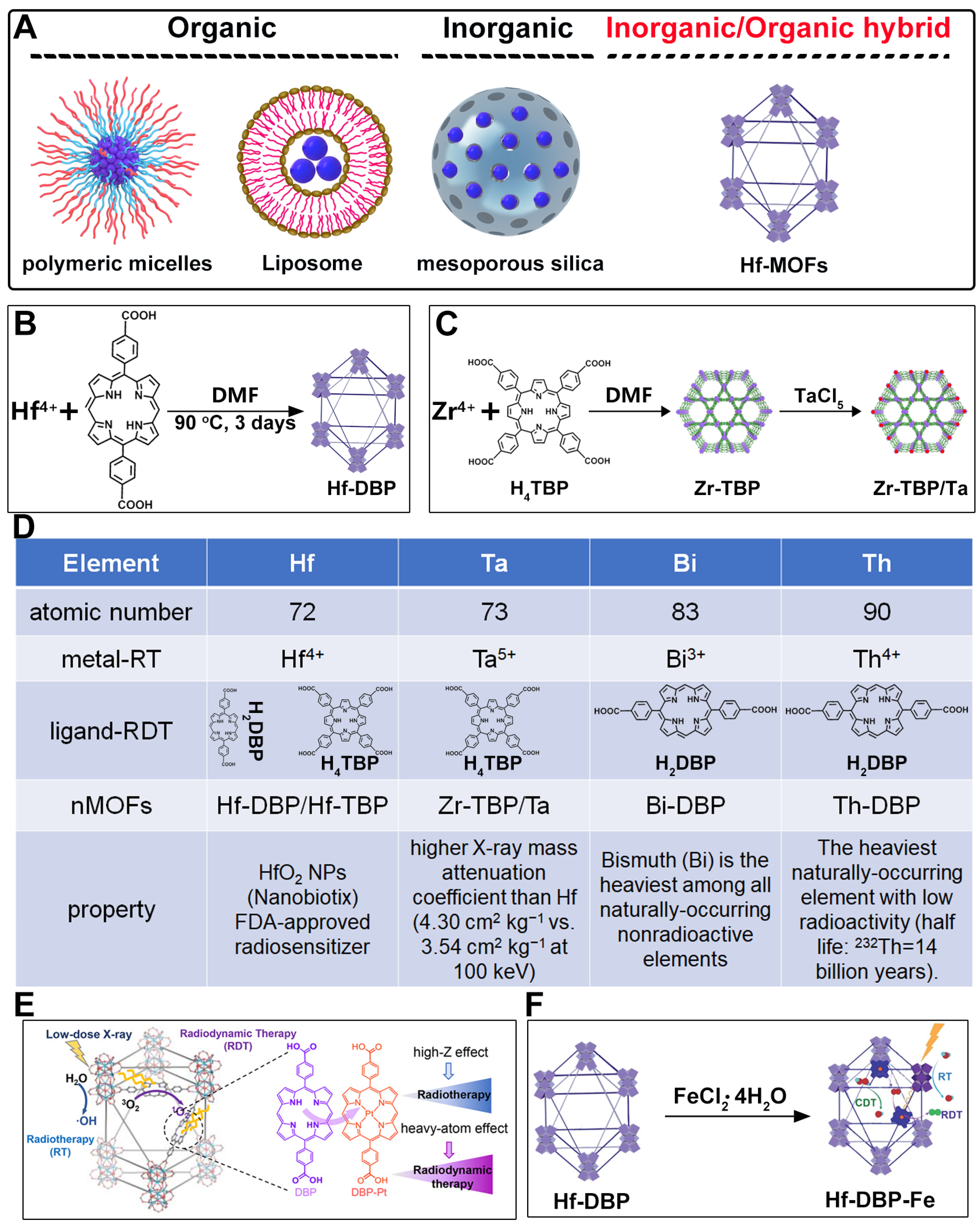
2. Design Rationale and Advantages of Porphyrin–MOFs for Combined RT-RDT

Metal nanoparticles have demonstrated significant potential in biomedical applications due to their unique physicochemical properties. However, their potential in vivo toxicity remains a major concern [41]. To enhance their stability and biocompatibility, various carrier materials are commonly employed (Figure 2A). Traditional carriers mainly include organic materials, such as PEGylated polymers (e.g., DSPE-PEG) or liposomes [42,43,44,45], as well as inorganic materials like mesoporous silica (Figure 2A) [46,47]. These carriers have proven effective in reducing toxicity and improving cellular internalization efficiency. Nevertheless, most of these carrier systems adopt a spherical morphology. Studies have shown that, based on Monte Carlo simulation results, layered structure-shaped nanoparticles outperform cubic and spherical morphologies in terms of radiation dose enhancement [48]. Moreover, conventional polymeric or silica-based carriers are typically inert and lack additional biological functionalities, which limits their capacity for synergistic therapeutic effects. Additionally, the potential long-term toxicity of mesoporous silica poses a barrier to its further clinical translation [49].
Therefore, the development of novel and multifunctional metal carrier materials is of great research significance. MOFs, as hybrid inorganic/organic materials, offer distinct advantages in this field. First, through the adjustment of synthesis conditions such as solvent composition or catalysts (e.g., acid), MOFs can be tailored into nanoscale layered structures, which are beneficial for enhancing radiation dose deposition. Second, their intrinsic porous structure facilitates the generation of hydroxyl radicals during radiotherapy, thereby enhancing therapeutic efficacy. Third, MOFs can be constructed using high-Z metals and porphyrin-based photosensitizers, enabling a synergistic combination of radiotherapy and radiodynamic therapy, offering a more effective strategy for treating refractory malignant tumors. Furthermore, the periodic and ordered architecture of MOFs effectively prevents the aggregation of both metal nanoparticles and photosensitizers, making them more responsive to irradiation. Finally, the structural diversity and facile functionalization capability of MOFs provide broad opportunities for the development of multifunctional therapeutic platforms incorporating various high-Z metals.
The preparation of MOF materials through the coordination of heavy atoms with porphyrin ligands typically involves two approaches: (1) Heavy atoms, such as Hf, can be directly used as metal nodes that participate in the formation of MOF structures (Figure 2B) [28]. This method achieves a higher content of Hf atoms, providing more effective radiosensitization. (2) Alternatively, MOF materials can be prepared first, followed by the introduction of high-Z metals through co-doping (Figure 2C) [50]. This approach not only endows MOF materials with RDT functionality but also expands the variety of radiosensitizers. Studies have shown that the ability of heavy atom metals to effectively attenuate and absorb X-ray energy is strongly correlated with their atomic numbers. This review summarizes research efforts on the preparation of MOF materials using four heavy atom metals—Hf (atomic number: 72) [28], Ta (atomic number: 73) [50], Bi (atomic number: 83) [29], and Th (atomic number: 90) [51]—with dicarboxyl or tetracarboxyl porphyrin ligands for combined RT and RDT to suppress cancer cell proliferation (Figure 2D). Furthermore, this review compares the advantages of these four heavy atom metals and their potential for future preclinical applications.
HfO2 nanoparticles (NPs) based on Hf have already received clinical approval, making research on combined RT and RDT treatments using Hf elements worthy of further time and effort (Figure 2D) [52,53,54,55]. Ta has attracted significant attention due to its higher X-ray mass attenuation coefficient compared to Hf, and its excellent performance as a CT contrast agent and photoacoustic imaging agent keeps related research at the forefront (Figure 2D) [56,57,58,59,60]. Bi not only exhibits superior radiosensitization compared to Hf but is also the heaviest non-radioactive element in nature, potentially offering new opportunities for radiotherapy (Figure 2D) [18,41,61,62,63,64,65,66]. Th is a naturally occurring heavy atom metal; despite its low-level radioactivity, its extremely long half-life makes related research highly promising for bioapplication (Figure 2D) [67,68,69,70,71,72].
In addition to enhancing the combined therapeutic effects of RT and RDT by using metals with higher atomic numbers, the structure of porphyrin ligands can also be optimized. Platinum (Pt)-coordinated porphyrin derivatives have been found to exhibit significantly enhanced photodynamic and photocatalytic performance [73,74,75,76,77,78]. Therefore, the rational design and synthesis of Pt-coordinated porphyrin materials with specific functional groups for the preparation of nMOFs are expected to further enhance their ability to generate ROS under photoexcitation or X-ray irradiation. For example, Pt-coordinated porphyrins can be used to form MOFs, where the heavy atom effect of Pt significantly enhances the efficiency of singlet oxygen generation during RDT (Figure 2E) [79]. Alternatively, porphyrin ligands can coordinate with Fe3+ ions, whose redox capability enables MOF materials to react with excess H2O2 in cancer cells to generate O2, thereby enhancing RDT efficacy [80]. This process also produces more hydroxyl radicals, enabling CDT effects (Figure 2F). These diverse structural designs not only optimize therapeutic outcomes but also further expand the application scope of MOF materials prepared with heavy atom metals and porphyrins.
This review comprehensively summarizes research efforts on porphyrin-based MOFs for combined RT and RDT treatments. To further highlight and compare the structural components of these different MOFs, the size and morphology of the prepared materials, and their mechanisms for inhibiting cancer cells, detailed information on the MOFs discussed in this review is compiled in Table 1. Through a comparison of the morphology and size of these materials and the types of cancer cells inhibited by them, it is evident that porphyrin-based MOFs not only exhibit excellent performance but also possess significant potential for further application expansion.

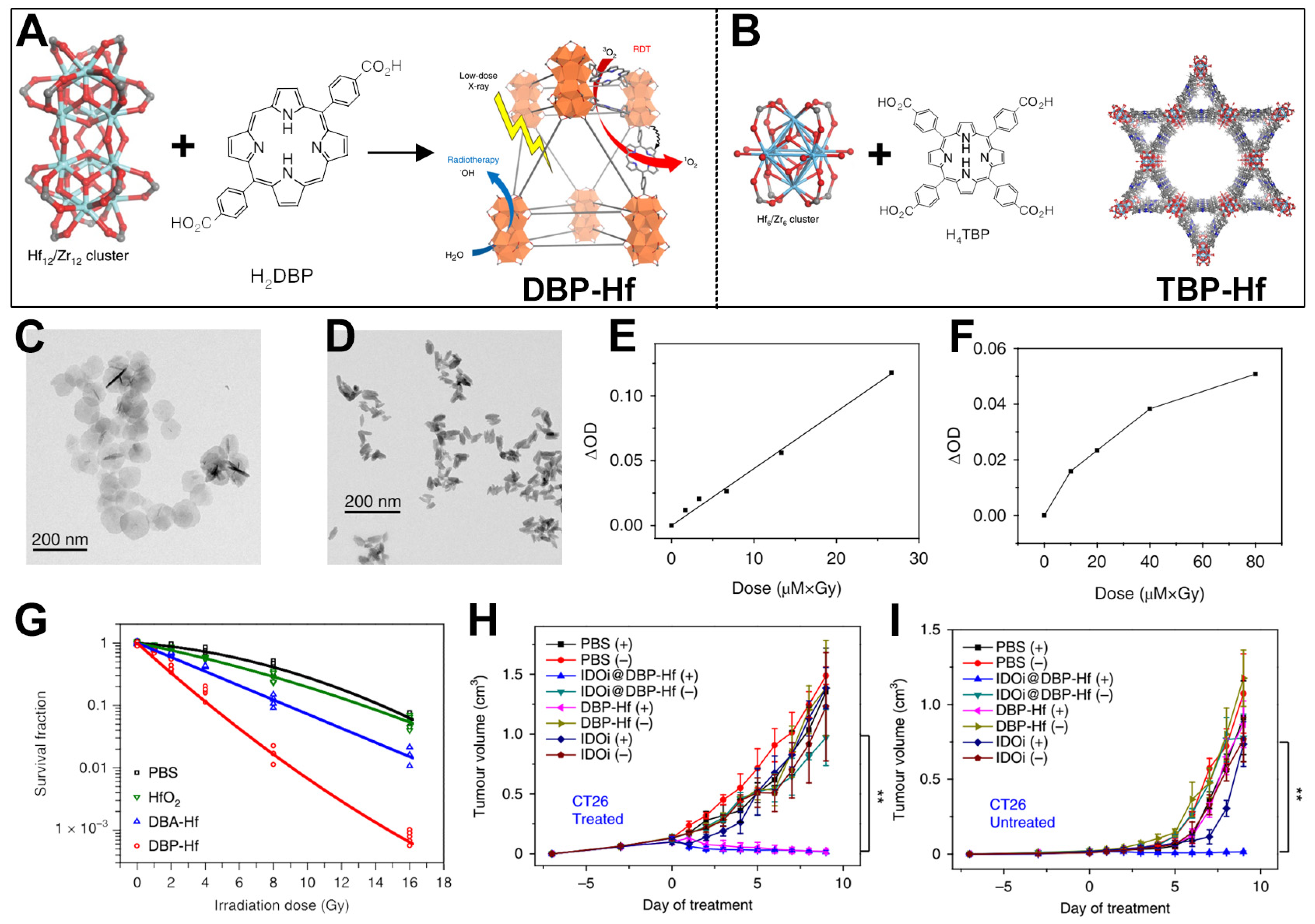
3. Hf-Based nMOFs for Enhanced RT-RDT and Synergistic Immunotherapy

Zr and Hf are elements in Group IVB of the periodic table [83,84,85]. Due to the influence of lanthanide contraction, the atomic and ionic radii of hafnium are very close to those of zirconium, resulting in extremely similar chemical properties. In chemical reactions, hafnium also mainly exhibits an oxidation state of +4. Both Zr and Hf elements can coordinate with porphyrin ligands to form MOF materials [86,87,88,89,90,91]. Differently from Zr, Hf has a relatively high Z of 72, which enables it to absorb X-ray photons for radiotherapy and RDT treatment mediated by porphyrin photosensitizer ligands [28]. Based on this, Lu et al. prepared two types of nMOF materials and used the pore structures of the nMOFs to load IDO inhibitors, achieving the goal of combined treatment of breast cancer and colorectal cancer through X-ray-excited RT, RDT, and checkpoint blockade therapy [28]. The specific structures of the two nMOFs are as follows: 5,15-bis(p-benzoic acid)porphyrin-Hf (DBP-Hf) (Figure 3A) and 5,10,15,20-tetrakis(p-benzoic acid)porphyrin-Hf (TBP-Hf) (Figure 3B). Subsequently, the authors analyzed the morphology and properties of the prepared nMOFs. Transmission electron microscopy (TEM) results showed that DBP-Hf presented a disk-like shape, while TBP-Hf showed a rod-like shape. Then, the authors determined the ability of DBP-Hf and TBP-Hf to generate 1O2 under X-ray irradiation using the 4-nitro-N,N-dimethylaniline assay (Figure 3E,F). As shown in Figure 3E, the ability of DBP-Hf to generate 1O2 showed a certain linear relationship with the product of the nMOF and X-ray doses. Similar results indicated that as the doses of nMOF and X-ray increased, the change in the optical density of 4-nitroso-N,N-dimethylaniline at 439 nm in the TBP-Hf detection system increased, suggesting an increase in the generated 1O2 (Figure 3F). After that, the authors measured the survival response of TUBO cells under different irradiation doses through a colony-forming assay. To highlight the excellent combined radiotherapy and RDT effects of the prepared DBP-Hf, the authors also used HfO2 NPs and DBA-Hf nMOFs prepared from Hf4+ and non-photosensitizer ligands as controls (Figure 3G). The experimental results showed that under the same irradiation dose, the radiosensitization effect of DBP-Hf was better than that of HfO2. The authors speculated that this was because the porosity of nMOFs was beneficial for the formation and diffusion of free radicals. DBP-Hf had the best radiosensitization effect, which originated from the combined effect of Hf-mediated radiotherapy and porphyrin photosensitizer-mediated RDT. Subsequently, taking DBP-Hf as an example, the authors used its pore structure to load an IDO inhibitor (a widely studied immunomodulatory enzyme) [92] and conducted an inhibition experiment on a bilateral tumor model in mice. As shown in Figure 3H, compared with the group injected with the IDO inhibitor alone, both DBP-Hf and IDOi@DBP-Hf under X-ray irradiation could effectively inhibit the growth of CT26 cancer cells. For distant tumors, only IDOi@DBP-Hf plus X-ray irradiation could inhibit the growth of cancer cells in the experimental group through the systemic immune response (Figure 3I). Immunotherapy is a widely researched strategy proven highly effective for cancer treatment [93,94,95]. The porous structure of nMOFs provides significant advantages for the implementation of combination therapies involving immunotherapy. Moreover, the work provides an important reference for the preclinical application of porphyrin-based nMOFs. Differently from PDT with limited penetration depth, this combined RT-RDT treatment method can effectively treat tumors in deep-seated tissues. Even with a 4.5 cm thick beef block in between, X-rays could still effectively activate the Hf–porphyrin nMOF materials, resulting in a cancer cell death rate of over 50%.

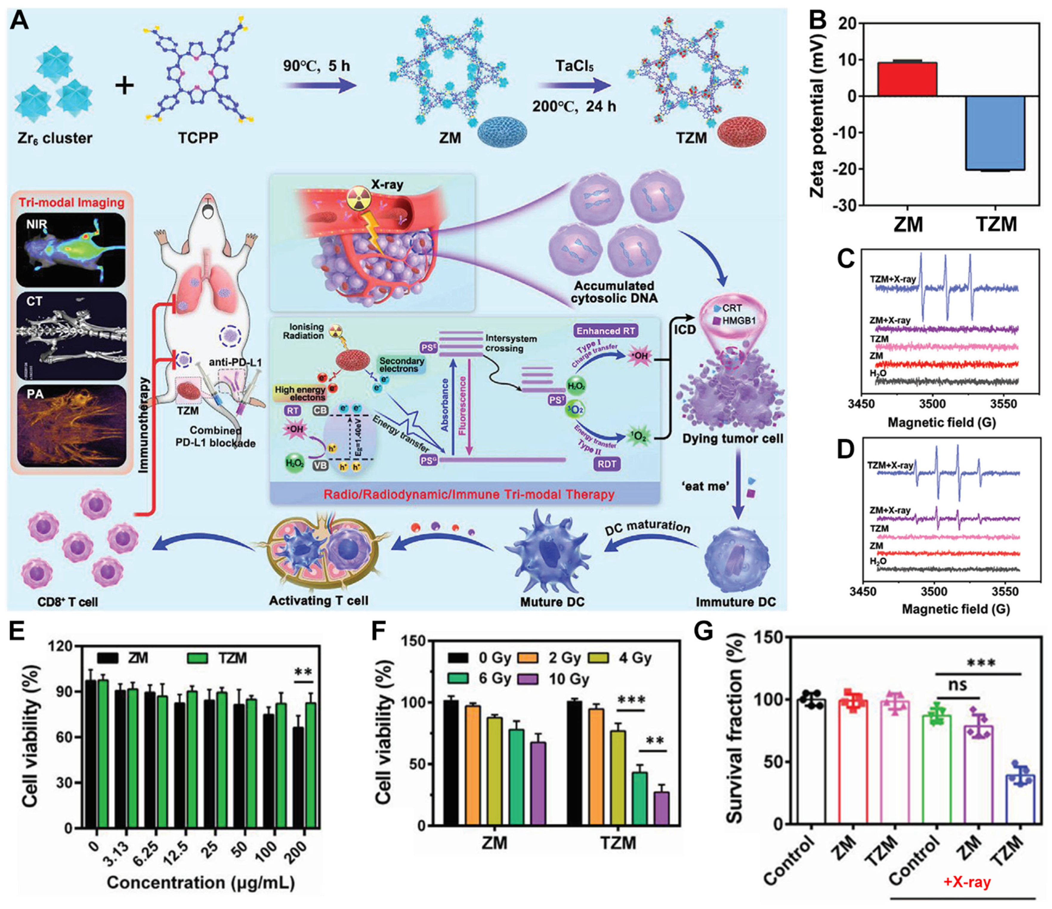
4. Ta-Zr-Co-Doped nMOFs for Combined RT and RDT

In addition to directly using Hf with a high Z to form nMOFs with porphyrins for combined RT-RDT treatment, the method of doping can also be adopted to introduce atoms with high Z into conventional nMOF structures [50]. This approach not only enriches the types of porphyrin-based nMOFs for combined RT-RDT treatment but also provides a new method for combining metals that cannot form nMOFs directly. For instance, Li et al. first synthesized a Zr-TCPP MOF (referred to as ZM) (Figure 4A) [50]. They then doped ZM with TaCl5 to prepare TZM, a Ta-doped Zr-TCPP MOF (Figure 4A). The prepared TZM composite could effectively generate RT and porphyrin-mediated RDT functions under X-ray irradiation, thereby inhibiting the growth and metastasis of osteosarcoma by inducing immunogenic cell death (Figure 4A). Subsequently, the authors found that the surface charge of ZM was 9.2 mV, which changed to −20.2 mV for TZM. This result is consistent with the reported negative charge of Ta NPs and also proves that the Ta element was successfully doped into the Zr-TCPP MOF. Electron spin resonance (ESR) test results showed that only TZM containing high-atomic-number elements could generate 1O2 (Figure 4C), and TZM produced significantly more ·OH than ZM (Figure 4D), indicating the effective sensitization ability of the Ta element. Since ZM does not contain high-atomic-number elements, it cannot effectively exert the functions of RT and RDT. Then, the authors used the CCK-8 assay (a commonly used method for evaluating the cytotoxicity of drug molecules) [96,97,98,99] to verify the inhibitory effects of ZM and TZM on K7M2 cells. Without X-ray irradiation, both ZM and TZM showed good biocompatibility (Figure 4E). As the X-ray intensity increased from 0 Gy to 10 Gy, it was found that the survival rate of K7M2 cells treated with TZM decreased significantly, which was due to the radiosensitization effect of Ta (Figure 4F). Thereafter, the authors co-incubated K7M2 cells with PBS, ZM, and TZM and measured their survival fractions. The results showed that under X-ray irradiation, only the survival fraction of the TZM experimental group was less than 50% (Figure 4G). This result further proves that the radiosensitization ability of the Ta element can effectively endow the Zr-TCPP MOF with RT-RDT functions. This system provides a reference for the further biomedical application of the Ta metal. It also offers a new example for the clinical application of radiosensitizers. However, it is important to note that this system incorporates two metallic elements, Zr and Ta. While each of these metals may exhibit good biocompatibility when used individually, the potential synergistic toxic effects arising from their coexistence within the same nanoparticle system have not been thoroughly investigated. This lack of understanding could represent a significant barrier to their clinical application.

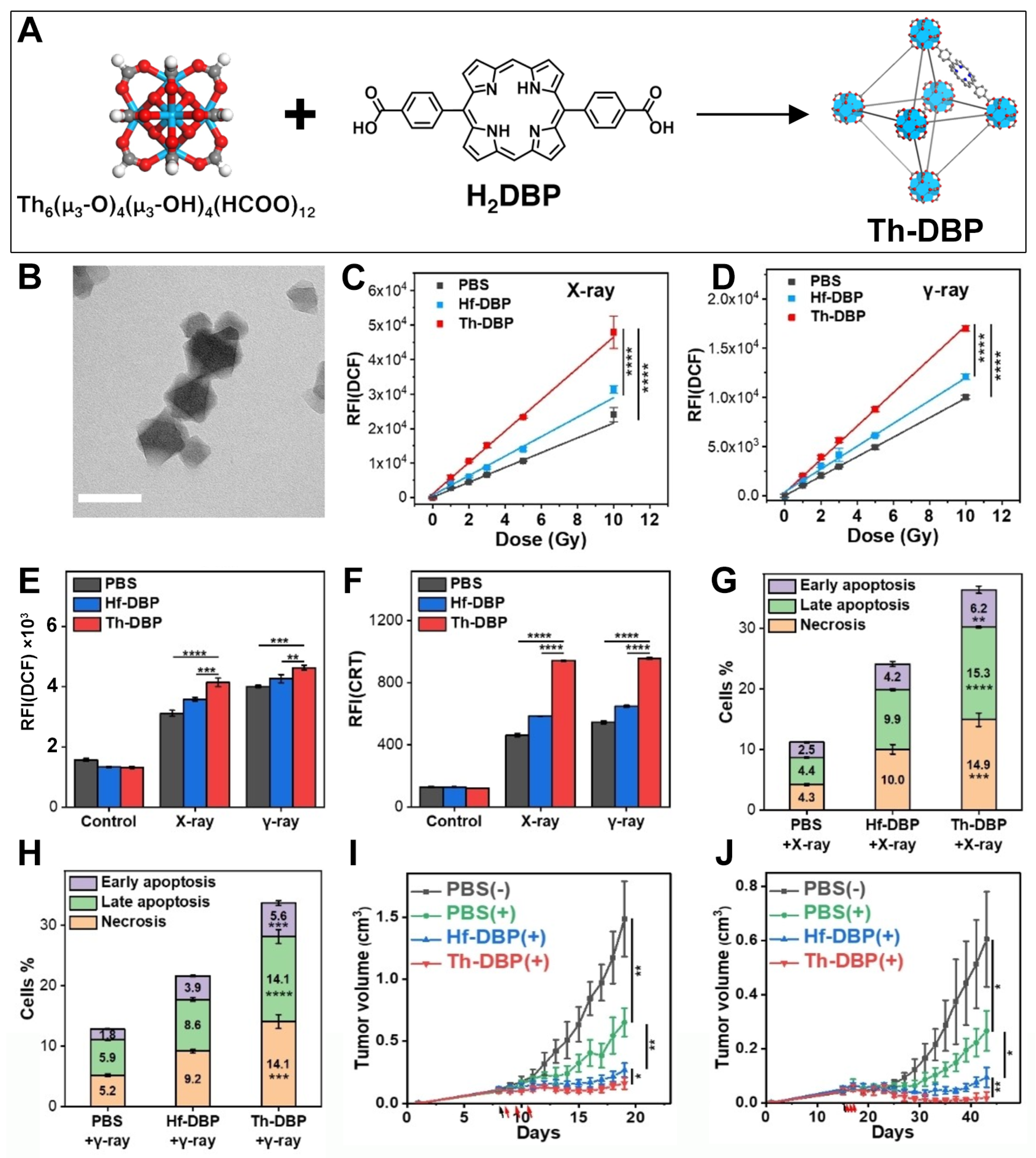
5. Th-Based nMOFs for Combined RT and RDT

The doping method can be used to introduce high-atomic-number radiosensitizers into nMOFs prepared from common metals. However, this method cannot guarantee the maximum loading of radiosensitizers. As radiosensitizers for clinical trials, HfO2 NPs have been approved by the US FDA, widely studied, and used as a control material. There are also many types of Hf-based nMOF materials [89,100,101,102,103]. To further develop nMOF materials with stronger radiosensitization effects, some researchers have found that other metals with more energy deposition under X-rays can be used to form MOF radiosensitizers with porphyrin ligands. For example, Th is a naturally occurring metal element with relatively low radioactivity. Its high Z makes its use in the construction of MOF materials possible. Based on this, Xu et al. used the solvothermal method to prepare Th-DBP MOF (Figure 5A) [51]. TEM characterization results showed that a Th-DBP MOF presented a nano-octahedral morphology with a diameter of ~80 nm (Figure 5B). Thereafter, the authors used DCFH-DA as a probe to detect the ability of Hf-DBP and Th-DBP MOF materials to generate ROS under X-ray and γ-ray irradiation. As shown in Figure 5C,D, Th-DBP had a stronger ability to generate ROS than Hf-DBP under both X-ray and γ-ray irradiation. Moreover, as the ray intensity increased, the amount of ROS generated by Th-DBP also significantly increased. The comparison results of the fluorescence intensity of DCF also indicated that Th-DBP was a stronger radiosensitizer than Hf-DBP (Figure 5E). To further study the treatment results of Th-DBP and Hf-DBP in cell and animal experiments, the authors used PEG polymer materials to modify the surfaces of the two MOF materials. After modification, the two MOF materials had similar cell uptake behaviors. Then, the authors evaluated the expression of calreticulin on the surface of CT26 cancer cells induced by Th-DBP and Hf-DBP. As shown in Figure 5F, due to the stronger ROS-generating ability of Th-DBP under ray irradiation, its ability to induce CRT expression was also significantly improved. Next, the authors conducted cytotoxicity tests. As shown in Figure 5G,H, due to the enhanced combined RT/RDT effect, Th-DBP showed a better effect in killing CT26 cancer cells than Hf-DBP under both X-ray and γ-ray irradiation. Then, the authors further verified the superiority of Th-DBP over Hf-DBP through in vivo experiments. As shown in Figure 5I, under X-ray irradiation, Th-DBP could effectively inhibit the proliferation of CT26 tumor cells in the BALB/c mouse model, showing certain advantages over the Hf-DBP radiosensitizer material. Similarly, as shown in Figure 5J, Th-DBP could also effectively inhibit the proliferation of Panc02 cancer cells in the C57BL/6 mouse model, indicating the effectiveness of Th-MOFs as radiosensitizers for the treatment of malignant tumors. This system not only provides new methods for the development of porphyrin-based MOF materials through the development of new high-atomic-number metals to form porphyrin-based MOF materials but also provides new candidate radiosensitizers for the treatment of colon cancer and pancreatic cancer. However, it should be pointed out that although Monte Carlo simulations indicate superior performance of Th over Hf, there are uncertainties in practical applications. Firstly, Th is a relatively rare metallic element with limited availability and challenges in maintaining consistent purity, which may hinder the scalable fabrication of related materials. Secondly, variations in the hypoxic microenvironments among different solid tumors, as well as discrepancies between clinical radiotherapy equipment and the simulation conditions used in the study, may affect the translational relevance. Finally, tumor heterogeneity across different cancer types could lead to significant variations in therapeutic outcomes. Therefore, extensive experimental data are still required to support the clinical application of such materials.

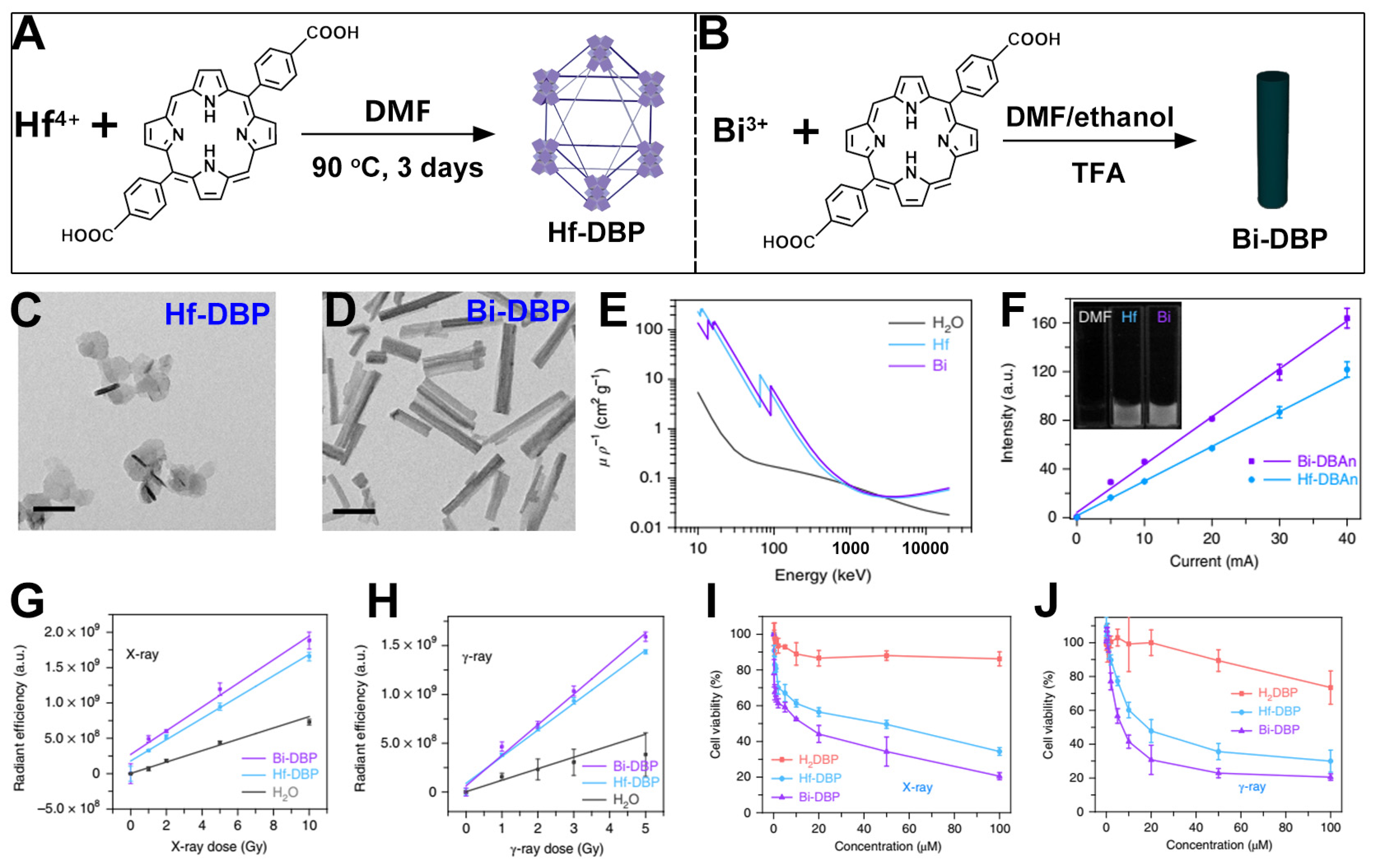
6. Bi-Based nMOFs for Combined RT and RDT

The effect of metals as radiosensitizers is related to their atomic numbers [104,105,106,107,108,109]. To ensure biomedical applications, the metals must also be non-radioactive elements. Currently, it is well known that Bi is the naturally occurring element with the highest atomic number among non-radioactive elements [110,111]. Therefore, using Bi as a metal cluster to construct MOF structures has great potential in the development of a radiosensitizer [112,113]. For example, Ni et al. first prepared Hf-DBP as a comparative reference (Figure 6A) [29]. Then, using the solvothermal method, Bi-DBP MOFs were prepared with Bi3+ and p-dicarboxyphenyl porphyrin as the building materials (Figure 6B). After that, the authors conducted property tests and functional characterizations of the two MOF materials. As shown in Figure 6C,D, Hf-DBP presented a disk shape as reported in the literature, while Bi-DBP showed a nanorod shape with a diameter * length of approximately 20 nm * 180 nm. Through testing, it was found that the K-edge energy of the Bi element at 90 keV is higher than that of the Hf element at 65 keV. This result indicates that the Bi element is more suitable for enhancing the secondary radiation of photons generated by a linear accelerator used in clinical settings (Figure 6E). In addition, the authors found through testing that the radiation signal of Bi-FBAn is 1.38 times that of Hf-DBAn, which shows that the Bi element has a higher X-ray absorption and energy transfer efficiency (Figure 6F). Next, the authors verified the ability of the two MOF materials to generate ·OH. As shown in Figure 6F,G, under both X-ray and γ-ray irradiation, the fluorescence detected by aminophenyl fluorescein (APF) for the Bi-DBP MOF was stronger than that for the Hf-DBP MOF. This indicates that Bi-based MOFs can exhibit a stronger sensitization effect. Then, the authors examined the cytotoxic effects of Hf-DBP and Bi-DBP on TRMP-C2 cells. As shown in Figure 6I,J, under an X-ray voltage with a dose of 2 Gy or 60Co γ-ray irradiation, the cell survival rate of the group treated with Bi-DBP was lower than that of the group treated with Hf-DBP. This result shows that under the same conditions, Bi-DBP MOFs can exert a stronger radiotherapy effect. Nevertheless, these studies are still at the laboratory level. In clinical practice, factors such as cost and the production yield of metal elements may need to be considered. Furthermore, although free heavy metals such as Au and Pb are generally associated with significant toxicity, the Bi metal itself is typically considered to have good biocompatibility [114,115,116,117]. However, it is important to note that Bi-MOFs may exhibit instability in the presence of phosphate or under other tumor microenvironmental conditions, leading to their gradual degradation and the release of soluble Bi3+ ions. These soluble Bi3+ ions may subsequently bind to biomacromolecules in vivo, affecting their distribution and metabolism. Therefore, efficient excretion of Bi3+ ions remains a key challenge for the clinical application of such materials.

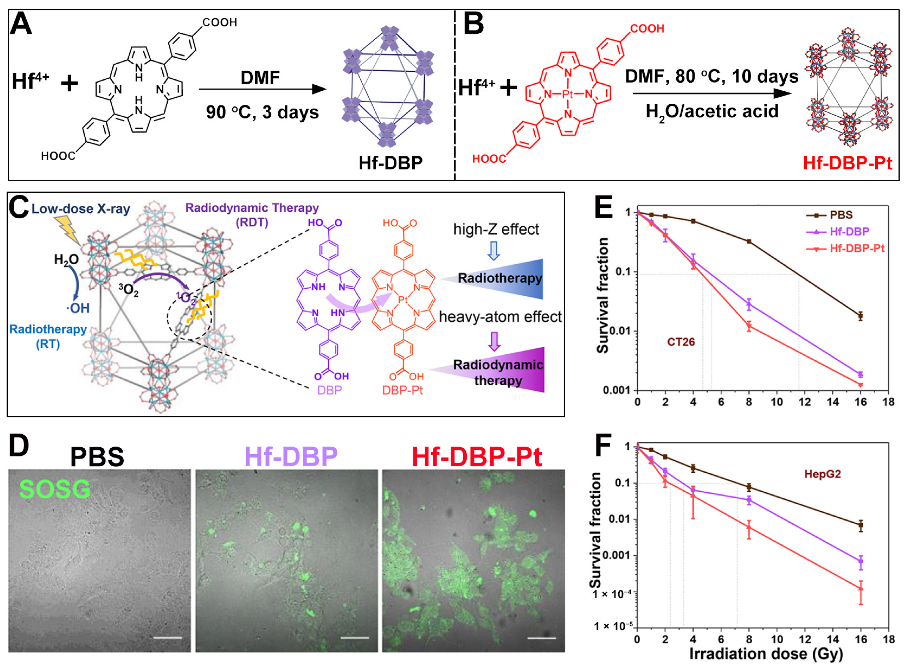
7. Hf-Based nMOFs: Boosting RT and RDT via Heavy Atom Effect

In addition to using metal elements with larger atomic numbers such as Th and Bi as participants in the construction of porphyrin-based nMOFs, which can enhance the radiotherapy effect of the sensitizer materials, there are also some methods to enhance the efficacy of RDT. It is well known that the heavy atom effect can enhance the ability of organic photosensitizers to generate 1O2 by promoting intersystem crossing between the singlet state and the triplet excited state of the photosensitizer [78,118,119,120,121]. Building on this, Guo et al. first synthesized a Hf-DBP MOF as a control (Figure 7A) [79]. They then prepared a platinum(II)-metallated DBP-Pt coordination complex and incorporated it into the Hf-DBP framework to obtain a Hf-DBP-Pt MOF (Figure 7B). The heavy atom effect of Pt2+ enhances the porphyrin photosensitizer’s ability to generate 1O2, as shown in Figure 7C. Subsequently, the authors used SOSG as a probe to detect the ability of Hf-DBP and Hf-DBP-Pt to generate 1O2 under the same X-ray irradiation conditions. As shown in Figure 7D, the green fluorescence of the group treated with Hf-DBP-Pt was significantly stronger than that of the Hf-DBP group. This result directly proves the enhancing effect of the heavy atom effect on porphyrin-mediated RDT. Then, the authors directly evaluated the radiosensitization effects of Hf-DBP and Hf-DBP-Pt on cancer cells in vitro. As shown in the results of the colony formation experiments in Figure 7E,F, for both CT26 cells and HepG2 cells, under the same X-ray dose, the survival fractions of cancer cells treated with Hf-DBP-Pt were lower. This directly proves the enhanced combined effect of RT-RDT induced by the heavy atom effect. This system endows the porphyrin-based nMOF with an enhanced ability to generate 1O2 by introducing the heavy atom Pt, thus achieving an enhanced combined effect of RT/RDT.

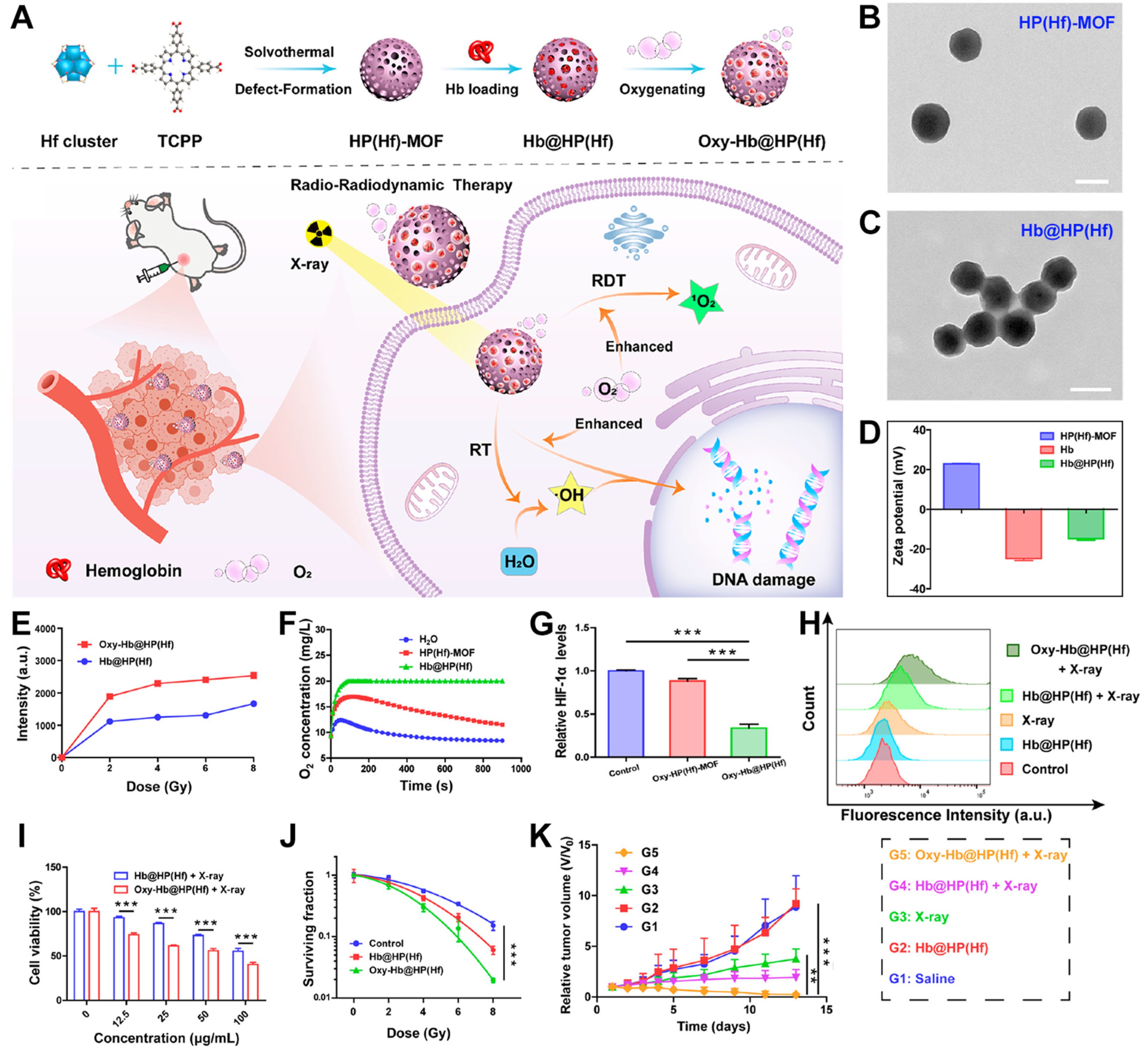
8. Hf-Based nMOFs: Boosting RT and RDT via Self-Oxygen-Carrying Function

The efficacy of PDT, sonodynamic therapy, and the combined RT-RDT treatment method may be hindered by the hypoxic environment of solid tumors [122,123]. Therefore, it is necessary to improve the hypoxic microenvironment of solid tumors while carrying out these treatment methods for enhanced therapeutic efficacy [124]. There are many ways to alleviate the hypoxic state of solid tumors [125,126]. Various strategies can be employed to increase intracellular O2 concentration, including the introduction of mitochondrial respiration inhibitors or the use of Pt NPs as catalysts to react with excess intracellular H2O2 [127,128,129,130,131,132,133]. Combination therapy strategies can also effectively overcome the limitations imposed by hypoxic environments on ROS-based treatment approaches [134,135,136]. In addition, proteins that transport oxygen, such as hemoglobin (Hb) in human red blood cells, can be used to coat and modify the materials. In the combined RT-RDT treatment protocol, using Hb to modify nMOF radiosensitizers can effectively improve the treatment effect. Based on this, Zhao et al. first prepared a Hf-MOF (Hb(Hf)-MOF) (Figure 8A) [81], which, due to its positive charge and porous structure, can adsorb hemoglobin (Hb) to form Hb@HP(Hf). Upon exposure to oxygen, the material enables Hb to transport oxygen, which is then released inside cells to alleviate hypoxia. Under X-ray excitation, Hb@HP can combine with water molecules, enhancing the formation of ·OH and exerting an enhanced RT effect. The increase in oxygen concentration helps the X-ray activation of porphyrin photosensitizers to generate 1O2, thus exerting an enhanced RDT effect and causing damage to the DNA of cancer cells (Figure 8A). As shown in Figure 8B, the prepared Hf-TCPP MOF presents a spherical shape and is well-dispersed. Figure 8C shows that the morphology of Hb@HP NPs obtained after modification with Hb remains essentially unchanged. As shown in Figure 8D, the surface of the Hf-TCPP MOF is charged, and the Hb@HP MOF obtained after being modified by negatively charged Hb molecules shows a negative charge similar to that of Hb. This result proves that the method of simply stirring and loading Hb molecules onto the surface of the Hf-TCPP MOF is effective. Afterward, the authors used SOSG as a probe to determine the ability of Hb@HP and Oxy-Hb@HP to generate 1O2 under the same X-ray irradiation. As shown in Figure 8E, after loading oxygen, the ability of the material to generate 1O2 under X-ray irradiation is significantly enhanced. Then, the authors used an oxygen detector to detect the change in the oxygen concentration of three groups of solutions, namely, H2O, Hf-MOF, and Hb@HP, after bubbling oxygen for 15 min. As shown in Figure 8F, the oxygen concentration of the Hb@HP experimental group was maintained, which proves that Hb can indeed normally perform the function of transporting oxygen. The results of hypoxia-inducible factor detection also indicate that the Hf-TCPP MOF modified by Hb (Hb@HP) can significantly improve the hypoxic state of cells (Figure 8G). The authors used flow cytometry to detect the fluorescence intensity of DCF in cells (DCF-DA can be oxidized by 1O2 to generate fluorescent DCF; the stronger the fluorescence, the more 1O2 is generated). The experimental results in Figure 8H show that after modifying Hf-MOF with O2-transporting Hb, an enhanced RDT effect can indeed be exhibited under X-ray irradiation. Following that, the authors evaluated the cytotoxicity of the prepared materials. Under the same concentration and X-ray irradiation, the survival rate of CT26 cells in the Oxy-Hb@HP group was significantly lower than that in the group without oxygen transportation (Figure 8I). The results of the colony survival experiment are consistent with those of the cytotoxicity test (Figure 8J). These results all indicate that improving the O2 concentration helps to enhance the combined effect of RT-RDT. Based on these results, the authors conducted an in vivo experiment to inhibit CT26 tumors. As shown in Figure 8K, the G5 group (Oxy-Hb@HP) exerted the most beneficial effect in inhibiting the volume of cancer cells under X-ray irradiation. This system achieves an enhanced combined effect of RT-RDT by modifying the MOF radiosensitizer with oxygen-transporting hemoglobin, providing an important reference for research on new radiosensitizers.

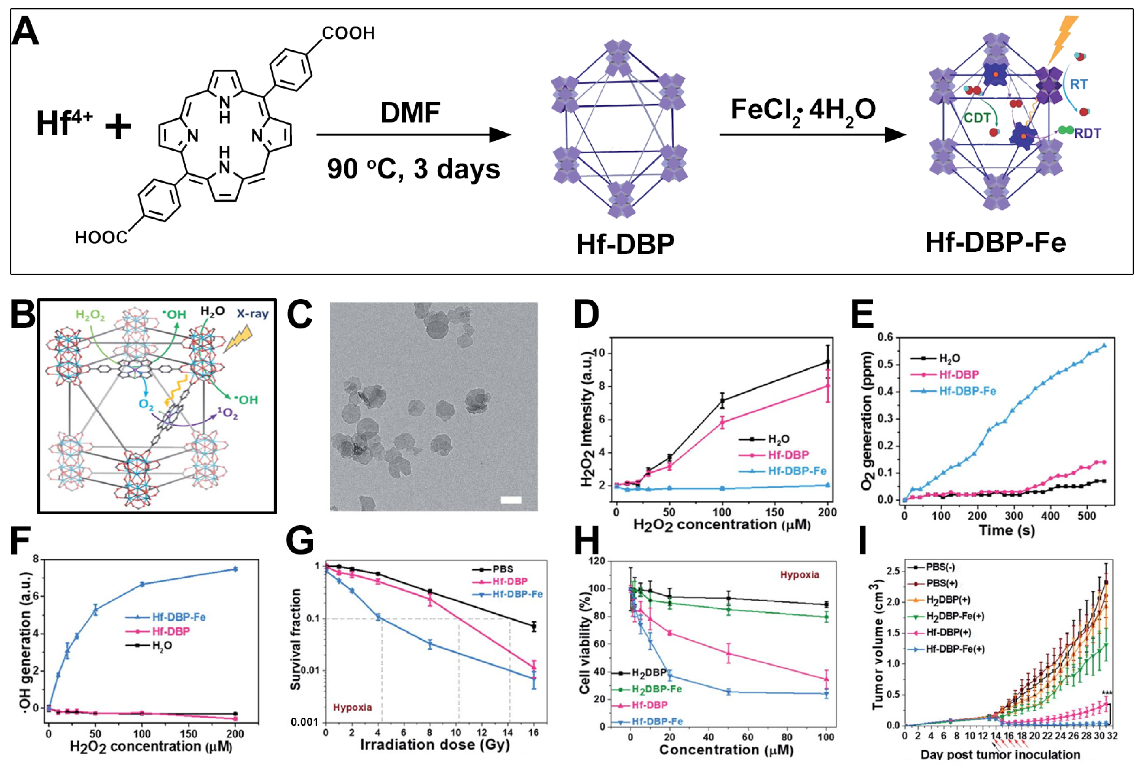
9. Hf-DBP-Fe nMOFs for RT-RDT Combined with CDT

In addition to the utilization of hemoglobin, using variable valence metals such as Fe3+/Fe2+ or Mn4+/Mn2+ as catalysts to react with the excessive H2O2 in cancer cells to generate O2 is also a very effective solution for addressing hypoxic tumors [137]. On the other hand, the porphyrin molecules in porphyrin-based MOF materials can interact with variable valence metals through the coordination of nitrogen elements on the pyrrole ring. Based on this, Lin et al. synthesized a Hf-DBP MOF and further functionalized it with the Fe element to obtain Hf-DBP-Fe (Figure 9A) [80]. The authors found that the Fe element could exhibit catalase-like activity. It could not only catalyze the generation of O2 from H2O2 but also exert a CDT effect under X-ray irradiation, enabling combined RT/RDT/CDT for a stronger antitumor effect (Figure 9B). The TEM test results showed that Hf-DBP-Fe presented a nanosheet morphology similar to that of Hf-DBP (Figure 9C). The H2O2 kit showed that Hf-DBP-Fe could effectively catalyze and reduce the concentration of H2O2 (Figure 9D), thereby generating O2 to improve the hypoxic state (Figure 9E). The APF probe also demonstrated that Hf-DBP-Fe could cause H2O2 to generate ·OH (Figure 9F). The colony formation experiments also proved that Hf-DBP-Fe could exhibit a stronger inhibitory effect on MC38 cells with a lower cell survival fraction under hypoxic conditions (Figure 9G). The cytotoxicity test revealed that both Hf-DBP and Hf-DBP-Fe showed good biocompatibility without X-ray irradiation. However, under X-ray irradiation, even under hypoxic conditions, both Hf-DBP and Hf-DBP-Fe could exhibit cytotoxicity (Figure 9H). But due to the catalase-like activity and CDT effect exerted by Hf-DBP-Fe formed with the participation of the Fe element, it showed a stronger inhibitory effect on MC38 cells with a lower IC50 value (Figure 9H). Subsequently, the authors verified the inhibitory effect on MC38 cancer cells in tumor-bearing mice. As shown in Figure 9I, under the same drug administration concentration condition, the group treated with Hf-DBP-Fe + X-ray irradiation achieved the best antitumor effect. Through the utilization of the structural characteristics of the porphyrin molecules in the MOF in coordination with the Fe element, the porphyrin-based MOF is endowed with more therapeutic functions and realizes the combined function of hypoxia relief and enhanced RT/RDT/CDT.

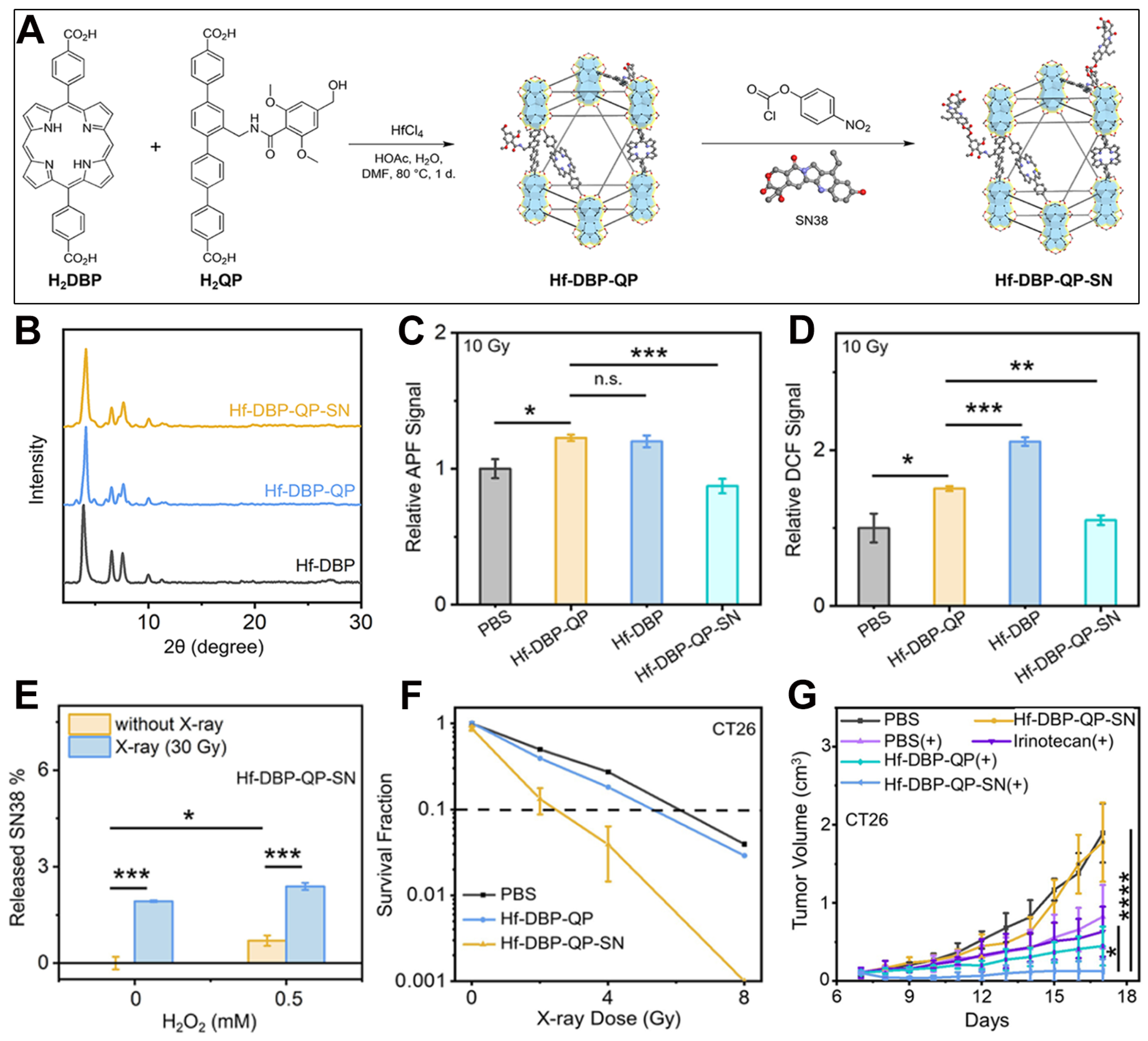
10. Mixed-Ligand nMOFs for RT-RDT Combined with Chemotherapy

In addition to strategies such as using metals with higher atomic numbers as MOF metal clusters, applying the heavy atom effect, and alleviating the hypoxic microenvironment of cancer cells to achieve enhanced RT-RDT efficacy, combining RT-RDT with treatment methods like chemotherapy can also enhance the therapeutic effect [138,139,140]. Differently from RT-RDT, most chemotherapy drug molecules are likely to cause systemic toxicity [141,142,143,144]. Therefore, it is of great significance to develop chemotherapy activated by special stimuli such as pH, hypoxia, and ROS, enabling it to act at the lesion site [145,146,147,148,149]. Through combination with the hydroxyl radicals generated during the RT-RDT process, the creation of a hydroxyl radical-activated release system can be attempted to develop a combined treatment protocol of RT-RDT and chemotherapy. Based on this, Zhen et al. incorporated quaterphenyl dicarboxylic acid derivative (H2QP) into the Hf-DBP structure to form Hf-DBP-QP [82]. Then, the 7-ethyl-10-hydroxycamptothecin (SN38) molecule was loaded to obtain Hf-DBP-QP-SN (Figure 10A). The bond between H2QP and the SN38 molecule can respond to the hydroxyl radicals generated during the RT-RDT process, thus releasing SN38 for chemotherapy. The PXRD test results proved that the formation of the MOF with the participation of H2QP and the subsequent loading of the SN38 molecule had little impact on the crystalline structure of Hf-DBP (Figure 10B). Thereafter, the authors evaluated the generation of hydroxyl radicals from the material by detecting the change in the APF signal intensity. As shown in Figure 10C, the total amount of hydroxyl radicals generated by Hf-DBP-QP was similar to that of Hf-DBP, but the amount of hydroxyl radicals generated by the Hf-DBP-QP-SN group decreased to a certain extent. This was because the generated hydroxyl radicals reacted with the chemical bonds formed by QP-SN, consuming some hydroxyl radicals and proving that SN38 could be effectively released (Figure 10C). Then, the authors evaluated the total amount of ROS generation by detecting the fluorescence change in DCF generated from the DCFH-DA probe. As shown in Figure 10D, under an X-ray irradiation of 10 Gy, the total amount of ROS generated by Hf-DBP-QP was less than that of the Hf-DBP group. This was because the doping of QA molecules reduced the amount of porphyrin molecules. Hf-DBP-QP-SN generated the least amount of total ROS due to its response to hydroxyl radicals. Since X-rays can promote the generation of hydroxyl radicals from H2O2, the authors conducted an experiment on the release of SN38 under the condition of adding additional H2O2 (Figure 10E). X-rays can stimulate water and H2O2 to generate hydroxyl radicals. Therefore, the release of SN38 molecules was most effective when both X-rays and hydrogen peroxide were present (Figure 10E). Following that, the authors evaluated the radiosensitization effect of the materials prepared in this system through a colony experiment. As shown in Figure 10F, under the excitation of the same X-ray dose, the survival fraction of CT26 cells in the group treated with Hf-DBP-QP-SN was significantly lower than that in the Hf-DBP-QP group. This result indicates that the introduction of the SN38 chemotherapy molecule significantly enhanced the combined RT/RDT treatment effect of Hf-DBP. Then, the authors determined the inhibitory effect of Hf-DBP-QP-SN on CT26 cancer cells in mice under X-ray irradiation. As shown in Figure 10G, compared with multiple other control groups, the tumor volume of the mice in the group treated with Hf-DBP-QP-SN + X-rays was the smallest. This result proves the effectiveness of introducing the SN38 chemotherapy molecule into this system for achieving an enhanced inhibitory effect on malignant cancer cells. This system also provides a unique design method for the combination of RT/RDT mediated by porphyrin-based nMOF materials and responsive chemotherapy.

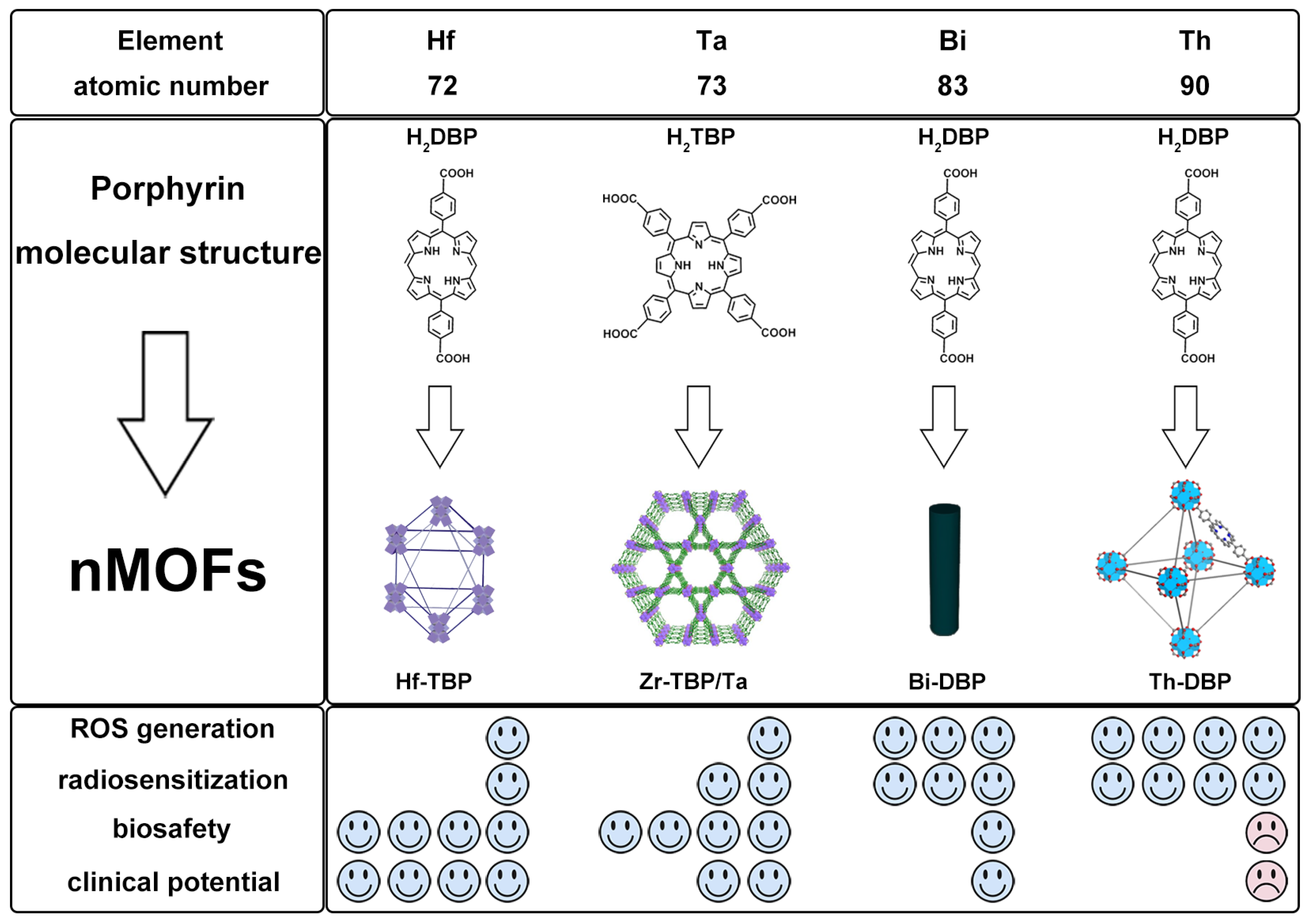
11. Comparative Study of nMOFs Derived from Hf, Ta, Bi, and Th

Although nMOFs based on the four metals have demonstrated excellent combined antitumor efficacy in RT-RDT at both cellular and murine levels, a more in-depth comparison of their ROS generation capacity and safety profiles is still needed to further advance their clinical translation. To address this, we present a systematic evaluation of these nMOFs in Figure 11, focusing on four key aspects: ROS generation efficiency, radiosensitization performance, toxicity, and potential for clinical translation.
In terms of ROS generation efficiency, although Ta has a higher atomic number and greater X-ray mass attenuation coefficient than Hf (and HfO2), it can only be incorporated into MOFs via doping. As a result, the amount of Ta in the MOF structure is limited by the initial feed ratio, making it difficult to confirm whether Ta-based MOFs exhibit stronger ROS generation capability than Hf-MOFs [50]. Experimental data indicate that the Bi-MOF shows significantly enhanced ROS generation compared to the Hf-MOF, while the Th-MOF demonstrates the highest ROS production efficiency [51]. Regarding radiosensitization performance, the sensitization effect of the four metals increases progressively with an increasing atomic number, from Hf, Ta, and Bi to Th. According to the literature data, under identical conditions, the radiosensitization ability of the Bi-MOF is 1.12 times that of the Hf-MOF (2.08 vs. 1.86) [29], while that of the Th-MOF reaches 1.45 times (1.77 vs. 1.22) [51]. In terms of biosafety and clinical application potential, the Hf-MOF exhibits the most promising translational prospects due to the excellent biocompatibility of Hf and the clinical use of HfO2 as a radiosensitizer. Tantalum (Ta) is also widely used in biomedical applications such as orthopedics and is typically non-radioactive in nature. However, studies on Ta-based materials remain limited, and more experimental data are needed to support their clinical translation. Bismuth (Bi) is considered a material with negligible radiotoxic risk and high radiosensitization efficiency. However, Bi-based MOFs often require functional modifications to mitigate potential toxicity. Thorium (Th), as a weakly radioactive element, requires careful safety evaluation for its clinical application. Due to concerns related to radioactivity, the clinical translation of Th-based MOFs may face significant challenges.

12. Conclusions

Malignant tumors have long been one of the most serious diseases threatening human survival, leading to the development and in-depth investigation of various treatment modalities [150,151,152,153,154,155]. Numerous studies have shown that combination therapy is a more effective strategy compared to single-mode treatments [156,157,158]. The structural tunability and diverse types of nMOFs make the implementation of combination therapy significantly more feasible. This review summarizes the relevant research progress in recent years on the combined effect of RT/RDT on cancer cells, which is generated by porphyrin-based nMOF materials under the excitation of X-rays or γ-rays. As porous materials (nMOFs) with excellent biocompatibility, nMOFs’ structural features facilitate the diffusion of 1O2 generated by X-ray activation and the realization of therapeutic effects. The structural diversity and easy modifiability of porphyrin-based MOFs endow them with potential for the development and application of nanomedicines excited by low-dose X-rays.
However, RT-RDT using materials similar to Hf–porphyrin MOFs still faces numerous challenges before clinical application. Firstly, the porphyrin molecules involved in coordination can only be synthesized through complex organic synthesis procedures in the laboratory, and the yield is relatively low. Secondly, some of the research work summarized in this review attempts to use metals with high Z such as Ta, Th, and Bi to prepare nMOF materials, but this may remain at the laboratory research stage. These metals with high Z may face the problem of high costs, which makes it difficult for them to be widely applied in clinical settings. Next, large amounts of detailed research data on various pharmacological and toxicological indicators, such as the in vivo metabolism and tissue distribution of nMOFs prepared from these metals with high Z, are still needed to support and prove the feasibility of their clinical application. Ultimately, the clinical translation of these materials will require collaborative efforts from researchers across multiple disciplines, including materials scientists, chemists, and medical experts, to ensure successful practical implementation. Solving these existing problems will contribute to the further promotion of the biomedical applications of porphyrin-based MOF materials.

Author Contributions

B.G.: writing—original draft, writing—review and editing. Q.Z.: writing—original draft, writing—review and editing. Y.Q.: writing—original draft, writing—review and editing. X.Z.: funding acquisition, writing—original draft, writing—review and editing. W.W.: writing—original draft, writing—review and editing. All authors have read and agreed to the published version of the manuscript.

Funding

This research was funded by the University-Enterprise Cooperation Project (133724623003 and 133725623006).

Acknowledgments

During the preparation of this manuscript, the authors used Qwen 3 for the purposes of language polishing, including word and grammar checking. The authors have reviewed and edited the output and take full responsibility for the content of this publication.

Conflicts of Interest

The authors declare no conflicts of interest.

References

  1. Wang, W.; Chen, D.; Li, F.; Xiao, X.; Xu, Q. Metal-organic-framework-based materials as platforms for energy applications. Chem 2024, 10, 86–133. [Google Scholar] [CrossRef]
  2. Dhakshinamoorthy, A.; Li, Z.; Yang, S.; Garcia, H. Metal–organic framework heterojunctions for photocatalysis. Chem. Soc. Rev. 2024, 53, 3002–3035. [Google Scholar] [CrossRef]
  3. Yu, Q.; Wang, X.; Wu, W.; Feng, X.; Kong, D.; Khan, U.; Ren, X.; Li, L. In Situ Encapsulation of Graphene Quantum Dots in Highly Stable Porphyrin Metal-Organic Frameworks for Efficient Photocatalytic CO2 Reduction. Molecules 2023, 28, 4703. [Google Scholar] [CrossRef]
  4. Chen, J.; Zhu, Y.; Kaskel, S. Porphyrin-Based Metal–Organic Frameworks for Biomedical Applications. Angew. Chem. Int. Ed. 2021, 60, 5010–5035. [Google Scholar] [CrossRef]
  5. Zhang, X.; Wasson, M.C.; Shayan, M.; Berdichevsky, E.K.; Ricardo-Noordberg, J.; Singh, Z.; Papazyan, E.K.; Castro, A.J.; Marino, P.; Ajoyan, Z.; et al. A historical perspective on porphyrin-based metal–organic frameworks and their applications. Coord. Chem. Rev. 2021, 429, 213615. [Google Scholar] [CrossRef]
  6. Xu, D.; Duan, Q.; Yu, H.; Dong, W. Photodynamic therapy based on porphyrin-based metal–organic frameworks. J. Mater. Chem. B 2023, 11, 5976–5989. [Google Scholar] [CrossRef]
  7. Qi, C.; Chen, J.; Qu, Y.; Luo, X.; Wang, W.; Zheng, X. Recent Advances in Porphyrin-Based Covalent Organic Frameworks for Synergistic Photodynamic and Photothermal Therapy. Pharmaceutics 2024, 16, 1625. [Google Scholar] [CrossRef]
  8. Zhang, Q.; Wang, X.; Chen, J.; Wu, J.; Zhou, M.; Xia, R.; Wang, W.; Zheng, X.; Xie, Z. Recent progress of porphyrin metal–organic frameworks for combined photodynamic therapy and hypoxia-activated chemotherapy. Chem. Commun. 2024, 60, 13641–13652. [Google Scholar] [CrossRef]
  9. Hong, G.; Antaris, A.L.; Dai, H. Near-infrared fluorophores for biomedical imaging. Nat. Biomed. Eng. 2017, 1, 0010. [Google Scholar] [CrossRef]
  10. Jurkovicova, D.; Neophytou, C.M.; Gašparović, A.Č.; Gonçalves, A.C. DNA Damage Response in Cancer Therapy and Resistance: Challenges and Opportunities. Int. J. Mol. Sci. 2022, 23, 14672. [Google Scholar] [CrossRef]
  11. Zhang, Z.; Liu, X.; Chen, D.; Yu, J. Radiotherapy combined with immunotherapy: The dawn of cancer treatment. Signal Transduct. Target. Ther. 2022, 7, 258. [Google Scholar] [CrossRef]
  12. Chen, X.; Li, M.; Lin, M.; Lu, C.; Kumar, A.; Pan, Y.; Liu, J.; Peng, Y. Current and promising applications of Hf(iv)-based MOFs in clinical cancer therapy. J. Mater. Chem. B 2023, 11, 5693–5714. [Google Scholar] [CrossRef]
  13. Mladenova, V.; Mladenov, E.; Stuschke, M.; Iliakis, G. DNA Damage Clustering after Ionizing Radiation and Consequences in the Processing of Chromatin Breaks. Molecules 2022, 27, 1540. [Google Scholar] [CrossRef]
  14. Haque, M.; Shakil, M.S.; Mahmud, K.M. The Promise of Nanoparticles-Based Radiotherapy in Cancer Treatment. Cancers 2023, 15, 1892. [Google Scholar] [CrossRef]
  15. He, L.; Yu, X.; Li, W. Recent Progress and Trends in X-ray-Induced Photodynamic Therapy with Low Radiation Doses. ACS Nano 2022, 16, 19691–19721. [Google Scholar] [CrossRef]
  16. Zhu, X.; Wu, J.; Liu, R.; Xiang, H.; Zhang, W.; Chang, Q.; Wang, S.; Jiang, R.; Zhao, F.; Li, Q.; et al. Engineering Single-Atom Iron Nanozymes with Radiation-Enhanced Self-Cascade Catalysis and Self-Supplied H2O2 for Radio-enzymatic Therapy. ACS Nano 2022, 16, 18849–18862. [Google Scholar] [CrossRef]
  17. Pan, S.; Huang, G.; Sun, Z.; Chen, X.; Xiang, X.; Jiang, W.; Xu, Y.; Chen, T.; Zhu, X. X-Ray-Responsive Zeolitic Imidazolate Framework-Capped Nanotherapeutics for Cervical Cancer-Targeting Radiosensitization. Adv. Funct. Mater. 2023, 33, 2213364. [Google Scholar] [CrossRef]
  18. Ren, S.; Zhang, Q.; Fu, H.; Cheng, J.; Xie, Y.; Liang, Q.; Xiao, X. A reactive oxygen species amplifier based on a Bi2WO6/BP heterojunction for high efficiency radiotherapy enhancement. J. Mater. Chem. B 2025, 13, 3128–3137. [Google Scholar] [CrossRef]
  19. Zhu, S.; Lin, S.; Han, R. Treating Deep-Seated Tumors with Radiodynamic Therapy: Progress and Perspectives. Pharmaceutics 2024, 16, 1135. [Google Scholar] [CrossRef]
  20. Ma, M.; Wang, J.; Jiang, H.; Chen, Q.; Xiao, Y.; Yang, H.; Lin, L. Transcranial deep-tissue phototherapy for Alzheimer’s disease using low-dose X-ray-activated long-afterglow scintillators. Acta Biomater. 2023, 155, 635–643. [Google Scholar] [CrossRef]
  21. Souris, J.S.; Leoni, L.; Zhang, H.J.; Pan, A.; Tanios, E.; Tsai, H.-M.; Balyasnikova, I.V.; Bissonnette, M.; Chen, C.-T. X-ray Activated Nanoplatforms for Deep Tissue Photodynamic Therapy. Nanomaterials 2023, 13, 673. [Google Scholar] [CrossRef]
  22. Chen, F.; Ruan, F.; Xie, X.; Lu, J.; Sun, W.; Shao, D.; Chen, M. Gold Nanocluster: A Photoelectric Converter for X-Ray-Activated Chemotherapy. Adv. Mater. 2024, 36, 2402966. [Google Scholar] [CrossRef]
  23. Li, H.; Zeng, J.; You, Q.; Zhang, M.; Shi, Y.; Yang, X.; Gu, W.; Liu, Y.; Hu, N.; Wang, Y.; et al. X-ray-activated nanoscintillators integrated with tumor-associated neutrophils polarization for improved radiotherapy in metastatic colorectal cancer. Biomaterials 2025, 316, 123031. [Google Scholar] [CrossRef]
  24. Li, Q.; Liu, Y.; Zhang, Y.; Jiang, W. Immunogenicity-boosted cancer immunotherapy based on nanoscale metal-organic frameworks. J. Control. Release 2022, 347, 183–198. [Google Scholar] [CrossRef]
  25. Wang, W.; Yu, Y.; Jin, Y.; Liu, X.; Shang, M.; Zheng, X.; Liu, T.; Xie, Z. Two-dimensional metal-organic frameworks: From synthesis to bioapplications. J. Nanobiotechnol. 2022, 20, 207. [Google Scholar] [CrossRef]
  26. Wei, Q.; Wu, Y.; Liu, F.; Cao, J.; Liu, J. Advances in antitumor nanomedicine based on functional metal–organic frameworks beyond drug carriers. J. Mater. Chem. B 2022, 10, 676–699. [Google Scholar] [CrossRef]
  27. Abánades Lázaro, I.; Chen, X.; Ding, M.; Eskandari, A.; Fairen-Jimenez, D.; Giménez-Marqués, M.; Gref, R.; Lin, W.; Luo, T.; Forgan, R.S. Metal–organic frameworks for biological applications. Nat. Rev. Methods Primers 2024, 4, 42. [Google Scholar] [CrossRef]
  28. Lu, K.; He, C.; Guo, N.; Chan, C.; Ni, K.; Lan, G.; Tang, H.; Pelizzari, C.; Fu, Y.-X.; Spiotto, M.T.; et al. Low-dose X-ray radiotherapy–radiodynamic therapy via nanoscale metal–organic frameworks enhances checkpoint blockade immunotherapy. Nat. Biomed. Eng. 2018, 2, 600–610. [Google Scholar] [CrossRef]
  29. Ni, K.; Xu, Z.; Culbert, A.; Luo, T.; Guo, N.; Yang, K.; Pearson, E.; Preusser, B.; Wu, T.; La Riviere, P.; et al. Synergistic checkpoint-blockade and radiotherapy–radiodynamic therapy via an immunomodulatory nanoscale metal–organic framework. Nat. Biomed. Eng. 2022, 6, 144–156. [Google Scholar] [CrossRef]
  30. Ding, S.; Chen, L.; Liao, J.; Huo, Q.; Wang, Q.; Tian, G.; Yin, W. Harnessing Hafnium-Based Nanomaterials for Cancer Diagnosis and Therapy. Small 2023, 19, 2300341. [Google Scholar] [CrossRef]
  31. Yang, S.; Gaohua, H.; Quan, C.; Lei, Y.; Peng, W.; Qi, Z.; Jiang, D.; Wei, Z.; Huang, J. Au-Pt Nanoparticle Formulation as a Radiosensitizer for Radiotherapy with Dual Effects. Int. J. Nanomed. 2021, 16, 239–248. [Google Scholar] [CrossRef]
  32. Ni, K.; Luo, T.; Nash, G.T.; Lin, W. Nanoscale Metal–Organic Frameworks for Cancer Immunotherapy. Acc. Chem. Res. 2020, 53, 1739–1748. [Google Scholar] [CrossRef]
  33. Xu, Z.; Luo, T.; Lin, W. Nanoscale Metal–Organic Layers for Biomedical Applications. Acc. Mater. Res. 2021, 2, 944–953. [Google Scholar] [CrossRef]
  34. Pan, Y.; Tang, W.; Fan, W.; Zhang, J.; Chen, X. Development of nanotechnology-mediated precision radiotherapy for anti-metastasis and radioprotection. Chem. Soc. Rev. 2022, 51, 9759–9830. [Google Scholar] [CrossRef]
  35. Zou, Y.; Wu, J.; Zhang, Q.; Chen, J.; Luo, X.; Qu, Y.; Xia, R.; Wang, W.; Zheng, X. Recent advances in cell membrane-coated porphyrin-based nanoscale MOFs for enhanced photodynamic therapy. Front. Pharmacol. 2024, 15, 1505212. [Google Scholar] [CrossRef]
  36. Zou, Y.; Chen, J.; Qu, Y.; Luo, X.; Wang, W.; Zheng, X. Evolution of nMOFs in photodynamic therapy: From porphyrins to chlorins and bacteriochlorins for better efficacy. Front. Pharmacol. 2025, 16, 1533040. [Google Scholar] [CrossRef]
  37. Mendes, R.F.; Figueira, F.; Leite, J.P.; Gales, L.; Almeida Paz, F.A. Metal–organic frameworks: A future toolbox for biomedicine? Chem. Soc. Rev. 2020, 49, 9121–9153. [Google Scholar] [CrossRef]
  38. Liu, J.; Huang, J.; Zhang, L.; Lei, J. Multifunctional metal–organic framework heterostructures for enhanced cancer therapy. Chem. Soc. Rev. 2021, 50, 1188–1218. [Google Scholar] [CrossRef]
  39. Elmehrath, S.; Nguyen, H.L.; Karam, S.M.; Amin, A.; Greish, Y.E. BioMOF-Based Anti-Cancer Drug Delivery Systems. Nanomaterials 2023, 13, 953. [Google Scholar] [CrossRef]
  40. Jiang, W.; Liang, C.-C.; Zhang, Y.-B. Multivariate Metal–Organic Frameworks for Programming Functions. Adv. Funct. Mater. 2024, 34, 2308946. [Google Scholar] [CrossRef]
  41. Zhang, X.-D.; Chen, J.; Min, Y.; Park, G.B.; Shen, X.; Song, S.-S.; Sun, Y.-M.; Wang, H.; Long, W.; Xie, J.; et al. Metabolizable Bi2Se3 Nanoplates: Biodistribution, Toxicity, and Uses for Cancer Radiation Therapy and Imaging. Adv. Funct. Mater. 2014, 24, 1718–1729. [Google Scholar] [CrossRef]
  42. Cruje, C.; Yang, C.; Uertz, J.; van Prooijen, M.; Chithrani, B.D. Optimization of PEG coated nanoscale gold particles for enhanced radiation therapy. RSC Adv. 2015, 5, 101525–101532. [Google Scholar] [CrossRef]
  43. Petersen, A.L.; Henriksen, J.R.; Binderup, T.; Elema, D.R.; Rasmussen, P.H.; Hag, A.M.; Kjær, A.; Andresen, T.L. In vivo evaluation of PEGylated 64Cu-liposomes with theranostic and radiotherapeutic potential using micro PET/CT. Eur. J. Nucl. Med. Mol. Imaging 2016, 43, 941–952. [Google Scholar] [CrossRef]
  44. Mohammadian, M.; Emamgholizadeh Minaei, S.; Shiralizadeh Dezfuli, A. Improve the cytotoxic effects of megavoltage radiation treatment by Fe3O4@Cus–PEG nanoparticles as a novel radiosensitizer in colorectal cancer cells. Cancer Nanotechnol. 2022, 13, 25. [Google Scholar] [CrossRef]
  45. Liu, H.; Cai, Y.; Zhang, Y.; Xie, Y.; Qiu, H.; Hua, L.; Liu, X.; Li, Y.; Lu, J.; Zhang, L.; et al. Development of a Hypoxic Radiosensitizer-Prodrug Liposome Delivery DNA Repair Inhibitor Dbait Combination with Radiotherapy for Glioma Therapy. Adv. Healthc. Mater. 2017, 6, 1601377. [Google Scholar] [CrossRef]
  46. Tang, X.; Wang, Z.; Xie, Y.; Liu, Y.; Yang, K.; Li, T.; Shen, H.; Zhao, M.; Jin, J.; Xiao, H.; et al. Radiation-Triggered Selenium-Engineered Mesoporous Silica Nanocapsules for RNAi Therapy in Radiotherapy-Resistant Glioblastoma. ACS Nano 2023, 17, 4062–4076. [Google Scholar] [CrossRef]
  47. Wu, H.; Yuan, Y.; Kang, S.; Zhou, G.; Gu, Y.; Yuan, X.; Li, J.; Gu, N. Autophagy inhibitor-loaded mesoporous AgNPs@SiO2 nanoplatform for synergistically enhanced glioma radiotherapy. Sci. China Mater. 2023, 66, 2902–2912. [Google Scholar] [CrossRef]
  48. Stewart, C.; Konstantinov, K.; McKinnon, S.; Guatelli, S.; Lerch, M.; Rosenfeld, A.; Tehei, M.; Corde, S. First proof of bismuth oxide nanoparticles as efficient radiosensitisers on highly radioresistant cancer cells. Phys. Medica 2016, 32, 1444–1452. [Google Scholar] [CrossRef]
  49. Croissant, J.G.; Fatieiev, Y.; Almalik, A.; Khashab, N.M. Mesoporous Silica and Organosilica Nanoparticles: Physical Chemistry, Biosafety, Delivery Strategies, and Biomedical Applications. Adv. Healthc. Mater. 2018, 7, 1700831. [Google Scholar] [CrossRef]
  50. Li, T.; Gao, M.; Wu, Z.; Yang, J.; Mo, B.; Yu, S.; Gong, X.; Liu, J.; Wang, W.; Luo, S.; et al. Tantalum–Zirconium Co-Doped Metal–Organic Frameworks Sequentially Sensitize Radio–Radiodynamic–Immunotherapy for Metastatic Osteosarcoma. Adv. Sci. 2023, 10, 2206779. [Google Scholar] [CrossRef]
  51. Xu, Z.; Luo, T.; Mao, J.; McCleary, C.; Yuan, E.; Lin, W. Monte Carlo Simulation-Guided Design of a Thorium-Based Metal–Organic Framework for Efficient Radiotherapy-Radiodynamic Therapy. Angew. Chem. Int. Ed. 2022, 61, e202208685. [Google Scholar] [CrossRef]
  52. Lu, M.; Wu, H.; Liu, D.; Wang, F.; Wang, Y.; Wang, M.; Cui, Q.; Zhang, H.; Zang, F.; Ma, M.; et al. Camouflaged Nanoreactors Mediated Radiotherapy-Adjuvant Chemodynamic Synergistic Therapy. ACS Nano 2023, 17, 24170–24186. [Google Scholar] [CrossRef]
  53. Hu, H.; Zheng, S.; He, C.; Zheng, Y.; Wei, Q.; Chen, S.; Wu, Z.; Xu, Y.; Zhao, B.; Yan, C. Radiotherapy-sensitized cancer immunotherapy via cGAS-STING immune pathway by activatable nanocascade reaction. J. Nanobiotechnol. 2024, 22, 234. [Google Scholar] [CrossRef]
  54. Liu, D.; Cao, F.; Xu, Z.; Zhao, C.; Liu, Z.; Pang, J.; Liu, Z.-X.; Moghiseh, M.; Butler, A.; Liang, S.; et al. Selective Organ-Targeting Hafnium Oxide Nanoparticles with Multienzyme-Mimetic Activities Attenuate Radiation-Induced Tissue Damage. Adv. Mater. 2024, 36, 2308098. [Google Scholar] [CrossRef]
  55. Wang, X.; Wang, D.; Liao, Y.; Guo, X.; Song, Q.; Liu, W.; Gu, C.; Du, S.; Sun, B.; Gu, Z. Hafnium oxide-based sensitizer with radiation-triggered cuproptosis for radiotherapy. Nano Today 2025, 61, 102626. [Google Scholar] [CrossRef]
  56. Ji, C.; Zhao, M.; Wang, C.; Liu, R.; Zhu, S.; Dong, X.; Su, C.; Gu, Z. Biocompatible Tantalum Nanoparticles as Radiosensitizers for Enhancing Therapy Efficacy in Primary Tumor and Metastatic Sentinel Lymph Nodes. ACS Nano 2022, 16, 9428–9441. [Google Scholar] [CrossRef]
  57. Li, R.; Zhao, W.; Wu, T.; Wang, A.; Li, Q.; Liu, Y.; Xiong, H. Tantalum-carbon-integrated nanozymes as a nano-radiosensitizer for radiotherapy enhancement. Front. Bioeng. Biotechnol. 2022, 10, 1042646. [Google Scholar] [CrossRef]
  58. Zhao, M.; Ji, C.; Dai, H.; Wang, C.; Liu, R.; Xie, J.; Wang, Y.; Gu, Z. Mussel-Inspired Tantalum Nanocomposite Hydrogels for In Situ Oral Cancer Treatment. ACS Appl. Mater. Interfaces 2023, 15, 4984–4995. [Google Scholar] [CrossRef]
  59. Ifijen, I.H.; Christopher, A.T.; Lekan, O.K.; Aworinde, O.R.; Faderin, E.; Obembe, O.; Abdulsalam_Akanji, T.F.; Igboanugo, J.C.; Udogu, U.; Ogidi, G.O.; et al. Advancements in tantalum based nanoparticles for integrated imaging and photothermal therapy in cancer management. RSC Adv. 2024, 14, 33681–33740. [Google Scholar] [CrossRef]
  60. Li, W.; Zhang, S.; Liu, L.; Li, M.; He, J.; Meng, Q.; Kang, J.; Zhou, D.; Gao, L.; Bai, J.; et al. Enhancing Chordoma Radiotherapy: Ta@PVP Nanoparticles as Potent Radiosensitizers. ACS Appl. Mater. Interfaces 2025, 17, 750–762. [Google Scholar] [CrossRef]
  61. Huang, H.; He, L.; Zhou, W.; Qu, G.; Wang, J.; Yang, N.; Gao, J.; Chen, T.; Chu, P.K.; Yu, X.-F. Stable black phosphorus/Bi2O3 heterostructures for synergistic cancer radiotherapy. Biomaterials 2018, 171, 12–22. [Google Scholar] [CrossRef]
  62. Zhang, F.; Liu, S.; Zhang, N.; Kuang, Y.; Li, W.; Gai, S.; He, F.; Gulzar, A.; Yang, P. X-ray-triggered NO-released Bi–SNO nanoparticles: All-in-one nano-radiosensitizer with photothermal/gas therapy for enhanced radiotherapy. Nanoscale 2020, 12, 19293–19307. [Google Scholar] [CrossRef]
  63. Liu, J.; Chen, J.; Liu, H.; Zhang, K.; Zeng, Q.; Yang, S.; Jiang, Z.; Zhang, X.; Chen, T.; Li, D.; et al. Bi/Se-Based Nanotherapeutics Sensitize CT Image-Guided Stereotactic Body Radiotherapy through Reprogramming the Microenvironment of Hepatocellular Carcinoma. ACS Appl. Mater. Interfaces 2021, 13, 42473–42485. [Google Scholar] [CrossRef]
  64. Huang, X.; Zha, F.; Zou, J.; Li, Y.; Wang, F.; Chen, X. Photoacoustic Imaging-Guided Synergistic Photothermal/Radiotherapy Using Plasmonic Bi/Bi2O3−x Nanoparticles. Adv. Funct. Mater. 2022, 32, 2113353. [Google Scholar] [CrossRef]
  65. Nosrati, H.; Ghaffarlou, M.; Salehiabar, M.; Mousazadeh, N.; Abhari, F.; Barsbay, M.; Ertas, Y.N.; Rashidzadeh, H.; Mohammadi, A.; Nasehi, L.; et al. Magnetite and bismuth sulfide Janus heterostructures as radiosensitizers for in vivo enhanced radiotherapy in breast cancer. Biomater. Adv. 2022, 140, 213090. [Google Scholar] [CrossRef]
  66. Zhu, L.; Chen, G.; Wang, Q.; Du, J.; Wu, S.; Lu, J.; Liu, B.; Miao, Y.; Li, Y. High-Z elements dominated bismuth-based heterojunction nano-semiconductor for radiotherapy-enhanced sonodynamic breast cancer therapy. J. Colloid Interface Sci. 2024, 662, 914–927. [Google Scholar] [CrossRef]
  67. Anderson, P.M.; Subbiah, V.; Trucco, M.M. Current and future targeted alpha particle therapies for osteosarcoma: Radium-223, actinium-225, and thorium-227. Front. Med. 2022, 9, 1030094. [Google Scholar] [CrossRef]
  68. Karlsson, J.; Schatz, C.A.; Wengner, A.M.; Hammer, S.; Scholz, A.; Cuthbertson, A.; Wagner, V.; Hennekes, H.; Jardine, V.; Hagemann, U.B. Targeted thorium-227 conjugates as treatment options in oncology. Front. Med. 2023, 9, 1071086. [Google Scholar] [CrossRef]
  69. Zhao, X.; Li, J.; Wang, Q.; Zhang, Z.; Liu, J.; Zhang, C.; Shi, J. Recent Progress on High- Metal-Based Nanomaterials for Cancer Radiosensitization. Chin. J. Chem. 2023, 41, 2545–2556. [Google Scholar] [CrossRef]
  70. Lunj, S.; Smith, T.A.D.; Reeves, K.J.; Currell, F.; Honeychurch, J.; Hoskin, P.; Choudhury, A. Immune effects of α and β radionuclides in metastatic prostate cancer. Nat. Rev. Urol. 2024, 21, 651–661. [Google Scholar] [CrossRef]
  71. Zhen, W.; Fan, Y.; Germanas, T.; Tillman, L.; Li, J.; Blenko, A.L.; Weichselbaum, R.R.; Lin, W. Digitonin-Loaded Nanoscale Metal–Organic Framework for Mitochondria-Targeted Radiotherapy-Radiodynamic Therapy and Disulfidptosis. Adv. Mater. 2024. ahead of print. [Google Scholar] [CrossRef]
  72. Woods, J.J.; Rigby, A.; Wacker, J.N.; Arino, T.; Alvarenga Vasquez, J.V.; Cosby, A.; Martin, K.E.; Abergel, R.J. Synthesis and Evaluation of a Bifunctional Chelator for Thorium-227 Targeted Radiotherapy. J. Med. Chem. 2025, 68, 1682–1692. [Google Scholar] [CrossRef]
  73. Bodedla, G.B.; Dong, Y.; Tang, G.; Zhao, J.; Zhang, F.; Zhu, X.; Wong, W.-Y. Long-lived excited states of platinum(ii)-porphyrins for highly efficient photocatalytic hydrogen evolution. J. Mater. Chem. A 2022, 10, 13402–13409. [Google Scholar] [CrossRef]
  74. Feng, H.; Li, H.; Liu, X.; Huang, Y.; Pan, Q.; Peng, R.; Du, R.; Zheng, X.; Yin, Z.; Li, S.; et al. Porphyrin-based Ti-MOFs conferred with single-atom Pt for enhanced photocatalytic hydrogen evolution and NO removal. Chem. Eng. J. 2022, 428, 132045. [Google Scholar] [CrossRef]
  75. Kou, M.; Qin, F.; Wang, Y.; Zhang, X.; Zhao, H.; Tian, Y.; Zhang, Z. Intrinsic Characterization Method on the Heavy Atom Effect of Metalloporphyrins. Inorg. Chem. 2022, 61, 15175–15181. [Google Scholar] [CrossRef]
  76. Scoditti, S.; Chiodo, F.; Mazzone, G.; Richeter, S.; Sicilia, E. Porphyrins and Metalloporphyrins Combined with N-Heterocyclic Carbene (NHC) Gold(I) Complexes for Photodynamic Therapy Application: What Is the Weight of the Heavy Atom Effect? Molecules 2022, 27, 4046. [Google Scholar] [CrossRef]
  77. Li, H.; Liu, X.; He, Y.; Feng, H.; Zhang, Y.; Liu, C.; Wu, Z. 2D porphyrin-based MOFs with highly dispersed Pt nanoparticles via in-situ partial reduction strategy from porphyrin embedded with single-atom Pt for enhancing photocatalytic hydrogen production. Fuel 2023, 338, 127369. [Google Scholar] [CrossRef]
  78. Tan, C.; Li, X.; Li, Z.; Lu, S.; Wang, F.; Liu, Y.; Wen, F.; Li, R.; Tu, D.; Chen, Z.; et al. Near-infrared-responsive nanoplatforms integrating dye-sensitized upconversion and heavy-atom effect for enhanced photodynamic therapy efficacy. Nano Today 2024, 54, 102089. [Google Scholar] [CrossRef]
  79. Guo, N.; Ni, K.; Luo, T.; Lan, G.; Arina, A.; Xu, Z.; Mao, J.; Weichselbaum, R.R.; Spiotto, M.; Lin, W. Reprogramming of Neutrophils as Non-canonical Antigen Presenting Cells by Radiotherapy–Radiodynamic Therapy to Facilitate Immune-Mediated Tumor Regression. ACS Nano 2021, 15, 17515–17527. [Google Scholar] [CrossRef]
  80. Ni, K.; Lan, G.; Song, Y.; Hao, Z.; Lin, W. Biomimetic nanoscale metal–organic framework harnesses hypoxia for effective cancer radiotherapy and immunotherapy. Chem. Sci. 2020, 11, 7641–7653. [Google Scholar] [CrossRef]
  81. Zhao, Y.; Liang, C.; Mei, Z.; Yang, H.; Wang, B.; Xie, C.; Xu, Y.; Tian, J. Oxygen-Enriched MOF-Hemoglobin X-ray Nanosensitizer for Enhanced Cancer Radio–Radiodynamic Therapy. ACS Mater. Lett. 2023, 5, 3237–3247. [Google Scholar] [CrossRef]
  82. Zhen, W.; Xu, Z.; Mao, Y.; McCleary, C.; Jiang, X.; Weichselbaum, R.R.; Lin, W. Nanoscale Mixed-Ligand Metal–Organic Framework for X-ray Stimulated Cancer Therapy. J. Am. Chem. Soc. 2024, 146, 33149–33158. [Google Scholar] [CrossRef]
  83. Li, Y.; Chen, Y.; Guo, Z.; Tang, C.; Sa, B.; Miao, N.; Zhou, J.; Sun, Z. Breaking the linear scaling relations in MXene catalysts for efficient CO2 reduction. Chem. Eng. J. 2022, 429, 132171. [Google Scholar] [CrossRef]
  84. Zhang, P.-X.; Ye, L.; Chen, F.-H.; Han, W.-J.; Wu, Y.-H.; Zhao, T. Stability, mechanical, and thermodynamic behaviors of (TiZrHfTaM)C (M = Nb, Mo, W, V, Cr) high-entropy carbide ceramics. J. Alloys Compd. 2022, 903, 163868. [Google Scholar] [CrossRef]
  85. Min, R.; Gao, Y.; Jiang, X.; Yang, X.; Li, L.; Kang, H.; Guo, E.; Chen, Z.; Wang, T. Enhancement in the thermoelectric performance of ZrNiSn-based alloys through extra Zr-rich nanoprecipitates with superstructures. Chem. Eng. J. 2023, 464, 142531. [Google Scholar] [CrossRef]
  86. Neuer, A.L.; Jessernig, A.; Gerken, L.R.H.; Gogos, A.; Starsich, F.H.L.; Anthis, A.H.C.; Herrmann, I.K. Cellular fate and performance of group IV metal organic framework radioenhancers. Biomater. Sci. 2022, 10, 6558–6569. [Google Scholar] [CrossRef]
  87. Zhou, W.; Liu, Z.; Wang, N.; Chen, X.; Sun, X.; Cheng, Y. Hafnium-Based Metal–Organic Framework Nanoparticles as a Radiosensitizer to Improve Radiotherapy Efficacy in Esophageal Cancer. ACS Omega 2022, 7, 12021–12029. [Google Scholar] [CrossRef]
  88. Fu, Z.; Liu, Z.; Wang, J.; Deng, L.; Wang, H.; Tang, W.; Ni, D. Interfering biosynthesis by nanoscale metal-organic frameworks for enhanced radiation therapy. Biomaterials 2023, 295, 122035. [Google Scholar] [CrossRef]
  89. Jerozal, R.T.; Pitt, T.A.; MacMillan, S.N.; Milner, P.J. High-Concentration Self-Assembly of Zirconium- and Hafnium-Based Metal–Organic Materials. J. Am. Chem. Soc. 2023, 145, 13273–13283. [Google Scholar] [CrossRef]
  90. Kang, L.-L.; Xing, C.; Jin, Y.-X.; Xie, L.-X.; Li, Z.-F.; Li, G. Two Dual-Function Zr/Hf-MOFs as High-Performance Proton Conductors and Amines Impedance Sensors. Inorg. Chem. 2023, 62, 3036–3046. [Google Scholar] [CrossRef]
  91. Hong, Y.-L.; Xu, Z.; Du, J.; Shi, Z.-Q.; Zuo, Y.-H.; Hu, H.-L.; Li, G. Prominent Intrinsic Proton Conduction in Two Robust Zr/Hf Metal–Organic Frameworks Assembled by Bithiophene Dicarboxylate. Inorg. Chem. 2024, 63, 10786–10797. [Google Scholar] [CrossRef]
  92. Hu, M.; Zhou, W.; Wang, Y.; Yao, D.; Ye, T.; Yao, Y.; Chen, B.; Liu, G.; Yang, X.; Wang, W.; et al. Discovery of the first potent proteolysis targeting chimera (PROTAC) degrader of indoleamine 2,3-dioxygenase 1. Acta Pharm. Sin. B 2020, 10, 1943–1953. [Google Scholar] [CrossRef]
  93. Guo, R.; Li, J.; Hu, J.; Fu, Q.; Yan, Y.; Xu, S.; Wang, X.; Jiao, F. Combination of epidrugs with immune checkpoint inhibitors in cancer immunotherapy: From theory to therapy. Int. Immunopharmacol. 2023, 120, 110417. [Google Scholar] [CrossRef]
  94. Wei, S.; Zhang, F.; Wang, W.; Du, G.; Yu, P.; Ye, L.; Wang, H.; Yang, Y.; Tian, J. Design, synthesis, and biological evaluation of novel molecules as potent inhibitors of indoleamine 2,3-dioxygenase 1. Mol. Divers. 2024. ahead of print. [Google Scholar] [CrossRef]
  95. Zhang, M.-R.; Lin-Lin, F.; Yang, G.; Qin, W.; You-Jie, L.; Hong-Fang, S.; Shu-Yang, X.; Liang, Y. Advancements in Stimulus-Responsive Co-Delivery Nanocarriers for Enhanced Cancer Immunotherapy. Int. J. Nanomed. 2024, 19, 3387–3404. [Google Scholar] [CrossRef]
  96. Xiong, S.; Song, D.; Xiang, Y.; Li, Y.; Zhong, Y.; Li, H.; Zhang, P.; Zhou, W.; Zeng, X.; Zhang, X. Reactive oxygen species, not Ca2+, mediates methotrexate-induced autophagy and apoptosis in spermatocyte cell line. Basic Clin. Pharmacol. Toxicol. 2020, 126, 144–152. [Google Scholar] [CrossRef]
  97. Cheng, Q.; Chen, J.; Guo, H.; Lu, J.-l.; Zhou, J.; Guo, X.-y.; Shi, Y.; Zhang, Y.; Yu, S.; Zhang, Q.; et al. Pyrroloquinoline quinone promotes mitochondrial biogenesis in rotenone-induced Parkinson’s disease model via AMPK activation. Acta Pharmacol. Sin. 2021, 42, 665–678. [Google Scholar] [CrossRef]
  98. Hu, Y.; Li, D.; Wei, H.; Zhou, S.; Chen, W.; Yan, X.; Cai, J.; Chen, X.; Chen, B.; Liao, M.; et al. Neurite Extension and Orientation of Spiral Ganglion Neurons Can Be Directed by Superparamagnetic Iron Oxide Nanoparticles in a Magnetic Field. Int. J. Nanomed. 2021, 16, 4515–4526. [Google Scholar] [CrossRef]
  99. Wei, H.; Yangnan, H.; Junguo, W.; Xia, G.; Xiaoyun, Q.; Tang, M. Superparamagnetic Iron Oxide Nanoparticles: Cytotoxicity, Metabolism, and Cellular Behavior in Biomedicine Applications. Int. J. Nanomed. 2021, 16, 6097–6113. [Google Scholar] [CrossRef]
  100. de Krafft, K.E.; Boyle, W.S.; Burk, L.M.; Zhou, O.Z.; Lin, W. Zr- and Hf-based nanoscale metal–organic frameworks as contrast agents for computed tomography. J. Mater. Chem. 2012, 22, 18139–18144. [Google Scholar] [CrossRef]
  101. Hu, Z.; Wang, Y.; Zhao, D. The chemistry and applications of hafnium and cerium(iv) metal–organic frameworks. Chem. Soc. Rev. 2021, 50, 4629–4683. [Google Scholar] [CrossRef]
  102. Wang, J.; Pan, J.; Tang, Y.; Chen, J.; Fei, X.; Xue, W.; Liu, X. Advances of hafnium based nanomaterials for cancer theranostics. Front. Chem. 2023, 11, 1283924. [Google Scholar] [CrossRef]
  103. Li, Q.-R.; Niu, M.-T.; Liu, L.-M.; Zeng, J.-Y.; Ji, P.; Zhou, H.; He, J.-L.; Chen, W.-H.; Zhang, X.-Z. Bioinspired Hf-based metal-organic framework radiosensitizer for nitric oxide-assisted radio-immunotherapy. Nano Today 2024, 58, 102447. [Google Scholar] [CrossRef]
  104. Gong, L.; Yujie, Z.; Chengcheng, L.; Mingzhen, Z.; Han, S. Application of Radiosensitizers in Cancer Radiotherapy. Int. J. Nanomed. 2021, 16, 1083–1102. [Google Scholar] [CrossRef]
  105. Jia, S.; Shengfang, G.; Xianqun, F.; Leong, K.W.; Ruan, J. Promoting Reactive Oxygen Species Generation: A Key Strategy in Nanosensitizer-Mediated Radiotherapy. Nanomedicine 2021, 16, 759–778. [Google Scholar] [CrossRef]
  106. Dubey, P.; Sertorio, M.; Takiar, V. Therapeutic Advancements in Metal and Metal Oxide Nanoparticle-Based Radiosensitization for Head and Neck Cancer Therapy. Cancers 2022, 14, 514. [Google Scholar] [CrossRef]
  107. Wang, Y.; Zhang, H.; Liu, Y.; Younis, M.H.; Cai, W.; Bu, W. Catalytic radiosensitization: Insights from materials physicochemistry. Mater. Today 2022, 57, 262–278. [Google Scholar] [CrossRef]
  108. Zhang, Y.; Han, X.; Liu, Y.; Wang, S.; Han, X.; Cheng, C. Research progress on nano-sensitizers for enhancing the effects of radiotherapy. Mater. Adv. 2022, 3, 3709–3725. [Google Scholar] [CrossRef]
  109. Taheri, A.; Khandaker, M.U.; Rabus, H.; Moradi, F.; Bradley, D.A. The influence of atomic number on the radiosensitization efficiency of metallic nanorods: A Monte Carlo simulation study. Radiat. Phys. Chem. 2025, 230, 112589. [Google Scholar] [CrossRef]
  110. Wen, S.; Ovais, M.; Li, X.; Ren, J.; Liu, T.; Wang, Z.; Cai, R.; Chen, C. Tailoring bismuth-based nanoparticles for enhanced radiosensitivity in cancer therapy. Nanoscale 2022, 14, 8245–8254. [Google Scholar] [CrossRef]
  111. Wang, Q.; Liu, J.; Chen, D.; Miao, S.; Wen, J.; Liu, C.; Xue, S.; Liu, Y.; Zhang, Q.; Shen, Y. “Cluster Bomb” Based Bismuth Nano-in-Micro Spheres Formed Dry Powder Inhalation for Thermo-Radio Sensitization Effects of Lung Metastatic Breast Cancer. Adv. Funct. Mater. 2023, 12, 2202622. [Google Scholar] [CrossRef]
  112. Wang, Q.-X.; Li, G. Bi(iii) MOFs: Syntheses, structures and applications. Inorg. Chem. Front. 2021, 8, 572–589. [Google Scholar] [CrossRef]
  113. Chen, B.; Wang, J.; Peng, L.; Wang, Q.; Wang, Y.; Xu, X. Radiation-Responsive Metal–Organic Frameworks: Fundamentals and Applications. Adv. Funct. Mater. 2024, 34, 2310270. [Google Scholar] [CrossRef]
  114. Shahbazi, M.-A.; Faghfouri, L.; Ferreira, M.P.A.; Figueiredo, P.; Maleki, H.; Sefat, F.; Hirvonen, J.; Santos, H.A. The versatile biomedical applications of bismuth-based nanoparticles and composites: Therapeutic, diagnostic, biosensing, and regenerative properties. Chem. Soc. Rev. 2020, 49, 1253–1321. [Google Scholar] [CrossRef]
  115. Badrigilan, S.; Fatemeh, H.; Jalal, C.; Mehdi, J.; Hadi, S.; Mojtaba, H.-G.; Webster, T.J.; Tayebi, L. A Review on the Biodistribution, Pharmacokinetics and Toxicity of Bismuth-Based Nanomaterials. Int. J. Nanomed. 2020, 15, 7079–7096. [Google Scholar] [CrossRef]
  116. Griffith, D.M.; Li, H.; Werrett, M.V.; Andrews, P.C.; Sun, H. Medicinal chemistry and biomedical applications of bismuth-based compounds and nanoparticles. Chem. Soc. Rev. 2021, 50, 12037–12069. [Google Scholar] [CrossRef]
  117. Gomez, C.; Hallot, G.; Laurent, S.; Port, M. Medical Applications of Metallic Bismuth Nanoparticles. Pharmaceutics 2021, 13, 1793. [Google Scholar] [CrossRef]
  118. Wu, Y.; Li, S.; Chen, Y.; He, W.; Guo, Z. Recent advances in noble metal complex based photodynamic therapy. Chem. Sci. 2022, 13, 5085–5106. [Google Scholar] [CrossRef]
  119. Lee, S.; Wu, H.; Yeo, S.; Lee, W.K.; Yoon, I. Transition metal-dependent heavy-atom effect of metallochlorin photosensitizers for enhanced photodynamic therapy. Inorg. Chem. Commun. 2023, 152, 110693. [Google Scholar] [CrossRef]
  120. Wu, L.-L.; Meng, X.; Zhang, Q.; Han, X.; Yang, F.; Wang, Q.; Hu, H.-Y.; Xing, N. Heavy-atom engineered hypoxia-responsive probes for precisive photoacoustic imaging and cancer therapy. Chin. Chem. Lett. 2024, 35, 108663. [Google Scholar] [CrossRef]
  121. Li, Q.; Pang, Y.; Mei, L.; Liang, S.; Wang, H.; Jiao, Y.; Qiu, S.; Chen, H.; Xing, X.; Sun, Y. Heavy atom engineering of Ru(ii) complex based sonosensitizers for enhancing antifungal therapy. Inorg. Chem. Front. 2025, 12, 2668–2677. [Google Scholar] [CrossRef]
  122. Sheng, Y.; Nesbitt, H.; Callan, B.; Taylor, M.A.; Love, M.; McHale, A.P.; Callan, J.F. Oxygen generating nanoparticles for improved photodynamic therapy of hypoxic tumours. J. Control. Release 2017, 264, 333–340. [Google Scholar] [CrossRef]
  123. Li, X.; Kwon, N.; Guo, T.; Liu, Z.; Yoon, J. Innovative Strategies for Hypoxic-Tumor Photodynamic Therapy. Angew. Chem. Int. Ed. 2018, 57, 11522–11531. [Google Scholar] [CrossRef]
  124. Huang, L.; Zhao, S.; Wu, J.; Yu, L.; Singh, N.; Yang, K.; Lan, M.; Wang, P.; Kim, J.S. Photodynamic therapy for hypoxic tumors: Advances and perspectives. Coord. Chem. Rev. 2021, 438, 213888. [Google Scholar] [CrossRef]
  125. Shen, Z.; Ma, Q.; Zhou, X.; Zhang, G.; Hao, G.; Sun, Y.; Cao, J. Strategies to improve photodynamic therapy efficacy by relieving the tumor hypoxia environment. NPG Asia Mater. 2021, 13, 39. [Google Scholar] [CrossRef]
  126. Wan, Y.; Fu, L.-H.; Li, C.; Lin, J.; Huang, P. Conquering the Hypoxia Limitation for Photodynamic Therapy. Adv. Mater. 2021, 33, 2103978. [Google Scholar] [CrossRef]
  127. Yuan, P.; Deng, F.-A.; Liu, Y.-B.; Zheng, R.-R.; Rao, X.-N.; Qiu, X.-Z.; Zhang, D.-W.; Yu, X.-Y.; Cheng, H.; Li, S.-Y. Mitochondria Targeted O2 Economizer to Alleviate Tumor Hypoxia for Enhanced Photodynamic Therapy. Adv. Healthc. Mater. 2021, 10, 2100198. [Google Scholar] [CrossRef]
  128. Huang, J.-Q.; Zhao, L.-P.; Zhou, X.; Liu, L.-S.; Zheng, R.-R.; Deng, F.-A.; Liu, Y.-B.; Yu, X.-Y.; Li, S.-Y.; Cheng, H. Carrier Free O2-Economizer for Photodynamic Therapy Against Hypoxic Tumor by Inhibiting Cell Respiration. Small 2022, 18, 2107467. [Google Scholar] [CrossRef]
  129. Cen, Y.; Chen, X.; Liu, Y.; Yu, B.; Yan, M.; Yang, N.; Kong, R.; Li, S.; Ti, H.; Cheng, H. Drug induced mitochondria dysfunction to enhance photodynamic therapy of hypoxic tumors. J. Control. Release 2023, 358, 654–666. [Google Scholar] [CrossRef]
  130. Yu, J.; Li, Q.; Wei, Z.; Fan, G.; Wan, F.; Tian, L. Ultra-stable MOF@MOF nanoplatform for photodynamic therapy sensitized by relieved hypoxia due to mitochondrial respiration inhibition. Acta Biomater. 2023, 170, 330–343. [Google Scholar] [CrossRef]
  131. Wei, J.; Li, J.; Sun, D.; Li, Q.; Ma, J.; Chen, X.; Zhu, X.; Zheng, N. A Novel Theranostic Nanoplatform Based on Pd@Pt-PEG-Ce6 for Enhanced Photodynamic Therapy by Modulating Tumor Hypoxia Microenvironment. Adv. Funct. Mater. 2018, 28, 1706310. [Google Scholar] [CrossRef]
  132. Xu, S.; Zhu, X.; Zhang, C.; Huang, W.; Zhou, Y.; Yan, D. Oxygen and Pt(II) self-generating conjugate for synergistic photo-chemo therapy of hypoxic tumor. Nat. Commun. 2018, 9, 2053. [Google Scholar] [CrossRef]
  133. Zhu, Y.; Yang, Y.; Li, N.; Luo, C.; Hou, X. Anthracene-based fluorescent MOFs decorated by platinum nanozymes as a multifunctional nanoplatform for enhanced photodynamic therapy and self-monitoring of real-time singlet oxygen. Chem. Eng. J. 2022, 446, 137333. [Google Scholar] [CrossRef]
  134. Gu, X.; Wu, B.; Feng, G.; Chen, Z.; Ren, F.; Chen, X.; Hong, W.; Li, W. PD-L1 Blockade Peptide-Modified Polymeric Nanoparticles for Oxygen-Independent-Based Hypoxic Tumor Photo/Thermodynamic Immunotherapy. Mol. Pharm. 2023, 20, 4007–4020. [Google Scholar] [CrossRef]
  135. Liang, Y.; Ping-Yu, W.; Ze-Yun, L.; Hong-Fang, S.; Qin, W.; Guang-Bin, S.; Xia, Z.; You-Jie, L.; Xie, S.-Y. Dual Stimuli-Responsive Micelles for Imaging-Guided Mitochondrion-Targeted Photothermal/Photodynamic/Chemo Combination Therapy-Induced Immunogenic Cell Death. Int. J. Nanomed. 2023, 18, 4381–4402. [Google Scholar] [CrossRef]
  136. Fu, L.; Huang, Y.; Shan, X.; Sun, X.; Wang, X.; Wang, X.; Chen, L.; Yu, S. NIR-activatable nitric oxide generator based on nanoparticles loaded small-molecule photosensitizers for synergetic photodynamic/gas therapy. J. Nanobiotechnol. 2024, 22, 595. [Google Scholar] [CrossRef]
  137. Gong, B.; Zhang, Q.; Chen, J.; Qu, Y.; Luo, X.; Wang, W.; Zheng, X. Recent Advances in Glutathione Depletion-Enhanced Porphyrin-Based nMOFs for Photodynamic Therapy. Pharmaceutics 2025, 17, 244. [Google Scholar] [CrossRef]
  138. Arabzadeh, A.; Mortezazadeh, T.; Aryafar, T.; Gharepapagh, E.; Majdaeen, M.; Farhood, B. Therapeutic potentials of resveratrol in combination with radiotherapy and chemotherapy during glioblastoma treatment: A mechanistic review. Cancer Cell Int. 2021, 21, 391. [Google Scholar] [CrossRef]
  139. Chen, Y.-P.; Ismaila, N.; Chua, M.L.K.; Colevas, A.D.; Haddad, R.; Huang, S.H.; Wee, J.T.S.; Whitley, A.C.; Yi, J.-L.; Yom, S.S.; et al. Chemotherapy in Combination With Radiotherapy for Definitive-Intent Treatment of Stage II-IVA Nasopharyngeal Carcinoma: CSCO and ASCO Guideline. J. Clin. Oncol. 2021, 39, 840–859. [Google Scholar] [CrossRef]
  140. Mortezaee, K.; Narmani, A.; Salehi, M.; Bagheri, H.; Farhood, B.; Haghi-Aminjan, H.; Najafi, M. Synergic effects of nanoparticles-mediated hyperthermia in radiotherapy/chemotherapy of cancer. Life Sci. 2021, 269, 119020. [Google Scholar] [CrossRef]
  141. Huang, L.; Zhao, S.; Fang, F.; Xu, T.; Lan, M.; Zhang, J. Advances and perspectives in carrier-free nanodrugs for cancer chemo-monotherapy and combination therapy. Biomaterials 2021, 268, 120557. [Google Scholar] [CrossRef]
  142. Li, G.; Sun, B.; Li, Y.; Luo, C.; He, Z.; Sun, J. Small-Molecule Prodrug Nanoassemblies: An Emerging Nanoplatform for Anticancer Drug Delivery. Small 2021, 17, 2101460. [Google Scholar] [CrossRef]
  143. Liu, S.; Khan, A.R.; Yang, X.; Dong, B.; Ji, J.; Zhai, G. The reversal of chemotherapy-induced multidrug resistance by nanomedicine for cancer therapy. J. Control. Release 2021, 335, 1–20. [Google Scholar] [CrossRef]
  144. Kuderer, N.M.; Desai, A.; Lustberg, M.B.; Lyman, G.H. Mitigating acute chemotherapy-associated adverse events in patients with cancer. Nat. Rev. Clin. Oncol. 2022, 19, 681–697. [Google Scholar] [CrossRef]
  145. Wei, D.; Sun, Y.; Zhu, H.; Fu, Q. Stimuli-Responsive Polymer-Based Nanosystems for Cancer Theranostics. ACS Nano 2023, 17, 23223–23261. [Google Scholar] [CrossRef]
  146. Zhuang, F.; Xiang, H.; Huang, B.; Chen, Y. Ultrasound-Triggered Cascade Amplification of Nanotherapy. Adv. Mater. 2023, 35, 2303158. [Google Scholar] [CrossRef]
  147. Liu, Z.-S.; Wen, J.; Huang, C.-Y.; Zhang, P.-W.; Miao, Y.-L.; Cheng, H.; Li, S.-Y. Nanomedicines Based on Responsive Nanocarriers for Cancer Therapy. Adv. Ther. 2024, 7, 2300223. [Google Scholar] [CrossRef]
  148. Negut, I.; Bita, B. Polymersomes as Innovative, Stimuli-Responsive Platforms for Cancer Therapy. Pharmaceutics 2024, 16, 463. [Google Scholar] [CrossRef]
  149. Pang, Q.; Xu, Z.; Sun, T.; Yue, S.; Yu, Y.; Lu, H.; He, M.; Chen, X.; Lu, Y.; Li, J. Strategic chemical synthesis and application of nanocarriers responsive to the tumor microenvironment. Nano Today 2024, 58, 102421. [Google Scholar] [CrossRef]
  150. Wang, N.; Zhang, Y.; Liu, H.; Wang, A.; Ren, T.; Gou, J.; Zhang, Y.; Yin, T.; He, H.; Tang, X. Toxicity Reduction and Efficacy Promotion of Doxorubicin in the Treatment of Breast Tumors Assisted by Enhanced Oral Absorption of Curcumin-Loaded Lipid–Polyester Mixed Nanoparticles. Mol. Pharm. 2020, 17, 4533–4547. [Google Scholar] [CrossRef]
  151. Yong, L.; Ji, L.; Jianqiang, Q.; Chi, M.; Jing, G.; Weijie, G.; Biao, X.; Changchun, L.; Yanan, Z. Recent Advances in Multi-target Drugs Targeting Protein Kinases and Histone Deacetylases in Cancer Therapy. Curr. Med. Chem. 2020, 27, 7264–7288. [Google Scholar] [CrossRef]
  152. Fan, Z.; Wu, C.; Chen, M.; Jiang, Y.; Wu, Y.; Mao, R.; Fan, Y. The generation of PD-L1 and PD-L2 in cancer cells: From nuclear chromatin reorganization to extracellular presentation. Acta Pharm. Sin. B 2022, 12, 1041–1053. [Google Scholar] [CrossRef]
  153. Wu, Z.-X.; Teng, Q.-X.; Yang, Y.; Acharekar, N.; Wang, J.-Q.; He, M.; Yoganathan, S.; Lin, J.; Wang, J.; Chen, Z.-S. MET inhibitor tepotinib antagonizes multidrug resistance mediated by ABCG2 transporter: In vitro and in vivo study. Acta Pharm. Sin. B 2022, 12, 2609–2618. [Google Scholar] [CrossRef]
  154. Guo, J.; Niu, Z.; Lv, R.; Yuan, J.; Zhang, Z.; Guan, X.; Li, D.; Zhang, H.; Zhao, A.; Feng, J.; et al. A novel GARP humanized mouse model for efficacy assessment of GARP-targeting therapies. Int. Immunopharmacol. 2024, 130, 111782. [Google Scholar] [CrossRef]
  155. Ye, Z.; Liu, J.; Liu, Y.; Zhao, Y.; Li, Z.; Xu, B.; Chen, D.; Wang, B.; Wang, Q.; Shen, Y. Hybrid nanopotentiators with dual cascade amplification for glioma combined interventional therapy. J. Control. Release 2024, 372, 95–112. [Google Scholar] [CrossRef]
  156. Fan, W.; Yung, B.; Huang, P.; Chen, X. Nanotechnology for Multimodal Synergistic Cancer Therapy. Chem. Rev. 2017, 117, 13566–13638. [Google Scholar] [CrossRef]
  157. Sang, M.; Dong, A.; Wu, S.; Li, F.; Wang, J.; Griffin, C.; Wu, R. A graph model of combination therapies. Drug Discov. Today 2022, 27, 1210–1217. [Google Scholar] [CrossRef]
  158. Xu, Y.; Dong, X.; Xu, H.; Jiao, P.; Zhao, L.-X.; Su, G. Nanomaterial-Based Drug Delivery Systems for Pain Treatment and Relief: From the Delivery of a Single Drug to Co-Delivery of Multiple Therapeutics. Pharmaceutics 2023, 15, 2309. [Google Scholar] [CrossRef]
Figure 1. Synthesis routes of Hf-DBP, Bi-DBP, and Th-DBP nMOF materials, along with material modification strategies to enhance therapeutic efficacy for combined RT and RDT treatment.
Pharmaceutics 17 00883 g001
Figure 2. (A) Schematic illustration of high-Z-metal-loaded carriers, including organic carriers (polymers, liposomes), inorganic carriers (mesoporous silica nanoparticles), and inorganic/organic hybrid materials (metal–organic frameworks). (B) Schematic illustration of the synthesis of Hf-DBP. (C) Schematic illustration of the synthesis of Zr-TBP/Ta. (D) Atomic numbers of four heavy atoms (Hf, Ta, Bi, and Th), structures of porphyrin ligands required for the formation of four MOFs, and characteristics of the prepared nanomaterials in radiotherapy (RT). (E) Influence of porphyrin ligands with and without a central Pt element on RT/RDT. (F) Synthetic route of the Hf-DBP-Fe MOF material.
Pharmaceutics 17 00883 g002
Figure 3. (A) Synthesis schematic of DBP-Hf nMOFs. (B) Synthesis schematic of TBP-Hf nMOFs. (C) TEM diagrams of DBP-Hf. (D) TEM diagrams of TBP-Hf. (E) 1O2 generation ability of DBP-Hf. (F) 1O2 generation ability of TBP-Hf. (G) Survival fractions of various groups (PBS, HfO2, DBA-Hf, or DBP-Hf). (H) Tumor volume changes after various treatments toward CT26 cells. ** p < 0.01. (I) Volume changes from untreated tumors of CT26. Reproduced with permission from [28]. Copyright (2018), Springer Nature.
Pharmaceutics 17 00883 g003
Figure 4. (A) Preparation schematic of TZM and its anticancer mechanism toward metastatic osteosarcoma. (B) Zeta potential of ZM and TZM. (C,D) ESR spectra for (C) 1O2 generation ability of various groups by electron spin resonance. (D) ·OH generation ability of various groups by electron spin resonance. (E) Cell viability of K7M2 cells after addition of ZM and TZM. (F) Cell viability of K7M2 cells after addition of ZM and TZM under various X-ray irradiation intensities. (G) Survival fraction of K7M2 cells after various treatments. ns: no significant difference; ** p < 0.01, *** p < 0.001. Reproduced with permission from [50]. Copyright (2023), Wiley-VCH Verlag.
Pharmaceutics 17 00883 g004
Figure 5. (A) Preparation procedure of Th-DBP. (B) TEM diagram of Th-DBP. (C,D) ROS generation ability of PBS, Hf-DBP, and Th-DBP detected by DCFH-DA probe after X-ray (C) or γ-ray (D) treatment. (E,F) Comparative results of ROS generation ability (E) and surface CRT expressions (F) of CT26 cells after treatment with Hf-DBP and Th-DBP. (G,H) Proportions of CT26 cells undergoing early apoptosis, late apoptosis, and necrosis following exposure to Hf-DBP or Th-DBP, subsequently irradiated with 2 Gy of X-ray (G) or 4 Gy of γ-ray (H). (I,J) Alterations in the volumes of CT26 tumors (I) and Panc02 tumors (J) post treatment with Th-DBP or Hf-DBP and subsequent X-ray irradiation. * p < 0.1, ** p < 0.01, *** p < 0.001, **** p < 0.0001. Reproduced with permission from [51]. Copyright (2022), Wiley-VCH Verlag.
Pharmaceutics 17 00883 g005
Figure 6. (A) Preparation schematic of Hf-DBP. (B) Preparation schematic of Bi-DBP. (C,D) TEM diagrams of (C) Hf-DBP and (D) Bi-DBP (scale bar = 100 nm). (E) Mass attenuation coefficients of three experimental groups (H2O, Hf, and Bi). (F) Optical images (shown in inset) along with linear fitting of radioluminescence intensities against X-ray tube current for Bi-DBP and Hf-DBP. (G,H) Enhancement of APF fluorescence in Bi-DBP and Hf-DBP compared to H2O at equivalent concentrations of 20 μM, under X-ray (G) and γ-ray (H) irradiation. (I,J) Cell viability of TRAMP-C2 cells after treatment with H2DBP, Hf-DBP, and Bi-DBP under X-ray (I) and γ-ray (J) irradiation. Reproduced with permission from [29]. Copyright (2022), Springer Nature.
Pharmaceutics 17 00883 g006
Figure 7. (A) Preparation procedure of Hf-DBP. (B) Preparation procedure of Hf-DBP-Pt. (C) Schematic of Pt metalation boosting energy deposition and intersystem crossing, enhancing RT-RDT. (D) 1O2 generation ability after addition of Hf-DBP and Hf-DBP-Pt using SOSG assay. (E,F) Survival fraction after the addition Hf-DBP and Hf-DBP-Pt into CT26 (E) and HepG2 (F) cells under X-ray irradiation. Reproduced with permission from [79]. Copyright (2021), American Chemical Society.
Pharmaceutics 17 00883 g007
Figure 8. (A) Preparation schematic of Hb@HP(Hf) nanosensitizer and its anticancer mechanism. (B) TEM picture of HP(Hf)-MOF. (C) TEM picture of Hb@HP(Hf). (D) ζ-potentials of Hb, HP(Hf)-MOF, and Hb@HP(Hf) in solution. (E) 1O2 generation ability of two groups under different doses of X-ray. (F) O2 concentration changes in three groups after oxygenation. (G) The changes in relative HIF-1α levels for three groups. (H) Intracellular ROS generation ability after various treatments detected by flow cytometry analysis. (I) Cell viabilities of CT26 cells after incubation with Hb@HP(Hf) and Oxy-Hb@HP(Hf) under X-ray irradiation. (J) Survival fraction of CT26 cells after various treatments. (K) Tumor volume changes for five groups (G1–G5). ** p < 0.01, *** p < 0.001. Reproduced with permission from [81]. Copyright (2023), American Chemical Society.
Pharmaceutics 17 00883 g008
Figure 9. (A) Preparation procedures of Hf-DBP-Fe nMOFs. (B) CDT and RT-RDT mechanism schematic of Hf-DBP-Fe nMOFs. (C) TEM picture of Hf-DBP-Fe. (D) H2O2 concentration changes for various groups detected by a H2O2 fluorescence kit. (E) O2 generation ability of three groups detected by an O2 sensor. (F) ·OH generation ability of three groups. (G) Clonogenic assay assessing radioenhancement of Hf-DBP-Fe, Hf-DBP, and PBS under hypoxic conditions with X-ray irradiation. (H) Cell viability of MC38 cells after incubation with H2DBP, H2DBP-Fe, Hf-DBP, or Hf-DBP-Fe (X-ray irradiation + hypoxic conditions). (I) Tumor volume changes in MC38 tumor-bearing mice after various treatments. Reproduced with permission from [80]. Copyright (2020), The Royal Society of Chemistry.
Pharmaceutics 17 00883 g009
Figure 10. (A) Preparation procedures of Hf-DBP-QP and Hf-DBP-QP-SN. (B) PXRD results of Hf-DBP, Hf-DBP-QP, and Hf-DBP-QP-SN. (C) ·OH generation ability and (D) ROS generation ability under X-ray irradiation. (E) Release efficiency of SN38 from Hf-DBP-QP-SN under X-ray irradiation, with and without H2O2. (F) Survival fraction of CT26 cells after treatment with Hf-DBP-QP and Hf-DBP-QP-SN under X-ray irradiation. (G) Volume changes in CT26 tumors after various treatments. (ns: no significant difference, * p < 0.1, ** p < 0.01, *** p < 0.001, **** p < 0.0001). Reproduced with permission from [82]. Copyright (2024), American Chemical Society.
Pharmaceutics 17 00883 g010
Figure 11. Comparative study of nMOFs derived from Hf, Ta, Bi, and Th.
Pharmaceutics 17 00883 g011
Table 1. Synthesis of porphyrin-based nMOFs and their integrated RT/RDT mechanisms for enhanced antitumor applications.
MaterialStructureProperties and
Therapeutic Advantages
Ref.
PorphyrinnMOF
Hf-DBP,
Hf-TBP
Pharmaceutics 17 00883 i001Pharmaceutics 17 00883 i002Hf-DBP, nanoplate size = 72.3 nm, 630 nm, RT (1O2)-RDT (·OH); Hf-TBP, fusiform, size = 72.7 nm, 650 nm, RT (1O2)-RDT (·OH), RT/RDT for CT26[28]
Zr-TBP/Ta,
Zr/Ta co-doped MOFs (TZM)
Pharmaceutics 17 00883 i003Pharmaceutics 17 00883 i004
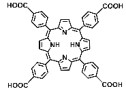
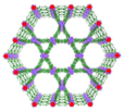
120 nm in width and 300 nm in length, RT (1O2)-RDT (·OH), RT/RDT for metastatic osteosarcoma[50]
Th-DBP,
Th-DBP@PEG
Pharmaceutics 17 00883 i005Pharmaceutics 17 00883 i006
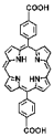
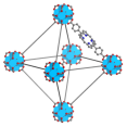
nano-octahedron, 80 nm,
RT (1O2)-RDT (·OH), RT/RDT for CT26 and Panc02 cells
[51]
Bi-DBPPharmaceutics 17 00883 i007Pharmaceutics 17 00883 i008nanorod, diameter (~20 nm) and length (~180 nm), RT (1O2)-RDT (·OH), RT/RDT for TRAMP-C2 cells[29]
Pt—heavy atom effect,
Hf-DBP-Pt
Pharmaceutics 17 00883 i009Pharmaceutics 17 00883 i010
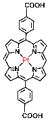
nanoplate, diameter (~100 nm) and thickness (~8 nm),
RT (1O2)-RDT (·OH), RT/RDT for CT26 and HepG2 cells
[79]
Hf-TBP,
HP(Hf)-MOF,
Hb@HP(Hf)
Pharmaceutics 17 00883 i011Pharmaceutics 17 00883 i012
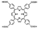
spherical structure, 200 nm,
O2-carrying, RT (1O2)-RDT (·OH), RT/RDT for 4T1 and CT26 cells
[81]
Fe(III)-mediated CDT,
Hf-DBP,
Hf-DBP-Fe
Pharmaceutics 17 00883 i013Pharmaceutics 17 00883 i014nanoplate, ~100 nm, CDT (O2 generation, ·OH),
RT (1O2)-RDT (·OH), RT/RDT for MC38 cells
[80]
Hf-DBP-QP,
Hf-DBP-QP-SN
Pharmaceutics 17 00883 i015Pharmaceutics 17 00883 i016
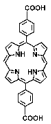
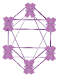
nanoplate, diameter (~120 nm) and thickness (~20 nm), chemotherapy, RT (1O2)-RDT (·OH), RT/RDT for CT26 cells[82]
DBP (5,15-di(p-benzoato)porphyrin); TBP (5,10,15,20-tetra(p-benzoato)porphyrin); Hb (hemoglobin); QP (quaterphenyl dicarboxylic acid); SN (7-ethyl-10-hydroxycamptothecin).
Disclaimer/Publisher’s Note: The statements, opinions and data contained in all publications are solely those of the individual author(s) and contributor(s) and not of MDPI and/or the editor(s). MDPI and/or the editor(s) disclaim responsibility for any injury to people or property resulting from any ideas, methods, instructions or products referred to in the content.

