Article

Salicylic Acid-Mediated Silver Nanoparticle Green Synthesis: Characterization, Enhanced Antimicrobial, and Antibiofilm Efficacy

by
Jingqing Zhang
,
Yuxu Chen
,
Yuanyu Xu
,
Zhimin Zhao
and
Xinjun Xu
*
School of Pharmaceutical Sciences, Sun Yat-sen University, Guangzhou 510006, China
*
Author to whom correspondence should be addressed.
Pharmaceutics 2025, 17(4), 532; https://doi.org/10.3390/pharmaceutics17040532
Submission received: 17 March 2025 / Revised: 9 April 2025 / Accepted: 15 April 2025 / Published: 18 April 2025
(This article belongs to the Special Issue Nanotechnology in Antibacterial Drug Delivery)

Abstract

:
Objectives: Silver nanoparticles (AgNPs) were synthesized via an easy and rapid biogenic synthesis approach, utilizing the dual capabilities of salicylic acid as both a reducing and capping agent. Methods: The characterization of Salicylic Acid-Mediated Silver Nanoparticle (SA-AgNPs) was conducted using a variety of techniques, including ultraviolet-visible spectroscopy, dynamic light scattering, scanning electron microscopy combined with energy dispersive X-ray spectroscopy, transmission electron microscopy, X-ray diffraction, Fourier transform infrared spectroscopy, as well as thermogravimetric analysis paired with differential scanning calorimetry. Results: SA-AgNPs demonstrated significant antibacterial properties against both Gram-positive (methicillin-resistant Staphylococcus epidermidis, Staphylococcus aureus, Cutibacterium acnes, methicillin-resistant Staphylococcus aureus) and Gram-negative (Escherichia coli), with minimum inhibitory concentrations (MICs) of 8, 9, 8, 4, and 6 μg/mL, respectively. At a concentration of 32 μg/mL, SA-AgNPs exhibited 99.9% killing efficiency against Escherichia coli (E. coli), Cutibacterium acnes (C. acnes), and methicillin-resistant Staphylococcus aureus (MRSA), within 4, 16, and 12 h, respectively. At the same concentration, SA-AgNPs effectively inhibited 95.61% of MRSA biofilm formation. SA-AgNPs induced the leakage of intracellular macromolecular substances by increasing the membrane permeability, which ultimately caused bacterial apoptosis. Conclusions: Overall, this study presents a fast and environmentally friendly approach for synthesizing SA-AgNPs, with potential applications as nano antibiotics antibacterial coatings for implantable medical devices and wound dressings.

Graphical Abstract

1. Introduction

Bacterial resistance to antibiotics has emerged as a worldwide public health concern, particularly regarding Methicillin-resistant Staphylococcus aureus (MRSA) [1,2,3,4]. Conventional antibiotics’ efficacy has reduced as resistant bacteria evolve [5]. Therefore, new alternative therapies are required to avoid drug-resistant bacterial infections.
Nanoparticles are defined as particles measuring between 1 and 100 nm [6]. Because of their distinct physical, chemical, and biological characteristics, nanoparticles have become increasingly popular in various applications, including antibacterial coatings for medicinal materials [7], cancer diagnosis and therapies [8], drug delivery [9] and imaging [10]. In particular, silver nanoparticles (AgNPs) have taken the leading position in these fields [11]. AgNPs also show promise as a potential strategy for overcoming the antimicrobial resistance challenge [12,13]. AgNPs can be manufactured using chemical [14,15], physical [16,17], and green techniques [18,19]. Green synthesis produces stable functionalized AgNPs by reducing and capping them with plant extracts, plant-derived compounds, or microorganisms [20]. Green synthesis is gaining popularity because of its environmental and economic benefits [21].
Notably, recent studies have demonstrated that phenolic compounds can enhance AgNPs’ bactericidal efficacy by synergizing with silver ions: AgNPs synthesized using proanthocyanidins and cinnamic acid exhibit great bactericidal efficacy against both bacteria and fungi [22,23]. Kaempferol-synthesized AgNPs demonstrated enhanced antibacterial efficacy against MRSA relative to commercial AgNPs and kaempferol individually [24]. AgNPs coated with gallic acid have strong antibacterial properties and low cytotoxicity to normal cells [25]. Curcumin-modified AgNPs exhibit enhanced bacterial membrane binding of silver and Ag⁺ release compared to unmodified counterparts, coupled with increased reactive oxygen species (ROS) generation, collectively driving synergistic antibacterial effects [26]. In contrast to chlorogenic acid alone, AgNPs synthesized with chlorogenic acid showed nearly eight times as much antibacterial activity against Pseudomonas aeruginosa [27]. A minimum inhibitory concentration (MIC) of 1.0 μg/mL was demonstrated by quercetin-mediated synthesized AgNPs with an 8 nm diameter against Streptococcus sp., Escherichia coli and Candida sp. Microorganisms [28].
However, no study has explored salicylic acid (SA)—a natural phenolic acid with intrinsic anti-inflammatory and antimicrobial properties [29,30,31]—as both a reducing and functionalizing agent for AgNPs and systematically investigated its potential to combat MRSA biofilms. While the poor aqueous solubility of SA has historically restricted its practical applications, this limitation is circumvented in the synthesis of AgNPs, where SA does not need to be fully dissolved to serve its intended functions. Salicylic acid, as a natural phenolic acid, can convert Ag (I) ions into Ag (0) [31]. Meanwhile, SA’s bioactive properties contribute to the membrane-disrupting and antibacterial activities of AgNPs, creating a synergistic effect [21]. This approach not only resolves the solubility issue but also transforms it into a beneficial characteristic in nanoparticle-based antibacterial strategies.
In this study, we synthesized AgNPs utilizing salicylic acid as both a reducing and capping agent, optimized the synthesis conditions (ultrasonic time, volume ratio and pH) and characterized it using a range of techniques such as UV–Visible spectrophotometer, transmission electron microscopy, scanning electron microscopy, X-ray diffraction and Fourier transform infrared spectroscopy. In terms of biological activity, we evaluated its antibacterial activity, bactericidal activity, and bactericidal efficiency against various kinds of bacterial strains. Furthermore, the antibiofilm activity of SA-AgNPs against MRSA was examined.

2. Materials and Methods

2.1. Materials

Silver nitrate (AgNO3), commercial AgNPs (≥99.5%, 60–120 nm), and ampicillin were from Shanghai Aladdin Biochemical Technology Co., Ltd. (Shanghai, China); Salicylic acid and sodium hydroxide were from Tianjin ZhiYuan Reagent Co., Ltd. (Tianjin, China). Mueller Hinton II broth (Cation-Adjusted) and Mueller-Hinton agar (MHA) were obtained from Solarbio Technology Co., Ltd. (Beijing, China); Brain Heart Infusion (BHI) Broth and Reinforced Clostridium Agar were purchased from HuanKai Microbial (Guangzhou, China). Trypticase soy broth was from Qingdao Hope Bio-Technology Co., Ltd. (Qingdao, China). Luria–Bertani (LB) broth was from Sangon Biotech (Shanghai) Co., Ltd. (Shanghai, China) Crystal violet (CV) was from Macklin Co., Ltd. (Shanghai, China). Phosphate buffered saline (PBS) was from Thermo Fisher Scientific (Waltham, MA, USA).
Gram-negative bacterial strains: Escherichia coli (E. coli, ATCC25922), Gram-positive bacterial strains: Staphylococcus aureus (S. aureus, ATCC25923), methicillin-resistant Staphylococcus aureus (MRSA, ATCC43300), methicillin-resistant Staphylococcus epidermidis (MRSE, ATCC35984) and Cutibacterium acnes (C. acnes, ATCC6919) were obtained from Guangdong Microbial Strain Preservation Center (Guangzhou, China).

2.2. Green Synthesis of SA-AgNPs

A solution of 7.24 μmol/mL SA in ultrapure water (H2O) was mixed with 10 mmol/mL AgNO3. After adding sodium hydroxide to correct the pH, the liquid was sonicated for 12 min with an ultrasonic power of 360 W and a frequency of 40 kHz. The solution was observed to change from colorless and transparent to dark brown, signaling the creation of AgNPs [32].

2.3. Study of the Effect of Different Factors on SA-AgNPs Biosynthesis

2.3.1. Effect of Ultrasonic Time

The experimental conditions were optimized for ultrasonic time, volume ratio, and pH, with the samples’ surface plasmon resonance (SPR) absorbance and particle size serving as reference indexes.
The volume ratio of AgNO3:SA was 2:1, and the pH was maintained at 9. To evaluate the impact of ultrasonic time on AgNP synthesis, the samples were exposed to various ultrasonic times (4 min, 8 min, 12 min, 16 min, and 20 min)

2.3.2. Effect of Volume Ratio

The ultrasonic time was 12 min, and the pH was maintained at 9. Different volume ratios of AgNO3:SA (1:2, 1:1, 2:1, 3:1, and 4:1) were used to assess the influence of volume ratio on AgNP synthesis.

2.3.3. Effect of pH

It was efficient in lengthening the steady dispersion time of AgNPs by reaching the optimal pH [33] AgNO3:SA was fed at a 2:1 volume ratio, and the ultrasonic period was 12 min. The reaction solution’s pH was modified to 6, 7, 8, 9, and 10 to examine the impact of pH on the synthesis of AgNPs.

2.4. Characterization of SA-AgNP

A UV-visible spectrophotometer (UV-Vis) (UV-2600, Shimadzu, Kyoto, Japan) was used to measure absorbance from 300 nm to 800 nm, confirming AgNP biosynthesis [34].
The samples were dispersed in water. A Zetasizer Nano ZS instrument (Malvern Instruments, Malvern, UK) was used to measure the polydispersity index (PDI), mean hydrodynamic size, and zeta potential.
To determine the shapes, sizes, and particle morphology of SA-AgNPs, the samples were diluted using ultrapure water and dripped on a carbon-coated copper film until dry at room temperature. The images were taken with transmission electron microscopy (TEM (JEM-2010HR, JEOL, Tokyo, Japan)) at 200 kV accelerating voltage.
The surface morphology and elemental composition of SA-AgNPs were analyzed using SEM (EVO MA10, ZEISS, Oberkochen, Germany) at 5 kV and EDX (Bruker, Berlin, Germany). The SA-AgNPs’ surface morphology was photographed and documented at different magnifications. The EDX result revealed the silver composition percentages in SA-AgNPs.
To analyze the crystalline structure of SA-AgNPs, the powder was flattened into a thin film on a slide, and the X-ray diffraction (XRD) (Empyrean Alpha 1, Malvern Panalytical, Malvern, UK) was operated in the 2θ region (10° to 80°) at a rate of 0.02°/min.
Nitrogen was used as a protective gas, and thermogravimetric analysis paired with differential scanning calorimetry (TG-DSC) (STA449F3, NETZSCH, Selb, Germany) spectra were recorded for SA-AgNPs heated from 33 °C to 900 °C.
The functional groups in SA-AgNPs were identified through Fourier transform infrared spectroscopy (FTIR) (Spectrum two, Perkin Elmer, Springfield, IL, USA), which scans ranging from 4000 to 400 cm−1 at a resolution of 0.5 cm−1.

2.5. Assay of Salicylic Acid

The assay of salicylic acid was conducted based on the approach detailed in prior studies, with certain modifications [35,36]. SA content in SA-AgNPs was evaluated indirectly using reversed-phase high-performance liquid chromatography (RP-HPLC) (LC-20AD, Shimadzu, Kyoto, Japan) on a C18 column (250 mm × 4.6 mm, COSMOSIL). The mobile phase consisted of a buffer solution made from sodium dihydrogen phosphate, with its pH fine-tuned to 2.2 using phosphoric acid, combined with methanol in a 50:50 volume ratio. Prior to application, it was passed through a 0.22 μm membrane filter utilizing a vacuum filtration setup and degassed in a sonication bath. A stock solution of 1 mg/mL SA was made using HPLC-grade methanol. Furthermore, reference substance solutions of 0.01 to 0.15 mg/mL were then generated from the stock solution using methanol as a diluent. The reacted SA-AgNPs solution was centrifuged to obtain a clear supernatant. The supernatant was subsequently passed through a 0.22 μm membrane filter to eliminate any remaining silver nanoparticles. Once filtered, the supernatant, which contained unreacted SA (not interacting with Ag+), was separated and acidified with sulfuric acid. Test samples were obtained by diluting the acidified supernatant with methanol. After filtering through a 0.22 μm membrane, the samples were measured at 310 nm. The SA content in the supernatant was measured in triplicate (n = 3) using the equation below.
ω = ρ × V m × 100 %
where w, ρ, V, and m are the mass fraction of SA in the sample (%), SA concentration from the standard curve (mg/mL), test sample volume (mL), and total mass of SA (mg).

2.6. Assay of Silver Ions

The silver ions content was determined by inductively coupled plasma-atomic emission spectroscopy (ICP-AES) (Optima8300, PerkinElmer, Springfield, IL, USA) [37,38]. A certain amount of SA-AgNPs powder was dissolved in a 5% v/v nitric acid solution to prepare silver ion solutions at concentrations between 4 and 8 mg/L. The test samples were then analyzed by ICP-AES, and the silver ion content in SA-AgNPs was calculated using the equation below.
ω = c × V 1000 × m × 100 %
where w, c, V, and m are the mass fraction of silver in the sample (%), silver concentration from the standard curve (mg/L), test sample volume (mL), and sample weight of SA-AgNPs (mg).

2.7. Stability of SA-AgNPs

To evaluate the stability of SA-AgNPs, the samples were kept in a refrigerator at 4 °C, shielded from light exposure.
Over specific time intervals—0, 2, 5, 7, 14, 28, 35, 49, and 65 days—the particle size, polydispersity index (PDI), and zeta potential were measured. Additionally, UV-Vis spectroscopy was employed to scan the samples across a wavelength range of 300–800 nm [38].

2.8. Antimicrobial Activity of SA-AgNPs

2.8.1. Minimum Inhibitory Concentration and Minimum Bactericidal Concentration (MIC and MBC)

The broth microdilution assay was performed in a 96-well plate to assess the minimum inhibitory concentration (MIC) and minimum bactericidal concentration (MBC), following the Clinical and Laboratory Standards Institute (CLSI) guidelines (M07) [39]. SA-AgNPs, commercial AgNPs, and ampicillin dispersions were all formulated to a specific concentration employing ultrapure water, while SA dispersions were prepared with 20% ethanol. The bacterial suspensions were adjusted to the appropriate optical density at 600 nm (OD600) and then spread onto Mueller-Hinton agar (MHA) plates. These plates were incubated at 37 °C for a period of 24 h to verify that the concentration of the bacterial suspensions reached 1 × 10⁸ colony-forming units (CFU)/mL.
The bacterial suspensions (1 × 10⁸ CFU/mL) were diluted one hundred-fold with cation-adjusted Mueller-Hinton broth (CAMHB), and BHI medium was used for C. acnes. Finally, each well contained 100 μL of diluted bacterial dispersions and 100 μL of sample dispersions (SA-AgNPs, commercial AgNPs, SA, ampicillin, and 20% ethanol) for a final volume of 200 μL. Sterile CAMHB served as the negative control, ampicillin as the positive control, and 20% ethanol as the solvent control.
The 96-well plates were placed in an incubator at 37 °C for 24 h. (72 h for C. acnes). C. acnes were incubated under anaerobic conditions using an AnaeroPack (Japan Mitsubishi MGC, Saitama, Japan). The results were visually assessed, with the MIC determined as the lowest dose at which no bacterial growth could be detected with the naked eye. To determine the MBC, 100 μL of the mixture from MIC wells showing no bacterial growth was then transferred and cultured on MH agar plates, incubated at 37 °C for 24 h.

2.8.2. Time-Kill Assay

CLSI guidelines (M26h) [40] were followed for performing Time-Kill assays. Bacteria were inoculated into sterile CAMH broth (BHI medium for C. acnes) to achieve a final concentration of 1 × 10⁶ CFU/mL. Diluted bacteria suspensions were added to each tube containing SA-AgNPs in sterile CAMH broth. Time-kill assays were conducted by applying final concentrations of SA-AgNPs at 1/2 × MIC, 1 × MIC, 2 × MIC, and 4 × MIC levels. After incubation at 37 °C, 100 μL of the diluted mixture was cultivated on MH agar plates (Reinforced Clostridium agar plates for C. acnes) at each time point. The plates were subsequently incubated at 37 °C for 24 h (72 h for C. acnes). The colonies on each plate were counted to assess the bacterial concentration at each time point.
Sterilization was regarded as complete when the bacterial concentration reached one-thousandth of the initial concentration.

2.9. Antibiofilm Ability Studies

2.9.1. Inhibition of Biofilm Formation

The biofilm formation of MRSA was assessed in 96-well microplates using microtiter plate assay, following a previously reported method with modifications [41]. Bacterial suspension diluted in Trypticase soy broth (TSB) with 1% (w/v) glucose and varying sample concentrations were transferred to each well of the 96-well plates. Positive controls consisted of wells containing only TSB and MRSA, while negative controls were wells filled solely with sterile phosphate-buffered saline (PBS, pH 7.4). The plates were then incubated at 37 °C for 48 h. Following incubation, the contents of each well were carefully discarded, and the wells were rinsed three times using 300 μL of PBS. Once the plate was dried, the biofilms on the bottom of the wells were fixed with 150 μL of 95% methanol. To visualize the biofilms, 0.1% crystal violet (CV) was introduced and left to incubate for 15 min at room temperature. Excess CV was removed by washing the wells with 300 μL of sterile distilled water until the wash solution ran clear. Subsequently, 200 μL of 33% glacial acetic acid was added to each well to dissolve the CV bound to the stained biofilm. A 150 μL aliquot from each well was then transferred to a fresh 96-well plate for further tests. The absorbance of each well was measured at 562 nm using a microplate reader to quantify biofilm formation. The cutoff optical density (ODc) was determined as the mean OD of the negative control plus three standard deviations [42]. The percentage of biofilm inhibition was then calculated using the following formula:
O D   p o s i t i v e   c o n t r o l O D   s a m p l e O D   p o s i t i v e   c o n t r o l × 100 %

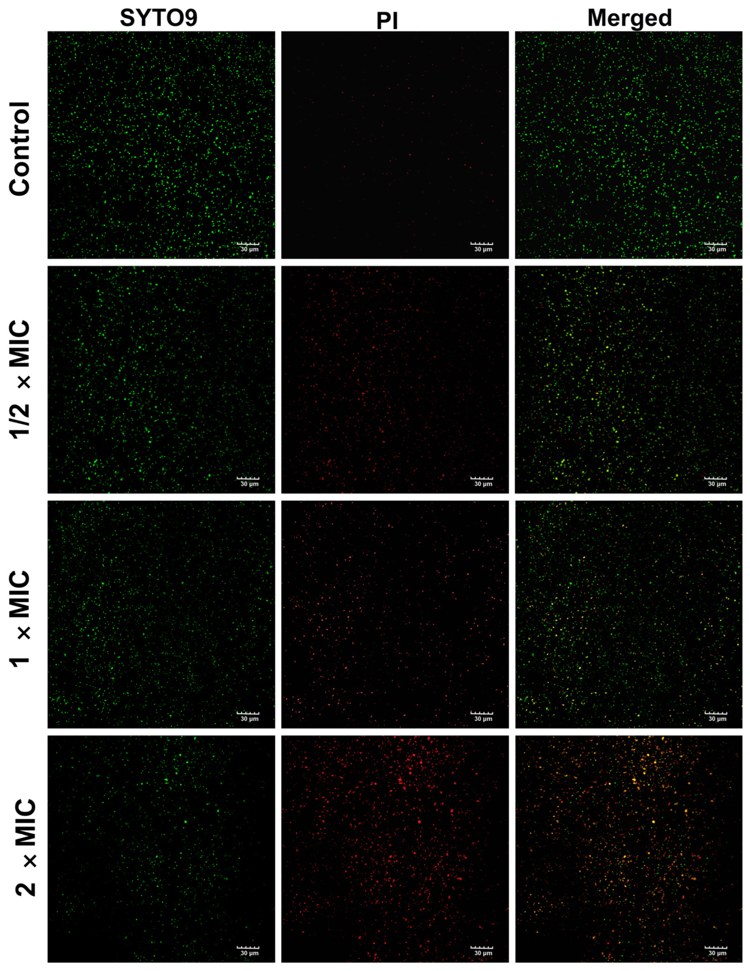
2.9.2. Live and Dead Bacteria Assay

To assess the viability of MRSA exposed to various concentrations of SA-AgNPs, live/dead bacterial assays were performed using confocal laser scanning microscopy (FV3000, Olympus, Tokyo, Japan). MRSA was cultured overnight in an LB medium. The bacterial suspensions were washed in sterile PBS (pH = 7.4) three times to remove the medium and then resuspended in sterile PBS to OD600 of 2.0. The bacteria were incubated with SA-AgNPs (1:1, v/v) at 37 °C for 1 h. Following this step, the suspension was stained and incubated at room temperature for 15 min, shielded from light. Bacterial viability was observed using the SYTO 9/propidium iodide (PI) double-stain kit (Maokang, Shanghai, China), following the guidelines provided by the manufacturer.

2.10. Determination of Leakage of DNA, RNA, and Protein Concentrations in Bacterial Cell

The bacteria (MRSA) were washed and resuspended in an M9 minimal medium (containing 10 mM NaAc, 2 mM MgSO4, 0.1 mM CaCl2). OD600 of the bacterial suspension was set to 0.6, then incubated with different concentrations (8 × MIC and 16 × MIC) SA-AgNPs at 37 °C and 160 rpm for 6 h, respectively. Bacteria without SA-AgNPs treatment as a control group. The supernatant was isolated by centrifugation (8000× g) for 5 min at 4 °C. The concentrations of DNA and RNA leaked from MRSA were measured at 260 nm by NanoDrop 2000 UV–vis spectrophotometer (Thermo Fisher Scientific, Waltham, MA, USA). The concentrations of proteins in the supernatant were determined using the dilution-free rapid gold BCA protein assay kit (Thermo Fisher Scientific, Waltham, MA, USA). The absorbance was measured at 490 nm.

2.11. Statistical Analysis

The study was conducted using three independent experimental groups. All findings reported in this research are presented as mean values accompanied by their standard deviations (SD). Data analysis was performed using Origin 2021 (OriginLab, Northampton, MA, USA) and GraphPad Prism 8.0.1 (GraphPad Software Inc., San Diego, CA, USA). Statistical comparisons were made using both one-way and two-way analysis of variance (ANOVA). p < 0.05 indicated the difference was statistically significant. * represents p < 0.05; ** represents p < 0.01; *** represents p < 0.001; **** represents p < 0.0001.

3. Results and Discussion

3.1. Green Synthesis of Salicylic Acid-Silver Nanoparticles (SA-AgNPs)

SA and AgNO3 were sonicated at room temperature, resulting in a dark brown solution. After changing the pH of the AgNO3 solution and adding salicylic acid for ultrasonication, the sample solution changed from colorless and translucent to dark brown (Figure S1A,B), indicating that the silver ions were reduced to Ag0 and silver nanoparticles were generated [24,43]. The sample was subsequently centrifuged, rinsed three times with ultrapure water, and freeze-dried to yield a brown-black powder (Figure S1C).
In this study, salicylic acid was used as a reducing and capping agent. AgNO3 was utilized as a precursor for the AgNPs, ultrapure water as a solvent for dissolution, and sodium hydroxide to modify pH. The entire synthesis process was easy and ecologically friendly. Phenolic chemicals can act as electron or hydrogen donors in the chemical reaction that produces AgNPs [31,44]. Unlike previous studies relying on complex plant extracts [45,46,47,48,49], our method employs pure SA to ensure reproducibility and mechanistic clarity.

3.2. Study of the Effect of Different Factors on the Biosynthesis of SA-AgNPs

3.2.1. Effect of Ultrasonic Time

As demonstrated in Figure 1A, SA-AgNPs were synthesized at various ultrasonic times (4, 8, 12, 16 and 20 min). By comparing the UV-Vis spectra corresponding to the SA-AgNPs solutions, Figure 1B makes it clear that the peak intensity of the SPR absorption bands reaches its maximum when the ultrasonic time is 12 min. After the ultrasonic time exceeds 12 min, the peak intensity decreases with further increases in ultrasonic duration, while the wavelength of the SPR peaks remains relatively unchanged. This suggests that ultrasonic treatment has little effect on the size of SA-AgNPs, aligning with hydrodynamic size measurements [50].
The SA-AgNPs particle size ranged from 120 nm to 180 nm during the 4–20 min ultrasonication interval, as shown in Figure 1C. After 12 min of sonication, the SA-AgNPs particle size was 149.37 ± 4.55 nm, and their PDI was the least, measuring less than 0.3 (0.231 ± 0.010). Thus, one of the ideal experimental settings is a 12-min ultrasonic time.

3.2.2. Effect of Volume Ratio

SA-AgNPs were synthesized with various volume ratios (1:2, 1:1, 2:1, 3:1, and 4:1), as shown in Figure 1D. The volume ratio (AgNO3:SA) was the primary experimental element affecting the concentration and size of SA-AgNPs. This was verified by the UV-vis spectra of SA-AgNPs synthesized with different volume ratios. Figure 1E demonstrates that the peak intensity of the SPR peak reaches its maximum when the volume ratio (AgNO3:SA) is increased from 1:2 to 2:1. The peak intensity of the SPR peak diminishes as the volume ratio (AgNO3:SA) increases from 2:1 to 5:1. This implies that the maximum concentration of nanoparticles was generated using a volume ratio of 2:1 (AgNO3:SA).
Furthermore, when the volume ratio (AgNO3:SA) was higher than 2:1, the SPR peak wavelength redshifted, and the peak shape broadened and flattened, indicating that the particle size of SA-AgNPs increased [51]. In Figure 1F, SA-AgNPs had a particle size of 129.2 ± 1.98 nm at a 2:1 volume ratio (AgNO3:SA) and a PDI of 0.296 ± 0.018. The optimal volume ratio is (AgNO3:SA) 2:1.

3.2.3. Effect of pH

SA-AgNPs were generated at various pH levels (6, 7, 8, 9, and 10), as indicated in Figure 1G. pH significantly influences silver nanoparticles’ formation and dimensions [49,52]. Figure 1H demonstrates that SPR peaks did not form in the UV-Vis spectra when the solution was acidic (pH = 6). The SPR peaks began to appear at pH 7 and reached their maximum intensity as the pH climbed to 9. Combined with Figure 1I, at pH 10, the peak intensity decreased again while the particle size and PDI increased, demonstrating that too high a pH not only decreases the creation of SA-AgNPs but also increases their size and induces the nanoparticles to assemble [49].
The solution’s absorption intensity was highest at pH 9, with an even dispersion of nanoparticles (PDI = 0.270 ± 0.006). As a result, the optimal pH was 9. Similar results have been obtained in previous studies [53,54,55,56]. It also has demonstrated that the optimal pH for green synthesis of silver nanoparticles varies with the reducing agent [57].

3.3. Characterization of SA-AgNPs

3.3.1. UV-Visible Spectrophotometer Analysis

UV-Vis Spectrophotometer is an important tool for characterizing the optical properties of AgNPs. Because of their unique optical properties, metal nanoparticles form SPR peaks that can be absorbed between 350 and 500 nm. As shown in Figure 2A, SA and AgNO3 have no absorbance peaks near 400 nm, while the synthesized SA-AgNPs exhibit a new absorbance peak at 387 nm. This is a typical SPR band for silver nanoparticles, indicating their formation [58].
Based on Mie-type scattering theory, the UV-Vis peak position reveals the size of AgNPs [50]. The narrower the shape of AgNPs’ SPR peak, the more evenly distributed they are, and vice versa [59]. A blueshift of SPR bands shows a decrease in the particle size of the AgNPs generated, while a redshift of SPR bands indicates an increase in particle size [51]. A higher SPR peak absorption value indicates a higher concentration of AgNPs in the solution [60]. This principle was applied in single-factor experiments and confirmed by hydrodynamic particle size and PDI measurements.

3.3.2. Hydrodynamic Particle Size and Zeta Potential Measurement

The diluted SA-AgNPs samples’ hydrodynamic particle size and zeta potential were determined by dynamic light scattering (DLS), and the results are given in Figure 2B. The SA-AgNPs produced under optimal experimental conditions had a hydrodynamic particle size of 166.8 nm, a zeta potential of −31 mV, and a PDI of 0.237, as determined by a Zetasizer Nano ZS instrument. The PDI of the SA-AgNPs was less than 0.3, indicating that the silver nanoparticles were distributed equally. The higher the absolute value of the zeta potential, the stronger the repulsion between particles and the lower the chance of aggregation [61]. As a result, SA-AgNPs are difficult to aggregate in solution and remain rather stable. Additionally, for stronger antibacterial action, SA-AgNPs’ negative charge on the surface helps them adhere to germs.

3.3.3. Transmission Electron Microscopy (TEM)

The size and surface morphology of SA-AgNPs were observed using TEM. According to Figure 2C,E, SA-AgNPs have a nearly spherical form with some particle agglomeration, which could be brought on by freeze-drying. Particle size statistics of SA-AgNPs in TEM images were analyzed using ImageJ 2.0. Figure 2D,F show histograms of the average diameter of the SA-AgNPs, with average particle sizes of 24.42 ± 5.57 nm and 28.90 ± 10.91 nm.
The particle sizes determined by the DLS and the ones counted in the TEM pictures differ significantly. This is because the SA-AgNPs’ hydrodynamic particle size in a liquid (water) is measured by DLS. The hydrodynamic particle size usually appears much larger than the TEM particle size [62], which is the actual particle size. Since the hydrodynamic size comprises not only the inorganic particles in the core but also the hydration layer on the surface and the attached organic components, the hydration layer on the AgNPs’ surface is what causes the difference between the two.

3.3.4. Scanning Electron Microscope-Energy Dispersive X-Ray Spectroscopy (SEM-EDX)

The surface morphology and elemental composition of SA-AgNPs were determined using SEM-EDX. Based on Figure 2G,H and Figure S2, the SA-AgNPs are elliptical or sub-spherical in shape and primarily composed of Ag. EDX spectra (Figure S3) revealed the presence of C and O, indicating that SA successfully capped AgNPs. Silver nanoparticles typically have a strong signal peak at 3 keV because of SPR. This shows the existence of silver as a component element, verifying the synthesis of SA-AgNPs. SA-AgNPs contain the largest atomic percentage of Ag (46.84%), followed by C (29.70%) and O (23.02%). C and O are obtained from salicylic acid, while O may also be derived from airborne contaminants adsorbed on the samples’ surfaces. The presence of C and O atoms verifies salicylic acid’s role as a capping and stabilizing agent on the surface of silver nanoparticles [63]. The remaining very minor amount of sodium (0.44%) came from sodium hydroxide.

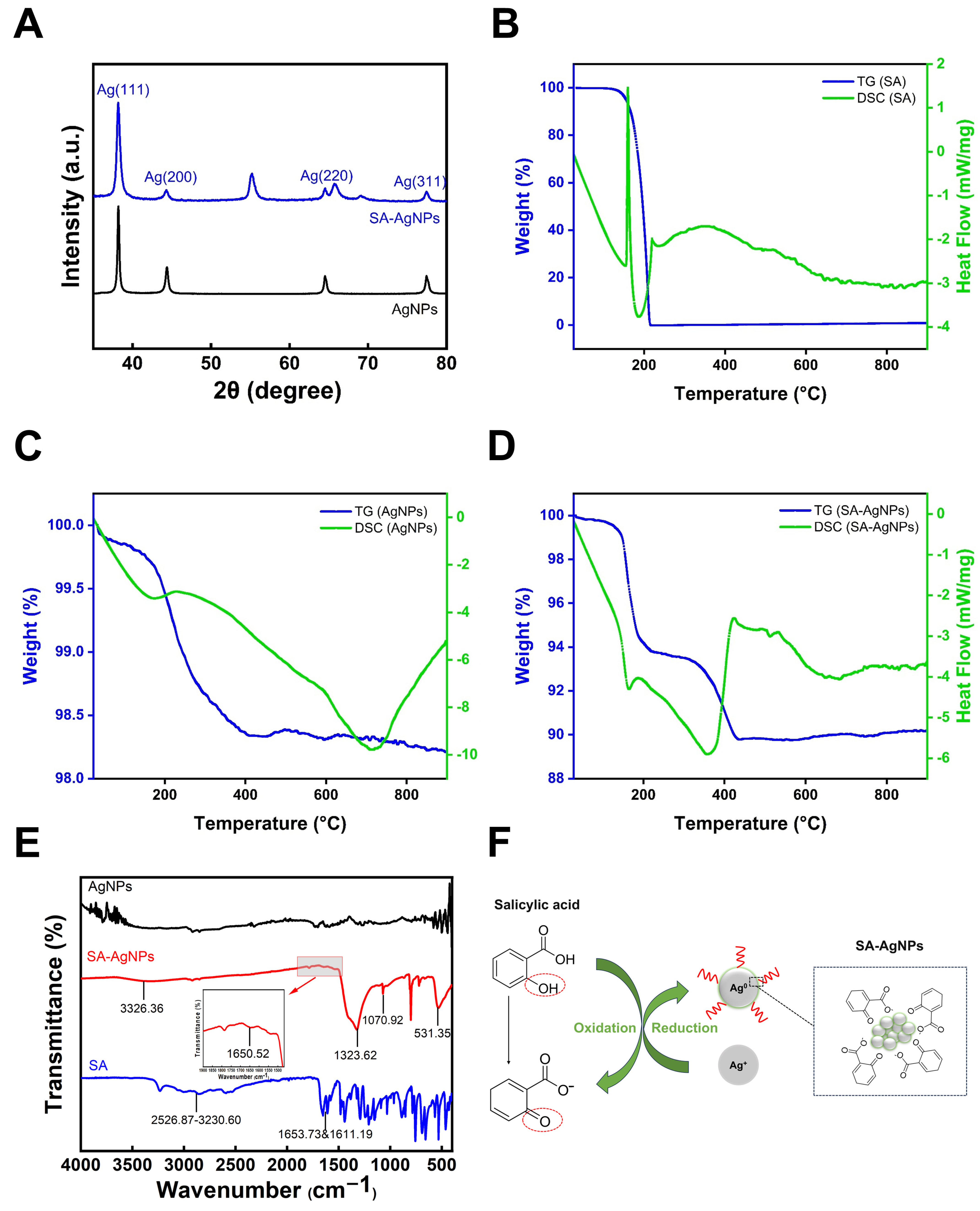
3.3.5. X-Ray Diffraction Analysis

Figure 3A shows the XRD results of synthesized SA-AgNPs. SA-AgNPs exhibited four distinct diffraction peaks at 38.12°, 44.22°, 64.50°, and 77.35°, which correspond to lattice planes (111), (200), (220), and (311), respectively. These peaks are also consistent with the four distinct peaks of commercial AgNPs at 38.12°, 44.35°, 64.46°, and 77.39°. The XRD spectrum of the SA-AgNPs was compared to the Joint Committee on Powder Diffraction Standards (JCPDS) file (No. 04–0783), which revealed the crystalline nature of SA-AgNPs [64].
However, there is a little shift in peak positions, which could be due to strain in the crystal structure. The peaks measured at 55.19° and 69.04° are similar to those of silver oxide [63]. The sharp peaks indicate strong crystallinity in AgNPs. This result validates the presence of silver in the prepared samples, as well as the crystallinity of SA-AgNPs.

3.3.6. Thermogravimetric-Differential Scanning Calorimetry (TG-DSC)

The thermal stability of SA-AgNPs was evaluated with TG-DSC. TG-DSC results are displayed in Figure 3B–D. As seen in the picture, the SA powder loses mass by approximately 100% after 215 °C, which is related to the fact that SA is an organic compound. The weight loss of commercial AgNPs without SA capping was approximately 1.70% between 32 °C and 407 °C. SA-AgNPs lost approximately 6.23% between 32 °C and 221 °C, and 3.98% between 221 °C and 434 °C, possibly due to water evaporation and loss of organic matter on the silver nanoparticles’ surfaces [65].
The overall weight loss of SA-AgNPs was 10.21%, and the melting point of silver is 960.54 °C [66], implying that the weight loss in the tested range is not due to silver; hence, the silver content can be determined to be 89.79%. This is consistent with the silver content estimated by EDX (87.30 ± 2.21%).

3.3.7. Fourier Transform Infrared Spectroscopy (FTIR)

IR analysis was applied to identify potential functional groups and formation mechanisms in SA-AgNPs. Figure 3E presents a comparison between the blank samples (commercial AgNPs and salicylic acid) and SA-AgNPs.
As demonstrated, the broad O–H stretching band of salicylic acid at 2526.87–3230.60 cm−1 results from the superposition of carboxylic acid and phenolic hydroxyl groups [67]. The two strong peaks at 1653.73 cm−1 and 1611.19 cm−1 represent the carbonyl group in salicylic acid.
The O–H stretching band of SA-AgNPs shifted to 3326.36 cm−1 compared to salicylic acid, indicating the loss of proton hydrogen [67]. This suggests that the O–H stretching of SA was likely to contribute to the synthesis of AgNPs [63]. The peak at 1650.52 cm−1 is attributed to the stretching of C=O groups or C=C stretching of aromatic ring [68]; the peak at 1323.62 cm−1 may be due to C–O stretching vibration in phenols [69]; the peak at 1070.92 cm−1 corresponds to the C–C or C–O stretching [70]; and the band at 531.35 cm−1 can be ascribed to the transition-metal Ag energy band [71]. The IR spectra suggest that the phenolic hydroxyl group (–OH) in salicylic acid reduces Ag+ to Ag0 while the phenolic hydroxyl group on the benzene ring is oxidized [22,72]. The capping alteration of AgNPs by salicylic acid was eventually achieved. According to the literature, phenolic compounds can serve as reducing and capping agents, enabling the synthesis of stable silver nanoparticles [73]. The phenolic hydroxyl groups in phenolic acids ionize under alkaline conditions to form phenolate ions, which act as electron donors to reduce Ag⁺ ions [74,75]. This redox process simultaneously oxidizes the phenolate into a quinonoid form [74,76]. Figure 3F illustrates a possible mechanism of SA-AgNPs formation according to the IR spectra.

3.4. Assay of Salicylic Acid

The content of SA was determined using the RP-HPLC technique. Linearity was seen at concentrations between 0.01 and 0.15 mg/mL. The sample solution’s peak area was estimated using a linear equation (Figure S4), yielding the SA content in the supernatant of 68.09%. Specifically, the total mass of SA is 1.0 mg, so the precipitated SA-AgNPs contained 0.3191 mg of SA, indicating that 31.91% of total SA was attached to the silver nanoparticles’ surface.

3.5. Assay of Silver Ions

SA-AgNPs had 90.61 ± 1.57% (Wt%) silver, as determined by ICP-AES analysis. This result corresponds to the EDX measurement of 87.30 ± 2.21% silver and the TG-DSC result of 89.79% silver.

3.6. Stability of SA-AgNPs

Stability tests are an essential step before the further application of nanoparticles. Thus, the stability of SA-AgNPs at 4 °C was investigated. UV-vis spectrophotometry can identify changes in the SPR peaks of silver nanoparticles, while PDI and zeta potential analyses reflect their stability in solution [38]. The higher the absolute value of the potential and the lower the PDI value, the more stable SA-AgNPs are in the solution [77,78]. As indicated in Table 1 and Figure 4A, the wavelength and peak intensity of SA-AgNPs’ SPR peak changed very little over 65 days. Figure 4B demonstrates an overall increase in particle size and PDI of SA-AgNPs, suggesting that nanoparticles may have agglomerated over this period. However, Figure 4C demonstrates that the zeta potential of SA-AgNPs remains almost unaltered after 65 days, and the absolute value is high. As a result, SA-AgNPs maintain good stability at 4 °C for 65 days.

3.7. Antibacterial Activity of SA-AgNPs

3.7.1. Minimum Inhibitory Concentration (MIC) and Minimum Bactericidal Concentration (MBC)

Measuring MIC and MBC determined the minimal concentrations at which SA-AgNPs inhibited bacterial growth and killed bacteria. The MIC and MBC for SA-AgNPs, SA, commercial AgNPs, and ampicillin against various bacteria are shown in Table 2 and Table 3, respectively.
The MIC and MBC of SA-AgNPs for MRSA and S. aureus are recorded at 8 μg/mL and 16 μg/mL, respectively. For E. coli, the MIC and MBC values stand at 6 μg/mL and 12 μg/mL. When it comes to MRSE, the MIC is 4 μg/mL while the MBC is 8 μg/mL. For C. acnes., the MIC and MBC are 9 μg/mL and 18 μg/mL, respectively. The MICs of SA-AgNPs against S. aureus and MRSA were approximately half that of AgNPs synthesized by Scutellaria baicalensis polysaccharide [79], indicating that SA-AgNPs have excellent antibacterial properties.
Salicylic acid (≥800 μg/mL) and commercial AgNPs (≥64 μg/mL) had significantly higher MIC values than synthetic SA-AgNPs. For the drug-resistant bacteria MRSE, the MIC of SA-AgNPs is lower than that of the positive drug ampicillin.
According to the MIC and MBC values, SA-AgNPs had significantly better bacteriostatic and antibacterial activity than those of salicylic acid and commercial AgNPs. This indicates the synergistic action of AgNPs combined with salicylic acid. The study found that the size, shape, concentration, and exposure period of silver nanoparticles influence their bactericidal action [80]. Furthermore, silver nanoparticles are more efficient at killing Gram-negative bacteria than Gram-positive bacteria, primarily because Gram-positive bacteria have thicker cell walls [81]. The antibacterial property of silver nanoparticles is primarily achieved by acting on the cell membrane and increasing its permeability; thus, thick cell walls reduce its antimicrobial activity.

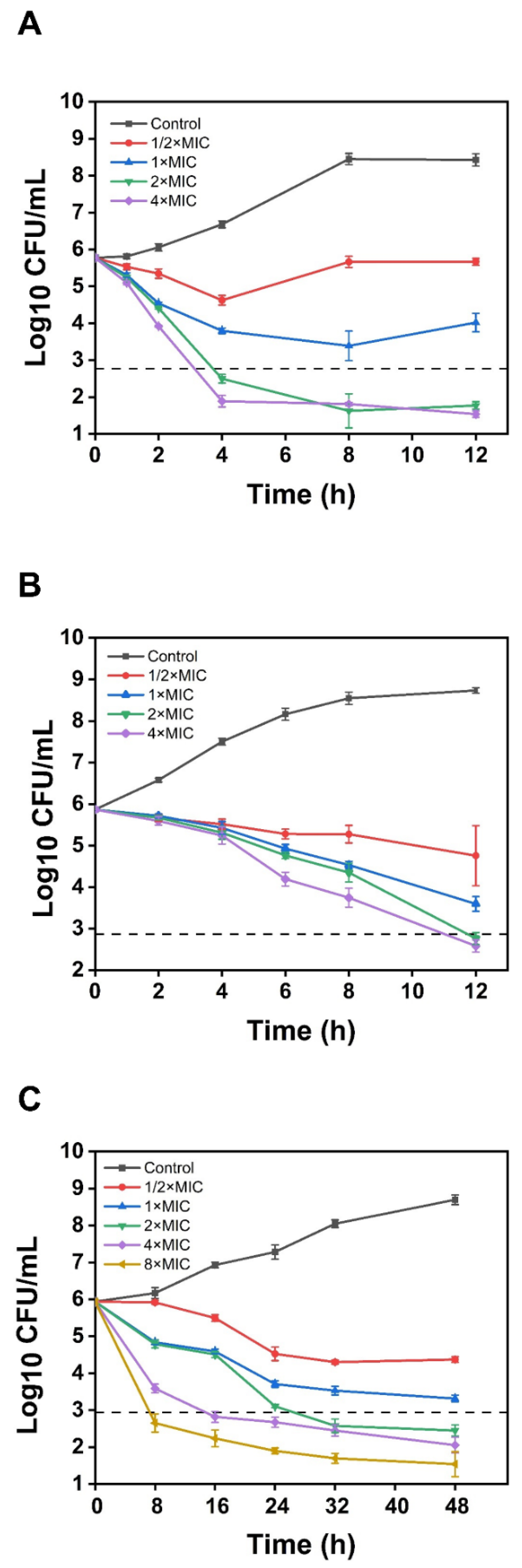
3.7.2. Time-Kill Assay

Time-kill assays were conducted to investigate the killing effect of Gram-positive bacteria (MRSA and C. acnes) and Gram-negative bacteria (E. coli) at various concentrations of SA-AgNPs (1/2 × MIC, MIC, 2 × MIC, and 4 × MIC). Figure 5A–C exhibits the bacterial concentration (log10 CFU/mL) versus time. SA-AgNPs at 2 × MIC and 4 × MIC levels killed E. coli and MRSA within 4 and 12 h, respectively, which means the bactericidal rate reached 99.9%. SA-AgNPs at 2 × MIC, 4 × MIC, and 8 × MIC concentrations killed C. acnes within 32 h, 16 h, and 8 h, respectively. SA-AgNPs (2 × MIC and 4 × MIC) killed E. coli in just 4 h, faster than killing Gram-positive bacteria (MRSA and C. acnes). Compared to other green synthesized silver nanoparticles, SA-AgNPS showed significantly higher bactericidal efficiency against MRSA. After 24 h of treatment with silver nanoparticles-kaempferol (1.25 mg/mL), MRSA growth decreased by 1.30 log10CFU/mL [24], while SA-AGNPS (16 µg/mL) reduced growth by 3.1 log10CFU/mL in 12 h.

3.8. Antibiofilm Ability Studies

3.8.1. Biofilm Formation Inhibition

Biofilms serve as a critical factor in the development of more than 80% of bacterial infections [82], playing a crucial role in the defense against antibacterial agents. Therefore, inhibiting biofilm formation effectively can reduce drug-resistant bacterial infections. Crystal violet dye (0.1%) was used to quantitatively determine the biofilm formation of MRSA treated with various concentrations of SA, AgNPs, and SA-AgNPs (Figure 6A–C). As shown in Figure 6B, SA-AgNPs at the lowest concentration of 1/4 × MIC (2 μg/mL) significantly reduced MRSA biofilm formation (p < 0.0001). Additionally, SA, AgNPs, and SA-AgNPs inhibited biofilm formation in MRSA in a concentration-dependent manner. The negative control wells had a mean OD of 0.1558 and a standard deviation of 0.0072, resulting in an ODc of 0.1774. Biofilm production was categorized as follows: OD ≤ ODc, no biofilm; ODc < OD ≤ 2 × ODc, weak biofilm; 2 × ODc < OD ≤ 4 × ODc, moderate biofilm; and OD > 4 × ODc, strong biofilm [83]. According to Figure 6B, SA at 6.4, 3.2, 1.6, and 0.8 mg/mL caused weak biofilm, while 0.4 and 0.2 mg/mL caused strong biofilm. AgNPs at 2048 and 1024 μg/mL caused weak and moderate biofilm, respectively, and at 512, 256, 128, and 64 μg/mL caused strong biofilm. SA-AgNPs at 64 and 32 μg/mL resulted in no biofilm, 16 μg/mL caused moderate biofilm, and 8, 4, and 2 μg/mL caused strong biofilm.
As shown in Figure 6C, SA at the concentrations of 6.4, 3.2, 1.6, 0.8, 0.4, and 0.2 mg/mL decreased biofilm formation by 93.56%, 94.03%, 94.35%, 95.09%, 50.84%, and 14.50%, respectively. AgNPs at 2048, 1024, 512, 256, 128, and 64 μg/mL decreased biofilm formation by 91.10%, 85.79%, 75.59%, 63.63%, 48.16%, and 36.77%, respectively. SA-AgNPs at 64, 32, 16, 8, 4, and 2 μg/mL reduced biofilm formation by 95.86%, 95.61%, 86.35%, 66.17%, 46.37%, and 37.55%, respectively. Compared to SA and AgNPs, SA-AgNPs had superior anti-biofilm activity at lower concentrations (64 and 32 μg/mL), completely inhibiting biofilm formation. At about the same dose, AgNPs using the extract of aerial parts of the Anthemis pseudocotula Boiss. plant (39 μg/mL) inhibits biofilm formation of MRSA by only 67.81% [84], whereas SA-AgNPs (32 μg/mL) inhibit 95.61%.

3.8.2. Live and Dead Bacteria Assay

SYTO 9 penetrates both intact and damaged bacterial cell membranes, attaching to DNA and emitting green fluorescence, whereas PI can only enter cells with broken membranes and emit red fluorescence. As demonstrated in Figure 7, untreated MRSA showed extensive green fluorescence and limited red fluorescence, indicating a high density of live cells with intact membranes [79]. Compared to the control group, SA-AgNPs treatment resulted in a decrease in green fluorescence, with wide areas of red and orange fluorescence (red-green fluorescence overlap), indicating a large number of apoptotic cells. In addition, as the concentration of SA-AgNPs grew, green fluorescence reduced, and red fluorescence increased. In conclusion, SA-AgNPs significantly increased bacterial death, preventing biofilm formation.

3.9. Effect of SA-AgNPs on the Leakage of DNA, RNA, and Protein in Bacterial Cell

The leakage of intracellular compounds reflects cell membrane permeability. Extracellular DNA, RNA, and protein levels were measured to evaluate the effect of SA-AgNPs on bacterial membrane permeability. As shown in Figure 8A, SA-AgNPs significantly increased protein leakage compared to the control. Similar results were observed for DNA and RNA leakage in Figure 8B. SA-AgNPs dose-dependently increased membrane permeability and compound leakage, which is one of its mechanisms of antibacterial action.

4. Conclusions

A green synthesis method for silver nanoparticles (SA-AgNPs) mediated by salicylic acid was developed successfully. A major benefit of this approach is its simplicity, requiring only two reagents (silver nitrate and salicylic acid), and it can be performed at room temperature and pressure. Additionally, it does not involve toxic chemicals, making it an environmentally friendly approach. SA acts as both a reducing agent and a bioactive ligand during the synthesis of silver nanoparticles. This unique dual role not only facilitates the reduction of silver ions to form AgNPs but also endows the nanoparticles with enhanced antibacterial properties. This study demonstrates that, despite SA’s poor solubility, its role in AgNP synthesis is unaffected and can be optimized to enhance the antibacterial activity of the nanoparticles.
Consequently, SA-AgNPs demonstrated superior antimicrobial properties, with MIC values ranging from 4 to 9 μg/mL. The antibacterial activity of SA-AgNPs was significantly enhanced, 33-fold (for E. coli and C. acnes), 32-fold (for MRSA and S. aureus), and 16-fold (for MRSE) higher than that of commercial AgNPs, respectively. Moreover, SA-AgNPs inhibited nearly 96% of MRSA biofilm formation at a lower concentration (64 μg/mL), showing a 60% increase compared to commercial AgNPs at the same concentration. The effect of SA-AgNPs on bacterial membrane permeability led to protein, DNA, and RNA leakage, ultimately leading to cell rupture and apoptosis. Silver nanoparticles show antibacterial and antibiofilm activity against drug-resistant bacteria and are less likely to develop resistance, making them a potential alternative therapy for multidrug-resistant bacterial infections.
However, silver nanoparticles confront several limitations in therapeutic applications, such as potential toxicity and impacts on the environment. Additionally, current studies are constrained by a narrow pathogen testing scope and lack validation in biologically complex systems, hindering translational relevance. Further research should focus on refining the silver nanoparticle preparation method and minimizing its toxicity, as well as investigating its possible application in antimicrobial coatings for medical devices or wound dressing materials.
In conclusion, SA-AgNPs, as an emerging antimicrobial agent, have great potential in addressing the problem of bacterial drug resistance due to their wide-ranging and effective antimicrobial ability; however, further studies are required to confirm their safe and effective clinical application.

Supplementary Materials

The following supporting information can be downloaded at: https://www.mdpi.com/article/10.3390/pharmaceutics17040532/s1, Figure S1. (A) Pre-reaction mixture, (B) mixture after reaction, and (C) freeze-dried powder of SA-AgNPs. Figure S2. SEM images of SA-AgNPs at different magnifications. Figure S3. Energy spectrum scanning element (Ag and O) of SA-AgNPs. Figure S4. The standard curve of SA between 0.01 and 0.15 mg/mL.

Author Contributions

Conceptualization, J.Z.; Data curation, J.Z.; Formal analysis, J.Z.; Investigation, J.Z.; Methodology, J.Z.; Software, J.Z.; Supervision, Z.Z. and X.X.; Validation, J.Z.; Visualization, J.Z.; Writing—original draft, J.Z.; Writing—review and editing, J.Z., Y.C., Y.X. and X.X. All authors have read and agreed to the published version of the manuscript.

Funding

This research received no external funding.

Institutional Review Board Statement

Not applicable.

Informed Consent Statement

Not applicable.

Data Availability Statement

Data is contained within the article.

Conflicts of Interest

The authors declare no conflicts of interest.

References

  1. Lee, A.S.; de Lencastre, H.; Garau, J.; Kluytmans, J.; Malhotra-Kumar, S.; Peschel, A.; Harbarth, S. Methicillin-resistant Staphylococcus aureus. Nat. Rev. Dis. Primers 2018, 4, 18033. [Google Scholar] [CrossRef] [PubMed]
  2. Murray, C.J.L.; Ikuta, K.S.; Sharara, F.; Swetschinski, L.; Aguilar, G.R.; Gray, A.; Han, C.; Bisignano, C.; Rao, P.; Wool, E.; et al. Global burden of bacterial antimicrobial resistance in 2019: A systematic analysis. Lancet 2022, 399, 629–655. [Google Scholar] [CrossRef]
  3. Naghavi, M.; Vollset, S.E.; Ikuta, K.S.; Swetschinski, L.R.; Gray, A.P.; Wool, E.E.; Aguilar, G.R.; Mestrovic, T.; Smith, G.; Han, C.; et al. Global burden of bacterial antimicrobial resistance 1990-2021: A systematic analysis with forecasts to 2050. Lancet 2024, 404, 1199–1226. [Google Scholar] [CrossRef] [PubMed]
  4. Rehman, S. A parallel and silent emerging pandemic: Antimicrobial resistance (AMR) amid COVID-19 pandemic. J. Infect. Public Health 2023, 16, 611–617. [Google Scholar] [CrossRef]
  5. Hutchings, M.I.; Truman, A.W.; Wilkinson, B. Antibiotics: Past, present and future. Curr. Opin. Microbiol. 2019, 51, 72–80. [Google Scholar] [CrossRef] [PubMed]
  6. Khan, I.; Saeed, K.; Khan, I. Nanoparticles: Properties, applications and toxicities. Arab. J. Chem. 2019, 12, 908–931. [Google Scholar] [CrossRef]
  7. Wang, L.L.; Hu, C.; Shao, L.Q. The antimicrobial activity of nanoparticles: Present situation and prospects for the future. Int. J. Nanomed. 2017, 12, 1227–1249. [Google Scholar] [CrossRef]
  8. Shi, J.J.; Kantoff, P.W.; Wooster, R.; Farokhzad, O.C. Cancer nanomedicine: Progress, challenges and opportunities. Nat. Rev. Cancer 2017, 17, 20–37. [Google Scholar] [CrossRef]
  9. Mitchell, M.J.; Billingsley, M.M.; Haley, R.M.; Wechsler, M.E.; Peppas, N.A.; Langer, R. Engineering precision nanoparticles for drug delivery. Nat. Rev. Drug Discov. 2021, 20, 101–124. [Google Scholar] [CrossRef]
  10. Khan, Y.; Sadia, H.; Shah, S.Z.A.; Khan, M.N.; Shah, A.A.; Ullah, N.; Ullah, M.F.; Bibi, H.; Bafakeeh, O.T.; Ben Khedher, N.; et al. Classification, synthetic, and characterization approaches to nanoparticles, and their applications in various fields of nanotechnology: A review. Catalysts 2022, 12, 1386. [Google Scholar] [CrossRef]
  11. Ahmad, S.; Ahmad, S.; Ali, S.; Esa, M.; Khan, A.; Yan, H. Recent advancements and unexplored biomedical applications of green synthesized Ag and Au nanoparticles: A review. Int. J. Nanomed. 2024, 19, 3187–3215. [Google Scholar] [CrossRef] [PubMed]
  12. Wang, C.Y.; Wei, X.Y.; Zhong, L.; Chan, C.L.; Li, H.Y.; Sun, H.Z. Metal-based approaches for the fight against antimicrobial resistance: Mechanisms, opportunities, and challenges. J. Am. Chem. Soc. 2025, 147, 12361–12380. [Google Scholar] [CrossRef]
  13. Wang, H.B.; Wang, M.J.; Xu, X.H.; Gao, P.; Xu, Z.L.; Zhang, Q.; Li, H.Y.; Yan, A.X.; Kao, R.Y.T.; Sun, H.Z. Multi-target mode of action of silver against Staphylococcus aureus endows it with capability to combat antibiotic resistance. Nat. Commun. 2021, 12, 3331. [Google Scholar] [CrossRef]
  14. Raza, M.A.; Kanwal, Z.; Rauf, A.; Sabri, A.N.; Riaz, S.; Naseem, S. Size- and shape-dependent antibacterial studies of silver nanoparticles synthesized by wet chemical routes. Nanomaterials 2016, 6, 74. [Google Scholar] [CrossRef] [PubMed]
  15. Wang, H.S.; Qiao, X.L.; Chen, J.G.; Wang, X.J.; Ding, S.Y. Mechanisms of PVP in the preparation of silver nanoparticles. Mater. Chem. Phys. 2005, 94, 449–453. [Google Scholar] [CrossRef]
  16. Boutinguiza, M.; Comesaña, R.; Lusquiños, F.; Riveiro, A.; del Val, J.; Pou, J. Production of silver nanoparticles by laser ablation in open air. Appl. Surf. Sci. 2015, 336, 108–111. [Google Scholar] [CrossRef]
  17. Yaqoob, A.A.; Umar, K.; Ibrahim, M.N.M. Silver nanoparticles: Various methods of synthesis, size affecting factors and their potential applications-a review. Appl. Nanosci. 2020, 10, 1369–1378. [Google Scholar] [CrossRef]
  18. Behravan, M.; Panahi, A.H.; Naghizadeh, A.; Ziaee, M.; Mandavi, R.; Mirzapour, A. Facile green synthesis of silver nanoparticles using Berberis vulgaris leaf and root aqueous extract and its antibacterial activity. Int. J. Biol. Macromol. 2019, 124, 148–154. [Google Scholar] [CrossRef]
  19. Zhang, X.F.; Liu, Z.G.; Shen, W.; Gurunathan, S. Silver Nanoparticles: Synthesis, Characterization, Properties, Applications, and Therapeutic Approaches. Int. J. Mol. Sci. 2016, 17, 1534. [Google Scholar] [CrossRef]
  20. Rónavari, A.; Igaz, N.; Adamecz, D.I.; Szerencsés, B.; Molnar, C.; Kónya, Z.; Pfeiffer, I.; Kiricsi, M. Green silver and gold nanoparticles: Biological synthesis approaches and potentials for biomedical applications. Molecules 2021, 26, 844. [Google Scholar] [CrossRef]
  21. Ahmed, S.; Ahmad, M.; Swami, B.L.; Ikram, S. A review on plants extract mediated synthesis of silver nanoparticles for antimicrobial applications: A green expertise. J. Adv. Res. 2016, 7, 17–28. [Google Scholar] [CrossRef] [PubMed]
  22. Biao, L.H.; Tan, S.N.; Zhang, X.W.; Gao, J.; Liu, Z.G.; Fu, Y.J. Synthesis and characterization of proanthocyanidins-functionalized Ag nanoparticles. Colloids Surf. B-Biointerfaces 2018, 169, 438–443. [Google Scholar] [CrossRef]
  23. Sun, C.C.; Zhi, H.X.; Li, H.; Li, J.C.; Shao, K.; Lin, Y.M.; Fu, Y.J.; Liu, Z.G. Synthesis, characterization and antimicrobial study of cinnamic acid functionalized Ag nanoparticles. Nanocomposites 2022, 8, 95–101. [Google Scholar] [CrossRef]
  24. Hairil Anuar, A.H.; Abd Ghafar, S.A.; Hanafiah, R.M.; Lim, V.; Mohd Pazli, N.F.A. Critical evaluation of green synthesized silver nanoparticles-kaempferol for antibacterial activity against methicillin-resistant Staphylococcus aureus. Int. J. Nanomed. 2024, 19, 1339–1350. [Google Scholar] [CrossRef] [PubMed]
  25. Li, D.; Liu, Z.J.; Yuan, Y.; Liu, Y.W.; Niu, F.L. Green synthesis of gallic acid-coated silver nanoparticles with high antimicrobial activity and low cytotoxicity to normal cells. Process Biochem. 2015, 50, 357–366. [Google Scholar] [CrossRef]
  26. Song, Z.; Wu, Y.; Wang, H.; Han, H. Synergistic antibacterial effects of curcumin modified silver nanoparticles through ROS-mediated pathways. Mater. Sci. Eng. C 2019, 99, 255–263. [Google Scholar] [CrossRef]
  27. Noh, H.J.; Kim, H.S.; Jun, S.H.; Kang, Y.H.; Cho, S.; Park, Y. Biogenic silver nanoparticles with chlorogenic acid as a bioreducing agent. J. Nanosci. Nanotechnol. 2013, 13, 5787–5793. [Google Scholar] [CrossRef]
  28. Tasca, F.; Antiochia, R. Biocide Activity of Green Quercetin-Mediated Synthesized Silver Nanoparticles. Nanomaterials 2020, 10, 909. [Google Scholar] [CrossRef]
  29. Jones, A.W. Early drug discovery and the rise of pharmaceutical chemistry. Drug Test. Anal. 2011, 3, 337–344. [Google Scholar] [CrossRef]
  30. Adamczak, A.; Ozarowski, M.; Karpinski, T.M. Antibacterial activity of some flavonoids and organic acids widely distributed in plants. J. Clin. Med. 2020, 9, 109. [Google Scholar] [CrossRef]
  31. Amini, S.M.; Akbari, A. Metal nanoparticles synthesis through natural phenolic acids. IET Nanobiotechnol. 2019, 13, 771–777. [Google Scholar] [CrossRef] [PubMed]
  32. Tyavambiza, C.; Elbagory, A.M.; Madiehe, A.M.; Meyer, M.; Meyer, S. The Antimicrobial and Anti-Inflammatory Effects of Silver Nanoparticles Synthesised from Cotyledon orbiculata Aqueous Extract. Nanomaterials 2021, 11, 1343. [Google Scholar] [CrossRef] [PubMed]
  33. Liu, Y.-L.; Li, Y.; Si, Y.-F.; Fu, J.; Dong, H.; Sun, S.-S.; Zhang, F.; She, Y.-H.; Zhang, Z.-Q. Synthesis of nanosilver particles mediated by microbial surfactants and its enhancement of crude oil recovery. Energy 2023, 272, 127123. [Google Scholar] [CrossRef]
  34. Deng, S.P.; Zhang, J.Y.; Ma, Z.W.; Wen, S.W.; Tan, S.Z.; Cai, J.Y. Facile Synthesis of long-term stable silver nanoparticles by kaempferol and their enhanced antibacterial activity against Escherichia coli and Staphylococcus aureus. J. Inorg. Organomet. Polym. Mater. 2021, 31, 2766–2778. [Google Scholar] [CrossRef]
  35. Kuzma, M.; Nyúl, E.; Mayer, M.; Fischer, E.; Perjési, P. HPLC analysis of in vivo intestinal absorption and oxidative metabolism of salicylic acid in the rat. Biomed. Chromatogr. 2016, 30, 2044–2052. [Google Scholar] [CrossRef]
  36. Cheng, S.; Xu, X.; Kong, X.; Jiang, Y.; Mo, L.; Li, M.; Jin, Y.; Han, Y.; Li, X.L.; Jin, T.; et al. Monitoring of salicylic acid content in human saliva and its relationship with plasma concentrations. J. Pharm. Biomed. Anal. 2022, 219, 114961. [Google Scholar] [CrossRef] [PubMed]
  37. Katta, C.; Shaikh, A.S.; Bhale, N.; Jyothi, V.; Kaki, V.R.; Dikundwar, A.G.; Singh, P.K.; Shukla, R.; Mishra, K.; Madan, J. Naringenin-Capped Silver Nanoparticles Amalgamated Gel for the Treatment of Cutaneous Candidiasis. AAPS Pharmscitech 2023, 24, 126. [Google Scholar] [CrossRef]
  38. Ahn, E.Y.; Jin, H.; Park, Y. Assessing the antioxidant, cytotoxic, apoptotic and wound healing properties of silver nanoparticles green-synthesized by plant extracts. Mater. Sci. Eng. C-Mater. Biol. Appl. 2019, 101, 204–216. [Google Scholar] [CrossRef]
  39. M07-A9; Methods for Dilution Antimicrobial Susceptibility Tests for Bacteria That Grow Aerobically. CLSI: Wayne, PA, USA, 2012.
  40. M26-A; Methods for Determining Bactericidal Activity of Antimicrobial Agents; Approved Guideline. CLSI: Wayne, PA, USA, 1999.
  41. Barabadi, H.; Mojab, F.; Vahidi, H.; Marashi, B.; Talank, N.; Hosseini, O.; Saravanan, M. Green synthesis, characterization, antibacterial and biofilm inhibitory activity of silver nanoparticles compared to commercial silver nanoparticles. Inorg. Chem. Commun. 2021, 129, 108647. [Google Scholar] [CrossRef]
  42. Barabadi, H.; Mohammadzadeh, A.; Vahidi, H.; Rashedi, M.; Saravanan, M.; Talank, N.; Alizadeh, A. Penicillium chrysogenum-derived silver nanoparticles: Exploration of their antibacterial and biofilm inhibitory activity against the standard and pathogenic Acinetobacter baumannii compared to tetracycline. J. Clust. Sci. 2022, 33, 1929–1942. [Google Scholar] [CrossRef]
  43. Sonbol, H.; Mohammed, A.E.; Korany, S.M. Soil fungi as biomediator in silver nanoparticles formation and antimicrobial efficacy. Int. J. Nanomed. 2022, 17, 2843–2863. [Google Scholar] [CrossRef] [PubMed]
  44. Arif, M.; Ullah, R.; Ahmad, M.; Ali, A.; Ullah, Z.; Ali, M.; Al-Joufi, F.A.; Zahoor, M.; Sher, H. Green synthesis of silver nanoparticles using Euphorbia wallichii leaf extract: Its antibacterial action against citrus canker causal agent and antioxidant potential. Molecules 2022, 27, 3525. [Google Scholar] [CrossRef] [PubMed]
  45. Choi, J.S.; Lee, J.W.; Shin, U.C.; Lee, M.W.; Kim, D.J.; Kim, S.W. Inhibitory activity of silver nanoparticles synthesized using Lycopersicon Esculentum against biofilm formation in Candida species. Nanomaterials 2019, 9, 1512. [Google Scholar] [CrossRef] [PubMed]
  46. Deen, G.R.; Hannan, F.A.; Henari, F.; Akhtar, S. Effects of different parts of the okra plant (Abelmoschus esculentus) on the Phytosynthesis of silver nanoparticles: Evaluation of synthesis conditions, nonlinear optical and antibacterial properties. Nanomaterials 2022, 12, 4174. [Google Scholar] [CrossRef]
  47. Zuñiga-Miranda, J.; Carrera-Pacheco, S.E.; Gonzalez-Pastor, R.; Mayorga-Ramos, A.; Rodríguez-Pólit, C.; Heredia-Moya, J.; Vizuete, K.; Debut, A.; Barba-Ostria, C.; Coyago-Cruz, E.; et al. Phytosynthesis of silver nanoparticles using Mansoa alliacea (Lam.) A.H. gentry (Bignoniaceae) leaf extract: Characterization and their biological activities. Pharmaceutics 2024, 16, 1247. [Google Scholar] [CrossRef]
  48. Lima, A.K.O.; Vieira, Í.R.S.; Souza, L.M.d.S.; Florêncio, I.; Silva, I.G.M.d.; Tavares Junior, A.G.; Machado, Y.A.A.; Santos, L.C.d.; Taube, P.S.; Nakazato, G.; et al. Green synthesis of silver nanoparticles using Paullinia cupana Kunth leaf extract collected in different seasons: Biological studies and catalytic properties. Pharmaceutics 2025, 17, 356. [Google Scholar] [CrossRef]
  49. Kurra, H.; Velidandi, A.; Sarvepalli, M.; Pabbathi, N.P.P.; Godishala, V. Aqueous cymbopogon citratus extract mediated silver nanoparticles: Part I. influence of synthesis parameters, characterization, and biomedical studies. Nanomaterials 2025, 15, 328. [Google Scholar] [CrossRef]
  50. Pal, S.; Tak, Y.K.; Song, J.M. Does the antibacterial activity of silver nanoparticles depend on the shape of the nanoparticle?: A study of the gram-negative bacterium Escherichia coli. Appl. Environ. Microbiol. 2007, 73, 1712–1720. [Google Scholar] [CrossRef]
  51. Smith, S.L.; Nissamudeen, K.M.; Philip, D.; Gopchandran, K.G. Studies on surface plasmon resonance and photoluminescence of silver nanoparticles. Spectrochim. Acta Part A-Mol. Biomol. Spectrosc. 2008, 71, 186–190. [Google Scholar] [CrossRef]
  52. Sathishkumar, M.; Sneha, K.; Won, S.W.; Cho, C.W.; Kim, S.; Yun, Y.S. Cinnamon zeylanicum bark extract and powder mediated green synthesis of nano-crystalline silver particles and its bactericidal activity. Colloids Surf. B-Biointerfaces 2009, 73, 332–338. [Google Scholar] [CrossRef]
  53. Riaz, M.; Mutreja, V.; Sareen, S.; Ahmad, B.; Faheem, M.; Zahid, N.; Jabbour, G.; Park, J. Exceptional antibacterial and cytotoxic potency of monodisperse greener AgNPs prepared under optimized pH and temperature. Sci. Rep. 2021, 11, 2866. [Google Scholar] [CrossRef] [PubMed]
  54. Singh, J.; Dhaliwal, A.S. Novel green synthesis and characterization of the antioxidant activity of silver nanoparticles prepared from Nepeta leucophylla root extract. Anal. Lett. 2019, 52, 213–230. [Google Scholar] [CrossRef]
  55. Abdel-Mohsen, A.M.; Abdel-Rahman, R.M.; Fouda, M.M.G.; Vojtova, L.; Uhrova, L.; Hassan, A.F.; Al-Deyab, S.S.; El-Shamy, I.E.; Jancar, J. Preparation, characterization and cytotoxicity of schizophyllan/silver nanoparticle composite. Carbohydr. Polym. 2014, 102, 238–245. [Google Scholar] [CrossRef] [PubMed]
  56. Ahani, M.; Khatibzadeh, M. Green synthesis of silver nanoparticles using gallic acid as reducing and capping agent: Effect of pH and gallic acid concentration on average particle size and stability. Inorg. Nano-Met. Chem. 2022, 52, 234–240. [Google Scholar] [CrossRef]
  57. Khandel, P.; Shahi, S.K.; Soni, D.K.; Yadaw, R.K.; Kanwar, L. Alpinia calcarata: Potential source for the fabrication of bioactive silver nanoparticles. Nano Converg. 2018, 5, 37. [Google Scholar] [CrossRef] [PubMed]
  58. Mondal, A.H.; Yadav, D.; Mitra, S.; Mukhopadhyay, K. Biosynthesis of silver nanoparticles using culture supernatant of Shewanella sp. ARY1 and their antibacterial activity. Int. J. Nanomed. 2020, 15, 8295–8310. [Google Scholar] [CrossRef]
  59. Khan, M.; Khan, M.; Adil, S.F.; Tahir, M.N.; Tremel, W.; Alkhathlan, H.Z.; Al-Warthan, A.; Siddiqui, M.R.H. Green synthesis of silver nanoparticles mediated by Pulicaria glutinosa extract. Int. J. Nanomed. 2013, 8, 1507–1516. [Google Scholar] [CrossRef]
  60. Mat Yusuf, S.N.A.; Che Mood, C.N.A.; Ahmad, N.H.; Sandai, D.; Lee, C.K.; Lim, V. Optimization of biogenic synthesis of silver nanoparticles from flavonoid-rich Clinacanthus nutans leaf and stem aqueous extracts. R. Soc. Open Sci. 2020, 7, 200065. [Google Scholar] [CrossRef]
  61. Mandal, D.; Dash, S.K.; Das, B.; Chattopadhyay, S.; Ghosh, T.; Das, D.; Roy, S. Bio-fabricated silver nanoparticles preferentially targets Gram positive depending on cell surface charge. Biomed. Pharmacother. 2016, 83, 548–558. [Google Scholar] [CrossRef]
  62. Bhattacharjee, S. DLS and zeta potential—What they are and what they are not? J. Control. Release 2016, 235, 337–351. [Google Scholar] [CrossRef]
  63. Liu, Y.; Kang, S.L.; Li, K.; Chen, J.; Bae, B.; Hwang, I.; Ahn, E.Y.; Park, Y.; Chun, K.H.; Lee, J. Ecofriendly and enhanced biogenic synthesis of silver nanoparticles using deep eutectic solvent-based green tea extracts. J. Clean. Prod. 2022, 379, 134655. [Google Scholar] [CrossRef]
  64. Devanesan, S.; Ponmurugan, K.; AlSalhi, M.S.; Al-Dhabi, N.A. Cytotoxic and Antimicrobial efficacy of silver nanoparticles synthesized using a traditional phytoproduct, Asafoetida Gum. Int. J. Nanomed. 2020, 15, 4351–4362. [Google Scholar] [CrossRef] [PubMed]
  65. Jing, Y.S.; Cheng, W.J.; Ma, Y.F.; Zhang, Y.M.; Li, M.S.; Zheng, Y.G.; Zhang, D.S.; Wu, L.F. Structural characterization, antioxidant and antibacterial activities of a novel polysaccharide from Zingiber officinale and its application in synthesis of silver nanoparticles. Front. Nutr. 2022, 9, 917094. [Google Scholar] [CrossRef] [PubMed]
  66. Bharathi, S.; Ramesh, B.; Kumaran, S.; Radhakrishnan, M.; Saravanan, D.; Saravanan, P.; Pugazhvendan, S.R.; Nalinasundari, M.S. Development of nanobiomaterial for wound healing based on silver nanoparticles loaded on chitosan hydrogel. 3 Biotech 2021, 11, 490. [Google Scholar] [CrossRef]
  67. Wang, M.; Wang, Z.Y.; Zhang, J.C.; Zhang, L.G.; Wang, W.; Zhan, J.B.; Liao, Y.; Wu, C.Y.; Yu, W.; Zhang, J.H. A matrine-based supramolecular ionic salt that enhances the water solubility, transdermal delivery, and bioactivity of salicylic acid. Chem. Eng. J. 2023, 468, 143480. [Google Scholar] [CrossRef]
  68. Moshikur, R.M.; Chowdhury, M.R.; Wakabayashi, R.; Tahara, Y.; Moniruzzaman, M.; Goto, M. Characterization and cytotoxicity evaluation of biocompatible amino acid esters used to convert salicylic acid into ionic liquids. Int. J. Pharm. 2018, 546, 31–38. [Google Scholar] [CrossRef]
  69. Begum, I.; Ameen, F.; Soomro, Z.; Shamim, S.; AlNadhari, S.; Almansob, A.; Al-Sabri, A.; Arif, A. Facile fabrication of malonic acid capped silver nanoparticles and their antibacterial activity. J. King Saud Univ.-Sci. 2021, 33, 101231. [Google Scholar] [CrossRef]
  70. Setyorini, D.A.; Noviandri, I.; Amran, M.B.; Rizki, W.O.S.; Serunting, M.A. A green-micro-synthesis of curcumin functionalized silver nanoparticles for bacteria inhibition and glucose sensor electrode modifier. Mater. Today Sustain. 2024, 25, 100648. [Google Scholar] [CrossRef]
  71. Al Baloushi, K.S.Y.; Senthilkumar, A.; Kandhan, K.; Subramanian, R.; Kizhakkayil, J.; Ramachandran, T.; Shehab, S.; Kurup, S.S.; Alyafei, M.A.M.; Dhaheri, A.S.A.; et al. Green synthesis and characterization of silver nanoparticles using moringa peregrina and their toxicity on MCF-7 and Caco-2 human cancer cells. Int. J. Nanomed. 2024, 19, 3891–3905. [Google Scholar] [CrossRef]
  72. Ahmed, A.; Usman, M.; Ji, Z.J.; Ra, M.; Yu, B.; Shen, Y.Q.; Cong, H.L. Nature-inspired biogenic synthesis of silver nanoparticles for antibacterial applications. Mater. Today Chem. 2023, 27, 101339. [Google Scholar] [CrossRef]
  73. Baruah, K.; Haque, M.; Langbang, L.; Das, S.; Aguan, K.; Roy, A.S. Ocimum sanctum mediated green synthesis of silver nanoparticles: A biophysical study towards lysozyme binding and anti-bacterial activity. J. Mol. Liq. 2021, 337, 116422. [Google Scholar] [CrossRef]
  74. Wang, H.Y.; Li, Y.F.; Huang, C.Z. Detection of ferulic acid based on the plasmon resonance light scattering of silver nanoparticles. Talanta 2007, 72, 1698–1703. [Google Scholar] [CrossRef]
  75. Selvakannan, P.R.; Swami, A.; Srisathiyanarayanan, D.; Shirude, P.S.; Pasricha, R.; Mandale, A.B.; Sastry, M. Synthesis of Aqueous Au Core–Ag shell nanoparticles using tyrosine as a pH-dependent reducing agent and assembling phase-transferred silver nanoparticles at the air–water interface. Langmuir 2004, 20, 7825–7836. [Google Scholar] [CrossRef]
  76. Bhutto, A.A.; Kalay, Ş.; Sherazi, S.T.H.; Culha, M. Quantitative structure–activity relationship between antioxidant capacity of phenolic compounds and the plasmonic properties of silver nanoparticles. Talanta 2018, 189, 174–181. [Google Scholar] [CrossRef] [PubMed]
  77. Kosmulski, M.; Maczka, E. Zeta potential and particle size in dispersions of alumina in 50-50 w/w ethylene glycol-water mixture. Colloids Surf. A Physicochem. Eng. Asp. 2022, 654, 130168. [Google Scholar] [CrossRef]
  78. Sebaaly, C.; Jraij, A.; Fessi, H.; Charcosset, C.; Greige-Gerges, H. Preparation and characterization of clove essential oil-loaded liposomes. Food Chem. 2015, 178, 52–62. [Google Scholar] [CrossRef]
  79. Yan, Y.; Li, G.; Su, M.; Liang, H. Scutellaria baicalensis polysaccharide-mediated green synthesis of smaller silver nanoparticles with enhanced antimicrobial and antibiofilm activity. ACS Appl. Mater. Interfaces 2024, 16, 45289–45306. [Google Scholar] [CrossRef]
  80. Shao, Y.; Luan, Y.; Hao, C.; Song, J.; Li, L.; Song, F. Antimicrobial protection of two controlled release silver nanoparticles on simulated silk cultural relic. J. Colloid Interface Sci. 2023, 652, 901–911. [Google Scholar] [CrossRef]
  81. Chowdhury, N.R.; MacGregor-Ramiasa, M.; Zilm, P.; Majewski, P.; Vasilev, K. ‘Chocolate’ silver nanoparticles: Synthesis, antibacterial activity and cytotoxicity. J. Colloid Interface Sci. 2016, 482, 151–158. [Google Scholar] [CrossRef]
  82. Beitelshees, M.; Hill, A.; Jones, C.H.; Pfeifer, B.A. Phenotypic variation during biofilm formation: Implications for anti-biofilm therapeutic design. Materials 2018, 11, 1086. [Google Scholar] [CrossRef]
  83. Stepanovic, S.; Vukovic, D.; Hola, V.; Di Bonaventura, G.; Djukic, S.; Cirkovic, I.; Ruzicka, F. Quantification of biofilm in microtiter plates: Overview of testing conditions and practical recommendations for assessment of biofilm production by staphylococci. Apmis 2007, 115, 891–899. [Google Scholar] [CrossRef] [PubMed]
  84. Ajlouni, A.W.; Hamdan, E.H.; Alshalawi, R.A.E.; Shaik, M.R.; Khan, M.; Kuniyil, M.; Alwarthan, A.; Ansari, M.A.; Khan, M.; Alkhathlan, H.Z.; et al. Green synthesis of silver nanoparticles using aerial part extract of the Anthemis pseudocotula boiss. Plant and their biological activity. Molecules 2023, 28, 246. [Google Scholar] [CrossRef] [PubMed]
Figure 1. (A) Photograph of SA-AgNPs synthesized at different ultrasonic times, (D) of SA-AgNPs synthesized at different volume ratios, and (G) of SA-AgNPs synthesized at various pH. (B) UV-vis spectra of SA-AgNPs produced at various ultrasonic times, (E) of SA-AgNPs synthesized at various volume ratios, and (H) of SA-AgNPs synthesized at different pH. (C) Particle size and PDI of SA-AgNPs synthesized at various ultrasonic times, (F) of SA-AgNPs synthesized at different volume ratios, and (I) of SA-AgNPs synthesized at different pH.
Pharmaceutics 17 00532 g001
Figure 2. (A) UV-Vis spectrum for SA-AgNPs, SA, and AgNO3. (B) particle size and zeta potential of SA-AgNPs. (C,E) TEM images of SA-AgNPs. (D,F) diameter measurements of SA-AgNPs represented as histogram graphs. (G,H) SEM-EDX images of SA-AgNPs.
Pharmaceutics 17 00532 g002
Figure 3. (A) XRD spectra of SA-AgNPs. (B) TG-DSC results for SA, (C) for commercial AgNPs, and (D) for SA-AgNPs. (E) FTIR spectrum (SA-AgNPs, SA and commercial AgNPs). (F) Possible mechanism of SA-AgNPs formation.
Pharmaceutics 17 00532 g003
Figure 4. (A) UV-vis spectra of SA-AgNPs on different days. (B) Particle size and PDI of SA-AgNPs on different days. (C) Zeta potential of SA-AgNPs at different days.
Pharmaceutics 17 00532 g004
Figure 5. (A) Time-kill curves for various concentrations of SA-AgNPs against E. coli. (B) Time-kill curves for various concentrations of SA-AgNPs against MRSA. (C) Time-kill curves for various concentrations of SA-AgNPs against C. acnes.
Pharmaceutics 17 00532 g005
Figure 6. (A) Images of residual biofilm of MRSA. (B) Effects of different SA, AgNPs, and SA-AgNPs concentrations on MRSA biofilm formation. **** represents p < 0.0001. (C) Biofilm inhibition percentage of MRSA treated with various concentrations of SA, AgNPs, and SA-AgNPs.
Pharmaceutics 17 00532 g006
Figure 7. The confocal laser scanning microscopy images of MRSA were treated with SA-AgNPs (1/2 × MIC, 1 × MIC and 2 × MIC) and sterile PBS (control), respectively.
Pharmaceutics 17 00532 g007
Figure 8. (A) Protein leakage in MRSA after 6 h of treatment with SA-AgNPs. (B) DNA and RNA leakage in MRSA after 6 h of treatment with SA-AgNPs. *** represents p < 0.001, **** represents p < 0.0001.
Pharmaceutics 17 00532 g008
Table 1. Wavelengths and absorption values of SPR peaks of SA-AgNPs at different days.
0 d2 d5 d7 d14 d28 d35 d49 d65 d
λmax (nm)411415406405407406408407409
Absorbance0.8940.8830.8780.8830.8650.8420.8370.820.789
Table 2. MIC values of SA-AgNPs, SA, commercial AgNPs and positive drug (ampicillin) against C. acnes, E. coli, MRSA, MRSE, and S. aureus, respectively.
CompoundMIC (µg/mL)
C. acnesE. coliMRSAMRSES. aureus
SA-AgNPs96848
Salicylic Acid1800800800800800
Commercial AgNPs30020025664256
Ampicillin0.5->5121281
Table 3. MBC values of SA-AgNPs against C. acnes, E. coli, MRSA, MRSE, and S. aureus, respectively.
CompoundMBC (µg/mL)
C. acnesE. coliMRSAMRSES. aureus
SA-AgNPs181216816
Disclaimer/Publisher’s Note: The statements, opinions and data contained in all publications are solely those of the individual author(s) and contributor(s) and not of MDPI and/or the editor(s). MDPI and/or the editor(s) disclaim responsibility for any injury to people or property resulting from any ideas, methods, instructions or products referred to in the content.

