
Enhanced Nanogel Formulation Combining the Natural Photosensitizer Curcumin and Pectis brevipedunculata (Asteraceae) Essential Oil for Synergistic Daylight Photodynamic Therapy in Leishmaniasis Treatment

 , , ,
, , ,  and
and 
Abstract
1. Introduction
2. Materials and Methods
2.1. Materials
2.2. Plant Material
2.3. Extraction Procedure
2.4. CG-MS Analyses of EOPb
2.5. Preparation of Nanogels
2.6. Stability Assay of Nanogels
2.7. FTIR Analysis
2.8. Scanning Electron Microscope (SEM)
2.9. DLS Analysis
2.10. Rheological Analysis
2.11. In Vitro Assay Against LLa Promastigote Cells
2.12. Statistical Analysis
3. Results and Discussion
3.1. Development of Nanogels
3.2. Characterization of Nanogels
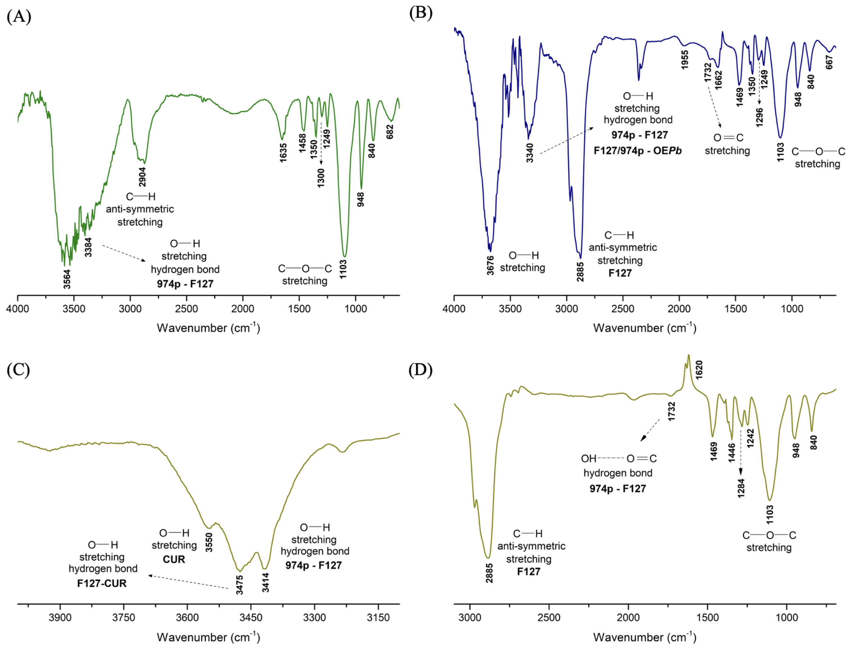
3.2.1. FTIR
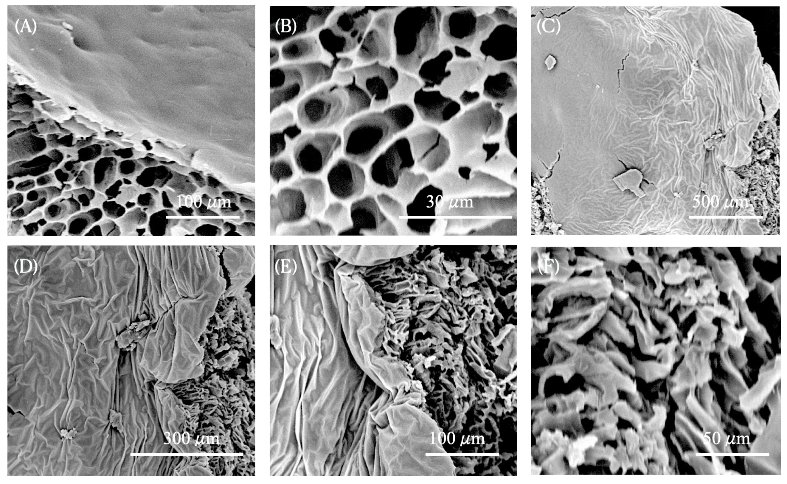
3.2.2. SEM
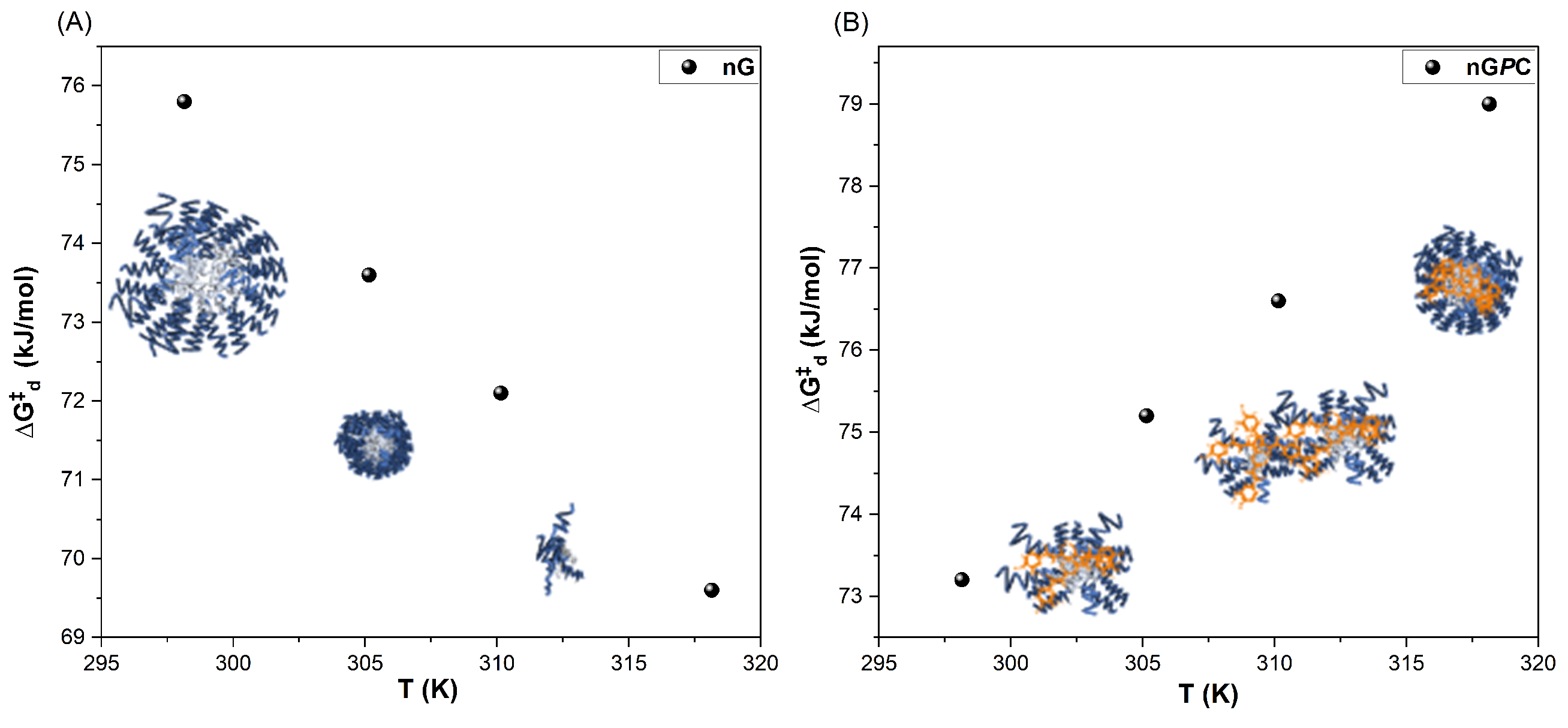
3.2.3. DLS
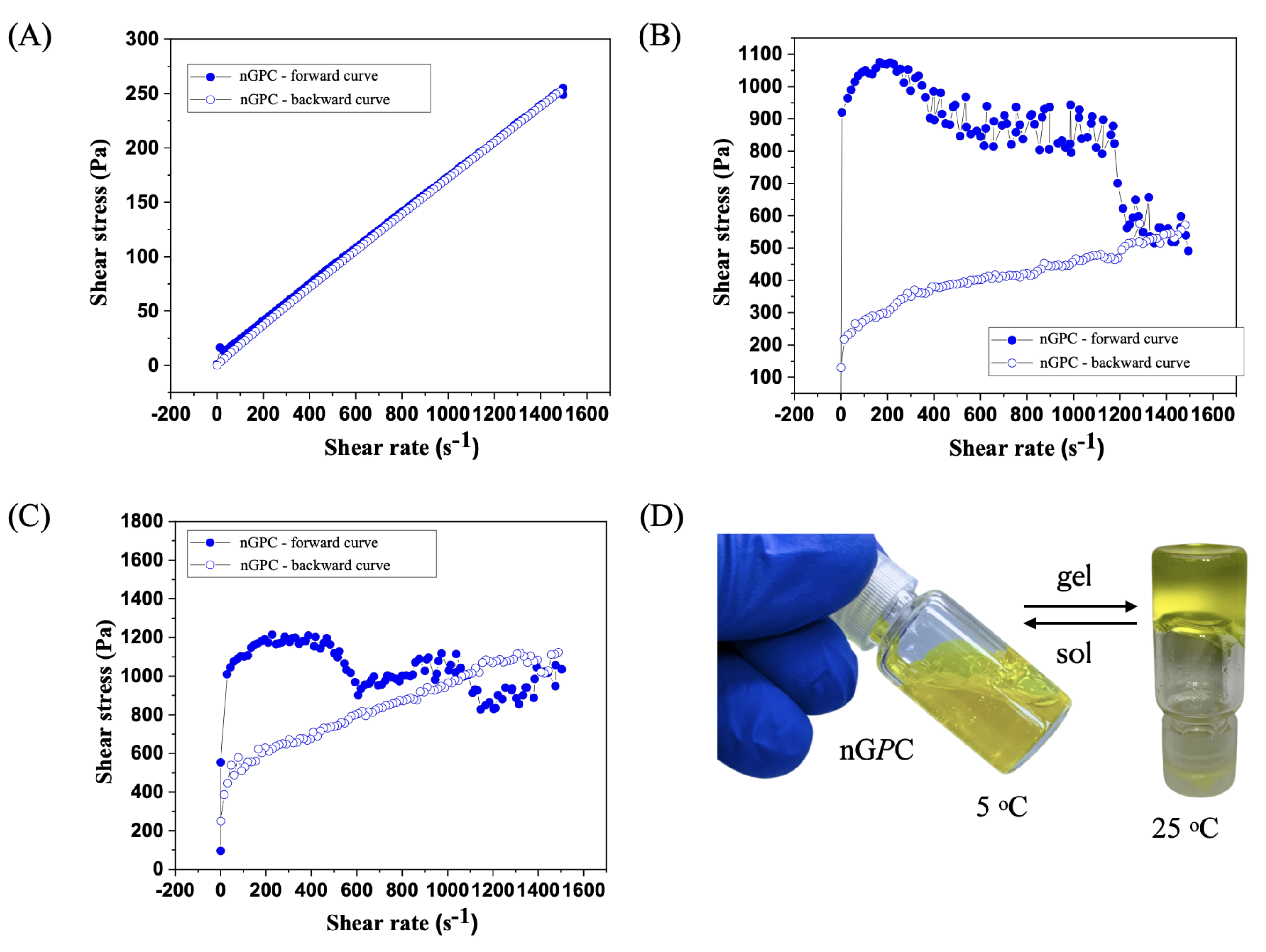
3.3. Rheological Analysis
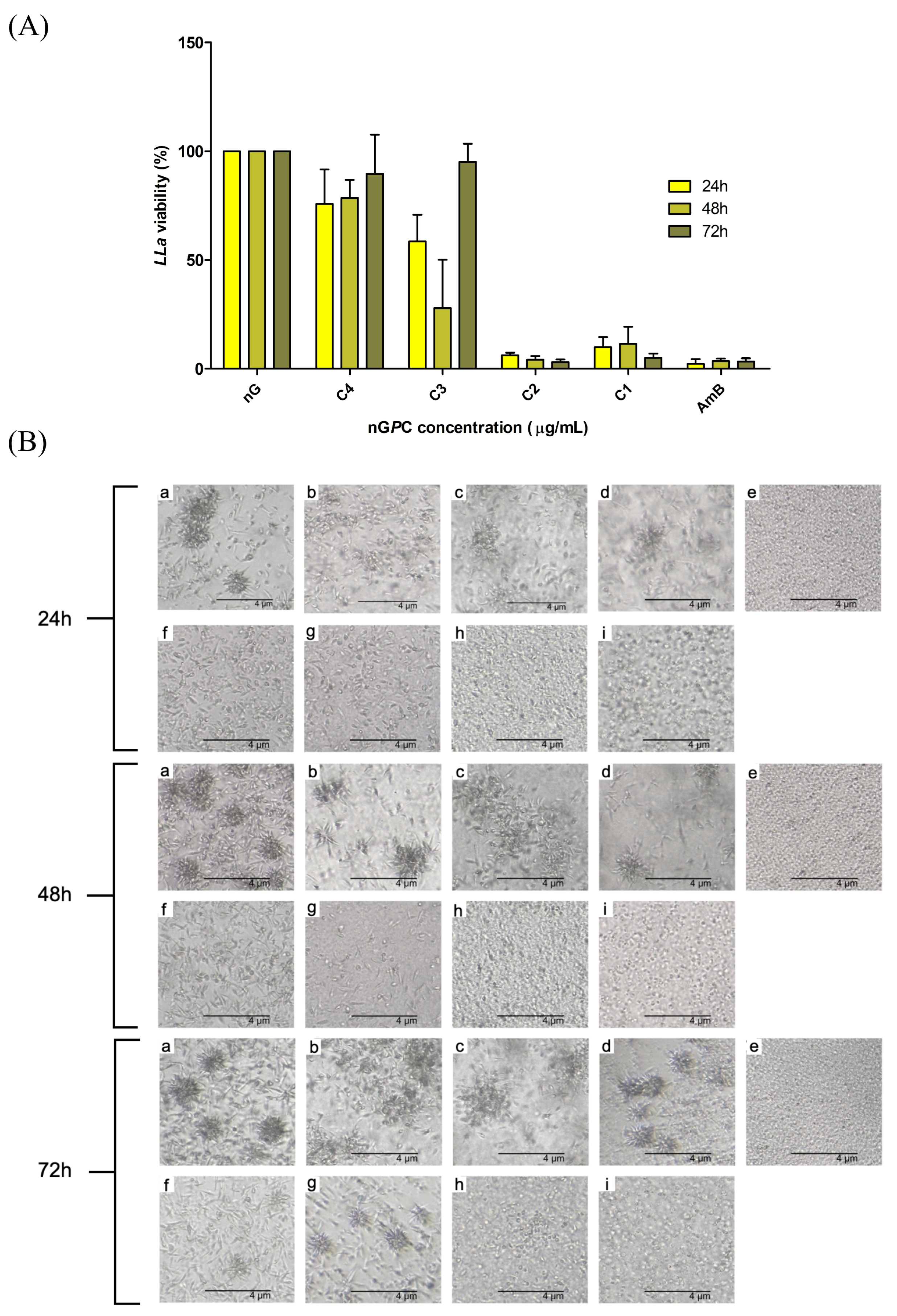
3.4. In Vitro Assay Against LLa Promastigotes
4. Conclusions
Author Contributions
Funding
Institutional Review Board Statement
Informed Consent Statement
Data Availability Statement
Acknowledgments
Conflicts of Interest
References
- World Health Organization (WHO). Neglected Tropical Diseases; World Health Organization: Geneva, Switzerland, 2025; Available online: https://www.who.int/news-room/fact-sheets/detail/neglected-tropical-diseases (accessed on 2 February 2025).
- Hotez, P.J.; Kamath, A. Neglected tropical diseases in sub-Saharan Africa: Review of their prevalence, distribution, and disease burden. PLoS Negl. Trop. Dis. 2009, 3, e412. [Google Scholar] [CrossRef]
- Molyneux, D.H.; Savioli, L.; Engels, D. Neglected tropical diseases: Progress towards addressing the chronic pandemic. Lancet 2017, 389, 312–325. [Google Scholar] [CrossRef] [PubMed]
- Fitzpatrick, C.; Nwankwo, U.; Lenk, E.; de Vlas, S.J.; Bundy, D.A.P. An Investment Case for Ending Neglected Tropical Diseases. In Major Infectious Diseases, 3rd ed.; Holmes, K.K., Bertozzi, S., Bloom, B.R., Jha, P., Eds.; The International Bank for Reconstruction and Development/The World Bank: Washington, DC, USA, 2017; Chapter 17. [Google Scholar]
- Hotez, P.; Aksoy, S. PLOS Neglected Tropical Diseases: Ten years of progress in neglected tropical disease control and elimination … More or less. PLoS Negl. Trop. Dis. 2017, 11, e0005355. [Google Scholar] [CrossRef] [PubMed]
- Ezike, T.C.; Okpala, U.S.; Onoja, U.L.; Nwike, C.P.; Ezeako, E.C.; Okpara, O.J.; Okoroafor, C.C.; Eze, S.C.; Kalu, O.L.; Odoh, E.C.; et al. Advances in drug delivery systems, challenges and future directions. Heliyon 2023, 9, e17488. [Google Scholar] [CrossRef] [PubMed]
- Zhang, Y.; Chan, H.F.; Leong, K.W. Advanced materials and processing for drug delivery: The past and the future. Adv. Drug Deliv. Rev. 2013, 65, 104–120. [Google Scholar] [CrossRef]
- Cupolillo, E.; Grimaldi, G., Jr.; Momen, H. A general classification of New World Leishmania using numerical zymotaxonomy. Am. J. Trop. Med. Hyg. 1994, 50, 296–311. [Google Scholar] [CrossRef]
- Kato, H.; Uezato, H.; Gomez, E.A.; Terayama, Y.; Calvopiña, M.; Iwata, H.; Hashiguchi, Y. Establishment of a mass screening method of sand fly vectors for Leishmania infection by molecular biological methods. Am. J. Trop. Med. Hyg. 2007, 77, 324–329. [Google Scholar] [CrossRef]
- Mann, S.; Frasca, K.; Scherrer, S.; Henao-Martínez, A.F.; Newman, S.; Ramanan, P.; Suarez, J.A. A Review of Leishmaniasis: Current Knowledge and Future Directions. Curr. Trop. Med. Rep. 2021, 8, 121–132. [Google Scholar] [CrossRef]
- da Costa, C.S.; Marques, E.M.; do Nascimento, J.R.; Lima, V.A.S.; Santos-Oliveira, R.; Figueredo, A.S.; de Jesus, C.M.; de Souza Nunes, G.C.; Brandão, C.M.; de Jesus, E.T.; et al. Design of Liquid Formulation Based on F127-Loaded Natural Dimeric Flavonoids as a New Perspective Treatment for Leishmaniasis. Pharmaceutics 2024, 16, 252. [Google Scholar] [CrossRef]
- Marques, E.M.; Rocha, R.L.; Brandão, C.M.; Xavier, J.K.A.M.; Camara, M.B.P.; Mendonça, C.J.S.; de Lima, R.B.; Souza, M.P.; Costa, E.V.; Gonçalves, R.S. Development of an Eco-Friendly Nanogel Incorporating Pectis brevipedunculata Essential Oil as a Larvicidal Agent Against Aedes aegypti. Pharmaceutics 2024, 16, 1337. [Google Scholar] [CrossRef]
- Marques, E.M.; Santos Andrade, L.G.; Rebelo Alencar, L.M.; Dias Rates, E.R.; Ribeiro, R.M.; Carvalho, R.C.; de Souza Nunes, G.C.; Sara Lopes Lera-Nonose, D.S.; Gonçalves, M.J.S.; Lonardoni, M.V.C.; et al. Nanotechnological formulation incorporating Pectis brevipedunculata (Asteraceae) essential oil: An ecofriendly approach for leishmanicidal and anti-inflammatory therapy. Polymers 2025, 17, 379. [Google Scholar] [CrossRef] [PubMed]
- Carvalho, C.E.; Sobrinho-Junior, E.P.; Brito, L.M.; Nicolau, L.A.; Carvalho, T.P.; Moura, A.K.; Rodrigues, K.A.; Carneiro, S.M.; Arcanjo, D.D.; Citó, A.M.; et al. Anti-Leishmania activity of essential oil of Myracrodruon urundeuva (Engl.) Fr. All.: Composition, cytotoxicity and possible mechanisms of action. Exp. Parasitol. 2017, 175, 59–67. [Google Scholar] [CrossRef] [PubMed]
- Ferreira, O.O.; Cruz, J.N.; de Moraes, Â.A.B.; de Jesus Pereira Franco, C.; Lima, R.R.; Anjos, T.O.D.; Siqueira, G.M.; Nascimento, L.D.D.; Cascaes, M.M.; de Oliveira, M.S.; et al. Essential oil of the plants growing in the Brazilian Amazon: Chemical composition, antioxidants, and biological applications. Molecules 2022, 27, 4373. [Google Scholar] [CrossRef] [PubMed]
- de Lara da Silva, C.E.; Oyama, J.; Ferreira, F.B.P.; de Paula Lalucci-Silva, M.P.; Lordani, T.V.A.; de Lara da Silva, R.C.; de Souza Terron Monich, M.; Teixeira, J.J.V.; Lonardoni, M.V.C. Effect of essential oils on Leishmania amazonensis: A systematic review. Parasitology 2020, 147, 1392–1407. [Google Scholar] [CrossRef]
- Alves, A.B.; da Silva Bortoleti, B.T.; Tomiotto-Pellissier, F.; Ganaza, A.F.M.; Gonçalves, M.D.; Carloto, A.C.M.; Rodrigues, A.C.J.; Silva, T.F.; Nakazato, G.; Kobayashi, R.K.T.; et al. Synergistic Antileishmanial Effect of Oregano Essential Oil and Silver Nanoparticles: Mechanisms of Action on Leishmania amazonensis. Pathogens 2023, 12, 660. [Google Scholar] [CrossRef]
- Alanazi, A.D.; Alghabban, A.J. Antileishmanial and synergic effects of Rhanterium epapposum essential oil and its main compounds alone and combined with glucantime against Leishmania major infection. Int. J. Parasitol. Drugs Drug Resist. 2024, 26, 100571. [Google Scholar] [CrossRef]
- Monzote, L.; Geroldinger, G.; Tonner, M.; Scull, R.; De Sarkar, S.; Bergmann, S.; Bacher, M.; Staniek, K.; Chatterjee, M.; Rosenau, T.; et al. Interaction of ascaridole, carvacrol, and caryophyllene oxide from essential oil of Chenopodium ambrosioides L. with mitochondria in Leishmania and other eukaryotes. Phytother. Res. 2018, 32, 1729–1740. [Google Scholar] [CrossRef]
- Essid, R.; Damergi, B.; Fares, N.; Jallouli, S.; Limam, F.; Tabbene, O. Synergistic combination of Cinnamomum verum and Syzygium aromaticum treatment for cutaneous leishmaniasis and investigation of their molecular mechanism of action. Int. J. Environ. Health Res. 2024, 34, 2687–2701. [Google Scholar] [CrossRef]
- Santana, R.C.; Rosa, A.D.S.; Mateus, M.H.D.S.; Soares, D.C.; Atella, G.; Guimarães, A.C.; Siani, A.C.; Ramos, M.F.S.; Saraiva, E.M.; Pinto-da-Silva, L.H. In vitro leishmanicidal activity of monoterpenes present in two species of Protium (Burseraceae) on Leishmania amazonensis. J. Ethnopharmacol. 2020, 259, 112981. [Google Scholar] [CrossRef]
- Pereira, S.L.; Marques, A.M.; Sudo, R.T.; Kaplan, M.A.; Zapata-Sudo, G. Vasodilator Activity of the Essential Oil from Aerial Parts of Pectis brevipedunculata and Its Main Constituent Citral in Rat Aorta. Molecules 2013, 18, 3072–3085. [Google Scholar] [CrossRef]
- Santos, S.R.; Melo, M.A.; Cardoso, A.V.; Santos, R.L.; de Sousa, D.P.; Cavalcanti, S.C. Structure-Activity Relationships of Larvicidal Monoterpenes and Derivatives against Aedes aegypti Linn. Chemosphere 2011, 84, 150–153. [Google Scholar] [CrossRef] [PubMed]
- Limane, B.B.; Ezzine, O.; Dhahri, S.; Ben Jamaa, M.L. Essential Oils from Two Eucalyptus from Tunisia and Their Insecticidal Action on Orgyia trigotephras (Lepidoptera, Lymantriidae). Biol. Res. 2014, 47, 29. [Google Scholar] [CrossRef]
- Varzandeh, M.; Mohammadinejad, R.; Esmaeilzadeh-Salestani, K.; Dehshahri, A.; Zarrabi, A.; Aghaei-Afshar, A. Photodynamic therapy for leishmaniasis: Recent advances and future trends. Photodiagn. Photodyn. Ther. 2021, 36, 102609. [Google Scholar] [CrossRef] [PubMed]
- Dourado, D.; Silva Medeiros, T.; do Nascimento Alencar, É.; Matos Sales, E.; Formiga, F.R. Curcumin-loaded nanostructured systems for treatment of leishmaniasis: A review. Beilstein J. Nanotechnol. 2024, 15, 37–50. [Google Scholar] [CrossRef] [PubMed]
- Khan, M.; Nadhman, A.; Sehgal, S.A.; Siraj, S.; Yasinzai, M.M. Formulation and Characterization of a Self-Emulsifying Drug Delivery System (SEDDS) of Curcumin for the Topical Application in Cutaneous and Mucocutaneous Leishmaniasis. Curr. Top. Med. Chem. 2018, 18, 1603–1609. [Google Scholar] [CrossRef]
- Suhail, M.; Rosenholm, J.M.; Minhas, M.U.; Badshah, S.F.; Naeem, A.; Khan, K.U.; Fahad, M. Nanogels as drug-delivery systems: A comprehensive overview. Ther. Deliv. 2019, 10, 697–717. [Google Scholar] [CrossRef]
- Camara, M.B.P.; Lima, A.S.; Jumbo, L.O.V.; Tavares, C.P.; Mendonça, C.J.S.; Monteiro, O.S.; Araújo, S.H.C.; Oliveira, E.E.; Lima Neto, J.S.; Maia, J.G.S.; et al. Seasonal and Circadian Evaluation of the Essential Oil from Pectis brevipedunculata and Its Acaricidal Activity against Rhipicephalus microplus (Acari: Ixodidae). J. Braz. Chem. Soc. 2023, 34, 1020–1029. [Google Scholar]
- United States Pharmacopeia and National Formulary (USP 41-NF 36); United States Pharmacopeial Convention: Rockville, MD, USA, 2016.
- Alexander, S.; Cosgrove, T.; Prescott, S.W.; Castle, T.C. Flurbiprofen Encapsulation Using Pluronic Triblock Copolymers. Langmuir 2011, 27, 8054–8060. [Google Scholar] [CrossRef]
- Haillant, O.; Dumbleton, D.; Zielnik, A. An Arrhenius Approach to Estimating Organic Photovoltaic Module Weathering Acceleration Factors. Sol. Energy Mater. Sol. Cells 2011, 95, 1889–1895. [Google Scholar] [CrossRef]
- Griffiths, P.C.; Stilbs, P.; Yu, G.E.; Booth, C. Role of Molecular Architecture in Polymer Diffusion: A PGSE-NMR Study of Linear and Cyclic Poly(ethylene Oxide). J. Phys. Chem. 1995, 99, 16752–16756. [Google Scholar] [CrossRef]
- De, M.; Bhattacharya, S.C.; Moulik, S.P.; Panda, A.K. Interfacial Composition, Structural and Thermodynamic Parameters of Water/(Surfactant+n-Butanol)/n-Heptane Water-in-Oil Microemulsion Formation in Relation to the Surfactant Chain Length. J. Surfactants Deterg. 2010, 13, 475–484. [Google Scholar] [CrossRef]
- Hwang, D.; Ramsey, J.D.; Kabanov, A.V. Polymeric micelles for the delivery of poorly soluble drugs: From nanoformulation to clinical approval. Adv. Drug Deliv. Rev. 2020, 156, 80–118. [Google Scholar] [CrossRef] [PubMed]
- Chakraborty, M.; Panda, A.K. Spectral Behaviour of Eosin Y in Different Solvents and Aqueous Surfactant Media. Spectrochim. Acta Part Mol. Biomol. Spectrosc. 2011, 81, 458–465. [Google Scholar] [CrossRef] [PubMed]
- Alexandridis, P.; Hatton, T.A. Poly(ethylene Oxide)-poly(propylene Oxide)-poly(ethylene Oxide) Block Copolymer Surfactants in Aqueous Solutions and at Interfaces: Thermodynamics, Structure, Dynamics, and Modeling. Colloids Surfaces Physicochem. Eng. Asp. 1995, 96, 1–46. [Google Scholar] [CrossRef]
- Wanka, G.; Hoffmann, H.; Ulbricht, W. Phase Diagrams and Aggregation Behavior of Poly(oxyethylene)-Poly(oxypropylene)-Poly(oxyethylene) Triblock Copolymers in Aqueous Solutions. Macromolecules 1994, 27, 4145–4159. [Google Scholar] [CrossRef]
- Alexander, S.; Cosgrove, T.; Castle, T.C.; Grillo, I.; Prescott, S.W. Effect of Temperature, Cosolvent, and Added Drug on Pluronic–Flurbiprofen Micellization. J. Phys. Chem. B 2012, 116, 11545–11551. [Google Scholar] [CrossRef]
- Brown, W.; Schillin, K. Triblock Copolymers in Aqueous Solution Studied by Static and Dynamic Light Scattering and Oscillatory Shear Measurements: Influence of Relative Block Sizes. Am. Chem. Soc. 1992, 6044, 6038–6044. [Google Scholar] [CrossRef]
- Sharma, P.K.; Bhatia, S.R. Effect of Anti-Inflammatories on Pluronic® F127: Micellar Assembly, Gelation and Partitioning. Int. J. Pharm. 2004, 278, 361–377. [Google Scholar] [CrossRef]
- Nilsson, M.; Håkansson, B.; Söderman, O.; Topgaard, D. Influence of Polydispersity on the Micellization of Triblock Copolymers Investigated by Pulsed Field Gradient Nuclear Magnetic Resonance. Macromolecules 2007, 40, 8250–8258. [Google Scholar] [CrossRef]
- Landazuri, G.; Fernandez, V.V.A.; Soltero, J.F.A.; Rharbi, Y. Kinetics of the Sphere-to-Rod like Micelle Transition in a Pluronic Triblock Copolymer. J. Phys. Chem. B 2012, 116, 11720–11727. [Google Scholar] [CrossRef]
- Brown, W.; Stilbs, P. On the Solution Conformation of Poly(ethylene Oxide): An FT-Pulsed Field Gradient NMR Self-Diffusion Study. Polymer 1982, 23, 1780–1784. [Google Scholar] [CrossRef]
- Espenson, J.H. Chemical Kinetics and Reaction Mechanisms, 2nd ed.; McGraw-Hill Series in Advanced Chemistry; McGraw-Hill: New York, NY, USA, 1995. [Google Scholar]
- Dash, S.K.; Benival, D.; Jindal, A.B. Formulation strategies to overcome amphotericin B-induced toxicity. Mol. Pharm. 2024, 21, 5392–5412. [Google Scholar] [CrossRef] [PubMed]
- Kuti, J.L. Optimizing Antimicrobial Pharmacodynamics: A Guide for Your Stewardship Program. Rev. Med. Clin. Condes 2016, 27, 615–624. [Google Scholar] [CrossRef]


| Component % (w/w) | ||||||
|---|---|---|---|---|---|---|
| Code | Water | F127 | 974P | EOPb | CUR | Stability a |
| nGPC 1 | 78.99 | 20 | 0.2 | 1 | 0.01 | S |
| nGPC 2 | 78.78 | 20 | 0.2 | 1 | 0.02 | S |
| nGPC 3 | 78.47 | 20 | 0.2 | 1 | 0.03 | S |
| nGPC 4 | 78.36 | 20 | 0.2 | 1 | 0.04 | S |
| nGPC 5 | 78.25 | 20 | 0.2 | 1 | 0.05 | C |
| nG | nGPC | |
|---|---|---|
| Temperature (°C) | Rh (nm)/PDI | |
| 25 | 661.00 ± 6.00/0.34 | 288.00 ± 74.00/0.29 |
| 32 | 124.00 ± 5.00/0.24 | 296.00 ± 24.00/0.44 |
| 37 | 17.00 ± 3.00/0.25 | 237.00 ± 17.00/0.42 |
| 45 | 16.93 ± 3.00/0.26 | 333.50 ± 22.00/0.45 |
| nG | nGPC | ||
|---|---|---|---|
| Parameter | Value | Parameter | Value |
| (kJ mol−1) | 208.53 | (kJ mol−1) | −10.76 |
| (kJ K−1 mol−1) | 0.31 | (kJ K−1 mol−1) | −0.29 |
| (kJ mol−1) | 168.20 | (kJ mol−1) | −13.30 |
| (kJ mol−1) at different temperatures | |||
| Temperature (K) | nG | Temperature (K) | nGPC |
| 298.15 | 75.80 | 298.15 | 73.20 |
| 303.15 | 73.60 | 303.15 | 75.20 |
| 305.15 | 72.10 | 305.15 | 76.60 |
| 310.15 | 69.60 | 310.15 | 79.00 |
Disclaimer/Publisher’s Note: The statements, opinions and data contained in all publications are solely those of the individual author(s) and contributor(s) and not of MDPI and/or the editor(s). MDPI and/or the editor(s) disclaim responsibility for any injury to people or property resulting from any ideas, methods, instructions or products referred to in the content. |
© 2025 by the authors. Licensee MDPI, Basel, Switzerland. This article is an open access article distributed under the terms and conditions of the Creative Commons Attribution (CC BY) license (https://creativecommons.org/licenses/by/4.0/).
Share and Cite
Campos, L.M.O.; Marques, E.M.; Lera-Nonose, D.S.S.L.; Gonçalves, M.J.S.; Lonardoni, M.V.C.; Nunes, G.C.d.S.; Braga, G.; Gonçalves, R.S. Enhanced Nanogel Formulation Combining the Natural Photosensitizer Curcumin and Pectis brevipedunculata (Asteraceae) Essential Oil for Synergistic Daylight Photodynamic Therapy in Leishmaniasis Treatment. Pharmaceutics 2025, 17, 286. https://doi.org/10.3390/pharmaceutics17030286
Campos LMO, Marques EM, Lera-Nonose DSSL, Gonçalves MJS, Lonardoni MVC, Nunes GCdS, Braga G, Gonçalves RS. Enhanced Nanogel Formulation Combining the Natural Photosensitizer Curcumin and Pectis brevipedunculata (Asteraceae) Essential Oil for Synergistic Daylight Photodynamic Therapy in Leishmaniasis Treatment. Pharmaceutics. 2025; 17(3):286. https://doi.org/10.3390/pharmaceutics17030286
Chicago/Turabian StyleCampos, Lara Maria Oliveira, Estela Mesquita Marques, Daniele Stéfanie Sara Lopes Lera-Nonose, Maria Julia Schiavon Gonçalves, Maria Valdrinez Campana Lonardoni, Glécilla Colombelli de Souza Nunes, Gustavo Braga, and Renato Sonchini Gonçalves. 2025. "Enhanced Nanogel Formulation Combining the Natural Photosensitizer Curcumin and Pectis brevipedunculata (Asteraceae) Essential Oil for Synergistic Daylight Photodynamic Therapy in Leishmaniasis Treatment" Pharmaceutics 17, no. 3: 286. https://doi.org/10.3390/pharmaceutics17030286
APA StyleCampos, L. M. O., Marques, E. M., Lera-Nonose, D. S. S. L., Gonçalves, M. J. S., Lonardoni, M. V. C., Nunes, G. C. d. S., Braga, G., & Gonçalves, R. S. (2025). Enhanced Nanogel Formulation Combining the Natural Photosensitizer Curcumin and Pectis brevipedunculata (Asteraceae) Essential Oil for Synergistic Daylight Photodynamic Therapy in Leishmaniasis Treatment. Pharmaceutics, 17(3), 286. https://doi.org/10.3390/pharmaceutics17030286