Review

Camptothecin in Cancer Therapy: Current Challenges and Emerging Strategies with Nanoemulsions

by
Heber Uriel Pérez-Ortega
1,
Rubén Ricardo Córdova-Espíritu
2,
Sebastian Cano-Serrano
1,
Eduardo García-González
3,4,
Micael Gerardo Bravo-Sánchez
2,
Ma. del Carmen Orozco-Mosqueda
2,
Hugo Jiménez-Islas
2,
Gabriel Luna-Bárcenas
5 and
Francisco Villaseñor-Ortega
2,*
1
División de Estudios de Posgrado e Investigación (DEPI), Tecnológico Nacional de México (TecNM), Instituto Tecnológico de Celaya (ITC), Celaya 38010, Guanajuato, Mexico
2
Departamento de Ingeniería Bioquímica y Ambiental, Tecnológico Nacional de México (TecNM), Instituto Tecnológico de Celaya (ITC), Celaya 38010, Guanajuato, Mexico
3
Tecnológico Nacional de México (TecNM), Instituto Tecnológico de Roque (ITR), Celaya 38110, Guanajuato, Mexico
4
Programa de Ingeniería en Biotecnología, Campus Celaya-Salvatierra, Universidad de Guanajuato, Mutualismo #303, Col. La Suiza, Celaya 36060, Guanajuato, Mexico
5
Tecnológico de Monterrey, Instituto de Materiales Avanzados para una Manufactura Sostenible, Epigmenio González 500 Fracc. San Pablo, Querétaro 76130, Querétaro, Mexico
*
Author to whom correspondence should be addressed.
Pharmaceutics 2025, 17(11), 1414; https://doi.org/10.3390/pharmaceutics17111414
Submission received: 30 August 2025 / Revised: 18 October 2025 / Accepted: 29 October 2025 / Published: 31 October 2025

Abstract

Camptothecin (CPT) is a natural alkaloid with potent antiproliferative activity, mediated by the inhibition of Topoisomerase I (Topo I), an essential enzyme for deoxyribonucleic acid (DNA) replication. However, its clinical application has been limited by low solubility and the instability of the lactone ring under physiological conditions, both of which decrease its efficacy. Semi-synthetic analogs such as irinotecan (CPT-11) and topotecan (TPT) have been developed and approved for the treatment of various types of cancer; however, challenges related to drug resistance and side effects continue to arise. Therefore, nanomedicine and nanoparticle-based delivery systems, including nanoemulsions, liposomes, and antibody–drug conjugates (ADCs), emerge as promising strategies to improve the stability, bioavailability, and effectiveness of CPT, despite significant challenges such as scalability, pharmacokinetic variability, and regulatory requirements. This review discusses recent advances in CPT, its analogs, and these delivery platforms, highlighting its potential to optimize cancer therapy and reduce toxicity while outlining translational challenges such as scalability, pharmacokinetic variability, and regulatory requirements.

Graphical Abstract

1. Introduction

The World Health Organization (WHO) describes cancer as a group of diseases that can occur in any organ or body tissue. This disease is marked by the uncontrolled growth of damaged cells that divide, grow, and spread uncontrollably, invading other tissues [1]. The formation of solid tumors involves six specific capabilities: sustained proliferative signaling, evasion of growth suppressors, disabling replicative mortality, induction of angiogenesis, cancer invasion or metastasis, and resistance to apoptosis or cell death [2].
Cancer represents one of the greatest health, social, and economic challenges of the 21st century. According to estimates from the International Agency for Research on Cancer (GLOBOCAN), nearly 20 million new cases and 9.7 million deaths attributable to this disease were recorded in 2022 [3]. Lung cancer remained the leading cause of cancer-related death, with around 1.8 million deaths (18.7%), followed by colorectal cancers (9.3%), liver cancer (7.8%), breast cancer (6.9%), and stomach cancer (6.8%) [4]. Given this situation, it is essential to seek more effective treatments that utilize anticancer agents with high antiproliferative activity and minimal side effects. Fortunately, nature offers a rich diversity of natural products that can be harnessed for the development of new drugs in the fight against this disease [5].
More than 65% of the medications used in cancer treatment are derived from plants and their derivatives. These compounds have been essential in the production of antiproliferative agents such as vincristine, vinblastine, doxorubicin, paclitaxel, among others, which have had a profound impact on the lives of countless cancer patients [6,7]. In 1966, Wall and Wani discovered camptothecin (CPT), initially extracted from the bark of Camptotheca acuminata, a plant species in the Camptotheca genus and Nyssaceae family, native to southern China [8]. Camptotheca acuminata is known for its fast growth in temperate climates. However, it does not tolerate cold well and requires a humid environment and fertile soil to flourish. CPT can be obtained from the fruit, roots, bark, branches, and leaves. However, in 1985, this compound was identified as an inhibitor of the enzyme topoisomerase I, an enzyme involved in the DNA replication process in eukaryotic cells [9]. It shows a wide range of antiproliferative activity, with more potent effects on colorectal, ovarian, breast, lung, and renal cancers, including those resistant to chemotherapy [10].
By the late 1990s, improved understanding of camptothecin (CPT)’s mechanism of action, chemistry, and pharmacology led to the development of analogs with better properties for clinical use [11], making it the second most important antiproliferative medicinal plant in the world, after Taxus baccata [12]. Additionally, it has been found that camptothecin and its derivatives exhibit other pharmacological effects, including anti-obesity, antiviral, and antifungal activities [13]. The global interest in camptothecin (CPT) and its derivatives has steadily increased over the past few decades, primarily driven by their clinical significance. Nearly twenty years ago, combined sales of CPT-11 and TPT already surpassed $750 million, setting a record in the oncology market [14]. Since 2018, this trend has sharpened, establishing CPT and its analogs as highly significant therapeutic and clinical agents worldwide [15].
Currently, commercial numbers indicate a strong market, supported by active trade of exports and imports between countries such as India, the USA, China, and Taiwan. This reflects the increasing international demand for camptothecin and its derivatives. However, its production remains a significant challenge, as roughly 1000 to 1500 tons of plant material are needed to produce just one ton of purified CPT. Globally, annual sales of CPT and related compounds are estimated at nearly $1 billion, confirming its significance as a key component of the current oncology pharmaceutical market [16].
In this review, we surveyed the literature in PubMed/MEDLINE, Scopus, and Web of Science (covering from 1966 to 2025; focusing on 2002 to 2025) using terms related to camptothecin, its clinical analogs (irinotecan/CPT-11, SN-38, topotecan), topoisomerase I inhibition, and delivery platforms (nanoemulsions, SNEDDS, lipid systems), including antibody–drug conjugates with CPT-type payloads. Using the keyword “camptothecin,” the initial results yielded 19,615 records in PubMed, 22,487 in Scopus, and 10,938 in Web of Science (totaling 53,040). Records from all databases were exported and de-duplicated in Mendeley (via DOI/title/year matching) to create a consolidated set. Screening was conducted in stages: by year (2002–2025), document type (original research, reviews, clinical studies), topical relevance to oncology/Topo I, formulation/delivery focus, and language/full-text availability, followed by full-text assessment against predefined inclusion/exclusion criteria. Citation counts were also used to prioritize influential and widely referenced studies. This process ultimately resulted in 224 studies that served as references.

2. Chemical Structure and Physicochemical Properties

CPT is an alkaloid characterized by a planar pentacyclic structure composed of five fused rings. Its structure includes a pyrrole-(3,4-β)-quinoline system corresponding to rings A, B, and C, attached to a pyridone ring (ring D) as illustrated in Figure 1. Additionally, it has a chiral center at position C20 in the α-hydroxylactone ring (ring E) [17]. One of the main obstacles to the use of CPT is its low solubility and stability in water, as its structure is predominantly hydrophobic, which prevents its dissolution in this medium [18,19]. Although the molecule contains some polar functional groups, such as the lactone group, these are not sufficient to counteract the large hydrophobic surface of the molecule [17]. Furthermore, CPT has a partition coefficient (Log P) ranging from 1.30 to 2.60, indicating that it has a highly lipophilic solubility. This property enables it to cross the cell membrane [20,21].
To optimize its solubility without affecting its biological activity, modifications can be made to rings A, B, and C. However, rings D and E must remain practically intact, as they play a fundamental role in inhibiting Topo I, a key mechanism in the antiproliferative activity of CPT [22]. In an acidic environment, the E ring remains closed, allowing it to retain its efficacy [23]. When the pH exceeds 5.5, the lactone ring opens through a hydrolysis process, transforming into its carboxylate form, which results in a 90% reduction in its biological activity [24,25]. The half-life (t1/2) of the conversion of the lactone ring into its carboxylate form is 11 min, and only 5.3% of the total camptothecin remains in the lactone form, rendering it ineffective against cancer [26,27].
This pH-related behavior also affects its interaction with plasma proteins after administration. In the bloodstream, camptothecin binds to transport proteins such as albumin and alpha-1-acid glycoprotein (AAG), facilitating its distribution throughout the system [28,29,30]. At a pH of 5.5 or higher, the non-enzymatic hydrolysis of the lactone ring occurs rapidly and reversibly, generating an open-ring hydroxyl carboxylic acid. Human serum albumin has a higher affinity for the carboxylate form, favoring its conversion and further decreasing the amount of CPT in its active form [26,31], which limits its bioavailability and therapeutic efficacy, posing an obstacle to its clinical use [32,33]. Unfavorable pharmacokinetic conditions compromise biological activity, requiring higher doses or continuous applications to achieve a therapeutic effect [34]. To maximize efficacy and minimize side effects, some analogs have been developed by modifying the CPT structure [35,36].

3. Mechanism of Action

CPT exerts its antiproliferative activity by directly inhibiting Topo I, an essential enzyme in DNA replication, transcription, and repair processes [37,38]. Topo I modulates DNA supercoiling by generating transient single-strand breaks, allowing torsional relaxation through controlled rotation of the strand and subsequent re-ligation [39,40,41]. CPT, through its lactone ring, specifically binds to the Topo I–DNA complex via hydrogen bonds with amino acid residues of the enzyme, such as Arg364, Asn722, and Asp533, stabilizing this complex and blocking DNA re-ligation [39,40]. Although Topo I functions at a remarkably high speed (up to 6000 cycles per minute), it is highly susceptible to inhibition by agents like CPT [42]. In this way, CPT converts the standard catalytic action of Topo I into a stable complex that interferes with the progression of the replication fork, causing lethal DNA breaks and leading to apoptosis in tumor cells. Figure 2 schematically illustrates how CPT binds to the Topo I–DNA complex, inducing programmed cell death in cancer cells.
This inhibition converts the regular catalytic activity of Topo I into a stable Topo I–DNA–CPT complex, which interferes with the progression of the replication fork, causing lethal DNA breaks and leading to genotoxic stress, especially in cancer cells during the S phase [43]. The accumulation of these covalent Topo I–DNA–CPT complexes is cytotoxic and triggers apoptosis, a type of programmed cell death that primarily occurs through the mitochondrial intrinsic pathway, activated by DNA damage [44,45].
The intrinsic pathway is activated in response to DNA damage caused by CPT [46,47]. The p53 gene, known as the ‘guardian of the genome,’ detects this damage and coordinates the cellular response, promoting apoptosis [48]. Activation of p53 induces the transcription of proapoptotic proteins such as Puma, Noxa, and Bad, which antagonize antiapoptotic proteins of the BCL-2 family (BCL-XL, MCL-1, BFL-1, BCL-W, and BCL2L10) located in the mitochondria [49]. This allows the activation of Bax and Bak, responsible for forming pores in the mitochondrial membrane (VDAC), facilitating the release of proapoptotic proteins such as cytochrome c and Smac/DIABLO into the cytosol [50,51,52].
In the cytosol, cytochrome c, released because of mitochondrial permeabilization induced by CPT, binds to Apaf-1, forming the apoptosome that recruits and activates caspase-9, while RAIDD facilitates the activation of caspase-2 [53,54,55]. Subsequently, caspase-9 activates caspase-3, which in turn triggers the activation of caspase-activated DNase (CAD). This enzyme fragments DNA into segments of 180–200 base pairs and degrades structural proteins such as actin and nuclear lamins. This process ensures the controlled disintegration of the cell, while still maintaining the integrity of the cytoskeleton and nuclear membrane [56,57,58,59]. Additionally, mitochondrial stress mediated by CPT promotes the release of endonuclease G (Endo G), which migrates to the nucleus and contributes to DNA fragmentation and the formation of apoptotic bodies that are later eliminated by phagocytosis, thus protecting neighboring cells from damage [60,61].
In parallel, the apoptosis-inducing factor (AIF) is also released in response to CPT exposure, causing chromatin condensation and fragmentation independently of caspases, ensuring complete cell death [62]. Figure 3 schematically illustrates the intrinsic apoptosis pathway triggered by CPT.
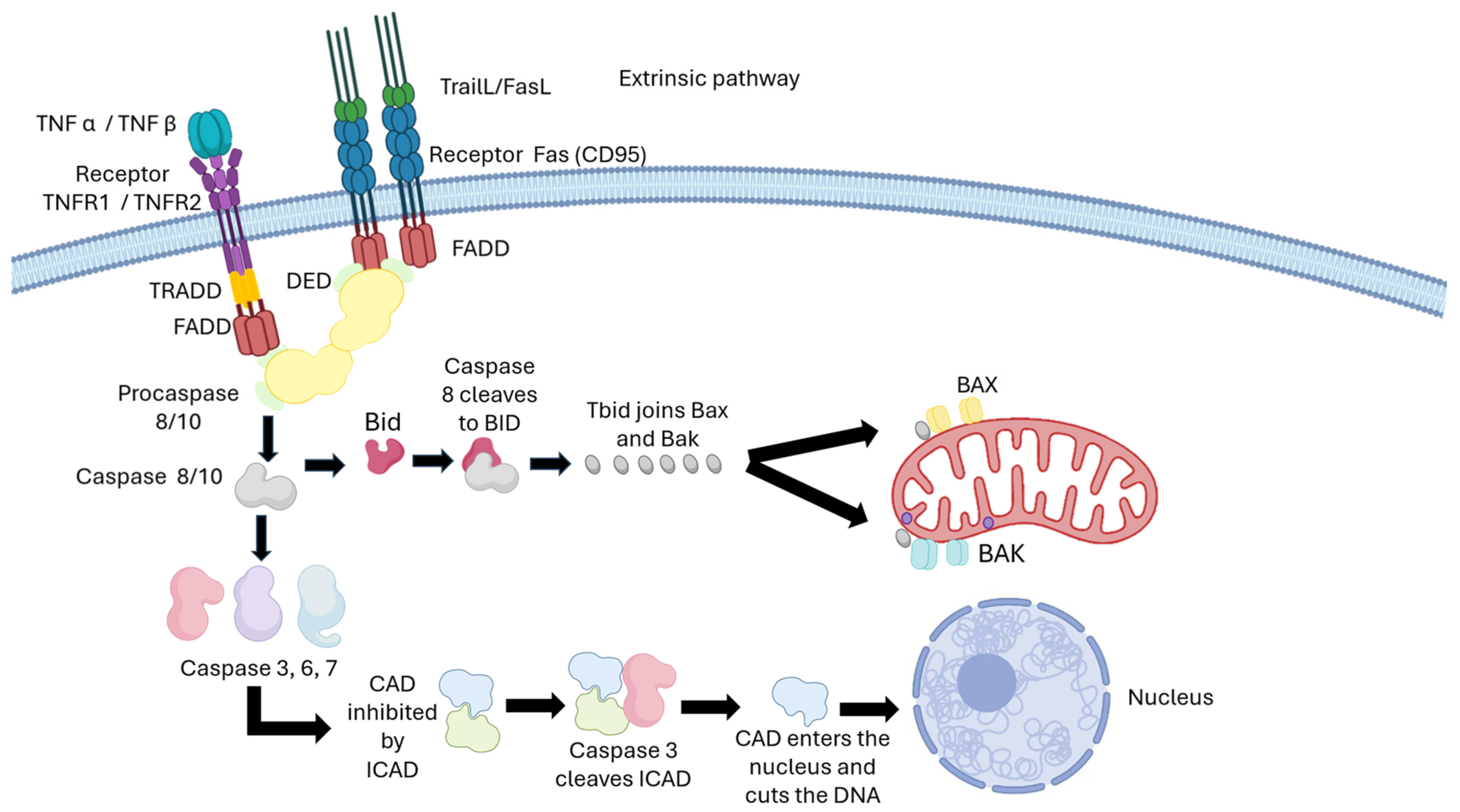
The extrinsic pathway of apoptosis is activated by pro-apoptotic signals from the extracellular environment that interact with death receptors on the cell membrane, such as Fas (CD95) and TNFR1/TNFR2 [63,64]. However, CPT primarily exerts its effect through the mitochondrial intrinsic pathway by causing DNA damage. Activation of death receptors can reinforce CPT-induced apoptosis, but the drug does not need to exert its effect. When FasL or TNF-α ligands bind to their receptors, receptor trimerization occurs, leading to the recruitment of the adaptor protein FADD (Fas-Associated Death Domain), forming the death signaling complex (DISC) [65,66,67]. FADD, through its death effector domain (DED), recruits initiator procaspases such as procaspase-8 and procaspase-10, allowing their activation via autoproteolysis [68,69,70,71,72]. Activated caspase-8 initiates the apoptotic cascade by cleaving and directly activating effector caspases such as caspase-3 and caspase-7, thereby executing apoptosis through DNA fragmentation and cellular protein degradation [73,74,75]. Additionally, FADD associates with TRADD in TNFR1 signaling, promoting caspase-8 activation [76,77]. The protein Bid acts as a critical bridge between the extrinsic and intrinsic pathways, allowing signals initiated by death receptors to reinforce CPT-induced apoptosis. Caspase-8 cleaves and activates Bid, producing tBid, which translocates to the mitochondria and promotes outer mitochondrial membrane permeabilization. This action induces the release of cytochrome c and activates Bax and Bak, converging with the intrinsic pathway to amplify apoptosis [78,79,80]. In this way, CPT’s action, which primarily initiates the intrinsic pathway, is enhanced by integration with the extrinsic pathway, intensifying programmed cell death.
Figure 4 schematically illustrates the extrinsic pathway mediated by death receptors and its integration with the intrinsic pathway.
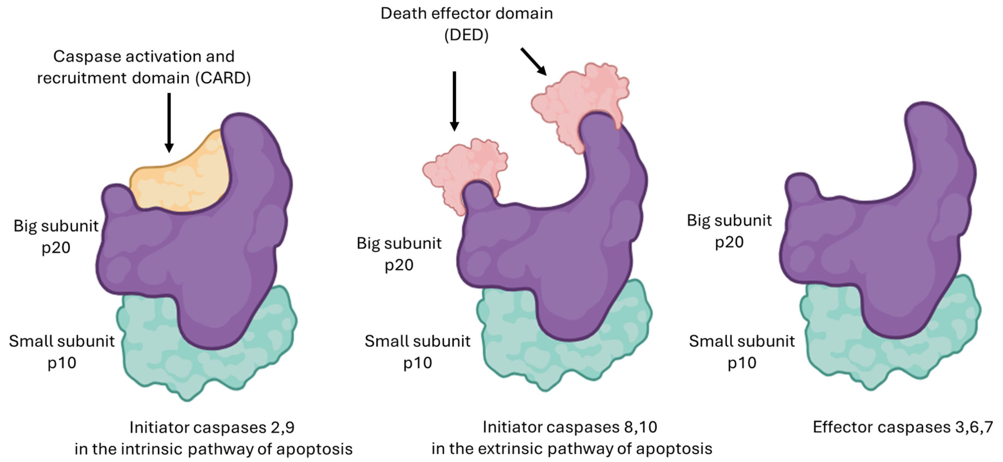
In mammals, caspases 2, 3, 6, 7, 8, 9, and 10 have been identified as key players in the apoptotic process [81]. These cysteine proteases, which are specific to aspartate, are expressed as proenzymes composed of an N-terminal domain, a large subunit (~20 kDa), and a small subunit (~10 kDa). These proenzymes are evolutionarily conserved and present in most cells [82].
During apoptosis induced by CPT, DNA damage and the release of cytochrome c from mitochondria sequentially activate caspases, which are divided into initiator and effector caspases based on the protein interaction domains at their N-terminal end [83]. Caspases 8 and 10 contain a death effector domain (DED). In contrast, caspases 2 and 9 have a caspase recruitment domain (CARD), which facilitates their activation through dimerization or assembly into complexes such as the apoptosome, formed after CPT action. Caspase-9 acts as the initiator in the intrinsic pathway, while caspase-8 can amplify apoptosis by cleaving Bid to generate tBid, enhancing mitochondrial permeabilization. Finally, the effector caspases 3, 6, and 7 execute cell death by degrading essential structural and regulatory proteins [84,85]. Figure 5 shows the structure and function of initiator and effector caspases in CPT-mediated apoptosis.
DNA fragmentation, a characteristic stage of the final phase of camptothecin (CPT)-induced apoptosis, is mediated by caspase-activated DNase (CAD). Under normal conditions, CAD remains inactive by being bound to its inhibitor ICAD (Inhibitor of Caspase-Activated DNase) [86]. During apoptosis, the activation of caspase-3, as a result of the cascade initiated by CPT, leads to the cleavage of ICAD, releasing CAD and allowing controlled DNA fragmentation into fragments of approximately 180–200 base pairs [87,88].
Apoptosis results in a reduction in cell size, the formation of blisters in the membrane, chromatin condensation, DNA fragmentation, and the disassembly of cells into apoptotic bodies surrounded by a membrane [89]. Both apoptotic cells and apoptotic bodies are eliminated through phagocytosis by macrophages and non-specialized phagocytes, such as epithelial cells [90]. Figure 6 illustrates the process of apoptosis execution and elimination of apoptotic bodies.

4. Structural Changes in Camptothecin

4.1. Modifications on Rings A and B

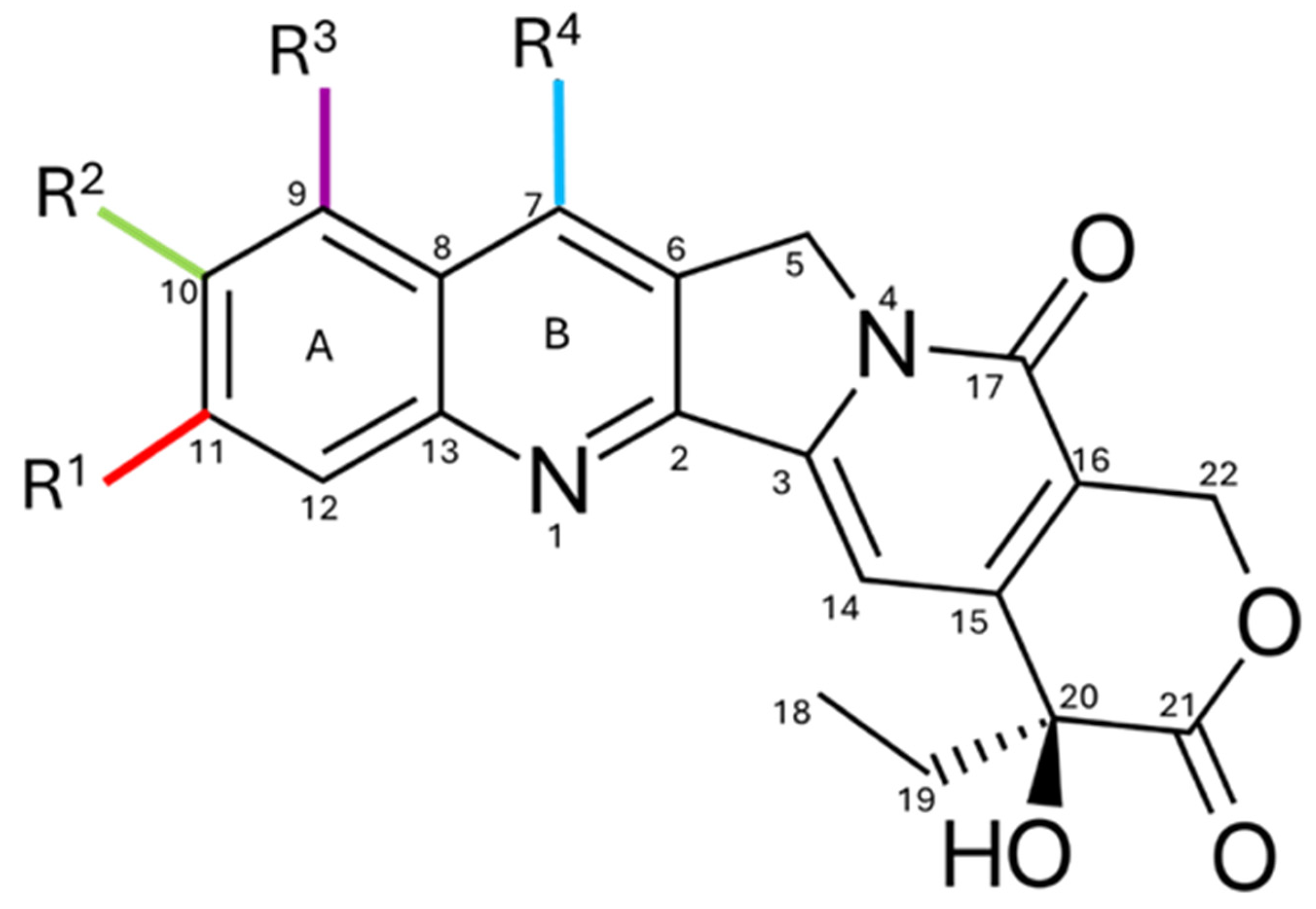
Figure 7 shows the substitutions on the A and B rings of CPT. These modifications include the addition of amino and/or hydrophilic hydroxyl groups to improve solubility and potency [91]. Substitution at positions C7, C9, C10, and C11 enhances anticancer activity without compromising cytotoxicity [92]. CPT-11 and TPT are examples of semi-synthetic CPT derivatives that exhibit better water solubility due to structural modifications at position C9, thereby increasing their solubility under physiological pH conditions [93].
Studies also show that alkyl groups, such as ethyl or chloromethyl at C7, boost antitumor activity. This effect is linked to the favorable lipophilic interaction between these alkyl groups and the Topo I-DNA complex [92]. Extending the carbon chain length at C7 increases the molecule’s lipid solubility and is directly related to higher potency and greater stability of the E ring in plasma [94]. Substituents like amines, nitro groups, hydroxyl, chlorine, or bromine on ring A enhance the antitumor effectiveness of camptothecin, as seen with rubitecan, a 9-nitrocamptothecin analog with increased water solubility [92].
The molecular weight of CPT and its derivatives is approximately 400 Da, such as the 10-hydrocamptothecin (10-HCPT) analog, which has a substitution at the C10 position with a hydroxyl group (OH), substantially reducing the tendency for the lactone ring to open and increasing solubility [95], as well as the 7-ethyl-10-hydrocamptothecin (SN-38) analog, which has a hydroxyl group at the C10 position and an ethyl group at C7. Other analogs have been developed through modifications on rings A and B, such as lurtotecan (GG211) and exatecan (DX-8951f) [96].
The introduction of O-substituted oxime substituents at C7 of the B ring has led to the development of other potent compounds, such as gimatecan (ST1481) and namitecan (ST-1968), which are now in clinical studies [94,97].

4.2. Modifications on Rings C and D

As mentioned earlier, modifying the C and D rings of camptothecin results in a loss of activity because they are in the central region of the molecule and cause a decrease in its structural planarity [93]. It has been observed that substitutions at the C5 and C14 positions reduce the amide group and the carbonyl ester, making the molecule inactive. This is because the pyridone group and the carbonyl are crucial for stabilizing the ternary complex of Topo I, DNA, and CPT [98,99].
Modified CPT analogs have been developed on the C and D rings. However, none of them have been approved for clinical trials due to their low ability to inhibit topoisomerase I and their reduced antitumor activity [91]. Reports on substituents on the C/D rings are limited due to the scarcity of accessible carbons for modification and the difficulty of the synthetic routes required to obtain potential analogs. Figure 8 shows the carbons where substitutions can be made on the C and D rings in the CPT molecule.

4.3. Modifications on Ring E

The E ring of CPT is the most crucial part for inhibiting the enzyme Topo I. It binds to the enzyme through hydrogen bonds, so the E ring can only tolerate minimal modifications [100]. Various studies were conducted to replace the oxygen in the lactone ring with other atoms, such as nitrogen or sulfur. These efforts resulted in the synthesis of compounds like hydroxamic acid and 20-deoxifluorocamptothecin, which had reduced susceptibility to E ring opening, but their antiproliferative activity against cancer cells was compromised [91,101].
However, Wall and colleagues demonstrated that the E ring of the molecule is essential in drug development. Therefore, they propose that the lactone ring remains intact. The activity of these compounds was reduced due to the loss of a hydrogen bond between the C20 group and the OH in the carbonyl group of the E ring [102,103]. These results led to one of the most significant conclusions in the field: the six-membered ring must remain intact for anti-tumor activity to occur. Figure 9 shows various molecules with substitutions on the E ring.
Among all the derivatives of CPT, only CPT-11 and TPT have successfully finished Phase I and II trials and received FDA approval. They have demonstrated clinical effectiveness and reduced toxicity compared to the original molecule in therapies for colon, lung, and ovarian cancer [99].

5. FDA-Approved Camptothecin Analogs

In the late 1960s, preclinical trials with sodium CPT, a water-soluble derivative, were initiated. However, severe toxic effects were observed, including sterile hemorrhagic cystitis, leukopenia, thrombocytopenia, and gastrointestinal toxicity, leading to the suspension of clinical trials with sodium CPT in the 1970s [104]. Over the next 20 years, researchers studied the drug in the laboratory, developing synthetic and semi-synthetic analogs of CPT aimed at improving its anticancer efficacy and safety [104,105].

5.1. Topotecan

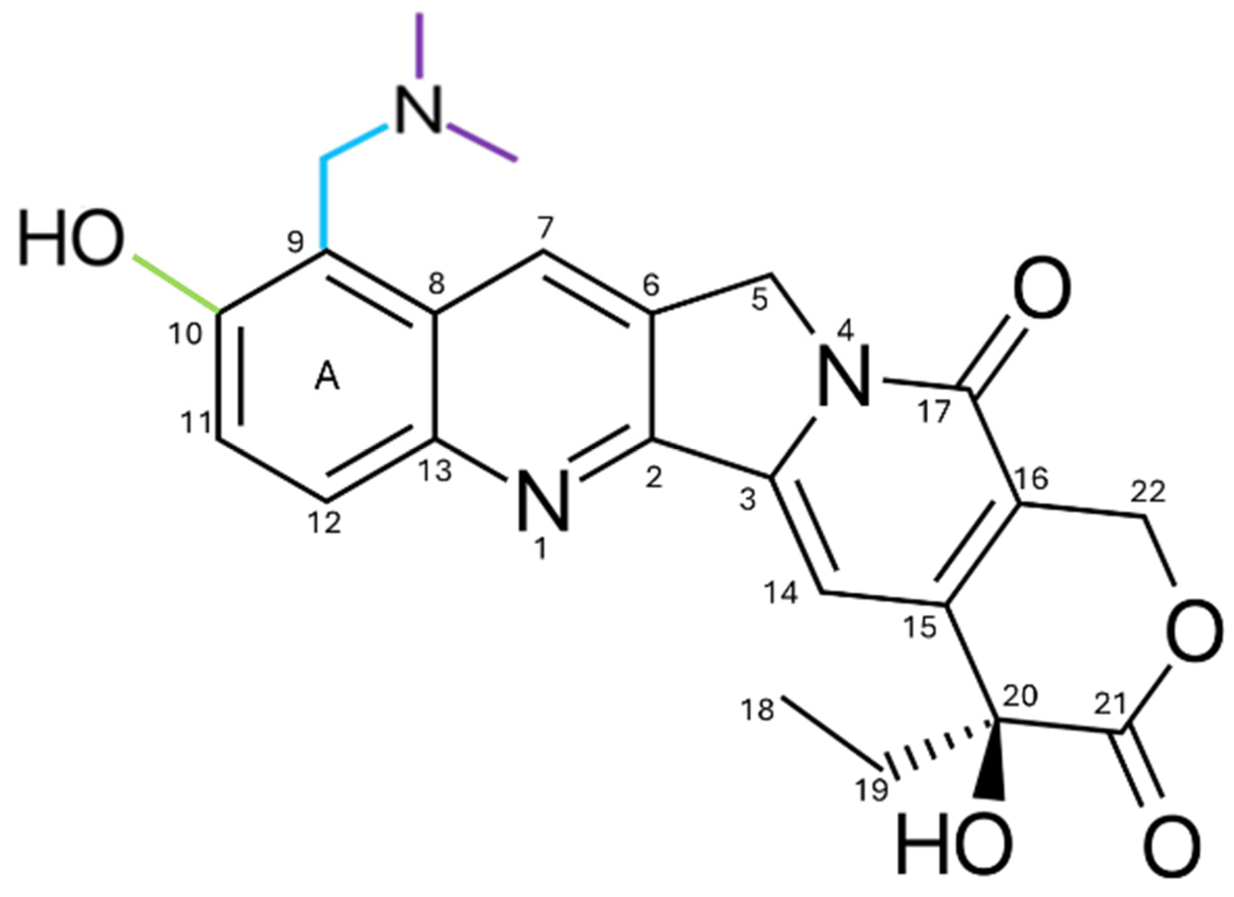
Topotecan (TPT) is a hydrophilic CPT analog approved by the FDA in 1996 [106] for second-line treatment of metastatic ovarian cancer [107]. TPT incorporates a basic side chain at the C9 position of the A ring of 10-hydroxycamptothecin, as illustrated in Figure 10. The basic side chain contains amines that can accept protons (H+), forming ammonium ions. These ions can interact with negative ions (such as chlorides or sulfates) to form ammonium salts, thereby improving water solubility compared to CPT [93].
The CPT analogs are specifically targeted at Topo I [108]. Cancer cells are destroyed by damaging both ribonucleic acid (RNA) and DNA, which are essential components for cell division and proliferation [109]. In the USA, TPT is used for metastatic ovarian, cervical, and small-cell lung cancer (SCLC) [110]. Additionally, it is administered orally in capsule form or intravenously [111].
Pharmacokinetic analysis showed that the biological half-life of TPT is approximately 2–3 h. Doctors have recommended administering TPT at 1.5 mg/m2 of body surface area intravenously daily for 30 min over 5 days, with a 3-week interval between the start of each cycle [112]. As side effects, it usually causes severe neutropenia, as well as acute and delayed diarrhea [113]. Excretion primarily occurs through the renal and fecal routes. About 20% is recovered as the original drug (total TPT) in the urine, and 33% of the oral dose has been identified unchanged (total TPT) in the feces [114].
TPT remains a key treatment option for patients with Small Cell Lung Cancer (SCLC) who relapse or do not respond to initial chemotherapy and are not suitable for reinduction. A recent retrospective study involving 44 patients treated from 2015 to 2022 reported a median progression-free survival (mPFS) of 1.9 months and a median overall survival (mOS) of 5.6 months. By stratifying patients based on critical prognostic markers, including the Eastern Cooperative Oncology Group (ECOG-PS) scale, the presence of active brain metastases, and the systemic inflammation index (SII), researchers identified those most likely to benefit, achieving a median progression-free survival (mPFS) of 5.7 months [115]. This study provides current data on the clinical performance of approved CPT analogs, aiding in improving their effectiveness in clinical practice. The side effects of TPT may resolve after stopping treatment, making it easier to manage compared to drugs that cause permanent damage. Its primary toxicity does not significantly impact the blood system, unlike many chemotherapeutic agents that lead to anemia or severe immunosuppression [116].

5.2. Irinotecan

Irinotecan, also known as CPT-11, has been a crucial component in the treatment of solid tumors worldwide. This derivative remains a key component in combination therapies such as FOLFIRI (folinic acid, fluorouracil, and irinotecan) and FOLFIRINOX (leucovorin, fluorouracil, irinotecan, and oxaliplatin), which are types of combination chemotherapy used to treat pancreatic cancer that has spread to other parts of the body or is inoperable due to vascular involvement [116,117,118]. These therapies have proven effective in managing metastatic or advanced tumors, including colon, gastric, and pancreatic cancers [119].
CPT-11 is a water-soluble prodrug, which means it needs to be transformed within the body to become active. Due to its basic side chain, activation occurs through hydrolysis by hepatic carboxylesterases, releasing SN-38, the true cytotoxic agent responsible for the therapeutic effect against cancer [120], as shown in Figure 11. With these modifications, it behaves as a hydrophilic compound with a large volume of distribution estimated at nearly 400 L/m2 at steady state [121]. Treatment with CPT-11 may consist of intravenous infusions of 30 or 90 min at 350 mg/m2 every three weeks. Common adverse effects include neutropenia, diarrhea, nausea, vomiting, alopecia, and fatigue [122].
The elimination of CPT-11 and SN-38 is a process that depends on cellular membrane transporters of the ABC (ATP-Binding Cassette) family; the primary transporters involved in this process are the multidrug resistance gene (MDR1), P-glycoprotein (P-gp), MRP1, MRP2, and BCRP, with the most relevant being the canalicular multispecific organic anion transporter (cMOAT), also known as MRP2 (Multidrug Resistance-associated Protein 2). These transporters are overexpressed in tumor cell lines and, under physiological conditions, are expressed in epithelial cells of normal tissues such as the intestine, kidney, and liver, where they mediate the elimination of CPT by actively expelling it out of the cells, facilitating its excretion via bile, urine, or the intestine [120,123,124,125]. Table 1 summarizes the main clinical trials that have evaluated combinations with CPT derivatives in different oncological contexts.
The data show that these therapeutic strategies have demonstrated signs of efficacy. Additionally, the combination was associated with considerable toxicity, highlighting hematologic and hepatic effects, which limit treatment tolerability. These results suggest that, although integrating tyrosine kinase inhibitors with TPT is a promising approach, further optimizations in patient selection, therapeutic regimen design, and toxicity management are still necessary to solidify their role in clinical practice.

6. Unapproved Camptothecin Analogs

Beyond topotecan and irinotecan, several camptothecin analogs have been developed to enhance solubility, stability, and therapeutic effectiveness. While some have shown promising preclinical or early clinical results, most remain unapproved due to toxicity, limited efficacy, or pharmacokinetic issues. The following sections briefly highlight notable examples, including belotecan, exatecan, lurtotecan, and rubitecan.

6.1. Belotecan

7-[2-(N-isopropylamine)ethyl]-(20S)-camptothecin or Belotecan (CKD-602) has emerged as a promising alternative to TPT, with studies indicating a favorable toxicity profile. In a randomized phase II trial, CKD-602 was compared with TPT in 164 patients with relapsed, susceptible SCLC. The response rate (ORR) was found to be higher with CKD-602 (33%) compared with TPT (21%, p = 0.09). Furthermore, the median overall survival (OS) was significantly longer with CKD-602, at 13.2 months, compared to 8.2 months with TPT (HR = 0.69, 95% CI: 0.48–0.99). Although progression-free survival (PFS) did not show significant differences between the two groups, CKD-602 potentially offered a better tolerability profile. As second-line therapy for relapsed, refractory SCLC, the overall objective response (ORR) for CKD-602 was reported at 14%, with a PFS of 1.6 months, suggesting modest clinical benefits [129].

6.2. Exatecan

Exatecan mesylate (DX-8951f) is a water-soluble derivative of camptothecin, exhibiting greater potency and antitumor activity than other analogs. Currently, it is used as a payload in antibody–drug conjugates (ADCs), where the DXd variant binds to antibodies via tetrapeptide linkers. This compound exhibits high cytotoxicity, good cell permeability, and a bystander effect; however, its marked hydrophobicity makes direct conjugation with antibodies difficult, limiting its application. In phase I clinical trials, free formulations of DX-8951f showed dose-limiting toxicity, primarily severe myelosuppression, which reduced its potential as a standalone agent [130].

6.3. Lurtotecan

Lurtotecan (GG211) is a semi-synthetic derivative of CPT that stands out for its water solubility and potent antitumor activity. It has been primarily evaluated for the treatment of epithelial ovarian cancer. To optimize its efficacy and pharmacokinetic profile, GG211 has been formulated in unilamellar liposomes, resulting in prolonged plasma residence time in preclinical models and a 1500-fold increase in the area under the curve (AUC) compared to the free form. However, despite these advances, clinical trials did not show superior efficacy compared to other available analogs, and the appearance of neutropenia and thrombocytopenia limited its development. For these reasons, its clinical use has been limited, and it has not been adopted as a standard option in oncology [131].

6.4. Rubitecan

9-nitro-20(S)-camptothecin or Rubitecan (RFS-2000) is a semi-synthetic derivative of camptothecin. In preclinical studies, xenografts of human cancer implanted in nude mice, administered at maximum tolerated doses, showed complete inhibition of tumor growth in all 30 evaluated models, and total regression in 24 of them. These models included the most common types of cancer: lung, colon, breast, pancreas, ovary, prostate, stomach, melanoma, and leukemia. However, despite these encouraging results in the preclinical phase, early clinical trials (phase I/II) showed limited benefits against solid tumors, except for pancreatic cancer. Larger clinical studies are expected to clarify in the coming years whether rubitecan can establish itself as a therapeutic option in oncology [132].
Figure 12 shows the molecular structure of non-approved CPT analogs with modifications in rings A and B. These analogs have been evaluated in clinical studies and are still in the research phase.
Table 2 summarizes clinical trials of CPT derivatives evaluated (belotecan, exatecan, and rubitecan), which show improvements in tolerability and specificity. However, response inconsistency and toxicity persist, preventing their approval by the FDA. Therefore, safer formulations with approaches that maximize antitumor activity are needed.
In addition to their function as individual agents, they have demonstrated considerable benefit when used alongside other chemotherapeutic drugs. Their capacity to boost antitumor effects through synergistic mechanisms helps to achieve better clinical outcomes, slow down resistance, and reduce toxicity by allowing smaller doses of each drug.

7. Conjugates (ADC) Based on Camptothecin Derivatives

The main limitation of drugs used to eliminate cancer cells is that they also destroy normal cells. Researchers devised a plausible solution: combining these drugs with an antibody (conjugate). The drug remains inactive until the antibody enters the targeted cancer cell, at which point it is released inside the cell. The FDA in USA has approved thirteen ADCs, including two derivatives of camptothecin: trastuzumab deruxtecan (T-DXd, Enhertu®) and sacituzumab govitecan (Trodelvy®) [135]. These conjugates represent an innovative therapeutic strategy that combines three essential elements: a monoclonal antibody, a linker, and a cytotoxic agent. The monoclonal antibody functions as a vehicle, recognizing overexpressed antigens on cancer cells and minimizing damage to healthy tissues. Because of this specificity, the cytotoxic payload is released locally, which helps reduce systemic toxicity and improve the clinical safety profile [136].
IMMU-132, called sacituzumab govitecan (SG, Trodelvy®), is a third-generation ADC composed of a humanized monoclonal antibody directed against TROP2, a transmembrane glycoprotein overexpressed in various epithelial tumors. This antibody is conjugated, via a hydrolysable linker, to SN-38, the active metabolite of irinotecan (CPT-11) [137]. His therapeutic action is based on the inhibition of Topo I, which induces DNA damage and triggers selective apoptosis in tumor cells. In the clinical setting, SG has received FDA approval for handling patients with previously treated metastatic triple-negative breast cancer (TNBC), demonstrating superior efficacy and a more favorable safety profile compared to standard therapies [138].
Trastuzumab deruxtecan (T-DXd, DS-8201; Enhertu®) is an anti-HER2 ADC developed by Daiichi Sankyo® in collaboration with AstraZeneca®. This conjugate combines the antibody trastuzumab with a derivative of CPT linked via a cleavable linker, ensuring efficient drug release inside HER2-positive tumor cells. T-DXd has demonstrated remarkable antitumor activity in patients with previously treated metastatic HER2-positive breast cancer, which led to its accelerated approval by the FDA in 2019 [139]. Currently, it is being evaluated in various clinical trials for HER2-positive gastric cancer, colorectal cancer with HER2 mutations or overexpression, and NSCLC with HER2 alterations, reporting promising preliminary results.

8. Resistance to Camptothecin

Cell death resistance has been identified as one of the key mechanisms in cancer progression, and its pathways are considered therapeutic targets [140]. Resistance to CPT and its analogs is a multifactorial phenomenon that limits their clinical efficacy. The main mechanisms include reduced intracellular accumulation of the drug, alterations in Topo I, and activation of DNA repair pathways [141]. In the case of TPT in cisplatin-resistant epithelial ovarian cancer, response rates are low (10–15%) due to decreased expression of Topo I and overexpression of ABC transporters such as ABCB1/P-gp and ABCG2/BCRP, which actively expel the drug and promote cross-resistance to multiple chemotherapeutic agents [142,143,144,145,146,147,148,149].
In the case of CPT-11, its effectiveness relies on its conversion to the active metabolite, SN-38, by carboxylesterase. The absence of conversion in the tumor decreases cytotoxicity, while selectively enhancing it may improve the response [150,151].
However, both irinotecan and SN-38 can be inactivated by UGT1A1, CYP3A4, and β-glucuronidase, which limits their clinical activity. In patients with colorectal cancer, a strong activation of NF-κB has been observed, which regulates pro-inflammatory factors and proto-oncogenes. Also, it induces the expression of PD-L1, thereby promoting immunosuppression and resistance to immunotherapy. Additionally, proteins such as osteopontin (OPN), survivin, and ISG15 are overexpressed, promoting proliferation, metastasis, and resistance. Silencing these proteins partially reverses resistance and increases sensitivity to SN-38 [152,153].
Genomic and transcriptomic studies have enabled precise mapping of the molecular basis of resistance. In human HAP1 cells exposed to TPT, in vitro evolution revealed that resistance is associated with a limited number of genomic variants. These variants were validated through RNAi and CRISPR-Cas9, confirming their functional role in modulating drug sensitivity [154]. It has been demonstrated that both mutations and overexpression of ABC transporters facilitate the expulsion of irinotecan and SN-38, reducing their efficacy. Additionally, transcriptomic networks mediated by OPN, NF-κB, PD-L1, survivin, and ISG15 reinforce cell survival and chemoresistance [153].
Resistance to CPT also involves mechanisms of DNA repair and degradation: repair of double-strand breaks mediated by BRCA1/2 and RAD51, along with the degradation of the trapped Topo I complex via ubiquitin/proteasome, reduces accumulated damage and compromises therapeutic efficacy. Overexpression of ABCG2/BCRP and other transporters, such as MRP1, is associated with lower intracellular CPT accumulation and poorer clinical response in multiple tumors, including breast, glioma, hepatocellular, and non-small cell lung cancers [155,156,157,158,159,160,161,162].
Among the most relevant mechanisms of clinical resistance are the constitutive activation of HIF, which promotes angiogenesis and tumor progression, generating resistance even against HIF-2α inhibitors; p53 dysfunction, which reduces apoptosis and facilitates resistance to multiple therapies; and the compensatory activation of the Akt/mTOR pathway, which has driven the development of next-generation inhibitors and combined strategies with tyrosine kinase inhibitors (TKI). Additionally, activation of the MEK/ERK and Wnt/β-catenin pathways has been linked to resistance to TKI, although experimental studies have demonstrated that inhibiting these pathways can overcome this resistance [163].
Resistance to CPT and its derivatives arises from the interaction between alterations in Topo I, overexpression of ABC transporters, activation of DNA repair mechanisms, and changes in cell signaling. This knowledge is essential for designing strategies to overcome it, including the development of transporter inhibitors, rational combinations with immunotherapy, and innovative formulations that optimize delivery and intracellular accumulation, thereby increasing therapeutic efficacy [164,165,166].

9. Management of Side Effects of Camptothecin

The adverse effects of CPT and its analogs include diarrhea, nausea, vomiting, neutropenia, and hemorrhagic cystitis [167]. Chemotherapy-induced diarrhea is typically treated with loperamide and atropine, which slow intestinal motility and improve patient comfort [168,169,170,171]. Hemorrhagic cystitis requires intravenous hydration, bladder irrigation, and, in severe cases, blood transfusion [172,173]. Antiemetics help prevent and control nausea and vomiting, although their effectiveness varies depending on individual response [174]. Neutropenia is prevented or treated with granulocyte colony-stimulating factors (G-CSF), especially in high-risk chemotherapy regimens [175,176].
Despite the potential of CPT for therapy, its effectiveness is limited by issues such as high toxicity, unstable lactone rings, and poor water solubility, which reduce its bioavailability in the bloodstream [177]. Consequently, developing a more effective delivery method is essential, and nanotechnology is among the most promising approaches.

10. Research and Current Advances

This section outlines the framework and sequence of topics: the biological basis for nanoscale systems interacting with membranes and organelles to locate cancer cells and control drug release; an overview of platforms (such as nanogels, polymeric nanoparticles—like micelles, liposomes, dendrimers—and inorganic materials) and the features that influence their performance; a detailed discussion of nanoemulsions—including formulation, characterization, and administration routes (oral and parenteral)—along with SNEDDS and related lipid systems; targeting strategies, comparing passive EPR-driven accumulation with ligand-mediated active uptake and stimulus-responsive release; and oncology applications, covering detection, imaging, and clinical experiences with CPT derivatives.

10.1. Nanotechnology in Medicine

Nanotechnology in the medical field is a rapidly growing area focused on detecting, treating, and preventing diseases using materials at the nanoscale. These systems, about the size of one thousand human cells and similar in dimension to biological molecules like enzymes and receptors, can interact directly with cell membranes and intracellular structures. Because of this ability, nanoparticles (NPs) can precisely locate disease sites and deliver therapeutic agents to targeted areas [178]. This technology has transformed the diagnosis and treatment of cancer, heart diseases, autoimmune disorders, and infections by surpassing biological barriers that traditional drugs cannot cross. As a result, it has led to the creation of more sensitive medical tools, highly accurate drug delivery systems, and personalized therapies [179]. These innovations improve treatment effectiveness by enhancing medication delivery directly to their intended targets [180]. Furthermore, NPs-based delivery systems have been engineered to enhance pharmacokinetic and pharmacodynamic profiles, increasing bioavailability, lowering systemic toxicity, and enabling a more controlled therapeutic response [181].

10.2. Types of Nanoparticles

Each type of nanotechnological platform has specific structural and functional features that determine its clinical potential. In the case of nanogels, their ability to gradually release the drug from the matrix allows for maintaining consistent plasma levels over long periods. This property, along with their controlled degradation, positions them as a suitable therapeutic strategy for chronic diseases that require the sustained, effective, and safe release of the active ingredient [182].
Polymeric nanoparticles (PNPs), including polymeric micelles, liposomes, and dendrimers, have proven to be effective systems for improving the solubility and stability of hydrophobic compounds, thereby helping to reduce systemic toxicity and enhance therapeutic efficacy. Recent advancements in polymer chemistry have enabled the creation of smart PNPs that can respond to specific physiological stimuli, such as pH changes, temperature shifts, or enzymatic activity, allowing for controlled and site-specific drug release [183].
Among inorganic nanomaterials, gold nanoparticles are notable for their relative biocompatibility and their ability to modulate optical properties, which allows them to be used in both imaging diagnostic techniques and photomedicine therapies. Additionally, functionalized nanotubes with antibodies that target tumor antigens enable highly selective release; when exposed to laser radiation, they absorb the energy and convert it into heat, destroying malignant cells without harming the surrounding healthy tissue [184].
Nanoparticle-based delivery systems (NPs) enable the encapsulation of hydrophobic drugs, shielding them from enzymatic degradation and extending their half-life in circulation. This advantage mainly stems from the enhanced permeability and retention (EPR) effect, which causes NPs to accumulate principally in tumor tissue due to the abnormal and highly permeable microvasculature typical of tumors. Consequently, the drug’s bioavailability is improved, and its therapeutic effectiveness is increased [185].
There are different types of systems, such as nanoemulsions (NEs), polymeric nanoparticles (PNPs), liposomes, and others, characterized by particle sizes ranging from 1 to 200 nm [186,187]. Figure 13 shows several examples of drug delivery systems.

10.3. Formulation and Characterization of Nanoemulsions

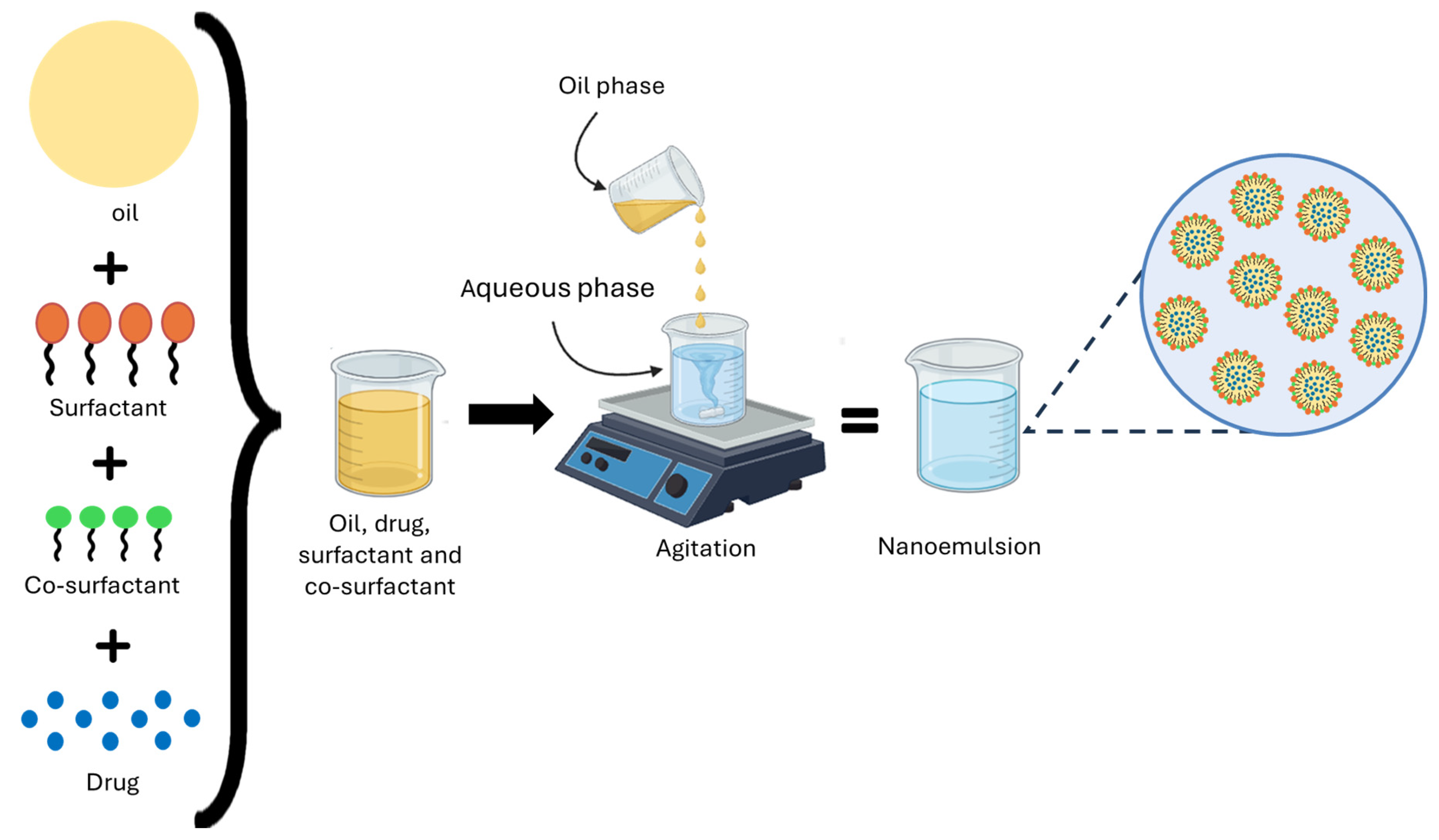
These systems can achieve the desired therapeutic effect, a feature that current technologies lack [188]. Within this approach, NEs have emerged as an innovative strategy for drug delivery, due to their unique properties that enhance the solubility and bioavailability of hydrophobic active ingredients [189]. NEs consist of immiscible liquids, such as oil and water, which form a single phase through an emulsifier, like surfactants and co-surfactants [190]. NEs are lipid-based and serve as a promising tool for delivering and improving the bioavailability of hydrophobic drugs. Most drugs are hydrophobic (lipophilic) in nature, which causes low solubility and bioavailability issues [191]. These systems can be used for developing pharmaceutical formulations due to their small particle sizes, making them suitable for topical, ocular, and intravenous administration [192]. Figure 14 shows the NEs structure.
Lipid formulations are divided into four types. Type I formulations include only lipids, such as triglycerides and drugs. Type II includes oils and O/W soluble surfactants. Type III combines lipids with surfactants and co-solvents to further facilitate self-emulsification, thereby increasing efficiency during the digestion process. Finally, Type IV contains only water-soluble surfactants and co-solvents (no oils) [193]. Transmission electron microscopy (TEM), scanning electron microscopy (SEM), Fourier Transform Infrared Spectroscopy (FTIR), dynamic light scattering (DLS), and Zeta potential analysis are some of the most valuable and straightforward techniques used to study the shape, size, chemical composition, and surface charge of particles [194].
For this reason, NEs are expected to be administered orally. This approach is mainly used to enhance the solubility and bioavailability of lipophilic drugs, as the nanoemulsion protects the active ingredient and helps its absorption in the intestine. However, oral administration faces significant challenges, such as low solubility, limited permeability, rapid metabolism, and instability in the gastrointestinal environment. Despite these issues, the oral route remains the preferred method for patients due to additional benefits such as reducing the risk of infections, thrombosis, and complications related to catheters [195,196,197].
To overcome these barriers, various nanocarrier systems have been developed, such as liposomes, lipid nanoparticles, double emulsions, cyclodextrins, proteins, and polymers like chitosan [198]. These systems protect active ingredients from the hostile environment of the gastrointestinal tract and enzymatic degradation, increasing their stability in biological fluids and their bioavailability [199]. Lipid nanocarriers are promising because they allow the encapsulation of hydrophobic compounds, reduce adverse effects, and prolong systemic circulation [200]. Furthermore, the evolution toward next-generation smart NPs has opened the possibility of evading immune recognition and selectively directing drugs toward tumor cells or their receptors [201], thereby increasing their effectiveness when combined with other therapeutic strategies [202].

10.4. Targeted Delivery of Nanoemulsions

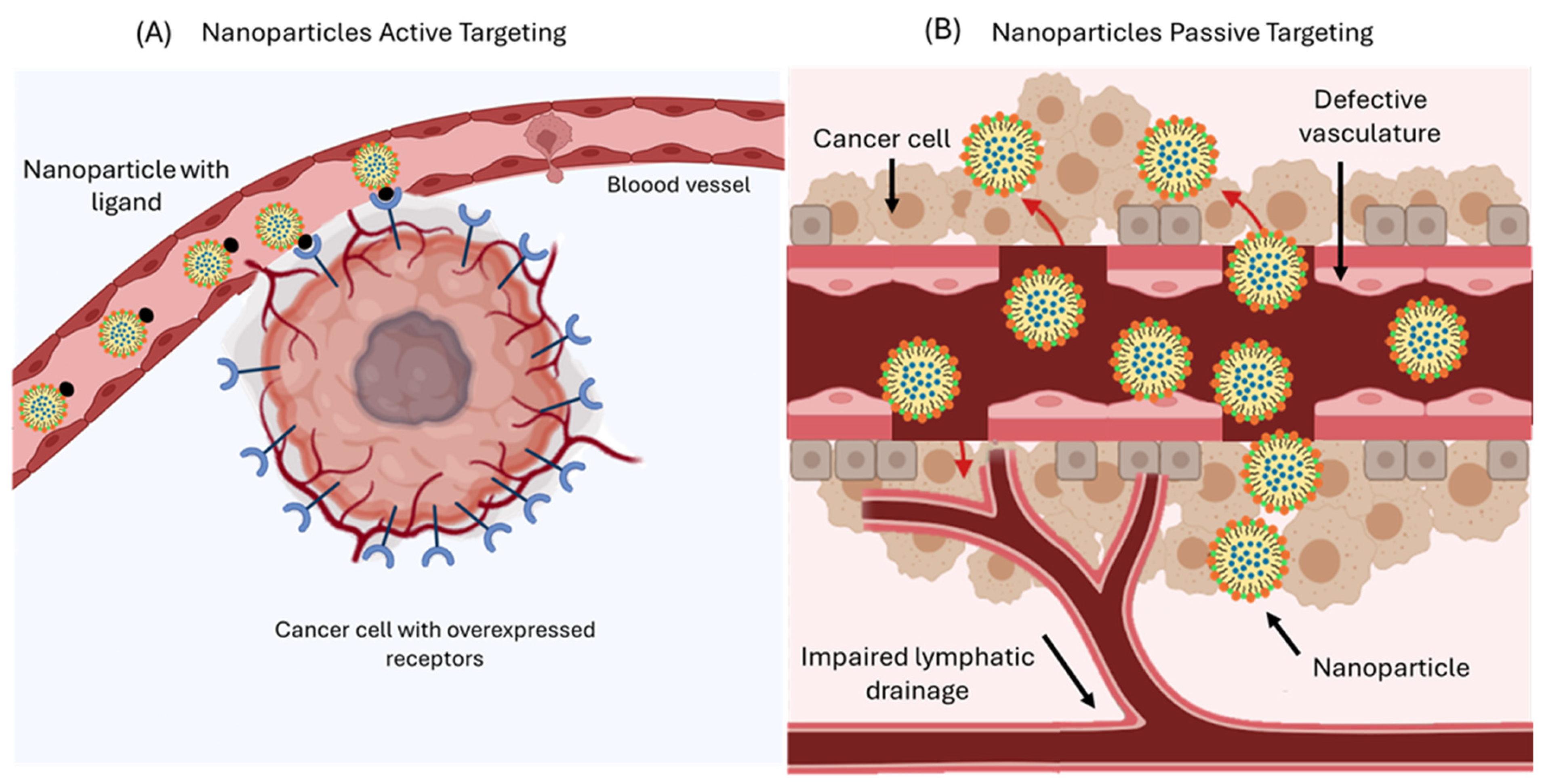
Drug delivery systems can be incorporated into cancer therapies, offering improvements in the pharmacokinetic parameters of therapeutic agents, reducing systemic toxicity to normal tissue, and minimizing adverse effects associated with conventional cancer treatments [203]. With this approach, nanoparticles can enter cancer cells through two distinct mechanisms, passive targeting and active targeting, as illustrated in Figure 15.
In passive targeting, these systems benefit from the characteristics of tumor tissues, such as the EPR effect, which allows NPs to concentrate in cancerous tissue [204]. Many tumors exhibit abnormal angiogenesis, resulting in permeable blood vessels with fenestrations and poor lymphatic drainage, characterized by elevated interstitial fluid pressure. This environment facilitates the penetration of NPs into the interstitial pores, which range in size from 100 to 780 nm [203]. NPs whose size typically ranges from 20 to 200 nm are suitable for taking advantage of this effect, as their size allows them to pass through these pores and accumulate in tumor tissue. Additionally, their retention is prolonged due to the low lymphatic drainage of the tumor. This targeted delivery can be further optimized through internal stimuli, such as acidic pH, specific enzymes, or alterations in redox potential, as well as external stimuli like temperature, magnetic fields, or radiation, thereby enhancing controlled drug release and reducing systemic side effects [205].
Active targeting exploits the biofunctionalization of NPs surfaces by using ligands with high affinity and specificity for receptors that are overexpressed on tumor cells [206]. This approach reduces systemic toxicity by avoiding healthy cells [207]. Controlled drug release should accumulate selectively and quantitatively at the therapeutic target, regardless of the route of administration [205].
Molecules such as folic acid, transferrin, biotin, and hyaluronic acid show a high affinity for receptors that are often overexpressed on tumor cells. Folic acid exhibits differences in its internalization pathway: in normal cells, it crosses the membrane via transmembrane transport, while in tumor cells, it enters through folate receptors that facilitate endocytosis. This basis has allowed the functionalization of the surface of nanoemulsions or other delivery systems with folic acid, thereby promoting selective internalization into cancer cells and intracellular drug release [208].
Nowadays, encapsulating active ingredients in NPs is a key strategy for directing drugs toward tumors or diseased organs, reducing damage to healthy tissues, and increasing therapeutic efficacy [209]. These nanoparticles can even penetrate the cell nucleus, release the active ingredient, and induce cell death [209,210].

10.5. Nanotechnology in the Oncology Field

Early diagnosis of cancer continues to be one of the main clinical challenges, as most cases are identified at advanced stages (III or IV), when the chances of curative treatment are limited. In response to this issue, nanotechnology has emerged as a promising tool to facilitate the early detection of tumors in different organs [211]. This discipline allows for the identification of tumor biomarkers in the extracellular microenvironment, as well as the application of in vivo bioimaging techniques [212].
NPs, because of their size, can cross cell membranes, making them excellent tools for detecting cancer cells and accurately locating affected areas [213]. These NPs can be engineered to specifically bind to receptors that are overexpressed on cancer cells, aiding their visualization and analysis. This method enhances the accuracy of early detection, tracking tumor progression, and reducing the time needed for clinical diagnosis [214].
The development of nanotechnology-based biosensors represents another prominent application in this field. These devices can identify specific biomarkers in blood or other bodily fluids, enabling the detection of molecular alterations associated with cancer in its early stages [215]. Nanotechnology aimed at cancer diagnosis offers a more precise, non-invasive, and efficient way to identify the disease. However, challenges related to standardization, sensitivity, and reproducibility of these methods persist, making it essential to continue optimizing nanotechnology-based diagnostic systems to improve prognosis and early detection of cancer and other pathologies in the future [212,214].
Table 3 presents the clinical trials conducted with NPs with CPT and their derivatives, designed to improve their stability, bioavailability, and therapeutic profile. These studies reflect advances in the use of nanostructured systems as a strategy to optimize drug release and reduce adverse effects associated with the free molecule.
CPT derivatives formulated in nanotechnology-based delivery systems demonstrate a superior toxicological profile compared to their non-encapsulated counterparts. As summarized in Table 1, Table 2 and Table 3, liposomal CPT-11, nanoparticle conjugates of CPT (CRLX101 and NLG207), and CPT-11-loaded nanomicelles (IH-NM) have shown a significant reduction in systemic and hematological toxicity, as well as improved overall tolerability and safety, even at higher doses. These findings suggest that nanostructured systems not only enhance the stability and bioavailability of the drug but also substantially mitigate adverse effects, representing a promising platform for safer and more effective clinical application of CPT derivatives.
Despite technological advances, NEs still face limitations that hinder their clinical use. The main challenges include the difficulty of ensuring scalable and reproducible production processes. Large-scale manufacturing requires specialized equipment and can lead to variations in particle size, increased costs, and waste. Additionally, uncertainties remain regarding their long-term safety, potential to trigger immune responses from specific components, and limited understanding of their pharmacokinetics and biodistribution, which can result in unpredictable therapeutic effects [220].
The regulatory environment still poses a significant challenge, as the lack of uniform guidelines and uncertainty regarding their long-term safety continue to delay approval by agencies such as the FDA [221]. Although lipid NE and polymer-based systems have demonstrated adequate biocompatibility and low toxicity in vivo models, a comprehensive chronic safety evaluation and the generation of solid clinical evidence in humans are still needed to support their incorporation into routine practice [222].
In the specific case of CPT, different systems have demonstrated improved solubility and enhanced antiproliferative activity; however, solid clinical evidence is still needed to support its implementation. Meanwhile, next-generation nanocarriers offer an emerging approach that should be explored in combination with immunotherapy and targeted therapies to optimize the clinical response [223]. Recently, countries such as the United States, the United Kingdom, Japan, and Germany have spearheaded the development of this technology due to their scientific advancements. Meanwhile, nations like Mexico, Colombia, and Brazil are still in the consolidation phase. Nonetheless, nanomedicine holds the potential to revolutionize the treatment of chronic and cancer-related diseases, which have negatively impacted the quality of life for millions over many decades [224].

11. Prospects

CPT derivatives remain essential anticancer agents, and advanced delivery methods such as nanoemulsions, liposomes, and ADCs improve their effectiveness and reduce toxicity. New techniques, such as reverse targeting and the use of anti-idiotypic distribution enhancers, combined with targeted therapies and immunotherapies, are being studied in clinical trials and could further enhance the effectiveness, selectivity, and clinical utility of CPTs.
Future research should optimize the nanoemulsion composition (droplet size, interfacial film, and zeta potential) and integrate drugs with immunotherapy to maximize tumor cytotoxicity and the antitumor immune response. However, the clinical translation of these platforms requires overcoming additional challenges, such as scaling up their production, harmonizing manufacturing protocols, obtaining regulatory approval under international standards, and assessing long-term toxicities to ensure solid safety profiles.
Together, these innovations establish CPTs and their advanced delivery platforms as novel support of modern oncology, offering more effective, safer, and more precise treatments that improve patients’ quality of life and accelerate their clinical implementation within a rigorous regulatory and translational framework.

12. Conclusions

CPT remains one of the most iconic molecules in the history of anticancer drug development, due to its highly specialized mechanism for targeting Topo I, which causes irreversible DNA damage and triggers cell apoptosis. Its clinical use has been limited by its chemical instability, low solubility, and systemic toxicity related to traditional administration. FDA-approved semi-synthetic analogs, such as CPT-11 and TPT, have made significant advances in efficacy and pharmacokinetics. However, they still face issues like acquired resistance, limited bioavailability, and treatment-related adverse effects.
The study of new structural modifications and conjugates, such as ADCs, offers promising alternatives to improve selectivity against cancer cells and expand the therapeutic range of CPT and its derivatives. Simultaneously, the incorporation of nanotechnological approaches, particularly nanoemulsions, has opened a new chapter in the development of CPT. These drug delivery systems protect the active form of the molecule, enhance its physicochemical stability, increase solubility in biological media, and direct its release toward tumor tissues through passive (EPR effect) or active (surface ligands) mechanisms. NEs not only provide an effective way to reduce toxicity and prolong plasma half-life but also facilitate co-administration of synergistic agents and treatment personalization. The rational combination of chemical synthesis of new derivatives and engineering of delivery systems based on nanoemulsions could mark the beginning of a new generation of anticancer formulations with greater efficacy, optimized safety, and significant clinical impact, thereby contributing to improved quality of life for patients. Advancing these strategies requires close multidisciplinary collaboration, precise regulatory guidelines, and solid clinical evidence to validate their therapeutic performance. This establishes CPT as a promising support in the evolution of modern oncological therapies and future precision medicine.

Author Contributions

Writing—original draft, review, editing, conceptualization, H.U.P.-O.; resources, G.L.-B., H.J.-I. and F.V.-O.; writing, review, and editing, M.d.C.O.-M., R.R.C.-E., S.C.-S., E.G.-G. and F.V.-O.; writing, H.J.-I. and M.d.C.O.-M.; supervision, M.G.B.-S., G.L.-B. and F.V.-O. All authors have read and agreed to the published version of the manuscript.

Funding

This article results from the research project “Design, characterization, and evaluation of self-emulsifying systems with the active substance camptothecin for antiproliferative efficacy in MC38 cancer cell line”, grant 21837.25-P. The Spanish title of the referred project is “Diseño, caracterización y evaluación de sistemas autoemulsionantes del principio activo camptotecina para la eficiencia antiproliferativa en la línea celular cancerosa MC38,” developed with support from Tecnológico Nacional de México (TecNM), to which the authors express their gratitude.

Institutional Review Board Statement

Not applicable.

Informed Consent Statement

Not applicable.

Data Availability Statement

Not applicable.

Conflicts of Interest

The authors declare no conflicts of interest.

Abbreviations

The following abbreviations are used in this manuscript:
10-HCPT10-hydroxycamptothecin
AAGAlpha-1-acid glycoprotein
ABCATP-binding cassette
ADCAntibody–drug conjugate
AIFApoptosis-inducing factor
AktAKT serine/threonine kinase
Apaf-1Apoptotic protease-activating factor 1
AUCArea under the curve
BakBCL-2 homologous antagonist/killer (pro-apoptotic effector)
BaxBCL-2–associated X protein (pro-apoptotic effector)
BCL-2A proto-oncogene that promotes cell survival
BCRPBreast cancer resistance protein
BI354 (IBI354)Anti-HER2 antibody–drug conjugate (investigational code)
BRCA1/2Breast Cancer Gene 1/2
CADCaspase-activated DNase
CARDCaspase recruitment domain
CESCarboxylesterases
CIConfidence interval
CKD-602Belotecan
cMOATCanalicular multispecific organic anion transporter
CPTCamptothecin
CPT-11Irinotecan
CRISPR-Cas9Clustered Regularly Interspaced Short Palindromic Repeats–Cas9
CYP3A4Cytochrome P450 3A4
DCRDisease control rate
DEDDeath effector domain
DISCDeath-inducing signaling complex
DLSDynamic light scattering
DNADeoxyribonucleic acid
DoRDuration of response
DS-8201Trastuzumab deruxtecan (development code)
DX-8951fExatecan
DXdDeruxtecan payload
ECOG-PSEastern Cooperative Oncology Group Performance Status
Endo GEndonuclease G
EPREnhanced permeation and retention
ERKExtracellular signal-regulated kinase
FADDFas-associated death domain
FasLFas ligand
FDAFood and Drug Administration
FL118Novel camptothecin derivative (research code)
FOLFIRIFolinic acid, fluorouracil, and irinotecan
FOLFIRINOXFluorouracil, leucovorin, irinotecan, and oxaliplatin
FTIRFourier Transform Infrared Spectroscopy
G-CSFGranulocyte colony-stimulating factor
GG211Lurtotecan
GIGastrointestinal tract
GLOBOCANInternational Agency for Research on Cancer (Global Cancer Observatory)
HAP1Near-haploid human cell line
HER2Human Epidermal Growth Factor Receptor 2
HIFHypoxia-inducible factor
HRHazard ratio
ICADInhibitor of caspase-activated DNase
IMMU-132 (SG)Sacituzumab govitecan
ISG15Interferon-stimulated gene 15
Log PPartition coefficient
MDR1Multidrug resistance protein 1
MEKMitogen-activated extracellular signal-regulated kinase
mOSMedian overall survival
mPFSMedian progression-free survival
MC38Murine colon adenocarcinoma cell line
MRP1Multidrug resistance-associated protein 1
MRP2Multidrug resistance-associated protein 2
mTOR
mTTP
Mechanistic Target of Rapamycin
Median Time To Progression
Nal-IRINanoliposomal irinotecan
NEsNanoemulsions
NF-κBNuclear factor kappa-light-chain-enhancer of activated B cells
Noxa Phorbol-12-Myristate-13-Acetate-Induced Protein 1 (PMAIP1)
NPsNanoparticles
NSCLCNon-small cell lung cancer
OHHydroxyl
OPNOsteopontin
ORROverall/Objective response rate
O/WOil-in-water (emulsion)
p53Tumor suppressor protein
PFSProgression-free survival
P-gpP-glycoprotein
PK-toxPharmacokinetics–toxicity correlation
PNPsPolymeric nanoparticles
PPProgression parameters
RAD51RAD51 recombinase
RAIDDReceptor-interacting protein with death domain
RFS 2000Rubitecan
RNARibonucleic acid
RNAiRNA interference
SCLCSmall-cell lung cancer
SEMScanning electron microscopy
SGSacituzumab govitecan
SIISystemic immune-inflammatory index
SmacSecond mitochondrial caspase activator
SN-387-ethyl-10-hydroxycamptothecin
SNEDDSSelf-nanoemulsifying drug delivery systems
ST1481Gimatecan
ST-1968Namitecan
tBidTruncated Bid
T-DXdTrastuzumab deruxtecan
TEMTransmission electron microscopy
TKITyrosine kinase inhibitors
TNBCTriple-negative breast cancer
TNFTumor necrosis factor
TNFR1Tumor necrosis factor receptor 1
TNFR2Tumor necrosis factor receptor 2
Topo ITopoisomerase I
TPTTopotecan
TRADDTumor necrosis factor receptor type 1-associated death domain protein
TRAILTNF-related apoptosis-inducing ligand
TROP-2Trophoblast Cell Surface Antigen 2
UGT1A1UDP-glucuronosyltransferase 1A1
VDACVoltage-dependent anion channel
WHOWorld Health Organization
WntWingless-related integration site pathway

References

  1. López-Plaza, B.; Loria-Kohen, V.; González-Rodríguez, L.G.; Fernández-Cruz, E. Alimentación y estilo de vida en la prevención del cáncer. Nutr. Hosp. 2022, 39, 74–77. [Google Scholar] [CrossRef] [PubMed]
  2. Zhou, Y.; Tao, L.; Qiu, J.; Xu, J.; Yang, X.; Zhang, Y.; Tian, X.; Guan, X.; Cen, X.; Zhao, Y. Tumor biomarkers for diagnosis, prognosis and targeted therapy. Signal Transduct. Target. Ther. 2024, 9, 132. [Google Scholar] [CrossRef] [PubMed]
  3. Singla, R.K.; Bishayee, A. Anticancer drug discovery based on natural products: From computational approaches to clinical studies. Cancers 2025, 17, 2507. [Google Scholar] [CrossRef] [PubMed]
  4. Bray, F.; Laversanne, M.; Sung, H.; Ferlay, J.; Siegel, R.L.; Soerjomataram, I.; Jemal, A. Global cancer statistics 2022: GLOBOCAN estimates of incidence and mortality worldwide for 36 cancers in 185 countries. CA Cancer J. Clin. 2024, 74, 229–263. [Google Scholar] [CrossRef]
  5. Khaiwa, N.; Maarouf, N.R.; Darwish, M.H.; Alhamad, D.W.; Sebastián, A.; Hamad, M.; Omar, H.A.; Orive, G.; Al-Tel, T.H. Camptothecin’s journey from discovery to WHO Essential Medicine: Fifty years of promise. Eur J. Med. Chem. 2021, 223, 113639. [Google Scholar] [CrossRef] [PubMed]
  6. Yu, L.; Jin, Y.; Song, M.; Zhao, Y.; Zhang, H. When natural compounds meet nanotechnology: Nature-inspired nanomedicines for cancer immunotherapy. Pharmaceutics 2022, 14, 1589. [Google Scholar] [CrossRef]
  7. Asma, S.T.; Acaroz, U.; Imre, K.; Morar, A.; Shah, S.R.A.; Hussain, Z.S.; Arslan-Acaroz, D.; Demirbas, H.; Hajrulai-Musliu, Z.; Istanbullugil, F.R.; et al. Natural products/bioactive compounds as a source of anticancer drugs. Cancers 2022, 14, 6203. [Google Scholar] [CrossRef]
  8. Odeniran, P.O.; Madlala, P.; Mkhwanazi, N.P.; Soliman, M.E.S. Camptothecin and Its Derivatives from Traditional Chinese Medicine in Combination with Anticancer Therapy Regimens: A Systematic Review and Meta-Analysis. Cancers 2024, 16, 3802. [Google Scholar] [CrossRef]
  9. Feng, L.; Sun, J.; Shi, Y.; Wang, G.; Wang, T. Predicting suitable habitats of Camptotheca acuminata considering both climatic and soil variables. Forests 2020, 11, 891. [Google Scholar] [CrossRef]
  10. Strzelecka, K.; Piotrowska, U.; Sobczak, M.; Oledzka, E. The advancement of biodegradable polyesters as delivery systems for camptothecin and its analogues—A status report. Int. J. Mol. Sci. 2023, 24, 1053. [Google Scholar] [CrossRef]
  11. Bacherikov, V.A. Total synthesis, mechanism of action, and antitumor efficacy of camptothecin and some of its analogues. Anticancer. Agents Med. Chem. 2022, 22, 3438–3465. [Google Scholar] [CrossRef]
  12. Peng, X.; Cai, X.; Tang, J.; Ge, J.; Chen, G. Rescuing and utilizing anticancer Nothapodytes species: Integrated studies from plant resources to natural medicines. Clin. Transl. Med. 2024, 14, e70110. [Google Scholar] [CrossRef]
  13. Fan, X.; Lin, X.; Ruan, Q.; Wang, J.; Yang, Y.; Sheng, M.; Zhou, W.; Kai, G.; Hao, X. Research progress on the biosynthesis and metabolic engineering of the anti-cancer drug camptothecin in Camptotheca acuminata. Ind. Crops Prod. 2022, 186, 115270. [Google Scholar] [CrossRef]
  14. Jadhav, R.R.; Sharma, S.; Singh, S.; Saurabh Sarma, J.; Khare, D. Enhancing camptothecin yields: Innovative approaches for sustainable production of anticancer drugs. Biotechnol. Bioprocess Eng. 2025, 30, 423–445. [Google Scholar] [CrossRef]
  15. Nguyen, T.M.; Nguyen, T.D.; Leung, Y.Y.; McConnachie, M.; Sannikov, O.; Xia, Z.; Dang, T.T. Discovering and harnessing oxidative enzymes for chemoenzymatic synthesis and diversification of anticancer camptothecin analogues. Commun. Chem. 2021, 4, 60. [Google Scholar] [CrossRef]
  16. Banadka, A.; Narasimha, S.W.; Dandin, V.S.; Naik, P.M.; Vennapusa, A.R.; Melmaiee, K.; Vemanna, R.S.; Al-Khayri, J.M.; Thiruvengadam, M.; Nagella, P. Biotechnological approaches for the production of camptothecin. Appl. Microbiol. Biotechnol. 2024, 108, 382. [Google Scholar] [CrossRef]
  17. Madkour, M.M.; Sebastian, A.; Ramadan, W.S.; Menon, V.; Lozon, L.; Srikanth, G.; Tarazi, H.; El-Gamal, M.I.; Al-Tel, T.H.; El-Awady, R. Synthesis and biological evaluation of new Camptothecin-like compounds as anticancer agents. J. Mol. Struct. 2025, 1328, 141271. [Google Scholar] [CrossRef]
  18. González-Ruiz, V.; Cores, Á.; Martín-Cámara, O.; Orellana, K.; Cervera-Carrascón, V.; Michalska, P.; Olives, A.I.; León, R.; Martín, M.A.; Menéndez, J.C. Enhanced stability and bioactivity of natural anticancer topoisomerase I inhibitors through cyclodextrin complexation. Pharmaceutics 2021, 13, 1609. [Google Scholar] [CrossRef] [PubMed]
  19. Swamy, M.K.; Purushotham, B.; Sinniah, U.R. Camptothecin: Occurrence, chemistry and mode of action. In Bioactive Natural Products for Pharmaceutical Applications; Springer: Cham, Switzerland, 2021; pp. 311–327. [Google Scholar] [CrossRef]
  20. Fan, S.; Cao, Y.-X.; Li, G.-Y.; Lei, H.; Attiogbe, M.K.I.; Yao, J.-C.; Yang, X.-Y.; Liu, Y.-J.; Hei, Y.-Y.; Zhang, H.; et al. F10, a new camptothecin derivative, was identified as a new orally–bioavailable, potent antitumor agent. Eur. J. Med. Chem. 2020, 202, 112528. [Google Scholar] [CrossRef]
  21. Nanavati, C.; Mager, D.E. Calculated Log D Is Inversely Correlated with Select Camptothecin Clearance and Efficacy in Colon Cancer Xenografts. J. Pharm. Sci. 2016, 105, 1561–1566. [Google Scholar] [CrossRef]
  22. Shaikh, R.; O’Brien, D.P.; Croker, D.M.; Walker, G.M. The development of pharmaceutical oral solid dosage forms. In Computer Aided Chemical Engineering; Elsevier: Amsterdam, The Netherlands, 2018; Volume 41, pp. 27–65. [Google Scholar] [CrossRef]
  23. Botella, P.; Rivero-Buceta, E. Safe approaches for camptothecin delivery: Structural analogues and nanomedicines. J. Control. Release 2017, 247, 28–54. [Google Scholar] [CrossRef]
  24. Mastrangelo, S.; Attina, G.; Triarico, S.; Romano, A.; Maurizi, P.; Ruggiero, A. The DNA-topoisomerase inhibitors in cancer therapy. Biomed. Pharmacol. J. 2022, 15, 553–562. [Google Scholar] [CrossRef]
  25. Khuroo, T.; Khuroo, A.; Hussain, A.; Mirza, A.M.; Panda, A.K.; Wani, J. QbD-based and Box-Behnken design assisted oral delivery of stable lactone (active) form of topotecan as PLGA nanoformulation: Cytotoxicity, pharmacokinetic, in vitro, and ex vivo gut permeation studies. J. Drug Deliv. Sci. Technol. 2022, 77, 103850. [Google Scholar] [CrossRef]
  26. Zhang, H.; Wang, X.; Dai, W.; Gemeinhart, R.A.; Zhang, Q.; Li, T. Pharmacokinetics and treatment efficacy of camptothecin nanocrystals on lung metastasis. Mol. Pharm. 2014, 11, 226–233. [Google Scholar] [CrossRef]
  27. Almeida, A.; Fernandes, E.; Sarmento, B.; Lúcio, M. A biophysical insight of camptothecin biodistribution: Towards a molecular understanding of its pharmacokinetic issues. Pharmaceutics 2021, 13, 869. [Google Scholar] [CrossRef]
  28. Yu, L.; Hua, Z.; Luo, X.; Zhao, T.; Liu, Y. Systematic interaction of plasma albumin with the efficacy of chemotherapeutic drugs. Biochim. Biophys. Acta Rev. Cancer 2022, 1877, 188655. [Google Scholar] [CrossRef]
  29. Ahmed, H.; Bergmann, F.; Zeitlinger, M. Protein binding in translational antimicrobial development—Focus on interspecies differences. Antibiotics 2022, 11, 923. [Google Scholar] [CrossRef]
  30. Hoogenboezem, E.N.; Duvall, C.L. Harnessing albumin as a carrier for cancer therapies. Adv. Drug Deliv. Rev. 2018, 130, 73–89. [Google Scholar] [CrossRef]
  31. Alshammari, M.K.; Alshehri, M.M.; Alshehri, A.M.; Alshlali, O.M.; Mahzari, A.M.; Almalki, H.H.; Kulaybi, O.Y.; Alghazwni, M.K.; Kamal, M.; Imran, M. Camptothecin loaded nano-delivery systems in the cancer therapeutic domains: A critical examination of the literature. J. Drug Deliv. Sci. Technol. 2023, 79, 104034. [Google Scholar] [CrossRef]
  32. Adak, D.; Ray, P.; Setua, S. Unlocking therapeutic precision: Camptotheca acuminata, a traditional Chinese herb tailored for phytonano-cancer theranostics. Pharmacol. Res. Mod. Chin. Med. 2024, 11, 100447. [Google Scholar] [CrossRef]
  33. Behera, A.; Padhi, S. Passive and active targeting strategies for the delivery of the camptothecin anticancer drug: A review. Environ. Chem. Lett. 2020, 18, 1557–1567. [Google Scholar] [CrossRef]
  34. Chen, Z.; Liu, M.; Wang, N.; Xiao, W.; Shi, J. Unleashing the Potential of Camptothecin: Exploring Innovative Strategies for Structural Modification and Therapeutic Advancements. J. Med. Chem. 2024, 67, 3244–3273. [Google Scholar] [CrossRef]
  35. Fonseca, L.C.; De Sousa, M.; Maia, D.L.S.; Visani de Luna, L.; Alves, O.L. Understanding the driving forces of camptothecin interactions on the surface of nanocomposites based on graphene oxide decorated with silica nanoparticles. Nanoscale Adv. 2020, 2, 1290–3000. [Google Scholar] [CrossRef] [PubMed]
  36. Kamle, M.; Pandhi, S.; Mishra, S.; Barua, S.; Kurian, A.; Mahato, D.K.; Rasane, P.; Büsselberg, D.; Kumar, P.; Calina, D.; et al. Camptothecin and its derivatives: Advancements, mechanisms and clinical potential in cancer therapy. Med. Oncol. 2024, 41, 1–18. [Google Scholar] [CrossRef]
  37. Yakkala, P.A.; Penumallu, N.R.; Shafi, S.; Kamal, A. Prospects of topoisomerase inhibitors as promising anti-cancer agents. Pharmaceuticals 2023, 16, 1456. [Google Scholar] [CrossRef] [PubMed]
  38. Kostjukov, V.V. Theoretical analysis of lactone and carboxylate forms of camptothecin in aqueous solution: Electronic states, absorption spectra, and hydration. J. Mol. Liq. 2021, 344, 117804. [Google Scholar] [CrossRef]
  39. El-Sayed, A.S.A.; Hassan, W.H.B.; Sweilam, S.H.; Alqarni, M.H.S.; El Sayed, Z.I.; Abdel-Aal, M.M.; Abdelsalam, E.; Abdelaziz, S. Production, bioprocessing, and antiproliferative activity of camptothecin from Penicillium chrysogenum, a marine sponge endosymbiont, as a metabolically stable camptothecin producer. Molecules 2022, 27, 3033. [Google Scholar] [CrossRef] [PubMed]
  40. Talukdar, A.; Kundu, B.; Sarkar, D.; Goon, S.; Mondal, M.A. Topoisomerase I inhibitors: Challenges, progress and the road ahead. Eur. J. Med. Chem. 2022, 236, 114304. [Google Scholar] [CrossRef]
  41. Shen, G.; Li, S.; Zhu, Y.; Xu, Z.; Liu, X.; Lv, C.; Xing, Z.; Cui, L.; Li, W. Recent progress in topoisomerase inhibitors as anticancer agents: Research and design strategies for Topo I and II inhibitors via structural optimization. Bioorg. Chem. 2025, 165, 109040. [Google Scholar] [CrossRef]
  42. Tesauro, C.; Simonsen, A.K.; Bech Andersen, M.; Wandsoe Petersen, K.; Laust Kristoffersen, E.; Algreen, L. Topoisomerase I activity and sensitivity to camptothecin in breast cancer-derived cells: A comparative study. BMC Cancer 2019, 19, 1158. [Google Scholar] [CrossRef]
  43. Patil, M.A.; Sarkate, A.P.; Nirmal, N.P.; Sakhale, B.K. Alkaloids as potential anticancer agents. Recent Front. Phytochem. 2023, 203–224. [Google Scholar] [CrossRef]
  44. Mustafa, M.; Ahmad, R.; Tantry, I.Q.; Ahmad, W.; Siddiqui, S.; Alam, M.; Abbas, K.; Moinuddin Hassan, M.I.; Habib, S.; Islam, S. Apoptosis: Una descripción general completa de las vías de señalización, los cambios morfológicos y la importancia fisiológica y las implicaciones terapéuticas. Cells 2024, 13, 1838. [Google Scholar] [CrossRef]
  45. Zhang, W.; Hu, X.; Shen, Q.; Xing, D. Mitochondria-specific drug release and reactive oxygen species burst induced by polyprodrug nanoreactors can enhance chemotherapy. Nat. Commun. 2019, 10, 1704. [Google Scholar] [CrossRef] [PubMed]
  46. Lim, Y.; Dorstyn, L.; Kumar, S. The p53-caspase-2 axis in the cell cycle and DNA damage response. Exp. Mol. Med. 2021, 53, 517–527. [Google Scholar] [CrossRef] [PubMed]
  47. Lossi, L. The concept of intrinsic versus extrinsic apoptosis. Biochem. J. 2022, 479, 357–384. [Google Scholar] [CrossRef]
  48. Jentsch, M.; Snyder, P.; Sheng, C.; Cristiano, E.; Loewer, A. P53 dynamics in single cells are temperature-sensitive. Sci. Rep. 2020, 10, 1481. [Google Scholar] [CrossRef]
  49. Opferman, J.; Kothari, A. Anti-apoptotic BCL-2 family members in development. Cell Death Differ. 2018, 25, 37–45. [Google Scholar] [CrossRef] [PubMed]
  50. Wei, H.; Qu, L.; Dai, S.; Li, Y.; Wang, H.; Feng, Y.; Chen, X.; Jiang, L.; Guo, M.; Li, J.; et al. Structural insight into the molecular mechanism of p53-mediated mitochondrial apoptosis. Nat. Commun. 2021, 12, 2280. [Google Scholar] [CrossRef]
  51. Roufayel, R.; Younes, K.; Al-Sabi, A.; Murshid, N. BH3-only proteins Noxa and Puma are key regulators of induced apoptosis. Life 2022, 12, 256. [Google Scholar] [CrossRef]
  52. Hohorst, L.; Ros, U.; Garcia-Saez, A.J. Mitochondrial dynamics and pore formation in regulated cell death pathways. Trends Biochem. Sci. 2025, 1–14. [Google Scholar] [CrossRef]
  53. Park, H.H. Caspase recruitment domains for protein interactions in cellular signaling (Review). Int. J. Mol. Med. 2019, 43, 1119–1127. [Google Scholar] [CrossRef]
  54. Vigneswara, V.; Ahmed, Z. The role of caspase-2 in regulating cell fate. Cells 2020, 9, 1259. [Google Scholar] [CrossRef] [PubMed]
  55. Dho, S.H.; Cho, M.; Woo, W.; Kim, D.; Park, S.; Lee, J.; Lee, H.; Choi, K.; Han, J. Caspases as master regulators of programmed cell death: Apoptosis, pyroptosis and beyond. Exp. Mol. Med. 2025, 57, 1121–1132. [Google Scholar] [CrossRef]
  56. Jan, R.; Chaudhry, G.-E. Understanding apoptosis and apoptotic pathways: Targeted cancer therapies. Adv. Pharm. Bull. 2019, 9, 205–218. [Google Scholar] [CrossRef] [PubMed]
  57. Eskandari, E.; Eaves, C.J. Paradoxical roles of caspase-3 in regulating cell survival, proliferation, and tumorigenesis. J. Cell Biol. 2022, 221, e202201159. [Google Scholar] [CrossRef]
  58. Lindenboim, L.; Zohar, H.; Worman, H.J.; Stein, R. The nuclear envelope: Target and mediator of the apoptotic process. Cell Death Discov. 2020, 6, 29. [Google Scholar] [CrossRef]
  59. Gheyas, R.; Menko, A.S. The involvement of caspases in the process of nuclear removal during lens fiber cell differentiation. Cell Death Discov. 2023, 9, 386. [Google Scholar] [CrossRef]
  60. Choi, Y.N.; Seo, W.T.; Lee, T.Y.; Jeon, H.D.; Yoo, J.S. Nuclear endonuclease G regulates cell proliferation in ovarian cancer. FEBS Open Bio. 2023, 13, 655–669. [Google Scholar] [CrossRef]
  61. Schoeniger, A.; Wolf, P.; Edlich, F. How do hexokinases inhibit receptor-mediated apoptosis? Biology 2022, 11, 412. [Google Scholar] [CrossRef] [PubMed]
  62. Melloul, O.; Zabit, S.; Lichtenstein, M.; Duran, D.; Grunewald, M.; Lorberboum-Galski, H. Targeted and caspase-independent apoptosis induction with novel chimeric proteins for the treatment of solid cancers. Cancers 2025, 17, 1179. [Google Scholar] [CrossRef]
  63. Risso, V.; Lafont, E.; Le Gallo, M. Therapeutic approaches targeting CD95L/CD95 signaling in cancer and autoimmune diseases. Cell Death Dis. 2022, 13, 248. [Google Scholar] [CrossRef] [PubMed]
  64. Pfeffer, C.M.; Singh, A.T.K. Apoptosis: A target for cancer therapy. Int. J. Mol. Sci. 2018, 19, 448. [Google Scholar] [CrossRef]
  65. Carneiro, B.A.; El-Deiry, W.S. Targeting apoptosis in cancer therapy. Nat. Rev. Clin. Oncol. 2020, 17, 395–417. [Google Scholar] [CrossRef]
  66. Marikar, F.; Zi-Chun, H. Adapter protein—FADD bridges the apoptosis. Int. J. Med. Biochem. 2023, 6, 124–132. [Google Scholar] [CrossRef]
  67. Guerrache, A.; Micheau, O. TNF-related apoptosis-inducing ligand: Non-apoptotic signalling. Cells 2024, 13, 521. [Google Scholar] [CrossRef]
  68. Van Opdenbosch, N.; Lamkanfi, M. Caspases in cell death, inflammation, and disease. Immunity 2019, 50, 1352–1364. [Google Scholar] [CrossRef]
  69. Park, Y.H.; Han, C.W.; Jeong, M.S.; Jang, S.B. DED interaction of FADD and caspase-8 in the induction of apoptotic cell death. J. Microbiol. Biotechnol. 2022, 32, 1034–1040. [Google Scholar] [CrossRef] [PubMed]
  70. Coria-Paredes, D.M.; Wilkins-Rodríguez, A.A.; Gutiérrez-Kobeh, L. Apoptosis: A key process that Trypanosoma cruzi modulates as a strategy to perpetuate infection. J. Parasitol. Res. 2025, 2025, 2093615. [Google Scholar] [CrossRef] [PubMed]
  71. Pathak, C.; Ansari, A.; Kumar, A.; Ranjan, K. Molecular perspective of proteases in modeling of cell death signaling. Preprints 2025, 2025010084. [Google Scholar] [CrossRef]
  72. Fu, Q.; Fu, T.-M.; Cruz, A.C.; Sengupta, P.; Thomas, S.K.; Wang, S.; Siegel, R.M.; Wu, H.; Chou, J.J. Structural basis and functional role of intramembrane trimerization of the Fas/CD95 death receptor. Mol. Cell 2016, 61, 602–613. [Google Scholar] [CrossRef]
  73. Zhang, W.; Zhu, C.; Liao, Y.; Zhou, M.; Xu, W.; Zou, Z. Caspase-8 in inflammatory diseases: A potential therapeutic target. Cell Mol. Biol. Lett. 2024, 29, 130. [Google Scholar] [CrossRef]
  74. Nadendla, E.K.; Tweedell, R.E.; Kasof, G.; Kanneganti, T.-D. Caspases: Structural and molecular mechanisms and functions in cell death, innate immunity, and disease. Cell Discov. 2025, 11, 42. [Google Scholar] [CrossRef]
  75. Tummers, B.; Mari, L.; Guy, C.S.; Heckmann, B.L.; Rodriguez, D.A.; Ruhl, S. Caspase-8-dependent inflammatory responses are controlled by its adaptor, FADD, and necroptosis. Immunity 2020, 52, 994–1006. [Google Scholar] [CrossRef]
  76. Han, Y.H.; Wang, Y.; Lee, S.J.; Jin, M.H.; Sun, H.N.; Kwon, T. Regulation of anoikis by extrinsic death receptor pathways. Cell Commun. Signal. 2023, 21, 227. [Google Scholar] [CrossRef]
  77. Li, Z.; Yuan, W.; Lin, Z. Functional roles in cell signaling of adaptor protein TRADD from a structural perspective. Comput. Struct. Biotechnol. J. 2020, 18, 2867–2876. [Google Scholar] [CrossRef]
  78. Wyżewski, Z.; Gregorczyk-Zboroch, K.P.; Mielcarska, M.B.; Świtlik, W.; Niedzielska, A. Bid protein: A participant in the apoptotic network with roles in viral infections. Int. J. Mol. Sci. 2025, 26, 2385. [Google Scholar] [CrossRef]
  79. Makinwa, Y.; Luo, Y.; Musich, P.R.; Zou, Y. Canonical and non-canonical functions of BH3-only protein in apoptosis, oncogenesis, cancer therapeutics, and aging. Cancers 2024, 16, 2199. [Google Scholar] [CrossRef]
  80. Flores-Romero, H.; Hohorst, L.; John, M.; Albert, M.; E King, L.; Beckmann, L.; Szabo, T.; Hertlein, V.; Luo, X.; Villunger, A.; et al. The BCL-2 family protein tBID acts as a BAX-like apoptosis effector. EMBO J. 2022, 41, e108690. [Google Scholar] [CrossRef]
  81. Dhani, S.; Zhao, Y.; Zhivotovsky, B. A long way to go: Caspase inhibitors in clinical use. Cell Death Dis. 2021, 12, 949. [Google Scholar] [CrossRef]
  82. Mandal, R.; Compte Barrón, J.; Kostova, I.; Becker, S.; Strebhardt, K. Caspase-8: The double-edged sword. Biochim. Biophys. Acta Rev. Cancer 2020, 1873, 188357. [Google Scholar] [CrossRef]
  83. Hussar, P. Apoptosis regulators Bcl-2 and caspase-3. Encyclopedia 2022, 2, 1624–1636. [Google Scholar] [CrossRef]
  84. Wang, Z. Regulation of cell cycle progression by growth factor-induced cell signaling. Cells 2021, 10, 3327. [Google Scholar] [CrossRef]
  85. Orning, P.; Lien, E. Multiple roles of caspase-8 in cell death, inflammation, and innate immunity. J. Leukoc. Biol. 2021, 109, 121–141. [Google Scholar] [CrossRef]
  86. Moeed, A.; Thilmany, N.; Beck, F.; Puthussery, B.K.; Ortmann, N.; Haimovici, A.; Badr, M.T.; Haghighi, E.B.; Boerries, M.; Öllinger, R.; et al. The caspase-activated DNase drives inflammation and contributes to defense against viral infection. Cell Death Differ. 2024, 31, 924–937. [Google Scholar] [CrossRef]
  87. Kari, S.; Subramanian, K.; Altomonte, I.A.; Murugesan, A.; Yli-Harja, O.; Kandhavelu, M. Programmed Cell Death Detection Methods: A Systematic Review and a Categorical Comparison. Apoptosis 2022, 27, 482–508. [Google Scholar] [CrossRef]
  88. Cai, Z.; Cheng, Z.; Zhang, L.; Zhang, Y.; Shi, J.; Jin, J.; Mi, Z.; Yuan, Z.; Wu, Z. CAD hijacks STING to impair antitumor immunity and radiotherapy efficacy of colorectal cancer. Cell Death Dis. 2025, 16, 641. [Google Scholar] [CrossRef]
  89. Ahmad Bhat, I.; Maqsood Bhat, A.; Tasduq Abdullah, S. Apoptosis—Mechanisms, Regulation in Pathology, and Therapeutic potential. In Biochemistry; IntechOpen: London, UK, 2025. [Google Scholar] [CrossRef]
  90. Han, C.Z.; Juncadella, I.J.; Kinchen, J.M.; Buckley, M.W.; Klibanov, A.L.; Dryden, K.; Onengut-Gumuscu, S.; Erdbrügger, U.; Turner, S.D.; Shim, Y.M.; et al. Macrophages redirect phagocytosis by non-professional phagocytes and influence inflammation. Nature 2016, 539, 570–574. [Google Scholar] [CrossRef]
  91. Wang, X.; Zhuang, Y.; Wang, Y.; Jiang, M.; Yao, L. The recent developments of camptothecin and its derivatives as potential anti-tumor agents. Eur. J. Med. Chem. 2023, 260, 115710. [Google Scholar] [CrossRef]
  92. Liu, Y.-Q.; Li, W.-Q.; Morris-Natschke, S.L.; Qian, K.; Yang, L.; Zhu, G.-X.; Wu, X.-B.; Chen, A.-L.; Zhang, S.-Y.; Nan, X.; et al. Perspectives on biologically active camptothecin derivatives. Med. Res. Rev. 2015, 35, 753–789. [Google Scholar] [CrossRef]
  93. Liu, Z.; Yuan, Y.; Wang, N.; Yu, P.; Teng, Y. Drug combinations of camptothecin derivatives promote the antitumor properties. Eur. J. Med. Chem. 2024, 279, 116872. [Google Scholar] [CrossRef]
  94. Dai, Y.; Qian, M.; Li, Y. Structural Modification Endows Small-Molecular SN38 Derivatives with Multifaceted Functions. Molecules 2023, 28, 4931. [Google Scholar] [CrossRef]
  95. Li, G.; Cai, C.; Qi, Y.; Tang, X. Hydroxyethyl starch–10-hydroxycamptothecin conjugate: Synthesis, pharmacokinetics, cytotoxicity and pharmacodynamic investigation. Drug Deliv. 2014, 23, 277–284. [Google Scholar] [CrossRef]
  96. Avendaño, C.; Menéndez, J.C. DNA intercalation and topoisomerase inhibition. In Medicinal Chemistry of Anticancer Drugs, 2nd ed.; Elsevier: Amsterdam, The Netherlands, 2023; pp. 325–387. [Google Scholar] [CrossRef]
  97. Pullaiah, T.; Raveendran, V. Camptothecin: Chemistry, biosynthesis, analogs, and chemical synthesis. In Camptothecin and Camptothecin Producing Plants: Botany, Chemistry, Anticancer Activity and Biotechnology; Academic Press: Cambridge, MA, USA, 2020; pp. 47–103. [Google Scholar] [CrossRef]
  98. Virupaksha, B.; Alpana, G. CoMFA QSAR models of camptothecin analogues based on the distinctive SAR features of combined ABC, CD and E ring substitutions. Comput. Biol. Med. 2012, 42, 890–897. [Google Scholar] [CrossRef]
  99. Liu, J.; Geng, G.; Liang, G.; Wang, L.; Luo, K.; Yuan, J.; Zhao, S. A novel topoisomerase I inhibitor DIA-001 induces DNA damage mediated cell cycle arrest and apoptosis in cancer cell. Ann. Transl. Med. 2020, 8, 89. [Google Scholar] [CrossRef]
  100. Zhang, M.; Zhu, L.-Z.; Yang, C.-J.; Yan, J.-X.; Wang, Z.-P.; Bai, Y.-P.; Peng, L.-Z.; Luo, H.-B.; Zhang, Z.-J.; Li, L.; et al. Improved anti-tumor activity of fluorinated camptothecin derivatives 9-fluorocamptothecin and 7-ethyl-9-fluorocamptothecin on hepatocellular carcinoma by targeting topoisomerase I. Bioorg. Chem. 2023, 139, 106652. [Google Scholar] [CrossRef]
  101. Zhu, L.; Zhuang, C.; Lei, N.; Guo, Z.; Sheng, C.; Dong, G.; Wang, S.; Zhang, Y.; Yao, J.; Miao, Z.; et al. Synthesis and preliminary bioevaluation of novel E-ring modified acetal analog of camptothecin as cytotoxic agents. Eur. J. Med. Chem. 2012, 56, 1–9. [Google Scholar] [CrossRef]
  102. Zi, C.-T.; Yang, L.; Xu, F.-Q.; Dong, F.-W.; Ma, R.-J.; Li, Y.; Zhou, J.; Ding, Z.-T.; Jiang, Z.-H.; Hu, J.-M. Synthesis and antitumor activity of biotinylated camptothecin derivatives as potent cytotoxic agents. Bioorg. Med. Chem. Lett. 2019, 29, 234–237. [Google Scholar] [CrossRef] [PubMed]
  103. Dai, X.; Wu, G.; Zhang, Y.; Zhang, X.; Yin, R.; Qi, X.; Li, J.; Jiang, T. Design, synthesis, and in vitro/in vivo anti-cancer activities of novel (20S)-10,11-methylenedioxy-camptothecin heterocyclic derivatives. Int. J. Mol. Sci. 2020, 21, 8495. [Google Scholar] [CrossRef]
  104. Cragg, G.M.; Pezzuto, J.M. Natural products as a vital source for the discovery of cancer chemotherapeutic and chemopreventive agents. Med. Princ. Pract. 2016, 25 (Suppl. 2), 41–59. [Google Scholar] [CrossRef]
  105. Gerwick, W.H. Plant sources of drugs and chemicals. In Encyclopedia of Biodiversity, 2nd ed.; Levin, S.A., Ed.; Elsevier: Amsterdam, The Netherlands, 2013; pp. 129–139. [Google Scholar] [CrossRef]
  106. Saraf, S.; Jain, A.; Tiwari, A.; Verma, A.; Jain, S.K. Engineered liposomes bearing camptothecin analogue for tumour targeting: In vitro and ex-vivo studies. J. Liposome Res. 2020, 31, 326–341. [Google Scholar] [CrossRef]
  107. Lichota, A.; Gwozdzinski, K. Anticancer activity of natural compounds from plant and marine environment. Int. J. Mol. Sci. 2018, 19, 3533. [Google Scholar] [CrossRef] [PubMed]
  108. Shang, X.; Morris-Natschke, S.L.; Liu, Y.; Guo, X.; Xu, X.; Goto, M.; Li, J.; Yang, G.; Lee, K. Biologically active quinoline and quinazoline alkaloids, part I. Med. Res. Rev. 2018, 38, 775–828. [Google Scholar] [CrossRef] [PubMed]
  109. Di Nunzio, M.R.; Douhal, A. Robust inclusion complex of topotecan comprised within a rhodamine-labeled β-cyclodextrin: Competing proton and energy transfer processes. Pharmaceutics 2023, 15, 1620. [Google Scholar] [CrossRef]
  110. Kyle, A.H.; Baker, J.H.E.; Gandolfo, M.J.; Reinsberg, S.A.; Minchinton, A.I. Tissue penetration and activity of camptothecins in solid tumor xenografts. Mol. Cancer Ther. 2014, 13, 2727–2737. [Google Scholar] [CrossRef]
  111. Jeong, S.H.; Jang, J.H.; Lee, Y.B. Pharmacokinetic comparison of three different administration routes for topotecan hydrochloride in rats. Pharmaceuticals 2020, 13, 231. [Google Scholar] [CrossRef] [PubMed]
  112. Varsha, K.; Sharma, A.; Kaur, A.; Madan, J.; Pandey, R.S.; Jain, U.K.; Chandra, R. Natural plant-derived anticancer drugs nanotherapeutics: A review on preclinical to clinical success. In Nanostructures for Cancer Therapy; Micro and Nano Technologies; Elsevier: Amsterdam, The Netherlands, 2017; pp. 775–809. [Google Scholar] [CrossRef]
  113. Samare-Najaf, M.; Samareh, A.; Jamali, N.; Abbasi, A.; Clark, C.C.T.; Khorchani, M.J.; Zal, F. Adverse effects and safety of etirinotecan pegol, a novel topoisomerase inhibitor, in cancer treatment: A systematic review. Curr. Cancer Ther. Rev. 2021, 17, 234–243. [Google Scholar] [CrossRef]
  114. Devriese, L.A.; Witteveen, P.E.; Mergui-Roelvink, M.; Smith, D.A.; Lewis, L.D.; Mendelson, D.S.; Bang, Y.; Chung, H.C.; Dar, M.M.; Huitema, A.D.R.; et al. Pharmacodynamics and pharmacokinetics of oral topotecan in patients with advanced solid tumors and impaired renal function. Br. J. Clin. Pharmacol. 2015, 80, 253–266. [Google Scholar] [CrossRef]
  115. Lambrecht, L.; Arnold, P.; Behr, J.; Mertsch, P.; Tufman, A.; Kauffmann-Guerrero, D. Topotecan in a real-world small-cell lung cancer cohort: Prognostic biomarkers improve selection of patients for second-line treatment. Diagnostics 2024, 14, 1572. [Google Scholar] [CrossRef]
  116. Tiseo, M.; Ardizzoni, A. Current status of second-line treatment and novel therapies for small cell lung cancer. J. Thorac. Oncol. 2007, 2, 764–772. [Google Scholar] [CrossRef]
  117. Tezuka, S.; Ueno, M.; Kobayashi, S.; Hamaguchi, T.; Yamachika, Y.; Oishi, R.; Nagashima, S.; Fukushima, T.; Morimoto, M.; Shin, M. Nal-IRI/5-FU/LV versus modified FOLFIRINOX and FOLFIRI as second-line chemotherapy for unresectable pancreatic cancer: A single center retrospective study. Pancreatology 2022, 22, 789–796. [Google Scholar] [CrossRef]
  118. Dai, W.; Habbous, S.; Saluja, R.; Beca, J.; Raphael, M.; Arias, J.; Gavura, S.; Earle, C.; Biagi, J.; Coburn, N.; et al. Comparative effectiveness of FOLFIRINOX versus gemcitabine and nab-paclitaxel in initially unresectable locally advanced pancreatic cancer: A population-based study to assess subsequent surgical resection and overall survival. Clin. Oncol. 2023, 35, 303–311. [Google Scholar] [CrossRef]
  119. Bailly, C. Irinotecan: 25 years of cancer treatment. Pharmacol. Res. 2019, 148, 104398. [Google Scholar] [CrossRef] [PubMed]
  120. Qi, Q.R.; Tian, H.; Yue, B.S.; Zhai, B.T.; Zhao, F. Research progress of SN38 drug delivery system in cancer treatment. Int. J. Nanomedicine 2024, 19, 945–964. [Google Scholar] [CrossRef]
  121. De Man, F.M.; Goey, A.K.; Van Schaik, R.H.; Mathijssen, R.H.; Bins, S. Individualization of irinotecan treatment: A review of pharmacokinetics, pharmacodynamics, and pharmacogenetics. Clin. Pharmacokinet. 2018, 57, 1229–1254. [Google Scholar] [CrossRef]
  122. Fujita, K.; Kubota, Y.; Ishida, H.; Sasaki, Y. Irinotecan: A key chemotherapeutic agent for metastatic colorectal cancer. World J. Gastroenterol. 2015, 21, 12234–12248. [Google Scholar] [CrossRef]
  123. Mladenić, K.; Sedić, M. Chemoresistance mechanisms in colon cancer: Focus on conventional chemotherapy. Clin. Cancer Drugs. 2021, 8, 67–105. [Google Scholar] [CrossRef]
  124. Yang, X.; Chen, J.; Wang, Y.; Wu, Y.; Zhang, J. Managing irinotecan-induced diarrhea: A comprehensive review of therapeutic interventions in cancer treatment. Pharmaceuticals 2025, 18, 359. [Google Scholar] [CrossRef]
  125. Westover, D.; Ling, X.; Lam, H.; Welch, J.; Jin, C.; Gongora, C.; Del Rio, M.; Wani, M.; Li, F. FL118, a novel camptothecin derivative, is insensitive to ABCG2 expression and shows improved efficacy in comparison with irinotecan in colon and lung cancer models with ABCG2-induced resistance. Mol. Cancer 2015, 14, 92. [Google Scholar] [CrossRef] [PubMed]
  126. Spigel, D.R.; Dowlati, A.; Chen, Y.; Navarro, A.; Yang, J.C.-H.; Stojanovic, G.; Jove, M.; Rich, P.; Andric, Z.G.; Wu, Y.-L.; et al. RESILIENT Trial Investigators. RESILIENT Part 2: A randomized, open-label phase III study of liposomal irinotecan versus topotecan in adults with relapsed small cell lung cancer. J. Clin. Oncol. 2024, 42, 2317–2326. [Google Scholar] [CrossRef]
  127. Lemech, C.R.; Sun, Y.; Nagrial, A.; Wu, X.; Morris, M.F.; Ning, F.; Yang, J.; Pan, Y.; Cai, J.; Lu, P.; et al. IBI354 (anti-HER2 antibody-drug conjugate [ADC]) in patients with HER2-positive breast cancer and other solid tumors: Updates from a phase 1 study. J. Clin. Oncol. 2025, 43, 1029. [Google Scholar] [CrossRef]
  128. Chekerov, R.; Arndt, T.; Pietzner, K.; Canzler, U.; Wimberger, P.; Strauß, H.-G.; Mahner, S.; Woelber, L.; de Gregorio, N.; Stocker, G.; et al. Pazopanib with topotecan weekly for patients with platinum-resistant or intermediate-sensitive recurrent ovarian cancer: Results of a multicentre, open-label phase I/II study (TOPAZ). J. Cancer Res. Clin. Oncol. 2023, 149, 7637–7649. [Google Scholar] [CrossRef]
  129. Das, S.; Samaddar, S. Recent advances in the clinical translation of small-cell lung cancer therapeutics. Cancers 2025, 17, 255. [Google Scholar] [CrossRef] [PubMed]
  130. Xu, J.; Wen, J.; Ji, X.; Chen, J.; Yang, M.; Hong, M.; Deng, D. Preclinical development of a high affinity anti-exatecan monoclonal antibody and application in bioanalysis of antibody-exatecan conjugates. J. Pharm. Biomed. Anal. 2025, 262, 116843. [Google Scholar] [CrossRef] [PubMed]
  131. Pillai, G. Nanotechnology toward treating cancer: A comprehensive review. In Applications of Targeted Nano Drugs and Delivery Systems; Elsevier: Amsterdam, The Netherlands, 2019; pp. 221–256. [Google Scholar] [CrossRef]
  132. Olender, D.; Żwawiak, J.; Zaprutko, L. Multidirectional efficacy of biologically active nitro compounds included in medicines. Pharmaceuticals 2018, 11, 54. [Google Scholar] [CrossRef] [PubMed]
  133. Kang, J.-H.; Lee, K.-H.; Kim, D.-W.; Kim, S.-W.; Kim, H.R.; Kim, J.-H.; Choi, J.-H.; An, H.J.; Kim, J.-S.; Jang, J.-S.; et al. A randomized phase 2b study comparing the efficacy and safety of belotecan versus topotecan as monotherapy for sensitive-relapsed small-cell lung cancer. Br. J. Cancer. 2021, 124, 713–720. [Google Scholar] [CrossRef]
  134. Schöffski, P.; Wang, C.-C.; Schöffski, M.P.; Wozniak, A. Current role of topoisomerase I inhibitors for the treatment of mesenchymal malignancies and their potential future use as payload of sarcoma-specific antibody-drug conjugates. Oncol. Res. Treat. 2024, 47, 18–41. [Google Scholar] [CrossRef]
  135. Gong, J.; Zhang, W.; Balthasar, J.P. Camptothecin-based anti-cancer therapies and strategies to improve their therapeutic index. Cancers 2025, 17, 1032. [Google Scholar] [CrossRef]
  136. Tonon, G.; Rizzolio, F.; Visentin, F.; Scattolin, T. Antibody drug conjugates for cancer therapy: From metallodrugs to nature-inspired payloads. Int. J. Mol. Sci. 2024, 25, 8651. [Google Scholar] [CrossRef]
  137. Han, S.; Lim, K.S.; Blackburn, B.J.; Yun, J.; Putnam, C.W.; Bull, D.A.; Won, Y.W. The Potential of Topoisomerase Inhibitor-Based Antibody-Drug Conjugates. Pharmaceutics 2022, 14, 1707. [Google Scholar] [CrossRef]
  138. Khan, S.; Jandrajupalli, S.B.; Bushara, N.Z.A.; Raja, R.D.P.; Mirza, S.; Sharma, K.; Verma, R.; Kumar, A.; Lohani, M. Targeting Refractory Triple-Negative Breast Cancer with Sacituzumab Govitecan: A New Era in Precision Medicine. Cells 2024, 13, 2126. [Google Scholar] [CrossRef]
  139. Indini, A.; Rijavec, E.; Grossi, F. Trastuzumab deruxtecan: Changing the destiny of HER2 expressing solid tumors. Int. J. Mol. Sci. 2021, 22, 4774. [Google Scholar] [CrossRef]
  140. Molina Trinidad, E.M.; Becerril Flores, M.A.; Imbert Palafox, J.L. Apoptosis y cáncer. Prensa. Med. Argent. 2020, 9, 124–130. [Google Scholar] [CrossRef]
  141. Beretta, L.; Gatti, G.; Perego, P.; Zaffaroni, N. Camptothecin resistance in cancer: Insights into the molecular mechanisms of a DNA-damaging drug. Curr. Med. Chem. 2013, 20, 1541–1556. [Google Scholar] [CrossRef]
  142. Stasiak, P.; Sopel, J.; Płóciennik, A.; Musielak, O.; Lipowicz, J.M.; Rawłuszko-Wieczorek, A.A.; Sterzyńska, K.; Korbecki, J.; Januchowski, R. Elacridar inhibits BCRP protein activity in 2D and 3D cell culture models of ovarian cancer and re-sensitizes cells to cytotoxic drugs. Int. J. Mol. Sci. 2025, 26, 5800. [Google Scholar] [CrossRef]
  143. Godbey, W.T. Cellular transport. In An Introduction to Biotechnology, 1st ed.; Academic Press: London, UK, 2014; pp. 35–64. [Google Scholar] [CrossRef]
  144. Choi, Y.H.; Yu, A.M. ABC transporters in multidrug resistance and pharmacokinetics, and strategies for drug development. Curr. Pharm. Des. 2014, 20, 793–807. [Google Scholar] [CrossRef] [PubMed]
  145. Ozawa, S.; Miura, T.; Terashima, J.; Habano, W. Cellular irinotecan resistance in colorectal cancer and overcoming irinotecan refractoriness through various combination trials including DNA methyltransferase inhibitors: A review. Cancer Drug Resist. 2021, 4, 946–964. [Google Scholar] [CrossRef] [PubMed]
  146. Bircsak, K.M.; Aleksunes, L.M. Interaction of isoflavones with the drug transporter BCRP/ABCG2. Curr. Drug Metab. 2015, 16, 124–140. [Google Scholar] [CrossRef]
  147. Karthika, C.; Sureshkumar, R.; Zehravi, M.; Akter, R.; Ali, F.; Ramproshad, S.; Mondal, B.; Kundu, M.K.; Dey, A.; Rahman, M.H.; et al. Multidrug resistance in cancer cells: Focus on a possible strategy plan to address colon carcinoma cells. Life 2022, 12, 811. [Google Scholar] [CrossRef]
  148. Bahar, E.; Kim, J.Y.; Kim, H.S.; Yoon, H. Establishment of acquired cisplatin resistance in ovarian cancer cell lines characterized by enriched metastatic properties with increased Twist expression. Int. J. Mol. Sci. 2020, 21, 7613. [Google Scholar] [CrossRef]
  149. Ricci, J.W.; Lovato, D.M.; Severns, V.; Sklar, L.A.; Larson, R.S. Novel ABCG2 antagonists reverse topotecan-mediated chemotherapeutic resistance in ovarian carcinoma xenografts. Mol. Cancer Ther. 2016, 15, 2853–2862. [Google Scholar] [CrossRef]
  150. Ando, K.; Shah, A.K.; Sachdev, V.; Kleinstiver, B.P.; Taylor-Parker, J.; Welch, M.M.; Hu, Y.; Salgia, R.; White, F.M.; Parvin, J.D.; et al. Camptothecin resistance is determined by topoisomerase I degradation regulation through the ubiquitin proteasome pathway. Oncotarget 2017, 8, 43733–43751. [Google Scholar] [CrossRef] [PubMed]
  151. Kumar, S.; Sherman, M.Y. Resistance to TOP-1 inhibitors: Good old drugs still can surprise us. Int. J. Mol. Sci. 2023, 24, 7233. [Google Scholar] [CrossRef] [PubMed]
  152. Saurav, S.; Karfa, S.; Vu, T.; Liu, Z.; Datta, A.; Manne, U.; Samuel, T.; Datta, P.K. Overcoming irinotecan resistance by targeting its downstream signaling pathways in colon cancer. Cancers 2024, 16, 3491. [Google Scholar] [CrossRef]
  153. Sharma, N.K.; Bahot, A.; Sekar, G.; Bansode, M.; Khunteta, K.; Sonar, P.V.; Hebale, A.; Salokhe, V.; Sinha, B.K. Understanding Cancer’s Defense against Topoisomerase-Active Drugs: A Comprehensive Review. Cancers 2024, 16, 680. [Google Scholar] [CrossRef]
  154. Jado, J.C.; Dow, M.; Carolino, K.; Klie, A.; Fonseca, G.J.; Ideker, T.; Carter, H.; Winzeler, E.A. In vitro evolution and whole genome analysis to study chemotherapy drug resistance in haploid human cells. Sci. Rep. 2024, 14, 13989. [Google Scholar] [CrossRef]
  155. Tomicic, M.T.; Kaina, B. Topoisomerase degradation, DSB repair, p53 and IAPs in cancer cell resistance to camptothecin-like topoisomerase I inhibitors. Biochim. Biophys. Acta Rev. Cancer 2013, 1835, 11–27. [Google Scholar] [CrossRef]
  156. Álvarez-Fernández, L.; Millán-García, A.; Merino, G.; Blanco-Paniagua, E. ABCG2 transporter: From structure to function—Current insights and open questions. Int. J. Mol. Sci. 2025, 26, 6119. [Google Scholar] [CrossRef]
  157. Gavande, N.S.; VanderVere-Carozza, P.S.; Hinshaw, H.D.; Jalal, S.I.; Sears, C.R.; Pawelczak, K.S.; Turchi, J.J. DNA repair targeted therapy: The past or future of cancer treatment? Pharmacol. Ther. 2016, 160, 65–83. [Google Scholar] [CrossRef]
  158. Steurer, B.; Marteijn, J.A. Traveling rocky roads: The consequences of transcription-blocking DNA lesions on RNA polymerase II. J. Mol. Biol. 2017, 429, 3146–3155. [Google Scholar] [CrossRef]
  159. Chen, Y.F.; Xu, Y.Y.; Shao, Z.M.; Yu, K.D. Resistance to antibody-drug conjugates in breast cancer: Mechanisms and solutions. Cancer Commun. 2023, 43, 295–412. [Google Scholar] [CrossRef]
  160. Skok, Ž.; Zidar, N.; Kikelj, D.; Ilaš, J. Dual inhibitors of human DNA topoisomerase II and other cancer-related targets. J. Med. Chem. 2020, 63, 884–904. [Google Scholar] [CrossRef] [PubMed]
  161. Yamashita, M.; Tsukamoto, M.; Imai, R.; Muramatsu, H.; Nakagawa, H. rs2231142 (421 C>A, Q141K) Is More Functionally Influential than rs2231137 (34 G>A, V12M) on Anticancer Drug Resistance Mediated by the ABCG2 Haplotype In Vitro. Int. J. Mol. Sci. 2025, 26, 7428. [Google Scholar] [CrossRef]
  162. Cravo, D.L.d.M.; Pimentel, P.A.B.; Garcia, A.P.V.; Junqueira, A.L.d.M.; Soares, F.S.; Giuliano, A.; Almendros, A.; Horta, R.d.S. Comparative Analysis of Chemotherapy Resistance Mechanisms in Humans and Companion Animals. Vet. Sci. 2025, 12, 747. [Google Scholar] [CrossRef]
  163. Bianchi, N.; Ancona, P.; Aguiari, G. Molecular Mechanisms of Drug Resistance in Clear Cell Renal Cell Carcinoma. Cancers 2025, 17, 1613. [Google Scholar] [CrossRef]
  164. Gongora, C.; Vezzio-Vie, N.; Tuduri, S.; Denis, V.; Causse, A.; Auzanneau, C.; Collod-Beroud, G.; Coquelle, A.; Pasero, P.; Pourquier, P.; et al. New topoisomerase I mutations are associated with resistance to camptothecin. Mol. Cancer 2011, 10, 64. [Google Scholar] [CrossRef] [PubMed]
  165. Nitiss, J.L. Targeting DNA topoisomerase II in cancer chemotherapy. Nat. Rev. Cancer 2009, 9, 338–350. [Google Scholar] [CrossRef]
  166. Arakawa, Y.; Ozaki, K.; Okawa, Y.; Yamada, H. Three missense mutations of DNA topoisomerase I in highly camptothecin-resistant colon cancer cell sublines. Oncol. Rep. 2013, 30, 1053–1058. [Google Scholar] [CrossRef] [PubMed]
  167. Al Qadire, M.; Ballad, C.A.C.; Aljezawi, M.; Al Omari, O.; Alaloul, F.; Musa, A.; Al Sabei, S.; Khalaf, A. Nurses’ knowledge of chemotherapy-induced neutropenia and its management: A cross-sectional survey. J. Cancer Res. Clin. Oncol. 2023, 149, 2893–2901. [Google Scholar] [CrossRef]
  168. Pisani, R.P.d.L.; Altieri, G.; Stasio, R.C.; Lazzano, P.; Reni, M.; Falconi, M.; Vanella, G.; Arcidiacono, P.G.; Capurso, G. Gastrointestinal symptoms in the journey of pancreatic cancer patients. Expert Rev. Gastroenterol. Hepatol. 2025, 19, 883–900. [Google Scholar] [CrossRef]
  169. Karlović, D.; Klarica, L.; Kršul, D.; Simičić, N.; Jerković, A.; Milotić, M.; Zelić, M. Fecal Incontinence: How to Approach and Treat? IntechOpen: London, UK, 2025. [Google Scholar] [CrossRef]
  170. Jiang, W.; Wu, Y.; He, X.; Jiang, L.; Zhang, W.; Zheng, W.; Hu, M.; Zhu, C. Important role of gut microbiota in chemotherapy-induced diarrhea and therapeutics. J. Cancer 2025, 16, 648–659. [Google Scholar] [CrossRef]
  171. Chen, M.; Li, Y.; Chen, P. Restore intestinal steady-state: New advances in the clinical management of chemotherapy-associated diarrhea and constipation. J. Mol. Histol. 2025, 56, 101. [Google Scholar] [CrossRef] [PubMed]
  172. Mak, G.; Zhong, W.; Chan, V.; Leslie, S. Successful management of severe refractory haemorrhagic cystitis secondary to cyclophosphamide and BK virus with cystotomy and alum infusion. Urol. Case Rep. 2021, 39, 101781. [Google Scholar] [CrossRef] [PubMed]
  173. Long, B.; Koyfman, A. Red blood cell transfusion in the emergency department. J. Emerg. Med. 2016, 51, 120–130. [Google Scholar] [CrossRef]
  174. American Cancer Society. Náuseas y Vómitos por Quimioterapia. Atlanta: American Cancer Society. Available online: https://www.cancer.org/es/cancer/como-sobrellevar-el-cancer/efectos-secundarios/problemas-alimentarios/nauseas-y-vomito/quimio-nauseas-vomitos.html (accessed on 28 August 2024).
  175. Fischer, O.W.; Justesen, T.F.; Gögenur, D.S.; Madsen, M.T.; Mortensen, M.B.; Gögenur, I.; Orhan, A. Long-term oncological outcomes of granulocyte colony-stimulating factor (G-CSF) treatment in gastrointestinal cancers: A systematic review and meta-analysis. Cancers 2025, 17, 1313. [Google Scholar] [CrossRef]
  176. Aslam, S.; Li, E.; Bell, E.; Lal, L.; Anderson, A.J.; Peterson-Brandt, J.; Lyman, G. Risk of chemotherapy-induced febrile neutropenia in intermediate-risk regimens: Clinical and economic outcomes of granulocyte colony-stimulating factor prophylaxis. J. Manag. Care Spec. Pharm. 2023, 29, 128–138. [Google Scholar] [CrossRef]
  177. Ghanbari-Movahed, M.; Kaceli, T.; Mondal, A.; Hosein Farzaei, M.; Bishayee, A. Recent Advances in Improved Anticancer Efficacies of Camptothecin Nano-Formulations: A Systematic Review. Biomedicines 2021, 9, 480. [Google Scholar] [CrossRef]
  178. Fortune, A.; Aime, A.; Raymond, D.; Kumar, S. Nanotechnology in medicine: A double-edged sword for health outcomes. Health Nanotechnol. 2025, 1, 9. [Google Scholar] [CrossRef]
  179. Malik, S.; Muhammad, K.; Waheed, Y. Emerging Applications of Nanotechnology in Healthcare and Medicine. Molecules 2023, 28, 6624. [Google Scholar] [CrossRef]
  180. Soto Vazquez, R.; Záyago Lau, E.; López Maldonado, L.A. Gobernanza de la nanomedicina: Una revisión sistemática. Mundo Nano. 2021, 15, 1–25. [Google Scholar] [CrossRef]
  181. Elsharkasy, O.M.; Nordin, J.Z.; Hagey, D.W.; de Jong, O.G.; Schiffelers, R.M.; El Andaloussi, S.; Vader, P. Extracellular vesicles as drug delivery systems: Why and how? Adv. Drug Deliv. Rev. 2020, 159, 332–343. [Google Scholar] [CrossRef]
  182. Delgado-Pujol, E.J.; Martínez, G.; Casado-Jurado, D.; Vázquez, J.; León-Barberena, J.; Rodríguez-Lucena, D.; Torres, Y.; Alcudia, A.; Begines, B. Hydrogels and nanogels: Pioneering the future of advanced drug delivery systems. Pharmaceutics 2025, 17, 215. [Google Scholar] [CrossRef]
  183. Eltaib, L. Polymeric Nanoparticles in Targeted Drug Delivery: Unveiling the Impact of Polymer Characterization and Fabrication. Polymers 2025, 17, 833. [Google Scholar] [CrossRef]
  184. Kazi, R.N.A.; Hasani, I.W.; Khafaga, D.S.R.; Kabba, S.; Farhan, M.; Aatif, M.; Muteeb, G.; Fahim, Y.A. Nanomedicine: The effective role of nanomaterials in healthcare from diagnosis to therapy. Pharmaceutics 2025, 17, 987. [Google Scholar] [CrossRef] [PubMed]
  185. Deng, Z.; Al Siraj, A.; Lowry, I.; Ruan, E.; Patel, R.; Gao, W.; Kalin, T.V.; Kalinichenko, V.V. Nanoparticle-based delivery systems for synergistic therapy in lung cancers. Bioengineering 2025, 12, 968. [Google Scholar] [CrossRef]
  186. Russell, J.W.; Li, Y.; Yang, G.; Chun-Xia, Z. Nanoemulsions for drug delivery. Particuology 2022, 64, 85–97. [Google Scholar] [CrossRef]
  187. Najahi-Missaoui, W.; Arnold, R.D.; Cummings, B.S. Safe nanoparticles: Are we there yet? Int. J. Mol. Sci. 2021, 22, 385. [Google Scholar] [CrossRef] [PubMed]
  188. Jamkhande, P.G.; Ghule, N.W.; Bamer, A.H.; Kalaskar, M.G. Metal nanoparticles synthesis: An overview on methods of preparation, advantages and disadvantages, and applications. J. Drug Deliv. Sci. Technol. 2019, 53, 101174. [Google Scholar] [CrossRef]
  189. Holm, R.; Kuentz, M.; Ilie-Spiridon, A.R.; Griffin, B.T. Lipid based formulations as supersaturating oral delivery systems: From current to future industrial applications. Eur. J. Pharm. Sci. 2023, 189, 106556. [Google Scholar] [CrossRef]
  190. Ashaolu, T.J. Nanoemulsions for health, food, and cosmetics: A review. Environ. Chem. Lett. 2021, 19, 3381–3395. [Google Scholar] [CrossRef]
  191. Kumar, M.; Bishnoi, R.S.; Shukla, A.K.; Jain, C.P. Techniques for formulation of nanoemulsion drug delivery system: A review. Prev. Nutr. Food Sci. 2019, 24, 225–234. [Google Scholar] [CrossRef]
  192. Gupta, A. Nanoemulsions. In Nanoparticles for Biomedical Applications; Elsevier: Amsterdam, The Netherlands, 2020; pp. 371–384. [Google Scholar] [CrossRef]
  193. Preeti; Sambhakar, S.; Saharan, R.; Narwal, S.; Malik, R.; Gahlot, V.; Khalid, A.; Najmi, A.; Zoghebi, K.; Halawi, M.A.; et al. Exploring lipids for their potential to improve bioavailability of lipophilic drug candidates: A review. Saudi Pharm. J. 2023, 31, 101870. [Google Scholar] [CrossRef] [PubMed]
  194. Silvestri, B.; Armanetti, P.; Sanità, G.; Vitiello, G.; Lamberti, A.; Calì, G.; Pezzella, A.; Luciani, G.; Menichetti, L.; Luin, S.; et al. Silver nanoparticles as plasmon-resonant enhancers for eumelanin’s photoacoustic signal in a self-structured hybrid nanoprobe. Mater. Sci. Eng. C 2019, 102, 788–797. [Google Scholar] [CrossRef] [PubMed]
  195. Cueto, Y.L.; Ortega, W.L.; Sotomayor, R.G. Sistemas de entrega de fármacos autoemulsificables: Una plataforma de desarrollo alternativa para la industria farmacéutica colombiana. Rev. Colomb. Cienc. Quím. Farm. 2019, 48, 260–313. [Google Scholar] [CrossRef]
  196. Mohite, P.; Singh, S.; Pawar, A.; Sangale, A.; Prajapati, B.G. Lipid-based oral formulation in capsules to improve the delivery of poorly water-soluble drugs. Front. Drug Deliv. 2023, 3, 1232012. [Google Scholar] [CrossRef]
  197. Silva, A.C.; Moreira, J.N.; Sousa Lobo, J.M. Current insights on lipid-based nanosystems 2023. Pharmaceuticals 2023, 16, 1700. [Google Scholar] [CrossRef] [PubMed]
  198. Kashyap, D.; Tuli, H.S.; Yerer, M.B.; Sharma, A.; Sak, K.; Srivastava, S.; Pandey, A.; Garg, V.K.; Sethi, G.; Bishayee, A. Natural product-based nanoformulations for cancer therapy: Opportunities and challenges. Semin. Cancer Biol. 2019, 69, 5–23. [Google Scholar] [CrossRef]
  199. Almeida, A.; Castro, F.; Resende, C.; Lucio, M.; Schwartz, S., Jr.; Sarmento, B. Oral delivery of camptothecin-loaded multifunctional chitosan-based micelles is effective in reduce colorectal cancer. J. Control Release. 2022, 349, 731–743. [Google Scholar] [CrossRef]
  200. Mishra, D.K.; Shandilya, R.; Mishra, P.K. Lipid based nanocarriers: A translational perspective. Nanomedicine 2018, 14, 2023–2050. [Google Scholar] [CrossRef]
  201. Czerniel, J.; Gostyńska, A.; Jańczak, J.; Stawny, M. A critical review of the novelties in the development of intravenous nanoemulsions. Eur. J. Pharm. Biopharm. 2023, 191, 36–56. [Google Scholar] [CrossRef]
  202. García-Pinel, B.; Porras-Alcalá, C.; Ortega-Rodríguez, A.; Sarabia, F.; Prados, J.; Melguizo, C.; López-Romero, J.M. Nanopartículas basadas en lípidos: Aplicación y avances recientes en el tratamiento del cáncer. Nanomaterials 2019, 9, 638. [Google Scholar] [CrossRef]
  203. Maeda, H. Toward a full understanding of the EPR effect in primary and metastatic tumors as well as issues related to its heterogeneity. Adv. Drug Deliv. Rev. 2015, 91, 3–6. [Google Scholar] [CrossRef]
  204. Bajracharya, R.; Song, J.G.; Patil, B.R.; Lee, S.H.; Noh, H.M.; Kim, D.H. Functional ligands for improving anticancer drug therapy: Current status and applications to drug delivery systems. Drug Deliv. 2022, 29, 1959–1970. [Google Scholar] [CrossRef]
  205. Vagena, I.A.; Malapani, C.; Gatou, M.A.; Lagopati, N.; Pavlatou, E.A. Enhancement of EPR Effect for Passive Tumor Targeting: Current Status and Future Perspectives. Appl. Sci. 2025, 15, 3189. [Google Scholar] [CrossRef]
  206. Sanità, G.; Carrese, B.; Lamberti, A. Nanoparticle surface functionalization: How to improve biocompatibility and cellular internalization. Front. Mol. Biosci. 2020, 7, 587012. [Google Scholar] [CrossRef]
  207. Padhi, S.; Behera, A. Advanced drug delivery systems in the treatment of ovarian cancer. In Advanced Drug Delivery Systems in the Management of Cancer; Academic Press: Cambridge, MA, USA, 2021; pp. 127–139. [Google Scholar] [CrossRef]
  208. Chen, Y.; Tang, Y.; Li, Y.; Rui, Y.; Zhang, P. Enhancing the efficacy of active pharmaceutical ingredients in medicinal plants through nanoformulations: A promising field. Nanomaterials 2024, 14, 1598, Erratum in Nanomaterials 2025, 15, 1442. [Google Scholar] [CrossRef]
  209. Alrushaid, N.; Khan, F.A.; Al-Suhaimi, E.A.; Elaissari, A. Nanotechnology in cancer diagnosis and treatment. Pharmaceutics 2023, 15, 1025. [Google Scholar] [CrossRef]
  210. Farhoudi, L.; Hosseinikhah, S.M.; Vahdat-Lasemi, F.; Sukhorukov, V.N.; Kesharwani, P.; Sahebkar, A. Polymeric micelles paving the way: Recent breakthroughs in camptothecin delivery for enhanced chemotherapy. Int. J. Pharm. 2024, 659, 124292. [Google Scholar] [CrossRef] [PubMed]
  211. Dessale, M.; Mengistu, G.; Mengist, H.M. Nanotechnology: A promising approach for cancer diagnosis, therapeutics and theragnosis. Int. J. Nanomed. 2022, 17, 3735–3749. [Google Scholar] [CrossRef] [PubMed]
  212. Singh, A.; Amiji, M.M. Application of nanotechnology in medical diagnosis and imaging. Curr. Opin. Biotechnol. 2022, 74, 241–246. [Google Scholar] [CrossRef]
  213. Khan, M.I.; Hossain, M.I.; Hossain, M.K.; Rubel, M.H.K.; Hossain, K.M.; Mahfuz, A.M.U.B.; Anik, M.I. Recent progress in nanostructured smart drug delivery systems for cancer therapy: A review. ACS Appl. Bio Mater. 2022, 5, 971–1012. [Google Scholar] [CrossRef] [PubMed]
  214. Kher, C.; Kumar, S. The application of nanotechnology and nanomaterials in cancer diagnosis and treatment: A review. Cureus 2022, 14, e29059. [Google Scholar] [CrossRef]
  215. Liu, Z.; Zhou, Y.; Lu, J.; Gong, T.; Ibáñez, E.; Cifuentes, A.; Lu, W. Microfluidic Biosensors for Biomarker Detection in Body Fluids: A Key Approach for Early Cancer Diagnosis. Biomark. Res. 2024, 12, 153. [Google Scholar] [CrossRef]
  216. Schmidt, K.T.; Karzai, F.; Bilusic, M.; Cordes, L.M.; Chau, C.H.; Peer, C.J.; Wroblewski, S.; Huitema, A.D.R.; Schellens, J.H.M.; Gulley, J.L.; et al. A single-arm phase II study combining NLG207, a nanoparticle camptothecin, with enzalutamide in advanced metastatic castration-resistant prostate cancer post-enzalutamide. Oncol. 2022, 27, 718-e694. [Google Scholar] [CrossRef] [PubMed]
  217. Sachdev, J.C.; Munster, P.; Northfelt, D.W.; Han, H.S.; Ma, C.; Maxwell, F.; Wang, T.; Belanger, B.; Zhang, B.; Moore, Y.; et al. Phase I study of liposomal irinotecan in patients with metastatic breast cancer: Findings from the expansion phase. Breast Cancer Res. Treat. 2021, 185, 759–771. [Google Scholar] [CrossRef] [PubMed]
  218. Li, C.; Xu, J.; Gan, Y.; Liang, X.J. Innovative irinotecan-loaded nanomicelles will enter Phase I clinical trial in 2021. Transl. Patent 2020, 1, 100057. [Google Scholar] [CrossRef]
  219. Sanoff, H.K.; Moon, D.H.; Moore, D.T.; Boles, J.; Bui, C.; Blackstock, W.; O’Neil, B.H.; Subramaniam, S.; McRee, A.J.; Carlson, C.; et al. Phase I/II trial of CRLX101 nanocamptothecin with capecitabine and radiotherapy as neoadjuvant treatment for locally advanced rectal cancer. Nanomedicine 2019, 18, 189–195. [Google Scholar] [CrossRef] [PubMed]
  220. Izadiyan, Z.; Webster, T.J.; Kia, P.; Kalantari, K.; Misran, M.; Rasouli, E. Nanoemulsions based therapeutic strategies: Enhancing targeted drug delivery against breast cancer cells. Int. J. Nanomed. 2025, 20, 6133–6162. [Google Scholar] [CrossRef]
  221. Amparo, T.R.; Almeida, T.C.; Sousa, L.R.D.; Xavier, V.F.; da Silva, G.N.; Brandão, G.C.; dos Santos, O.D.H. Nanostructured formulations for a local treatment of cancer: A mini review about challenges and possibilities. Pharmaceutics 2025, 17, 205. [Google Scholar] [CrossRef]
  222. Parvin, N.; Aslam, M.; Joo, S.W.; Mandal, T.K. Nano-Phytomedicine: Harnessing Plant-Derived Phytochemicals in Nanocarriers for Targeted Human Health Applications. Molecules 2025, 30, 3177. [Google Scholar] [CrossRef]
  223. Galatage Sunil, T.; Trivedi, R.; Durgacharan, A.B. Oral self-emulsifying nanoemulsion systems for enhancing dissolution, bioavailability and anticancer effects of camptothecin. J. Drug Deliv. Sci. Technol. 2022, 78, 103929. [Google Scholar] [CrossRef]
  224. Martínez Torreblanca, A.; Tirado Hernández, J.; Villalpando Castro, D.; Villapudua Rodríguez, G. Nanomedicina desde una perspectiva tecnológica: Revisión de literatura. Rev. Investig. Tecnol. Inf. 2020, 8, 56–65. [Google Scholar] [CrossRef]
Figure 1. Chemical structure of CPT, highlighting the quinoline rings (A), conjugated (B and C), pyridone (D), and lactone (E), as well as key functional groups such as the hydroxyl (-OH). These structural regions are critical for its interaction with Topo I, and modifications introduced at specific positions, such as rings A, B, and E, have improved critical pharmacological properties, including solubility and stability, thereby enhancing its development as a basis for clinically useful analogs.
Pharmaceutics 17 01414 g001
Figure 2. (A) Under normal conditions, Topo I transiently cuts a strand of DNA to relax the supercoiling generated at the replication fork and then re-ligates it, allowing normal replication to proceed. (B) In the presence of CPT, the drug binds to DNA and Topo I, forming a ternary complex that prevents re-ligation and re-coiling of the DNA strand, leading to the accumulation of breaks in the chain and thence causing cell death.
Pharmaceutics 17 01414 g002
Figure 3. Activation of the intrinsic apoptosis pathway by CPT. The stabilization of the DNA–Topoisomerase I complex by CPT causes double-strand breaks during replication, which activates the p53-mediated DNA damage response. Activation of p53 regulates the expression of proapoptotic proteins of the Bcl-2 family, promoting mitochondrial permeabilization and the release of cytochrome c. This associates with Apaf-1 and procaspase-9 to form the apoptosome, which triggers the activation of effector caspases and ultimately leads to cell apoptosis.
Pharmaceutics 17 01414 g003
Figure 4. Extrinsic pathway of apoptosis and points of convergence with the mechanism of action of CPT. The activation of death receptors (TNFR, Fas/CD95, TRAIL) recruits adaptor complexes (TRADD, FADD) that activate caspases-8/10, initiating a signaling cascade that leads to the activation of effector caspases (3, 6, 7). Additionally, caspase-8 can cleave Bid, generating tBid, which activates Bax/Bak in the mitochondria and connects with the intrinsic pathway.
Pharmaceutics 17 01414 g004
Figure 5. Structure of executioner and initiator caspases. All caspases contain a large subunit (p20) and a small subunit (p10) that form the catalytic site. Executioner caspases lack additional domains, while initiator caspases have regulatory regions such as DED or CARD, which allow their recruitment to activation complexes (DISC or apoptosome).
Pharmaceutics 17 01414 g005
Figure 6. Apoptosis stages. The cell undergoes a series of changes, transitioning from a normal state to chromatin condensation and contraction. This is followed by nuclear fragmentation and the formation of apoptotic bodies, which are eliminated without triggering an inflammatory response.
Pharmaceutics 17 01414 g006
Figure 7. CPT structure with sites of structural modification. Substitutions on rings A and B, particularly, increase water solubility and antiproliferative potency, as seen in clinical derivatives such as topotecan and irinotecan.
Pharmaceutics 17 01414 g007
Figure 8. The C ring helps stabilize the planar conformation necessary for DNA intercalation without affecting the overall molecular planarity or its interaction with topoisomerase I (Topo I) and DNA. This structural feature has enabled the creation of clinical derivatives with better solubility and increased therapeutic effectiveness. In contrast, the D ring is part of the rigid core responsible for maintaining molecular planarity; any modification in this region tends to destabilize the interaction with Topo I and DNA, resulting in a significant loss of biological activity.
Pharmaceutics 17 01414 g008
Figure 9. CPT structure highlighting the lactone ring (blue), which is essential for biological activity. The integrity of the ring is crucial for stabilizing the ternary complex with Topo I and DNA; its opening under physiological conditions renders the molecule inactive. This chemical sensitivity limits direct modifications to the E ring; therefore, derivatives aim to improve solubility and stability by modifying other regions of the molecule while preserving this critical ring.
Pharmaceutics 17 01414 g009
Figure 10. Structure of the TPT, derived from CPT. The introduced modifications are highlighted in colors, consisting of a dimethylaminomethyl group (blue and purple) and an additional hydroxyl group (green). These substitutions increase water solubility, improving formulation and clinical bioavailability, without altering the integrity of ring E, which is essential for interaction with Topo I and DNA.
Pharmaceutics 17 01414 g010
Figure 11. Enzymatic conversion of irinotecan to SN-38. The prodrug irinotecan is primarily hydrolyzed by carboxylesterases (CES), which remove the carbamate group and produce the active metabolite SN-CI.
Pharmaceutics 17 01414 g011
Figure 12. Representative chemical structures of semi-synthetic camptothecin analogs in clinical studies, with modifications only in rings A and B. (A) Exatecan contains structural modifications in both rings A and B. (B) Rubitecan displays alterations exclusively in ring B. (C) Belotecan also presents modifications confined to ring B. (D) Lurtotecan exhibits structural changes in both rings A and B.
Pharmaceutics 17 01414 g012
Figure 13. Main nanotechnology systems applied in drug delivery. These systems improve the solubility, bioavailability, and controlled release of therapeutic agents, offering advantages over conventional formulations. (A) All organic nanocarriers are included, such as dendrimers, micelles, polymeric nanoparticles, liposomes, polymersomes, drug–polymer conjugates, and protein-based nanoparticles. (B) All inorganic nanocarriers are included, such as carbon nanotubes, gold nanoparticles, and mesoporous silica nanoparticles.
Pharmaceutics 17 01414 g013
Figure 14. Self-nanoemulsifying drug delivery systems (SNEDDS) preparation. The lipophilic drug is mixed with oil, surfactants, and co-surfactants to form an isotropic preconcentrate. Upon dispersing this preconcentrate in water under gentle agitation, stable nanoemulsions are generated, which improve solubility and oral bioavailability.
Pharmaceutics 17 01414 g014
Figure 15. Schematic representation of targeted drug delivery using functionalized nanoparticles. (A) the effect of enhanced permeability and retention (EPR) is shown, through which nanoparticles passively accumulate in tumor tissue due to abnormal vascularization. (B) active targeting is illustrated, where nanoparticles are coated with specific ligands that recognize and bind to overexpressed receptors on the surface of tumor cells, facilitating their internalization and increasing therapeutic efficacy.
Pharmaceutics 17 01414 g015
Table 1. Relevant clinical trials of camptothecin derivatives.
DrugCancer TypeClinical PhaseResultToxicity ProfileRef.
Nal-IRI (nanoliposomal Irinotecan) vs. TopotecanSmall Cell Lung Cancer (SCLC) relapsedPhase III, randomizedSimilar OS and PFS; Nal-IRI showed higher ORR in some analyses; did not consistently achieve superiorityNal-IRI: neutropenia, diarrhea (like irinotecan);
topotecan: intense myelosuppression
[126]
BI354 (ADC: trastuzumab- CPT derivative)HER2+ metastatic breast cancer and solid tumorsPhase I, globalORR 58%; DCR 90.9%; DoR 12 m: 71.8%; PFS/OS not achieved; OS 9 m: 96.2%Myelosuppression, anemia, nausea[127]
Pazopanib + TopotecanPlatinum-resistant ovarian cancerPhase II, randomizedImproved ORR in PP but with greater toxicity; pazopanib reduced bioavailability (−18.8%) but without impact on AUC; no PK-tox correlationDose-limiting toxicity in 23% (hematological and hepatic) in combination[128]
OS: overall survival, PFS: progression-free survival, ORR: objective response rate, DCR: disease control rate, PK-tox: pharmacokinetics-toxicity correlation, DoR: duration of response, PP: Progression Parameters, AUC: Area Under the Curve (pharmacokinetic exposure).
Table 2. Summary of clinical trials of camptothecin derivatives.
DrugCancer TypeClinical PhaseResultToxicity ProfileRef.
BelotecanSensitive-relapsed small-cell lung cancer (SCLC)Phase IIORR 33%; DCR 85%; median OS 13.2 months; PFS not significantly differentAcceptable safety profile; higher treatment completion rate (53% vs. 35%); well tolerated in patients < 65 years [133]
ExatecanSolid tumors, including sarcomasPhase ISome antitumor activity: partial responses reported in solid tumors,
including a patient with sarcoma
Neutropenia,
Thrombocytopenia hematological,
nausea, vomiting,
diarrhea, elevated
hepatic transaminases,
asthenia, alopecia
[134]
Exatecan mesylateAdvanced soft tissue sarcomas (STS)Phase IICBR: 60%
leiomyosarcoma; 53% non-leiomyosarcoma; 3-month PFS: 56% leiomyosarcoma; 26% non leiomyosarcoma; ORR 0%
Neutropenia,
thrombocytopenia,
anemia, dyspnea,
fatigue
[134]
RubitecanAdvanced soft tissue sarcomas (STS)Phase I/IIChordoma ORR 7%, mTTP 9.9 weeks.
soft tissue sarcomas ORR 4%, mTTP 8.0 weeks;
gastrointestinal
stromal tumors mTTP 8.3 weeks
Anemia,
hyperglycemia,
nausea, leukopenia
[134]
OS: overall survival, PFS: progression-free survival, ORR: objective response rate, DCR: disease control rate, CBR: clinical benefit rate, mTTP: median Time To Progression.
Table 3. Summary of clinical trials with camptothecin NPs and derivatives.
Drug & SponsorTargetClinical TrialCancer TypeToxicity ProfileRef.
NLG207
(Camptothecin
nanoparticle–drug
conjugate) +
Enzalutamide
NewLink Genetics Corporation
Topoisomerase I Androgen
receptor
inhibitor
Phase II Metastatic castration-resistant prostate cancer (mCRPC) post-enzalutamideNoninfectious cystitis (Grade 3) and
myelosuppression; poor tolerability at 12 mg/m2
[216]
Liposomal
Irinotecan
Ipsen
Biopharmaceuticals
Servier
Topoisomerase I Phase I Metastatic breast cancer, including
active brain
metastases
Diarrhea (27.6%),
nausea (17.2%),
fatigue (13.8%),
asthenia (10.3%),
hypokalemia (10.3%); no treatment-related deaths
[217]
IH-NM
(Irinotecan-loaded nanomicelles)
National Center for Nanoscience and
Technology of China
Topoisomerase I Phase IColorectal cancer
(CRC)
Improved tolerability (1.56× higher MTD vs. free CPT-11), reduced systemic toxicity, and no hematologic toxicity observed[218]
CRLX101
(Camptothecin nanoparticle–drug conjugate)
Cerulean Pharma Inc.
Topoisomerase I Phase I/II Locally advanced
rectal cancer (with
chemoradiotherapy)
Lymphopenia
(1 Grade 4 case); overall excellent safety and feasibility
[219]
Disclaimer/Publisher’s Note: The statements, opinions and data contained in all publications are solely those of the individual author(s) and contributor(s) and not of MDPI and/or the editor(s). MDPI and/or the editor(s) disclaim responsibility for any injury to people or property resulting from any ideas, methods, instructions or products referred to in the content.