Article

Long-Term Therapeutic Effects of 225Ac-DOTA-E[c(RGDfK)]2 Induced by Radiosensitization via G2/M Arrest in Pancreatic Ductal Adenocarcinoma

1
Division of Functional Imaging, Exploratory Oncology Research & Clinical Trial Center, National Cancer Center, Kashiwa 277-8577, Japan
2
Advanced Clinical Research Center, Fukushima Global Medical Science Center, Fukushima Medical University, Fukushima 960-1295, Japan
3
Division of Pathology, Exploratory Oncology Research & Clinical Trial Center, National Cancer Center, Kashiwa 277-8577, Japan
4
Department of Radiation Oncology, College of Medicine, University of Arizona, Tucson, AZ 85719, USA
5
National Institute of Radiological Sciences, National Institutes for Quantum Science and Technology, Chiba 263-8555, Japan
*
Author to whom correspondence should be addressed.
Pharmaceutics 2025, 17(1), 9; https://doi.org/10.3390/pharmaceutics17010009
Submission received: 29 November 2024 / Revised: 14 December 2024 / Accepted: 17 December 2024 / Published: 24 December 2024
(This article belongs to the Section Clinical Pharmaceutics)

Abstract

:
Background: Alpha radionuclide therapy has emerged as a promising novel strategy for cancer treatment; however, the therapeutic potential of 225Ac-labeled peptides in pancreatic cancer remains uninvestigated. Methods: In the cytotoxicity study, tumor cells were incubated with 225Ac-DOTA-RGD2. DNA damage responses (γH2AX and 53BP1) were detected using flowcytometry or immunohistochemistry analysis. Biodistribution and therapeutic studies were carried out in BxPC-3-bearing mice. Results: 225Ac-DOTA-RGD2 demonstrated potent cytotoxicity against cells expressing αvβ3 or αvβ6 integrins and induced G2/M arrest and γH2AX expression as a marker of double-stranded DNA breaks. 225Ac-DOTA-RGD2 (20, 40, 65, or 90 kBq) showed favorable pharmacokinetics and remarkable tumor growth inhibition without severe side effects in the BxPC-3 mouse model. In vitro studies revealed that 5 and 10 kBq/mL of 225Ac-DOTA-RGD2 swiftly induced G2/M arrest and elevated γH2AX expression. Furthermore, to clarify the mechanism of successful tumor growth inhibition for a long duration in vivo, we investigated whether short-term high radiation exposure enhances radiation sensitivity. Initially, a 4 h induction treatment with 5 and 10 kBq/mL of 225Ac-DOTA-RGD2 enhanced both cytotoxicity and γH2AX expression with 0.5 kBq/mL of 225Ac-DOTA-RGD2 compared to a treatment with only 0.5 kBq/mL of 225Ac-DOTA-RGD2. Meanwhile, the γH2AX expression induced by 5 or 10 kBq/mL of 225Ac-DOTA-RGD2 alone decreased over time. Conclusions: These findings highlight the potential of using 225Ac-DOTA-RGD2 in the treatment of intractable pancreatic cancers, as its ability to induce G2/M cell cycle arrest enhances radiosensitization, resulting in notable growth inhibition.

1. Introduction

Pancreatic ductal adenocarcinoma (PDAC) is an aggressive disease, with a 5-year survival rate of <10%, and it is the third leading cause of cancer deaths in the USA. In addition, the mortality rate of PDAC is much higher than its incidence rate [1,2]. While chemotherapy and radiation therapy are applied for PDAC at any stage, tumor resection is essential. Even in resectable cases, PDAC cells may persist and lead to a high rate of recurrence, which necessitates neoadjuvant or adjuvant therapy, including chemotherapy or chemoradiotherapy [3,4]. The poor treatment response of PDAC is a serious clinical problem that warrants novel therapeutic strategies and modalities, including radiosensitization, to improve outcomes in patients with PDAC [5].
PDAC is characterized by extensive fibrosis (desmoplasia) and hypovascularity, leading to poor drug penetration beyond the tumor’s vascularized regions; this unique microenvironment feature of PDAC preferentially contributes to chemoresistance by preventing the perfusion and delivery of chemotherapeutic drugs into the tumors [6,7]. In a study on a genetically engineered mouse model with hypovascular tumors relative to normal tissues, the tumors showed poor drug delivery [8]. Whatcott et al. identified desmoplasia as a poor prognostic factor, further emphasizing the difficulty in achieving effective drug concentrations for treating PDAC and underscoring the urgency for innovative treatment approaches [9].
Radiotherapy with alpha particles (targeted alpha therapy [TAT]) constitutes a promising strategy for cancer therapy [10]. Delivery agents, such as small compounds, peptides, and antibodies labeled with radioisotopes, systemically deliver radioisotopes to tumor tissues to irradiate tumor cells. Labeled drugs are used in the order of picomolar to micromolar concentrations. Unlike those of conventional chemotherapy, the therapeutic effects of TAT depend on radiation dosimetry inside the body rather than the intratumoral drug concentrations [11]. In addition, alpha particles are highly cytotoxic because of their high linear-energy transfer (LET). High LET radiation causes dense ionization within the cell nucleus, resulting in double-stranded DNA breaks (DSBs) and subsequent cell death. Another advantage of a high LET is that ionization is not dependent on the oxygen concentration. Among the therapeutic radionuclides, 225Ac is one of the most attractive candidates, as it has a relatively long half-life (9.9 days) and undergoes four alpha and two beta decays to become stable 209Bi at 28 MeV. Kratochwil et al. reported that 225Ac-PSMA drastically decreased prostate-specific antigen levels and induced a complete response without hematotoxicity [12]. However, the precise mechanism by which 225Ac small molecules, including peptides, kill tumor cells is poorly understood except for their high LET and long half-life. Thus, elucidating such mechanisms is critical for optimizing their therapeutic potential in pancreatic cancer and other malignancies.
To enable targeted therapy for PDAC, the αvβ3 and αvβ6 integrins are prominently expressed in PDAC cells [13,14], and both of these integrins recognize arginine–glycine–aspartate (RGD) motifs, making radiolabeled RGD peptides promising candidates for tumor targeting. Radiolabeled c(RGDfK) peptides are also candidate imaging agents for tumors that express αvβ3 integrin, such as glioblastoma and melanoma [15,16,17]. Additionally, 18F-FB-A20FMDV2 (NAVPNLRGDLQVLAQKVART), a 20-amino acid peptide that is derived from the foot-and-mouth disease virus, has been utilized as a selective imaging agent of αvβ6 integrin in a pancreatic cancer xenograft model [18]. We previously reported that 111In-DOTA-c(RGDfK) specifically accumulates in PDAC in a chemical carcinogenesis model, and we visualized it using single-photon emission computed tomography (SPECT)/computed tomography (CT) [19]. Moreover, 111In-DOTA-c(RGDfK) exhibited widespread distribution within tumors with large stroma. Furthermore, c(RGDfK) binds to both αvβ6 integrin and αvβ3 integrin [20], indicating the potential utility of radiolabeled c(RGDfK) peptides as candidates for TAT in PDAC. However, in terms of radiation dosimetry to tumors, dimeric RGD peptides are superior to monomeric RGD peptides such as 111In-DOTA-c(RGDfK).
In this study, we aimed to investigate the therapeutic potential of 225Ac-DOTA-E[c(RGDfK)]2 (225Ac-DOTA-RGD2) for treating PDAC. The mechanisms underlying the long-lasting therapeutic effects of 225Ac small molecules, distinct from antibodies, remain unclear in clinical settings and warrant further investigation. Further, we present the specific cytotoxicity of 225Ac-DOTA-RGD2 on tumor cells with αvβ3 or αvβ6 integrins and explore the remarkable therapeutic effects in PDAC mice models despite rapid tumor clearance.

2. Materials and Methods

2.1. Preparation of Radiolabeled DOTA-E[c(RGDfK)]2

The radiolabeling of DOTA-RGD2 with 111In was performed as described previously [21]. Briefly, 111InCl3 (Nihon Medi-Physics, Tokyo, Japan) and 3 M ammonium acetate (pH 6.0) were added to a microtube and incubated for 5 min. DOTA-E[c(RGDfK)]2 (Peptide International Inc., Louisville, KY, USA) was added to this solution and incubated at 95 °C for 10 min. 111In-DOTA-E[c(RGDfK)]2 was purified using high-performance liquid chromatography.
225Ac(NO3)3 (Oak Ridge National Laboratory, Oak Ridge, TN, USA) was dissolved to 20 MBq/mL in 0.2 M hydrochloric acid (Ultrapure grade HCl; Kanto Chemical Co., Tokyo, Japan) before use. The pH of the 225Ac solution was adjusted to approximately 9.0 using 0.2 M Tris buffer (pH 9.0). DOTA-E[c(RGDfK)]2 was added to the solution and incubated at 95 °C for 30 min. The radiochemical purity of 225Ac-DOTA-RGD2 was measured without further purification by thin-layer chromatography using a bioimaging analyzer (Fuji BAS-5000; Fuji Film Co., Tokyo, Japan).

2.2. Cell Culture and Animal Model

Human PDAC cell lines AsPC-1, Capan-1, and PANC-1 were purchased from the American Type Culture Collection (Manassas, VA, USA), and BxPC-3 and PSN-1 were purchased from the European Collection of Authenticated Cell Culture (UK Healthy Security Agency, London, UK). AsPC-1, BxPC-3, and PSN-1 cells were cultured in RPMI 1640 supplemented with 10% fetal bovine serum (FBS). Capan-1 cells were cultured in Iscove’s modified Dulbecco’s medium supplemented with 20% FBS, and PANC-1 cells were cultured in Dulbecco’s modified Eagle’s medium supplemented with 10% FBS. All cell lines were grown in a humidified atmosphere of 5% CO2 in air at 37 °C. Mycoplasma testing was regularly performed on all cell lines.
Human PDAC xenograft models were used for the experiments. Female BALB/c nude mice (4–5 weeks old, 12–18 g) were obtained from CLEA Japan (Tokyo, Japan). Before the experiments, the mice were acclimatized for ≥1 week. Each PDAC cell line (3–5 × 10⁶ cells) was suspended in a 1:1 ratio of culture media/Matrigel (Corning Inc., Corning, NY, USA) and subcutaneously injected into the right backs of the mice. The study protocol was approved by the Committee for Ethics of Animal Experimentation of the National Cancer Center (K18-012). Animal experiments were performed in accordance with the Guidelines for the Care and Use of Experimental Animals established by the committee.

2.3. Cell Viability Assay

The cells were seeded in a 96-well plate and incubated for 24 h (500–4000 cells/well). 225Ac-DOTA-RGD2, 225Ac-DOTA, or phosphate-buffered saline (PBS) as control was added to each well so that the final concentrations were as indicated in Figure 1. After 24 h incubation, the medium was replaced with 100 μL of fresh culture medium. After incubation for 6–10 days, cell viability was assayed using the CellTiter Glo-2.0 luminescent cell viability assay (Promega, Madison, WI, USA). Luminescence activity was measured using SpectraMax iD5 (Molecular Devices, San Jose, CA, USA). IC50 values were calculated using GraphPad Prism 9.3.1 (GraphPad Software, La Jolla, CA, USA). All assays were performed 3–4 times.

2.4. FCM Analysis

To estimate the expression of αvβ3, αvβ5, and αvβ6 integrins, the cells were trypsinized into a single-cell suspension. After washing in PBS, the cells (1 × 10⁶) were resuspended in 100 μL of reaction buffer (1% bovine serum albumin [BSA], 1 mM MgCl2, 0.1% NaN3, PBS) and incubated with primary antibodies at 4 °C for 30 min. Primary antibodies were detected using the corresponding secondary antibodies. Mouse anti-human αvβ3 integrin antibody (clone LM609, Cat. no. MAB1976; Millipore Sigma, Burlington, MA, USA), mouse anti-human αvβ5 integrin antibody (clone P1F6, Cat. no. MAB1961Z, Millipore Sigma), and mouse anti-human αvβ6 integrin antibody (clone 10D5, Cat. no. MAB2077Z, Millipore Sigma) were used as primary antibodies. FITC rat anti-mouse IgG1 antibody (Cat. no. 553443, BD Biosciences, Franklin Lakes, NJ, USA) and Alexa Fluor 647 donkey anti-mouse IgG antibody were used as secondary antibodies. The cells were analyzed using a FACS aria (BD Biosciences), and the data were analyzed using FlowJo v10 (FlowJo, Ashland, OR, USA).
Staining for γH2AX was conducted as described in previous reports [22,23]. The cells were treated with 225Ac-DOTA-RGD2 (0, 1, 5, and 10 kBq/mL) for 8, 16, or 24 h. The cells were fixed with 2% paraformaldehyde (PFA) at room temperature for 10 min and washed with PBS after removing the PFA. The cells were resuspended in ice-cold 70% ethanol and kept at −20 °C for 2 h. The cells were washed with BSA-T-PBS (1% BSA and 0.1% Triton-X in PBS) and resuspended in 100 μL BSA-T-PBS containing FITC mouse anti-H2A.X Phospho (Ser139) antibody (clone 2F3, Cat. no. 613404; BioLegend, San Diego, CA, USA) in the dark at 23–26 °C for 1 h and washed with BSA-T-PBS. The cells were simultaneously stained with 100 μL PI/RNase Staining Buffer (BD Biosciences) to analyze the cell cycle phase. Samples were analyzed using a Sony SH800 flow cytometer (Sony Biotechnology, San Jose, CA, USA).

2.5. Fluorescence Immunohistochemistry

Cells were treated with 225Ac-DOTA-RGD2 (0–10 kBq/mL) for 24 h and then fixed with 4% PFA/PBS for 15 min. After permeabilization with 0.1% Triton-X/PBS for 15 min, the cells were blocked with 1% BSA/PBS-T for 1 h and incubated with primary and secondary antibodies for 1 h at room temperature. The nuclei were stained with DAPI. Mouse anti-H2A.X Phospho (Ser139) antibody (Clone:2F3, Cat. no. 613401, 1:500; BioLegend) and rabbit-poly anti-53BP1 antibody (Cat. no. NB100-304SS, 1:800; Novus Biologicals, Centennial, CO, USA) were used as primary antibodies. Alexa Fluor-488 alpaca anti-mouse IgG (Cat. no. 615-545-214, 1:800; Jackson ImmunoResearch Inc., West Grove, PA, USA) and Alexa Fluor-594 donkey anti-rabbit IgG (Cat. no. A11007, 1:500; Thermo Fisher Scientific, Waltham, MA, USA) were used as secondary antibodies. Images were acquired using a BZ-9000 fluorescence microscope (Keyence Corp., Osaka, Japan). The foci of γH2AX and 53BP1 on the images were analyzed using Dynamic Cell Count (Keyence Corp.). The average number of foci per cell was >100 from three independent studies.

2.6. In Vitro Combination Treatment

For the combination treatment, BxPC-3 and PANC-1 cells were incubated with 225Ac-DOTA-RGD2 (0, 5, or 10 kBq/mL) for 4 h. The medium was replaced with a fresh culture medium containing 225Ac-DOTA-RGD2 (0, 0.1, and 0.5 kBq/mL). After 6 days of incubation, cell viability was measured as described in the section on cell viability assays.

2.7. Biodistribution and SPECT/CT Imaging of 111In-DOTA-RGD2

BxPC-3-bearing mice were injected with 74 kBq of 111In-DOTA-RGD2 via the tail vein. The mice were sacrificed under anesthesia at the indicated time points, and the tissue samples were excised. The tissue samples were weighed, and radioactivity was determined using a gamma counter (2480 Wizard2; PerkinElmer, Waltham, MA, USA). Uptake in the organs was expressed as % ID/g.
SPECT/CT was performed as described previously [19,24]. Briefly, the mice were injected with 7.3 MBq of 111In-DOTA-RGD2. SPECT/CT was performed using a NanoSPECT/CT scanner (Bioscan Inc., Washington, DC, USA), and the imaging data were processed using dedicated software (Nucline v 2.00, Bioscan Inc.) and VivoQuant 3.5 (Invicro LLC., Boston, MA, USA).

2.8. Biodistribution and Alpha Camera Imaging of 225Ac-DOTA-RGD2

Biodistribution analysis was performed as described previously. BxPC-3-bearing mice were injected with 25 kBq of 225Ac-DOTA-RGD2 via the tail vein. Radioactivity was counted after >12 h to equilibrate the daughter nuclides using a gamma counter (2480 Wizard2). Uptake in the organs was expressed as % ID/g.
The ex vivo imaging of tumors and kidneys was conducted using an ionizing radiation quantum imaging detector alpha camera (QScint Imaging Solutions, Tucson, AZ, USA) [25]. Briefly, BxPC-3-bearing mice were injected via the tail vein with 25–50 kBq/100 μL of 225Ac-DOTA-RGD2. The mice were sacrificed 1 h after injection, and the tumors were excised. The excised tumors were embedded in the Tissue-Tek O.C.T. compound (Sakura Finetek, Torrance, CA, USA) and rapidly frozen. Cryosections (5–10 μm) were mounted on a ZnS scintillation film (EJ-440; Eljen Technology, Sweetwater, TX, USA) for alpha camera imaging and on glass slides for hematoxylin and eosin staining.

2.9. Therapy with 225Ac-DOTA-RGD2

Mice bearing BxPC-3 tumors were randomized into four groups (n = 5–7/group). The mice were intravenously injected with 225Ac-DOTA-RGD2 (20, 40, or 65 kBq) or saline. During the observation period, the mice were weighed, and the tumor size was measured weekly using a caliper. The tumor volume was calculated as follows: tumor volume (mm3) = 1/2 × L × W2, where L is the long axis (mm), and W is the width (mm). For the hematotoxicity evaluation, blood was collected from the tail vein at the indicated date. The concentrations of white blood cells, red blood cells, hemoglobin, and platelets were determined using a hematological analyzer (Celltac MEK-6458; Nihon Kohden, Tokyo, Japan). To evaluate kidney injury caused by therapy, urine samples were collected, and neutrophil gelatinase-associated lipocalin (NGAL) was measured using the NGAL ELISA Kit (BioPorto, Hellerup, Denmark). The mice were sacrificed when the tumor volume reached a humane endpoint (weight loss of ≥20% compared with the previous week or tumor volume exceeding 2000 mm3). Blood was collected at the end of the study to evaluate alanine aminotransferase (ALT), aspartate transaminase (AST), blood urea nitrogen (BUN), and creatinine. Biochemical parameters were measured using a DRI-CHEM NX700 analyzer (Fujifilm, Tokyo, Japan).

2.10. Statistical Analyses

Data were analyzed using GraphPad Prism 9 (GraphPad Software). Differences between groups were assessed using a one-way analysis of variance with Dunn’s multiple-comparison test or a two-way analysis of variance with Dunnett’s test for multiple comparisons. Statistical significance was established at p < 0.05 (* p < 0.05, ** p < 0.01, *** p < 0.001, and **** p < 0.0001). Kaplan–Meier survival curve statistics were analyzed with a log-rank (Mantel–Cox) test.

3. Results

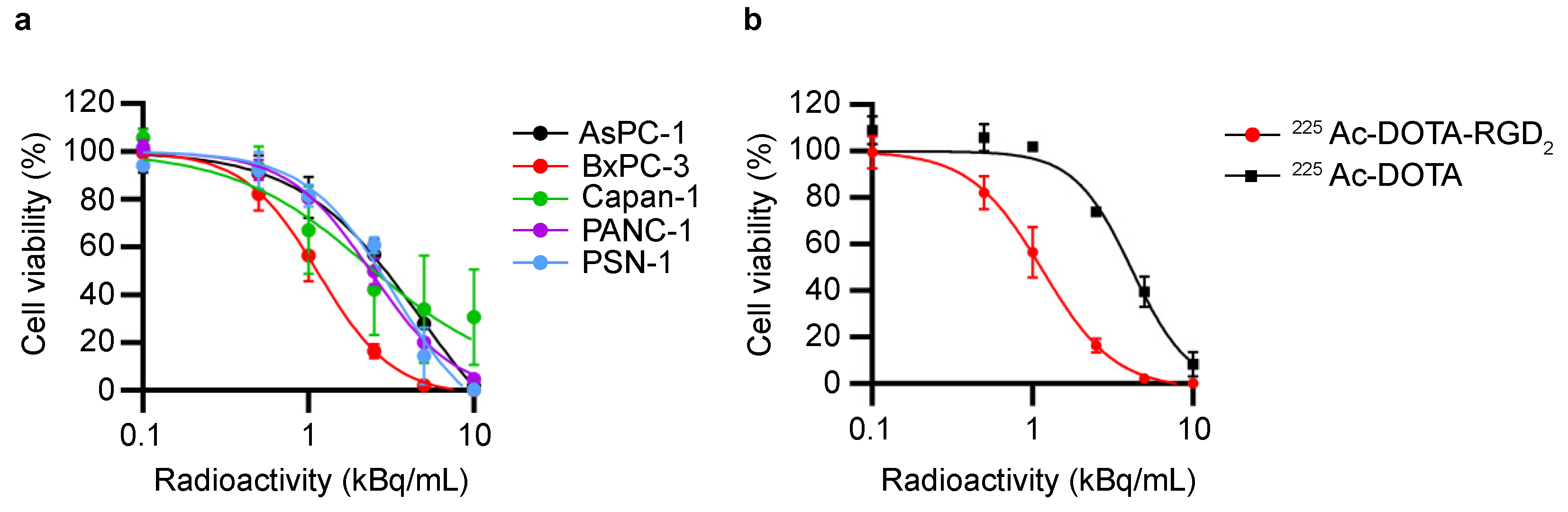
3.1. In Vitro Cytotoxicity Assessment by 225Ac-DOTA-RGD2 Binding

We evaluated the cytotoxicity of 225Ac-DOTA-RGD2 and the expression of αvβ3, αvβ5, and αvβ6 integrins in PDAC cell lines (Figure 1 and Figure S1). After 24 h of treatment, 225Ac-DOTA-RGD2 reduced the tumor cell viability in a dose-dependent manner (Figure 1a). The IC50 values of 225Ac-DOTA-RGD2 were 1.16 ± 0.09 kBq/mL for BxPC-3, 2.29 ± 0.69 kBq/mL for Capan-1, 2.41 ± 0.10 kBq/mL for PANC-1, 3.29 ± 0.63 kBq/mL for PSN-1, and 4.72 ± 1.73 kBq/mL for AsPC-1. Meanwhile, the IC50 value of 225Ac-DOTA for BxPC-3 was 4.17 ± 0.53 kBq/mL (Figure 1b). Flow cytometry (FCM) analysis revealed that these PDAC cells express αvβ3, αvβ5, and αvβ6 integrins (Figure S1). αvβ3 integrin was abundantly expressed in PANC-1, and αvβ6 integrin was expressed in BxPC-3 and Capan-1. αvβ5 integrin was expressed among all PDAC cell lines at the same level.

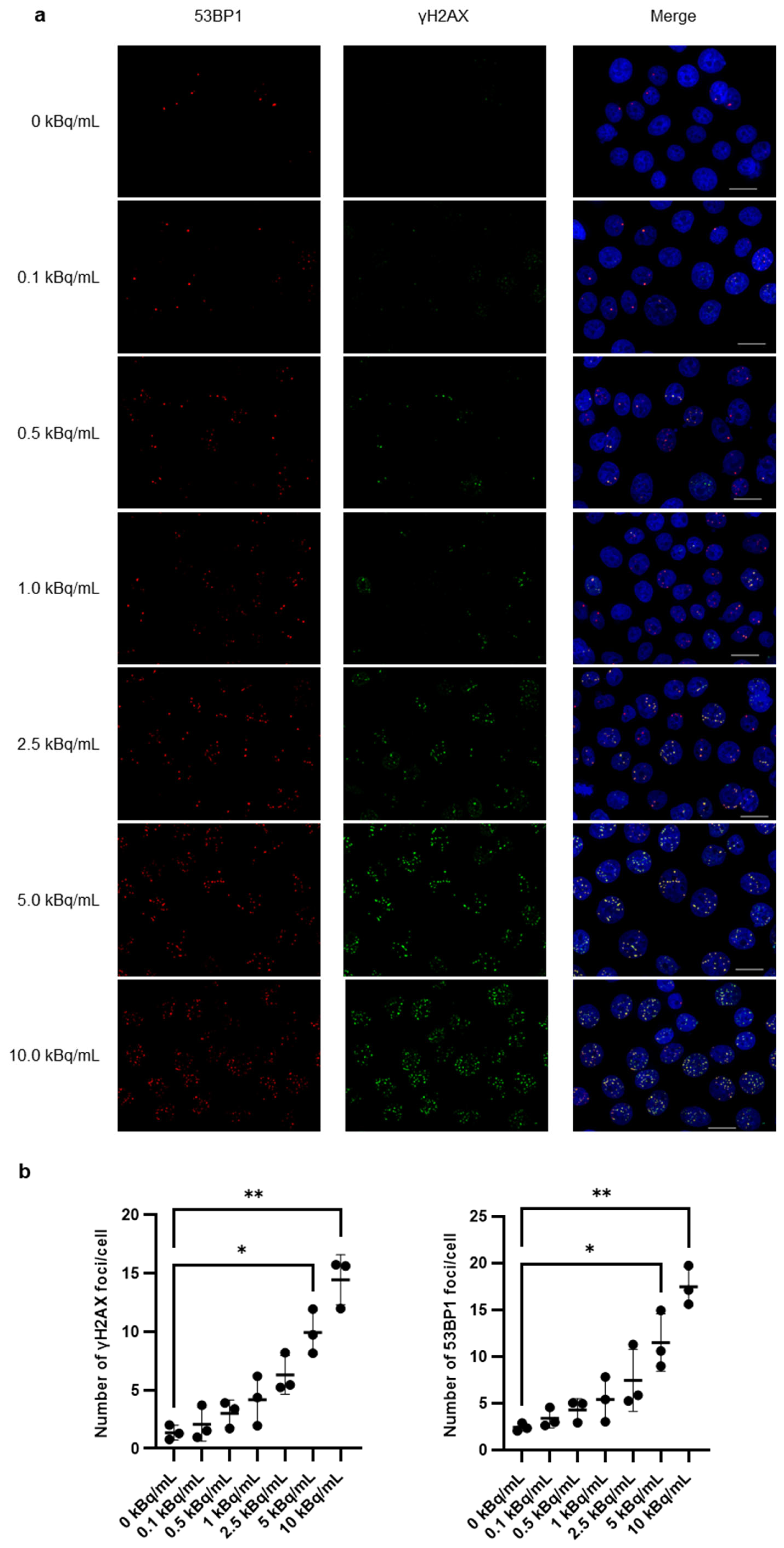
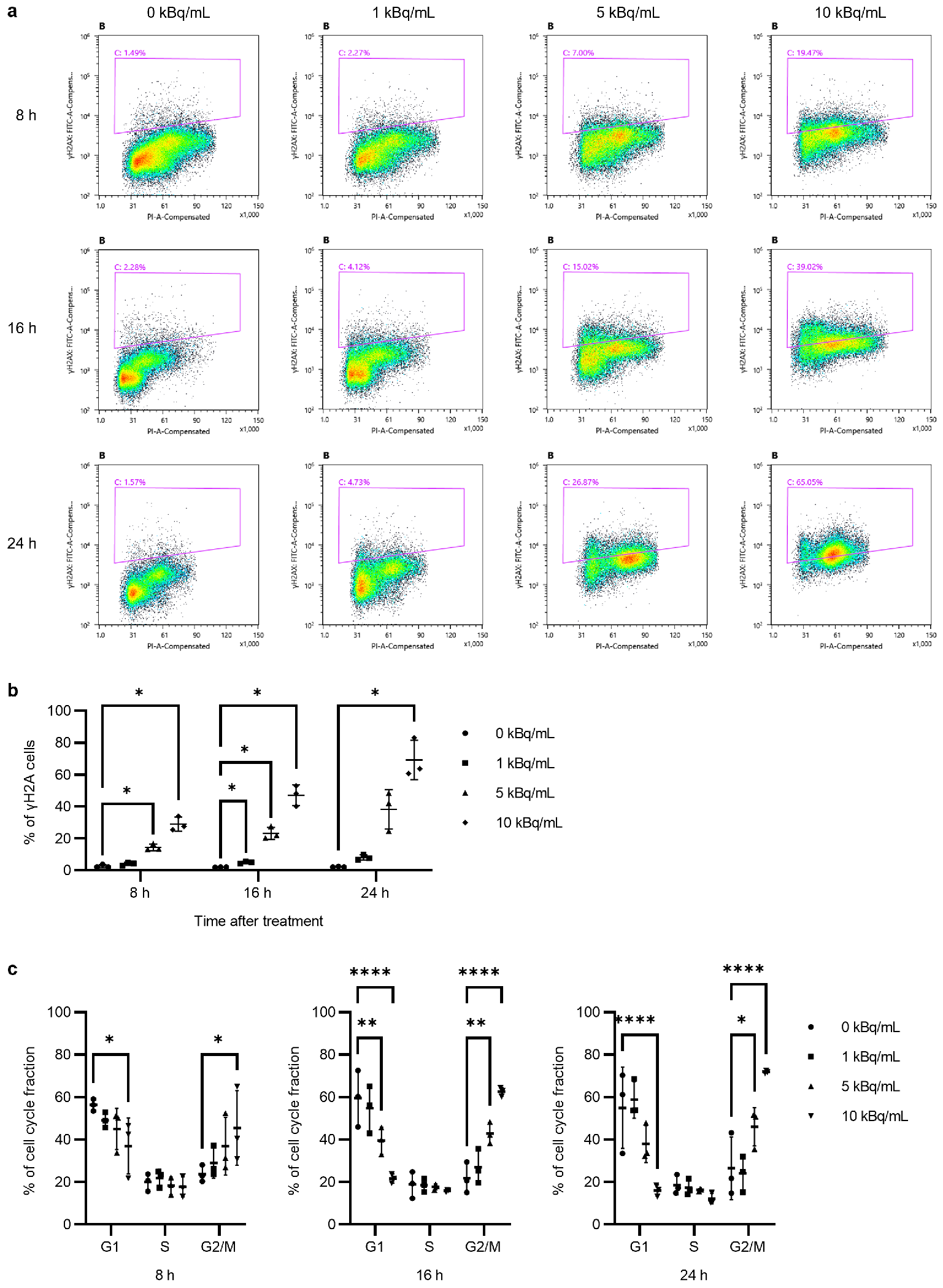
3.2. γH2AX/53BP1 Expression and G2/M Arrest After 225Ac-DOTA-RGD2

Fluorescence immunohistochemistry and FCM analysis were performed to detect the induction of DSBs and changes in the cell cycle. The number of γH2AX and 53BP1 foci significantly increased with an increase in the treatment dose of 225Ac-DOTA-RGD2, and both numbers were similar (Figure 2a,b). The size of the foci increased with the increase in the treatment dose. The 53BP1 foci were well merged with the γH2AX foci (Figure 2a).
Furthermore, FCM analysis revealed that, based on the duration and dose of 225Ac-DOTA-RGD2 treatment, γH2AX was upregulated (Figure 3). After 8 h of incubation with 5 and 10 kBq/mL of 225Ac-DOTA-RGD2, γH2AX expression in BxPC-3 cells was markedly induced (14.4% and 29.0%, respectively; Figure 3a,b); after 24 h of incubation, it reached 2.16% in the control group (0 kBq/mL), 8.03% in the 1 kBq/mL group, 38.39% in the 5 kBq/mL group, and 69.23% in the 10 kBq/mL group. In addition, significantly increased G2/M fractions were observed in the cells treated with 5 and 10 kBq/mL of 225Ac-DOTA-RGD2 (Figure 3a,c). At an early time point (8 h after incubation), the G2/M fraction in the 10 kBq/mL group increased to 45.4% (vs. 23.72% in controls).

3.3. DOTA-RGD2 Pharmacokinetics and Intratumoral Distribution

We assessed the pharmacokinetics of 225Ac-DOTA-RGD2 in BxPC-3 cell-bearing mice (Figure 4a). 225Ac-DOTA-RGD2 was highly taken up in BxPC-3 (4.80 ± 0.38%ID/g at 1 h) and was gradually washed out. Tumor uptake of 225Ac-DOTA-RGD2 was still retained up to 1.9 ± 0.63%ID/g at 168 h after injection. In contrast, 225Ac-DOTA-RGD2 was rapidly removed from the blood (<0.02% injected dose [ID]/g at 4 h). The highest uptake was found in the kidneys (12.87 ± 2.75% ID/g at 15 min), and the radioactivity was rapidly and preferentially excreted in urine (46.47% ID at 24 h and 65.29% ID at 168 h). Radioactivity in the feces was approximately 10%. This pharmacokinetic profile was similar to that of 111In-DOTA-RGD2, which exhibited rapid clearance from the blood and high accumulation in the kidneys (Figure S2a,b). The SPECT images clearly showed rapid excretion into the bladder. Moreover, we investigated the effect of the specific activity on the biodistribution of 111In-DOTA-RGD2 (Figure S2c). Co-injection of DOTA-RGD2 dose-dependently inhibited the uptake of 111In-DOTA-RGD2 in BxPC-3 cells and the kidneys. While the administration of 10 μg of DOTA-RGD2 significantly inhibited the uptake by more than 50%, the reduction observed with 1 μg of DOTA-RGD2 was more moderate, with decreases of 33.4% and 31.2% observed in BxPC-3 cells and the kidneys, respectively. Alpha camera imaging and hematoxylin and eosin staining revealed that 225Ac-DOTA-RGD2 accumulated in tumor areas with higher cellularity (Figure 4b).

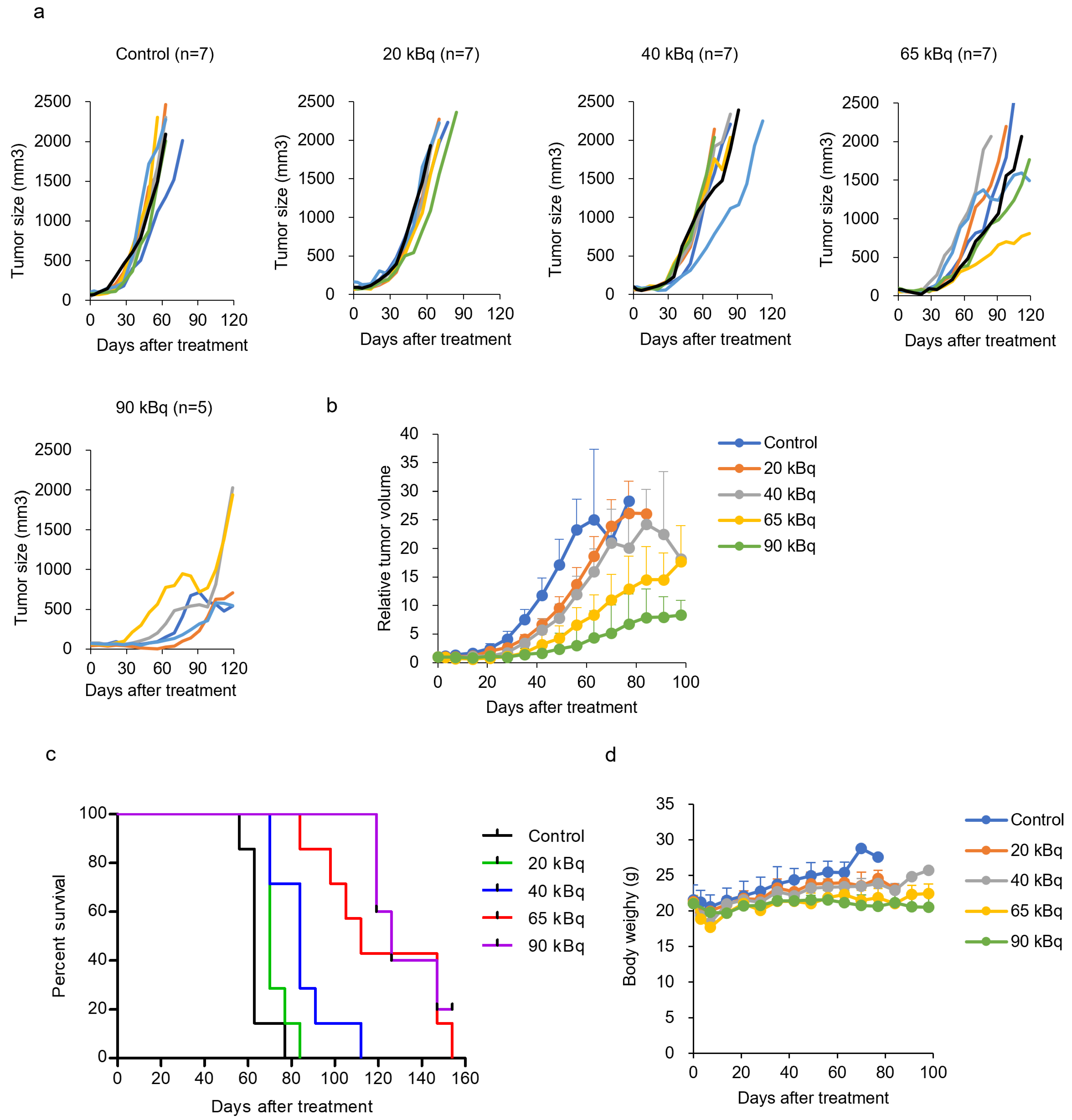
3.4. Treatment with 225Ac-DOTA-RGD2 in Mice Bearing BxPC-3

The therapeutic potential and adverse effects of 225Ac-DOTA-RGD2 were examined in mice bearing BxPC-3 cells. 225Ac-DOTA-RGD2 inhibited tumor growth in a dose-dependent manner (Figure 5a–c). No significant acute toxicity was observed for the administration of up to 90 kBq, as evaluated by monitoring bodyweight loss > 20%; however, transient body weight loss was observed within 2 weeks (Figure 5d). Low-dose administration (20 and 40 kBq) resulted in a limited inhibition of tumor growth. The inhibitory effects of the two compounds were similar. Doses of 65 and 90 kBq of 225Ac-DOTA-RGD2 preferentially suppressed tumor growth. Kaplan–Meier analysis revealed that 225Ac-DOTA-RGD2 significantly increased the median survival time compared with that in non-treated animals (control: 63 days, 20 kBq: 70 days, p < 0.05; 40 kBq: 84 days, p < 0.001; 65 kBq: 112 days, p < 0.001; 90 kBq: and 126 days, p < 0.001; Figure 5c). Hematological analysis revealed that the white blood cell counts transiently decreased and later recovered readily (Figure S3a). Similarly, platelet count decreased after the administration of 225Ac-DOTA-RGD2. In particular, 65 and 90 kBq of 225Ac-DOTA-RGD2 induced a long-lasting decrease in platelet count. Furthermore, we measured neutrophil gelatinase-associated lipocalin (NGAL) levels to evaluate acute kidney injury at 3, 7, and 14 days after the administration of 225Ac-DOTA-RGD2. There was no significant increase in NGAL by 225Ac-DOTA-RGD2 (Figure S3b). Alanine aminotransferase (ALT), aspartate transaminase (AST), blood urea nitrogen (BUN), and creatinine levels were measured at the endpoint. 225Ac-DOTA-RGD2 did not increase the ALT, AST, BUN, or creatinine levels (Figure S3c).

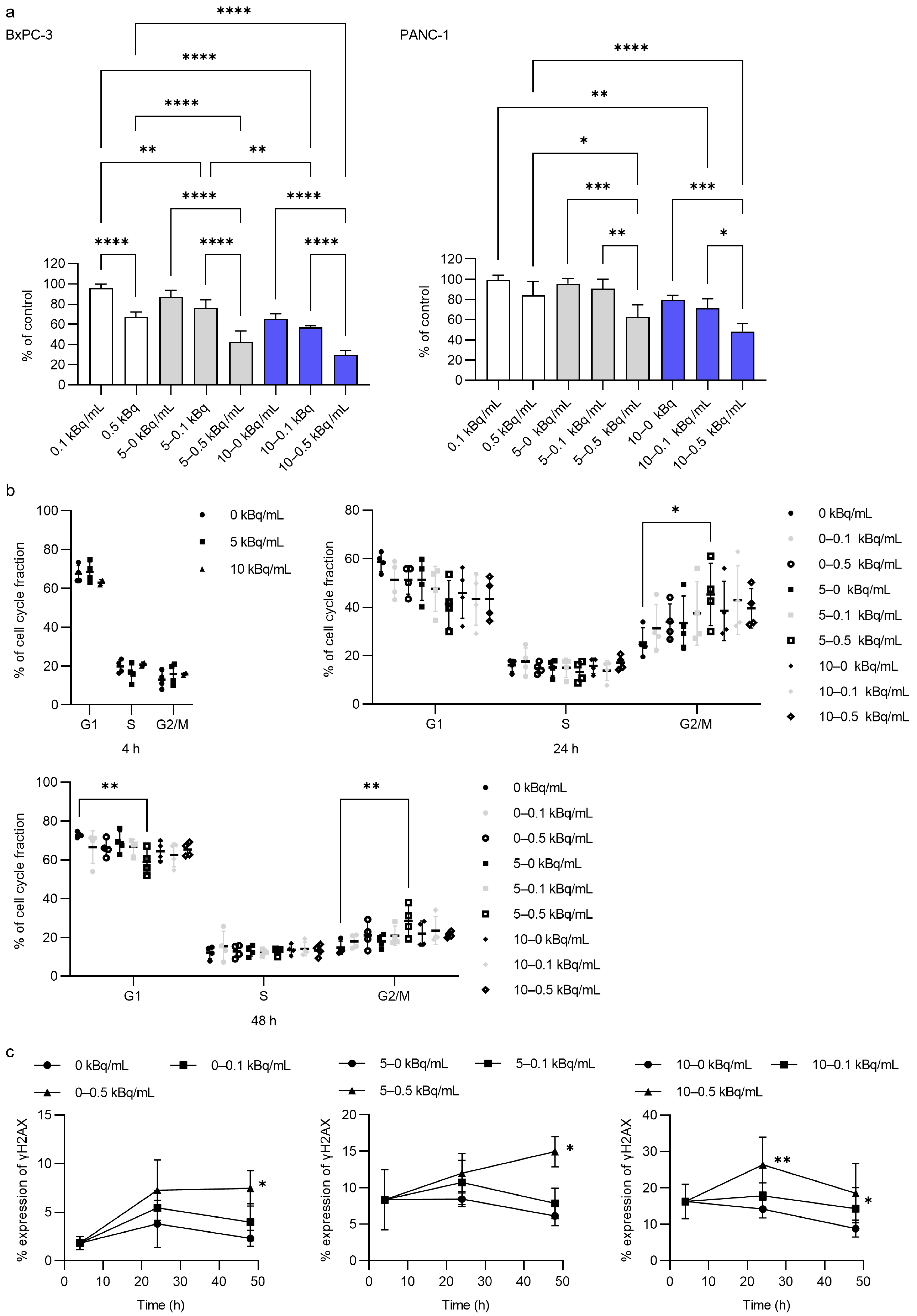
3.5. In Vitro Antitumor Activity of 225Ac-DOTA-RGD2 Combination

We investigated the effects of low-dose radiation treatment following transient high-dose radiation treatment on cell viability (Figure 6). Six days of incubation with 0.1 kBq/mL of 225Ac-DOTA-RGD2 did not affect cell viability (Figure 6a). The effect of 0.5 kBq/mL of 225Ac-DOTA-RGD2 was moderate. However, only the 4 h incubation with 5 or 10 kBq/mL of 225Ac-DOTA-RGD2 drastically enhanced the antitumor activity of 0.5 kBq/mL of 225Ac-DOTA-RGD2. In BxPC-3 cells, the cell viability following the combination treatment was 42.7 ± 10.8% for 5–0.5 kBq/mL and 29.5% ± 4.8% for 10–0.5 kBq/mL, whereas that after the transient treatment of 5 and 10 kBq/mL was 86.8% ± 6.8% and 65.4% ± 5.0%, respectively. In PANC-1 cells, the cell viability after the combination treatment was 62.9% ± 11.8% for 5–0.5 kBq/mL and 48.1% ± 8.1% for 10–0.5 kBq/mL, while that following the transient treatment of 5 and 10 kBq/mL was 95.7% ± 5.1% and 79.5% ± 4.7%, respectively.
The 4 h incubation period with 5 or 10 kBq/mL of 225Ac-DOTA-RGD2 did not alter the cell cycle distribution, although following a subsequent 16 h incubation with 0.1 or 0.5 kBq/mL of 225Ac-DOTA-RGD2 (totaling 24 h), there was an increase in the G2/M fraction (Figure 6b). By the 48 h mark, the cell cycle distribution had returned to levels comparable to the control, characterized by a predominance of the G1 fraction.
γH2AX expression was notably induced by incubation with 0.5 kBq/mL of 225Ac-DOTA-RGD2. In the absence of transient incubation with 5 or 10 kBq/mL of 225Ac-DOTA-RGD2, only 0.5 kBq/mL of the compound increased γH2AX expression (7.47% ± 1.82% for 0–0.5 kBq vs. 2.30% ± 0.82% for 0 kBq and 3.98% ± 1.91% for 0–0.1 kBq at 48 h). The 4 h incubation with 5 or 10 kBq/mL of 225Ac-DOTA-RGD2 significantly boosted γH2AX expression induced by 0.5 kBq/mL of 225Ac-DOTA-RGD2 and 0.1 kBq/mL of 225Ac-DOTA-RGD2. Meanwhile, the γH2AX expression induced by 5 or 10 kBq/mL of 225Ac-DOTA-RGD2 decreased over time.

4. Discussion

A single administration of 225Ac radiopharmaceuticals confers significant antitumor efficacy in preclinical and clinical studies. The first clinical study using 225Ac-PSMA-617 reported a drastic positive response even after a single administration, and finally, a complete response was achieved after three administrations [12]. In a preclinical study, 225Ac-DOTATATE delayed tumor growth in a lung neuroendocrine tumor model [26]. However, the mechanisms underlying these potent antitumor effects remain obscure. In general, alpha nuclides have been focused on for high LET, whereas the long half-life of 225Ac makes it one of the most attractive candidates for tumor treatment. Here, we performed efficacy and toxicity studies and investigated the mechanism underlying the continuous antitumor effect of 225Ac radiopharmaceuticals in a pancreatic tumor model using 225Ac-DOTA-RGD2.
Our study indicated that 225Ac-DOTA-RGD2 would be a possible candidate for TAT for pancreatic cancer with αvβ3vβ6 integrins. In vitro cytotoxicity experiments indicated that 225Ac-DOTA-RGD2 was more potent than 225Ac-DOTA in BxPC-3 cells. In addition, a significant strong correlation was observed between the in vitro cytotoxicity (IC50) and tumor uptake of 225Ac-DOTA-RGD2 (Figure S4; r = −0.906, p < 0.05). Therefore, the cytotoxicity of 225Ac was due to the binding of 225Ac-DOTA-RGD2 to tumor cells. However, 24 h of incubation with 10 kBq/mL of 225Ac-DOTA, which does not bind to αvβ3vβ6 integrins, was also lethal to the cells. This implies that radiation dosimetry to non-target tissues should be well managed in TAT, although the range of alpha rays is <100 μm.
This preclinical study revealed that 225Ac-DOTA-RGD2 (20–90 kBq), with a tumor accumulation of 2.0% ID/g after 48 h, significantly suppressed tumor growth. Our previous study using 90Y-DOTA-c(RGDfK) indicated that even with three injections of 11.1 MBq, tumor growth inhibition was limited [21]. A single high-dose injection of 90Y-DOTA-RGD2 (37 MBq) delayed tumor growth (median survival of 54 days vs. 19 days in the untreated group) in the OVCAR-3 xenograft model, although regrowth was observed 2 weeks after treatment. The prolongation of median survival time would be caused by the high LET and long half-time (9.9 days vs. 64 h of 90Y) of 225Ac. 225Ac-labeled antibodies have been reported to be more effective in tumor treatment than antibodies labeled with a beta radionuclide (90Y) or a short-half-life alpha nuclide (213Bi) [27,28]. Thus, our therapeutic experiments using 225Ac-DOTA-RGD2 suggest that 225Ac-labeled peptides could confer drastic therapeutic efficacy, although specific tumor-targeting ability is required.
Regarding side effects, kidney injury is debatable. Mice treated with 111 kBq of 225Ac-DOTATATE showed chronic nephropathy, although BUN and creatinine levels did not increase [26]. This side effect is characterized by pharmacokinetics unique to peptides that show preferential renal excretion [29]. However, in our study, 225Ac-DOTA-RGD2 was significantly taken up by the kidneys, although renal dysfunction was not detected by biochemical analysis. Additionally, TAT using 225Ac-DOTATATE has been attempted in patients with neuroendocrine tumors [30]. Yadav et al. reported that 225Ac-DOTATATE treatment is effective without severe hematological, renal, and hepatological toxicities and is beneficial to patients refractory to 177Lu treatment [31]. Dose planning based on dosimetry from preclinical studies is required to avoid radiation nephrotoxicity.
Considering the potent antitumor activity of 225Ac-DOTA-RGD2, we should focus on the fact that the cells were efficiently blocked at G2/M by 225Ac irradiation. FCM analysis revealed that treatment with >5 kBq/mL 225Ac-DOTA-RGD2 readily led cells to G2/M arrest, and γH2AX was sequentially found in the cells. Irradiation with high-LET carbon ions (12C6+) at 2 Gy is more lethal and induces G2/M arrest and γH2AX expression more efficiently compared with X-ray irradiation at the same dose [32]. It is well known that cells in the G2/M phase are sensitive to radiation [33]. Some radiosensitizing agents arrest cells in the G2/M phase [34,35,36]. Thus, targeting the cell cycle is a strategy to enhance radiosensitivity [37]. This may allow a lower radiation dose to severely damage the cells. Rapid clearance of peptides or peptide mimetics from normal organs enables the administration of high radioactivity to enhance radiosensitivity.
From our results and reports on 225Ac-PRRT thus far, we speculate that, after transient intense irradiation, continuous low-dose irradiation would contribute to successful therapeutic efficacy. Based on our in vitro results showing that G2/M arrest was induced by 225Ac-DOTA-RGD2, we examined the effect of combination treatment with 225Ac-DOTA-RGD2 on BxPC-3 and PANC-1 cells. Pretreatment with 5 or 10 kBq/mL of 225Ac-DOTA-RGD2 for only 4 h significantly enhanced the cytotoxicity of low-dose 225Ac-DOTA-RGD2 (0.1 or 0.5 kBq/mL). This suggests that an initial higher dose triggers sufficient cytotoxicity, even at subsequent lower doses. These results would explain the continuous suppression of tumor growth by 225Ac-DOTA-RGD2. Therefore, the pharmacokinetics of the peptides are suitable for TAT using radionuclides with long half-lives.
In addition to G2/M arrest, alterations in the DNA repair response could be another factor that induces radiosensitization. Generally, DNA damage is repaired by homologous recombination (HR) and non-homologous end joining (NHEJ). Severe DNA damage, such as clustered DSBs in heterochromatin, is repaired by HR following NHEJ at an early stage [38]. HR functions in the late S/G2 phase using a sister chromatid as a template to repair DSBs precisely [39]. Foci of the DNA damage response mediator 53BP1 were well merged with γH2AX foci in BxPC-3 and PANC-1 cells during 24 h treatment with 225Ac-DOTA-RGD2. 53BP1 is a key factor in regulating the balance between NHEJ and HR. 53BP1 suppresses HR and facilitates NHEJ, a fast repair process that occurs within a few hours of DNA damage [40]. The inhibition of HR by the Chk1/2 inhibitor AZD7762 induced radiosensitization and a prolonged tumor-volume doubling time in MiaPaCa-2 and patient-derived pancreatic tumor xenograft models [41]. Sustainable irradiation with 225Ac-DOTA-RGD2 maintains 53BP1 expression, resulting in the inhibition of HR.
In TAT, it is controversial whether antibodies or peptides are preferable. Antibodies that exhibit slow blood clearance and sustained tumor accumulation are potential candidates as 225Ac radiopharmaceuticals. Theoretically, antibodies labeled with long-half-life radionuclides, such as 225Ac and 177Lu, can continuously irradiate tumors. With the trend of antibody–drug development, labeled antibodies have been vigorously studied [42]. Unfortunately, only 90Y-labeled anti-CD20 antibody (Zevalin®) is currently approved as an antitumor drug. Slow blood clearance and non-specific intense uptake in the liver may result in hematotoxicity and liver toxicity, respectively [43,44,45]. Therefore, a lower dose is required to reduce side effects. In the case of peptides, the radioactivity per administration should be increased because they exhibit relatively rapid clearance. In addition, multiple doses are available (or mandatory) for adequate therapeutic effects while controlling side effects. 225Ac-PSMA-617 and 117Lu-DOTATATE (Lutathera®) are administrated multiple times [30,46].
Additionally, peptides are superior to antibodies in terms of intratumoral permeability. We previously reported that 111In-DOTA-c(RGDfK) was homogeneously distributed inside pancreatic cancer cells in a carcinogenesis model that was histologically similar to human pancreatic cancer [19]. Furthermore, alpha camera imaging confirmed the uniform distribution of 225Ac-DOTA-RGD2 in the viable region of BxPC-3 cells. However, radiolabeled antibodies accumulate in the rim, which corresponds to viable tumor tissue and areas with high blood vessel density and large blood vessels [47,48]. These features of radiolabeled peptides suggest that they are appropriate for treating PDAC.
This study has a limitation in that it has not demonstrated that combination treatment with high-dose administration followed by a low dose is effective. We need to investigate the dose and dosage interval and demonstrate the therapeutic effects and side effects of the dosing design. Unfortunately, however, the supply of 225Ac is extremely limited, and it is extremely difficult to obtain 225Ac for basic research. In fact, we currently cannot obtain it from overseas. The production of 225Ac is currently progressing in Japan, and we will examine effective dosing designs for clinical use in the future.
In conclusion, high-intensity irradiation with 225Ac-DOTA-RGD2 induced G2/M arrest, thereby inducing radiosensitivity. Our findings suggest that 225Ac-DOTA-RGD2 could confer a radiation dose sufficient to treat PDAC without undesirable radiation exposure to normal tissues. The high LET and long half-life of 225Ac make it suitable for the development of radionuclide therapies.

Supplementary Materials

The following supporting information can be downloaded at https://www.mdpi.com/article/10.3390/pharmaceutics17010009/s1. Figure S1: Expression of αvβ3, αvβ5, and αvβ6 in PDCA cell lines. Figure S2: Pharmacokinetics of 111In-DOTA-RGD2 in BxPC-3 xenograft model. Figure S3: Toxicity evaluation after treatment with 225Ac-DOTA-RGD2. Figure S4: Correlations between the in vitro cytotoxicity (IC50) and tumor uptake of 225Ac-DOTA-RGD2 (%ID/g).

Author Contributions

Conceptualization, M.Y.; investigation, M.Y., K.W., K.O., A.D., M.I., M.K., B.W.M. and Y.Y.; data curation, M.Y.; writing—original draft preparation, M.Y. and B.W.M.; writing—review and editing, M.Y., B.W.M., A.I. and H.F.; supervision, M.Y.; project administration, M.Y. and H.F.; funding acquisition, M.Y., K.W., Y.Y. and H.F. All authors have read and agreed to the published version of the manuscript.

Funding

This study was supported by grants from Grant-in-Aid for Scientific Research (C) (grant number KAKENHI 18K07698) and Project for Promotion of Cancer Research and Therapeutic Evolution (P-PROMOTE) from the Agency for Medical Research and Development (AMED) (grant number JP20cm0106279).

Institutional Review Board Statement

The study protocol was approved by the Committee for Ethics of Animal Experimentation of the National Cancer Center (K18-012). Animal experiments were performed in accordance with the Guidelines for the Care and Use of Experimental Animals established by the committee.

Informed Consent Statement

Not applicable.

Data Availability Statement

The data in this manuscript are available from the corresponding author upon reasonable request.

Conflicts of Interest

The authors declare no conflicts of interest.

References

  1. Siegel, R.L.; Miller, K.D.; Wagle, N.S.; Jemal, A. Cancer statistics, 2023. CA Cancer J. Clin. 2023, 73, 17–48. [Google Scholar] [CrossRef] [PubMed]
  2. Sung, H.; Ferlay, J.; Siegel, R.L.; Laversanne, M.; Soerjomataram, I.; Jemal, A.; Bray, F. Global cancer statistics 2020: GLOBOCAN estimates of incidence and mortality worldwide for 36 cancers in 185 countries. CA Cancer J. Clin. 2021, 71, 209–249. [Google Scholar] [CrossRef] [PubMed]
  3. Zhang, X.P.; Xu, S.; Gao, Y.X.; Zhao, Z.M.; Zhao, G.D.; Hu, M.G.; Tan, X.L.; Lau, W.Y.; Liu, R. Early and late recurrence patterns of pancreatic ductal adenocarcinoma after pancreaticoduodenectomy: A multicenter study. Int. J. Surg. 2023, 109, 785–793. [Google Scholar] [CrossRef] [PubMed]
  4. Li, X.; Wan, Y.; Lou, J.; Xu, L.; Shi, A.; Yang, L.; Fan, Y.; Yang, J.; Huang, J.; Wu, Y.; et al. Preoperative recurrence prediction in pancreatic ductal adenocarcinoma after radical resection using radiomics of diagnostic computed tomography. EClinicalMedicine 2022, 43, 101215. [Google Scholar] [CrossRef]
  5. Gillen, S.; Schuster, T.; Meyer Zum Büschenfelde, C.; Friess, H.; Kleeff, J. Preoperative/neoadjuvant therapy in pancreatic cancer: A systematic review and meta-analysis of response and resection percentages. PLoS Med. 2010, 7, e1000267. [Google Scholar] [CrossRef]
  6. Minchinton, A.I.; Tannock, I.F. Drug penetration in solid tumours. Nat. Rev. Cancer 2006, 6, 583–592. [Google Scholar] [CrossRef]
  7. Provenzano, P.P.; Hingorani, S.R. Hyaluronan, fluid pressure, and stromal resistance in pancreas cancer. Br. J. Cancer 2013, 108, 1–8. [Google Scholar] [CrossRef]
  8. Olive, K.P.; Jacobetz, M.A.; Davidson, C.J.; Gopinathan, A.; McIntyre, D.; Honess, D.; Madhu, B.; Goldgraben, M.A.; Caldwell, M.E.; Allard, D.; et al. Inhibition of Hedgehog signaling enhances delivery of chemotherapy in a mouse model of pancreatic cancer. Science 2009, 324, 1457–1461. [Google Scholar] [CrossRef]
  9. Whatcott, C.J.; Diep, C.H.; Jiang, P.; Watanabe, A.; LoBello, J.; Sima, C.; Hostetter, G.; Shepard, H.M.; Von Hoff, D.D.; Han, H. Desmoplasia in primary tumors and metastatic lesions of pancreatic cancer. Clin. Cancer Res. 2015, 21, 3561–3568. [Google Scholar] [CrossRef]
  10. Parker, C.; Lewington, V.; Shore, N.; Kratochwil, C.; Levy, M.; Linden, O.; Noordzij, W.; Park, J.; Saad, F. Targeted alpha therapy, an emerging class of cancer agents: A review. JAMA Oncol. 2018, 4, 1765–1772. [Google Scholar] [CrossRef]
  11. Sgouros, G.; Bodei, L.; McDevitt, M.R.; Nedrow, J.R. Radiopharmaceutical therapy in cancer: Clinical advances and challenges. Nat. Rev. Drug Discov. 2020, 19, 589–608. [Google Scholar] [CrossRef] [PubMed]
  12. Kratochwil, C.; Bruchertseifer, F.; Giesel, F.L.; Weis, M.; Verburg, F.A.; Mottaghy, F.; Kopka, K.; Apostolidis, C.; Haberkorn, U.; Morgenstern, A. 225Ac-PSMA-617 for PSMA-targeted α-radiation therapy of metastatic castration-resistant prostate cancer. J. Nucl. Med. 2016, 57, 1941–1944. [Google Scholar] [CrossRef] [PubMed]
  13. Hosotani, R.; Kawaguchi, M.; Masui, T.; Koshiba, T.; Ida, J.; Fujimoto, K.; Wada, M.; Doi, R.; Imamura, M. Expression of integrin αVβ3 in pancreatic carcinoma: Relation to MMP-2 activation and lymph node metastasis. Pancreas 2002, 25, e30–e35. [Google Scholar] [CrossRef] [PubMed]
  14. Grzesiak, J.J.; Ho, J.C.; Moossa, A.R.; Bouvet, M. The integrin-extracellular matrix axis in pancreatic cancer. Pancreas 2007, 35, 293–301. [Google Scholar] [CrossRef]
  15. Beer, A.J.; Haubner, R.; Goebel, M.; Luderschmidt, S.; Spilker, M.E.; Wester, H.J.; Weber, W.A.; Schwaiger, M. Biodistribution and pharmacokinetics of the αvβ3-selective tracer 18F-galacto-RGD in cancer patients. J. Nucl. Med. 2005, 46, 1333–1341. [Google Scholar]
  16. Haubner, R.; Wester, H.J.; Weber, W.A.; Mang, C.; Ziegler, S.I.; Goodman, S.L.; Senekowitsch-Schmidtke, R.; Kessler, H.; Schwaiger, M. Noninvasive imaging of αvβ3 integrin expression using 18F-labeled RGD-containing glycopeptide and positron emission tomography. Cancer Res. 2001, 61, 1781–1785. [Google Scholar]
  17. Schnell, O.; Krebs, B.; Carlsen, J.; Miederer, I.; Goetz, C.; Goldbrunner, R.H.; Wester, H.J.; Haubner, R.; Popperl, G.; Holtmannspotter, M.; et al. Imaging of integrin αvβ3 expression in patients with malignant glioma by [18F] Galacto-RGD positron emission tomography. Neuro Oncol. 2009, 11, 861–870. [Google Scholar] [CrossRef]
  18. Hausner, S.H.; DiCara, D.; Marik, J.; Marshall, J.F.; Sutcliffe, J.L. Use of a peptide derived from foot-and-mouth disease virus for the noninvasive imaging of human cancer: Generation and evaluation of 4-[18F]fluorobenzoyl A20FMDV2 for in vivo imaging of integrin αvβ6 expression with positron emission tomography. Cancer Res. 2007, 67, 7833–7840. [Google Scholar] [CrossRef]
  19. Yoshimoto, M.; Hayakawa, T.; Mutoh, M.; Imai, T.; Tsuda, K.; Kimura, S.; Umeda, I.O.; Fujii, H.; Wakabayashi, K. In vivo SPECT imaging with 111In-DOTA-c(RGDfK) to detect early pancreatic cancer in a hamster pancreatic carcinogenesis model. J. Nucl. Med. 2012, 53, 765–771. [Google Scholar] [CrossRef]
  20. Kapp, T.G.; Rechenmacher, F.; Neubauer, S.; Maltsev, O.V.; Cavalcanti-Adam, E.A.; Zarka, R.; Reuning, U.; Notni, J.; Wester, H.J.; Mas-Moruno, C.; et al. A comprehensive evaluation of the activity and selectivity profile of ligands for RGD-binding integrins. Sci. Rep. 2017, 7, 39805. [Google Scholar] [CrossRef]
  21. Yoshimoto, M.; Ogawa, K.; Washiyama, K.; Shikano, N.; Mori, H.; Amano, R.; Kawai, K. αvβ3 integrin-targeting radionuclide therapy and imaging with monomeric RGD peptide. Int. J. Cancer 2008, 123, 709–715. [Google Scholar] [CrossRef] [PubMed]
  22. Huang, X.; Darzynkiewicz, Z. Cytometric assessment of histone H2AX phosphorylation: A reporter of DNA damage. Methods Mol. Biol. 2006, 314, 73–80. [Google Scholar] [CrossRef]
  23. Muslimovic, A.; Ismail, I.H.; Gao, Y.; Hammarsten, O. An optimized method for measurement of gamma-H2AX in blood mononuclear and cultured cells. Nat. Protoc. 2008, 3, 1187–1193. [Google Scholar] [CrossRef] [PubMed]
  24. Hayakawa, T.; Mutoh, M.; Imai, T.; Tsuta, K.; Yanaka, A.; Fujii, H.; Yoshimoto, M. SPECT/CT of lung nodules using 111In-DOTA-c(RGDfK) in a mouse lung carcinogenesis model. Ann. Nucl. Med. 2013, 27, 640–647. [Google Scholar] [CrossRef] [PubMed]
  25. Miller, B.W.; Frost, S.H.; Frayo, S.L.; Kenoyer, A.L.; Santos, E.; Jones, J.C.; Green, D.J.; Hamlin, D.K.; Wilbur, D.S.; Fisher, D.R.; et al. Quantitative single-particle digital autoradiography with α-particle emitters for targeted radionuclide therapy using the iQID camera. Med. Phys. 2015, 42, 4094–4105. [Google Scholar] [CrossRef] [PubMed]
  26. Tafreshi, N.K.; Pandya, D.N.; Tichacek, C.J.; Budzevich, M.M.; Wang, Z.; Reff, J.N.; Engelman, R.W.; Boulware, D.C.; Chiappori, A.A.; Strosberg, J.R.; et al. Preclinical evaluation of [225Ac]Ac-DOTA-TATE for treatment of lung neuroendocrine neoplasms. Eur. J. Nucl. Med. Mol. Imaging 2021, 48, 3408–3421. [Google Scholar] [CrossRef]
  27. Song, H.; Hobbs, R.F.; Vajravelu, R.; Huso, D.L.; Esaias, C.; Apostolidis, C.; Morgenstern, A.; Sgouros, G. Radioimmunotherapy of breast cancer metastases with alpha-particle emitter 225Ac: Comparing efficacy with 213Bi and 90Y. Cancer Res. 2009, 69, 8941–8948. [Google Scholar] [CrossRef]
  28. Veach, D.R.; Storey, C.M.; Luckerath, K.; Braun, K.; von Bodman, C.; Lamminmaki, U.; Kalidindi, T.; Strand, S.E.; Strand, J.; Altai, M.; et al. PSA-targeted alpha-, beta-, and positron-emitting immunotheranostics in murine prostate cancer models and nonhuman primates. Clin. Cancer Res. 2021, 27, 2050–2060. [Google Scholar] [CrossRef]
  29. Vegt, E.; de Jong, M.; Wetzels, J.F.; Masereeuw, R.; Melis, M.; Oyen, W.J.; Gotthardt, M.; Boerman, O.C. Renal toxicity of radiolabeled peptides and antibody fragments: Mechanisms, impact on radionuclide therapy, and strategies for prevention. J. Nucl. Med. 2010, 51, 1049–1058. [Google Scholar] [CrossRef]
  30. Ballal, S.; Yadav, M.P.; Bal, C.; Sahoo, R.K.; Tripathi, M. Broadening horizons with 225Ac-DOTATATE targeted alpha therapy for gastroenteropancreatic neuroendocrine tumour patients stable or refractory to 177Lu-DOTATATE PRRT: First clinical experience on the efficacy and safety. Eur. J. Nucl. Med. Mol. Imaging 2020, 47, 934–946. [Google Scholar] [CrossRef]
  31. Yadav, M.P.; Ballal, S.; Sahoo, R.K.; Bal, C. Efficacy and safety of 225Ac-DOTATATE targeted alpha therapy in metastatic paragangliomas: A pilot study. Eur. J. Nucl. Med. Mol. Imaging 2022, 49, 1595–1606. [Google Scholar] [CrossRef] [PubMed]
  32. Zhang, Q.; Kong, Y.; Yang, Z.; Liu, Y.; Liu, R.; Geng, Y.; Luo, H.; Zhang, H.; Li, H.; Feng, S.; et al. Preliminary study on radiosensitivity to carbon ions in human breast cancer. J. Radiat. Res. 2020, 61, 399–409. [Google Scholar] [CrossRef] [PubMed]
  33. Pawlik, T.M.; Keyomarsi, K. Role of cell cycle in mediating sensitivity to radiotherapy. Int. J. Radiat. Oncol. Biol. Phys. 2004, 59, 928–942. [Google Scholar] [CrossRef] [PubMed]
  34. Dukaew, N.; Konishi, T.; Chairatvit, K.; Autsavapromporn, N.; Soonthornchareonnon, N.; Wongnoppavich, A. Enhancement of radiosensitivity by eurycomalactone in human NSCLC cells through G2/M cell cycle arrest and delayed DNA double-strand break repair. Oncol. Res. 2020, 28, 161–175. [Google Scholar] [CrossRef] [PubMed]
  35. Liu, X.; Sun, C.; Jin, X.; Li, P.; Ye, F.; Zhao, T.; Gong, L.; Li, Q. Genistein enhances the radiosensitivity of breast cancer cells via G2/M cell cycle arrest and apoptosis. Molecules 2013, 18, 13200–13217. [Google Scholar] [CrossRef]
  36. Schwarz, K.; Dobiasch, S.; Nguyen, L.; Schilling, D.; Combs, S.E. Modification of radiosensitivity by curcumin in human pancreatic cancer cell lines. Sci. Rep. 2020, 10, 3815. [Google Scholar] [CrossRef]
  37. Buckley, A.M.; Lynam-Lennon, N.; O’Neill, H.; O’Sullivan, J. Targeting hallmarks of cancer to enhance radiosensitivity in gastrointestinal cancers. Nat. Rev. Gastroenterol. Hepatol. 2020, 17, 298–313. [Google Scholar] [CrossRef]
  38. Shibata, A.; Conrad, S.; Birraux, J.; Geuting, V.; Barton, O.; Ismail, A.; Kakarougkas, A.; Meek, K.; Taucher-Scholz, G.; Lobrich, M.; et al. Factors determining DNA double-strand break repair pathway choice in G2 phase. EMBO J. 2011, 30, 1079–1092. [Google Scholar] [CrossRef]
  39. Moynahan, M.E.; Jasin, M. Mitotic homologous recombination maintains genomic stability and suppresses tumorigenesis. Nat. Rev. Mol. Cell Biol. 2010, 11, 196–207. [Google Scholar] [CrossRef]
  40. Bunting, S.F.; Callen, E.; Wong, N.; Chen, H.T.; Polato, F.; Gunn, A.; Bothmer, A.; Feldhahn, N.; Fernandez-Capetillo, O.; Cao, L.; et al. 53BP1 inhibits homologous recombination in BRCA1-deficient cells by blocking resection of DNA breaks. Cell 2010, 141, 243–254. [Google Scholar] [CrossRef]
  41. Morgan, M.A.; Parsels, L.A.; Zhao, L.; Parsels, J.D.; Davis, M.A.; Hassan, M.C.; Arumugarajah, S.; Hylander-Gans, L.; Morosini, D.; Simeone, D.M.; et al. Mechanism of radiosensitization by the Chk1/2 inhibitor AZD7762 involves abrogation of the G2 checkpoint and inhibition of homologous recombinational DNA repair. Cancer Res. 2010, 70, 4972–4981. [Google Scholar] [CrossRef] [PubMed]
  42. Parakh, S.; Lee, S.T.; Gan, H.K.; Scott, A.M. Radiolabeled antibodies for cancer imaging and therapy. Cancers 2022, 14, 1545. [Google Scholar] [CrossRef] [PubMed]
  43. Merkx, R.I.J.; Lobeek, D.; Konijnenberg, M.; Jimenez-Franco, L.D.; Kluge, A.; Oosterwijk, E.; Mulders, P.F.A.; Rijpkema, M. Phase I study to assess safety, biodistribution and radiation dosimetry for 89Zr-girentuximab in patients with renal cell carcinoma. Eur. J. Nucl. Med. Mol. Imaging 2021, 48, 3277–3285. [Google Scholar] [CrossRef]
  44. Tamura, K.; Kurihara, H.; Yonemori, K.; Tsuda, H.; Suzuki, J.; Kono, Y.; Honda, N.; Kodaira, M.; Yamamoto, H.; Yunokawa, M.; et al. 64Cu-DOTA-trastuzumab PET imaging in patients with HER2-positive breast cancer. J. Nucl. Med. 2013, 54, 1869–1875. [Google Scholar] [CrossRef]
  45. Ulaner, G.A.; Lyashchenko, S.K.; Riedl, C.; Ruan, S.; Zanzonico, P.B.; Lake, D.; Jhaveri, K.; Zeglis, B.; Lewis, J.S.; O’Donoghue, J.A. First-in-human human epidermal growth factor receptor 2-targeted imaging using 89Zr-pertuzumab PET/CT: Dosimetry and clinical application in patients with breast cancer. J. Nucl. Med. 2018, 59, 900–906. [Google Scholar] [CrossRef]
  46. Zacherl, M.J.; Gildehaus, F.J.; Mittlmeier, L.; Boning, G.; Gosewisch, A.; Wenter, V.; Unterrainer, M.; Schmidt-Hegemann, N.; Belka, C.; Kretschmer, A.; et al. First clinical results for PSMA-targeted alpha-therapy using 225Ac-PSMA-I&T in advanced-mCRPC patients. J. Nucl. Med. 2021, 62, 669–674. [Google Scholar] [CrossRef]
  47. Abbas, N.; Heyerdahl, H.; Bruland, O.S.; Borrebaek, J.; Nesland, J.; Dahle, J. Experimental α-particle radioimmunotherapy of breast cancer using 227Th-labeled p-benzyl-DOTA-trastuzumab. EJNMMI Res 2011, 1, 18. [Google Scholar] [CrossRef]
  48. Örbom, A.; Eriksson, S.E.; Elgstrom, E.; Ohlsson, T.; Nilsson, R.; Tennvall, J.; Strand, S.E. The intratumoral distribution of radiolabeled 177Lu-BR96 monoclonal antibodies changes in relation to tumor histology over time in a syngeneic rat colon carcinoma model. J. Nucl. Med. 2013, 54, 1404–1410. [Google Scholar] [CrossRef]
Figure 1. In vitro cytotoxicity. (a) Cytotoxicity of 225Ac-DOTA-RGD2 in human pancreatic tumor cell lines. (b) Comparison of cytotoxicity between 225Ac-DOTA-RGD2 and 225AcDOTA in BxPC-3. All assays were performed in triplicate. Data are presented as mean ± standard deviation.
Pharmaceutics 17 00009 g001
Figure 2. Induction of γH2AX and 53BP1 foci formation in response to increasing doses of 225Ac-DOTA-RGD2 at 24 h. (a) Representative images of γH2AX and 53BP1 foci obtained by immunofluorescence microscopy in BxPC-3 cells. Scale bar, 20 μm. (b) The number of γH2AX and 53BP1 foci per cell. Induction of γH2AX and 53BP1 foci in response to increasing doses of 225Ac-DOTA-RGD2 was monitored at 24 h. The number of γH2AX and 53BP1 foci per cell was counted, and 50–100 cells were analyzed. All assays were performed in triplicate. Data are presented as mean ± standard deviation and analyzed using a one-way analysis of variance with Dunn’s multiple-comparisons test (* p < 0.05, ** p < 0.01).
Pharmaceutics 17 00009 g002
Figure 3. Flow cytometric analysis of BxPC-3 after incubation with 225Ac-DOTA-RGD2. (a) Representative fluorescence-activated cell sorting plots for γH2AX. The y-axis indicates γH2AX staining, and the x-axis is the DNA content. (b) Percentage of cells with γH2AX staining. All assays were performed in triplicate. (c) Percentage of cell cycle distribution (G1, S, and G2/M). All assays were performed in triplicate. Data are presented as the mean ± standard deviation (* p < 0.05, ** p < 0.01, and **** p < 0.0001).
Pharmaceutics 17 00009 g003
Figure 4. Biodistribution of 225Ac-DOTA-RGD2 in BxPC-3-bearing mice. (a) Pharmacokinetics of 225Ac-DOTA-RGD2. Data are expressed as % ID/g for organs and blood and as % ID for carcass, urine, and feces. Data are shown as the mean ± standard deviation (n = 3–4). (b) Alpha camera imaging of intratumoral distribution and corresponding hematoxylin and eosin images. The scale bars indicate 100 μm. ID, injected dose.
Pharmaceutics 17 00009 g004
Figure 5. Therapeutic efficacy of 225Ac-DOTA-RGD2 in BxPC-3-bearing mice. (a) Individual tumor responses. Each solid color line represents a tumor from a single mouse. (b) Relative tumor growth of the mice groups treated with a single dose of 225Ac-DOTA-RGD2 compared to the control group (untreated). Data are shown as the mean ± standard deviation. (c) Kaplan–Meier survival curves of the mice treated with 225Ac-DOTA-RGD2. Log-rank (Mantel–Cox) test; p = 0.0192, hazard ratio [HR] 2.415, 95% CI 0.7639–7.636 (control vs. 20 kBq); p = 0.0014, HR 3.342, 95% CI 0.9631–11.60 (control vs. 40 kBq); p = 0.0002, HR 3.774, 95% CI 1.042–13.67 (control vs. 65 kBq); p = 0.0009, HR 3.786, 95% CI 1.062–13.49 (control vs. 90 kBq). (d) Change in body weight after administration of 225Ac-DOTA-RGD2. Data are shown as the mean ± standard deviation.
Pharmaceutics 17 00009 g005
Figure 6. Cytotoxicity, cell cycle, and γH2AX expression by low-dose 225Ac-DOTA-RGD2 after 4 h of treatment with high-dose (5 or 10 kBq/mL) of 225Ac-DOTA-RGD2 in BxPC-3 and PANC-1 cells. (a) Cell viability. The white, grey, and blue columns indicate the pretreatment with 0, 5, and 10 kBq/mL of 225Ac-DOTA-RGD2, respectively. (b) Percentage of cell cycle distribution. (c) Time course of γH2AX expression. The significance of γH2AX expression at each time point was compared to 0, 5, or 10 kBq/mL as the control in each graph. Data represent the mean ± standard deviation (n = 2–4). * p < 0.05, ** p < 0.01, *** p < 0.001, and **** p < 0.0001.
Pharmaceutics 17 00009 g006
Disclaimer/Publisher’s Note: The statements, opinions and data contained in all publications are solely those of the individual author(s) and contributor(s) and not of MDPI and/or the editor(s). MDPI and/or the editor(s) disclaim responsibility for any injury to people or property resulting from any ideas, methods, instructions or products referred to in the content.

