
A Visual Raman Nano−Delivery System Based on Thiophene Polymer for Microtumor Detection

Abstract
1. Introduction
1.1. Materials
1.2. Characterization
1.3. Preparation of PBDB-T NS and BDD NS
1.4. Preparation of PBDB-T and BDD Film
1.5. In Vitro Raman Characterization
1.6. Cell Culture
1.7. Pharmacokinetics and Cytotoxicity of the PBDB-T NS
1.8. Animal Studies
1.9. Establishment of Subcutaneous or Orthotopic CT26-Luc Colon Tumor Model
1.10. In Vivo Raman Imaging
1.11. Histologic Analysis
1.12. Data Processing
2. Results and Discussion
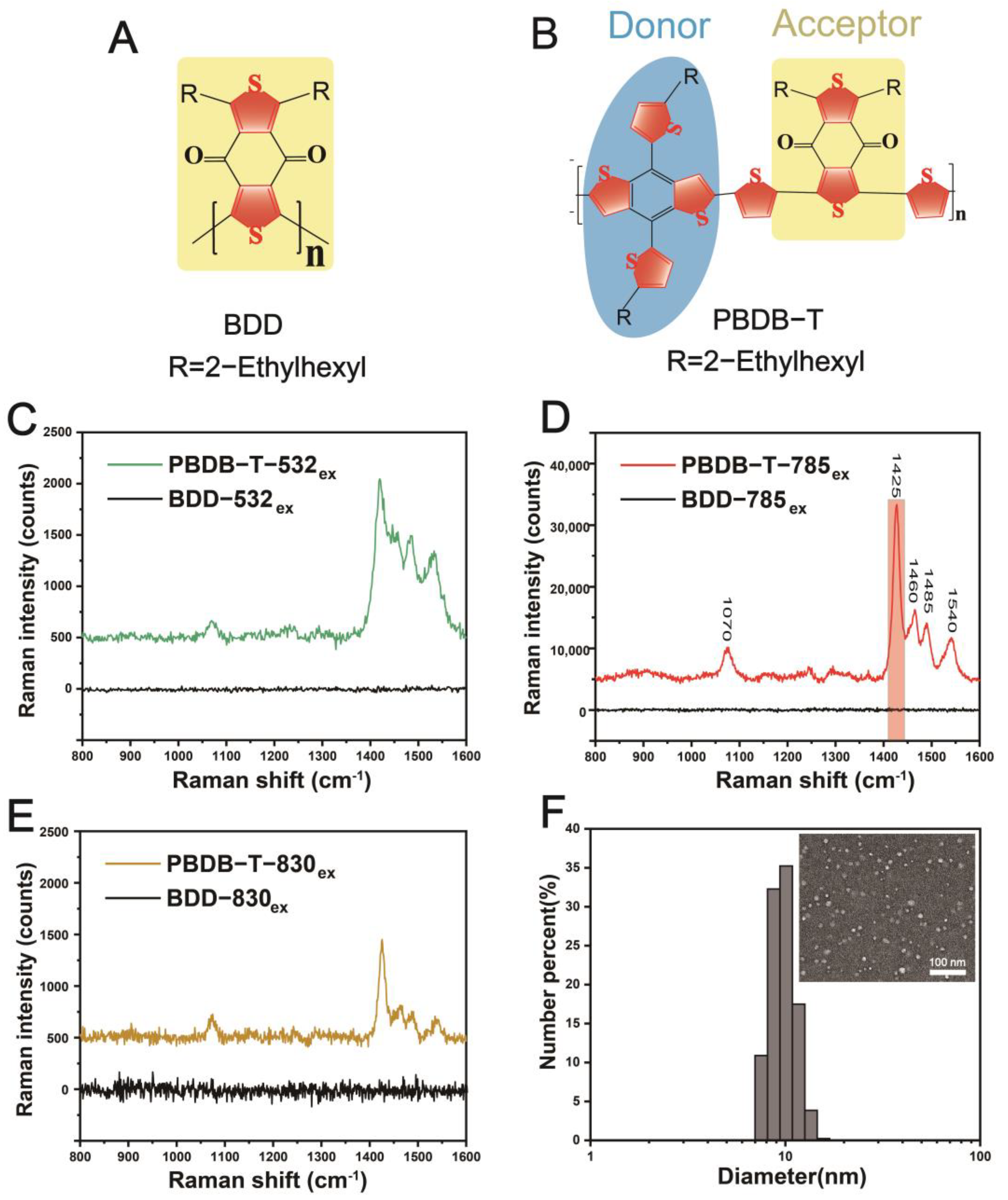
2.1. Preparation and Spectral Characterization of the PBDB-T NS and BDDNS
2.2. Donor–Acceptor(D-A) Molecular Regulation Mechanism of the PBDB-T NS for Raman Enhancement
2.3. Stability, Cytotoxicity, In Vivo Pharmacokinetics of the PBDB-T NS
2.4. In Vivo Intraoperative Raman Imaging of Tumors and Metastatic Microtumors, and In Vivo Non-Invasive Raman Imaging of Deep Tumor Tissue
3. Conclusions
Author Contributions
Funding
Institutional Review Board Statement
Informed Consent Statement
Data Availability Statement
Conflicts of Interest
References
- Eibir, T.E.I.F. Strategic research agenda for biomedical imaging. Insights Imaging 2019, 10, 7. [Google Scholar]
- Shaked, N.T.; Boppart, S.A.; Wang, L.V.; Popp, J. Label-free biomedical optical imaging. Nat. Photonics 2023, 17, 1031–1041. [Google Scholar] [CrossRef] [PubMed]
- Zhang, S.; Chen, H.; Wang, L.; Liu, C.; Liu, L.; Sun, Y.; Shen, X. A simple strategy for simultaneously enhancing photostability and mitochondrial-targeting stability of near-infrared fluorophores for multimodal imaging-guided photothermal therapy. J. Mater. Chem. B 2021, 9, 189–195. [Google Scholar] [CrossRef] [PubMed]
- Li, J.; Zhang, Y.; Ding, S.; Panneerselvam, R.; Tian, Z. Core–shell nanoparticle-enhanced raman spectroscopy. Chem. Rev. 2017, 117, 5002–5069. [Google Scholar] [CrossRef] [PubMed]
- Zong, C.; Xu, M.; Xu, L.; Wei, T.; Ma, X.; Zheng, X.; Hu, R.; Ren, B. Surface-enhanced raman spectroscopy for bioanalysis: Reliability and challenges. Chem. Rev. 2018, 118, 4946–4980. [Google Scholar] [CrossRef] [PubMed]
- Emmert, S.; Quargnali, G.; Thallmair, S.; Rivera-Fuentes, P. A locally activatable sensor for robust quantification of organellar glutathione. Nat. Chem. 2023, 15, 1415–1421. [Google Scholar] [CrossRef]
- Wang, X.; He, S.; Cheng, P.; Pu, K. A dual-locked tandem fluorescent probe for imaging of pyroptosis in cancer chemo-immunotherapy. Adv. Mater. 2023, 35, 2206510. [Google Scholar] [CrossRef] [PubMed]
- Etrych, T.; Janouskova, O.; Chytil, P. Fluorescence imaging as a tool in preclinical evaluation of polymer-based nano-dds systems intended for cancer treatment. Pharmaceutics 2019, 11, 471. [Google Scholar] [CrossRef]
- Cho, H.; Kwon, G.S. Polymeric micelles for neoadjuvant cancer therapy and tumor-primed optical imaging. ACS Nano 2011, 5, 8721–8729. [Google Scholar] [CrossRef]
- Kolitz-Domb, M.; Grinberg, I.; Corem-Salkmon, E.; Margel, S. Engineering of near infrared fluorescent proteinoid-poly (l-lactic acid) particles for in vivo colon cancer detection. J. Nanobiotechnol. 2014, 12, 30. [Google Scholar] [CrossRef]
- Du, Z.; Wang, W.; Luo, S.; Zhang, L.; Yuan, S.; Hei, Y.; Bao, Z.; Chen, C.; Lin, Y.; Chu, L. Self-renewable tag for photostable fluorescence imaging of proteins. J. Am. Chem. Soc. 2023, 145, 18968–18976. [Google Scholar] [CrossRef] [PubMed]
- Tang, Y.; Chen, X.; Zhang, S.; Smith, Z.J.; Gao, T. Vibrational fingerprint analysis of an azo-based resonance raman scattering probe for imaging proton distribution in cellular lysosomes. Anal. Chem. 2021, 93, 15659–15666. [Google Scholar] [CrossRef]
- Serebrennikova, K.V.; Berlina, A.N.; Sotnikov, D.V.; Zherdev, A.V.; Dzantiev, B.B. Raman scattering-based biosensing: New prospects and opportunities. Biosensors 2021, 11, 512. [Google Scholar] [CrossRef] [PubMed]
- Butler, H.J.; Ashton, L.; Bird, B.; Cinque, G.; Curtis, K.; Dorney, J.; Esmonde-White, K.; Fullwood, N.J.; Gardner, B.; Martin-Hirsch, P.L.; et al. Using raman spectroscopy to characterize biological materials. Nat. Protoc. 2016, 11, 664–687. [Google Scholar] [CrossRef]
- Hu, F.; Zeng, C.; Long, R.; Miao, Y.; Wei, L.; Xu, Q.; Min, W. Supermultiplexed optical imaging and barcoding with engineered polyynes. Nat. Methods 2018, 15, 194–200. [Google Scholar] [CrossRef]
- von Maltzahn, G.; Centrone, A.; Park, J.H.; Ramanathan, R.; Sailor, M.J.; Hatton, T.A.; Bhatia, S.N. Sers-coded gold nanorods as a multifunctional platform for densely multiplexed near-infrared imaging and photothermal heating. Adv. Mater. 2009, 21, 3175–3180. [Google Scholar] [CrossRef]
- Kang, J.W.; So, P.T.C.; Dasari, R.R.; Lim, D. High resolution live cell raman imaging using subcellular organelle-targeting sers-sensitive gold nanoparticles with highly narrow intra-nanogap. Nano Lett. 2015, 15, 1766–1772. [Google Scholar] [CrossRef]
- Kudelski, A. Analytical applications of raman spectroscopy. Talanta 2008, 76, 1–8. [Google Scholar] [CrossRef]
- Liu, C.H.; Zhou, Y.; Sun, Y.; Li, J.Y.; Zhou, L.X.; Boydston-White, S.; Masilamani, V.; Zhu, K.; Pu, Y.; Alfano, R.R. Resonance raman and raman spectroscopy for breast cancer detection. Technol Cancer Res. Treat. 2013, 12, 371–382. [Google Scholar] [CrossRef]
- Lin, L.; Bi, X.; Gu, Y.; Wang, F.; Ye, J. Surface-enhanced raman scattering nanotags for bioimaging. J. Appl. Phys. 2021, 129, 191101. [Google Scholar] [CrossRef]
- Wen, C.; Wang, L.; Liu, L.; Shen, X.C.; Chen, H. Surface-enhanced raman probes based on gold nanomaterials for in vivo diagnosis and imaging. Chem. Asian J. 2022, 17, e202200014. [Google Scholar] [CrossRef]
- Li, Y.; Heo, J.; Lim, C.; Pliss, A.; Kachynski, A.V.; Kuzmin, A.N.; Kim, S.; Prasad, P.N. Organelle specific imaging in live cells and immuno-labeling using resonance raman probe. Biomaterials 2015, 53, 25–31. [Google Scholar] [CrossRef] [PubMed]
- Yuan, Y.; Raj, P.; Zhang, J.; Siddhanta, S.; Barman, I.; Bulte, J.W.M. Furin-mediated self-assembly of olsalazine nanoparticles for targeted raman imaging of tumors. Angew. Chem. Int. Ed. 2021, 60, 3923–3927. [Google Scholar] [CrossRef] [PubMed]
- Myers Kelley, A. Resonance raman and resonance hyper-raman intensities: Structure and dynamics of molecular excited states in solution. J. Phys. Chem. A 2008, 112, 11975–11991. [Google Scholar] [CrossRef] [PubMed]
- Jakubek, R.S.; Handen, J.; White, S.E.; Asher, S.A.; Lednev, I.K. Ultraviolet resonance raman spectroscopic markers for protein structure and dynamics. Trend. Analyt. Chem. 2018, 103, 223–229. [Google Scholar] [CrossRef] [PubMed]
- Darby, B.L.; Etchegoin, P.G.; Le Ru, E.C. Single-molecule surface-enhanced raman spectroscopy with nanowatt excitation. Phys. Chem. Chem. Phys. 2014, 16, 23895–23899. [Google Scholar] [CrossRef] [PubMed]
- Chen, Y.; Wang, S.; Zhang, F. Near-infrared luminescence high-contrast in vivo biomedical imaging. Nat. Rev. Bioeng. 2023, 1, 60–78. [Google Scholar] [CrossRef]
- Altınoğlu, E.; Adair, J.H. Near infrared imaging with nanoparticles. WIREs Nanomed. Nanobiotechnol. 2010, 2, 461–477. [Google Scholar] [CrossRef] [PubMed]
- Li, L.; Xu, Y.; Chen, Y.; Zheng, J.; Zhang, J.; Li, R.; Wan, H.; Yin, J.; Yuan, Z.; Chen, H. A family of push-pull bio-probes for tracking lipid droplets in living cells with the detection of heterogeneity and polarity. Anal. Chim. Acta 2020, 1096, 166–173. [Google Scholar] [CrossRef]
- Zhang, W.; Huang, T.; Li, J.; Sun, P.; Wang, Y.; Shi, W.; Han, W.; Wang, W.; Fan, Q.; Huang, W. Facial control intramolecular charge transfer of quinoid conjugated polymers for efficient in vivo nir-ii imaging. ACS Appl. Mater. Inter. 2019, 11, 16311–16319. [Google Scholar] [CrossRef]
- Onorato, G.; Fardella, F.; Lewinska, A.; Gobbo, F.; Tommasini, G.; Wnuk, M.; Tino, A.; Moros, M.; Antognazza, M.R.; Tortiglione, C. Optical control of tissue regeneration through photostimulation of organic semiconducting nanoparticles. Adv. Healthc. Mater. 2022, 11, e2200366. [Google Scholar] [CrossRef] [PubMed]
- Yaghoobi Nia, N.; Bonomo, M.; Zendehdel, M.; Lamanna, E.; Desoky, M.M.H.; Paci, B.; Zurlo, F.; Generosi, A.; Barolo, C.; Viscardi, G.; et al. Impact of p3ht regioregularity and molecular weight on the efficiency and stability of perovskite solar cells. ACS Sustain. Chem. Eng. 2021, 9, 5061–5073. [Google Scholar] [CrossRef]
- Zhao, Q.; Li, D.; Peng, J. Interrogating polymorphism in conjugated poly(thieno)thiophene thin films for field-effect transistors. Macromolecules 2023, 56, 490–500. [Google Scholar] [CrossRef]
- Lu, H.; Zhang, X.; Li, C.; Wei, H.; Liu, Q.; Li, W.; Bo, Z. Performance enhancement of polymer solar cells by using two polymer donors with complementary absorption spectra. Macromol. Rapid Commun. 2015, 36, 1348–1353. [Google Scholar] [CrossRef] [PubMed]
- Jeong, M.; Oh, J.; Cho, Y.; Lee, B.; Jeong, S.; Lee, S.M.; Kang, S.H.; Yang, C. Triisopropylsilyl-substituted benzo[1,2-b:4,5-c′]-dithiophene-4,8-dione-containing copolymers with more than 17% efficiency in organic solar cells. Adv. Funct. Mater. 2021, 31, 2102371. [Google Scholar] [CrossRef]
- Zheng, B.; Huo, L.; Li, Y. Benzodithiophenedione-based polymers: Recent advances in organic photovoltaics. NPG Asia Mater. 2020, 12, 3. [Google Scholar] [CrossRef]
- Huo, L.; Liu, T.; Fan, B.; Zhao, Z.; Sun, X.; Wei, D.; Yu, M.; Liu, Y.; Sun, Y. Organic solar cells based on a 2d benzo[1,2-b:4,5-b′]-difuran-conjugated polymer with high-power conversion efficiency. Adv. Mater. 2015, 27, 6969–6975. [Google Scholar] [CrossRef]
- Duan, C.; Furlan, A.; van Franeker, J.J.; Willems, R.E.M.; Wienk, M.M.; Janssen, R.A.J. Wide-bandgap benzodithiophene–benzothiadiazole copolymers for highly efficient multijunction polymer solar cells. Adv. Mater. 2015, 27, 4461–4468. [Google Scholar] [CrossRef] [PubMed]
- Huo, L.; Hou, J. Benzo[1,2-b:4,5-b′]dithiophene-based conjugated polymers: Band gap and energy level control and their application in polymer solar cells. Polym. Chem. 2011, 2, 2453. [Google Scholar] [CrossRef]
- Brozek-Pluska, B.; Miazek, K.; Musia, J.; Kordek, R. Label-free diagnostics and cancer surgery raman spectra guidance for the human colon at different excitation wavelengths. RSC Adv. 2019, 9, 40445–40454. [Google Scholar] [CrossRef]
- Qiu, Y.; Zhang, Y.; Li, M.; Chen, G.; Fan, C.; Cui, K.; Wan, J.; Han, A.; Ye, J.; Xiao, Z. Intraoperative detection and eradication of residual microtumors with gap-enhanced raman tags. ACS Nano 2018, 12, 7974–7985. [Google Scholar] [CrossRef]
- Walters, C.M.; Pao, C.; Gagnon, B.P.; Zamecnik, C.R.; Walker, G.C. Bright surface-enhanced raman scattering with fluorescence quenching from silica encapsulated j-aggregate coated gold nanoparticles. Adv. Mater. 2018, 30, 1705381. [Google Scholar] [CrossRef] [PubMed]
- Liu, Z.; Davis, C.; Cai, W.; He, L.; Chen, X.; Dai, H. Circulation and long-term fate of functionalized, biocompatible single-walled carbon nanotubes in mice probed by raman spectroscopy. Proc. Natl. Acad. Sci. USA 2008, 105, 1410–1415. [Google Scholar] [CrossRef]
- Zavaleta, C.; de la Zerda, A.; Liu, Z.; Keren, S.; Cheng, Z.; Schipper, M.; Chen, X.; Dai, H.; Gambhir, S.S. Noninvasive raman spectroscopy in living mice for evaluation of tumor targeting with carbon nanotubes. Nano Lett. 2008, 8, 2800–2805. [Google Scholar] [CrossRef]
- Blanco, E.; Shen, H.; Ferrari, M. Principles of nanoparticle design for overcoming biological barriers to drug delivery. Nat. Biotechnol. 2015, 33, 941–951. [Google Scholar] [CrossRef]
- Wu, J. The enhanced permeability and retention (epr) effect: The significance of the concept and methods to enhance its application. J. Pers. Med. 2021, 11, 771. [Google Scholar] [CrossRef] [PubMed]
- Schneider, C.A.; Rasband, W.S.; Eliceiri, K.W. Nih image to imagej: 25 years of image analysis. Nat. Methods 2012, 9, 671–675. [Google Scholar] [CrossRef] [PubMed]
- Gao, S.; Wei, G.; Zhang, S.; Zheng, B.; Xu, J.; Chen, G.; Li, M.; Song, S.; Fu, W.; Xiao, Z.; et al. Albumin tailoring fluorescence and photothermal conversion effect of near-infrared-ii fluorophore with aggregation-induced emission characteristics. Nat. Commun. 2019, 10, 2206. [Google Scholar] [CrossRef]
- Díaz-López, R.; Tsapis, N.; Santin, M.; Bridal, S.L.; Nicolas, V.; Jaillard, D.; Libong, D.; Chaminade, P.; Marsaud, V.; Vauthier, C.; et al. The performance of pegylated nanocapsules of perfluorooctyl bromide as an ultrasound contrast agent. Biomaterials 2010, 31, 1723–1731. [Google Scholar] [CrossRef]
- Qian, D.; Ye, L.; Zhang, M.; Liang, Y.; Li, L.; Huang, Y.; Guo, X.; Zhang, S.; Tan, Z.A.; Hou, J. Design, application, and morphology study of a new photovoltaic polymer with strong aggregation in solution state. Macromolecules 2012, 45, 9611–9617. [Google Scholar] [CrossRef]
- Mu, X.; Guo, Y.; Li, Y.; Wang, Z.; Li, Y.; Xu, S. Analysis and design of resonance raman reporter molecules by density functional theory. J. Raman Spectrosc. 2017, 48, 1196–1200. [Google Scholar] [CrossRef]
- Xie, L.; Ling, X.; Fang, Y.; Zhang, J.; Liu, Z. Graphene as a substrate to suppress fluorescence in resonance raman spectroscopy. J. Am. Chem. Soc. 2009, 131, 9890–9891. [Google Scholar] [CrossRef]
- Tsoi, W.C.; James, D.T.; Kim, J.S.; Nicholson, P.G.; Murphy, C.E.; Bradley, D.D.C.; Nelson, J.; Kim, J. The nature of in-plane skeleton raman modes of p3ht and their correlation to the degree of molecular order in p3ht: Pcbm blend thin films. J. Am. Chem. Soc. 2011, 133, 9834–9843. [Google Scholar] [CrossRef] [PubMed]
- Petry, R.; Schmitt, M.; Popp, J. Raman spectroscopy-a prospective tool in the life sciences. ChemPhysChem 2003, 4, 14–30. [Google Scholar] [CrossRef]
- Zhang, M.; Guo, X.; Ma, W.; Ade, H.; Hou, J. A large-bandgap conjugated polymer for versatile photovoltaic applications with high performance. Adv. Mater. 2015, 27, 4655–4660. [Google Scholar] [CrossRef]
- Chen, C.; Chan, S.; Chao, T.; Ting, C.; Ko, B. Low-bandgap poly(thiophene-phenylene-thiophene) derivatives with broaden absorption spectra for use in high-performance bulk-heterojunction polymer solar cells. J. Am. Chem. Soc. 2008, 130, 12828–12833. [Google Scholar] [CrossRef] [PubMed]
- Kastantin, M.; Ananthanarayanan, B.; Karmali, P.; Ruoslahti, E.; Tirrell, M. Effect of the lipid chain melting transition on the stability of dspe-peg(2000) micelles. Langmuir 2009, 25, 7279–7286. [Google Scholar] [CrossRef]
- Li, Y.; Zeng, S.; Hao, J. Non-invasive optical guided tumor metastasis/vessel imaging by using lanthanide nanoprobe with enhanced down-shifting emission beyond 1500 nm. ACS Nano 2019, 13, 248–259. [Google Scholar] [CrossRef]
- Chen, D.; Li, B.; Cai, S.; Wang, P.; Peng, S.; Sheng, Y.; He, Y.; Gu, Y.; Chen, H. Dual targeting luminescent gold nanoclusters for tumor imaging and deep tissue therapy. Biomaterials 2016, 100, 1–16. [Google Scholar] [CrossRef]
- Georgiev, N.I.; Bakov, V.V.; Anichina, K.K.; Bojinov, V.B. Fluorescent probes as a tool in diagnostic and drug delivery systems. Pharmaceuticals 2023, 16, 381. [Google Scholar] [CrossRef]
- Ma, S.; Kim, J.H.; Chen, W.; Li, L.; Lee, J.; Xue, J.; Liu, Y.; Chen, G.; Tang, B.; Tao, W.; et al. Cancer cell-specific fluorescent prodrug delivery platforms. Adv. Sci. 2023, 10, e2207768. [Google Scholar] [CrossRef] [PubMed]
- Xie, Q.; Liu, J.; Chen, B.; Ge, X.; Zhang, X.; Gao, S.; Ma, Q.; Song, J. Nir-ii fluorescent activatable drug delivery nanoplatform for cancer-targeted combined photodynamic and chemotherapy. ACS Appl. Bio Mater. 2022, 5, 711–722. [Google Scholar] [CrossRef] [PubMed]


| Item | Reference Range | PBDB–T NS | Control | |
|---|---|---|---|---|
| WBC | 109/L | 0.8–10.6 | 6.43 ± 0.90 | 3.77 ± 0.77 |
| LYMPH | 109/L | 0.6–8.9 | 5.07 ± 0.65 | 2.87 ± 0.58 |
| MONO | 109/L | 0.04–1.4 | 0.13 ± 0.05 | 0.13 ± 0.05 |
| NEUT | 109/L | 0.23–3.6 | 1.23 ± 0.26 | 0.77 ± 0.19 |
| LYMPH | % | 40–92 | 78.40 ± 0.99 | 75.43 ± 1.48 |
| MONO | % | 0.9–18 | 2.50 ± 0.08 | 3.67 ± 0.78 |
| NEUT | % | 65–50 | 19.10 ± 1.04 | 20.90 ± 1.36 |
| RBC | 1012/L | 6.5–11.5 | 7.95 ± 0.34 | 8.18 ± 0.24 |
| HGB | g/L | 110–165 | 122.33 ± 2.62 | 132.00 ± 1.41 |
| HCT | % | 35–55 | 39.67 ± 1.58 | 42.00 ± 0.41 |
| MCV | fL | 41–55 | 49.97 ± 0.29 | 51.43 ± 1.02 |
| MCH | pg | 13–18 | 15.33 ± 0.50 | 16.07 ± 0.29 |
| MCHC | g/L | 300–360 | 308.33 ± 8.50 | 313.67 ± 1.89 |
| RDW | % | 12–19 | 14.85 ± 0.15 | 14.07 ± 0.46 |
| PLT | 109/L | 400–1600 | 683.67 ± 121.69 | 663.33 ± 67.71 |
| MPV | fL | 4.0–6.2 | 5.90 ± 0.16 | 5.83 ± 0.17 |
| PCT | % | 0.100–0.780 | 0.40 ± 0.08 | 0.41 ± 0.07 |
Disclaimer/Publisher’s Note: The statements, opinions and data contained in all publications are solely those of the individual author(s) and contributor(s) and not of MDPI and/or the editor(s). MDPI and/or the editor(s) disclaim responsibility for any injury to people or property resulting from any ideas, methods, instructions or products referred to in the content. |
© 2024 by the authors. Licensee MDPI, Basel, Switzerland. This article is an open access article distributed under the terms and conditions of the Creative Commons Attribution (CC BY) license (https://creativecommons.org/licenses/by/4.0/).
Share and Cite
Li, M.; Luo, A.; Xu, W.; Wang, H.; Qiu, Y.; Xiao, Z.; Cui, K. A Visual Raman Nano−Delivery System Based on Thiophene Polymer for Microtumor Detection. Pharmaceutics 2024, 16, 655. https://doi.org/10.3390/pharmaceutics16050655
Li M, Luo A, Xu W, Wang H, Qiu Y, Xiao Z, Cui K. A Visual Raman Nano−Delivery System Based on Thiophene Polymer for Microtumor Detection. Pharmaceutics. 2024; 16(5):655. https://doi.org/10.3390/pharmaceutics16050655
Chicago/Turabian StyleLi, Meng, Aoxiang Luo, Wei Xu, Haoze Wang, Yuanyuan Qiu, Zeyu Xiao, and Kai Cui. 2024. "A Visual Raman Nano−Delivery System Based on Thiophene Polymer for Microtumor Detection" Pharmaceutics 16, no. 5: 655. https://doi.org/10.3390/pharmaceutics16050655
APA StyleLi, M., Luo, A., Xu, W., Wang, H., Qiu, Y., Xiao, Z., & Cui, K. (2024). A Visual Raman Nano−Delivery System Based on Thiophene Polymer for Microtumor Detection. Pharmaceutics, 16(5), 655. https://doi.org/10.3390/pharmaceutics16050655