Abstract
Despite the well-documented health benefits of the probiotic Saccharomyces, its application in oral health has not been comprehensively assessed. Dental caries is a transmissible disease initiated by acid production of cariogenic bacteria and yeast, such as Streptococcus mutans and Candida albicans, on tooth enamel and followed by subsequent enamel demineralization. Here, we investigated the effect of two Saccharomyces strains (Saccharomyces boulardii and Saccharomyces cerevisiae) on S. mutans–C. albicans cross-kingdom interactions using a cariogenic planktonic model. Viable cells, pH changes, and gene expression were measured. S. cerevisiae and S. boulardii inhibited the growth of C. albicans in dual- and multi-species conditions at 4, 6, and 20 h. Saccharomyces also inhibited C. albicans hyphal formation. Furthermore, Saccharomyces reduced the acidity of the culture medium, which usually plummeted below pH 5 when S. mutans and C. albicans were present in the model. The presence of Saccharomyces maintained the culture medium above 6 even after overnight incubation, demonstrating a protective potential against dental enamel demineralization. S. boulardii significantly down-regulated S. mutans atpD and eno gene expression. Overall, our results shed light on a new promising candidate, Saccharomyces, for dental caries prevention due to its potential to create a less cariogenic environment marked by a neutral pH and reduced growth of C. albicans.
1. Introduction
Early childhood caries (ECC) is the most common chronic childhood disease worldwide [1]. Untreated ECC has a negative impact on the oral health-related quality of life of children and their families [2,3]. Oral microorganisms are associated with ECC etiopathogenesis; for example, Streptococcus mutans is the well-known pathogenic bacterium responsible for dental caries due to its acidogenicity and aciduric properties [4]. Recent advances in pediatric caries research also revealed the cariogenic role of fungi in ECC [5,6,7]. Specifically, Candida albicans has been shown to enhance cariogenicity through its synergistic interactions with S. mutans in producing acid, forming biofilms, and causing more severe caries [5,6,7]. Additionally, high levels of Candida species have been frequently reported in children with ECC [8,9,10].
Conventional measures, including oral hygiene management and pharmaceutical interventions, have been adopted for ECC prevention and treatment [3,11,12,13]. However, children remain at high risk for recurrent caries due to either low adherence to positive oral hygiene habits or the ineffectiveness of antimicrobial applications [14,15,16]. Moreover, a high relapse rate of oral candidiasis occurs among individuals wearing dentures, likely due to the conducive environment for Candida colonization created by the denture’s surface and the microclimate between the denture and oral mucosa [17,18]. Intriguingly, an increased adherence of both C. albicans and S. mutans to the denture acrylic base has been observed [19]. In addition to conventional antimicrobial approaches, alternative treatments, such as probiotics, have been investigated for their effects on oral health.
Probiotics are non-pathogenic live microorganisms that, when administered in appropriate quantities, can be beneficial to the health of the host [20]. Studies have shown beneficial effects of probiotic microorganisms in the oral cavity due to their inhibiting the abundance of pathogens [20]. For example, our previous work demonstrated the ability of Lactobacillus plantarum 14917 to inhibit the growth of S. mutans and C. albicans and cariogenic biofilm formation [21,22]. These studies elicited the potential of probiotics to inhibit cariogenic polymicrobial interactions and prevent ECC. However, the inhibitory effect of L. plantarum on S. mutans and C. albicans was dependent on a higher dosage of L. plantarum that poses challenges to clinical application [21]. Thus, given this background, it is worth exploring the potential of other probiotics in disrupting cariogenic cross-kingdom interactions.
Saccharomyces boulardii (S. boulardii) and Saccharomyces cerevisiae (S. cerevisiae) are two closely related strains commonly used as probiotics and as reagents in the preparation of food and wine. S. boulardii is stable over a wide range of pH levels, temperatures, and exposures to bile salts and gastrointestinal enzymes [23]. S. boulardii is also incapable of promoting antibiotic resistance, as the exchange of antibiotic-resistant genes between fungi and bacteria is unlikely [24,25]. Moreover, S. boulardii is absent from the natural gut microbiota but has been extensively studied in several gastrointestinal and systemic diseases. For example, studies have shown evidence that S. boulardii can prevent antibiotic-associated diarrhea [26] and prevent Clostridium difficile-associated colitis and traveler’s diarrhea [27,28]. S. boulardii has also demonstrated effectiveness in treating urinary tract and vaginal yeast infections, high cholesterol levels, lactose intolerance, teenage acne, and fever blisters [29,30,31].
Regarding oral health, two randomized controlled clinical studies [32,33] provide supporting evidence of using locally delivered probiotic S. boulardii as an adjunct to mechanical therapy that is used to manage periodontal disease. Moreover, Deshmukh et al. [34] assessed the impact of formulations with S. boulardii on oral health and found similar efficacy between chlorohexidine and probiotic mouthwashes in reducing dental plaque accumulation and promoting gingival health.
S. cerevisiae, commonly known as brewer’s yeast, is a unicellular fungus [35]. Studies have revealed the benefits of S. cerevisiae strains to both systemic and oral health. For example, daily supplements of S. cerevisiae delivered in single capsules were found to significantly reduce gastrointestinal symptoms of irritable bowel syndrome in both mice and humans [36,37]. S. cerevisiae-based intravaginal treatments also accelerated the clearance of C. albicans in mice with vaginal candidiasis [38]. Concerning oral health, administration of S. cerevisiae in the oral cavity has been shown to decrease C. albicans load and virulence in mice infected with oropharyngeal candidiasis [39]. Moreover, Premanathan et al. [40] observed a shorter recovery time from oral candidiasis in patients treated with topically applied S. cerevisiae.
Interestingly, S. cerevisiae shares several genes with S. boulardii that are involved in probiotic phenotypes [41]. These genes include HSP150 and YGP1, which regulate responses to stress and acidic pH tolerance; HSP26 and SSA4, which regulate heat responses; and ARO9 and ARO8, which are involved in the biosynthesis of aromatic alcohols, such as phenylethanol and tryptophol [41,42]. These aromatic alcohols can inhibit the virulence of C. albicans [43]. Moreover, S. boulardii has been reported to secrete medium-chain fatty acids, mainly capric acid, with bioactivity against C. albicans hyphae and biofilm formation [44,45].
With the above-mentioned characteristics of S. cerevisiae and S. boulardii, these two species demonstrate the potential to influence cariogenic microorganisms. However, the effect of S. boulardii and S. cerevisiae on cariogenic S. mutans and C. albicans cross-kingdom interactions has not been assessed. Our study aims to fill this gap by examining the effect of probiotic S. boulardii and S. cerevisiae on the growth of S. mutans and C. albicans in a cariogenic planktonic model that mimics a high-caries-risk clinical condition. The study results will provide insight into the influence of S. cerevisiae and S. boulardii on cariogenic cross-kingdom microorganisms and expand preventative and treatment options for dental caries, such as oral application of yeast probiotics for ECC.
2. Materials and Methods
2.1. Bacterial Strains and Starter Preparation
The microorganisms used in the study were S. mutans UA159, C. albicans SC5314, S. boulardii ATCC MYA796, and S. cerevisiae ATCC 204508. C. albicans, S. mutans, and Saccharomyces were recovered from frozen stock using YPD agar (BD Difco™, San Jose, CA, USA, 242720), blood agar (TSA with sheep blood, Thermo Scientific™, Waltham, MA, USA), and Yeast mold agar (BD Difco™, 271210), respectively. After 48 h incubation at 37 °C, 3–5 colonies of each species were inoculated into 10 mL of broth for overnight incubation (5% CO2, 37 °C). C. albicans, S. boulardii, and S. cerevisiae were cultured in YPD broth (BD Difco™, 242820); S. mutans was cultured in TSBYE broth (3% Tryptic Soy, 0.5% Yeast Extract Broth, BD Bacto™ 286220 and Gibco™ 212750, Thermo Scientific™, Waltham, MA, USA) with 1% glucose. The next day, 0.5 mL of the overnight starters was added to glass tubes containing fresh broth and incubated for 3–5 h until they reached the mid-exponential phase with desirable optical density. The morning starters were then ready to be used for the preparation of the planktonic model described below.
2.2. Planktonic Model
Interactions between C. albicans, S. mutans, and Saccharomyces species were first evaluated in planktonic conditions. The inoculation quantity of C. albicans (103 CFU/mL) and S. mutans (105 CFU/mL) was chosen to mimic a high-caries-risk condition in the clinical setting. Individuals who carry more than 105 CFU/mL of S. mutans in saliva are considered to be at high risk for caries [46], while individuals who have more than 400 CFU/mL of C. albicans in saliva could be diagnosed with oral candidiasis using the laboratory standard [47]. The inoculation quantity of the two Saccharomyces species (107 CFU/mL) is in the lower dose range of the probiotics used in commercial probiotic products (109–1010 CFU as a single dosage).
Mono-species, dual-species, and multi-species models were used to assess the interaction between C. albicans, S. mutans, and Saccharomyces (either S. boulardii or S. cerevisiae). The planktonic models used in this study consisted of three types: mono-species, dual-species, and multi-species conditions. In the mono-species model, one of the following microorganisms: C. albicans, S. mutans, or Saccharomyces was incubated in 10 mL of TSBYE broth with 1% glucose for 20 h at 37 °C and 5% CO2. In the dual-species model, either C. albicans or S. mutans was co-cultured with one of the Saccharomyces species for 20 h under the same conditions. In the multi-species models, C. albicans, S. mutans, and one of the Saccharomyces species were mixed and cultivated for 20 h under the same circumstances. At 0, 2, 4, 6, and 20 h, the colony-forming unit per milliliter (CFU/mL) and culture media pH value were measured for each model.
To evaluate the inhibition of pseudohyphae and hyphae formation in C. albicans at selected time points, we placed a quantity of 20 µL of the culture medium on a glass slide and immediately observed it under a light microscope (Olympus BX43, 214, Tokyo, Japan) with a 100× oil objective (Olympus UPlanFL N 100×, Tokyo, Japan).
2.3. PCR and Real-Time Quantitative PCR (Real-Time qPCR)
PCR was performed in a thermal cycler (Applied Biosystems, Waltham, MA, USA), following the instructions provided by the manufacturer to assess the amplification of genes of interest. The primers used in this study are shown in Table S1 [48]. First, the yeast DNA of Saccharomyces species and C. albicans was collected from their respective overnight cultures (15 h). DNA extractions were performed using the MasterPure Yeast DNA Purification Kit (LGC Genomics, Berlin, Germany). The PCR was performed in a 50-volume containing 25 μL PCR Master Mix (2×) (Thermo Fisher Scientific, Bermen, Germany), 1 μL DNA template, 5 μL for each primer, and 14 μL nuclease-free water. The reaction was performed at 95 °C for 5 min, followed by 35 cycles of denaturation at 95 °C for 15 s, annealing at 55 °C for 30 s, and polymerization at 72 °C for 1 min, with one final extension cycle at 72 °C for 10 min. The product of the PCR was run on a pre-cast 2% agarose gel (E-gel® Ex agarose gel from Invitrogen (Carlsbad, CA, USA) along with a DNA ladder (E-gel® 1 kb plus DNA ladder, Invitrogen, Carlsbad, CA, USA). The gel was run for 10 min and then visualized under UV light, and the picture was saved for documentation.
Real-time qPCR was conducted to validate the expression of particular genes related to C. albicans and S. mutans virulence factors or viability. The primers used in this study are shown in Table S1 in Supplementary Materials. First, cellular RNAs were extracted from 4 mL mixture at 20 h, and 1–4 μg of purified RNA was converted to synthesize cDNAs with an iScript cDNA Synthesis Kit (Bio-Rad Laboratories, Inc., Hercules, CA, USA). The resultant cDNA and negative controls were quantitatively amplified using a QuantStudio™ 3 Real-Time PCR System (Thermo Fisher Scientific, Wilmington, DE, USA) and applied Biosystems™ PowerTrack™ SYBR Green Master Mix. A 20-volume PCR reaction comprised 2 μL cDNA template, 1 μL for each primer, 10 μL 2× SYBR-Green mix (SYBR-Green and Taq DNA Polymerase), and 6 μL nuclease-free water. To determine gene expression, three replicates for each round were set up, and relative gene expressions were calculated using the comparative ΔΔCt method. Unique core genes of S. mutans and C. albicans, namely, gyrA and ACT1, respectively, were utilized as housekeeping genes.
2.4. Statistical Analysis
To compare the live abundance of C. albicans, S. mutans, and Saccharomyces species in the planktonic models, the CFU/mL values were first converted into natural log values for statistical purposes. Of note, zero values were retained as zero. Normality tests were used to evaluate the data distribution of variables, including pH value, natural log-converted CFU/mL value, and 2−ΔΔCT (real-time qRT-PCR value). To compare the difference between groups when data followed a normal distribution, the Student’s t-test for two groups and one-way ANOVA for more than two groups followed by a post hoc test were performed. Nevertheless, if data were not normally distributed, we used the Mann–Whitney U test to compare the results of the two groups and the Kruskal–Wallis test to compare the results for more than two groups. The statistical analysis was performed using SPSS Version 24 (SPSS Statistics for Windows, Version 24.0; IBM, Armonk, NY, USA) with a significance level of p < 0.05.
3. Results
3.1. Growth Profile of Saccharomyces Species
The growth curves of S. cerevisiae and S. boulardii in YPD or TSBYE with 1% glucose are shown in Figure 1. During the initial 8 h, S. cerevisiae grew faster than S. boulardi, and both reached a plateau at 10 h. S. cerevisiae showed similar growth curves in YPD and TSBYE with 1% glucose (Figure 1A). However, the growth of S. boulardi in TSBYE with 1% glucose was lower than the growth in YPD at 4 and 20 h (Figure 1B).
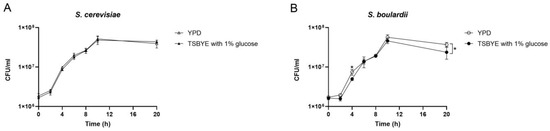
Figure 1.
Growth curves of planktonic Saccharomyces cerevisiae (S. cerevisiae) (A) and Saccharomyces boulardii (S. boulardii) (B) in two culture mediums: YPD and TSBYE with 1% glucose. Data are presented as means (± standard deviations) of three independent experiments performed in triplicate. * Indicates a significant difference in CFU/mL between different culture media, with p < 0.05.
3.2. The Impact of Saccharomyces Species on C. albicans and S. mutans in the Dual-Species Conditions
Both S. cerevisiae and S. boulardii significantly inhibited the growth of C. albicans by 1 log at 4 h, 2 logs at 6 h, and 6 logs at 20 h (Figure 2A). S. boulardii significantly inhibited the growth of S. mutans at 6 and 20 h. S. cerevisiae significantly inhibited the growth of S. mutans at 2 and 4 h but failed at a later stage (Figure 2B). In contrast to the inhibited growth of C. albicans and S. mutans, S. boulardii grew better in the presence of C. albicans or S. mutans at 20 h (Figure 2C). However, the growth of S. cerevisiae in the dual-species conditions declined at 20 h (Figure 2D), which may explain the loss of its inhibitory effect on S. mutans at a later stage.
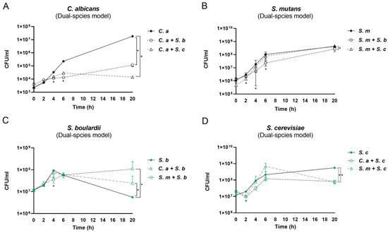
Figure 2.
Interactions between Saccharomyces species and C. albicans or S. mutans in dual-species conditions. (A) The growth of C. albicans cultured with or without Saccharomyces species. (B) The growth of S. mutans cultured with or without Saccharomyces species. (C) The growth of S. boulardii cultured with or without C. albicans/S. mutans. (D) The growth of S. cerevisiae cultured with or without C. albicans/S. mutans. * Indicates a significant difference in CFU/mL between mono- and dual-species conditions, with p < 0.05. C. a: C. albicans; S. m: S. mutans; S. c: S. cerevisiae; S. b: S. boulardii.
3.3. The Impact of Saccharomyces Species on C. albicans and S. mutans in the Multi-Species Conditions
Intriguingly, both S. cerevisiae and S. boulardii significantly inhibited the growth of C. albicans at all time points (2, 4, 6, and 20 h) in the multi-species conditions (Figure 3A). However, Saccharomyces species had a dampened inhibitory effect on S. mutans (Figure 3B). Compared to the C. albicans–S. mutans dual-species control, S. mutans grew faster when together with Saccharomyces at the early stage; however, it grew with a reduced speed between 12 and 20 h, although the viable counts at 20 h between the groups have no statistical significance (p > 0.05). Between the two Saccharomyces species in the multi-species model, S. cerevisiae had a slower growth speed than S. boulardii at 2 h, while it had a faster growth rate at the mid and late stages (6–20 h) (Figure 3C).
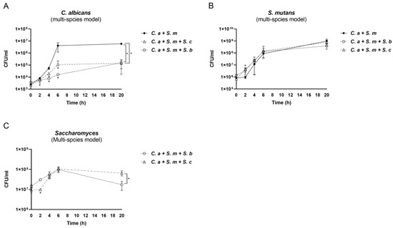
Figure 3.
Interactions among Saccharomyces species, C. albicans, and S. mutans in multi-species conditions. (A) The growth of C. albicans in control (C. a + S. m) and Saccharomyces species-treated groups. (B) The growth of S. mutans in control (C. a + S. m) and Saccharomyces species-treated groups. * Indicates that p < 0.05 when comparing control with Saccharomyces species-treated groups. (C) The growth of Saccharomyces species in multi-species conditions. * Indicates that p < 0.05 when comparing S. cerevisiae- and S. boulardii-treated groups.
3.4. Compositional Changes in Saccharomyces Species, C. albicans, and S. mutans in the Multi-Species Model
Next, we assessed the compositional changes in the multi-species model over time. When S. cerevisiae and C. albicans grew together, S. cerevisiae showed its dominance from the beginning to the end due to the initial concentration of S. cerevisiae (107 CFU/mL) being higher than that of C. albicans (103 CFU/mL) (Figure 4A). When S. cerevisiae and S. mutans grew together, the initial concentration of S. mutans was 105 CFU/mL. S. cerevisiae still seized its dominance from 2 h to 6 h; however, until 20 h, S. mutans was able to prevail after a fierce competition with S. cerevisiae (Figure 4B).
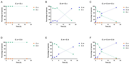
Figure 4.
Changes in species composition in dual- and multi-species conditions. (A–F) The proportional representation of each microorganism in dual- and multi-species conditions.
Next, when S. cerevisiae grew with C. albicans and S. mutans in the multi-species condition, a compositional switch occurred. As shown in Figure 4C, at the beginning, S. cerevisiae took the lead due to its highest initial concentration. Shortly, S. mutans displayed rapid growth rates, took over the race, and became the dominant species from 6 h to 20 h. C. albicans’ growth remained low over time. Intriguingly, compared to the S. mutans–S. cerevisiae dual-species condition, S. mutans in the multi-species model, when C. albicans was present, showed much stronger competitiveness against S. cerevisiae. This indicates an interspecies synergistic relationship between C. albicans and S. mutans, as well as synchronous antagonism between S. cerevisiae and S. mutans. A similar scenario was seen in the dual- and multi-species conditions when S. boulardii was present (Figure 4D–F).
3.5. Dynamic Changes in Culture pH in the Mono-, Dual-, and Multi-Species Conditions
Figure 5 shows the effect of Saccharomyces species on the environmental pH in the mono-, dual-, and multi-species models. Overall, the culture medium pH was lowered over time in all groups, particularly with a significant drop to pH 4.0 at 20 h in the S. mutans mono-species and C. albicans–S. mutans dual-species conditions. Significantly, the addition of either S. cerevisiae or S. boulardii neutralized the acidic environment and maintained the culture pH at 6.0 over the 20 h period, which is above the well-known critical pH of 5.5 for enamel demineralization.
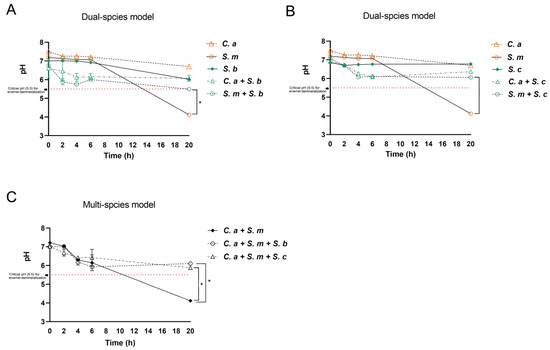
Figure 5.
Dynamic changes in pH in the culture medium. (A) pH in mono-species condition and S. boulardii present dual-species condition. * Indicates that p < 0.05 when comparing dual-species (S. b + S. m) with mono-species (S. m) conditions at 20 h. (B) pH in mono-species condition and S. cerevisiae present dual-species condition. * Indicates that p < 0.05 when comparing dual-species (S. c + S. m) with mono-species (S. m) conditions at 20 h. (C) pH in control (C. a + S. m) and Saccharomyces species-treated groups. * Indicates that p < 0.05 when comparing Saccharomyces species-treated groups with control at 20 h.
3.6. Regulation of S. mutans and C. albicans Virulence Genes by Saccharomyces Species
To evaluate the differential gene expression between the control and the experimental conditions with added Saccharomyces species, qPCR was conducted at 20 h. To minimize the bias from gene expression crosstalk between C. albicans and Saccharomyces species, we first examined the expressions of ACT1, EGR4, ECE1, and CHT2 in C. albicans, S. cerevisiae, or S. boulardii. PCR amplification products confirmed that the above genes are expressed by C. albicans only, not by any of the Saccharomyces species (Figure S1).
Next, as shown in Figure 6A, compared to the C. albicans–S. mutans dual-species control, S. boulardii reduced the expression of the S. mutans genes atpD (stress response gene related to ATPase complex and acid tolerance) and eno (associated with degradation of carbohydrates via glycolysis) by 1.4-fold (p < 0.01) and 2-fold (p < 0.001), respectively. In contrast, lacC and lacG, the genes involved in galactose metabolism, were significantly up-regulated when S. boulardii was added (p < 0.0001). The addition of S. cerevisiae had a negligible effect on the expression of S. mutans genes.
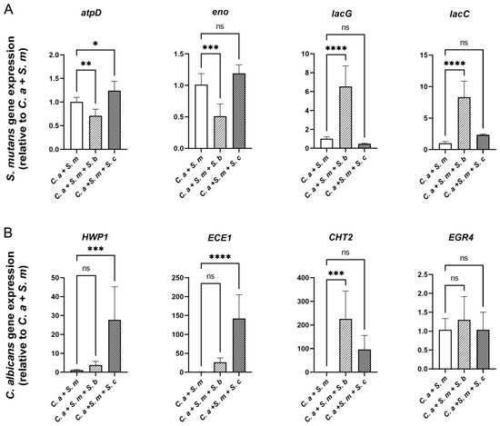
Figure 6.
Effect of Saccharomyces species on the expression of S. mutans and C. albicans genes in multi-species model. qRT-PCR was performed for S. mutans (A) and C. albicans (B) genes of interest for mixed-species culture at 20 h. Relative mRNA levels were presented as ratios relative to control group (C. a + S. m). Results are reported as the means ± SDs of three independent experiments. p values were determined by one-way ANOVA with post hoc tests. * p < 0.05, ** p < 0.01, *** p < 0.001, **** p < 0.0001.
For C. albicans gene expression (Figure 6B), S. cerevisiae up-regulated the expression of HWP1 and ECE1, which are associated with hyphal growth, by 27.2-fold and 74.63-fold, respectively (p < 0.001), whereas S. boulardii significantly up-regulated the expression of another C. albicans virulence gene, CHT2, which is associated with fungal wall remodeling. EGR4, however, related to antifungal medication resistance, was not statistically affected by either S. cerevisiae or S. boulardii.
3.7. Inhibition of C. albicans Hyphae/Pseudohyphae Formation by Saccharomyces Species
Inhibition of Candida hyphae or pseudohyphae formation was assessed by observing the culture mixture at 0 h, 6 h, and 20 h under a light microscope. In the C. albicans–S. mutans dual-species condition, C. albicans had a typical Candidal pseudohyphae formation at 6 h and elongated hyphal formations at 20 h. In comparison, the addition of S. cerevisiae or S. boulardii inhibited the growth of C. albicans in both yeast forms and the transition from yeast to hyphae or pseudohyphae form. The quantitative reduction in C. albicans by S. cerevisiae or S. boulardii observed in Figure 7 is consistent with the growth inhibition measured by CFUs.
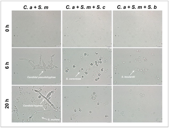
Figure 7.
Inhibition of Candidal hyphae formation by Saccharomyces species in multi-species model at ×100 magnification. C. albicans that contains yeast-form, pseudohyphal, and hyphal cells can be found in control group (C. a + S. m) but not in Saccharomyces species-treated groups. These are representative images of multiple fields of view. Scale bars = 10 μm.
4. Discussion
While various treatment options have been employed to control ECC, mainly by targeting cariogenic pathogens [21,22], limited studies have assessed probiotic yeast in interrupting cariogenic bacteria–fungi cross-kingdom interactions. Our study revealed novel findings that the oral health effects of S. boulardii and S. cerevisiae are not solely limited to inhibition of the growth of oral pathogens, such as C. albicans, but also extend to modulation of culture medium pH, influence on C. albicans and S. mutans virulence gene expression.
Kellis et al. and Wolfe proposed a hypothesis suggesting that the fermentative capability of this yeast complex might have developed during the period when sugar-rich fruit-bearing plants became prevalent in the environment [49,50]. The sequencing of the S. cerevisiae genome partially supports this theory, as it uncovered substantial genetic redundancy, with a significant number of genes dedicated to sugar metabolism [51]. Today, S. boulardii, a probiotic yeast, is well known to interact with its host and exhibits antimicrobial activity and antitoxin and immune regulatory effects and provides various health benefits in humans [52].
In our models, the inhibitory effect of Saccharomyces species on the growth of C. albicans was notable in both the dual- and multi-species conditions. In a study conducted by Krasowska et al., it was demonstrated that introducing live S. boulardii cells into a C. albicans culture has an adverse impact on two key virulence factors of this pathogenic fungus. S. boulardii released factors into the medium that exhibited antagonistic effects on both the adhesion and filamentation of C. albicans [44]. A S. boulardii strain has been observed to hinder the adhesion of C. albicans to mucosal cell lines. Additionally, its extract diminishes cytokine-induced inflammatory responses in Caco-2 cells, evident through the suppression of IL-8 expression [53]. S. boulardii strains have also been identified to reduce filamentation, impede biofilm formation, and inhibit the translocation of C. albicans [44,54]. In our study, both S. cerevisiae and S. boulardii significantly inhibited the growth of C. albicans at all the time points, especially in the multi-species conditions. The competition for resources was apparent. Saccharomyces species competed with C. albicans and S. mutans for available nutrients. By utilizing sugars in the environment, Saccharomyces may limit the substrate available for the acid production by C. albicans and potentially reduce the metabolic activities of C. albicans [55]. The reduced inhibitory effect of Saccharomyces on the growth of S. mutans may be attributed to the slower growth rate of Saccharomyces compared to that of S. mutans. We also speculate that in a mixed environment, Saccharomyces species may demonstrate less competitiveness in utilizing available nutrients, resulting in a better proliferation of S. mutans.
Saccharomyces species, including S. boulardii and S. cerevisiae, are known for their fermentation activities. They metabolize sugars and generate organic acids (such as acetic acid and lactic acid), along with carbon dioxide and ethanol [23]. This genus can be characterized as the “sugar fungus,” particularly since its members naturally thrive in substrates rich in sweetness, such as nectar and fruits. This may explain their similar growth curves in YPD and TSBYE with 1% glucose. The optimum pH for the growth of Saccharomyces species is 4.5–6.5, and oxygen is important to maintain viability, but they survive under microaerophilic conditions [56]. C. albicans inhabits various ecological niches within the host and needs to endure a broad spectrum of environmental pH levels. The primary regulator of cytosolic pH in fungi is the plasma membrane H⁺-ATPase Pma1p. The neutral pH maintained in the culture medium when S. boulardii and S. cerevisiae interacted with S. mutans and C. albicans could be attributed to several potential mechanisms, detailed below.
Firstly, Saccharomyces may limit the substrate available for the acid production by S. mutans and potentially reduce the metabolic activities of C. albicans [55]. Saccharomyces species have been reported to produce antimicrobial compounds that could potentially inhibit the growth or metabolic activities of S. mutans and C. albicans, indirectly contributing to pH regulation [57]. Secondly, Saccharomyces species could also influence the expression or activity of these virulence factors, indirectly impacting the acid production and biofilm formation of S. mutans and C. albicans [44]. This is verified by our PCR results, which showed that S. boulardii significantly reduced the virulence gene expression of S. mutans (atpD and eno). The gene atpD is acid-adaptive and related to the acid stress tolerance response, while eno is related to the degradation of carbohydrates via glycolysis. Lastly, the capability of Saccharomyces species for biofilm formation and matrix production may indirectly impact the local pH environment [23].
The expressions of several genes associated with S. mutans virulence were altered in the multi-species models when Saccharomyces species were added. S. boulardii up-regulated two virulence genes of S. mutans, lacC and lacG. The tagatose 6-phosphate kinase (lacC) and intracellular 6-phospho-β-galactosidase (lacG) both participated in galactose metabolism by S. mutans [58]. According to Liu et al., the S. boulardii strain can assimilate galactose, although at a significantly lower rate compared to other S. cerevisiae strains [59]. This lower galactose utilization by S. boulardii was attributed to a single point mutation, G1278A. However, the G1278A mutation enables S. boulardii cells to grow on glucose [59]. When S. boulardii and S. mutans were co-cultured for carbon source utilization, to maintain energy efficiency and competitiveness, S. boulardii selectively utilized more rapidly metabolizable glucose, while S. mutans favored galactose. This may explain why S. mutans expressed higher levels of galactose metabolism-related genes with S. boulardii rather than with S. cerevisiae.
Finally, we found that Saccharomyces has a strong inhibitory effect on the crucial virulence factors of C. albicans, i.e., the ability to form filaments. It is a distinctive ability of C. albicans that it can exist in three phases, budding yeast, pseudohyphae, and hyphae [60]. C. albicans is recognized for adopting a filamentous morphology when exposed to serum at 37 °C, and this capability is essential for the virulence of the organism. The adaptability of the mycelial form is a crucial factor influencing drug resistance and plays a significant role during the infection stage [61]. Moreover, the transition of C. albicans from yeast to hyphae aids the fungi in evading macrophage phagocytosis, thereby elevating the probability of invading host tissues and causing more extensive damage [62]. In this study, hyphae/pseudohyphae formation of C. albicans was assessed in the C. albicans–S. mutans dual-species condition; C. albicans had a typical pseudohyphae formation at 6 h and elongated hyphal formations at 20 h. The addition of S. cerevisiae or S. boulardii inhibited the growth of C. albicans in both yeast form and the transition from yeast to hyphae or pseudohyphae form. Like our current investigation, Krasowska et al. [44] also illustrated that the suppressive impact of live S. boulardii cells on the filamentation of C. albicans strains is directly correlated with the quantity of S. boulardii added. Live cells of S. boulardii and the extract from its culture filtrate exhibited a potent inhibitory influence on the filamentation and biofilm formation of C. albicans. It is worth noting that despite the reduction in C. albicans hyphae formation by S. cerevisiae or S. boulardii observed under the microscope, the virulence gene expression of C. albicans (HWP1, ECE1, and CHT2) was found to be up-regulated by Saccharomyces species. This discrepancy between the gene expression and the observed hyphae formation reduction phenomena could be explained by the fact that S. cerevisiae or S. boulardii might have impacted the translation and protein synthesis process, which deserves further investigation in future studies.
Overall, our results provided evidence to fill an important gap in dental caries research by examining the effect of probiotic S. boulardii and S. cerevisiae on the growth of S. mutans and C. albicans in a cariogenic planktonic model that mimics a high-caries-risk clinical condition. The following limitations are recognized with the intriguing findings: Although our study results indicated the interactions between Saccharomyces species, S. mutans and C. albicans, other cariogenic factors, such as biofilm formation and enamel demineralization, need to be assessed in biofilm and animal models. Second, our study introduced glucose as the sugar challenge in the planktonic model and future studies should assess other forms of carbohydrates, such as sucrose. Third, we used qRT-PCR to assess several virulence genes; however, high-throughput methods such as RNA sequencing would offer more comprehensive understanding of the global transcriptomic changes in S. mutans and C. albicans when they interact with Saccharomyces species, which we plan to assess in future investigations. Fourth, different methods for probiotic application, including oral rinse, and local and topical delivery have been reported in clinical trials. Animal studies and human clinical trials are warranted to further assess the preventive and therapeutic effects of probiotic S. boulardii and S. cerevisiae.
5. Conclusions
To the best of our knowledge, this is the first study to demonstrate the inhibitory effect of S. cerevisiae and S. boulardii on the growth of C. albicans in a cariogenic planktonic model. The results shed light on a new promising candidate, Saccharomyces, for dental caries prevention due to its potential to create a less cariogenic environment marked by a neutral pH and reduced growth of common cariogenic pathogens.
Supplementary Materials
The following supporting information can be downloaded at: https://www.mdpi.com/article/10.3390/pharmaceutics16020215/s1. Table S1: Primers used in PCR; Figure S1: PCR amplification products obtained with four types of primers.
Author Contributions
Conceptualization, D.Y., Y.Z. and J.X.; methodology, D.Y., Y.W., Y.Z. and J.X.; formal analysis, D.Y., Y.W., A.A.G., C.M., Y.Z. and J.X.; funding acquisition, J.X.; investigation, D.Y., Y.W., A.A.G., C.M., L.S., S.T. and T.L.; data curation, D.Y., Y.W., A.A.G., C.M., Y.Z., L.S., S.T. and T.L.; project administration, Y.W. and J.X.; resources, J.X.; supervision, Y.W. and J.X.; validation, Y.W. and J.X.; writing—original draft preparation, D.Y., Y.W., A.A.G. and C.M.; writing—review and editing, D.Y., Y.W., A.A.G., C.M., Y.Z., L.S., S.T., T.L. and J.X. All authors have read and agreed to the published version of the manuscript.
Funding
This research was funded by J.X. NIH/NIDCR K23DE027412 and J.X. R01DE03102.
Institutional Review Board Statement
Not applicable.
Informed Consent Statement
Not applicable.
Data Availability Statement
All data generated or analyzed during this study are included in this article. Further inquiries can be directed to the corresponding author.
Conflicts of Interest
The authors declare no conflicts of interest. The funding agencies had no role in the study design, data collection, analyses, decision to publish, or preparation of the manuscript.
References
- Meyer, F.; Enax, J. Early Childhood Caries: Epidemiology, Aetiology, and Prevention. Int. J. Dent. 2018, 2018, e1415873. [Google Scholar] [CrossRef]
- Alanzi, A.; Husain, F.; Husain, H.; Hanif, A.; Baskaradoss, J.K. Does the severity of untreated dental caries of preschool children influence the oral health-related quality of life? BMC Oral. Health 2023, 23, 552. [Google Scholar] [CrossRef]
- Anil, S.; Anand, P.S. Early Childhood Caries: Prevalence, Risk Factors, and Prevention. Front. Pediatr. 2017, 5, 157. [Google Scholar] [CrossRef]
- Zeng, Y.; Nikikova, A.; Abdelsalam, H.; Li, J.; Xiao, J. Activity of Quercetin and Kaemferol against Streptococcus mutans Biofilm. Arch. Oral. Biol. 2019, 98, 9–16. [Google Scholar] [CrossRef] [PubMed]
- Ellepola, K.; Truong, T.; Liu, Y.; Lin, Q.; Lim, T.K.; Lee, Y.M.; Cao, T.; Koo, H.; Seneviratne, C.J. Multi-omics Analyses Reveal Synergistic Carbohydrate Metabolism in Streptococcus mutans-Candida albicans Mixed-Species Biofilms. Infect. Immun. 2019, 87, e00339-19. [Google Scholar] [CrossRef] [PubMed]
- Falsetta, M.L.; Klein, M.I.; Colonne, P.M.; Scott-Anne, K.; Gregoire, S.; Pai, C.-H.; Gonzalez-Begne, M.; Watson, G.; Krysan, D.J.; Bowen, W.H.; et al. Symbiotic relationship between Streptococcus mutans and Candida albicans synergizes virulence of plaque biofilms in vivo. Infect. Immun. 2014, 82, 1968–1981. [Google Scholar] [CrossRef] [PubMed]
- Hwang, G.; Liu, Y.; Kim, D.; Li, Y.; Krysan, D.J.; Koo, H. Candida albicans mannans mediate Streptococcus mutans exoenzyme GtfB binding to modulate cross-kingdom biofilm development in vivo. PLoS Pathog. 2017, 13, e1006407. [Google Scholar] [CrossRef] [PubMed]
- Alkhars, N.; Zeng, Y.; Alomeir, N.; Al Jallad, N.; Wu, T.T.; Aboelmagd, S.; Youssef, M.; Jang, H.; Fogarty, C.; Xiao, J. Oral Candida Predicts Streptococcus mutans Emergence in Underserved US Infants. J. Dent. Res. 2022, 101, 54–62. [Google Scholar] [CrossRef]
- Raja, M.; Hannan, A.; Ali, K. Association of oral candidal carriage with dental caries in children. Caries Res. 2010, 44, 272–276. [Google Scholar] [CrossRef] [PubMed]
- Signoretto, C.; Burlacchini, G.; Faccioni, F.; Zanderigo, M.; Bozzola, N.; Canepari, P. Support for the role of Candida spp. in extensive caries lesions of children. New Microbiol. 2009, 32, 101–107. [Google Scholar]
- Colak, H.; Dulgergil, C.T.; Dalli, M.; Hamidi, M.M. Early childhood caries update: A review of causes, diagnoses, and treatments. J. Nat. Sci. Biol. Med. 2013, 4, 29–38. [Google Scholar] [CrossRef] [PubMed]
- Tinanoff, N.; Baez, R.J.; Diaz Guillory, C.; Donly, K.J.; Feldens, C.A.; McGrath, C.; Phantumvanit, P.; Pitts, N.B.; Seow, W.K.; Sharkov, N.; et al. Early childhood caries epidemiology, aetiology, risk assessment, societal burden, management, education, and policy: Global perspective. Int. J. Paediatr. Dent. 2019, 29, 238–248. [Google Scholar] [CrossRef] [PubMed]
- Razeghi, S.; Amiri, P.; Mohebbi, S.Z.; Kharazifard, M.J. Impact of Health Promotion Interventions on Early Childhood Caries Prevention in Children Aged 2-5 Years Receiving Dental Treatment Under General Anesthesia. Front. Public. Health 2020, 8, 6. [Google Scholar] [CrossRef] [PubMed]
- Alkhars, N.; Gaca, A.; Zeng, Y.; Al-Jallad, N.; Rustchenko, E.; Wu, T.T.; Eliav, E.; Xiao, J. Antifungal Susceptibility of Oral Candida Isolates from Mother-Infant Dyads to Nystatin, Fluconazole, and Caspofungin. J. Fungi 2023, 9, 580. [Google Scholar] [CrossRef] [PubMed]
- Badri, P.; Saltaji, H.; Flores-Mir, C.; Amin, M. Factors affecting children’s adherence to regular dental attendance: A systematic review. J. Am. Dent. Assoc. 2014, 145, 817–828. [Google Scholar] [CrossRef] [PubMed]
- Sharif, M.O.; Newton, T.; Cunningham, S.J. A systematic review to assess interventions delivered by mobile phones in improving adherence to oral hygiene advice for children and adolescents. Br. Dent. J. 2019, 227, 375–382. [Google Scholar] [CrossRef] [PubMed]
- Gendreau, L.; Loewy, Z.G. Epidemiology and etiology of denture stomatitis. J. Prosthodont. Implant. Esthet. Reconstr. Dent. 2011, 20, 251–260. [Google Scholar] [CrossRef] [PubMed]
- Mousa, M.; Lynch, E.; Kielbassa, A. Denture-related stomatitis in new complete denture wearers and its association with Candida species colonization: A prospective case-series. Quintessence Int. 2020, 51, 554–565. [Google Scholar] [CrossRef]
- Baena-Monroy, T.; Moreno-Maldonado, V.; Franco-Martínez, F.; Aldape-Barrios, B.; Quindós, G.; Sánchez-Vargas, L.O. Candida albicans, Staphylococcus aureus and Streptococcus mutans colonization in patients wearing dental prosthesis. Med. Oral Patol. Oral Cir. Bucal 2005, 10, E27–E39. [Google Scholar]
- Anilkumar, K.; Monisha, A.L.S. Role of friendly bacteria in oral health—A short review. Oral Health Prev. Dent. 2012, 10, 3–8. [Google Scholar]
- Bao, J.; Huang, X.; Zeng, Y.; Wu, T.; Lu, X.; Meng, G.; Ren, Y.; Xiao, J. Dose-Dependent Inhibitory Effect of Probiotic Lactobacillus plantarum on Streptococcus mutans-Candida albicans Cross-Kingdom Microorganisms. Pathogens 2023, 12, 848. [Google Scholar] [CrossRef]
- Zeng, Y.; Fadaak, A.; Alomeir, N.; Wu, Y.; Wu, T.T.; Qing, S.; Xiao, J. Effect of Probiotic Lactobacillus plantarum on Streptococcus mutans and Candida albicans Clinical Isolates from Children with Early Childhood Caries. Int. J. Mol. Sci. 2023, 24, 2991. [Google Scholar] [CrossRef] [PubMed]
- Edwards-Ingram, L.; Gitsham, P.; Burton, N.; Warhurst, G.; Clarke, I.; Hoyle, D.; Oliver, S.G.; Stateva, L. Genotypic and physiological characterization of Saccharomyces boulardii, the probiotic strain of Saccharomyces cerevisiae. Appl. Environ. Microbiol. 2007, 73, 2458–2467. [Google Scholar] [CrossRef] [PubMed]
- Moré, M.I.; Swidsinski, A. Saccharomyces boulardii CNCM I-745 supports regeneration of the intestinal microbiota after diarrheic dysbiosis–a review. Clin. Exp. Gastroenterol. 2015, 8, 237–255. [Google Scholar] [CrossRef] [PubMed]
- Neut, C.; Mahieux, S.; Dubreuil, L.J. Antibiotic susceptibility of probiotic strains: Is it reasonable to combine probiotics with antibiotics? Med. Mal. Infect. 2017, 47, 477–483. [Google Scholar] [CrossRef] [PubMed]
- Micklefield, G. Saccharomyces boulardii in the treatment and prevention of antibiotic-associated diarrhea. MMW-Fortschritte Med. 2014, 156, 18–22. [Google Scholar] [CrossRef]
- Na, X.; Kelly, C. Probiotics in Clostridium difficile infection. J. Clin. Gastroenterol. 2011, 45, S154. [Google Scholar] [CrossRef] [PubMed]
- Tung, J.M.; Dolovich, L.R.; Lee, C.H. Prevention of Clostridium difficile infection with Saccharomyces boulardii: A systematic review. Can. J. Gastroenterol. Hepatol. 2009, 23, 817–821. [Google Scholar] [CrossRef]
- Palma, M.L.; Zamith-Miranda, D.; Martins, F.S.; Bozza, F.A.; Nimrichter, L.; Montero-Lomeli, M.; Marques, E.T.A.; Douradinha, B. Probiotic Saccharomyces cerevisiae strains as biotherapeutic tools: Is there room for improvement? Appl. Microbiol. Biotechnol. 2015, 99, 6563–6570. [Google Scholar] [CrossRef]
- van der Aa Kühle, A.; Skovgaard, K.; Jespersen, L. In vitro screening of probiotic properties of Saccharomyces cerevisiae var. boulardii and food-borne Saccharomyces cerevisiae strains. Int. J. Food Microbiol. 2005, 101, 29–39. [Google Scholar] [CrossRef]
- Zanello, G.; Meurens, F.; Berri, M.; Salmon, H. Saccharomyces boulardii effects on gastrointestinal diseases. Curr. Issues Mol. Biol. 2009, 11, 47–58. [Google Scholar] [PubMed]
- Chandra, R.V.; Swathi, T.; Reddy, A.A.; Chakravarthy, Y.; Nagarajan, S.; Naveen, A. Effect of a locally delivered probiotic-prebiotic mixture as an adjunct to scaling and root planing in the management of chronic periodontitis. J. Int. Acad. Periodontol. 2016, 18, 67–75. [Google Scholar] [PubMed]
- Ranjith, A.; Nazimudeen, N.B.; Baiju, K.V. Probiotic mouthwash as an adjunct to mechanical therapy in the treatment of stage II periodontitis: A randomized controlled clinical trial. Int. J. Dent. Hyg. 2022, 20, 415–421. [Google Scholar] [CrossRef]
- Deshmukh, M.A.; Dodamani, A.S.; Karibasappa, G.; Khairnar, M.R.; Naik, R.G.; Jadhav, H.C. Comparative evaluation of the efficacy of probiotic, herbal and chlorhexidine mouthwash on gingival health: A randomized clinical trial. J. Clin. Diagn. Res. JCDR 2017, 11, ZC13. [Google Scholar] [CrossRef] [PubMed]
- Parapouli, M.; Vasileiadis, A.; Afendra, A.-S.; Hatziloukas, E. Saccharomyces cerevisiae and its industrial applications. AIMS Microbiol. 2020, 6, 1–31. [Google Scholar] [CrossRef] [PubMed]
- Pineton de Chambrun, G.; Neut, C.; Chau, A.; Cazaubiel, M.; Pelerin, F.; Justen, P.; Desreumaux, P. A randomized clinical trial of Saccharomyces cerevisiae versus placebo in the irritable bowel syndrome. Dig. Liver Dis. 2015, 47, 119–124. [Google Scholar] [CrossRef] [PubMed]
- Spiller, R.; Pélerin, F.; Cayzeele Decherf, A.; Maudet, C.; Housez, B.; Cazaubiel, M.; Jüsten, P. Randomized double blind placebo-controlled trial of Saccharomyces cerevisiae CNCM I-3856 in irritable bowel syndrome: Improvement in abdominal pain and bloating in those with predominant constipation. United Eur. Gastroenterol. J. 2016, 4, 353–362. [Google Scholar] [CrossRef] [PubMed]
- Pericolini, E.; Gabrielli, E.; Ballet, N.; Sabbatini, S.; Roselletti, E.; Cayzeele Decherf, A.; Pélerin, F.; Luciano, E.; Perito, S.; Jüsten, P.; et al. Therapeutic activity of a Saccharomyces cerevisiae-based probiotic and inactivated whole yeast on vaginal candidiasis. Virulence 2017, 8, 74–90. [Google Scholar] [CrossRef]
- Roselletti, E.; Sabbatini, S.; Ballet, N.; Perito, S.; Pericolini, E.; Blasi, E.; Mosci, P.; Cayzeele Decherf, A.; Monari, C.; Vecchiarelli, A. Saccharomyces cerevisiae CNCM I-3856 as a New Therapeutic Agent Against Oropharyngeal Candidiasis. Front. Microbiol. 2019, 10, 1469. [Google Scholar] [CrossRef]
- Premanathan, M.; Shakurfow, F.A.A.; Ismail, A.A.; Berfad, M.A.; Ebrahim, A.T.; Awaj, M.M. Treatment of oral candidiasis (thrush) by Saccharomyces cerevisiae. Int. J. Med. Med. Sci. 2011, 3, 83–86. [Google Scholar]
- Collins, J.H.; Kunyeit, L.; Weintraub, S.; Sharma, N.; White, C.; Haq, N.; Anu-Appaiah, K.A.; Rao, R.P.; Young, E.M. Genetic basis for probiotic yeast phenotypes revealed by nanopore sequencing. G3 Genes|Genomes|Genet. 2023, 13, jkad093. [Google Scholar] [CrossRef]
- Chen, H.; Fink, G.R. Feedback control of morphogenesis in fungi by aromatic alcohols. Genes Dev. 2006, 20, 1150–1161. [Google Scholar] [CrossRef] [PubMed]
- Kunyeit, L.; Kurrey, N.K.; Anu-Appaiah, K.A.; Rao, R.P. Secondary Metabolites from Food-Derived Yeasts Inhibit Virulence of Candida albicans. mBio 2021, 12, e01891-21. [Google Scholar] [CrossRef]
- Krasowska, A.; Murzyn, A.; Dyjankiewicz, A.; Łukaszewicz, M.; Dziadkowiec, D. The antagonistic effect of Saccharomyces boulardii on Candida albicans filamentation, adhesion and biofilm formation. FEMS Yeast Res. 2009, 9, 1312–1321. [Google Scholar] [CrossRef]
- Murzyn, A.; Krasowska, A.; Stefanowicz, P.; Dziadkowiec, D.; Łukaszewicz, M. Capric acid secreted by S. boulardii inhibits C. albicans filamentous growth, adhesion and biofilm formation. PLoS ONE 2010, 5, e12050. [Google Scholar] [CrossRef]
- Gabris, K.; Nagy, G.; Madlena, M.; Denes, Z.; Marton, S.; Keszthelyi, G.; Banoczy, J. Associations between microbiological and salivary caries activity tests and caries experience in Hungarian adolescents. Caries Res. 1999, 33, 191–195. [Google Scholar] [CrossRef]
- Epstein, J.B. Oral and pharyngeal candidiasis. Topical agents for management and prevention. Postgrad. Med. 1989, 85, 257–269. [Google Scholar] [CrossRef] [PubMed]
- Wasfi, R.; Abd El-Rahman, O.A.; Zafer, M.M.; Ashour, H.M. Probiotic Lactobacillus sp. inhibit growth, biofilm formation and gene expression of caries-inducing Streptococcus mutans. J. Cell Mol. Med. 2018, 22, 1972–1983. [Google Scholar] [CrossRef] [PubMed]
- Kellis, M.; Birren, B.W.; Lander, E.S. Proof and evolutionary analysis of ancient genome duplication in the yeast Saccharomyces cerevisiae. Nature 2004, 428, 617–624. [Google Scholar] [CrossRef]
- Wolfe, K.H. Comparative genomics and genome evolution in yeasts. Philos. Trans. R. Soc. Lond. B Biol. Sci. 2006, 361, 403–412. [Google Scholar] [CrossRef][Green Version]
- Goffeau, A.; Barrell, B.G.; Bussey, H.; Davis, R.W.; Dujon, B.; Feldmann, H.; Galibert, F.; Hoheisel, J.D.; Jacq, C.; Johnston, M.; et al. Life with 6000 genes. Science 1996, 274, 546–567. [Google Scholar] [CrossRef] [PubMed]
- McFarland, L.V. Systematic review and meta-analysis of Saccharomyces boulardii in adult patients. World J. Gastroenterol. 2010, 16, 2202–2222. [Google Scholar] [CrossRef] [PubMed]
- Murzyn, A.; Krasowska, A.; Augustyniak, D.; Majkowska-Skrobek, G.; Lukaszewicz, M.; Dziadkowiec, D. The effect of Saccharomyces boulardii on Candida albicans-infected human intestinal cell lines Caco-2 and Intestin 407. FEMS Microbiol. Lett. 2010, 310, 17–23. [Google Scholar] [CrossRef]
- Berg, R.; Bernasconi, P.; Fowler, D.; Gautreaux, M. Inhibition of Candida albicans translocation from the gastrointestinal tract of mice by oral administration of Saccharomyces boulardii. J. Infect. Dis. 1993, 168, 1314–1318. [Google Scholar] [CrossRef]
- Laurian, R.; Ravent, J.; Dementhon, K.; Lemaire, M.; Soulard, A.; Cotton, P. Candida albicans Hexokinase 2 Challenges the Saccharomyces cerevisiae Moonlight Protein Model. Microorganisms 2021, 9, 848. [Google Scholar] [CrossRef]
- Mendes-Ferreira, A.; Cosme, F.; Barbosa, C.; Falco, V.; Ines, A.; Mendes-Faia, A. Optimization of honey-must preparation and alcoholic fermentation by Saccharomyces cerevisiae for mead production. Int. J. Food Microbiol. 2010, 144, 193–198. [Google Scholar] [CrossRef]
- Morton, C.O.; Hayes, A.; Wilson, M.; Rash, B.M.; Oliver, S.G.; Coote, P. Global phenotype screening and transcript analysis outlines the inhibitory mode(s) of action of two amphibian-derived, alpha-helical, cationic peptides on Saccharomyces cerevisiae. Antimicrob. Agents Chemother. 2007, 51, 3948–3959. [Google Scholar] [CrossRef] [PubMed]
- Abranches, J.; Chen, Y.Y.; Burne, R.A. Galactose metabolism by Streptococcus mutans. Appl. Environ. Microbiol. 2004, 70, 6047–6052. [Google Scholar] [CrossRef]
- Liu, J.J.; Zhang, G.C.; Kong, I.I.; Yun, E.J.; Zheng, J.Q.; Kweon, D.H.; Jin, Y.S. A Mutation in PGM2 Causing Inefficient Galactose Metabolism in the Probiotic Yeast Saccharomyces boulardii. Appl. Environ. Microbiol. 2018, 84, e02858-1. [Google Scholar] [CrossRef]
- Saghrouni, F.; Ben Abdeljelil, J.; Boukadida, J.; Ben Said, M. Molecular methods for strain typing of Candida albicans: A review. J. Appl. Microbiol. 2013, 114, 1559–1574. [Google Scholar] [CrossRef]
- Thomson, D.D.; Wehmeier, S.; Byfield, F.J.; Janmey, P.A.; Caballero-Lima, D.; Crossley, A.; Brand, A.C. Contact-induced apical asymmetry drives the thigmotropic responses of Candida albicans hyphae. Cell Microbiol. 2015, 17, 342–354. [Google Scholar] [CrossRef] [PubMed]
- Uwamahoro, N.; Verma-Gaur, J.; Shen, H.H.; Qu, Y.; Lewis, R.; Lu, J.; Bambery, K.; Masters, S.L.; Vince, J.E.; Naderer, T.; et al. The pathogen Candida albicans hijacks pyroptosis for escape from macrophages. mBio 2014, 5, e00003–e00014. [Google Scholar] [CrossRef] [PubMed]


Disclaimer/Publisher’s Note: The statements, opinions and data contained in all publications are solely those of the individual author(s) and contributor(s) and not of MDPI and/or the editor(s). MDPI and/or the editor(s) disclaim responsibility for any injury to people or property resulting from any ideas, methods, instructions or products referred to in the content. |
© 2024 by the authors. Licensee MDPI, Basel, Switzerland. This article is an open access article distributed under the terms and conditions of the Creative Commons Attribution (CC BY) license (https://creativecommons.org/licenses/by/4.0/).