Article

Pharmacokinetic Interaction Between Olaparib and Regorafenib in an Animal Model

by
Danuta Szkutnik-Fiedler
1,*,
Agnieszka Karbownik
1,
Filip Otto
1,
Julia Maciejewska
2,
Alicja Kuźnik
2,
Tomasz Grabowski
3,
Anna Wolc
4,5,
Edmund Grześkowiak
1,
Joanna Stanisławiak-Rudowicz
1,6 and
Edyta Szałek
1
1
Department of Clinical Pharmacy and Biopharmacy, Poznań University of Medical Sciences, Rokietnicka 3, 60-806 Poznań, Poland
2
Students Scientific Association at the Department of Clinical Pharmacy and Biopharmacy, Faculty of Pharmacy, Poznań University of Medical Sciences, Rokietnicka 3, 60-806 Poznań, Poland
3
Department of Inorganic Chemistry, Faculty of Pharmacy, Medical University of Gdańsk, M. Skłodowskiej-Curie 3a, 80-210 Gdańsk, Poland
4
Department of Animal Science, Iowa State University, 239E Kildee Hall, Ames, IA 50011, USA
5
Hy-Line International, 2583 240th Street, Dallas Center, IA 50063, USA
6
University Clinical Hospital, Szamarzewskiego 84/86, 60-569 Poznań, Poland
*
Author to whom correspondence should be addressed.
Pharmaceutics 2024, 16(12), 1575; https://doi.org/10.3390/pharmaceutics16121575
Submission received: 3 October 2024 / Revised: 4 December 2024 / Accepted: 7 December 2024 / Published: 9 December 2024

Abstract

:
Background: Olaparib (OLA) and regorafenib (REG) are metabolized by the CYP3A4 isoenzyme of cytochrome P450. Both drugs are also substrates and inhibitors of the membrane transporters P-glycoprotein and BCRP. Therefore, the potential concomitant use of OLA and REG may result in clinically relevant drug–drug interactions. Knowledge of the influence of membrane transporters and cytochrome P450 enzymes on the pharmacokinetics of drugs makes it possible to assess their impact on the efficacy and safety of therapy. Purpose: The study aimed to evaluate the bilateral pharmacokinetic interactions of OLA and REG and its active metabolites after a single administration in healthy rats. Methods: The study was performed in male Wistar rats (n = 24) randomly divided into three groups: one study group, IREG+OLA (n = 8), received REG with OLA, and two control groups, IIREG (n = 8) and IIIOLA (n = 8), received REG and OLA, respectively. The concentrations of OLA, REG, REG-N-oxide (M-2), and N-desmethyl-REG-N-oxide (M-5) were determined by ultra-performance liquid chromatography–tandem mass spectrometry (UPLC-MS/MS). The values of the pharmacokinetic parameters of OLA, REG, M-2, and M-5 were determined by non-compartmental analysis with linear interpolation. Results: After OLA administration, the pharmacokinetic parameters of REG (AUC0–∞, tmax, and t0.5) increased significantly by 3.38-, 2.66-, and 1.82-fold, respectively. On the other hand, REG elimination parameters, i.e., kel and Cl/F, were significantly reduced in the study group by 1.77- and 1.70-fold, respectively. In the study group, Cmax and AUC0–t values were also 7.22- and 8.86-fold higher for M-2 and 16.32- and 17.83-fold higher for M-5, respectively. The Metabolite M-2/Parent and Metabolite M-5/Parent ratios for Cmax and AUC0–t increased by 6.52-, 10.74-, 28-, and 13-fold, respectively. After administration of OLA with REG, the Cmax, AUC0–t, and AUC0–∞ of OLA increased by 2.0-, 3.4-, and 3.4-fold, respectively, compared to the control group. Meanwhile, Cl/F and Vd/F of OLA were significantly decreased in the presence of REG. Conclusions: OLA was shown to significantly affect the pharmacokinetics of REG and its active metabolites M-2 and M-5 in rats after co-administration of both drugs. There was also a significant effect of REG on the pharmacokinetics of OLA, which may have clinical relevance. The AUC ratios (study group/control group) were 3.41 and 3.39 for REG and OLA, respectively, indicating that REG and OLA were moderate inhibitors in this preclinical study. The results obtained need to be confirmed in clinical studies. This study may provide guidance on the safety of using both drugs in clinical practice.

1. Introduction

Cancer patients are a group particularly susceptible to drug–drug interactions (DDIs) due to polypharmacotherapy. Serious consequences and clinical implications can result from pharmacodynamic interactions, but also from changes in drug pharmacokinetics [1,2,3]. Among the most commonly diagnosed cancers in both men and women are cancers of the gastrointestinal tract, including colorectal cancer (CRC) [4,5].
One of the drugs used in CRC is regorafenib (REG), which is recommended for patients with metastatic colorectal cancer (CRC), including those who have failed treatment with fluoropyrimidine-based chemotherapy or treatment with an anti-VEGF or anti-EGFR agent. REG is also indicated in patients with unresectable or metastatic submucosal gastrointestinal cancer who have progressed or are intolerant to prior treatment with imatinib and sunitinib, and in patients with hepatocellular carcinoma who have received prior treatment with sorafenib [6,7,8].
REG targets several protein kinases involved in tumor angiogenesis, oncogenesis, and metastasis. The drug has broad pharmacological activity, blocking the activity of vascular endothelial growth factor (VEGF), angiopoietin (TIE2), fibroblast growth factor (FGFR), platelet-derived growth factor (PDGFR), and the RET oncogene [6,7,8,9,10].
Preclinical studies confirm that inhibitors of poly-ADP-ribose polymerases (PARP-1, PARP-2, and PARP-3) may also be helpful in the treatment of CRC and other cancers associated with less common gene mutations such as ATM, CHECK2, PALB2, and RAD51 [11,12,13]. PARP inhibition alone is thought to be cytostatic but not cytotoxic against ATM-deficient tumor cells, so a combination of a PARP inhibitor and another chemotherapeutic agent is required to induce cell death [11,12,13,14].
An example of a PARP inhibitor that has been shown to have such an effect is olaparib (OLA) [11,12,15], which is approved for the maintenance treatment of breast cancer, platinum-sensitive ovarian cancer, pancreatic cancer, prostate cancer in patients with inherited mutations in the BRCA1 or BRCA2 genes [15], and endometrial cancer [16].
OLA has also been shown to be well-tolerated and highly effective in prolonging survival in patients with advanced hepatocellular carcinoma (HCC) in combination with other anticancer agents [17,18,19,20,21,22]. This is important information regarding new therapeutic options, as the median overall survival for patients with gastrointestinal cancers is only 1 year on average. In addition, only 3% of patients with stage IV disease at diagnosis achieve 5-year progression-free survival [23].
The combination of OLA with various kinase inhibitors [24,25,26,27,28,29,30,31,32] shows promising results in enhancing its efficacy and overcoming resistance in different cancer types. These combinations were generally well-tolerated and demonstrated significant clinical activity, warranting further investigation and potential clinical application.
In humans, both OLA [15,22,32] and REG [6,7,8,9,10] are metabolized by the cytochrome P450 isoenzyme CYP3A4, and REG is additionally metabolized by the uridine diphospho-glucuronotransferase UGT1A9. In addition to being a CYP3A4 substrate, OLA may also inhibit the activity of this isoenzyme in vitro. A similar effect may be expected under in vivo conditions. OLA [15,22,32] and REG, as well as the REG metabolites M-2 and M-5 [6,7,8,9,10], are substrates of the membrane transporter P-glycoprotein (P-gp). In addition, both drugs have P-gp inhibitory activity [6,7,8,9,10,15,22,32].
REG and its active metabolites M-2 and M-5 are also substrates of the breast cancer resistance protein (BCRP) membrane transporter, and REG itself is an inhibitor of BCRP. OLA also has inhibitory activity against BCRP. REG is not a substrate for OCT1, OCT2, MATE1, and MATE2-K, but is an inhibitor of these membrane transporters [6,7,8,9,10]. OLA is a substrate for the MATE1 and MATE2-K transporters involved in its renal excretion but is not a substrate for OCT1 and OCT2. Inhibitory activity of OLA was observed on OATP1B1, OAT3, OCT1/2, MATE1, and MATE2K (IC50 values ranging from 5.5 to 47.1 μM) [15,22,32].
The mechanism of pharmacokinetic interaction between OLA and REG is shown in Figure 1.
Given the literature reports on the therapeutic potential of both drugs [6,7,9,10,11,21], there is a likelihood of their combined use in the clinical setting, especially in patients with advanced gastrointestinal cancers, including CRC or HCC. However, due to the fact that the two drugs share common metabolic pathways [6,7,8,9,10,15,22,32], there is a risk of clinically relevant pharmacokinetic interaction, which could affect the final therapeutic effect or the severity of adverse effects [1,2,3,33,34,35,36,37]. In addition, REG and OLA are drugs that have only recently been introduced into the treatment of oncologic diseases, so there is insufficient clinical experience with their use.
Therefore, the proposed preclinical study to evaluate the effects of OLA on the pharmacokinetics of REG and its active metabolites M-2 and M-5, as well as of REG on the pharmacokinetics of OLA, may clarify much in this regard and guide clinical studies and observations in patients.

2. Materials and Methods

2.1. Reagents

OLA (CAS No. 763113-22-0), REG (CAS No. 755037-03-7), M-2, M-5, and REG-d3 (internal standard for the quantification of REG, M-2, and M-5) were purchased from LGC Standards Sp. z o.o. (Łomianki, Poland). OLA-d4 (internal standard for the quantification of OLA) was from LGC Standards (Toronto Research Chemicals Inc, North York, ON, Canada).
LC-MS grade methanol, acetonitrile, water, formic acid, ammonium acetate, and dimethyl sulfoxide (DMSO) were purchased from Sigma-Aldrich (Saint Louis, MI, USA). The water used in the mobile phase was deionized, distilled, and filtered through a Millipore system (Direct Q3, Millipore, Darmstadt, Germany, USA) before use. REG (Stivarga®, batch No. BXJRJJ1) was received from Bayer Poland Sp. z o.o (Warsaw, Poland). OLA (Lynparza®, batch number RR214) was purchased from AstraZeneca Pharma Poland Sp. z o.o. (Warsaw, Poland).
Drug-free rat plasma was obtained from the Anima Sp. z o. o. SK and ViVARI s.c. (Warszawa, Poland).

2.2. Animals

Healthy, 14-week-old adult male Wistar rats purchased from the Anima Sp. z o. o. SK and ViVARI s.c. (Warszawa, Poland) were used in the study. The animals were housed for one week in a temperature- and humidity-controlled room with a 12-h light/dark cycle and fed a standard diet with free access to fresh water.
The experiments were carried out according to the European Union Directive 2010/63/EU [38] and approved by the Local Ethics Committee (approval no 85/2022 of 16 December 2022) located in the Department of Animal Physiology and Biochemistry, Poznań University of Life Sciences, Wołyńska 35, 60-637 Poznań, Poland.

2.3. Study Protocol

Three groups of animals were used in the study: one study group, IREG+OLA (n = 8), received REG with OLA, and two control groups, IIREG (n = 8) and IIIOLA (n = 8), received REG and OLA, respectively.
As required by the Local Ethics Committee, the pharmacokinetic data from the control groups IIREG and the IIIOLA were adapted from the authors’ previous projects on pharmacokinetic interactions between REG and atorvastatin [39] and OLA and metformin [40]. This reduced the number of animals included in the experiment, which is consistent with the 3R concept [41]. The same drugs (same manufacturer, same dose) were administered in the study group IREG+OLA, but from different batches than in the control groups. No other experimental conditions were changed.
OLA and REG were solubilized in 10% dimethyl sulfoxide (DMSO) and administered using a gastric probe (1 mL of each solution) directly into the animals’ stomachs at doses of 100 mg/kg body weight (b.w.) [40] and 20 mg/kg b.w. [39], respectively.
Blood samples in the volume of 100 µL were collected from the tail vein into heparinized tubes prior to the administration of the drug, and at 0.25, 0.5, 1, 2, 3, 5, 7, 9, 24, 26, 48, and 72 h for REG and 0.08, 0.17, 0.33, 0.50, 0.75, 1, 2, 3, 4, 6, 8, 10, 12, and 24 h for OLA. The samples were centrifuged (2880 g, 10 min at 4 °C), and the plasma was stored at −80 °C until analyzed.

2.4. UPLC-MS/MS Analysis of OLA, REG, M-2, and M-5

The plasma concentrations of OLA, REG, M-2, and M-5 were analyzed using the UPLC-MS/MS method [42,43] and validated according to European Medicines Agency (EMA) guidelines [44].
OLA in animal plasma samples was quantified using an Acquity 1-class PLUS ultra-high performance liquid chromatograph coupled to a Xevo TQ-S micro triple quadrupole mass spectrometer (Waters Corporation, 34 Maple Street, Milford, MA, USA) [42]. A Cortecs UPLC C18 column (2.1 × 50 mm, 1.6 µm, Waters Corporation, 34 Maple Street, Milford, MA, USA) was used for the chromatographic separation. The column temperature and injection volume were set at 40 °C and 1 µL, respectively. The mobile phase consisted of acetonitrile (eluent A) and 2 mM ammonium acetate in water (eluent B) with 0.1% 98–100% formic acid. The flow rate was kept at 0.3 mL/min. The elution gradient was as follows: 0–3 min, 5% A; 4–5 min, 95% A; 3–5 min, linear from 5% to 95% A; 5–6 min, linear from 95% to 5% A. The mass spectrometer was operated in multiple reaction monitoring mode. Two transitions were monitored for OLA and OLA-d4 as an internal standard (IS): m/z 435.1→367.1 and 435.1→281.0 (qualifier transition) for OLA and m/z 439.1→367.1 and 439.1→281.0 for IS.
The determination of REG, M-2, and M-5 was performed using an established LC-MS/MS system consisting of a Xevo TQ-S-micro triple quadrupole mass spectrometer coupled with a UPLC Acquity I-class PLUS (Waters Corporation, Milford, MA, USA), and Waters MassLynx V4.2 SCN1017 Software [43]. A Cortecs UPLC C18 column (2.1 × 50 mm, 1.6 μm) was used in combination with a VanGuard Acquity UPLC BEH C18 pre-column (2.1 × 5 mm, 3/Pk, Waters Corporation). The injection volume was 5 μL, the autosampler temperature was 4 °C, and the column temperature was 40 °C. The composition of mobile phase A was 0.1% aqueous formic acid solution (v/v), and phase B was acetonitrile/methanol and 0.1% aqueous formic acid solution in a ratio of 1:3 (v/v). The total run time was 5 min. The gradient was as follows: 0–0.5 min with 10% B; 0.5–3 min, 95% B; 3–4 min, 95% B; 4–4.1 min, 10% B; 4.1–5 min, 10% B. The flow rate was 0.4 mL/min. Multiple reaction monitoring (MRM) experiments using positive electrospray ionization (ESI) were performed to analyze the samples.
The Xevo TQ-S micro mass spectrometer (Waters) was operated in the positive ion mode and configured in the multiple reaction monitoring mode for the detection of REG, REG-N-oxide (M-2), N-desmethyl-REG-N-oxide (M-5), and isotope-labelled REG-d3 as an internal standard (IS).
The following settings of the Xevo TQ-S micro mass spectrometer were used: source temperature 150 °C, desolvation temperature 600 °C, nitrogen gas flow 900 L/h, capillary voltage 3.6 kV. Transition ion pairs (parent m/z–daughter m/z) were identified using the MRM mode for the following compounds: 482.95 to 270.08 and 288.02 for REG; 499.00 to 304.01, 252.16, and 229.00 for M2; 485.94 to 202.02 and 228.98 for M5; and 486.02 to 273.07 for REG-d3.

2.5. Pharmacokinetic Assay

The values of the pharmacokinetic parameters of OLA, REG, and M-2 and M-5 were determined by non-compartmental analysis with linear interpolation. PKanalix 2023R1 software (Lixoft, Antony, France) was used to calculate the elimination rate constant (kel), the absorption rate constant (ka), the half-life in the elimination phase (t1/2), the area under the concentration–time curve from zero to the last measurable concentration (AUC0–t), the area under the plasma concentration–time curve from zero to infinity (AUC0–∞), the apparent plasma drug clearance (Cl/F), and the apparent volume of distribution (Vd/F). The maximum plasma concentration (Cmax) and the time to reach the Cmax (tmax) were obtained directly from the measured values. All these parameters were presented as arithmetic mean ± standard deviation (SD) and subjected to statistical analysis.

2.6. Statistical Analysis

Statistical analysis was performed using SAS software, version 9.4 (SAS Institute Inc., Cary, NC 27513, USA). The Shapiro–Wilk test was used to determine normality. Two pairs of groups were analyzed independently: IREG+OLA vs. IIREG and IREG+OLA vs. IIIOLA. The differences between the normally distributed variables were determined with the Student’s t-test. The non-normally distributed variables were analyzed with the Mann–Whitney test. A p-value of <0.05 was considered significant.

3. Results

3.1. Analytical Methods

The validation procedure followed the latest EMA [44] guidelines.

3.1.1. OLA Analytical Method Validation

The calibration curves for OLA were prepared within a range of 0.025–35 µg/mL with a correlation coefficient r = 0.9957.
The lower limit of quantification (LLOQ) was determined at 0.025 µg/mL with acceptable precision and accuracy and S/N > 10.
The intra- and inter-run accuracy, determined as %bias, was ≤ 8% across three quality control (QC) levels (7.43 and −2.56, −0.48 and 5.04, −6.44 and 2.71% for 0.06, 9, and 20 µg/mL, respectively), and <20% for the LLOQ (−4.6 and 14.25%).
The intra- and inter-run precision of the assay (coefficient of variation, CV) was within 15% for the QC samples (5.33 and 9.98; 8.24 and 8.31; 11.39 and 13.50% for 0.06, 9, and 20 µg/mL, respectively), and below 20% for the LLOQ (4.23 and 17.56%).

3.1.2. REG, M-2, and M-5 Analytical Method Validation

The linear correlation coefficient (r) values of the regression equations were as follows: for REG, r = 0.9980 (in the concentration range of 50–8000 ng/mL); for M-2, r = 0.9946 (in the concentration range of 25–2500 ng/mL); and for M-5, r = 0.9927 (in the concentration range of 25–175 ng/mL).
The lower limit of quantification (LLOQ) for REG, M-2, and M-5 was determined at 50, 25, and 25 ng/mL, respectively, with acceptable precision and accuracy and S/N > 10.
The intra- and inter-run precision of the assay (coefficient of variation, CV) for REG, M-2, and M-5 was within 15% for the QC samples and <20% for the LLOQ, and for REG were 5.73 and 1.93, 3.81 and −8.39, 0.23 and −2.15% for 150, 3000, and 6000 ng/mL, respectively, and 6.77 and 13.38% for 50 ng/mL; for M-2, were 3.18 and −6.8, 2.44 and 11.16, 2.57 and −5.25% for 75, 750 and 2000 ng/mL, respectively, and 0.59 and −4.8% for 25 ng/mL; for M-5, were 6.66 and 8.27, 4.06 and −3.9, 1.25 and −3.43% for 75, 100 and 150 ng/mL, and 0.26 and 8.72% for 25 ng/mL.
No carry-over effect was observed after injecting a blank sample into the LC system following analysis of the high concentration sample.
The stability of OLA, REG, M-2, and M-5 was evaluated using three concentrations of QCs. The concentrations were diluted in five replicates.
Short-term and long-term stability were tested at −20 °C storage conditions and ambient temperature at processing conditions. For the short term, three cycles of freezing and thawing within three days were applied.
The long-term test was performed after 181 days of storage for OLA and 180 days for REG, M-2, and M-5. All met ICH acceptance criteria. OLA was stable in plasma for at least 181 days and REG, M-2, and M-5 for 180 days.

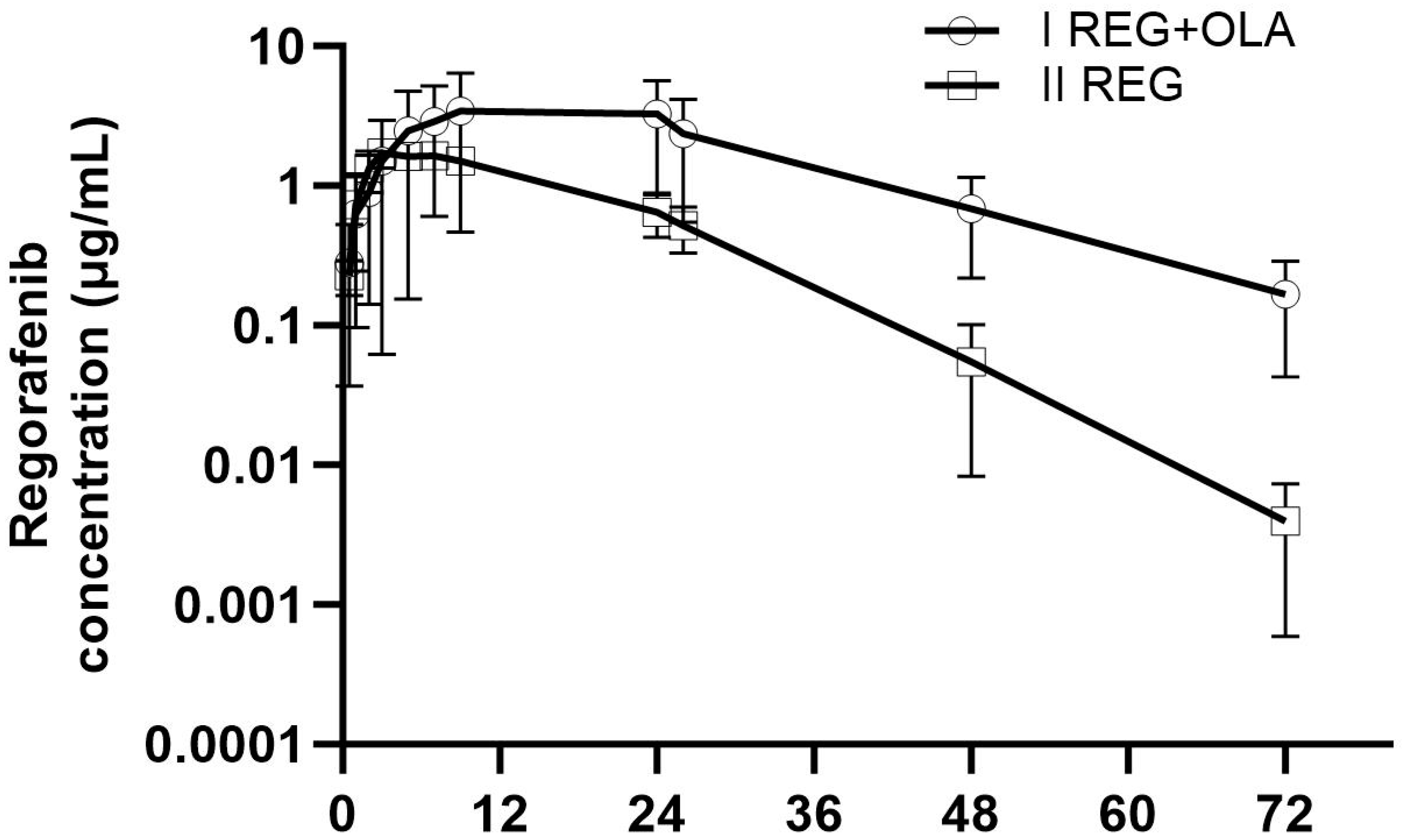
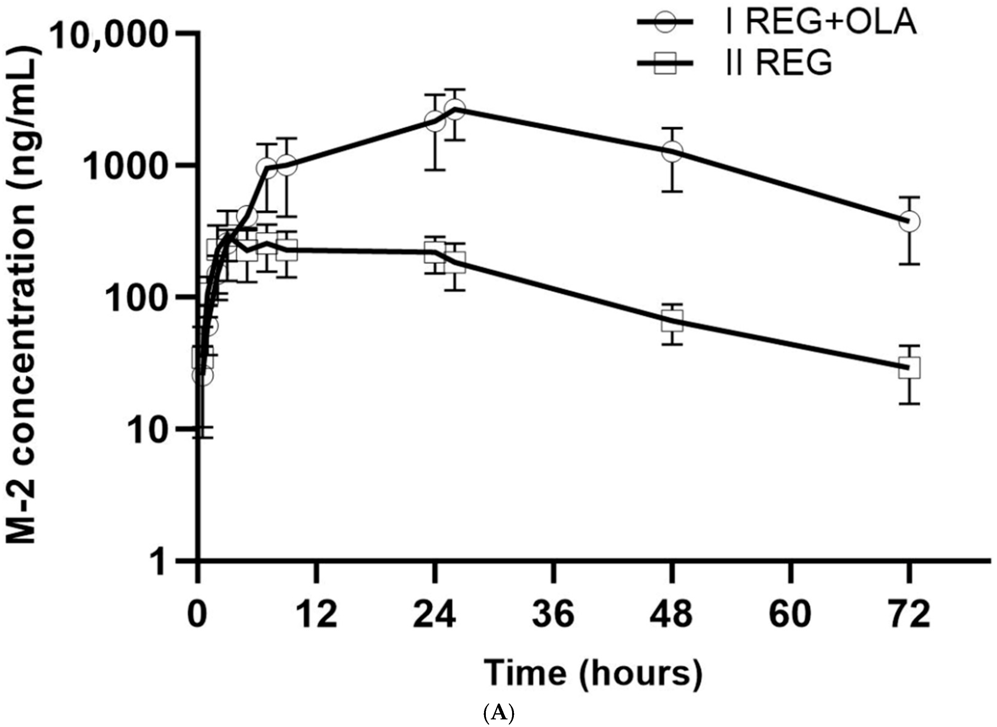
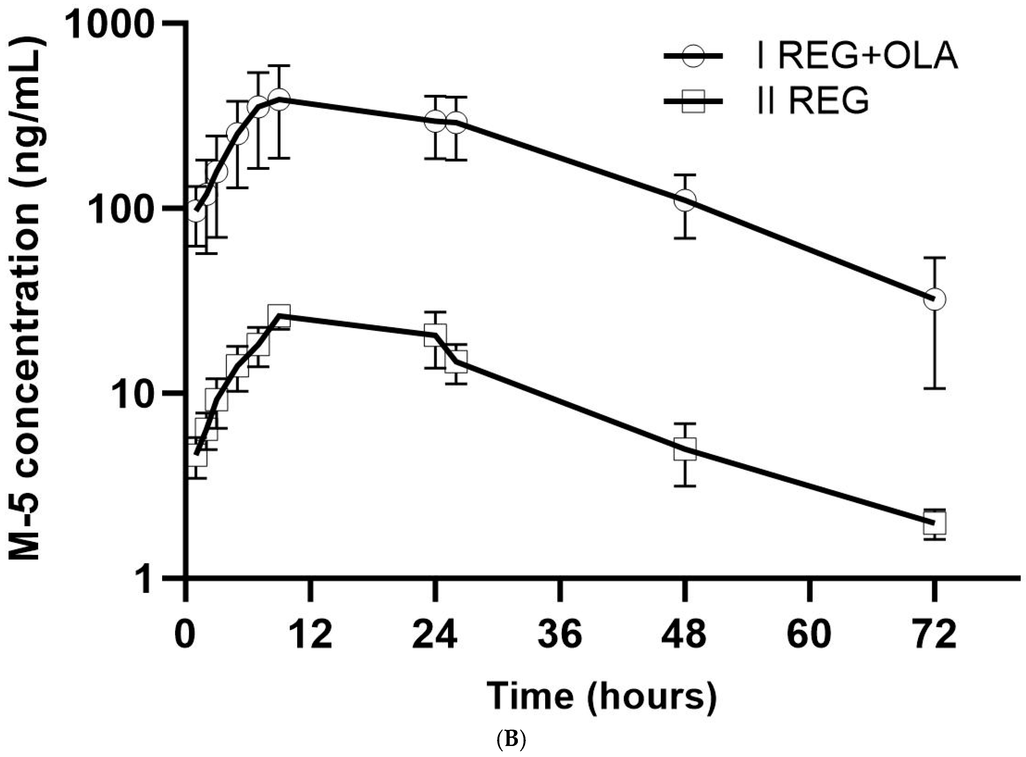
3.2. The Influence of OLA on the Pharmacokinetics of REG, M-2, and M-5

The mean plasma concentrations of REG, M-2, and M-5 in the study and control groups are shown in Figure 2 and Figure 3A,B. The pharmacokinetic profile of REG, M-2, and M-5 was shown to be significantly altered during co-administration of OLA (Table 1). After OLA administration, the pharmacokinetic parameters of REG (AUC0–∞, tmax, and t0.5) increased significantly by 3.38-, 2.66-, and 1.82-fold, respectively (p-value < 0.05). On the other hand, REG elimination parameters, i.e., kel and Cl/F, were significantly reduced in the study group compared to the control group by 1.77- and 1.70-fold, respectively.
During combined administration of OLA and REG, Cmax and AUC0–t values for M-2 were 7.22 and 8.86 times higher, respectively, and for M-5, Cmax, AUC0–t, and AUC0–∞ were 16.32, 17.83, and 18.82 times higher, respectively.
The Metabolite M-2/Parent and Metabolite M-5/Parent ratios for the Cmax and AUC0–t increased by 6.52-, 10.74-, 28-, and 13-fold, respectively.
The sum of the Cmax values for REG, M-2, and M-5 in the study group IREG+OLA (5844.08 ± 3793.77 ng/mL; CV = 64.9%) was significantly greater (p = 0.0089) than in the control IIREG group (2078.95 ± 691.36 ng/mL; CV = 33.3%).

3.3. The Influence of REG on the Pharmacokinetics of OLA

The mean plasma concentration–time profiles and the pharmacokinetic parameters of OLA in the control (IIIOLA) and study (IREG+OLA) group are shown in Figure 4 and Table 2, respectively. After the administration of OLA with REG, the Cmax, AUC0–t, and AUC0–∞ of OLA increased 2.0, 3.4, and 3.4-fold, respectively, compared to the control group. Meanwhile, Cl/F and Vd/F of OLA in the presence of REG significantly decreased (p = 0.0006 and 0.0281, respectively). The value of the AUC0–∞ ratio (study group/control group) was 3.41.

4. Discussion

Data from the literature suggest that OLA may be used in combination with other anticancer drugs to treat cancers associated with different gene mutations [11,12,13,14]. For example, OLA is proving to be effective in the treatment of cancers with ATM mutations, such as colorectal cancer, for which REG is used [12,13]. OLA has also been shown to be well-tolerated and highly effective in prolonging survival in patients with advanced gastrointestinal cancers, including hepatocellular carcinoma, when used in combination with other anticancer drugs [17,18,19,20,21].
OLA and REG are metabolized by the CYP3A4 isoenzyme of cytochrome P450. Both drugs are also substrates and inhibitors of the membrane transporters P-glycoprotein and BCRP [6,7,8,9,10,15,22,32,33,36,37]. Therefore, the potential concomitant use of OLA and REG may result in clinically relevant drug–drug interactions.
Knowledge of the influence of membrane transporters and cytochrome P450 enzymes on the pharmacokinetics of drugs makes it possible to assess their impact on the efficacy and safety of therapy [45,46,47].
In this regard, the study aimed to evaluate the bilateral pharmacokinetic interactions of OLA and REG and its active metabolites after a single administration in healthy rats.

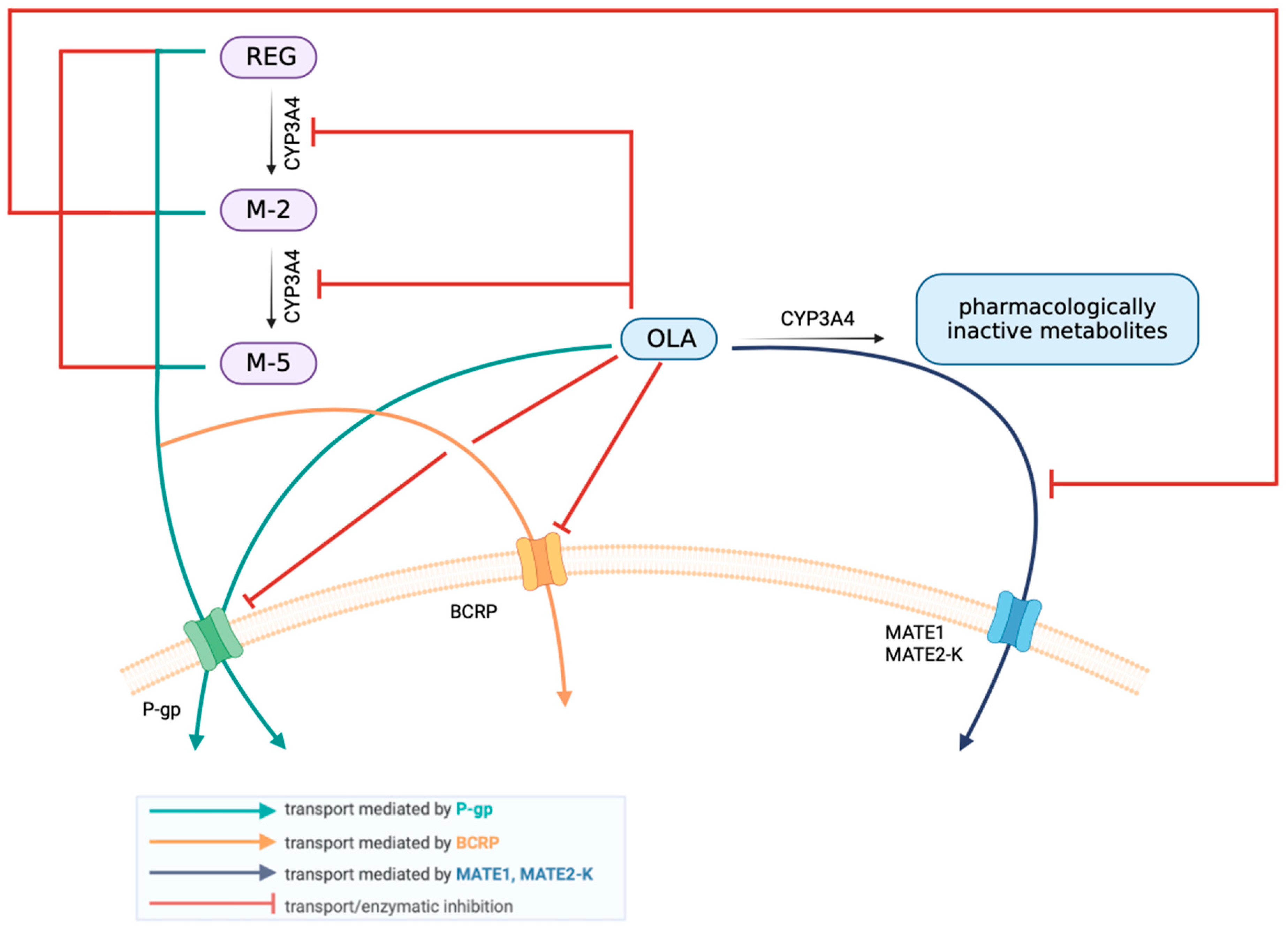
4.1. The Effect of OLA on the Pharmacokinetics of REG, M-2, and M-5

We found a significant 3.38-fold increase in AUC0–∞ (p = 0.0289) of REG (victim) when co-administered with OLA (perpetrator) compared to the control group. Parameters such as tmax (p = 0.0069) and t0.5 (p = 0.0003) were also significantly greater than in the control group.
However, no significant differences were observed for REG regarding Cmax, AUC0–t, and Vd/F in the study group compared to the control group. Co-administration of REG with OLA also affected the Cmax and AUC0–t of M-2 and M-5, which increased significantly, compared to the group of animals receiving REG alone (Table 1).
One reason for the observed increase in the AUC of REG and its active metabolites in the presence of OLA may be the inhibition by OLA of the P-gp efflux pump and BCRP in the small intestine [33,35,36].
Also, an inhibitory effect of OLA on the BCRP transporter and the CYP3A4 isoenzyme in the liver, of which REG and its active metabolites are substrates, cannot be excluded [8,32,33,35,37]. Although the inhibitory effect of OLA on P-gp and BCRP is rather insignificant for hepatic efflux, it is potentially important for intestinally expressed transporters [15].
Inhibition of the renal P-gp pump by OLA may also be the reason for the increase in plasma Cmax of M-2 and M-5, which are P-gp substrates [8,33,35,36]. Human studies have shown that REG, as well as M-2 and M-5, tend to accumulate in the blood after administration of REG at doses above 80 mg per day [48].
The increase in exposure to REG, M-2, and M-5 can also be explained by increased absorption as seen on plasma concentration–time profiles (Figure 2 and Figure 3A,B).
OLA, by inhibiting the CYP3A4 isoenzyme in the liver, may also reduce the metabolism of M-2, resulting in an increase in its plasma concentration, as M-2 is further metabolized by CYP3A4 [48].
The value of AUCin the presence of perpetrator/AUCin the absence of perpetrator is 3.39 and therefore seems to indicate that OLA was a moderate inhibitor in this animal model [47,49].
In contrast, the elimination process parameters of REG, i.e., kel and Cl/F, were significantly reduced compared to the control group, which may indicate reduced and prolonged elimination of REG from the body.
The Metabolite M-2/Parent and Metabolite M-5/Parent ratios for the Cmax and AUC0–t increased significantly in the IREG+OLA group. Also, the sum of the Cmax values for REG, M-2, and M-5 in the study group IREG+OLA (5844.08 ± 3793.77 ng/mL) was significantly greater (p = 0.0089) than in the control IIREG group (2078.95 ± 691.36 ng/mL).
The percentage of active metabolite to parent drug is important in determining dosing regimens. If it is greater than 10%, the potency of the pharmacological effect should be considered [50].
According to Steinbronn et al. [35], the recommendation to use AUCMetabolite/AUCParent drug ≥ 0.25 as the cutoff point for stratifying metabolite screening for CYP inhibition in vitro adequately identifies metabolites with the potential to cause DDIs in vivo. In our study, the ratio of the AUC M-2 to the AUC REG was 0.27.
When a single dose of REG is administered to humans, the peak plasma concentrations of M-2 and M-5 are much lower than those of the parent compound. However, at steady state, the concentrations of both REG and its metabolites are comparable [8].
Preclinical studies have shown that increased bioavailability of REG resulting from pharmacokinetic interactions may be associated with increased anti-tumor efficacy [51,52]. A study by Fukudo et al. [53] showed that adequate blood levels of REG, but also of its metabolites, were significant for progression-free survival (PSF) in patients with advanced metastatic colorectal cancer, gastrointestinal stromal tumor, and hepatocellular carcinoma.
Increased exposure of the body to REG may result in an increase in its pharmacological effect [51,52] but also in the occurrence of side effects, particularly in the form of liver dysfunction, rash, general weakness, the occurrence of hand-foot syndrome, or the development of hypertension [8,10].
OLA as an inhibitor of P-gp, BCRP, OATP1B1, OCT1, OCT2, OAT3, MATE1, and MATE2-K (IC50 values ranging from 5.5 to 47.1 μM) may interact with many drugs [15,22,32,33,36,37]. According to the Australian Public Assessment Report for Olaparib [15], OLA may alter the exposures of co-administered drugs that are substrates of CYP3A (in the intestine), P-glycoprotein (in the intestine), BCRP (in the intestine), OATP1B1, OAT3, OCT2, MATE1, and MATE2K. Regarding the ability of OLA to inhibit P-gp and BCRP transporters, the IC50 values are higher than 10 μM [15], which means that OLA has a low potential to inhibit P-gp and BCRP.
REG is not a substrate of OCT2, MATE1, and MATE2-K [8]. Therefore, inhibition of these transporters by OLA does not affect the pharmacokinetics of REG.
It is not yet known whether OLA will have clinically relevant DDIs when co-administered with substrates of these transporters, but this cannot be excluded. Among other things, it has been shown that caution should be exercised when OLA is co-administered with statins [54].
Zhao et al. [54] report that not only OLA but also other PARP inhibitors, i.e., niraparib, rucaparib, and veliparib, have inhibitory effects on many membrane transporters. The authors note that close monitoring for adverse effects is recommended when PARP inhibitors are administered concomitantly with some membrane transporter substrates due to the risk of clinically significant interactions.
In the authors’ previous preclinical study [40], it was shown that OLA significantly alters the pharmacokinetics of metformin. Co-administration of OLA and metformin in rats resulted in a 2.8-fold increase in metformin Cmax and a 2.6-fold increase in AUC. This increase in metformin exposure under clinical conditions may result in a risk of adverse effects, particularly in the gastrointestinal tract [40]. As reported by McCormick et al. [32,33], OLA (17–500 μM) inhibited CYP3A4/5 with an IC50 of 119 μM. In time-dependent CYP inhibition assays, OLA (10 μM) had little effect on CYP3A4/5, whereas at 2–200 μM, it acted as a time-dependent CYP3A4/5 inhibitor [32,33]. As the IC50 for OLA is 119 µM for reversible inhibition, systemic exposure is unlikely to inhibit hepatic CYP3A activity. However, given the much higher concentrations of OLA in the gastrointestinal tract (3683 µM from a 400 mg dose) [15], inhibition of intestinal CYP3A activity is possible. CYP3A1 is the rat orthologue of CYP3A4, having 73% amino acid homology with human CYP3A4. It is also regarded as the most metabolically relevant isoform in rats [55]. The activity of CYP3A in male rats is closest to that in humans [56,57].
Therefore, clinically significant drug–drug interactions due to OLA inhibition or induction of hepatic or intestinal CYP3A4/5 cannot be excluded.
The authors concluded that OLA should be used with caution in patients with a narrow therapeutic range or sensitive CYP3A substrates.
However, Deng et al. [36] observed that OLA is a weak inhibitor of atorvastatin transport in P-gp vesicles and would have at most a minor effect on atorvastatin and rosuvastatin efflux in the small intestine.
A significant increase in tmax REG was seen in the presence of OLA. Both drugs, REG and OLA, are lipophilic [8,10], which means that they may compete for the rate and extent of dissolution in the gastrointestinal tract. This may have slowed the absorption of REG, in particular, which is more sensitive to pH changes. As both drugs are substrates of P-gp present in the gut [8,10], mutual competition may slow the absorption of REG, leading to a delayed achievement of maximum blood concentration. Co-administration of both drugs may also stress metabolic pathways, delaying intestinal absorption and the onset of tmax of REG.

4.2. The Effect of REG on the Pharmacokinetics of OLA

The values of OLA concentrations were higher in the study group than in the control group (Figure 4). It was also shown that REG increased the PK parameters of OLA: AUC0–t and AUC0–∞ by 241.7% and 240.6%, respectively, and Cmax by 104.9%.
Both OLA [15,22,32] and REG [6,7,8,9,10] are substrates of the CYP3A4 isoenzyme, so the increased bioavailability of OLA may be due to the higher affinity of REG for CYP3A4. Like REG [8], OLA is a substrate of P-glycoprotein [15].
Both drugs also have an inhibitory effect on this membrane transporter. In vitro data suggest that REG is an inhibitor of P-glycoprotein with an IC50 value of approximately 2 μM [10], which means that REG is a moderate inhibitor of P-glycoprotein. REG may have the potential to inhibit intestinal absorption of a co-administered P-gp substrate, as a 160 mg REG dose can be estimated to have a duodenal concentration of 1.3 mM (based on an assumed duodenal volume of 250 mL), which is significantly higher than the IC50 for P-gp inhibition of 2.2 µM [8,10].
The increase in OLA levels may, therefore, be due to the inhibitory effect of REG on the P-gp efflux pump in the small intestine [8,9,10]. The two drugs thus compete for both CYP3A4 affinity and affinity for membrane transporters.
REG is also an inhibitor of OCT1, OCT2, MATE1, and MATE2-K [8]. As OLA is a substrate of MATE2-K [15], the increase in its blood concentration in rats after co-administration with REG may also be due to reduced renal elimination as a result of renal inhibition of MATE1/2-K by REG. This may confirm the reduction in OLA clearance values in the study group.
In the authors’ previous study investigating the interaction of REG and atorvastatin, a significant effect of REG on the pharmacokinetics of atorvastatin metabolites, which is also a CYP3A4 substrate, was confirmed [39].
However, according to Jin et al. [37], kinase inhibitors, including REG, did not cause a significant IC50 shift for CYP3A inhibition. The authors found in an in vitro study that REG, masitinib, and vatalanib were competitive inhibitors of CYP3A with Ki values of 20.7, 1.3, and 0.2 μM, respectively. REG and other kinase inhibitors such as axitinib, nintedanib, tozasertib, and trametinib were predicted to cause very small AUC increases (≤1.2-fold) for (S)- and/or (R)-warfarin, suggesting a low risk of DDIs with warfarin via CYP inhibition. REG was also predicted to have little or no effect on the AUCs of apixaban and rivaroxaban. In vitro studies with human hepatic microsomes or recombinant enzymes showed that REG competitively inhibits CYP2C8, CYP2B6, CYP2C9, CYP2C19, and CYP3A4 with R1 values > 1.1; M-2 inhibits CYP2C9, CYP2C8, CYP2D6, and CYP3A4 with R1 values > 1.1 and M-5 inhibits CYP2C8 with a R1 value > 1.1. CYP3A4 contributes significantly to REG metabolism but works in conjunction with UGT1A9 [58].
The value of the AUC0–∞ ratio (study group/control group) was 3.41, indicating that REG was a moderate inhibitor [47] in this preclinical study.
Drug–drug interaction studies with OLA have shown that concomitant use of strong and moderate CYP3A4 inhibitors, which can significantly increase the AUC and Cmax of this drug, should be avoided. These include itraconazole (170 and 42% increase in AUC and Cmax of OLA, respectively) [59] or fluconazole (121 and 14% increase in AUC and Cmax of OLA, respectively) [54]. Therefore, according to Zhao et al. [54], if a concomitant CYP3A4 inhibitor is required, the dose of OLA should be reduced to 100 mg twice daily for a strong CYP3A4 inhibitor, or to 150 mg twice daily for a moderate CYP3A4 inhibitor.

4.3. Summary Including Interspecies Differences

Due to interspecies differences, the translation of data from rats to humans often requires adjustments and meticulous interpretation. It is known that CYP enzymes and UGTs show significant interspecies variability, particularly in their hepatic and intestinal expression. P-gp function appears consistent across species, while BCRP and MRPs exhibit marked differences in activity and substrate specificity. These variations highlight the importance of considering species-specific characteristics when extrapolating preclinical data to humans [56,60].
Comparison of the biotransformation of REG in humans and various animal species revealed significant differences in the phase 1 and phase 2 reactions in vitro and in vivo. In humans, N-oxidation of pyridine (M-2 formation) was much more pronounced than methyl hydroxylation, both in vitro and in vivo. Glucuronidation as a primary biotransformation pathway of REG played an important role only in humans. M-2 appears to be one of the major oxidative metabolites of REG in rats. M-2 formation in the small intestine is higher in humans than in rodents. However, in all species, more M-2 was formed in the liver than in the small intestine [61].
In humans, UGT1A9 and UGT1A1 are the primary UGT enzymes involved in the glucuronidation of REG. UGT1A9 is predominantly expressed in the liver and kidney, while UGT1A1 is also present in the liver and intestine. Both enzymes contribute to the formation of REG glucuronide, the major glucuronidated metabolite in humans.
In rats, the UGT isoforms differ slightly from those in humans, with UGT1A1 and UGT2B1 being key enzymes in glucuronidation [62].
The choice of the optimal animal species to reproduce the pharmacokinetics of a drug in humans is challenging, especially in studies of DDI. The most commonly used animals are mice, rats, rabbits, dogs, monkeys, and, to a lesser extent, hamsters and guinea pigs. However, none of these models are perfect counterparts of human metabolic pathways [57,60].
However, rats are a common model for studying drug pharmacokinetics, including drug–drug interactions [63].
P-gp in rats has similar localization to human tissue, though differences in expression levels are reported. The activity of P-gp at the blood–brain barrier (BBB) is generally higher in rodents (rats and mice) compared to humans [64].
There are no significant species differences in P-gp function across hepatocytes between humans and rats [65].
Generally, in rats, P-gp abundance is higher in the intestine (124.1 pmol/g) compared to that in the liver (26.6 pmol/g) [66].
BCRP in rats is similarly localized, but its expression levels can vary from humans, often being lower in the intestines and kidneys. Lower BCRP levels in rat intestines lead to higher oral bioavailability of BCRP substrate drugs in rats compared to humans, while lower BCRP expression in rat kidneys results in slower renal elimination and longer systemic circulation times for these drugs in rats [67,68,69].
Absolute quantification of BCRP/Bcrp protein in the liver of different species, and the rank order as dog > rat > monkey ≈ human, was established by Li et al. [70].
Protein expression levels (fmol/cm2) of MDR1 and BCRP at the BBB in humans are 9.88-fold and 5.23-fold smaller than those in rats, respectively [71]. Generally, BCRP in rats is most abundant in the intestinal segments, while OATP 1a1, 1a4, 1b2, and 2a1 and MRP 2 and 6 are mainly detected in the liver [66].
Cytochrome P450 and UDP-glycosyltransferase enzymes in rats were detected mainly in the liver [66].
Cao et al. demonstrated that rats and humans show similar drug intestinal absorption profiles and transporter expression patterns in the small intestine, but distinct metabolizing enzyme expression levels and patterns make it challenging to predict human drug metabolism or oral bioavailability. The expression of CYP3A4/CYP3A9 and UDPG enzymes showed a 12- to 193-fold difference between human and rat intestine [72].
Although REG disposition varies between rats and humans due to differences in protein-binding and glucuronidation, it is significantly due to enterohepatic circulation. REG is not a substrate for OATP1 and OATP2, which means that ABCC2-mediated hepatobiliary excretion and OATP1/OATP2-mediated hepatic uptake do not play important roles in the disposition of REG [73].
Of course, the anatomical and physiological differences between rats and humans also need to be considered. In general, the anatomical structure of the gastrointestinal tract of white rats is similar to that of humans [74,75].
However, rats have relatively higher portal blood flow in relation to body weight. This may lead to faster drug metabolism and clearance from the liver. In rats, bile secretion is continuous at a constant rate due to the lack of a gallbladder. This may result in continuous recirculation of the drug, albeit at potentially lower concentrations, as bile is not concentrated in the same way as in humans. Rats have a higher activity of certain intestinal enzymes (such as β-glucuronidase) involved in the breakdown of drug conjugates excreted in the bile. This enzymatic activity allows the drug to be deconjugated and reabsorbed, improving the process of intestinal-hepatic recirculation [76].
There is a good correlation between the protein binding observed for drugs in human plasma and that for rats, although compounds tend to be slightly more bound to human plasma proteins compared to those from rats [77].

4.4. Limitations of the Study

The study performed has certain limitations. The differences among species regarding CYP450 should be considered. However, the activity of CYP3A in male rats is closest to that in humans, with CYP3A1, the rat orthologue of CYP3A4, exhibiting 73% amino acid homology with its human counterpart. In order to obtain reliable results and to verify the effect of the pathological condition on the pharmacokinetics of OLA and REG, a future preclinical study should be performed in animals with induced cancer. The pharmacokinetics of these drugs after repeated dosing were also not evaluated. OLA metabolites were also not determined.

5. Conclusions

OLA was shown to significantly affect the pharmacokinetics of REG and its active metabolites M-2 and M-5 in rats after co-administration of both drugs. There was also a significant effect of REG on the pharmacokinetics of OLA, which may have clinical relevance. The interaction observed in our study was mainly due to metabolic inhibition by the effects of both drugs on the CYP3A isoenzyme. The results obtained need to be confirmed in clinical studies. This study may provide guidance on the safety of using both drugs in clinical practice.

Author Contributions

Conceptualization, A.K. (Agnieszka Karbownik), D.S.-F. and E.S.; methodology, A.K. (Agnieszka Karbownik), D.S.-F. and E.S.; software, F.O., A.W. and T.G.; validation, J.M., A.K. (Alicja Kuźnik), A.K. (Agnieszka Karbownik) and F.O.; formal analysis, D.S.-F., A.K. (Agnieszka Karbownik) and E.S.; investigation, D.S.-F., A.K. (Alicja Kuźnik), J.M., F.O., J.S.-R. and A.K. (Agnieszka Karbownik); resources, A.K. (Agnieszka Karbownik) and D.S.-F.; data curation, D.S.-F., A.K. (Agnieszka Karbownik), F.O., T.G. and A.W.; writing—original draft preparation, D.S.-F., A.K. (Agnieszka Karbownik), F.O. and E.S.; writing—review and editing, D.S.-F., A.K. (Agnieszka Karbownik), E.S. and T.G.; visualization, D.S.-F., F.O., T.G. and A.K. (Agnieszka Karbownik); supervision, E.S. and E.G.; project administration, E.S.; funding acquisition, J.M., A.K. (Alicja Kuźnik), E.S. and D.S.-F. All authors have read and agreed to the published version of the manuscript.

Funding

This research was funded by Poznan University of Medical Sciences, grant number NSB0000025. The funding source had no effect on any part of the study, preparation, or submission of the manuscript.

Institutional Review Board Statement

The study was conducted according to the guidelines of the Declaration of Helsinki and approved by the Ethics Committee of Poznań University of Life Sciences, Department of Animal Physiology and Biochemistry, Wołyńska 35 Str., 60-637 Poznań, Poland (consent No. 85/2022).

Informed Consent Statement

Not applicable.

Data Availability Statement

The data presented in this study are available on request from the corresponding author.

Acknowledgments

The figure illustrating the interaction between regorafenib and olaparib was created using the BioRender.com platform (free access version). Figure 2, Figure 3 and Figure 4 were generated using GraphPad Prism 10.0.3. Part of the work was carried out with the support of the Center of Innovative Pharmaceutical Technology at Poznań University of Medical Sciences.

Conflicts of Interest

The authors declare no conflicts of interest. A.W. works for Hy-Line International. The company had no role in the design of the study, in the collection, analysis, or interpretation of data, in the writing of the manuscript, or in the decision to publish the results.

References

  1. Oliveira, R.F.; Oliveira, A.I.; Cruz, A.S.; Ribeiro, O.; Afreixo, V.; Pimentel, F. Polypharmacy and drug interactions in older patients with cancer receiving chemotherapy: Associated factors. BMC Geriatr. 2024, 24, 557. [Google Scholar] [CrossRef] [PubMed]
  2. Gholipourshahraki, T.; Aria, A.; Sharifi, M.; Moghadas, A.; Moghaddas, A. Potential Drug Interactions in Hospitalized Hematologic Cancer Patients: New Update with New Chemotherapy Regimens. J. Res. Pharm. Pract. 2024, 12, 115–122. [Google Scholar] [CrossRef] [PubMed]
  3. Lam, C.S.; Hua, R.; Au-Doung, P.L.W.; Wu, Y.K.; Koon, H.K.; Zhou, K.R.; Loong, H.H.; Chung, V.C.; Cheung, Y.T. Association between potential supplement-drug interactions and liver diseases in patients with cancer: A large prospective cohort study. Clin. Nutr. ESPEN 2023, 58, 152–159. [Google Scholar] [CrossRef]
  4. Xi, Y.; Xu, P. Global colorectal cancer burden in 2020 and projections to 2040. Transl. Oncol. 2021, 14, 101174. [Google Scholar] [CrossRef]
  5. Klimeck, L.; Heisser, T.; Hoffmeister, M.; Brenner, H. Colorectal cancer: A health and economic problem. Best Pract. Res. Clin. Gastroenterol. 2023, 66, 101839. [Google Scholar] [CrossRef]
  6. Xu, D.; Liu, Y.; Tang, W.; Xu, L.; Liu, T.; Jiang, Y.; Zhou, S.; Qin, X.; Li, J.; Zhao, J.; et al. Regorafenib in Refractory Metastatic Colorectal Cancer: A Multi-Center Retrospective Study. Front. Oncol. 2022, 12, 838870. [Google Scholar] [CrossRef]
  7. Xu, X.; Yu, Y.; Liu, M.; Liang, L.; Liu, T. Efficacy and safety of regorafenib and fruquintinib as third-line treatment for colorectal cancer: A narrative review. Transl. Cancer Res. 2022, 11, 276–287. [Google Scholar] [CrossRef] [PubMed]
  8. Australian Public Assessment Report for Regorafenib, April 2019. Available online: https://www.tga.gov.au/sites/default/files/auspar-regorafenib-190404.pdf (accessed on 1 September 2024).
  9. Granito, A.; Marinelli, S.; Forgione, A.; Renzulli, M.; Benevento, F.; Piscaglia, F.; Tovoli, F. Regorafenib Combined with Other Systemic Therapies: Exploring Promising Therapeutic Combinations in HCC. J. Hepatocell. Carcinoma 2021, 8, 477–492. [Google Scholar] [CrossRef]
  10. Rey, J.B.; Launay-Vacher, V.; Tournigand, C. Regorafenib as a single-agent in the treatment of patients with gastrointestinal tumors: An overview for pharmacists. Target. Oncol. 2015, 10, 199–213. [Google Scholar] [CrossRef]
  11. Papageorgiou, G.I.; Fergadis, E.; Skouteris, N.; Christakos, E.; Tsakatikas, S.A.; Lianos, E.; Kosmas, C. Case Report: Combination of Olaparib with Chemotherapy in a Patient with ATM-Deficient Colorectal Cancer. Front. Oncol. 2021, 11, 788809. [Google Scholar] [CrossRef]
  12. Wang, C.; Jette, N.; Moussienko, D.; Bebb, D.G.; Lees-Miller, S.P. ATM-Deficient Colorectal Cancer Cells Are Sensitive to the PARP Inhibitor Olaparib. Transl. Oncol. 2017, 10, 190–196. [Google Scholar] [CrossRef] [PubMed]
  13. Jette, N.R.; Kumar, M.; Radhamani, S.; Arthur, G.; Goutam, S.; Yip, S.; Kolinsky, M.; Williams, G.J.; Bose, P.; Lees-Miller, S.P. ATM-Deficient Cancers Provide New Opportunities for Precision Oncology. Cancers 2020, 12, 687. [Google Scholar] [CrossRef] [PubMed]
  14. Gong, H.; Su, Y.; Zhao, L.; Ma, L.; Zhang, L.; Hou, L.; Li, T.; Niu, S.; Zhang, H.; Li, C.; et al. Efficacy and safety of targeted drugs in advanced or metastatic gastric and gastroesophageal junction cancer: A network meta-analysis. J. Clin. Pharm. Ther. 2022, 47, 493–506. [Google Scholar] [CrossRef]
  15. Australian Public Assessment Report for Olaparib, February 2018. Available online: https://www.tga.gov.au/sites/default/files/auspar-olaparib-210415.pdf (accessed on 1 September 2024).
  16. EMA Recommends Extension of Therapeutic Indications for Olaparib to Patients with Endometrial Cancer. 17 July 2024. Available online: https://www.esmo.org/oncology-news/ema-recommends-extension-of-therapeutic-indications-for-olaparib-to-patients-with-endometrial-cancer (accessed on 20 August 2024).
  17. Yang, X.D.; Kong, F.E.; Qi, L.; Lin, J.X.; Yan, Q.; Loong, J.H.C.; Xi, S.Y.; Zhao, Y.; Zhang, Y.; Yuan, Y.F.; et al. PARP inhibitor Olaparib overcomes Sorafenib resistance through reshaping the pluripotent transcriptome in hepatocellular carcinoma. Mol. Cancer 2021, 20, 20. [Google Scholar] [CrossRef]
  18. Lai, Y.H.; Tung, K.C.; Chen, S.C. Durable response to olaparib combined low-dose cisplatin in advanced hepatocellular carcinoma with FANCA mutation: A case report. Medicine 2022, 101, e30719. [Google Scholar] [CrossRef]
  19. Sun, G.; Miao, G.; Li, Z.; Zheng, W.; Zhou, C.; Sun, G.; Cao, H.; Li, Z.; Tang, W. Inhibition of PARP Potentiates Immune Checkpoint Therapy through miR-513/PD-L1 Pathway in Hepatocellular Carcinoma. J. Oncol. 2022, 2022, 6988923. [Google Scholar] [CrossRef]
  20. Xu, H.; Ma, Z.; Mo, X.; Chen, X.; Xu, F.; Wu, F.; Chen, H.; Zhou, G.; Xia, H.; Zhang, C. Inducing Synergistic DNA Damage by TRIP13 and PARP1 Inhibitors Provides a Potential Treatment for Hepatocellular Carcinoma. J. Cancer 2022, 13, 2226–2237. [Google Scholar] [CrossRef] [PubMed]
  21. Zhao, J.; Sun, Y.; Lin, H.; Chou, F.; Xiao, Y.; Jin, R.; Cai, X.; Chang, C. Olaparib and enzalutamide synergistically suppress HCC progression via the AR-mediated miR-146a-5p/BRCA1 signaling. FASEB J. 2020, 34, 5877–5891. [Google Scholar] [CrossRef]
  22. Zeng, Y.; Arisa, O.; Peer, C.J.; Fojo, A.; Figg, W.D. PARP inhibitors: A review of the pharmacology, pharmacokinetics, and pharmacogenetics. Semin. Oncol. 2024, 51, 19–24. [Google Scholar] [CrossRef]
  23. Dopazo, C.; Søreide, K.; Rangelova, E.; Mieog, S.; Carrion-Alvarez, L.; Diaz-Nieto, R.; Primavesi, F.; Stättner, S. Hepatocellular carcinoma. Eur. J. Surg. Oncol. 2024, 50, 107313. [Google Scholar] [CrossRef]
  24. Do, K.T.; Kochupurakkal, B.; Kelland, S.; de Jonge, A.; Hedglin, J.; Powers, A.; Quinn, N.; Gannon, C.; Vuong, L.; Parmar, K.; et al. Phase 1 Combination Study of the CHK1 Inhibitor Prexasertib and the PARP Inhibitor Olaparib in High-grade Serous Ovarian Cancer and Other Solid Tumors. Clin. Cancer Res. 2021, 27, 4710–4716. [Google Scholar] [CrossRef] [PubMed]
  25. Shah, P.D.; Wethington, S.L.; Pagan, C.; Latif, N.; Tanyi, J.; Martin, L.P.; Morgan, M.; Burger, R.A.; Haggerty, A.; Zarrin, H.; et al. Combination ATR and PARP Inhibitor (CAPRI): A phase 2 study of ceralasertib plus olaparib in patients with recurrent, platinum-resistant epithelial ovarian cancer. Gynecol. Oncol. 2021, 163, 246–253. [Google Scholar] [CrossRef] [PubMed]
  26. Wethington, S.L.; Shah, P.D.; Martin, L.; Tanyi, J.L.; Latif, N.; Morgan, M.; Torigian, D.A.; Rodriguez, D.; Smith, S.A.; Dean, E.; et al. Combination ATR (ceralasertib) and PARP (olaparib) Inhibitor (CAPRI) Trial in Acquired PARP Inhibitor-Resistant Homologous Recombination-Deficient Ovarian Cancer. Clin. Cancer Res. 2023, 29, 2800–2807. [Google Scholar] [CrossRef] [PubMed]
  27. Lloyd, R.L.; Wijnhoven, P.W.G.; Ramos-Montoya, A.; Wilson, Z.; Illuzzi, G.; Falenta, K.; Jones, G.N.; James, N.; Chabbert, C.D.; Stott, J.; et al. Combined PARP and ATR inhibition potentiates genome instability and cell death in ATM-deficient cancer cells. Oncogene 2020, 39, 4869–4883. [Google Scholar] [CrossRef]
  28. Konstantinopoulos, P.A.; Barry, W.T.; Birrer, M.; Westin, S.N.; Cadoo, K.A.; Shapiro, G.I.; Mayer, E.L.; O’Cearbhaill, R.E.; Coleman, R.L.; Kochupurakkal, B.; et al. Olaparib and α-specific PI3K inhibitor alpelisib for patients with epithelial ovarian cancer: A dose-escalation and dose-expansion phase 1b trial. Lancet Oncol. 2019, 20, 570–580. [Google Scholar] [CrossRef]
  29. Zimmer, A.S.; Nichols, E.; Cimino-Mathews, A.; Peer, C.; Cao, L.; Lee, M.J.; Kohn, E.C.; Annunziata, C.M.; Lipkowitz, S.; Trepel, J.B.; et al. A phase I study of the PD-L1 inhibitor, durvalumab, in combination with a PARP inhibitor, olaparib, and a VEGFR1-3 inhibitor, cediranib, in recurrent women’s cancers with biomarker analyses. J. Immunother. Cancer 2019, 7, 197. [Google Scholar] [CrossRef]
  30. Fok, J.H.L.; Ramos-Montoya, A.; Vazquez-Chantada, M.; Wijnhoven, P.W.G.; Follia, V.; James, N.; Farrington, P.M.; Karmokar, A.; Willis, S.E.; Cairns, J.; et al. AZD7648 is a potent and selective DNA-PK inhibitor that enhances radiation, chemotherapy and olaparib activity. Nat. Commun. 2019, 10, 5065. [Google Scholar] [CrossRef]
  31. Li, J.; Wang, R.; Kong, Y.; Broman, M.M.; Carlock, C.; Chen, L.; Li, Z.; Farah, E.; Ratliff, T.L.; Liu, X. Targeting Plk1 to Enhance Efficacy of Olaparib in Castration-Resistant Prostate Cancer. Mol. Cancer Ther. 2017, 16, 469–479. [Google Scholar] [CrossRef]
  32. McCormick, A.; Swaisland, H.; Reddy, V.P.; Learoyd, M.; Scarfe, G. In vitro evaluation of the inhibition and induction potential of olaparib; a potent poly(ADP-ribose) polymerase inhibitor; on cytochrome P450. Xenobiotica 2018, 48, 555–564. [Google Scholar] [CrossRef]
  33. McCormick, A.; Swaisland, H. In vitro assessment of the roles of drug transporters in the disposition and drug-drug interaction potential of olaparib. Xenobiotica 2017, 47, 903–915. [Google Scholar] [CrossRef]
  34. Kobayashi, K.; Sugiyama, E.; Shinozaki, E.; Wakatsuki, T.; Tajima, M.; Kidokoro, H.; Aoyama, T.; Nakano, Y.; Kawakami, K.; Hashimoto, K.; et al. Associations among plasma concentrations of regorafenib and its metabolites; adverse events; and ABCG2 polymorphisms in patients with metastatic colorectal cancers. Cancer Chemother. Pharmacol. 2021, 87, 767–777. [Google Scholar] [CrossRef] [PubMed]
  35. Steinbronn, C.; Yang, X.; Yu, J.; Dimova, H.; Huang, S.M.; Ragueneau-Majlessi, I.; Isoherranen, N. Do Inhibitory Metabolites Impact DDI Risk Assessment? Analysis of in vitro and in vivo Data from NDA Reviews Between 2013 and 2018. Clin. Pharmacol. Ther. 2021, 110, 452–463. [Google Scholar] [CrossRef]
  36. Deng, F.; Sistonen, J.; Neuvonen, M.; Niemi, M. Inhibition of efflux transporters by poly ADP-ribose polymerase inhibitors. Basic Clin. Pharmacol. Toxicol. 2023, 133, 428–436. [Google Scholar] [CrossRef]
  37. Jin, S.; Paludetto, M.N.; Kurkela, M.; Kahma, H.; Neuvonen, M.; Xiang, X.; Cai, W.; Backman, J.T. In vitro assessment of inhibitory effects of kinase inhibitors on CYP2C9; 3A and 1A2: Prediction of drug-drug interaction risk with warfarin and direct oral anticoagulants. Eur. J. Pharm. Sci. 2024, 203, 106884. [Google Scholar] [CrossRef]
  38. Directive 2010/63/EU of the European Parliament and of the Council of 22 September 2010 on the Protection of Animals Used for Scientific Purposes. Available online: https://eur-lex.europa.eu/LexUriServ/LexUriServ.do?uri=OJ:L:2010:276:0033:0079:EN:PDF (accessed on 6 October 2023).
  39. Szkutnik-Fiedler, D.; Szałek, E.; Otto, F.; Czyrski, A.; Karaźniewicz-Łada, M.; Wolc, A.; Grześkowiak, E.; Lewandowski, K.; Karbownik, A. Pharmacokinetic interaction between regorafenib and atorvastatin in rats. Pharmacol. Rep. 2024, 76, 1184–1195. [Google Scholar] [CrossRef]
  40. Stanisławiak-Rudowicz, J.; Karbownik, A.; Szkutnik-Fiedler, D.; Otto, F.; Grabowski, T.; Wolc, A.; Grześkowiak, E.; Szałek, E. Bidirectional pharmacokinetic drug interactions between olaparib and metformin. Cancer Chemother. Pharmacol. 2024, 93, 79–88. [Google Scholar] [CrossRef] [PubMed]
  41. Burden, N.; Chapman, K.; Sewell, F.; Robinson, V. Pioneering better science through the 3Rs: An introduction to the national centre for the replacement; refinement; and reduction of animals in research (NC3Rs). J. Am. Assoc. Lab. Anim. Sci. 2015, 54, 198–208. [Google Scholar] [PubMed]
  42. Su, G.; Qin, L.; Su, X.; Tao, C.; Wei, Y. Gender-dependent pharmacokinetics of olaparib in rats determined by ultra-high performance liquid chromatography/electrospray ionization tandem mass spectrometry. Biomed. Chromatogr. 2020, 34, e4791. [Google Scholar] [CrossRef]
  43. Gu, E.M.; Liu, Y.N.; Pan, L.; Hu, Y.; Ye, X.; Luo, P. A high throughput method for Monitoring of Sorafenib; regorafenib; cabozantinib and their metabolites with UPLC-MS/MS in rat plasma. Front. Pharmacol. 2022, 13, 955263. [Google Scholar] [CrossRef]
  44. ICH Guideline M10 on Bioanalytical Method Validation and Study Sample Analysis. 25 July 2022. Available online: https://www.ema.europa.eu/en/ich-m10-bioanalytical-method-validation-scientific-guideline (accessed on 15 November 2023).
  45. Cole, S.; Kerwash, E.; Andersson, A. A summary of the current drug interaction guidance from the European Medicines Agency and considerations of future updates. Drug Metab. Pharmacokinet. 2020, 35, 2–11. [Google Scholar] [CrossRef]
  46. Choi, Y.H. Interpretation of Drug Interaction Using Systemic and Local Tissue Exposure Changes. Pharmaceutics 2020, 12, 417. [Google Scholar] [CrossRef]
  47. In Vitro Drug Interaction Studies—Cytochrome P450 Enzyme- and Transporter-Mediated Drug Interactions. Guidance for Industry. U.S. Department of Health and Human Services Food and Drug Administration Center for Drug Evaluation and Research (CDER). January 2020. Clinical Pharmacology. Available online: https://www.xenotech.com/access-adme-research-resources/resources/2020-fda-guidance-for-industry-2/ (accessed on 11 December 2023).
  48. Rousseau, B.; Boukerma, A.K.; Henriques, J.; Cohen, R.; Lucidarme, O.; Borg, C.; Tournigand, C.; Kim, S.; Bachet, J.B.; Mazard, T.; et al. Impact of trough concentrations of regorafenib and its major metabolites M-2 and M-5 on overall survival of chemorefractory metastatic colorectal cancer patients: Results from a multicentre GERCOR TEXCAN phase II study. Eur. J. Cancer 2022, 168, 99–107. [Google Scholar] [CrossRef] [PubMed]
  49. Yu, J.; Zhou, Z.; Tay-Sontheimer, J.; Levy, R.H.; Ragueneau-Majlessi, I. Risk of clinically relevant pharmacokinetic-based drug-drug interactions with drugs approved by the U.S. Food and Drug Administration between 2013 and 2016. Drug Metab. Dispos. 2018, 46, 835–845. [Google Scholar] [CrossRef] [PubMed]
  50. Molenaar-Kuijsten, L.; Van Balen, D.E.M.; Beijnen, J.H.; Steeghs, N.; Huitema, A.D.R. A review of CYP3A drug–drug interaction studies: Practical guidelines for patients using targeted oral anticancer drugs. Front. Pharmacol. 2021, 12, 670862. [Google Scholar] [CrossRef] [PubMed]
  51. Fu, Q.; Chen, M.; Anderson, J.T.; Sun, X.; Hu, S.; Sparreboom, A.; Baker, S.D. Interaction Between Sex and Organic Anion-Transporting Polypeptide 1b2 on the Pharmacokinetics of Regorafenib and Its Metabolites Regorafenib-N-Oxide and Regorafenib-Glucuronide in Mice. Clin. Transl. Sci. 2019, 12, 400–407. [Google Scholar] [CrossRef]
  52. Khachatryan, V.; Muazzam, A.; Hamal, C.; Velugoti, L.S.D.R.; Tabowei, G.; Gaddipati, G.N.; Mukhtar, M.; Alzubaidee, M.J.; Dwarampudi, R.S.; Mathew, S.; et al. The Role of Regorafenib in the Management of Advanced Gastrointestinal Stromal Tumors: A Systematic Review. Cureus 2022, 14, e28665. [Google Scholar] [CrossRef]
  53. Fukudo, M.; Asai, K.; Tani, C.; Miyamoto, M.; Ando, K.; Ueno, N. Pharmacokinetics of the oral multikinase inhibitor regorafenib and its association with real-world treatment outcomes. Investig. New Drugs 2021, 39, 1422–1431. [Google Scholar] [CrossRef]
  54. Zhao, D.; Long, X.; Wang, J. Transporter-mediated drug-drug interactions involving poly (ADP-ribose) polymerase inhibitors (Review). Oncol. Lett. 2023, 25, 161. [Google Scholar] [CrossRef]
  55. Sun, Z.; Zhang, Z.; Ji, M.; Yang, H.; Cromie, M.; Gu, J.; Wang, C.; Yang, L.; Yu, Y.; Gao, W.; et al. BDE47 induces rat CYP3A1 by targeting the transcriptional regulation of miR-23b. Sci. Rep. 2016, 6, 31958. [Google Scholar] [CrossRef]
  56. Bogaards, J.J.; Bertrand, M.; Jackson, P.; Oudshoorn, M.J.; Weaver, R.J.; van Bladeren, P.J.; Walther, B. Determining the best animal model for human cytochrome P450 activities: A comparison of mouse, rat, rabbit, dog, micropig, monkey and man. Xenobiotica 2000, 30, 1131–1152. [Google Scholar] [CrossRef]
  57. Zuber, R.; Anzenbacherová, E.; Anzenbacher, P. Cytochromes P450 and experimental models of drug metabolism. J. Cell. Mol. Med. 2002, 6, 189–198. [Google Scholar] [CrossRef] [PubMed]
  58. Stivarga (regorafenib) Tablets. Drug Approval Package. Available online: https://www.accessdata.fda.gov/drugsatfda_docs/nda/2012/203085Orig1s000TOC.cfm (accessed on 10 November 2024).
  59. Dirix, L.; Swaisland, H.; Verheul, H.M.; Rottey, S.; Leunen, K.; Jerusalem, G.; Rolfo, C.; Nielsen, D.; Molife, L.R.; Kristeleit, R.; et al. Effect of Itraconazole and Rifampin on the Pharmacokinetics of Olaparib in Patients with Advanced Solid Tumors: Results of Two Phase I Open-label Studies. Clin. Ther. 2016, 38, 2286–2299. [Google Scholar] [CrossRef]
  60. Pasanen, M. Species differences in CYP enzymes. In Citocromo P40; Agnosto, M.C., Comez-Lechon, M.J., Eds.; Instituto de Espana, Real academia Nacional de Farmacia: Madrid, Spain, 2004; pp. 63–90. [Google Scholar]
  61. Kojima, A.; Sogabe, A.; Nadai, M.; Katoh, M. Species differences in oxidative metabolism of regorafenib. Xenobiotica 2021, 51, 1400–1407. [Google Scholar] [CrossRef] [PubMed]
  62. Furukawa, T.; Naritomi, Y.; Tetsuka, K.; Nakamori, F.; Moriguchi, H.; Yamano, K.; Terashita, S.; Tabata, K.; Teramura, T. Species differences in intestinal glucuronidation activities between humans, rats, dogs and monkeys. Xenobiotica 2014, 44, 205–216. [Google Scholar] [CrossRef]
  63. Kazuki, Y.; Kobayashi, K.; Hirabayashi, M.; Abe, S.; Kajitani, N.; Kazuki, K.; Takehara, S.; Takiguchi, M.; Satoh, D.; Kuze, J.; et al. Humanized UGT2 and CYP3A transchromosomic rats for improved prediction of human drug metabolism. Proc. Natl. Acad. Sci. USA 2019, 116, 3072–3081. [Google Scholar] [CrossRef] [PubMed]
  64. Verscheijden, L.F.M.; Koenderink, J.B.; de Wildt, S.N.; Russel, F.G.M. Differences in P-glycoprotein activity in human and rodent blood-brain barrier assessed by mechanistic modelling. Arch. Toxicol. 2021, 95, 3015–3029. [Google Scholar] [CrossRef] [PubMed]
  65. Li, M.; Yuan, H.; Li, N.; Song, G.; Zheng, Y.; Baratta, M.; Hua, F.; Thurston, A.; Wang, J.; Lai, Y. Identification of interspecies difference in efflux transporters of hepatocytes from dog, rat, monkey and human. Eur. J. Pharm. Sci. 2008, 35, 114–126. [Google Scholar] [CrossRef]
  66. Sharma, S.; Singh, D.K.; Mettu, V.S.; Yue, G.; Ahire, D.; Basit, A.; Heyward, S.; Prasad, B. Quantitative Characterization of Clinically Relevant Drug-Metabolizing Enzymes and Transporters in Rat Liver and Intestinal Segments for Applications in PBPK Modeling. Mol. Pharm. 2023, 20, 1737–1749. [Google Scholar] [CrossRef]
  67. Kawahara, I.; Nishikawa, S.; Yamamoto, A.; Kono, Y.; Fujita, T. Assessment of contribution of BCRP to intestinal absorption of various drugs using portal-systemic blood concentration difference model in mice. Pharmacol. Res. Perspect. 2020, 8, e00544. [Google Scholar] [CrossRef]
  68. Sharma, S.; Mettu, V.S.; Prasad, B. Interplay of Breast Cancer Resistance Protein (Bcrp/Abcg2), Sex, and Fed State in Oral Pharmacokinetic Variability of Furosemide in Rats. Pharmaceutics 2023, 15, 542. [Google Scholar] [CrossRef]
  69. Nakanishi, T.; Tamai, I. Interaction of Drug or Food with Drug Transporters in Intestine and Liver. Curr. Drug Metab. 2015, 16, 753–764. [Google Scholar] [CrossRef]
  70. Li, N.; Palandra, J.; Nemirovskiy, O.V.; Lai, Y. LC-MS/MS mediated absolute quantification and comparison of bile salt export pump and breast cancer resistance protein in livers and hepatocytes across species. Anal. Chem. 2009, 81, 2251–2259. [Google Scholar] [CrossRef] [PubMed]
  71. Uchida, Y.; Yagi, Y.; Takao, M.; Tano, M.; Umetsu, M.; Hirano, S.; Usui, T.; Tachikawa, M.; Terasaki, T. Comparison of absolute protein abundances of transporters and receptors among blood-brain barriers at different cerebral regions and blood-spinal cord barrier in human and rat. Mol. Pharm. 2020, 17, 2006–2020. [Google Scholar] [CrossRef]
  72. Cao, X.; Gibbs, S.; Fang, L.; Miller, H.; Landowski, C.; Shin, H.; Lennernas, H.; Zhong, Y.; Amidon, G.; Yu, L.; et al. Why is it Challenging to Predict Intestinal Drug Absorption and Oral Bioavailability in Human Using Rat Model. Pharm. Res. 2006, 23, 1675–1686. [Google Scholar] [CrossRef] [PubMed]
  73. Hotta, K.; Ueyama, J.; Tatsumi, Y.; Tsukiyama, I.; Sugiura, Y.; Saito, H.; Matsuura, K.; Hasegawa, T. Lack of Contribution of Multidrug Resistance-associated Protein and Organic Anion-transporting Polypeptide to Pharmacokinetics of Regorafenib, a Novel Multi-Kinase Inhibitor, in Rats. Anticancer Res. 2015, 35, 4681–4689. [Google Scholar] [PubMed]
  74. Hryn, V.; Kostylenko, Y.; Yushchenko, Y.; Lavrenko, A.; Ryabushko, O. General comparative anatomy of human and white rat digestive systems: A bibliographic analysis. Wiad. Lek. 2018, 71, 1599–1602. [Google Scholar]
  75. Wu, T.C.; Ma, C.; Jeong, H.; Yao, H.M.; Chiou, W.L. Comparison of gastrointestinal absorption between human and rat: The role of intestinal absorptive surface area and unstirred aqueous layer. Food Chem. Toxicol. 2002, 40, 1727–1728. [Google Scholar] [CrossRef]
  76. DeSesso, J.M.; Jacobson, C.F. Anatomical and physiological parameters affecting gastrointestinal absorption in humans and rats. Food Chem. Toxicol. 2001, 39, 209–228. [Google Scholar] [CrossRef]
  77. Colclough, N.; Ruston, L.; Wood, J.M.; MacFaul, P.A. Species differences in drug plasma protein binding. Med. Chem. Commun. 2014, 5, 963–967. [Google Scholar] [CrossRef]
Figure 1. Pharmacokinetic interaction between OLA and REG.
Pharmaceutics 16 01575 g001
Figure 2. The REG plasma concentration–time profiles in rats after oral administration of a single dose of REG (20 mg/kg b.w.) to the IIREG group and REG + OLA (20 mg/kg b.w. + 100 mg/kg b.w.) to the IREG+OLA group.
Pharmaceutics 16 01575 g002
Figure 3. (A). The M-2 plasma concentration–time profiles in rats receiving REG (IIREG) and REG+OLA (IREG+OLA). (B). The M-5 plasma concentration–time profiles in rats receiving REG (IIREG) and REG+OLA (IREG+OLA).
Pharmaceutics 16 01575 g003aPharmaceutics 16 01575 g003b
Figure 4. The OLA plasma concentration–time profiles in rats after oral administration of a single dose of OLA (100 mg/kg b.w.) to the IIIOLA group and REG + OLA (20 mg/kg b.w. + 100 mg/kg b.w.) to the IREG+OLA group.
Pharmaceutics 16 01575 g004
Table 1. Pharmacokinetic parameters of REG and its metabolites M-2 and M-5 after oral administration of a single dose of REG (20 mg/kg b.w.) to the IIREG group and REG + OLA (20 mg/kg b.w. + 100 mg/kg b.w.) to the IREG+OLA group.
Pharmacokinetic ParametersIREG+OLA
(n = 8)
IIREG
(n = 8)
p-Value
IREG+OLA vs. IIREG
Gmean Ratio **
(90% CI)
IREG+OLA
vs. IIREG
REG
Cmax (µg/mL)3.69 ± 2.97
(80.5)
1.87 ± 0.31
(16.5)
0.50541.37
(0.73; 2.58)
AUC0–t
(ng × h/mL)
106.92 ± 84.63
(79.2)
36.76 ± 5.84
(15.9)
0.05181.71
(0.73; 4.04)
AUC0–∞ (ng × h/mL)124.47 ± 79.63
(64.0)
36.85 ± 5.83
(15.8)
0.02892.54
(1.38; 4.67)
tmax (h)14.1 ± 8.2
(58.2)
5.8 ± 2.1
(36.9)
0.00692.27
(1.47; 3.51)
kel (h−1)0.06 ± 0.02
(26.1)
0.11 ± 0.03
(22.8)
0.0015 *0.56
(0.44; 0.71)
t0.5 (h)11.65 ± 3.70
(31.7)
6.37 ± 1.33
(20.8)
0.00031.79
(1.41; 2.26)
Cl/F (mL/h × kg)129.86 ± 160.06
(123.1)
220.83 ± 29.74
(13.5)
0.02050.37
(0.20; 0.68)
Vd/F (mL)2025.57 ± 2097.73
(103.5)
2062.53 ± 615.85
(29.7)
0.33570.66
(0.34; 1.27)
M-2
Cmax (ng/mL)2469.92 ± 1257.44
(50.9)
342.10 ± 132.87
(38.8)
0.00076.83
(4.52; 10.33)
AUC0–t (ng × h/mL)85,176.80 ± 35,788.00
(42.0)
9615.36 ± 2275.41
(23.7)
0.00078.38
(5.92; 11.87)
AUC0–∞ (ng × h/mL)2469.92 ± 1257.44
(50.9)
11,469.93 ± 2133.05
(18.6)
0.00076.83
(4.52; 10.33)
Metabolite M-2/Parent ratio
Cmax1.24 ± 0.89
(71.9)
0.44 ± 0.73
(39.76)
0.00075.73
(3.34; 9.83)
AUC0–t1.44 ± 1.09
(75.9)
0.27 ± 0.08
(29.77)
0.00076.39
(3.07; 13.29)
M-5
Cmax (ng/mL)428.18 ± 194.48
(45.4)
26.23 ± 3.98
(15.2)
0.000714.23
(10.39; 19.63)
AUC0–t (ng × h/mL)14,324.20 ± 5574.43
(38.9)
803.19 ± 177.02
(22.0)
0.000716.13
(12.03; 21.62)
AUC0–∞ (ng × h/mL)15,959.41 ± 4837.53
(30.31)
847.88 ± 178.97
(21.1)
0.001618.43
(14.19; 23.93)
Metabolite M-5/Parent ratio
Cmax0.28 ± 0.22
(78.1)
0.03 ± 0.06
(170.1)
0.000711.93
(5.47; 26.01)
AUC0–t0.26 ± 0.17
(66.2)
0.02 ± 0.01
(23.33)
0.000712.29
(4.22; 35.74)
AUC0–∞0.37 ± 0.19
(50.9)
0.02 ± 0.01
(22.82)
0.00409.61
(4.41; 20.93)
Cmax, maximum observed plasma concentration; AUC0–t, area under the plasma concentration–time curve from zero to the time of last measurable concentration; AUC0–∞, area under the plasma concentration–time curve from zero to infinity; tmax, time to the first occurrence of Cmax; kel, elimination rate constant; t0.5, half-life in the elimination phase; Cl/F, apparent plasma drug clearance; Vd/F, apparent volume of distribution. Arithmetic means and standard deviations (SDs) are shown with coefficients of variation CVs (%) in brackets. * Student’s t-test; the remaining data were analyzed using the Mann–Whitney test for non-normality. ** Geometric mean (Gmean), ratio between the IREG+OLA and IIREG groups (%) with a 90% confidence interval (CI) in the brackets.
Table 2. Pharmacokinetic parameters of OLA after oral administration of a single dose of OLA (100 mg/kg b.w.) to the IIIOLA group and REG + OLA (20 mg/kg b.w. + 100 mg/kg b.w.) to the IREG+OLA group.
Pharmacokinetic ParametersIREG+OLA
(n = 8)
IIIOLA
(n = 8)
IREG+OLA vs. IIIOLA
p-Value
Gmean Ratio **
(90% CI)
IREG+OLA
vs. IIIOLA
Cmax (µg/mL)17.87 ± 7.66 (42.9)8.72 ± 6.80 (78.0)0.02412.86
(1.36; 6.03)
AUC0–t (µg × h/mL)96.53 ± 33.23 (34.4)28.28 ± 28.16 (99.6)0.00065.98
(2.42; 14.77)
AUC0–∞ (µg × h/mL)97.14 ± 33.26 (34.2)28.58 ± 28.31 (99.1)0.00065.93
(2.41; 14.62)
kel (1/h)0.25 ± 0.06 (23.2)0.44 ± 0.34 (77.5)0.0583 *0.59
(0.30; 1.13)
tmax (h)1.28 ± 1.11 (86.3)1.16 ± 0.92 (78.9)0.9120 *1.32
(0.62; 2.80)
t0.5 (h)3.01 ± 0.86 (28.6)4.79 ± 6.06 (126.5)0.0650 *1.71
(0.88; 3.30)
Cl/F (L/h)0.43 ± 0.16 (38.3)7.84 ± 15.21 (193.9)0.0006 *0.15
(0.06; 0.37)
Vd/F (L)1.88 ± 1.05 (55.9)22.49 ± 24.81 (110.3)0.0281 *0.26
(0.10; 0.65)
Cmax, maximum observed plasma concentration; AUC0–t, area under the plasma concentration–time curve from zero to the time of last measurable concentration; AUC0–∞, area under the plasma concentration–time curve from zero to infinity; tmax, time to the first occurrence of Cmax; kel, elimination rate constant; t0.5, half-life in the elimination phase; Cl/F, apparent plasma drug clearance; Vd/F, apparent volume of distribution. Arithmetic means and standard deviations (SDs) are shown with coefficients of variation CVs (%) in brackets. * Mann–Whitney test for non-normality; other data were analyzed using Student’s t-test. ** Geometric mean (Gmean), ratio between the IREG+OLA and IIIOLA groups (%) with a 90% confidence interval (CI) in the brackets.
Disclaimer/Publisher’s Note: The statements, opinions and data contained in all publications are solely those of the individual author(s) and contributor(s) and not of MDPI and/or the editor(s). MDPI and/or the editor(s) disclaim responsibility for any injury to people or property resulting from any ideas, methods, instructions or products referred to in the content.

