Article

In Vitro and In Vivo Antimelanogenesis Effects of Leaf Essential Oil from Agathis dammara

1
School of Forestry and Resource Conservation, National Taiwan University, Taipei 10617, Taiwan
2
Department of Animal Science and Technology, National Taiwan University, Taipei 10617, Taiwan
3
Department of Forestry, National Chung Hsing University, Taichung 40227, Taiwan
4
Special Crop and Metabolome Discipline Cluster, Academy Circle Economy, National Chung Hsing University, Taichung 40227, Taiwan
*
Author to whom correspondence should be addressed.
Pharmaceutics 2023, 15(9), 2269; https://doi.org/10.3390/pharmaceutics15092269
Submission received: 4 August 2023 / Revised: 28 August 2023 / Accepted: 30 August 2023 / Published: 2 September 2023
(This article belongs to the Topic Bioactive Phytochemicals from Plant Essential Oils)

Abstract

:
Agathis species are widely distributed around Southeast Asia, Australasia, South Pacific islands, and etc. Traditionally, Agathis species have been used as the folk medicines, the common ethnopharmacological uses of Agathis genus are the treatments of headache and myalgia. This study aims to investigate the chemical composition of Agathis dammara (Lamb.) Rich. leaf essential oil and to explore its antimelanogenesis effect. The chemical constituents of leaf essential oil are analyzed using gas chromatography-mass spectrometry (GC-MS), the major constituents of leaf essential oil are sesquiterpenoids. The major constituents are δ-cadinene (16.12%), followed by γ-gurjunene (15.57%), 16-kaurene (12.43%), β-caryophyllene (8.58%), germacrene D (8.53%), and γ-cadinene (5.33%). As for the in vitro antityrosinase activity, leaf essential oil inhibit the tyrosinase activity of mushroom when the substrate is 3,4-dihydroxyphenylalanine (L-DOPA). Leaf essential oil prevents tyrosinase from acting as diphenolase and catalyzing L-DOPA to dopaquinone, and converting into dark melanin pigments. A. dammara leaf essential oil also exhibits the in vivo antimelanogenesis effect, leaf essential oil reduces 43.48% of melanin formation in zebrafish embryos at the concentration of 50 μg/mL. Results reveal A. dammara leaf essential oil has the potential for developing the skin whitening drug and depigmentation ingredient for hyperpigmentary disorders.

1. Introduction

Araucariaceae is an ancient family and widespread in the Jurassic period, distribution in both hemispheres including numerous taxa. Araucariaceae species are present in South America, Australasia, South Pacific islands, Southeast Asia and etc. Araucariaceae family contains four genera, including Agathis, Araucaria, Columbea, and Wollemia, com-monly known as kauri or dammar. The ethnopharmacological uses of the Araucariaceae family are versatile and numerous, including emollient and antiseptic properties, and to treat headache, myalgia, respiratory infections, rheumatisms, skin wounds, insomnia, and so on [1,2,3,4,5].
Genus Agathis are widely distributed in Australia, New Zealand, South Pacific islands, and Southeast Asia, the species are characterized by their brownish black bark, leathery and ovate-lanceolate leaves, circular branch scars on their trunks, and their milky resins. Agathis genus contains 18 accepted species and 4 unresolved species [1,2,3,4]. To date, the timbers of Agathis spp. are recognized as good materials for processing, the timber is straight-grained, heavier and harder wood, knot free, with a fine textured and lustrous surface [3]. Gum copal is harvested by tapping or cutting the living Agathis trees. Resins and gums from Agathis spp. have been used as varnishes, lacquers, adhesive, and etc. [1,5]. According to the related research reports, α-pinene, β-pinene, camphene, α-copaene, α-cubebene, β-caryophyllene, δ-cadinene, allo-aromadendrene, aromadendrene, germacrene D, limonene, myrcene, sabinene, spathulenol, and 16-kaurene are the most abundant constituents found in the essential oils of Agathis species [2,3]. The biological activities of genus Agathis include antibacterial, antifungal, anti-inflammatory, antileishmanial, antiplasmodial, cytotoxic activities, etc. in literatures [3,5,6,7,8]. Verma et al. investigated the antibacterial activity of resin essential oil from A. robusta (C. Moore ex F. Muell.) Bailey (Queensland Kauri), resin essential oil showed moderate activity against Staphylococcus epidermidis and good activity against S. aureus in the disc diffusion assay [5]. The leaf essential oil of A. dammara (Lamb.) Rich. was also found to have significant antibacterial activity against Staphylococcus aureus (Gram positive bacterium) and Pseudomonas aeruginosa (Gram negative bacterium) in the disc diffusion method and microwell dilution assay [6]. A. atropurpurea B. Hyland bark resin exhibited effective antileishmanial activity against Leishmania amazonensis promastigotes and amastigotes [7]. Resins from A. atropurpurea showed slight antifungal activity against Aspergillus niger and Rhizopus stolonifera, which are the important and widespread human and agricultural harmful pathogens [8].
Melanin, the products of melanogenesis, is produced in melanocytes and stored in melanosomes. Normally, melanin prevents skin from ultraviolet (UV) damage, photocarcinogenesis, and interference of vitamin D3 formation [9,10,11,12]. However, abnormal melanin production and accumulation could induce hyperpigmentation (age spots, freckles and melasma), and melanoma, the skin tumor [9,13,14,15]. In order to prevent from hyperpigmentation, suppressing tyrosinase activity is one of the most effective strategies. Tyrosinase (EC 1.14.18.1), a copper containing enzyme, is a key enzyme in melanogenesis. The site of copper ion pair (central domain) coordinating with 6 histidine residues formed an active site of tyrosinase [9,16,17]. Depending on the substrates binding to the active site, tyrosinase catalyzes two reactions. When tyrosinase binds with L-tyrosine (monophenol), it acts as monophenolase and helps hydroxylation of L-tyrosine. When tyrosinase (diphenolase) binds with 3,4-dihydroxyphenylalanine (L-DOPA), further oxidizes of L-DOPA to dopaquinone occurs, and leads to the formation of eumelanin and pheomelanin [16,17,18].
Currently, the well-known ingredients, hydroquinone, kojic acid, ascorbic acid, and arbutin, prevent L-tyrosine or L-DOPA converting to dopaquinone by inhibiting tyrosinase activity [17,19,20]. These compounds may bring about some disadvantages. For example, hydroquinone could cause skin irritation and cell mutation; kojic acid is carcinogenic; ascorbic acid and arbutin has poor storability [14,17,21,22,23]. Due to the drawbacks and obstacles, alternatives from natural sources have been increasingly researched [24,25,26,27,28,29]. Elaeocarpus serratus Linn. (Tiliaceae) leaf extract and its active compounds, gallic acid, myricetin and mearnsetin, present tyrosinase inhibitory activity and antimelanogenesis effect on zebrafish (Danio rerio) embryos [25]. Tetrahydrocurcumin, 1,7-bis(4-hydroxy-3-methoxyphenyl)heptane-3,5-dione, is the metabolite of curcumin, derived from the Curcuma longa L. rhizome. Tetrahydrocurcumin can inhibit the melanin production in B16F10 melanoma cells induced by melanocyte-stimulating hormone (α-MSH), and modulate the expressions of important cellular enzymes, tyrosinase, tyrosinase-related protein 1 (TRP-1), and tyrosinase-related protein 2 (TRP-2), which involved the biosynthetic production of melanin [26]. Green tea (Camellia sinensis (L.) Kuntze) extract and its constituents, including gallocatechin-3-O-gallate (GCG), epigallocatechin-3-O-gallate (EGCG), and epicatechin-3-O-gallate (ECG) exhibited the effective antityrosinase efficacy [27]. Several plant natural products were reported to possess the antityrosinase activity and antimelanogenesis effect, including citrus essential oils, sesamol, haginin A, and etc. [28,29]. Bioactive ginsenosides, including ginsenoside Rh6, vina-ginsenoside R4, vina-ginsenoside R13, ginsenoside Rh23, and floralginsenoside A, isolated from Panax ginseng C. A. Meyer (Araliaceae) leaf and berry exhibit the antimelanogenesis effect in zebrafish model [30,31,32]. The aims of the study are to investigate the chemical composition and antimelanogenesis effects (in vitro and in vivo) of A. dammara leaf essential oil.

2. Materials and Methods

2.1. Plant Material

The fresh and mature leaves (dark green) of Agathis dammara (Lamb.) Rich. were collected from the campus (25°01′03.0″ N 121°32′21.1″ E) of National Taiwan University, Taipei, Taiwan in March 2022. The diameter at breast height (DBH) of the tree is 31.9 cm, and the height is 11.6 m. The species was identified by Dr. Chih-Chieh Yu, Xishuangbanna Tropical Botanical Garden, Chinese Academy of Sciences. Voucher specimen (AD0322) has been kept in the laboratory of Chemical Utilization of Biomaterials, School of Forestry and Resource Conservation, National Taiwan University.

2.2. Hydrodistillation of Essential Oil

The fresh and mature leaves of A. dammara were hydrodistilled for 6 h by using the Clevenger apparatus. After the hydrodistillation, the leaf essential oil was placed in a dark glass bottle and stored in a refrigerator at 4 °C for further investigation [33,34,35].

2.3. Gas Chromatography-Mass Spectrometry (GC-MS) Analysis

To investigate the chemical composition of the essential oil, the analysis of A. dammara leaf oil was carried out on a Trace GC Ultra (Thermo Fisher Scientific, Waltham, MA, USA) equipped with a DB-5 MS column (Crossbond 5% methylpolysiloxane, 30.0 m length × 0.25 mm diameter, thickness 0.25 μm; Agilent Technologies, Palo Alto, CA, USA). The oven temperature started from 60 °C for 3 min, programed at 3 °C/min to 120 °C, 5 °C/min to 240 °C for 3 min. The injector temperature was held at 250 °C, and split ratio was 10:1. The carrier gas was helium; 1.0 mL/min flow rate; ion source temperature 250 °C; mass range 50–650 amu. The constituents of essential oil were characterized by National Institute of Standards and Technology (NIST) V.2.0 and Wiley 7.0 GC-MS libraries and Kovats indexes in the reference [36]. Kovats indexes of the constituents are determined by retention times of n-alkanes (C7–C30) on the DB-5MS column. The relative contents of constituents were determined by integrating the peaks on total ion chromatograms (TIC) [37,38].

2.4. Antityrosinase Assay

Tyrosinase is an essential enzyme in melanogenesis. The antimelanogeneic effect of specimen can be preliminarily determined by its tyrosinase inhibitory activity. The activity was determined by previous assays with minor modification [24,39]. The specimen-buffer mixture (110 μL) was added in a 96 well microplate. The specimen was diluted in DMSO and mixed with 0.1 M potassium phosphate buffer (pH = 6.8). Kojic acid (Sigma, St. Louis, MO, USA) and arbutin (Sigma, St. Louis, MO, USA) were used as the positive controls. Subsequently, 50 μL of 200 U/mL mushroom tyrosinase (EC 1.14.18.1; Sigma, St. Louis, MO, USA) was added and mixed with the specimen-buffer mixture. After mixed with 40 μL of substrate (L-tyrosine and L-DOPA), the mixture was incubated for 10 min at room temperature. Next, the 96-well microplate was sent into SPEOTROstar, the microplate reader (BMG Labtech, Ortenberg, Germany), and the absorbance (475 nm) of each well was measured. The tyrosinase inhibitory activity was presented as inhibition rate and half maximal inhibitory concentration (IC50). The equation of inhibition rate is as followed: Inhibition (%) = [1 − (Aspecimen − Aspecimen’s blank)/(Acontrol − Acontrol’s blank)] × 100. The IC50 was determined by concentration-response curve. The experiments were performed in triplicate.

2.5. Antimelanogenesis Effect in Zebrafish Embryos

Wild-type AB zebrafish (Danio rerio) were maintained under synchronized conditions (26–30 °C; pH = 6.5–7.5; 14 h/10 h of light/dark cycle). 9 h-post-fertilization (hpf) embryos were collected and arrayed in 24 well plate. Each well (1 mL) contains 5 embryos, E3 medium, and specimen diluted in 1% DMSO. After that, the plate was incubated at 28 °C for 48 h, and the 57 hpf zebrafish embryo were photographed under a stereomicroscope (Hamlet SEM-H, Taiwan) at 40× magnification. The melanin content of zebrafish embryos was quantified by ImageJ (V1.52a) and presented as inhibition rate. The calculation of inhibition is: Inhibition (%) = [1 − (Intensity specimen/Intensity control)] × 100. 1-Phenyl-2-thiourea (PTU), kojic acid, and arbutin were the positive controls [24,27,40]. The protocol was approved by the Institutional Animal Care and Use Committee (IACUC) in National Taiwan University (IACUC Approval No: NTU-112-EL-00072), all zebrafish were handled by the 3R’s principles of laboratory animal care and use.

2.6. Statistical Analysis

The statistical analysis of data obtained in the study was analyzed by SPSS (Statistical Product and Service Solutions) (Chicago, IL, USA) Version 16 with the Scheffe’s multiple comparison test, a post-hoc multiple comparison method. The confidence interval was set at the level of 95%.

3. Results and Discussion

3.1. Chemical Constituents of A. dammara Leaf Essential Oil

In this study, leaf essential oil was hydrodistilled from the mature dark green leaves of A. dammara. The yield of leaf essential oil was 0.15 ± 0.05%, on dry matter basis of leaf. Figure 1 showed the gas chromatogram of A. dammara leaf essential oil. The constituents of A. dammara leaf essential oil were analyzed by using GC-MS, and there are 21 constituents found in the chromatogram. According to Table 1, the leaf essential oil contained sesquiterpene hydrocarbons (69.35 ± 2.18%), diterpene hydrocarbons (13.64 ± 1.46%), and oxygenated sesquiterpenes (3.10 ± 0.21%). Except for 16-kaurene (12.43 ± 1.32%), most of the major constituents were sesquiterpenoids, including δ-cadinene (16.12 ± 0.53%), γ-gurjunene (15.57 ± 0.49%), β-caryophyllene (8.58 ± 0.94%), germacrene D (8.53 ± 0.20%), and γ-cadinene (5.33 ± 0.16%) (Figure 2). Chen et al. analyzed the chemical constituents of A. dammara leaf essential oil, the major components were limonene (36.81%), β-bisabolene (33.43%) and β-myrcene (25.48%) [6]; the content of monoterpenoids, limonene and β-myrcene, was over 50%. The result of the study is inconsistent with our analysis of the chemical compositions of leaf essential oil. New or young leaves of A. dammara are light green color, later turn dark green after a few days, and keep dark green color. Differences between two studies of chemical constituents of leaf essential oil may be due to the maturity of leaf, collected region, environment temperature, sunlight, season, and etc.
The sesquiterpenoids of leaf essential oil were dominated by compounds with cadinane skeleton, including α-muurolene, α-cadinene, γ-cadinene, δ-cadinene, trans-1,4-cadinadiene, α-calacorene, β-calacorene, 1-epi-cubenol, α-cadinol, and δ-cadinol. The compounds with copane skeleton were α-copaene and α-ylangene. α-Cubebene and β-cubebene were classified as compounds with cubebane skeleton. α-Caryophyllene and β-caryophyllene were characterized as caryophyllane skeleton. Other compounds, δ-elemene, γ-gurjunene, and germacrene D, were characterized as elemane skeleton, guaiane skeleton, and germacrene skeleton, respectively. As for the diterpenes, rimuene and 16-kaurene, they were classified as rosane and kaurane skeleton compounds.
The major constituents of A. dammara leaf essential oil were likewise observed in those of A. robusta F. M. Bailey and A. macrophylla (Lindl.) Mast., which share a same clade with A. dammara in the phylogenetic tree [41,42]. Brophy et al. reported that constituents of A. robusta leaf essential oil were spathulenol (36.7%), rimuene (5.6%), α-pinene (3.7%), caryophyllene oxide (3.1%), δ-cadinene (1.6%), β-caryophyllene (1.4%), β-pinene (1.2%), and germacrene D (1.1%) [2]. Verma et al. also reported the chemical constituents of A. robusta leaf essential oil was predominated by sesquiterpenoids (75.6%), containing β-selinene (18.1%), caryophyllene oxide (11.5%), spathulenol (10.5%), α-selinene (9.8%), γ -muurolene (5.8%), and etc.; the leaf essential oil also contained diterpenoid, rimuene (14.2%, second major component) [5]. The main constituents of A. atropurpurea the principal components were the monoterpene α-pinene (7.9%), the sesquiterpenes δ-cadinene (9.0%) and the diterpenes phyllocladene (12.5%) and 16-kaurene (19.4%) [2]. The identified constituents of A. dammara leaf essential oil were somewhat similar to those of of A. robusta, A. macrophylla, and A. atropurpurea leaf essential oils.

3.2. Tyrosinase Inhibitory Activity of A. dammara Leaf Essential Oil

Tyrosinase which catalyzes L-tyrosine and L-DOPA into dopaquinone and dopachrome, respectively, is the essential enzyme in melanogenesis. To inhibit the melanogenesis process, suppressing the tyrosinase activity is one of the most common and effective strategies [43].
Figure 3 shows that leaf essential oil could prevent tyrosinase from acting as diphenolase and catalyzing L-DOPA to dopaquinone. Tyrosinase inhibitory activity of A. dammara leaf essential oil presented the dose-dependent effect. The inhibition rates were 6.93 ± 1.37%, 18.36 ± 1.82%, and 28.38 ± 3.95% at the concentration of 100, 200, and 400 μg/mL, respectively. Kojic acid, positive control, presented inhibition of diphenolase. Kojic acid showed 69.15% inhibition at the concentration of 12.5 μg/mL, followed by 48.20%, 24.94%, 10.57% at the concentrations of 6.25, 3.13, and 1.56 μg/mL. The IC50 values of leaf essential oil and kojic acid were 690.02 ± 18.85 and 6.74 ± 0.09 μg/mL, respectively (Table 2). When the substrate was L-tyrosine, no significant inhibitory activity of leaf essential oil was observed. As for positive control, kojic acid (IC50 = 2.46 ± 0.06 μg/mL) presented the tyrosinase inhibitory activity (Table 2).
Huang et al. examined the tyrosinase inhibitory activity of Vitex negundo Linn. (Lamiaceae) leaf essential oil, the inhibition rate of leaf essential oil against tyrosinase were 26.6% at the concentration of 5 mg/mL, using L-DOPA as the substrate [44]. Cheraif et al. evaluated the antityrosinase activity of essential oils from six Algerian plants, Juniperus oxycedrus L. (Cupressaceae) essential oil displayed the best tyrosinase inhibitory activity with a tyrosinase inhibition rate of 39.65% at the concentration of 1 mg/mL [45]. Etlingera elatior (Jack) R. M. Sm. (Zingiberaceae) leaf essential oil exhibited a moderate activity against tyrosinase, with an IC50 value of 2.34 ± 0.04 mg/mL when using L-DOPA as the substrate [46]. Salleh et al. investigated the antityrosinase effects of leaf essential oil and bark essential oil of Beilschmiedia madang Blume (Lauraceae), using L-DOPA as the substrate, inhibition rates of leaf essential oil and bark essential oil were 53.1 ± 0.2% and 51.2 ± 0.2%, respectively, at the concentration of 1 mg/mL [47]. Pogostemon plectranthoides Desf. (Lamiaceae) leaf essential oils collected from three bio-geographical regions also had the antityrosinase activity. Leaf essential oils were rich in sesquiterpenes, and the active leaf essential oil possessed the tyrosinase inhibitory effect with the inhibition rate of ca. 50% at the concentration of 1 mg/mL, using L-DOPA as the substrate [48]. These related researches illustrated that A. dammara leaf essential oil exhibited the tyrosinase inhibition potential.

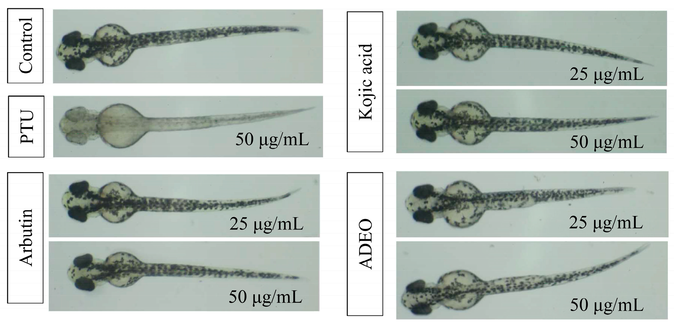
3.3. Antimelanogenesis Effect of A. dammara Leaf Essential Oil in Zebrafish

Zebrafish (D. rerio) has been a powerful and wide used model organism and the efficient alternative to other animal models in multiple researches. In the present study, zebrafish is a suitable vertebrate model for melanogenesis study because of its easily observed pigmentation and the similarity of gene sequences and organ systems [21,49,50]. The antimelanogenesis effects in the zebrafish model caused by different treatments were shown in Figure 4. In control group, the patterns of pigmentation presented normal dispersion. After treated with A. dammara leaf essential oil, kojic acid, and arbutin, the zebrafish embryos displayed less amount of pigmentation. As for PTU treatment, the zebrafish embryos became transparent after 48 h incubation, inhibition rate of melanin was 98.23 ± 1.01% at the concentration of 50 μg/mL.
Table 3 shows the inhibitory effect of each treatment on melanogenesis of zebrafish embryo, including leaf essential oil, arbutin, kojic acid, and PTU. The melanin contents of zebrafish embryos could decrease by 21.03%, 37.36% and 43.48% with treatment of leaf essential oil at a concentration of 12.5, 25 and 50 μg/mL, with a dose-dependent manner. In comparison with the treatment of arbutin, leaf essential oil presented higher antimelanogenesis effect at the concentrations of 25 and 50 μg/mL. Significant statistical differences (p < 0.05) were observed between the treatment of leaf essential oil (43.48%) and the treatment of arbutin (21.49%) at the concentration of 50 μg/mL. Another positive control, kojic acid, inhibited 18.53% and 20.44% of melanin production of zebrafish embryos at the concentrations of 25 and 50 μg/mL, respectively. Kojic acid exhibited the lower melanin inhibition in zebrafish embryos than leaf essential oil treatments did.
Chelly et al. (2021) evaluated the antimelanogenesis activities of methanolic extracts of Rhanterium suaveolens Desf. (Asteraceae) flower, stem, and leaf, with in vitro antityrosinase inhibition assay and in vivo zebrafish embryo assay [51]. Among these part extracts of R. suaveolens, flower extract exhibited the best antityrosinase effect on tyrosinase induced L-DOPA oxidation. Flower extract also possessed the statistically significant decrease of melanin formation in zebrafish embryo at the concentrations of 0.5 and 1 mg/mL. The reduction of melanin formation in zebrafish embryo was less than 50% after 48 h treatment of 1 mg/mL concentration of R. suaveolens flower extract. Dalbergia pinnata (Lour.) Prain essential oil inhibited 18.77% of melanin production of zebrafish embryos at a concentration of 30 μg/mL [52]. Bletilla striata (Thunb.) Rchb. f. tuber extract reduced 18.35% and 24.39% of melanin formation of zebrafish embryos at the concentrations of 10 μg/mL and 30 μg/mL, respectively [53]. The results in this study revealed that A. dammara leaf essential oil possessed a potent melanogenesis inhibition activity in zebrafish embryos.
Yang et al. investigated the antimelanogenesis effect of lime mint (Mentha aquatica × M. suaveolens) essential oil and its main constituents in B16F10 murine melanoma cells. The main constituents of lime mint essential oil were D-limonene (41.10%), D-carvone (8.58%), δ-selinene (6.73%), and β-caryophyllene (6.24%). Among these main constituents, only β-caryophyllene exhibited the antimelanogenesis activity in a dose dependent manner. Results revealed that β-caryophyllene can modulate the expression of melanogenesis related proteins, including tyrosinase, microphthalmia-associated transcription factor (MITF), TRP-1, and TRP-2. β-Caryophyllene reduces cellular melanogenesis by down regulating the expression of these melanogenesis related proteins, resulting in the decrease in melanin content of B16F10 murine melanoma cells at the concentration of 150 μM [54]. According this related research, compound β-caryophyllene might contribute the melanogenesis effect of A. dammar leaf essential oil.

4. Conclusions

For the assessment of antimelanogenesis effect of essential oil from A. dammara leaf, both in vitro and in vivo antimelanogenesis assays were conducted in this study. The chemical constituents of A. dammara leaf essential oil was dominated by sesquiterpenoids through the GC-MS analysis. The most abundant constituent was δ-cadinene (16.12%), followed by γ-gurjunene (15.57%), 16-kaurene (12.43%), β-caryophyllene (8.58%), germacrene D (8.53%), and γ-cadinene (5.33%). Among all the constituents, most of the sesquiterpenoids were the compounds with cadinane skeleton. In vitro antityrosinase assay, A. dammara leaf essential oil showed diphenolase inhibitory activity. When the substrate was L-DOPA, the IC50 value of leaf essential oil was 690.02 μg/mL. According to the in vivo zebrafish embryo assay, leaf essential oil has shown efficacy to reduce the melanin formation in zebrafish embryos. At a concentration of 50 μg/mL, leaf essential oil, arbutin, and kojic acid inhibited 43.48%, 21.49%, and 20.44%, respectively, of melanin production of zebrafish embryos. Antimelanogenesis activity of leaf essential oil was better than those of arbutin and kojic acid in zebrafish embryos. These results revealed A. dammara leaf essential oil has in vitro and in vivo antimelanogenesis activities, and has the potential to be the skin whitening drug and depigmentation ingredient for hyperpigmentary disorders.

Author Contributions

Conceptualization, H.-T.C., I.-H.L. and S.-T.C.; methodology, I.-H.L., S.-Y.W. and H.-T.C.; software, Y.-T.H. and I.-H.L.; formal analysis, Y.-T.H., S.-Y.W. and H.-T.C.; investigation, Y.-T.H., I.-H.L. and H.-T.C.; writing—original draft preparation, H.-T.C. and Y.-T.H.; writing—review and editing, H.-T.C. and S.-T.C. All authors have read and agreed to the published version of the manuscript.

Funding

This research was funded by Council of Agriculture, Executive Yuan, Taipei, Taiwan (111AS-1.6.1-ST-aO and 112AS-1.6.1-ST-a9).

Institutional Review Board Statement

The zebrafish protocol was approved by the Institutional Animal Care and Use Committee (IACUC) in National Taiwan University (IACUC Approval No: NTU-112-EL-00072). Animal experiments were conducted in accordance with the 3R’s principles of laboratory animal care and use.

Informed Consent Statement

Informed consent was obtained from all subjects involved in the study.

Data Availability Statement

The data are available are available from the corresponding author on reasonable request.

Acknowledgments

The authors thank the National Taiwan University, Taiwan, for the financial support.

Conflicts of Interest

The authors declare no conflict of interest.

References

  1. Smith, R.M.; Marty, R.A.; Peters, C.F. The diterpene acids in the bled resins of three pacific kauri, Agathis vitiensis, A. lanceolata and A. macrophylla. Phytochemistry 1981, 20, 2205–2207. [Google Scholar] [CrossRef]
  2. Brophy, J.J.; Goldsack, R.J.; Wu, M.Z.; Fookes, C.J.R.; Forster, P.I. The steam volatile oil of Wollemia nobilis and its comparison with other members of the Araucariaceae (Agathis and Araucaria). Biochem. Syst. Ecol. 2000, 28, 563–578. [Google Scholar] [CrossRef]
  3. Frezza, C.; Venditti, A.; De Vita, D.; Toniolo, C.; Franceschin, M.; Ventrone, A.; Tomassini, L.; Foddai, S.; Guiso, M.; Nicoletti, M.; et al. Phytochemistry, chemotaxonomy, and biological activities of the Araucariaceae family—A review. Plants 2020, 9, 888. [Google Scholar] [CrossRef] [PubMed]
  4. Balocchi, F.; Wingfield, M.J.; Paap, T.; Ahumada, R.; Barnes, I. Pathogens of the Araucariaceae: How much do we know? Curr. For. Rep. 2022, 8, 124–147. [Google Scholar] [CrossRef]
  5. Verma, R.S.; Padalia, R.C.; Goswami, P.; Verma, S.K.; Chauhan, A.; Darokar, M.P. Chemical composition and antibacterial activity of the essential oil of Kauri Pine [Agathis robusta (C. Moore ex F. Muell.) F.M. Bailey] from India. J. Wood Chem. Technol. 2016, 36, 270–277. [Google Scholar] [CrossRef]
  6. Chen, Z.; He, D.; Deng, J.; Zhu, J.; Mao, Q. Chemical composition and antibacterial activity of the essential oil from Agathis dammara (Lamb.) Rich fresh leaves. Nat. Prod. Res. 2015, 29, 2050–2053. [Google Scholar] [CrossRef]
  7. Monzote, L.; Piñón, A.; Setzer, W.N. Antileishmanial potential of tropical rainforest plant extracts. Medicines 2014, 1, 32–55. [Google Scholar] [CrossRef]
  8. Surapuram, V.; Setzer, W.N.; McFeeters, R.L.; McFeeters, H. Antifungal activity of plant extracts against Aspergillus niger and Rhizopus stolonifer. Nat. Prod. Commun. 2014, 9, 1603–1605. [Google Scholar] [CrossRef]
  9. Kim, Y.J.; Uyama, H. Tyrosinase inhibitors from natural and synthetic sources: Structure, inhibition mechanism and perspective for the future. Cell Mol. Life Sci. 2005, 62, 1707–1723. [Google Scholar] [CrossRef]
  10. Solano, F.; Briganti, S.; Picardo, M.; Ghanem, G. Hypopigmenting agents: An updated review on biological, chemical and clinical aspects. Pigment Cell Res. 2006, 19, 550–571. [Google Scholar] [CrossRef]
  11. Pillaiyar, T.; Manickam, M.; Jung, S.H. Inhibitors of melanogenesis: A patent review (2009–2014). Expert Opin. Ther. Pat. 2015, 25, 775–788. [Google Scholar] [CrossRef]
  12. D’Mello, S.A.N.; Finlay, G.J.; Baguley, B.C.; Askarian-Amiri, M.E. Signaling pathways in melanogenesis. Int. J. Mol. Sci. 2016, 17, 1144. [Google Scholar] [CrossRef] [PubMed]
  13. Chang, C.J.; Dai, R.Y.; Leu, Y.L.; Tsai, T.Y. Effects of the melanogenic inhibitor, uracil, derived from Lactobacillus plantarum TWK10-fermented soy milk on anti-melanogenesis in B16F0 mouse melanoma cells. J. Funct. Foods 2015, 17, 314–327. [Google Scholar] [CrossRef]
  14. Lee, R.; Ko, H.J.; Kim, K.; Sohn, Y.; Min, S.Y.; Kim, J.A.; Na, D.; Yeon, J.H. Anti-melanogenic effects of extracellular vesicles derived from plant leaves and stems in mouse melanoma cells and human healthy skin. J. Extracell. Vesicles 2019, 9, 1703480. [Google Scholar] [CrossRef]
  15. Tasneem, R.; Khan, H.M.S.; Rasool, F.; Khan, K.U.R.; Umair, M.; Esatbeyoglu, T.; Korma, S.A. Development of phytocosmeceutical microemulgel containing flaxseed extract and its in vitro and in vivo characterization. Pharmaceutics 2022, 14, 1656. [Google Scholar] [CrossRef]
  16. Matoba, Y.; Kumagai, T.; Yamamoto, A.; Yoshitsu, H.; Sugiyama, M. Crystallographic evidence that the dinuclear copper center of tyrosinase is flexible during catalysis. J. Biol. Chem. 2006, 281, 8981–8990. [Google Scholar] [CrossRef]
  17. Pillaiyar, T.; Manickam, M.; Namasivayam, V. Skin whitening agents: Medicinal chemistry perspective of tyrosinase inhibitors. J. Enzyme Inhib. Med. Chem. 2017, 32, 403–425. [Google Scholar] [CrossRef]
  18. Ramsden, C.A.; Riley, P.A. Tyrosinase: The four oxidation states of the active site and their relevance to enzymatic activation, oxidation and inactivation. Bioorg. Med. Chem. 2014, 22, 2388–2395. [Google Scholar] [CrossRef]
  19. Oh, T.I.; Jung, H.J.; Lee, Y.M.; Lee, S.; Kim, G.H.; Kan, S.Y.; Kang, H.; Oh, T.; Ko, H.M.; Kwak, K.C.; et al. Zerumbone, a tropical ginger sesquiterpene of Zingiber officinale roscoe, attenuates α-MSH-induced melanogenesis in B16F10 cells. Int. J. Mol. Sci. 2018, 19, 3149. [Google Scholar] [CrossRef]
  20. Zolghadri, S.; Bahrami, A.; Hassan Khan, M.T.; Munoz-Munoz, J.; Garcia-Molina, F.; Garcia-Canovas, F.; Saboury, A.A. A comprehensive review on tyrosinase inhibitors. J. Enzyme Inhib. Med. Chem. 2019, 34, 279–309. [Google Scholar] [CrossRef]
  21. Chen, W.C.; Tseng, T.S.; Hsiao, N.W.; Lin, Y.L.; Wen, Z.H.; Tsai, C.C.; Lee, Y.C.; Lin, H.H.; Tsai, K.C. Discovery of highly potent tyrosinase inhibitor, T1, with significant anti-melanogenesis ability by zebrafish in vivo assay and computational molecular modeling. Sci. Rep. 2015, 5, 7995. [Google Scholar] [CrossRef] [PubMed]
  22. Liu, F.; Qu, L.; Li, H.; He, J.; Wang, L.; Fang, Y.; Yan, X.; Yang, Q.; Peng, B.; Wu, W.; et al. Advances in biomedical functions of natural whitening substances in the treatment of skin pigmentation diseases. Pharmaceutics 2022, 14, 2308. [Google Scholar] [CrossRef] [PubMed]
  23. Li, Y.; Xiang, H.; Xue, X.; Chen, Y.; He, Z.; Yu, Z.; Zhang, L.; Miao, X. Dual antimelanogenic effect of nicotinamide-stabilized phloretin nanocrystals in larval zebrafish. Pharmaceutics 2022, 14, 1825. [Google Scholar] [CrossRef]
  24. Matsuura, R.; Ukeda, H.; Sawamura, M. Tyrosinase inhibitory activity of citrus essential oils. J. Agric. Food Chem. 2006, 54, 2309–2313. [Google Scholar] [CrossRef]
  25. Huang, C.Y.; Liu, I.H.; Huang, X.Z.; Chen, H.J.; Chang, S.T.; Chang, M.L.; Ho, Y.T.; Chang, H.T. Antimelanogenesis effects of leaf extract and phytochemicals from ceylon olive (Elaeocarpus serratus) in zebrafish model. Pharmaceutics 2021, 13, 1059. [Google Scholar] [CrossRef] [PubMed]
  26. Tang, X.; Dong, Q.; Li, J.; Li, F.; Michniak-Kohn, B.B.; Zhao, D.; Ho, C.-T.; Huang, Q. Anti-melanogenic mechanism of tetrahydrocurcumin and enhancing its topical delivery efficacy using a lecithin-based nanoemulsion. Pharmaceutics 2021, 13, 1185. [Google Scholar] [CrossRef] [PubMed]
  27. No, J.K.; Soung, D.Y.; Kim, Y.J.; Shim, K.H.; Jun, Y.S.; Rhee, S.H.; Yokozawa, T.; Chung, H.Y. Inhibition of tyrosinase by green tea components. Life Sci. 1999, 65, 241–246. [Google Scholar] [CrossRef]
  28. Ferreira, A.M.; de Souza, A.A.; Koga, R.d.C.R.; Sena, I.d.S.; Matos, M.d.J.S.; Tomazi, R.; Ferreira, I.M.; Carvalho, J.C.T. Anti-melanogenic potential of natural and synthetic substances: Application in zebrafish model. Molecules 2023, 28, 1053. [Google Scholar] [CrossRef]
  29. Yang, J.; Lee, S.Y.; Jang, S.K.; Kim, K.J.; Park, M.J. Inhibition of melanogenesis by essential oils from the Citrus cultivars peels. Int. J. Mol. Sci. 2023, 24, 4207. [Google Scholar] [CrossRef]
  30. Lee, D.Y.; Cha, B.J.; Lee, Y.S.; Kim, G.S.; Noh, H.J.; Kim, S.Y.; Kang, H.C.; Kim, J.H.; Baek, N.I. The potential of minor ginsenosides isolated from the leaves of Panax ginseng as inhibitors of melanogenesis. Int. J. Mol. Sci. 2015, 16, 1677–1690. [Google Scholar] [CrossRef]
  31. Lee, D.Y.; Kim, H.G.; Lee, Y.G.; Kim, J.H.; Lee, J.W.; Choi, B.R.; Jang, I.B.; Kim, G.S.; Baek, N.I. Isolation and quantification of ginsenoside rh23, a new anti-melanogenic compound from the leaves of Panax ginseng. Molecules 2018, 23, 267. [Google Scholar] [CrossRef] [PubMed]
  32. Lee, D.Y.; Lee, J.S.; Jeong, Y.T.; Byun, G.H.; Kim, J.H. Melanogenesis inhibition activity of floralginsenoside A from Panax ginseng berry. J. Ginseng Res. 2017, 41, 602–607. [Google Scholar] [CrossRef]
  33. Chang, H.T.; Chang, M.L.; Chen, Y.T.; Chang, S.T.; Hsu, F.L.; Wu, C.C.; Ho, C.K. Evaluation of motor coordination and anti-depressant activities of Cinnamomum osmophloeum ct. linalool leaf oil in rodent model. Molecules 2021, 26, 3037. [Google Scholar] [CrossRef] [PubMed]
  34. Chen, G.R.; Chang, M.L.; Chang, S.T.; Ho, Y.T.; Chang, H.T. Cytotoxicity and apoptosis induction of 6,7-dehydroroyleanone from Taiwania cryptomerioides bark essential oil in hepatocellular carcinoma cells. Pharmaceutics 2022, 14, 351. [Google Scholar] [CrossRef] [PubMed]
  35. Moreira, P.; Sousa, F.J.; Matos, P.; Brites, G.S.; Gonçalves, M.J.; Cavaleiro, C.; Figueirinha, A.; Salgueiro, L.; Batista, M.T.; Branco, P.C.; et al. Chemical composition and effect against skin alterations of bioactive extracts obtained by the hydrodistillation of Eucalyptus globulus leaves. Pharmaceutics 2022, 14, 561. [Google Scholar] [CrossRef] [PubMed]
  36. Adams, R.P. Identification of Essential Oil Components by Gas Chromatography/Mass Spectrometry; Allured Publishing Corporation: Carol Stream, IL, USA, 2007; p. 804. ISBN 978-1932633214. [Google Scholar]
  37. Zhang, N.; Bian, Y.; Yao, L. Essential oils of Gardenia jasminoides J. Ellis and Gardenia jasminoides f. longicarpa Z.W. Xie & M. Okada flowers: Chemical characterization and assessment of anti-inflammatory effects in alveolar macrophage. Pharmaceutics 2022, 14, 966. [Google Scholar] [CrossRef]
  38. Huang, C.Y.; Chang, Y.Y.; Chang, S.T.; Chang, H.T. Xanthine oxidase inhibitory activity and chemical composition of Pistacia chinensis leaf essential oil. Pharmaceutics 2022, 14, 1982. [Google Scholar] [CrossRef]
  39. Masuda, T.; Yamashita, D.; Takeda, Y.; Yonemori, S. Screening for tyrosinase inhibitors among extracts of seashore plants and identification of potent inhibitors from Garcinia subelliptica. Biosci. Biotechnol. Biochem. 2005, 69, 197–201. [Google Scholar] [CrossRef]
  40. Chaita, E.; Lambrinidis, G.; Cheimonidi, C.; Agalou, A.; Beis, D.; Trougakos, I.; Mikros, E.; Skaltsounis, A.L.; Aligiannis, N. Anti-melanogenic properties of greek plants. a novel depigmenting agent from Morus alba wood. Molecules 2017, 22, 514. [Google Scholar] [CrossRef]
  41. Setoguchi, H.; Asakawa Osawa, T.; Pintaud, J.C.; Jaffré, T.; Veillon, J.M. Phylogenetic relationships within Araucariaceae based on rbcL gene sequences. Am. J. Bot. 1998, 85, 1507–1516. [Google Scholar] [CrossRef]
  42. Escapa, I.H.; Iglesias, A.; Wilf, P.; Catalano, S.A.; Caraballo-Ortiz, M.A.; Rubén Cúneo, N. Agathis trees of Patagonia’s Cretaceous-Paleogene death landscapes and their evolutionary significance. Am. J. Bot. 2018, 105, 1345–1368. [Google Scholar] [CrossRef] [PubMed]
  43. Chang, T.S. Natural melanogenesis inhibitors acting through the down-regulation of tyrosinase activity. Materials 2012, 5, 1661–1685. [Google Scholar] [CrossRef]
  44. Huang, H.C.; Chang, T.Y.; Chang, L.Z.; Wang, H.F.; Yih, K.H.; Hsieh, W.Y.; Chang, T.M. Inhibition of melanogenesis versus antioxidant properties of essential oil extracted from leaves of Vitex negundo Linn and chemical composition analysis by GC-MS. Molecules 2012, 17, 3902–3916. [Google Scholar] [CrossRef]
  45. Cheraif, K.; Bakchiche, B.; Gherib, A.; Bardaweel, S.K.; Çol Ayvaz, M.; Flamini, G.; Ascrizzi, R.; Ghareeb, M.A. Chemical composition, antioxidant, anti-tyrosinase, anti-cholinesterase and cytotoxic activities of essential oils of six Algerian plants. Molecules 2020, 25, 1710. [Google Scholar] [CrossRef]
  46. Sangthong, S.; Promputtha, I.; Pintathong, P.; Chaiwut, P. Chemical constituents, antioxidant, anti-tyrosinase, cytotoxicity, and anti-melanogenesis activities of Etlingera elatior (Jack) leaf essential oils. Molecules 2022, 27, 3469. [Google Scholar] [CrossRef] [PubMed]
  47. Salleh, W.M.N.H.W.; Ahmad, F.; Yen, K.H. Chemical compositions and biological activities of the essential oils of Beilschmiedia madang Blume (Lauraceae). Arch. Pharm. Res. 2015, 38, 485–493. [Google Scholar] [CrossRef]
  48. Suganya, P.; Jeyaprakash, K.; Mallavarapu, G.R.; Murugan, R. Comparison of the chemical composition, tyrosinase inhibitory and anti-inflammatory activities of the essential oils of Pogostemon plectranthoides from India. Ind. Crops Prod. 2015, 69, 300–307. [Google Scholar] [CrossRef]
  49. Choi, T.Y.; Kim, J.H.; Ko, D.H.; Kim, C.H.; Hwang, J.S.; Ahn, S.; Kim, S.Y.; Kim, C.D.; Lee, J.H.; Yoon, T.J. Zebrafish as a new model for phenotype-based screening of melanogenic regulatory compounds. Pigment Cell Res. 2007, 20, 120–127. [Google Scholar] [CrossRef]
  50. Lin, F.J.; Li, H.; Wu, D.T.; Zhuang, Q.G.; Li, H.B.; Geng, F.; Gan, R.Y. Recent development in zebrafish model for bioactivity and safety evaluation of natural products. Crit. Rev. Food Sci. Nutr. 2022, 62, 8646–8674. [Google Scholar] [CrossRef]
  51. Chelly, S.; Chelly, M.; Occhiuto, C.; Cimino, F.; Cristani, M.; Saija, A.; Molonia, M.S.; Ruberto, G.; D’Angelo, V.; Germanò, M.P.; et al. Evaluation of antioxidant, anti-inflammatory and antityrosinase potential of extracts from different aerial parts of Rhanterium suaveolens from Tunisia. Chem. Biodivers. 2021, 18, e2100316. [Google Scholar] [CrossRef]
  52. Zhou, W.; He, Y.; Lei, X.; Liao, L.; Fu, T.; Yuan, Y.; Huang, X.; Zou, L.; Liu, Y.; Ruan, R.; et al. Chemical composition and evaluation of antioxidant activities, antimicrobial, and anti-melanogenesis effect of the essential oils extracted from Dalbergia pinnata (Lour.) Prain. J. Ethnopharmacol. 2020, 254, 112731. [Google Scholar] [CrossRef] [PubMed]
  53. Luo, Y.; Wang, J.; Li, S.; Wu, Y.; Wang, Z.; Chen, S.; Chen, H. Discovery and identification of potential anti-melanogenic active constituents of Bletilla striata by zebrafish model and molecular docking. BMC Complement. Med. Ther. 2022, 22, 9. [Google Scholar] [CrossRef] [PubMed]
  54. Yang, C.H.; Huang, Y.C.; Tsai, M.L.; Cheng, C.Y.; Liu, L.L.; Yen, Y.W.; Chen, W.L. Inhibition of melanogenesis by β-caryophyllene from lime mint essential oil in mouse B16 melanoma cells. Int. J. Cosmet. Sci. 2015, 37, 550–554. [Google Scholar] [CrossRef] [PubMed]
Figure 1. Gas chromatogram of Agathis dammara leaf essential oil.
Pharmaceutics 15 02269 g001
Figure 2. Chemical structures of major constituents of A. dammara leaf essential oil.
Pharmaceutics 15 02269 g002
Figure 3. Mushroom tyrosinase inhibitory activity of A. dammara leaf essential oil. When using L-DOPA as the substrate. (a) A. dammara leaf essential oil; (b) kojic acid. Different letters (a–d; a–e) represent significantly different at the level of p < 0.05 according to the Scheffe’s test.
Pharmaceutics 15 02269 g003
Figure 4. Effects of specimens on melanogenesis of zebrafish embryos. PTU: 1-Phenyl-2-thiourea; ADEO: A. dammara leaf essential oil.
Pharmaceutics 15 02269 g004
Table 1. Constituents of essential oil from Agathis dammara leaves.
RT a (min)KI brKI cConstituentM.F.M.W.Relative Content (%)Identified Method d
23.3413351338δ-ElemeneC15H242040.23 ± 0.03MS, KI
23.8413481351α-CubebeneC15H242041.65 ± 0.21MS, KI
24.6913691371α-YlangeneC15H242042.84 ± 0.39MS, KI
24.9413751376α-CopaeneC15H242042.47 ± 0.31MS, KI
25.4013861388β-CubebeneC15H242040.62 ± 0.06MS, KI
26.5014161419β-CaryophylleneC15H242048.58 ± 0.94MS, KI
27.6814541454α-CaryophylleneC15H242044.37 ± 0.31MS, KI
28.3514751477γ-GurjuneneC15H2420415.57 ± 0.49MS, KI
28.5014791481Germacrene DC15H242048.53 ± 0.20MS, KI
29.0314951500α-MuuroleneC15H242041.71 ± 0.01MS, KI
29.4615101513γ-CadineneC15H242045.33 ± 0.16MS, KI
29.6415171522δ-CadineneC15H2420416.12 ± 0.53MS, KI
30.0015311534trans-1,4-CadinadieneC15H242040.58 ± 0.03MS, KI
30.1215351538α-CadineneC15H242040.46 ± 0.03MS, KI
30.2415401545α-CalacoreneC15H202000.25 ± 0.02MS, KI
30.8115601565β-CalacoreneC15H202000.10 ± 0.01MS, KI
32.54162616281-epi-CubenolC15H26O2220.41 ± 0.03MS, KI
32.9116411646δ-CadinolC15H26O2221.31 ± 0.09MS, KI
33.2316541654α-CadinolC15H26O2221.39 ± 0.10MS, KI
38.6918981896RimueneC20H362761.21 ± 0.14MS, KI
41.632047204316-KaureneC20H3227212.43 ± 1.32MS, KI
Sesquiterpene Hydrocarbons 69.35 ± 2.18
Oxygenated Sesquiterpenes 3.10 ± 0.21
Diterpene Hydrocarbons 13.64 ± 1.46
Total Identified 86.09 ± 0.51
a RT: Retention time; b KI: Kovats index relative to n-alkanes (C7–C30) on a DB-5MS column; c rKI: reference Kovats index from other research [36]; d MS: NIST and Wiley libraries spectra and literature; KI: Kovats index.
Table 2. IC50 values of A. dammara leaf essential oil against mushroom tyrosinase.
SpecimenIC50 (μg/mL)
L-Tyrosine as the SubstrateL-DOPA as the Substrate
Leaf essential oil- *690.02 ± 18.85
Kojic acid **2.46 ± 0.066.74 ± 0.09
*: >800 μg/mL; **: Positive control.
Table 3. Inhibitory effect of A. dammara leaf essential oil on melanogenesis of zebrafish embryos.
SpecimenConcentration (μg/mL)Inhibition (%)
PTU5098.23 ± 1.01 d
Arbutin2519.62 ± 9.10 a,b
5021.49 ± 5.98 a,b
Kojic acid2518.53 ± 3.56 a
5020.44 ± 7.27 a,b
Leaf essential oil12.521.03 ± 10.02 a,b
2537.36 ± 9.79 b,c
5043.48 ± 7.30 c
PTU: 1-Phenyl-2-thiourea. Different letters (a–d) in the table indicate significantly different inhibition values between specimens at the level of p < 0.05 according to the Scheffe’s test.
Disclaimer/Publisher’s Note: The statements, opinions and data contained in all publications are solely those of the individual author(s) and contributor(s) and not of MDPI and/or the editor(s). MDPI and/or the editor(s) disclaim responsibility for any injury to people or property resulting from any ideas, methods, instructions or products referred to in the content.

