Article

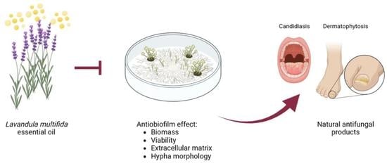
Antibiofilm Effect of Lavandula multifida Essential Oil: A New Approach for Chronic Infections

by
Jorge Alves-Silva
1,2,3,4,
Mónica Zuzarte
1,2,3,4,*,
Carlos Cavaleiro
1,5 and
Lígia Salgueiro
1,5,*
1
Univ Coimbra, Faculty of Pharmacy, Azinhaga de S. Comba, 3000-548 Coimbra, Portugal
2
Univ Coimbra, Coimbra Institute for Clinical and Biomedical Research (iCBR), Faculty of Medicine, Azinhaga de S. Comba, 3000-548 Coimbra, Portugal
3
Univ Coimbra, Center for Innovative Biomedicine and Biotechnology (CIBB), 3000-548 Coimbra, Portugal
4
Clinical Academic Centre of Coimbra (CACC), 3004-561 Coimbra, Portugal
5
Univ Coimbra, Chemical Process Engineering and Forest Products Research Centre (CIEPQPF), Department of Chemical Engineering, Faculty of Sciences and Technology, Rua Sílvio Lima, 3030-790 Coimbra, Portugal
*
Authors to whom correspondence should be addressed.
Pharmaceutics 2023, 15(8), 2142; https://doi.org/10.3390/pharmaceutics15082142
Submission received: 23 June 2023 / Revised: 21 July 2023 / Accepted: 7 August 2023 / Published: 15 August 2023

Abstract

:
Fungal infections are associated with high morbidity and mortality rates, being highly prevalent in patients with underlying health complications such as chronic lung disease, HIV, cancer, and diabetes mellitus. To mitigate these infections, the development of effective antifungals is imperative, with plants standing out as promising sources of bioactive compounds. In the present study, we focus on the antibiofilm potential of Lavandula multifida essential oil (EO) against dermatophyte strains and Candida albicans. The EO was characterized using GC and GC–MS, and its antifungal effect was assessed on both biofilm formation and disruption. Biofilm mass, extracellular matrix, and viability were quantified using crystal violet, safranin, and XTT assays, respectively, and morphological alterations were confirmed using optical and scanning electron microscopy. L. multifida EO showed very high amounts of carvacrol and was very effective in inhibiting and disrupting fungal biofilms. The EO significantly decreased biofilm mass and viability in all tested fungi. In addition, a reduction in dermatophytes’ extracellular matrix was observed, particularly during biofilm formation. Morphological alterations were evident in mature biofilms, with a clear decrease in hypha diameter. These promising results support the use of L. multifida EO in the development of effective plant-based antifungal products.

Graphical Abstract

1. Introduction

Fungal infections, or mycosis, represent a huge health and economic burden affecting over 1 billion individuals worldwide and accounting for around 13 million infections per year [1]. Indeed, the Global Action Fund for Fungal Infections (GAFFI) estimates that over 300 million individuals are affected by a severe fungal infection every year [2]. Moreover, patients with underlying health concerns or a weakened immune system, including chronic lung disease, prior tuberculosis, HIV, cancer, and diabetes mellitus, are at higher risk [3]. In addition, in patients with HIV, the relapse rates of onychomycosis are higher than in healthy individuals [4], reinforcing the relevance of these infections in debilitated patients.
The main etiologic agents of superficial fungal infections belong to Epidermophyton, Microsporum, and Trichophyton filamentous fungi genera that affect the skin, nails, and hair [5]. These infections, collectively named dermatophytosis, although not lethal, are the most disseminated type of mycosis, affecting between 20 and 25% of the world’s population [6]. Dermatophytosis is generally neglected but is a major cause of morbidity-associated superficial mycoses, with frequent relapses and high resistance to therapy [7]. Indeed, therapy duration is usually very long, leading to poor patient adherence and consequent infection relapses. Moreover, these fungi are known to produce biofilms [8,9], thus further contributing to their resistance to antifungal therapy and the consequent persistence of infections.
On the other hand, invasive infections, although less prevalent than superficial mycosis, account for higher mortality rates, being responsible for 1.5 million annual deaths [1], with candidiasis being the most predominant type [10]. Despite the emergence of non-albicans Candida infections, Candida albicans remains the most widespread strain in humans [11]. An overgrowth of this fungi causes candidiasis with vaginal yeast infection, diaper rash, and thrush being the most common types of infection. More severe cases called invasive candidiasis target the whole body, including the blood, bones, brain, and heart. Similarly, to dermatophytes, C. albicans also has the capacity to form biofilms that pose a threat to the individual, as these structures are very resistant to treatment and lead to chronic and persistent infections [12,13,14].
Both dermatophytosis and candidiasis constitute a very serious health concern that requires effective therapeutic strategies. Despite the recent developments in antifungals with the increasing use of echinocandins and third-generation azoles, invasive fungal infections continue to impact patients’ quality of life, and mortality rates remain high. These poor outcomes are usually associated with antifungals’ high toxicity, unpleasant side effects, drug interactions, and, most importantly, the emergence of resistant strains [15,16]. Indeed, the extensive use of non-selective antifungals has contributed to the emergence of fungi resistance with associated off-target toxicity and treatment failure. Global warming is also thought to lead to the emergence of new fungal diseases [17] as the gap between environmental and host temperatures narrows [18] and the fungi seem to easily adapt [17], thus pushing the burden of these infections.
Aromatic plants represent a valuable source of antifungal compounds, and particularly those from the genus Lavandula have shown very promising antifungal properties [19,20,21,22,23,24,25,26,27]. In a previous study, we reported the antifungal potential of fernleaf lavender (Lavandula multifida) against several pathogenic fungi and pointed out a possible mechanism of action through initial metabolic arrest followed by cell death in C. albicans [27]. In the present study, we intend to deepen our knowledge by exploring the effect of this essential oil on dermatophytes and C. albicans biofilms. These highly organized complex microbial communities, involved by self-produced extracellular matrix, are known to protect the fungi from the surrounding environment and constitute reservoirs for persistent infections [28]. In addition, the fungi growing within biofilms present phenotypic alterations that promote resistance to antifungal drugs compared to planktonic cells [29,30,31]. Indeed, biofilms are very difficult to eradicate due to their high resistance to conventional therapy and host defenses [32]. Therefore, targeting these virulence factors is of utmost importance and represents a very attractive strategy for the development of effective antifungal drugs.
In the present study, we highlight the antibiofilm potential of L. multifida essential oil against several strains of dermatophytes and C. albicans. Our studies point out promising results for the majority of the strains tested, with Epidermophyton floccosum being the most susceptible. Overall, the antivirulence potential of L. multifida essential oil, together with its inhibitory effect on fungal growth, as previously reported, justifies the development of effective essential oil-based antifungal products.

2. Materials and Methods

2.1. Plant Material and Essential Oil Characterization

Lavandula multifida essential oil was obtained using hydrodistillation for 3 h from fresh flowering aerial parts of plants collected in Sesimbra region, Portugal. Plant authenticity was verified by the taxonomist Dr. Jorge Paiva (University of Coimbra). Voucher specimens were deposited at the Herbarium of the Department of Life Sciences of the University of Coimbra, Portugal. The essential oil was characterized using gas-chromatography (GC, Agilent Technologies, Palo Alto, CA, USA) coupled with mass spectrometry (GC–MS, Agilent Technologies), as previously described [33].

2.2. Antifungal Effect of L. multifida Essential Oil

2.2.1. Fungal Strains

The antibiofilm potential of L. multifida essential oil was assessed on yeasts and filamentous fungi. Dermatophyte species included both collection strains such as Microsporum gypseum CECT 2908, Trichophyton mentagrophytes var. interdigitale CECT 2958, and T. rubrum CECT 2794, and clinical strains, namely Epidermophytom floccosum FF9, M. canis FF1, and T. mentagrophytes FF7, whereas C. albicans ATCC 10231 was the representative yeast strain tested. Strains were cultured in potato dextrose agar (Oxoid Limited, Hampshire, UK) or Sabouraud agar (Oxoid Limited) at 37 °C for 48 h (C. albicans) or 30 °C for 7 days (dermatophytes).

2.2.2. Fungal Inoculum

Dermatophyte inoculums were prepared from 7-day-old cultures in Sabouraud agar by adding sterile 0.9% NaCl and vigorously vortexed to detach conidia. The saline was then transferred to a new sterile tube and left to settle for 5 min to allow the separation of the conidia from hyphae. The supernatant was collected to a sterile tube and turbidity was adjusted to 1 unit of McFarland scale, containing approximately 1 × 10⁶ conidia/mL.
C. albicans was grown in a yeast peptone dextrose (YPD) medium (1% yeast extract, 2% peptone, and 2% dextrose, Oxoid Limited) inoculated from 24 h old cultures in Sabouraud agar for 24 h at 37 °C. Afterward, the medium was removed, and yeasts were washed twice with PBS (0.8% NaCl, 0.02% KH2PO4. 0.31% Na2HPO4.12 H2O and 0.02% KCl; pH 7.4), with centrifugations of 3000× g for 10 min between each washing step. Cell density was adjusted to 1 × 10⁶ CFU/mL in RPMI 1640 pH 7.0 (with L-glutamine, without bicarbonate, Sigma-Aldrich, Burlington, MA, USA) supplemented with MOPS (Sigma-Aldrich).

2.2.3. Effect of L. multifida Essential Oil on Biofilm Formation

To assess the effect of L. multifida essential oil on biofilm formation, the method described by Ali and colleagues was used [34], with some modifications. For dermatophytes, 200 µL of conidia suspension was added to sterile 96-well flat-bottom polystyrene microtiter plates and incubated at 37 °C for 3 h to allow for conidia to adhere. Then, saline was removed, and wells were washed with PBS (pH 7.4) to remove non-adherent cells. Afterward, 200 µL of RPMI medium containing different concentrations of the essential oil (0.32–0.04 µL/mL) was added and incubated for 72 h at 37 °C. Negative and positive controls containing non-inoculated medium and EO-free inoculated medium were included, respectively.
For C. albicans, 100 µL of the cell suspension was added to sterile 96-well flat-bottom polystyrene microtiter plates, and then 100 µL of RPMI medium containing different concentrations of the essential oil was added in order to achieve the final essential oil concentration ranging from 0.64 to 0.08 µL/mL. Plates were then incubated for 24 or 48 h. Negative and positive controls containing non-inoculated medium or EO-free inoculated medium were included, respectively.

2.2.4. Effect of the L. multifida Essential Oil on Mature Biofilms

The capacity of the essential oil to disrupt mature biofilms was determined using the method reported by Ali and colleagues [34], with slight adaptations. Dermatophyte conidia suspensions (200 µL) were left to adhere for 3 h at 37 °C in sterile 96-well flat-bottom polystyrene microtiter plates. After saline removal, cells were washed with PBS to remove non-adherent cells, and 200 µL of sterile RPMI medium was added to the plates and left to incubate for 72 h at 37 °C. Then, the medium was removed, and following a washing step with PBS, 200 µL of RPMI with different concentrations of the essential oil (0.32–0.04 µL/mL) was added to the respective well and plates were then further incubated for 24 h at 37 °C. Negative and positive controls were considered as referred to in Section 2.2.3.
A cell suspension of C. albicans (100 µL) was added to a sterile 96-well flat-bottom polystyrene microtiter plate containing 100 µL of sterile RPMI medium and incubated for 24 h at 37 °C. Then, the medium was removed, and after a washing step with PBS, 200 µL of RPMI medium containing different concentrations of the essential oil (0.64–0.08 µL/mL) was added to the respective well and plates were then further incubated for 24 h at 37 °C. Negative and positive controls were considered as referred to in Section 2.2.3.

2.2.5. Biofilm Mass

Crystal violet staining was carried out to quantify biofilm mass. Dermatophyte biofilms were stained according to the method reported by Castelo-Branco and colleagues [35], with slight modifications. Briefly, following medium removal, cells were washed with PBS to remove non-adherent cells. Then, biofilms were fixed with absolute methanol for 10 min. Afterward, 100 µL of 0.5% crystal violet solution was added and left to stain the biofilms for 15 min. Following crystal violet removal, biofilms were washed twice with sterile water. Crystals were dissolved using 150 µL of 33% acetic acid. The volume was transferred to new wells, and the absorbance was read at 620 nm.
C. albicans biofilms were stained using the procedure reported by Raut and colleagues [36]. Briefly, after medium removal, biofilms were air-dried and fixed with absolute methanol for 15 min. Then, plates were dried and 0.02% of crystal violet was added for 15 min. Excess solution was removed and wells were washed twice with sterile water, followed by the addition of 150 µL of 33% acetic acid to dissolve the crystals. The solution was transferred to new wells and the absorbance read at 620 nm.
Biomass reduction was calculated according to the following equation:
Biomass (%) = Abs treatment/Abs CT × 100
Abs CT and Abs treatment represent the absorbance at 620 nm for control and treated biofilms, respectively.

2.2.6. Extracellular Matrix

Dermatophyte biofilm’s extracellular matrix was quantified using safranin red according to Costa-Orlandi and colleagues [37] with slight modifications. Following medium removal, biofilms were washed with PBS to remove non-adherent cells, and 100 µL of 0.5% safranin red solution was added and left to stain the biofilms for 5 min. Then, the solution was removed, and biofilms were washed twice with sterile PBS to remove the unbounded dye. Safranin crystals were released from the biofilm using 33% acetic acid and after transference to new wells, the absorbance was read at 520 nm. The reduction in biofilm extracellular matrix was calculated using the following equation:
Extracellular matrix (%) = Abs treatment/Abs CT × 100
Abs CT and Abs treatment represent the absorbance at 520 nm for control and treated biofilms, respectively.

2.2.7. Biofilm Viability

The XTT (2.3-bis (2-methoxy-4-nitro-5-sulfophenyl)-5-[carbonyl (phenylamino)]-2H-tetrazolium hydroxide) reduction assay was used to determine the metabolic activity of biofilms, as previously reported [38]. After discarding the medium, biofilms were washed with PBS. Then, 100 µL of 1 mg/mL of XTT salt with 4 µM of menadione (from a solution of 10 mM made in acetone) was added, and biofilms were further incubated for 3 h at 37 °C. At the end of this period, the absorbance was read at 490 nm, and metabolic activity was measured using the following equation:
Metabolic activity (%) = Abs treatment/Abs CT × 100
Abs CT and Abs treatment represent the absorbance at 490 nm for control and treated biofilms, respectively.

2.2.8. Biofilm Morphology and Ultrastructure

Morphological differences in biofilms in the presence of the essential oil were assessed using bright-field microscopy. Briefly, after XTT incubation, z-stacks of the biofilms were acquired using a bright-field microscope Ziess Axio XPD IRE 2 (Carl Zeiss, Oberkochen, Germany) equipped with a 40× objective LD Plan-Neofluar 40x/0.6 Korr Ph 2 M27 (Carl Zeiss) using the inbuilt ‘optimal’ settings to determine the optimal intervals based on sample thickness. Z-projections with the minimum intensity or average intensity setting were obtained using ImageJ/Fiji ver. 1.53t for dermatophyte or C. albicans biofilms, respectively.
Dermatophyte biofilm hypha diameters were measured using the “Straight” line function in ImageJ/Fiji ver. 1.53t. A total of 50 hyphae were measured in each condition.
Scanning electron microscopy images were obtained for Epidermophyton floccosum. The fungal inoculum was grown on a glass side according to the procedure described in Section 2.2.4. Following essential oil treatments (0.64 and 0.32 µL/mL), glass slides were attached to SEM stubs with adhesive carbon substrates (12 mm, Agar Scientific, Essex, UK) and visualized using a variable-pressure scanning electron microscope (FlexSEM 1000, Hitachi, Tokyo, Japan) at an accelerating voltage of 10 kV.

2.3. Statistical Analysis

Results are shown as mean values ± SEM (standard error of the mean) from at least three independent experiments performed in duplicate. Sample normality was determined using the Shapiro–Wilk normality test. Statistical significance for dermatophytes was determined using mixed effect analysis followed by Dunnett’s multiple comparisons test, while for C. albicans, it was determined using ordinary one-way analysis of variance (ANOVA) followed by Dunnet’s multiple comparisons test, using GraphPad Prism version 9.5.0 (GraphPad Software, San Diego, CA, USA). Statistical significance is considered for p values lower than 0.05.

3. Results

3.1. Chemical Composition of L. multifida Essential Oil

The essential oil of L. multifida obtained from plants growing in Sesimbra region in Portugal was rich in oxygenated monoterpenes, being the phenolic compound carvacrol present in very high amounts (46.4.%). Other main compounds included cis-β-ocimene (12.7%) and β-bisabolene (10.1%), as shown in Table 1.

3.2. Effect of L. multifida Essential Oil on the Formation of Dermatophyte Biofilms

Dermatophytes are able to form biofilms that contribute to their higher resistance to antifungal drugs. In our studies, for the majority of the dermatophyte strains tested, two types of fungal growth were observed: aerial and attached. Bearing in mind the general definition of a biofilm, only attached fungi were considered for the evaluation of the effect of L. multifida essential oil on both biofilm formation and mature biofilm disruption. In both cases, biofilm mass and extracellular matrix were quantified using crystal violet and safranin staining, respectively, and biofilm viability was assessed using the XTT assay.
Regarding the effect of the oil on biofilm formation, a significant inhibition was attained at 0.32 µL/mL, with biofilm mass being greatly inhibited in all the tested strains and extracellular matrix and biofilm viability being significantly decreased in the majority of the strains (Figure 1A–F). Combining these features, the essential oil at 0.32 µL/mL was more effective against E. floccosum, inhibiting 90% of biofilm mass, 80% of extracellular matrix, and more than 50% of biofilm viability (Figure 1A). In addition, M. gypseum (Figure 1C), T. mentagrophytes (Figure 1D), and T. rubrum (Figure 1F) were quite susceptible, with all parameters reduced by more than 50%. In addition, at 0.16 µL/mL, the essential oil was also effective on dermatophyte biofilm mass, as a significant reduction was observed in several strains (Figure 1A–C,E), confirming its antibiofilm potential.

3.3. Effect of L. multifida Essential Oil on the Disruption of Dermatophyte Mature Biofilms

Considering the resistance of mature biofilms to antifungals, we assessed the capacity of L. multifida essential oil to disrupt these structures. As expected, the mature biofilms were more resistant to the activity of the essential oil; however, promising results were obtained for all the tested strains, except for Microsporum canis. In addition to the parameters assessed in Section 3.2, morphological alterations were considered, resorting to light microscopy observations and quantification of hypha diameters.
Similar to that observed in biofilm formation inhibition, E. floccosum was the most susceptible strain (Figure 2A), with 0.32 µL/mL of the essential oil significantly decreasing biofilm mass (46%), extracellular matrix (49%), and viability (30%). Morphological alterations were also obvious with hyphae being much thinner (Figure 2B) and more septate (Figure 2C).
The capacity of the essential oil to disrupt Microsporum canis and M. gypseum mature biofilms was also assessed (Figure 3). In the first, the oil (0.32 µL/mL) slightly decreased all parameters assessed, although no statistical significance was attained (Figure 3A). This weak activity was also observed using optical microscopy, where the amount of biofilm detected was very similar in all conditions; however, hypha diameter was clearly reduced in the treated biofilms (Figure 3E). Indeed, when hypha diameter was measured, a strong decrease was detected (Figure 3B). On the other hand, for M. gypseum, a significant reduction in biofilm mass and extracellular matrix was attained (Figure 3C), and morphological alterations were also observed, with the biofilm presenting a disorganized structure in comparison to the control biofilm (Figure 3E); nevertheless, no effect on hypha diameter was observed (Figure 3D).
For Trichophyton, three strains were tested, namely T. mentagrophytes, T. mentagrophytes var. interdigitale, and T. rubrum (Figure 4). Overall, the essential oil at 0.32 µL/mL showed a significant reduction in biofilm viability in all the tested strains (Figure 4A,C,E), being also able to significantly reduce extracellular matrix in the first two (Figure 4A,C). Morphological alterations were also observed, particularly for T. mentagrophytes, where hyphae appeared bloated, and for T. mentagrophytes var. interdigitale, where hypha formation seemed compromised (Figure 4G). In agreement with safranin staining, a decrease in the extracellular matrix was observed for T. mentagrophytes (Figure 4G, arrowheads). For T. rubrum, a reduction in hypha density was observed in the highest dose tested, and, in addition, even though no statistical difference was attained in safranin staining, a reduction in extracellular matrix deposition was observed (Figure 4G, arrowheads). Regarding hypha diameters, significant reductions were only observed in T. rubrum (Figure 4F).

3.4. Morphological Effect of L. multifida Essential Oil on E. floccosum

Epidermophyton floccosum was selected to carry out scanning electron microscopy observations, as it was overall the most susceptible strain to L. multifida essential oil. As observed in Figure 5, the essential oil at 0.32 µL/mL induced evident morphological alterations, namely an increase in septate hyphae (arrowhead) with flattened surface (arrows), and less packed mycelia.

3.5. Effect of L. multifida Essential Oil on the Formation of Candida albicans Biofilms

Considering the role that C. albicans biofilms play in persistent infections, the effect of L. multifida essential oil on its biofilm formation and disruption was assessed, through the quantification of biofilm mass and viability.
Regarding the effect of the oil on biofilm formation, two time-points were considered: 24 h (Figure 6A–C) and 48 h (Figure 6D–F), thus allowing to assess the effect of the oil on an intermediate (12–30 h) and mature (38–72 h) stage of biofilm development [39].
Overall, the addition of 0.64 µL/mL of the essential oil for 24 h or 48 h was very effective, leading to a reduction in both biofilm mass and viability (Figure 6A,B,D,E). Optical microscopy observations corroborated these results as an evident decrease in biofilms was observed. Indeed, the yeast form of C. albicans was more prevalent, indicating a decrease in the dimorphic transition, thus suggesting a reduction in fungal virulence (Figure 6C,F).

3.6. Effect of L. multifida Essential Oil on the Disruption of Candida albicans Mature Biofilms

Mature biofilms, allowed to grow for 24 h, were treated with the essential oil for an additional period of 24 h. As expected, these biofilms were more resilient to the essential oil, with no effects observed on biofilm mass (Figure 7A). Nevertheless, biofilm viability was significantly reduced at 0.64 µL/mL, thus showing a clear effect of the oil before biomass reduction is visible (Figure 7B). Indeed, microscopy observations confirmed similar amounts of biofilm in all conditions, but a closer observation showed quite evident morphological differences (Figure 7C, zoom-ins). Indeed, similar to what was observed during biofilm formation (Figure 6), dimorphic transition was impaired in the presence of 0.64 µL/mL of the essential oil (Figure 7C), reinforcing the effect of the oil on virulence reduction.

4. Discussion

Dermatophytosis tends to be neglected as it generally is not life-threatening. However, these infections deserve more attention due to frequent relapses, recalcitrance, and resistance to therapy. Moreover, although predominantly superficial, in certain patients, such as those undergoing immunosuppression therapy, with genetic predisposition, or with chronic pathologies, dermatophytes become invasive, infecting the dermis, and in some cases internal organs such as the brain [40,41]. Their recalcitrance has been associated with long treatment durations that lead to poor patient compliance, host-specific characteristics, the emergence of resistant strains, and the formation of biofilms [8]. Despite the impact of biofilms, primarily on antifungal resistance, studies on dermatophytes are scarce, and the majority report the formation of biofilms in Trichophyton species, and only a few assess this capacity in Microsporum canis and M. gypseum, as reviewed elsewhere [8]. In the present study, besides these strains, we included Epidermophyton floccosum, which is also able to produce biofilms in vitro.
Overall, and similarly to that reported by Brilhante and colleagues [9], we observed that M. canis was the weakest biofilm producer, as confirmed by optical microscopy analysis (Figure 3) and crystal violet staining (Figure S1). Furthermore, T. mentagrophytes var. interdigitale also showed a low biofilm formation capacity in comparison to the remaining strains, with E. floccosum being the most effective (Figure S1). C. albicans was also considered in the present study due to its high prevalence and capacity to form biofilms.
Bearing in mind the need for effective therapeutic strategies able to mitigate the negative impact of dermatophytosis and candidiasis, we sought to explore the antifungal potential of L. multifida essential oil rich in carvacrol. The reported chemical composition is in agreement with previous studies using plants from different regions that demonstrate that carvacrol is the predominant compound in this species [27,42,43,44,45,46]. Overall, our results show a significant antibiofilm potential with promising results attained on the prevention of both dermatophyte and C. albicans biofilm formation. These results are highly relevant since fungal biofilms are often associated with chronic infections [47]. Besides biofilm formation, the essential oil was also able to disrupt mature biofilms in all the tested strains (except M. canis), a feature of uttermost importance, as biofilm disruption increases fungal susceptibility to conventional antifungal drugs [47], and consequently contributes to the eradication of the infection. These results suggest that L. multifida essential oil could be considered in clinical practice and contribute to a more effective therapeutic response.
In our studies, L. multifida essential oil from 0.32 µL/mL onwards was able to inhibit biofilm formation in all the tested strains, with E. floccosum being the most susceptible strain, followed by T. mentagrophytes, M. gypseum, and T. rubrum. As far as we know, this is the first report on the antibiofilm effect against E. floccosum, a very relevant opportunist infectious agent in immunocompromised individuals [48]. Moreover, the interesting results obtained against T. rubrum, responsible for 70–90% of all dermatophyte infections [48,49], further highlight the potential of L. multifida essential oil, as this dermatophyte is responsible for more than 50% of all invasive dermatophytosis [41]. Indeed, in mature T. rubrum biofilm, we report a significant viability decrease in the presence of the essential oil before a significant decrease in biofilm mass and matrix was observed, thus suggesting a decrease in fungal virulence capacity.
C. albicans biofilm is associated with very high mortality rates, being responsible for fatal infections in up to 50% of adults and 30% of young individuals [12]. Furthermore, these biofilm-related infections have serious economic consequences. Indeed, in the US alone, these infections are associated with an excess of USD 6.5 billion in annual expenditure, often associated with the high resistance to antifungals observed in these structures [14]. In the present study, we showed that L. multifida essential oil is able to prevent C. albicans biofilm formation and eradicate its mature biofilms. These results are quite relevant, as fluconazole, the most widely used antifungal in the clinic, fails to inhibit biofilms even at doses 200× higher than its minimal inhibitory concentration [50]. The reduction in biofilm biomass during the formation phase suggests that the essential oil prevents the adhesion of yeasts, making them more susceptible to conventional antifungals, thus highlighting once again the essential oil’s relevance in clinical practice. On the other hand, the capacity of the essential oil to decrease the viability of mature biofilms without decreasing biofilm biomass suggests that, similarly to what we suggested for T. rubrum, the oil seems to penetrate the biofilm complex structure and compromise the pathogen´s viability and consequent virulence capacity.
Studies reporting the antibiofilm effect of natural products against dermatophytes are scarce, with only one reporting the effect of essential oils and their isolated compounds [34]. Furthermore, regarding Lavandula species essential oils, only a few studies have shown their antibiofilm potential against Candida spp. For example, the essential oil from L. dentata was able to inhibit C. albicans biofilm adhesion, proliferation, and viability [51]. The essential oil from L. stoechas presented strong antibiofilm activity against collection and clinical strains of Candida spp. [52]. The essential oil from L. x intermedia presented strong disruptive properties against C. albicans mature biofilms [53], and the essential oil of L. angustifolia presented relevant antibiofilm activity against several clinical isolates of C. albicans [54]. Contrarily to fungal species, studies on bacterial biofilms are more frequent, with several pointing out the antibiofilm potential of lavender species. Overall, L. angustifolia essential oil is the most studied and has shown promising disruptive effects against mature Campylobacter jejuni biofilms, in addition to preventing its adhesion [55], and against methicillin-resistant Staphylococcus aureus (MRSA) biofilms [56]. This oil was also able to eradicate S. aureus and Escherichia coli mature biofilms from the surface of medical materials [57]. The essential oils from other lavender species have also been studied, such as L. x intermedia, which was able to disrupt Streptococcus agalactiae mature biofilms [53]; L. mairei, which disrupted mature biofilms of Acinetobacter baumannii, prevented cell adhesion, and induced cell detachment [58]; and L. officinalis, which prevented the adhesion of Porphyromonas gingivalis and Streptococcus mutans [59].
Regarding the effect of isolated compounds, namely carvacrol, highly present in L. multifida essential oil, only one study has been performed on fungi showing its ability to inhibit and disrupt mature biofilms of Cryptococcus neoformans and C. laurentii [60]. In this study, the possible mechanism of action underlying the observed effect was suggested to be related to its capacity to decrease extracellular matrix and capsule thickness, alter lipid metabolism, inhibit ergosterol synthesis, and induce reactive oxygen species formation [61]. Similar to lavender essential oils, studies on the effect of essential oils´ isolated compounds on bacterial biofilms are quite frequent. Indeed, carvacrol has been widely assessed alone, in combination, or even encapsulated, and has shown effects, namely on biofilm formation inhibition and/or mature biofilm disruption. For example, carvacrol was able to decrease biofilm formation in Enterococcus faecalis and Klebsiella pneumoniae [62,63], in Pseudomonas aeruginosa and S. aureus formed on technical surfaces [64], and in P. gingivalis, Fusobacterium nucleatum, and S. mutans biofilm formed on titanium implants [65,66]. In addition, on mature biofilms, this compound was quite effective, being able to eradicate mature biofilms of Aeromonas hydrophyla [67], Staphylococcus epidermidis, P. aeruginosa [68], and Gardnerella spp., which, even after compound removal, did not recover their viability. Furthermore, carvacrol prevented the adhesion of Gardnerella to human vaginal epithelial cells [69], highlighting its therapeutic potential. Carvacrol was also able to prevent both biofilm formation and mature biofilm disruption in different Mycobacterium species [70]. The same effects were reported for Salmonella Enteritidis [71], S. aureus, and S. epidermidis [72]. When microencapsulated, this compound was also quite effective in inhibiting P. aeruginosa and E. faecalis biofilm formation [73,74]. In addition, carvacrol-loaded chitosan nanoparticles were effective in disrupting mature biofilms of P. aeruginosa [75], probably due to alterations in quorum sensing [76]. Treatment with free and encapsulated carvacrol prevented biofilm formation by Salmonella and S. aureus in stainless steel surfaces [77], while carvacrol-loaded polymeric systems impaired biofilm formation in S. aureus, S. epidermidis, E. coli, and Listeria monocytogenes [78,79]. Furthermore, poly-(DL-lactide-co-glycolide) nanoparticles loaded with carvacrol disrupted mature S. epidermidis biofilms [80], and micelles loaded with this compound disrupted mature E. coli and L. monocytogenes biofilms [81,82]. β-bisabolene, another main compound of L. multifida essential oil, was also described as having antibiofilm potential by inhibiting biofilm formation in Mycobacterium smegmatis [83]. Therefore, taking into account the reported antibiofilm effects of both carvacrol and β-bisabolene, it seems that the activity herein observed might be due to the presence of these compounds; however, synergistic effects with other compounds cannot be disregarded.

5. Conclusions

Herein, we report, for the first time, strong inhibitory effects of L. multifida essential oil on biofilm formation against dermatophytes and C. albicans. Of relevance, the essential oil was able to eradicate mature biofilms against all tested strains, with dermatophytes being more susceptible than C. albicans, particularly E. floccosum, T. mentagrophytes, M. gypseum, and T. rubrum. These results are quite relevant, as studies on dermatophytes biofilms are scarce, with this study being the first to consider E. floccosum biofilms. The disruptive capacity reported is highly pertinent as it highlights that L. multifida essential oil could be of relevance in clinical practice. Indeed, this essential oil has a pleasant aroma, and due to its lipophilicity, can be easily applied to several topical products.
Overall, the present study brings new insights to the field of dermatophytosis and paves the way for the development of complementary strategies to manage mycosis, as these infections are highly prevalent and tend to be neglected. This is quite relevant, as patients with underlying health concerns or a weakened immune system are at higher risk of frequent relapses and recurrent infections. Moreover, in some cases, these fungal infections can evolve into chronic disorders, thus strengthening the need for new therapeutic agents. Nevertheless, in order to validate the promising antibiofilm effects herein reported for L. multifida essential oil, further experiments should consider comparisons with reference antifungal drugs, deepen the knowledge on the mechanism of action underlying the antibiofilm effect, and consider pre-clinical studies. In addition, despite L. multifida´s wide distribution (Iberian Peninsula, Sicily, Northwest Africa, and the Canary Islands), cultivation techniques should be considered to avoid overexploitation in the wild and enable a standardized chemical composition relevant for industrial purposes.

Supplementary Materials

The following supporting information can be downloaded at: https://www.mdpi.com/article/10.3390/pharmaceutics15082142/s1, Figure S1: Dermatophytes biofilm biomass assessed by crystal violet assay. Dermatophytes were left to adhere for 3 h and further incubated for 72 h in culture medium. Values represent the mean ± SEM of the optical density at 620 nm of at least three independent assays.

Author Contributions

Conceptualization, M.Z. and L.S.; methodology, J.A.-S., C.C. and M.Z.; validation, M.Z., C.C. and L.S.; investigation, M.Z., C.C. and J.A.-S.; resources, M.Z. and L.S.; writing—original draft preparation, J.A.-S.; writing—review and editing, M.Z. and L.S.; supervision, L.S.; funding acquisition, L.S. and M.Z. All authors have read and agreed to the published version of the manuscript.

Funding

This work was supported by La Caixa Foundation and Foundation for Science and Technology, under the project PD21-00003. Support from PPBI-Portuguese Platform of BioImaging: POCI-01-0145-FEDER-022122 was also provided.

Institutional Review Board Statement

Not applicable.

Informed Consent Statement

Not applicable.

Data Availability Statement

The data presented in this study are available on request from the corresponding author.

Conflicts of Interest

The authors declare no conflict of interest.

References

  1. Bongomin, F.; Gago, S.; Oladele, R.; Denning, D. Global and Multi-National Prevalence of Fungal Diseases—Estimate Precision. J. Fungi 2017, 3, 57. [Google Scholar] [CrossRef] [Green Version]
  2. GAFFI. Global Action Fund for Fungal Infections. Available online: https://gaffi.org/why/fungal-disease-frequency/ (accessed on 6 April 2023).
  3. World Health Organization. WHO Fungal Priority Pathogens List to Guide Research, Development and Public Health Action; World Health Organization: Geneva, Switzerland, 2022.
  4. Daniel, C.R.; Norton, L.A.; Scher, R.K. The Spectrum of Nail Disease in Patients with Human Immunodeficiency Virus Infection. J. Am. Acad. Dermatol. 1992, 27, 93–97. [Google Scholar] [CrossRef]
  5. Zuzarte, M.; Lopes, G.; Pinto, E.; Salgueiro, L. Are natural products an alternative therapy for dermatophytosis? In Dermatophytes and Dermatophytoses; Springer International Publishing: Cham, Switzerland, 2021; pp. 473–519. [Google Scholar]
  6. Havlickova, B.; Czaika, V.A.; Friedrich, M. Epidemiological Trends in Skin Mycoses Worldwide. Mycoses 2008, 51, 2–15. [Google Scholar] [CrossRef] [PubMed]
  7. Gupta, A.K.; Cooper, E.A. Update in Antifungal Therapy of Dermatophytosis. Mycopathologia 2008, 166, 353–367. [Google Scholar] [CrossRef] [PubMed]
  8. Markantonatou, A.-M.; Samaras, K.; Vyzantiadis, T.-A. Dermatophytic Biofilms: Characteristics, Significance and Treatment Approaches. J. Fungi 2023, 9, 228. [Google Scholar] [CrossRef] [PubMed]
  9. Brilhante, R.S.N.; Correia, E.E.M.; de Guedes, G.M.M.; Pereira, V.S.; de Oliveira, J.S.; Bandeira, S.P.; de Alencar, L.P.; de Andrade, A.R.C.; de Castelo-Branco, D.S.C.M.; de Cordeiro, R.A.; et al. Quantitative and Structural Analyses of the in Vitro and Ex Vivo Biofilm-Forming Ability of Dermatophytes. J. Med. Microbiol. 2017, 66, 1045–1052. [Google Scholar] [CrossRef]
  10. Kullberg, B.J.; Arendrup, M.C. Invasive Candidiasis. N. Engl. J. Med. 2015, 373, 1445–1456. [Google Scholar] [CrossRef] [Green Version]
  11. Pappas, P.G.; Lionakis, M.S.; Arendrup, M.C.; Ostrosky-Zeichner, L.; Kullberg, B.J. Invasive Candidiasis. Nat. Rev. Dis. Primers 2018, 4, 18026. [Google Scholar] [CrossRef]
  12. Atriwal, T.; Azeem, K.; Husain, F.M.; Hussain, A.; Khan, M.N.; Alajmi, M.F.; Abid, M. Mechanistic Understanding of Candida albicans Biofilm Formation and Approaches for Its Inhibition. Front. Microbiol. 2021, 12, 638609. [Google Scholar] [CrossRef]
  13. Kuhn, D.M.; Ghannoum, M.A. Candida Biofilms: Antifungal Resistance and Emerging Therapeutic Options. Curr. Opin. Investig. Drugs 2004, 5, 186–197. [Google Scholar] [PubMed]
  14. Gulati, M.; Nobile, C.J. Candida albicans Biofilms: Development, Regulation, and Molecular Mechanisms. Microbes Infect. 2016, 18, 310–321. [Google Scholar] [CrossRef] [PubMed] [Green Version]
  15. Brown, G.D.; Denning, D.W.; Gow, N.A.R.; Levitz, S.M.; Netea, M.G.; White, T.C. Hidden Killers: Human Fungal Infections. Sci. Transl. Med. 2012, 4, 165rv13. [Google Scholar] [CrossRef] [Green Version]
  16. Rodrigues, M.L.; Nosanchuk, J.D. Fungal Diseases as Neglected Pathogens: A Wake-up Call to Public Health Officials. PLoS Negl. Trop. Dis. 2020, 14, e0007964. [Google Scholar] [CrossRef] [PubMed] [Green Version]
  17. Garcia-Solache, M.A.; Casadevall, A. Global Warming Will Bring New Fungal Diseases for Mammals. mBio 2010, 1, e00061-10. [Google Scholar] [CrossRef] [Green Version]
  18. Cohen, J.M.; Venesky, M.D.; Sauer, E.L.; Civitello, D.J.; McMahon, T.A.; Roznik, E.A.; Rohr, J.R. The Thermal Mismatch Hypothesis Explains Host Susceptibility to an Emerging Infectious Disease. Ecol. Lett. 2017, 20, 184–193. [Google Scholar] [CrossRef] [PubMed]
  19. Zuzarte, M.; Gonçalves, M.J.; Cruz, M.T.; Cavaleiro, C.; Canhoto, J.; Vaz, S.; Pinto, E.; Salgueiro, L. Lavandula Luisieri Essential Oil as a Source of Antifungal Drugs. Food Chem. 2012, 135, 1505–1510. [Google Scholar] [CrossRef]
  20. Zuzarte, M.; Gonçalves, M.J.; Cavaleiro, C.; Dinis, A.M.; Canhoto, J.M.; Salgueiro, L.R. Chemical Composition and Antifungal Activity of the Essential Oils of Lavandula Pedunculata (Miller) Cav. Chem. Biodivers. 2009, 6, 1283–1292. [Google Scholar] [CrossRef]
  21. El Abdali, Y.; Agour, A.; Allali, A.; Bourhia, M.; El Moussaoui, A.; Eloutassi, N.; Salamatullah, A.M.; Alzahrani, A.; Ouahmane, L.; Aboul-Soud, M.A.M.; et al. Lavandula Dentata L.: Phytochemical Analysis, Antioxidant, Antifungal and Insecticidal Activities of Its Essential Oil. Plants 2022, 11, 311. [Google Scholar] [CrossRef]
  22. D’Auria, F.D.; Tecca, M.; Strippoli, V.; Salvatore, G.; Battinelli, L.; Mazzanti, G. Antifungal Activity of Lavandula Angustifolia Essential Oil against Candida albicans Yeast and Mycelial Form. Med. Mycol. 2005, 43, 391–396. [Google Scholar] [CrossRef] [Green Version]
  23. Zuzarte, M.; Gonçalves, M.J.; Cavaleiro, C.; Cruz, M.T.; Benzarti, A.; Marongiu, B.; Maxia, A.; Piras, A.; Salgueiro, L. Antifungal and Anti-Inflammatory Potential of Lavandula Stoechas and Thymus Herba-Barona Essential Oils. Ind. Crops Prod. 2013, 44, 97–103. [Google Scholar] [CrossRef]
  24. Minooeianhaghighi, M.H.; Sepehrian, L.; Shokri, H. Antifungal Effects of Lavandula Binaludensis and Cuminum Cyminum Essential Oils against Candida albicans Strains Isolated from Patients with Recurrent Vulvovaginal Candidiasis. J. Mycol. Med. 2017, 27, 65–71. [Google Scholar] [CrossRef]
  25. Domingues, J.; Goulão, M.; Delgado, F.; Gonçalves, J.C.; Gonçalves, J.; Pintado, C.S. Essential Oils of Two Portuguese Endemic Species of Lavandula as a Source of Antifungal and Antibacterial Agents. Processes 2023, 11, 1165. [Google Scholar] [CrossRef]
  26. Puškárová, A.; Bučková, M.; Kraková, L.; Pangallo, D.; Kozics, K. The Antibacterial and Antifungal Activity of Six Essential Oils and Their Cyto/Genotoxicity to Human HEL 12469 Cells. Sci. Rep. 2017, 7, 8211. [Google Scholar] [CrossRef] [PubMed] [Green Version]
  27. Zuzarte, M.; Vale-Silva, L.; Gonçalves, M.J.; Cavaleiro, C.; Vaz, S.; Canhoto, J.; Pinto, E.; Salgueiro, L. Antifungal Activity of Phenolic-Rich Lavandula multifida L. Essential Oil. Eur. J. Clin. Microbiol. Infect. Dis. 2012, 31, 1359–1366. [Google Scholar] [CrossRef] [PubMed]
  28. Ramage, G.; Rajendran, R.; Sherry, L.; Williams, C. Fungal Biofilm Resistance. Int. J. Microbiol. 2012, 2012, 528521. [Google Scholar] [CrossRef] [PubMed]
  29. Ramage, G.; Vande Walle, K.; Wickes, B.L.; López-Ribot, J.L. Standardized Method for In Vitro Antifungal Susceptibility Testing of Candida albicans Biofilms. Antimicrob. Agents Chemother. 2001, 45, 2475–2479. [Google Scholar] [CrossRef] [Green Version]
  30. Yazdanpanah, S.; Sasanipoor, F.; Khodadadi, H.; Rezaei-Matehkolaei, A.; Jowkar, F.; Zomorodian, K.; Kharazi, M.; Mohammadi, T.; Nouripour-Sisakht, S.; Nasr, R.; et al. Quantitative Analysis of in Vitro Biofilm Formation by Clinical Isolates of Dermatophyte and Antibiofilm Activity of Common Antifungal Drugs. Int. J. Dermatol. 2023, 62, 120–127. [Google Scholar] [CrossRef]
  31. Brilhante, R.S.N.; Correia, E.E.M.; de Guedes, G.M.M.; de Oliveira, J.S.; de Castelo-Branco, D.S.C.M.; de Cordeiro, R.A.; de Pinheiro, A.Q.; Chaves, L.J.Q.; de Pereira Neto, W.A.; Sidrim, J.J.C.; et al. In Vitro Activity of Azole Derivatives and Griseofulvin against Planktonic and Biofilm Growth of Clinical Isolates of Dermatophytes. Mycoses 2018, 61, 449–454. [Google Scholar] [CrossRef]
  32. Kernien, J.F.; Snarr, B.D.; Sheppard, D.C.; Nett, J.E. The Interface between Fungal Biofilms and Innate Immunity. Front Immunol 2018, 8, 1968. [Google Scholar] [CrossRef] [Green Version]
  33. Cavaleiro, C.; Salgueiro, L.R.; Miguel, M.G.; Proença da Cunha, A. Analysis by Gas Chromatography–Mass Spectrometry of the Volatile Components of Teucrium Lusitanicum and Teucrium Algarbiensis. J. Chromatogr. A 2004, 1033, 187–190. [Google Scholar] [CrossRef] [Green Version]
  34. Ali, I.; Satti, N.K.; Dutt, P.; Prasad, R.; Khan, I.A. Hydroxychavicol: A Phytochemical Targeting Cutaneous Fungal Infections. Sci. Rep. 2016, 6, 37867. [Google Scholar] [CrossRef] [PubMed] [Green Version]
  35. de Castelo-Branco, D.S.C.M.; de Aguiar, L.; dos Araújo, G.S.; Lopes, R.G.P.; de Sales, J.A.; Pereira-Neto, W.A.; de Pinheiro, A.Q.; Paixão, G.C.; de Cordeiro, R.A.; Sidrim, J.J.C.; et al. In Vitro and Ex Vivo Biofilms of Dermatophytes: A New Panorama for the Study of Antifungal Drugs. Biofouling 2020, 36, 783–791. [Google Scholar] [CrossRef]
  36. Raut, J.S.; Shinde, R.B.; Chauhan, N.M.; Karuppayil, S.M. Terpenoids of Plant Origin Inhibit Morphogenesis, Adhesion, and Biofilm Formation by Candida albicans. Biofouling 2012, 29, 87–96. [Google Scholar] [CrossRef]
  37. Costa-Orlandi, C.B.; Sardi, J.C.O.; Santos, C.T.; Fusco-Almeida, A.M.; Mendes-Giannini, M.J.S. In Vitro Characterization of Trichophyton Rubrum and T. Mentagrophytes Biofilms. Biofouling 2014, 30, 719–727. [Google Scholar] [CrossRef]
  38. Alves, M.; Gonçalves, M.J.; Zuzarte, M.; Alves-Silva, J.M.; Cavaleiro, C.; Cruz, M.T.; Salgueiro, L. Unveiling the Antifungal Potential of Two Iberian Thyme Essential Oils: Effect on C. Albicans Germ Tube and Preformed Biofilms. Front. Pharmacol. 2019, 10, 446. [Google Scholar] [CrossRef]
  39. Jabra-Rizk, M.A.; Falkler, W.A.; Meiller, T.F. Fungal Biofilms and Drug Resistance. Emerg. Infect. Dis. 2004, 10, 14–19. [Google Scholar] [CrossRef]
  40. Martinez-Rossi, N.M.; Peres, N.T.A.; Bitencourt, T.A.; Martins, M.P.; Rossi, A. State-of-the-Art Dermatophyte Infections: Epidemiology Aspects, Pathophysiology, and Resistance Mechanisms. J. Fungi 2021, 7, 629. [Google Scholar] [CrossRef]
  41. Wang, R.; Huang, C.; Zhang, Y.; Li, R. Invasive Dermatophyte Infection: A Systematic Review. Mycoses 2021, 64, 340–348. [Google Scholar] [CrossRef] [PubMed]
  42. Elmakaoui, A.; Bourais, I.; Oubihi, A.; Nassif, A.; Bezhinar, T.; Shariati, M.A.; Blinov, A.V.; Hleba, L.; Hajjaji, S. El Chemical Composition and Antibacterial Activity of Essential Oil of Lavandula multifida. J. Microbiol. Biotechnol. Food Sci. 2022, 11, e7559. [Google Scholar] [CrossRef]
  43. Chograni, H.; Zaouali, Y.; Rajeb, C.; Boussaid, M. Essential Oil Variation among Natural Populations of Lavandula multifida L. (Lamiaceae). Chem. Biodivers. 2010, 7, 933–942. [Google Scholar] [CrossRef] [PubMed]
  44. Messaoud, C.; Chograni, H.; Boussaid, M. Chemical Composition and Antioxidant Activities of Essential Oils and Methanol Extracts of Three Wild Lavandula L. Species. Nat. Prod. Res. 2012, 26, 1976–1984. [Google Scholar] [CrossRef]
  45. Mostefa, M.B.; Kabouche, A.; Abaza, I.; Aburjai, T.; Touzani, R.; Kabouche, Z. Chemotypes Investigation of Lavandula Essential Oils Growing at Different North African Soils. J. Mater. Environ. Sci 2014, 5, 1896–1901. [Google Scholar]
  46. Sellam, K.; Ramchoun, M.; Alem, C.; El-Rhaffari, L. Biological Investigations of Antioxidant-Antimicrobial Properties and Chemical Composition of Essential Oil from Lavandula multifida. Oxid. Antioxid. Med. Sci. 2013, 2, 211–216. [Google Scholar] [CrossRef]
  47. Gupta, A.K.; Carviel, J.; Shear, N.H. Antibiofilm Treatment for Onychomycosis and Chronic Fungal Infections. Ski. Appendage Disord. 2018, 4, 136–140. [Google Scholar] [CrossRef] [PubMed]
  48. Dalla Lana, D.F.; Batista, B.G.; Alves, S.H.; Fuentefria, A.M. Dermatofitoses: Agentes Etiológicos, Formas Clínicas, Terapêutica e Novas Perspectivas de Tratamento. Clin. Biomed. Res. 2016, 36, 230–241. [Google Scholar] [CrossRef]
  49. Petrucelli, M.F.; de Abreu, M.H.; Cantelli, B.A.M.; Segura, G.G.; Nishimura, F.G.; Bitencourt, T.A.; Marins, M.; Fachin, A.L. Epidemiology and Diagnostic Perspectives of Dermatophytoses. J. Fungi 2020, 6, 310. [Google Scholar] [CrossRef] [PubMed]
  50. Alves-Silva, J.M.; Zuzarte, M.; Gonçalves, M.J.; Cruz, M.T.; Cavaleiro, C.; Salgueiro, L. Unveiling the Bioactive Potential of the Essential Oil of a Portuguese Endemism, Santolina Impressa. J. Ethnopharmacol. 2019, 244, 112–120. [Google Scholar] [CrossRef] [PubMed]
  51. Müller-Sepúlveda, A.; Chevecich, C.C.; Jara, J.A.; Belmar, C.; Sandoval, P.; Meyer, R.S.; Quijada, R.; Moura, S.; López-Muñoz, R.; Díaz-Dosque, M.; et al. Chemical Characterization of Lavandula Dentata Essential Oil Cultivated in Chile and Its Antibiofilm Effect against Candida albicans. Planta Med. 2020, 86, 1225–1234. [Google Scholar] [CrossRef] [PubMed]
  52. Karpiński, T.M.; Ożarowski, M.; Seremak-Mrozikiewicz, A.; Wolski, H. Anti-Candida and Antibiofilm Activity of Selected Lamiaceae Essential Oils. Front. Biosci. Landmark 2023, 28, 28. [Google Scholar] [CrossRef] [PubMed]
  53. Iseppi, R.; Tardugno, R.; Brighenti, V.; Benvenuti, S.; Sabia, C.; Pellati, F.; Messi, P. Phytochemical Composition and In Vitro Antimicrobial Activity of Essential Oils from the Lamiaceae Family against Streptococcus Agalactiae and Candida albicans Biofilms. Antibiotics 2020, 9, 592. [Google Scholar] [CrossRef]
  54. Dolatabadi, S.; Salari, Z.; Mahboubi, M. Antifungal Effects of Ziziphora Tenuior, Lavandula Angustifolia, Cuminum Cyminum Essential Oils against Clinical Isolates of Candida albicans from Women Suffering from Vulvovaginal Candidiasis. Infectio 2019, 23, 222. [Google Scholar] [CrossRef]
  55. Ramić, D.; Bucar, F.; Kunej, U.; Dogša, I.; Klančnik, A.; Smole Možina, S. Antibiofilm Potential of Lavandula Preparations against Campylobacter Jejuni. Appl. Environ. Microbiol. 2021, 87, AEM0109921. [Google Scholar] [CrossRef] [PubMed]
  56. Kot, B.; Wierzchowska, K.; Grużewska, A.; Lohinau, D. The Effects of Selected Phytochemicals on Biofilm Formed by Five Methicillin-Resistant Staphylococcus aureus. Nat. Prod. Res. 2018, 32, 1299–1302. [Google Scholar] [CrossRef] [PubMed]
  57. Budzyńska, A.; Wieckowska-Szakiel, M.; Sadowska, B.; Kalemba, D.; Rózalska, B. Antibiofilm Activity of Selected Plant Essential Oils and Their Major Components. Pol. J. Microbiol. 2011, 60, 35–41. [Google Scholar] [CrossRef] [PubMed]
  58. El Kheloui, R.; Laktib, A.; Elmegdar, S.; Fayzi, L.; Zanane, C.; Msanda, F.; Cherifi, K.; Latrache, H.; Mimouni, R.; Hamadi, F. Anti-Adhesion and Antibiofilm Activities of Lavandula Mairei Humbert Essential Oil against Acinetobacter Baumannii Isolated from Hospital Intensive Care Units. Biofouling 2022, 38, 953–964. [Google Scholar] [CrossRef]
  59. Takarada, K.; Kimizuka, R.; Takahashi, N.; Honma, K.; Okuda, K.; Kato, T. A Comparison of the Antibacterial Efficacies of Essential Oils against Oral Pathogens. Oral. Microbiol. Immunol. 2004, 19, 61–64. [Google Scholar] [CrossRef]
  60. Kumari, P.; Mishra, R.; Arora, N.; Chatrath, A.; Gangwar, R.; Roy, P.; Prasad, R. Antifungal and Anti-Biofilm Activity of Essential Oil Active Components against Cryptococcus neoformans and Cryptococcus Laurentii. Front. Microbiol. 2017, 8, 2161. [Google Scholar] [CrossRef] [Green Version]
  61. Kumari, P.; Arora, N.; Chatrath, A.; Gangwar, R.; Pruthi, V.; Poluri, K.M.; Prasad, R. Delineating the Biofilm Inhibition Mechanisms of Phenolic and Aldehydic Terpenes against Cryptococcus neoformans. ACS Omega 2019, 4, 17634–17648. [Google Scholar] [CrossRef]
  62. Yarkarami, F.; Kazemian, H.; Sadeghifard, N.; Pakzad, R.; Jalilian, F.; Asadollahi, P.; Hematian, A.; Pakzad, I. Inhibitory Effects of Carvacrol on Biofilm Formation and Expression of Biofilm Related Genes in Clinical Isolates of Enterococcus Faecalis. Clin. Lab. 2022, 68. [Google Scholar] [CrossRef] [PubMed]
  63. Kwiatkowski, P.; Sienkiewicz, M.; Pruss, A.; Łopusiewicz, Ł.; Arszyńska, N.; Wojciechowska-Koszko, I.; Kilanowicz, A.; Kot, B.; Dołęgowska, B. Antibacterial and Anti-Biofilm Activities of Essential Oil Compounds against New Delhi Metallo-β-Lactamase-1-Producing Uropathogenic Klebsiella Pneumoniae Strains. Antibiotics 2022, 11, 147. [Google Scholar] [CrossRef] [PubMed]
  64. Walczak, M.; Michalska-Sionkowska, M.; Olkiewicz, D.; Tarnawska, P.; Warżyńska, O. Potential of Carvacrol and Thymol in Reducing Biofilm Formation on Technical Surfaces. Molecules 2021, 26, 2723. [Google Scholar] [CrossRef]
  65. Maquera-Huacho, P.M.; Tonon, C.C.; Correia, M.F.; Francisconi, R.S.; Bordini, E.A.F.; Marcantonio, É.; Spolidorio, D.M.P. In Vitro Antibacterial and Cytotoxic Activities of Carvacrol and Terpinen-4-Ol against Biofilm Formation on Titanium Implant Surfaces. Biofouling 2018, 34, 699–709. [Google Scholar] [CrossRef] [PubMed] [Green Version]
  66. Ciandrini, E.; Campana, R.; Federici, S.; Manti, A.; Battistelli, M.; Falcieri, E.; Papa, S.; Baffone, W. In Vitro Activity of Carvacrol against Titanium-Adherent Oral Biofilms and Planktonic Cultures. Clin. Oral Investig. 2014, 18, 2001–2013. [Google Scholar] [CrossRef]
  67. Wang, J.; Qin, T.; Chen, K.; Pan, L.; Xie, J.; Xi, B. Antimicrobial and Antivirulence Activities of Carvacrol against Pathogenic Aeromonas Hydrophila. Microorganisms 2022, 10, 2170. [Google Scholar] [CrossRef]
  68. Walsh, D.J.; Livinghouse, T.; Goeres, D.M.; Mettler, M.; Stewart, P.S. Antimicrobial Activity of Naturally Occurring Phenols and Derivatives Against Biofilm and Planktonic Bacteria. Front. Chem. 2019, 7, 653. [Google Scholar] [CrossRef] [PubMed]
  69. Sousa, L.G.V.; Castro, J.; Cavaleiro, C.; Salgueiro, L.; Tomás, M.; Palmeira-Oliveira, R.; Martinez-Oliveira, J.; Cerca, N. Synergistic Effects of Carvacrol, α-Terpinene, γ-Terpinene, ρ-Cymene and Linalool against Gardnerella Species. Sci. Rep. 2022, 12, 4417. [Google Scholar] [CrossRef] [PubMed]
  70. Marini, E.; Di Giulio, M.; Ginestra, G.; Magi, G.; Di Lodovico, S.; Marino, A.; Facinelli, B.; Cellini, L.; Nostro, A. Efficacy of Carvacrol against Resistant Rapidly Growing Mycobacteria in the Planktonic and Biofilm Growth Mode. PLoS ONE 2019, 14, e0219038. [Google Scholar] [CrossRef]
  71. Čabarkapa, I.; Čolović, R.; Đuragić, O.; Popović, S.; Kokić, B.; Milanov, D.; Pezo, L. Anti-Biofilm Activities of Essential Oils Rich in Carvacrol and Thymol against Salmonella Enteritidis. Biofouling 2019, 35, 361–375. [Google Scholar] [CrossRef]
  72. Nostro, A.; Cellini, L.; Zimbalatti, V.; Blanco, A.R.; Marino, A.; Pizzimenti, F.; Di Giulio, M.; Bisignano, G. Enhanced Activity of Carvacrol against Biofilm of Staphylococcus aureus and Staphylococcus Epidermidis in an Acidic Environment. APMIS 2012, 120, 967–973. [Google Scholar] [CrossRef]
  73. Mechmechani, S.; Gharsallaoui, A.; El Omari, K.; Fadel, A.; Hamze, M.; Chihib, N.-E. Hurdle Technology Based on the Use of Microencapsulated Pepsin, Trypsin and Carvacrol to Eradicate Pseudomonas Aeruginosa and Enterococcus Faecalis Biofilms. Biofouling 2022, 38, 903–915. [Google Scholar] [CrossRef]
  74. Mechmechani, S.; Gharsallaoui, A.; Fadel, A.; El Omari, K.; Khelissa, S.; Hamze, M.; Chihib, N.-E. Microencapsulation of Carvacrol as an Efficient Tool to Fight Pseudomonas Aeruginosa and Enterococcus Faecalis Biofilms. PLoS ONE 2022, 17, e0270200. [Google Scholar] [CrossRef] [PubMed]
  75. Bernal-Mercado, A.T.; Juarez, J.; Valdez, M.A.; Ayala-Zavala, J.F.; Del-Toro-Sánchez, C.L.; Encinas-Basurto, D. Hydrophobic Chitosan Nanoparticles Loaded with Carvacrol against Pseudomonas Aeruginosa Biofilms. Molecules 2022, 27, 699. [Google Scholar] [CrossRef]
  76. Tapia-Rodriguez, M.R.; Bernal-Mercado, A.T.; Gutierrez-Pacheco, M.M.; Vazquez-Armenta, F.J.; Hernandez-Mendoza, A.; Gonzalez-Aguilar, G.A.; Martinez-Tellez, M.A.; Nazzaro, F.; Ayala-Zavala, J.F. Virulence of Pseudomonas Aeruginosa Exposed to Carvacrol: Alterations of the Quorum Sensing at Enzymatic and Gene Levels. J. Cell Commun. Signal. 2019, 13, 531–537. [Google Scholar] [CrossRef]
  77. Engel, J.B.; Heckler, C.; Tondo, E.C.; Daroit, D.J.; da Silva Malheiros, P. Antimicrobial Activity of Free and Liposome-Encapsulated Thymol and Carvacrol against Salmonella and Staphylococcus aureus Adhered to Stainless Steel. Int. J. Food. Microbiol. 2017, 252, 18–23. [Google Scholar] [CrossRef] [PubMed]
  78. Nostro, A.; Scaffaro, R.; Botta, L.; Filocamo, A.; Marino, A.; Bisignano, G. Effect of Temperature on the Release of Carvacrol and Cinnamaldehyde Incorporated into Polymeric Systems to Control Growth and Biofilms of Escherichia Coli and Staphylococcus aureus. Biofouling 2015, 31, 639–649. [Google Scholar] [CrossRef]
  79. Nostro, A.; Scaffaro, R.; D’Arrigo, M.; Botta, L.; Filocamo, A.; Marino, A.; Bisignano, G. Study on Carvacrol and Cinnamaldehyde Polymeric Films: Mechanical Properties, Release Kinetics and Antibacterial and Antibiofilm Activities. Appl. Microbiol. Biotechnol. 2012, 96, 1029–1038. [Google Scholar] [CrossRef]
  80. Iannitelli, A.; Grande, R.; Di Stefano, A.; Di Giulio, M.; Sozio, P.; Bessa, L.J.; Laserra, S.; Paolini, C.; Protasi, F.; Cellini, L. Potential Antibacterial Activity of Carvacrol-Loaded Poly(DL-Lactide-Co-Glycolide) (PLGA) Nanoparticles against Microbial Biofilm. Int. J. Mol. Sci. 2011, 12, 5039–5051. [Google Scholar] [CrossRef] [PubMed]
  81. Pérez-Conesa, D.; Cao, J.; Chen, L.; McLandsborough, L.; Weiss, J. Inactivation of Listeria Monocytogenes and Escherichia Coli O157:H7 Biofilms by Micelle-Encapsulated Eugenol and Carvacrol. J. Food Prot. 2011, 74, 55–62. [Google Scholar] [CrossRef]
  82. Pérez-Conesa, D.; Mclandsborough, L.; Weiss, J. Inhibition and Inactivation of Listeria Monocytogenes and Escherichia Coli O157:H7 Colony Biofilms by Micellar-Encapsulated Eugenol and Carvacrol. J. Food Prot. 2006, 69, 2947–2954. [Google Scholar] [CrossRef] [PubMed]
  83. Castagliuolo, G.; Di Napoli, M.; Vaglica, A.; Badalamenti, N.; Antonini, D.; Varcamonti, M.; Bruno, M.; Zanfardino, A.; Bazan, G. Thymus richardii subsp. Nitidus (Guss.) Jalas Essential Oil: An Ally against Oral Pathogens and Mouth Health. Molecules 2023, 28, 4803. [Google Scholar] [CrossRef]
Figure 1. Effect of L. multifida essential oil on dermatophyte biofilm formation. Dermatophytes were allowed to adhere to a surface for 3 h and then incubated for 72 h in the presence or absence of the essential oil. The parameters assessed on Epidermophyton floccosum (A), Microsporum canis (B), M. gypseum (C), Trichophyton mentagrophytes (D), T. mentagrophytes var. interdigitale (E) and T. rubrum (F) included biofilm mass using crystal violet assay (dark bars), extracellular matrix by safranin staining (gray bars), and biofilm viability using XTT assay (dotted bars). Results are expressed as a percentage relative to the control of a minimum of three independent experiments (mean ± SEM; * p < 0.05, ** p < 0.01; *** p < 0.001, compared to control).
Pharmaceutics 15 02142 g001
Figure 2. Effect of L. multifida essential oil on Epidermophyton floccosum biofilm disruption. Dermatophytes adhered for 3 h to a surface and were allowed to grow for 72 h and then exposed to the essential oil for an additional 24 h. The parameters assessed included biofilm mass using crystal violet assay (dark bars), extracellular matrix using safranin staining (gray bars), and biofilm viability using XTT assay (dotted bars) (A); hypha diameter (B); and optical microscopy observations (C). Results are expressed as a percentage relative to the control of a minimum of three independent experiments (mean ± SEM; * p < 0.05, ** p < 0.01; **** p < 0.001, compared to control); scale bar = 20 µm; close-up: 10 µm.
Pharmaceutics 15 02142 g002
Figure 3. Effect of L. multifida essential oil on Microsporum canis and Microsporum gypseum biofilm disruption. Dermatophytes adhered for 3 h to a surface and were allowed to grow for 72 h and then exposed to the essential oil for an additional 24 h. The parameters assessed included biofilm mass using crystal violet assay (dark bars), extracellular matrix by safranin staining (gray bars), and biofilm viability using XTT assay (dotted bars) (A,C); hypha diameter (B,D); and optical microscopy observations (E). Results are expressed as a percentage relative to control of a minimum of three independent experiments (mean ± SEM; * p < 0.05, ** p < 0.01, *** p < 0.001, **** p < 0.0001, compared to control); scale bar = 20 µm; close-up: 10 µm.
Pharmaceutics 15 02142 g003
Figure 4. Effect of L. multifida essential oil on Trichophyton mentagrophytes, Trichophyton mentagrophytes var. interdigitale, and Trichophyton rubrum biofilm disruption. Dermatophytes adhered for 3 h to a surface and were allowed to grow for 72 h and then exposed to the essential oil for an additional 24 h. The parameters assessed included biofilm mass using crystal violet assay (dark bars), extracellular matrix by safranin staining (gray bars), and biofilm viability using XTT assay (dotted bars) (A,C,E); hypha diameter (B,D,F); and optical microscopy observations (G). Results are expressed as a percentage relative to the control of a minimum of three independent experiments (mean ± SEM; * p < 0.05, ** p < 0.01, **** p < 0.0001, compared to control); scale bar = 20 µm; close-up: 10 µm.
Pharmaceutics 15 02142 g004aPharmaceutics 15 02142 g004b
Figure 5. Effect of L. multifida essential oil on mature Epidermophyton floccosum biofilm morphology. The dermatophyte adhered for 3 h to a glass coverslip and was allowed to grow for 72 h and then exposed to the essential oil for an additional 24 h. Septate (arrowheads) and flattened (arrows) hyphae were observed in the treated fungi, in comparison to the control. Scale bars: 100 µm, close-ups: 40 µm.
Pharmaceutics 15 02142 g005
Figure 6. Effect of L. multifida essential oil on C. albicans biofilm formation. Yeasts were incubated for 24 h (AC) or 48 h (DF) in the presence or absence of different concentrations of the essential oil. The parameters assessed included biofilm mass using crystal violet assay [(A,D) for 24 h and 48 h of treatment, respectively]; biofilm viability using XTT assay [(B,E), for 24 h and 48 h of treatment, respectively]; and morphological alterations through optical microscopy observations [(C,F) for 24 h and 48 h of treatment, respectively]. Results are expressed as a percentage relative to the control of a minimum of three independent experiments (mean ± SEM, * p < 0.05, ** p < 0.01, **** p < 0.0001, compared to control); scale bar = 20 μm; close-up = 10 µm.
Pharmaceutics 15 02142 g006
Figure 7. Effect of L. multifida essential oil on C. albicans mature biofilm disruption. Yeasts were allowed to grow for 24 h and then exposed to the essential oil for 24 h. The parameters assessed included biofilm mass using crystal violet assay (A); biofilm viability using XTT assay (B) and morphological alterations through optical microscopy observations (C). Results are expressed as a percentage relative to the control of a minimum of three independent experiments (mean ± SEM, ** p < 0.01, compared to control); scale bar = 20 μm, close-ups = 10 µm.
Pharmaceutics 15 02142 g007
Table 1. Chemical composition of L. multifida essential oil.
RI †RI ‡Compound%
9311029α-pinene1.5
96114411-octen-3-ol1.9
9721127β-pinene0.1
9821161myrcene5.9
100711523-carene0.7
10131272ρ-cymene1.0
10221205limonene0.5
10271233cis-β-ocimene12.7
10371249trans-β-ocimene0.7
1054 octanol0.6
10671401fenchone0.2
10731435cymenene0.7
10791285terpinolene1.3
10841381nonanal0.2
10841539linalool1.5
10991577fenchol0.2
11181370allo-ocimene0.3
11701689α-terpineol0.2
12812201carvacrol46.4
14111591trans-caryophyllene1.4
14511637allo-aromadendrene0.2
14511661trans-β-farnesene0.2
14691699germacrene D0.4
14951743(E,E)-α-farnesene2.3
14981720β-bisabolene10.1
15101748δ-cadinene0.3
15562107spathulenol1.7
15601966caryophyllene-oxide1.0
16192170T-muurolol0.3
16322216α-cadinol0.3
16622211α-bisabolol0.4
Monoterpene hydrocarbons25.1
Oxygen-containing monoterpenes48.8
Sesquiterpene hydrocarbons14.9
Oxygen-containing sesquiterpenes3.7
Others2.7
TOTAL95.2
Compounds listed in order of their elution on the SPB-1 column. † Experimental retention indices on the SPB-1 column relative to C8–C24 n-alkanes. ‡ Experimental retention indices on the SupelcoWax-10 column relative to C8 to C24 n-alkanes.
Disclaimer/Publisher’s Note: The statements, opinions and data contained in all publications are solely those of the individual author(s) and contributor(s) and not of MDPI and/or the editor(s). MDPI and/or the editor(s) disclaim responsibility for any injury to people or property resulting from any ideas, methods, instructions or products referred to in the content.

