

Recent Progress and Trends in the Development of Electrospun and 3D Printed Polymeric-Based Materials to Overcome Antimicrobial Resistance (AMR)


Abstract
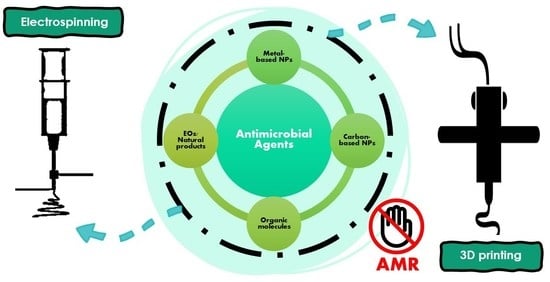
1. Introduction
2. Nanomaterial-Based Antimicrobial Agents
3. Antimicrobial Electrospun Nanofibrous Mats
3.1. Metallic Nanoparticles and Derivatives
3.2. Essential Oils
3.3. Carbon-Based Nanomaterials

3.4. Photoactivable Molecules and Nanomaterials
4. Antimicrobial 3D Printed Materials
4.1. Natural Polymers
4.2. Metallic Nanoparticles and Derivatives
4.3. Essential Oils
4.4. Other Strategies
5. Conclusions and Outlook
Author Contributions
Funding
Institutional Review Board Statement
Informed Consent Statement
Data Availability Statement
Acknowledgments
Conflicts of Interest
Abbreviations
| ΦΔ | Singlet oxygen quantum yield |
| β-Si3N4 | Silicon nitride |
| γ-MPS | 3-methacryloxypropyltrimethoxysilane |
| Ag NPs | Silver nanoparticles |
| Ag NWs | Silver nanowires |
| AG | Alginate |
| AlN | aluminum nitride |
| AM | Additive manufacturing |
| AMR | Antimicrobial resistance |
| ASTM | American Society for Testing and Materials |
| BS | Bacterial cellulose |
| CA | Cellulose acetate |
| CAD | Computer-aided design |
| CAR | Carvacrol |
| CBD | Chemical bath deposition |
| CEO | Cinnamon essential oil |
| CFU | Colony-Forming Unit |
| CG-MS | Gas Chromatography-Mass Spectrometry |
| ClInOCP | ClIn(III) octacarboxy phthalocyanine |
| CLV | Clove essential oil |
| CLXD | Chlorhexidine |
| CMC | carboxymethyl cellulose |
| CMC-GMA | carboxymethyl cellulose-glycidyl methacrylate |
| CNTs | Carbon nanotubes |
| CPP | Citrus peel pectin |
| CQDS | Carbon quantum dots |
| CS | Chitosan |
| CSEO | Citrus sinencis essential oil |
| Cu NPs | Copper nanoparticles |
| CuO NPs | Copper oxide (II) nanoparticles |
| DA | Dopamine |
| DIW | Direct ink writing |
| DLP | digital light processing |
| EC | Ethylcellulose |
| ECM | Extracellular matrix |
| EDC | 1-Ethyl-3-(3Dimethyl aminopropyl) carbodiimide |
| EDS | Energy-dispersive X-ray spectroscopy |
| EE | Encapsulation efficiency |
| EGDMA | ethyleneglycol dymethylacrylate |
| EOs | Essential oils |
| EPD | Electrophoretic deposition |
| EPL | ε-poly-L-lysine |
| EPL-MA | ε-poly-L-lysine-methacrylate |
| ER | Erythrosine B |
| FAO | Food and Agriculture Organization of the United Nations |
| FDA | Food and Drug Administration |
| FBS | Fetal bovine serum |
| FDA | Food and Drug Administration |
| FDM | Fused deposition modeling |
| FE-SEM | Field Emission Scanning Electron Microscopy |
| FFF | Fused filament fabrication |
| FGF | Fused granular fabrication |
| FRE | Freeform reversible embedding |
| FTIR | Fourier-Transform Infrared Spectroscopy |
| GC | Gas Chromatography |
| GEL | Gelatin |
| GEL/PDA | Gelatin/polydopamine |
| GMA | glycidyl methacrylate |
| GO | Graphene oxide |
| GQDs | Graphene quantum dots |
| GT | Gum tragacanth |
| GTMAC | Glycidyl trimethylammonium chloride |
| HA | Hydroxyapatite |
| HAc | Hyaluronic acid |
| HACC | Hydroxypropyltrimethyl ammonium chloride chitosan |
| HKUST-1 | Copper-based metal-organic framework |
| HNTs | Halloysite nanotubes |
| H&E | Hematoxylin and Eosin |
| ISO | International Organization for Standarization |
| MA | Maleic anhydride |
| MB | Methylene Blue |
| MBC | Minimum bactericidal concentration |
| MC | Momordica charantia |
| MIC | Minimum inhibitory concentration |
| MNP | Magnetic nanoparticles |
| MOF | Metallic organic frameworks |
| MRSA | Methicillin-resistant Staphylococcus aureus |
| MSN | mesoporous silica nanocarrier |
| Mw | Weight average molecular weight |
| MWCNT | Multi-walled carbon nanotubes |
| NC | nanocellulose |
| NF | Nanofibers |
| Nha | Nanohydroxyapatyte |
| NHDF | Normal human dermal fibroblast |
| NHS | N-hydroxysuccinimide |
| NHSMA | Methacrylic acid N-hydroxysuccinimide ester |
| NIPAM | N-isopropylacrylamide |
| NIR | Near Infrarred Radiation |
| NPs | Nanoparticles |
| OIE | World Organization for Animal Health |
| P1 | asymmetric triblock copolypeptide comprising EPL as central block, and polycysteine and polytyrosine as lateral blocks |
| P2 | asymmetric triblock copolypeptide comprising polyglutamic acid as central block, and polycysteine and polytyrosine as lateral blocks |
| PA | phytoncide oil type A extracted from Pinus densiflora |
| PAA | Poly(acrylic acid) |
| PACT | Photodynamic antimicrobial chemotherapy |
| PAH | Polyallylamine hydrochloride |
| PAN | Polyacrylonitrile |
| PB | phytoncide oil type B extracted from Chamaecyparis obtuse |
| PBS | Phosphate-buffered saline |
| PCL | Poly(ε-caprolactone) |
| PDA | Polydopamine |
| PDI | Photodynamic inactivation |
| PDMS | Poly(dimethyl siloxane) |
| PDT | Photodynamic Therapy |
| PEG | Poly(ethylene glycol) |
| PEO | Poly(ethylene oxide) |
| PEP | Peppermint essential oil |
| PEs | Pinus radiata bark extracts |
| PGA | Poly(glycolic acid) |
| PLA | Poly(lactic acid) |
| PLGA | Poly[(rac-lactide)-co-glycolide] |
| PMMA | Poly(methyl methacrylate) |
| PNIPAM | poly(N-isopropylacrylamide) |
| PPSu | Poly(1,3-propylene succinate) |
| PPTs | post-polymerization times |
| PS | Photosensitizer |
| PVA | Poly(vinyl alcohol) |
| PVP | Poly(vinylpyrrolidone) |
| ROS | Reactive Oxygen Species |
| RP | Rapid prototyping |
| SAXS | Small Angle X-ray Scattering |
| SBF | Simulated body fluid |
| SEM-EDS | Scanning Electron Microscopy/Energy-Dispersive X-ray Spectroscopy |
| SFF | Solid freeform fabrication |
| SLA | Stereolithography |
| SLM | selective laser melting |
| SLS | Selective laser sintering |
| SPAN | Sorbitan ester |
| SWCNT | Single-Walled Carbon Nanotubes |
| TEM | Transmission Electron Microscopy |
| THY | Thymol |
| TMPyP | Tetrakis(1-methyl-4-pyridinio) porphyrin tetra(p-toluenesulfonate) |
| TPU | thermoplastic polyurethane |
| UV | Ultraviolet visible |
| VRE | Vancomycin-Resistant Enterococci |
| WF | wood flour |
| WHO | World Health Organization |
| XPS | X-ray Photoelectron Spectroscopy |
| XRD | X-ray Diffraction |
| ZnO | Zinc oxide |
| ZnO NPs | ZnO nanoparticles |
| ZOI | Inhibition zones |
| ZrO2 NPs | zirconia nanoparticles |
References
- Pérez-Laguna, V.; Gilaberte, Y.; Millán-Lou, M.I.; Agut, M.; Nonell, S.; Rezusta, A.; Hamblin, M.R. A Combination of Photodynamic Therapy and Antimicrobial Compounds to Treat Skin and Mucosal Infections: A Systematic Review. Photochem. Photobiol. Sci. 2019, 18, 1020–1029. [Google Scholar] [CrossRef]
- Jamaledin, R.; Yiu, C.K.Y.; Zare, E.N.; Niu, L.-N.; Vecchione, R.; Chen, G.; Gu, Z.; Tay, F.R.; Makvandi, P. Advances in Antimicrobial Microneedle Patches for Combating Infections. Adv. Mater. 2020, 32, 2002129. [Google Scholar] [CrossRef] [PubMed]
- Ribeiro da Cunha, B.; Fonseca, L.P.; Calado, C.R.C. Antibiotic Discovery: Where Have We Come from, Where Do We Go? Antibiotics 2019, 8, 45. [Google Scholar] [CrossRef] [PubMed]
- Thombre, R.; Jangid, K.; Shukla, R.; Dutta, N.K. Editorial: Alternative Therapeutics Against Antimicrobial-Resistant Pathogens. Front. Microbiol. 2019, 10, 2173. [Google Scholar] [CrossRef] [PubMed]
- Stephens, L.J.; Werrett, M.V.; Sedgwick, A.C.; Bull, S.D.; Andrews, P.C. Antimicrobial Innovation: A Current Update and Perspective on the Antibiotic Drug Development Pipeline. Future Med. Chem. 2020, 12, 2035–2065. [Google Scholar] [CrossRef]
- Dhingra, S.; Rahman, N.A.A.; Peile, E.; Rahman, M.; Sartelli, M.; Hassali, M.A.; Islam, T.; Islam, S.; Haque, M. Microbial Resistance Movements: An Overview of Global Public Health Threats Posed by Antimicrobial Resistance, and How Best to Counter. Front. Public Health 2020, 8, 535668. [Google Scholar] [CrossRef]
- Hutchings, M.I.; Truman, A.W.; Wilkinson, B. Antibiotics: Past, Present and Future. Curr. Opin. Microbiol. 2019, 51, 72–80. [Google Scholar] [CrossRef]
- Murugaiyan, J.; Kumar, P.A.; Rao, G.S.; Iskandar, K.; Hawser, S.; Hays, J.P.; Mohsen, Y.; Adukkadukkam, S.; Awuah, W.A.; Jose, R.A.M.; et al. Progress in Alternative Strategies to Combat Antimicrobial Resistance: Focus on Antibiotics. Antibiotics 2022, 11, 200. [Google Scholar] [CrossRef]
- Annunziato, G. Strategies to Overcome Antimicrobial Resistance (AMR) Making Use of Non-Essential Target Inhibitors: A Review. Int. J. Mol. Sci. 2019, 20, 5844. [Google Scholar] [CrossRef]
- Acharya, K.P.; Wilson, R.T. Antimicrobial Resistance in Nepal. Front. Med. 2019, 6, 105. [Google Scholar] [CrossRef]
- Walsh, C. Molecular Mechanisms That Confer Antibacterial Drug Resistance. Nature 2000, 406, 775–781. [Google Scholar] [CrossRef] [PubMed]
- Pulingam, T.; Parumasivam, T.; Gazzali, A.M.; Sulaiman, A.M.; Chee, J.Y.; Lakshmanan, M.; Chin, C.F.; Sudesh, K. Antimicrobial Resistance: Prevalence, Economic Burden, Mechanisms of Resistance and Strategies to Overcome. Eur. J. Pharm. Sci. 2022, 170, 106103. [Google Scholar] [CrossRef] [PubMed]
- Antimicrobial Resistance. Available online: https://www.who.int/news-room/fact-sheets/detail/antimicrobial-resistance (accessed on 8 May 2023).
- Salleh, A.; Naomi, R.; Utami, N.D.; Mohammad, A.W.; Mahmoudi, E.; Mustafa, N.; Fauzi, M.B. The Potential of Silver Nanoparticles for Antiviral and Antibacterial Applications: A Mechanism of Action. Nanomaterials 2020, 10, 1566. [Google Scholar] [CrossRef]
- Fan, X.; Yahia, L.; Sacher, E. Antimicrobial Properties of the Ag, Cu Nanoparticle System. Biology 2021, 10, 137. [Google Scholar] [CrossRef]
- Cao, G.; Yan, J.; Ning, X.; Zhang, Q.; Wu, Q.; Bi, L.; Zhang, Y.; Han, Y.; Guo, J. Antibacterial and Antibiofilm Properties of Graphene and Its Derivatives. Colloids Surf. B Biointerfaces 2021, 200, 111588. [Google Scholar] [CrossRef]
- Raul, P.K.; Thakuria, A.; Das, B.; Devi, R.R.; Tiwari, G.; Yellappa, C.; Kamboj, D.V. Carbon Nanostructures as Antibacterials and Active Food-Packaging Materials: A Review. ACS Omega 2022, 7, 11555–11559. [Google Scholar] [CrossRef]
- Gallarato, L.A.; Mulko, L.E.; Dardanelli, M.S.; Barbero, C.A.; Acevedo, D.F.; Yslas, E.I. Synergistic Effect of Polyaniline Coverage and Surface Microstructure on the Inhibition of Pseudomonas Aeruginosa Biofilm Formation. Colloids Surf. B Biointerfaces 2017, 150, 1–7. [Google Scholar] [CrossRef] [PubMed]
- Abel, S.B.; Yslas, E.I.; Rivarola, C.R.; Barbero, C.A. Synthesis of Polyaniline (PANI) and Functionalized Polyaniline (F-PANI) Nanoparticles with Controlled Size by Solvent Displacement Method. Application in Fluorescence Detection and Bacteria Killing by Photothermal Effect. Nanotechnology 2018, 29, 125604. [Google Scholar] [CrossRef]
- Abel, S.B.; Gallarato, L.A.; Dardanelli, M.S.; Barbero, C.A.; Rivarola, C.R.; Yslas, E.I. Photothermal Lysis of Pseudomonas Aeruginosa by Polyaniline Nanoparticles under near Infrared Irradiation. Biomed. Phys. Eng. Express 2018, 4, 045037. [Google Scholar] [CrossRef]
- Tariq, S.; Wani, S.; Rasool, W.; Shafi, K.; Bhat, M.A.; Prabhakar, A.; Shalla, A.H.; Rather, M.A. A Comprehensive Review of the Antibacterial, Antifungal and Antiviral Potential of Essential Oils and Their Chemical Constituents against Drug-Resistant Microbial Pathogens. Microb. Pathog. 2019, 134, 103580. [Google Scholar] [CrossRef]
- Kolahalam, L.A.; Kasi Viswanath, I.V.; Diwakar, B.S.; Govindh, B.; Reddy, V.; Murthy, Y.L.N. Review on Nanomaterials: Synthesis and Applications. Mater. Today Proc. 2019, 18, 2182–2190. [Google Scholar] [CrossRef]
- Yang, D.-L.; Faraz, F.; Wang, J.-X.; Radacsi, N. Combination of 3D Printing and Electrospinning Techniques for Biofabrication. Adv. Mater. Technol. 2022, 7, 2101309. [Google Scholar] [CrossRef]
- Kozior, T.; Mamun, A.; Trabelsi, M.; Sabantina, L. Comparative Analysis of Polymer Composites Produced by FFF and PJM 3D Printing and Electrospinning Technologies for Possible Filter Applications. Coatings 2022, 12, 48. [Google Scholar] [CrossRef]
- Abel, S.B.; Ballarin, F.M.; Abraham, G.A. Combination of Electrospinning with Other Techniques for the Fabrication of 3D Polymeric and Composite Nanofibrous Scaffolds with Improved Cellular Interactions. Nanotechnology 2020, 31, 172002. [Google Scholar] [CrossRef] [PubMed]
- Chen, K.; Hu, H.; Zeng, Y.; Pan, H.; Wang, S.; Zhang, Y.; Shi, L.; Tan, G.; Pan, W.; Liu, H. Recent Advances in Electrospun Nanofibers for Wound Dressing. Eur. Polym. J. 2022, 178, 111490. [Google Scholar] [CrossRef]
- Clarissa, W.H.-Y.; Chia, C.H.; Zakaria, S.; Evyan, Y.C.-Y. Recent Advancement in 3-D Printing: Nanocomposites with Added Functionality. Prog. Addit. Manuf. 2022, 7, 325–350. [Google Scholar] [CrossRef]
- Lores, N.J.; Hung, X.; Talou, M.H.; Abraham, G.A.; Caracciolo, P.C. Novel Three-Dimensional Printing of Poly(Ester Urethane) Scaffolds for Biomedical Applications. Polym. Adv. Technol. 2021, 32, 3309–3321. [Google Scholar] [CrossRef]
- Yu, W.; Li, X.; He, J.; Chen, Y.; Qi, L.; Yuan, P.; Ou, K.; Liu, F.; Zhou, Y.; Qin, X. Graphene Oxide-Silver Nanocomposites Embedded Nanofiber Core-Spun Yarns for Durable Antibacterial Textiles. J. Colloid Interface Sci. 2021, 584, 164–173. [Google Scholar] [CrossRef]
- Mallakpour, S.; Azadi, E.; Hussain, C.M. Recent Breakthroughs of Antibacterial and Antiviral Protective Polymeric Materials during COVID-19 Pandemic and after Pandemic: Coating, Packaging, and Textile Applications. Curr. Opin. Colloid Interface Sci. 2021, 55, 101480. [Google Scholar] [CrossRef]
- Karagoz, S.; Kiremitler, N.B.; Sarp, G.; Pekdemir, S.; Salem, S.; Goksu, A.G.; Onses, M.S.; Sozdutmaz, I.; Sahmetlioglu, E.; Ozkara, E.S.; et al. Antibacterial, Antiviral, and Self-Cleaning Mats with Sensing Capabilities Based on Electrospun Nanofibers Decorated with ZnO Nanorods and Ag Nanoparticles for Protective Clothing Applications. ACS Appl. Mater. Interfaces 2021, 13, 5678–5690. [Google Scholar] [CrossRef]
- Wang, D.; Li, K.; Zhou, C.; Lei, L.; de Rancourt de Mimérand, Y.; Jin, X.; Guo, J. Bi2MoO6 and Ag Nanoparticles Immobilized on Textile by Plasma-Derived Innovative Techniques to Generate Antimicrobial Activity. Appl. Surf. Sci. 2022, 585, 152591. [Google Scholar] [CrossRef]
- Preda, M.D.; Popa, M.L.; Neacșu, I.A.; Grumezescu, A.M.; Ginghină, O. Antimicrobial Clothing Based on Electrospun Fibers with ZnO Nanoparticles. Int. J. Mol. Sci. 2023, 24, 1629. [Google Scholar] [CrossRef] [PubMed]
- Larrañeta, E.; Dominguez-Robles, J.; Lamprou, D.A. Additive Manufacturing Can Assist in the Fight Against COVID-19 and Other Pandemics and Impact on the Global Supply Chain. 3D Print Addit. Manuf. 2020, 7, 100–103. [Google Scholar] [CrossRef] [PubMed]
- Mallakpour, S.; Azadi, E.; Hussain, C.M. State-of-the-Art of 3D Printing Technology of Alginate-Based Hydrogels—An Emerging Technique for Industrial Applications. Adv. Colloid Interface Sci. 2021, 293, 102436. [Google Scholar] [CrossRef]
- Ahmed, W.; Al-Marzouqi, A.H.; Nazir, M.H.; Rizvi, T.A.; Zaneldin, E.; Khan, M.; Aziz, M. Investigating the Properties and Characterization of a Hybrid 3D Printed Antimicrobial Composite Material Using FFF Process: Innovative and Swift. Int. J. Mol. Sci. 2023, 24, 8895. [Google Scholar] [CrossRef]
- Nishal, M.; Ram Prasad, K.; Salman Dasthageer, M.; Ragunath, A.G. Significance of Additive Manufacturing amidst the Pandemic. Mater. Today Proc. 2023, 72, 2540–2546. [Google Scholar] [CrossRef] [PubMed]
- Obasi, H.C.; Ijaz, K.; Akhtar, H.; Ali, A.; Khalid, H.; Khan, A.F.; Chaudhry, A.A. Fabrication of Antimicrobial Electrospun Mats Using Polyvinyl Alcohol–Zinc Oxide Blends. Polym. Bull. 2023, 80, 2681–2695. [Google Scholar] [CrossRef]
- Sekyere, J.O.; Asante, J. Emerging Mechanisms of Antimicrobial Resistance in Bacteria and Fungi: Advances in the Era of Genomics. Future Microbiol. 2018, 13, 241–262. [Google Scholar] [CrossRef]
- Becerra, G.; Plascencia, A.; Luévanos, A.; Domínguez, M.; Hernández, I. Mecanismo de resistencia a antimicrobianos en bacterias. Enfermedades Infecc. Microbiol. 2009, 29, 70–76. [Google Scholar]
- Durán, L. Resistencia antimicrobiana e implicancias para el manejo de infecciones del tracto urinario. Rev. Méd. Clín. Las Condes 2018, 29, 213–221. [Google Scholar] [CrossRef]
- Abushaheen, M.A.; Muzaheed; Fatani, A.J.; Alosaimi, M.; Mansy, W.; George, M.; Acharya, S.; Rathod, S.; Divakar, D.D.; Jhugroo, C.; et al. Antimicrobial Resistance, Mechanisms and Its Clinical Significance. Dis. Mon. 2020, 66, 100971. [Google Scholar] [CrossRef]
- Rojas, G.C.; Ulate, L.A. Resistencia antimicrobiana: Microorganismos más resistentes y antibióticos con menor actividad. Rev. Medica Costa Rica Centroam. 2017, 73, 757–763. [Google Scholar]
- Pfaller, M.A. Antifungal Drug Resistance: Mechanisms, Epidemiology, and Consequences for Treatment. Am. J. Med. 2012, 125, S3–S13. [Google Scholar] [CrossRef] [PubMed]
- Xie, M.; Gao, M.; Yun, Y.; Malmsten, M.; Rotello, V.M.; Zboril, R.; Akhavan, O.; Kraskouski, A.; Amalraj, J.; Cai, X.; et al. Antibacterial Nanomaterials: Mechanisms, Impacts on Antimicrobial Resistance and Design Principles. Angew. Chem. Int. Ed. 2023, 62, e202217345. [Google Scholar] [CrossRef] [PubMed]
- Garg, P.; Attri, P.; Sharma, R.; Chauhan, M.; Chaudhary, G.R. Advances and Perspective on Antimicrobial Nanomaterials for Biomedical Applications. Front. Nanotechnol. 2022, 4, 898411. [Google Scholar] [CrossRef]
- Wang, Y.; Sun, H. Polymeric Nanomaterials for Efficient Delivery of Antimicrobial Agents. Pharmaceutics 2021, 13, 2108. [Google Scholar] [CrossRef]
- Spizzirri, U.G.; Aiello, F.; Carullo, G.; Facente, A.; Restuccia, D. Nanotechnologies: An Innovative Tool to Release Natural Extracts with Antimicrobial Properties. Pharmaceutics 2021, 13, 230. [Google Scholar] [CrossRef] [PubMed]
- Hochvaldová, L.; Večeřová, R.; Kolář, M.; Prucek, R.; Kvítek, L.; Lapčík, L.; Panáček, A. Antibacterial Nanomaterials: Upcoming Hope to Overcome Antibiotic Resistance Crisis. Nanotechnol. Rev. 2022, 11, 1115–1142. [Google Scholar] [CrossRef]
- Sportelli, M.C.; Picca, R.A.; Cioffi, N. Nano-Antimicrobials Based on Metals. In Novel Antimicrobial Agents and Strategies; John Wiley & Sons, Ltd.: Hoboken, NJ, USA, 2014; pp. 181–218. ISBN 978-3-527-67613-2. [Google Scholar]
- Xin, Q.; Shah, H.; Nawaz, A.; Xie, W.; Akram, M.Z.; Batool, A.; Tian, L.; Jan, S.U.; Boddula, R.; Guo, B.; et al. Antibacterial Carbon-Based Nanomaterials. Adv. Mater. 2019, 31, 1804838. [Google Scholar] [CrossRef]
- Azizi-Lalabadi, M.; Hashemi, H.; Feng, J.; Jafari, S.M. Carbon Nanomaterials against Pathogens; the Antimicrobial Activity of Carbon Nanotubes, Graphene/Graphene Oxide, Fullerenes, and Their Nanocomposites. Adv. Colloid Interface Sci. 2020, 284, 102250. [Google Scholar] [CrossRef]
- Hammer, K.A.; Carson, C.F. Antibacterial and Antifungal Activities of Essential Oils. In Lipids and Essential Oils as Antimicrobial Agents; John Wiley & Sons, Ltd.: Hoboken, NJ, USA, 2011; pp. 255–306. ISBN 978-0-470-97662-3. [Google Scholar]
- Phoenix, D.A.; Dennison, S.R.; Harris, F. Photodynamic Antimicrobial Chemotherapy. In Novel Antimicrobial Agents and Strategies; John Wiley & Sons, Ltd.: Hoboken, NJ, USA, 2014; pp. 295–330. ISBN 978-3-527-67613-2. [Google Scholar]
- Joy, N.; Anuraj, R.; Viravalli, A.; Dixit, H.N.; Samavedi, S. Coupling between Voltage and Tip-to-Collector Distance in Polymer Electrospinning: Insights from Analysis of Regimes, Transitions and Cone/Jet Features. Chem. Eng. Sci. 2021, 230, 116200. [Google Scholar] [CrossRef]
- Li, Y.; Zhu, J.; Cheng, H.; Li, G.; Cho, H.; Jiang, M.; Gao, Q.; Zhang, X. Developments of Advanced Electrospinning Techniques: A Critical Review. Adv. Mater. Technol. 2021, 6, 2100410. [Google Scholar] [CrossRef]
- Xue, J.; Wu, T.; Dai, Y.; Xia, Y. Electrospinning and Electrospun Nanofibers: Methods, Materials, and Applications. Chem. Rev. 2019, 119, 5298–5415. [Google Scholar] [CrossRef] [PubMed]
- Angel, N.; Li, S.; Yan, F.; Kong, L. Recent Advances in Electrospinning of Nanofibers from Bio-Based Carbohydrate Polymers and Their Applications. Trends Food Sci. Technol. 2022, 120, 308–324. [Google Scholar] [CrossRef]
- Rahmati, M.; Mills, D.K.; Urbanska, A.M.; Saeb, M.R.; Venugopal, J.R.; Ramakrishna, S.; Mozafari, M. Electrospinning for Tissue Engineering Applications. Prog. Mater. Sci. 2021, 117, 100721. [Google Scholar] [CrossRef]
- Uhljar, L.É.; Ambrus, R. Electrospinning of Potential Medical Devices (Wound Dressings, Tissue Engineering Scaffolds, Face Masks) and Their Regulatory Approach. Pharmaceutics 2023, 15, 417. [Google Scholar] [CrossRef]
- Jain, R.; Shetty, S.; Yadav, K.S. Unfolding the Electrospinning Potential of Biopolymers for Preparation of Nanofibers. J. Drug Deliv. Sci. Technol. 2020, 57, 101604. [Google Scholar] [CrossRef]
- Patel, P.R.; Gundloori, R.V.N. A Review on Electrospun Nanofibers for Multiple Biomedical Applications. Polym. Adv. Technol. 2023, 34, 44–63. [Google Scholar] [CrossRef]
- De Vrieze, S.; Van Camp, T.; Nelvig, A.; Hagström, B.; Westbroek, P.; De Clerck, K. The Effect of Temperature and Humidity on Electrospinning. J. Mater. Sci. 2009, 44, 1357–1362. [Google Scholar] [CrossRef]
- Shangguan, W.; Li, S.; Cao, L.; Wei, M.; Wang, Z.; Xu, H. Electrospinning and Nanofibers: Building Drug Delivery Systems and Potential in Pesticide Delivery. Mater. Today Commun. 2022, 33, 104399. [Google Scholar] [CrossRef]
- Luraghi, A.; Peri, F.; Moroni, L. Electrospinning for Drug Delivery Applications: A Review. J. Control Release 2021, 334, 463–484. [Google Scholar] [CrossRef]
- Chen, Y.; Dong, X.; Shafiq, M.; Myles, G.; Radacsi, N.; Mo, X. Recent Advancements on Three-Dimensional Electrospun Nanofiber Scaffolds for Tissue Engineering. Adv. Fiber Mater. 2022, 4, 959–986. [Google Scholar] [CrossRef]
- Maliszewska, I.; Czapka, T. Electrospun Polymer Nanofibers with Antimicrobial Activity. Polymers 2022, 14, 1661. [Google Scholar] [CrossRef] [PubMed]
- Leung, C.M.; Dhand, C.; Dwivedi, N.; Xiao, A.; Ong, S.T.; Chalasani, M.L.S.; Sriram, H.; Balakrishnan, Y.; Dolatshahi-Pirouz, A.; Orive, G.; et al. Combating Microbial Contamination with Robust Polymeric Nanofibers: Elemental Effect on the Mussel-Inspired Cross-Linking of Electrospun Gelatin. ACS Appl. Bio Mater. 2019, 2, 807–823. [Google Scholar] [CrossRef] [PubMed]
- Mude, H.; Tata, P.; Jamma, T.; Ganesan, R.; Ray Dutta, J. Fabrication and Facile Surface Derivatization of Poly(ε-Caprolactone)-Based Wound Dressing Materials Imparting Anti-Infective, Excessive Biofluid Drainage, and Easy-to-Peel Characteristics. Adv. Ther. 2023, 6, 2300015. [Google Scholar] [CrossRef]
- Bandeira, M.; Chee, B.S.; Frassini, R.; Nugent, M.; Giovanela, M.; Roesch-Ely, M.; Crespo, J.d.S.; Devine, D.M. Antimicrobial PAA/PAH Electrospun Fiber Containing Green Synthesized Zinc Oxide Nanoparticles for Wound Healing. Materials 2021, 14, 2889. [Google Scholar] [CrossRef] [PubMed]
- Geetha, K.; Sivasangari, D.; Kim, H.-S.; Murugadoss, G.; Kathalingam, A. Electrospun Nanofibrous ZnO/PVA/PVP Composite Films for Efficient Antimicrobial Face Masks. Ceram. Int. 2022, 48, 29197–29204. [Google Scholar] [CrossRef]
- Salmeri, M.; Ognibene, G.; Saitta, L.; Lombardo, C.; Genovese, C.; Barcellona, M.; D’Urso, A.; Spitaleri, L.; Blanco, I.; Cicala, G.; et al. Optimization of ZnO Nanorods Growth on Polyetheresulfone Electrospun Mats to Promote Antibacterial Properties. Molecules 2020, 25, 1696. [Google Scholar] [CrossRef]
- Sarwar, M.N.; Ali, H.G.; Ullah, S.; Yamashita, K.; Shahbaz, A.; Nisar, U.; Hashmi, M.; Kim, I.-S. Electrospun PVA/CuONPs/Bitter Gourd Nanofibers with Improved Cytocompatibility and Antibacterial Properties: Application as Antibacterial Wound Dressing. Polymers 2022, 14, 1361. [Google Scholar] [CrossRef]
- Peng, R.; Zhang, S.; Yao, Y.; Wang, J.; Zhu, X.; Jiang, R.; Zhang, J.; Zhang, W.; Wang, C. MOFs Meet Electrospinning: New Opportunities for Water Treatment. Chem. Eng. J. 2023, 453, 139669. [Google Scholar] [CrossRef]
- Li, R.; Chen, T.; Pan, X. Metal–Organic-Framework-Based Materials for Antimicrobial Applications. ACS Nano 2021, 15, 3808–3848. [Google Scholar] [CrossRef] [PubMed]
- Zhang, S.; Ye, J.; Sun, Y.; Kang, J.; Liu, J.; Wang, Y.; Li, Y.; Zhang, L.; Ning, G. Electrospun Fibrous Mat Based on Silver (I) Metal-Organic Frameworks-Polylactic Acid for Bacterial Killing and Antibiotic-Free Wound Dressing. Chem. Eng. J. 2020, 390, 124523. [Google Scholar] [CrossRef]
- Wang, S.; Yan, F.; Ren, P.; Li, Y.; Wu, Q.; Fang, X.; Chen, F.; Wang, C. Incorporation of Metal-Organic Frameworks into Electrospun Chitosan/Poly (Vinyl Alcohol) Nanofibrous Membrane with Enhanced Antibacterial Activity for Wound Dressing Application. Int. J. Biol. Macromol. 2020, 158, 9–17. [Google Scholar] [CrossRef]
- Alven, S.; Buyana, B.; Feketshane, Z.; Aderibigbe, B.A. Electrospun Nanofibers/Nanofibrous Scaffolds Loaded with Silver Nanoparticles as Effective Antibacterial Wound Dressing Materials. Pharmaceutics 2021, 13, 964. [Google Scholar] [CrossRef]
- Unalan, I.; Slavik, B.; Buettner, A.; Goldmann, W.H.; Frank, G.; Boccaccini, A.R. Physical and Antibacterial Properties of Peppermint Essential Oil Loaded Poly (ε-Caprolactone) (PCL) Electrospun Fiber Mats for Wound Healing. Front. Bioeng. Biotechnol. 2019, 7, 346. [Google Scholar] [CrossRef] [PubMed]
- Gámez, E.; Mendoza, G.; Salido, S.; Arruebo, M.; Irusta, S. Antimicrobial Electrospun Polycaprolactone-Based Wound Dressings: An In Vitro Study About the Importance of the Direct Contact to Elicit Bactericidal Activity. Adv. Wound Care 2019, 8, 438–451. [Google Scholar] [CrossRef]
- García-Salinas, S.; Gámez, E.; Asín, J.; de Miguel, R.; Andreu, V.; Sancho-Albero, M.; Mendoza, G.; Irusta, S.; Arruebo, M. Efficiency of Antimicrobial Electrospun Thymol-Loaded Polycaprolactone Mats In Vivo. ACS Appl. Bio Mater. 2020, 3, 3430–3439. [Google Scholar] [CrossRef] [PubMed]
- Unalan, I.; Endlein, S.J.; Slavik, B.; Buettner, A.; Goldmann, W.H.; Detsch, R.; Boccaccini, A.R. Evaluation of Electrospun Poly(ε-Caprolactone)/Gelatin Nanofiber Mats Containing Clove Essential Oil for Antibacterial Wound Dressing. Pharmaceutics 2019, 11, 570. [Google Scholar] [CrossRef] [PubMed]
- Borges-Vilches, J.; Unalan, I.; Fernández, K.; Boccaccini, A.R. Fabrication of Biocompatible Electrospun Poly(ε-Caprolactone)/Gelatin Nanofibers Loaded with Pinus Radiata Bark Extracts for Wound Healing Applications. Polymers 2022, 14, 2331. [Google Scholar] [CrossRef]
- Abdollahi, A.; Zarenezhad, E.; Osanloo, M.; Ghaznavi, G.; Khalili Pour, M. Promising Antibacterial Activity of a Mat of Polycaprolactone Nanofibers Impregnated with a Green Nanogel. Nanomed. Res. J. 2020, 5, 192–201. [Google Scholar]
- Liakos, I.L.; Holban, A.M.; Carzino, R.; Lauciello, S.; Grumezescu, A.M. Electrospun Fiber Pads of Cellulose Acetate and Essential Oils with Antimicrobial Activity. Nanomaterials 2017, 7, 84. [Google Scholar] [CrossRef]
- Mishra, P.; Gupta, P.; Pruthi, V. Cinnamaldehyde Incorporated Gellan/PVA Electrospun Nanofibers for Eradicating Candida Biofilm. Mater. Sci. Eng. C 2021, 119, 111450. [Google Scholar] [CrossRef]
- Brandão, R.M.; Cardoso, M.d.G.; Batista, L.R.; Caetano, A.R.S.; Lemos, A.C.C.; Martins, M.A.; Nelson, D.L.; De Oliveira, J.E. Antifungal and Physicochemical Properties of Ocimum Essential Oil Loaded in Poly(Lactic Acid) Nanofibers. Lett. Appl. Microbiol. 2022, 74, 765–776. [Google Scholar] [CrossRef] [PubMed]
- De Toledo, L.G.; Ramos, M.A.D.S.; Spósito, L.; Castilho, E.M.; Pavan, F.R.; Lopes, É.D.O.; Zocolo, G.J.; Silva, F.A.N.; Soares, T.H.; Dos Santos, A.G.; et al. Essential Oil of Cymbopogon nardus (L.) Rendle: A Strategy to Combat Fungal Infections Caused by Candida Species. Int. J. Mol. Sci. 2016, 17, 1252. [Google Scholar] [CrossRef]
- Xin, Y.; Quan, L.; Zhang, H.; Ao, Q. Emerging Polymer-Based Nanosystem Strategies in the Delivery of Antifungal Drugs. Pharmaceutics 2023, 15, 1866. [Google Scholar] [CrossRef]
- El-Aassar, M.R.; El-Beheri, N.G.; Agwa, M.M.; Eltaher, H.M.; Alseqely, M.; Sadik, W.S.; El-Khordagui, L. Antibiotic-Free Combinational Hyaluronic Acid Blend Nanofibers for Wound Healing Enhancement. Int. J. Biol. Macromol. 2021, 167, 1552–1563. [Google Scholar] [CrossRef] [PubMed]
- Ghorbani, M.; Ramezani, S.; Rashidi, M.-R. Fabrication of Honey-Loaded Ethylcellulose/Gum Tragacanth Nanofibers as an Effective Antibacterial Wound Dressing. Colloids Surf. A Physicochem. Eng. Asp. 2021, 621, 126615. [Google Scholar] [CrossRef]
- Tamayo Marín, J.A.; Londoño, S.R.; Delgado, J.; Navia Porras, D.P.; Valencia Zapata, M.E.; Mina Hernandez, J.H.; Valencia, C.H.; Grande Tovar, C.D. Biocompatible and Antimicrobial Electrospun Membranes Based on Nanocomposites of Chitosan/Poly (Vinyl Alcohol)/Graphene Oxide. Int. J. Mol. Sci. 2019, 20, 2987. [Google Scholar] [CrossRef]
- Ruiz, S.; Tamayo, J.A.; Delgado Ospina, J.; Navia Porras, D.P.; Valencia Zapata, M.E.; Mina Hernandez, J.H.; Valencia, C.H.; Zuluaga, F.; Grande Tovar, C.D. Antimicrobial Films Based on Nanocomposites of Chitosan/Poly(Vinyl Alcohol)/Graphene Oxide for Biomedical Applications. Biomolecules 2019, 9, 109. [Google Scholar] [CrossRef]
- Wang, S.; Li, Y.; Zhao, R.; Jin, T.; Zhang, L.; Li, X. Chitosan Surface Modified Electrospun Poly(ε-Caprolactone)/Carbon Nanotube Composite Fibers with Enhanced Mechanical, Cell Proliferation and Antibacterial Properties. Int. J. Biol. Macromol. 2017, 104, 708–715. [Google Scholar] [CrossRef]
- Liu, Y.; Wang, S.; Lan, W.; Qin, W. Fabrication of Polylactic Acid/Carbon Nanotubes/Chitosan Composite Fibers by Electrospinning for Strawberry Preservation. Int. J. Biol. Macromol. 2019, 121, 1329–1336. [Google Scholar] [CrossRef]
- Kandel, R.; Jang, S.R.; Ghimire, U.; Shrestha, S.; Shrestha, B.K.; Park, C.H.; Kim, C.S. Engineered Nanostructure Fibrous Cell-Laden Biointerfaces Integrating Fe3O4/SrO2-FMWCNTs Induce Osteogenesis and Anti-Bacterial Effect. J. Ind. Eng. Chem. 2023, 120, 216–230. [Google Scholar] [CrossRef]
- Dehghani, N.; Haghiralsadat, F.; Yazdian, F.; Sadeghian-Nodoushan, F.; Ghasemi, N.; Mazaheri, F.; Pourmadadi, M.; Naghib, S.M. Chitosan/Silk Fibroin/Nitrogen-Doped Carbon Quantum Dot/α-Tricalcium Phosphate Nanocomposite Electrospinned as a Scaffold for Wound Healing Application: In Vitro and in Vivo Studies. Int. J. Biol. Macromol. 2023, 238, 124078. [Google Scholar] [CrossRef] [PubMed]
- de Faria, A.F.; Perreault, F.; Shaulsky, E.; Arias Chavez, L.H.; Elimelech, M. Antimicrobial Electrospun Biopolymer Nanofiber Mats Functionalized with Graphene Oxide–Silver Nanocomposites. ACS Appl. Mater. Interfaces 2015, 7, 12751–12759. [Google Scholar] [CrossRef] [PubMed]
- Sirelkhatim, N.; Parveen, A.; LaJeunesse, D.; Yu, D.; Zhang, L. Polyacrylonitrile Nanofibrous Mat from Electrospinning: Born with Potential Anti-Fungal Functionality. Eur. Polym. J. 2019, 119, 176–180. [Google Scholar] [CrossRef]
- Kurtz, I.S.; Schiffman, J.D. Current and Emerging Approaches to Engineer Antibacterial and Antifouling Electrospun Nanofibers. Materials 2018, 11, 1059. [Google Scholar] [CrossRef]
- Almeida, J.; Tomé, A.C.; Rangel, M.; Silva, A.M.G. Microwave-Assisted Synthesis and Spectral Properties of Pyrrolidine-Fused Chlorin Derivatives. Molecules 2023, 28, 3833. [Google Scholar] [CrossRef]
- Contreras, A.; Raxworthy, M.J.; Wood, S.; Schiffman, J.D.; Tronci, G. Photodynamically Active Electrospun Fibers for Antibiotic-Free Infection Control. ACS Appl. Bio Mater. 2019, 2, 4258–4270. [Google Scholar] [CrossRef]
- Contreras, A.; Raxworthy, M.J.; Wood, S.; Tronci, G. Hydrolytic Degradability, Cell Tolerance and On-Demand Antibacterial Effect of Electrospun Photodynamically Active Fibres. Pharmaceutics 2020, 12, 711. [Google Scholar] [CrossRef] [PubMed]
- Nyamu, S.N.; Ombaka, L.; Masika, E.; Ng’ang’a, M. Antimicrobial Photodynamic Activity of Phthalocyanine Derivatives. Adv. Chem. 2018, 2018, e2598062. [Google Scholar] [CrossRef]
- Strokov, K.; Schäfer, A.H.; Dobrindt, U.; Galstyan, A. Facile Fabrication of Silicon(IV)Phthalocyanine-Embedded Poly(Vinyl Alcohol)-Based Antibacterial and Antifouling Interfaces. ACS Appl. Bio Mater. 2020, 3, 3751–3760. [Google Scholar] [CrossRef] [PubMed]
- Sindelo, A.; Nyokong, T. Magnetic Nanoparticle—Indium Phthalocyanine Conjugate Embedded in Electrospun Fiber for Photodynamic Antimicrobial Chemotherapy and Photodegradation of Methyl Red. Heliyon 2019, 5, e02352. [Google Scholar] [CrossRef] [PubMed]
- Henke, P.; Dolanský, J.; Kubát, P.; Mosinger, J. Multifunctional Photosensitizing and Biotinylated Polystyrene Nanofiber Membranes/Composites for Binding of Biologically Active Compounds. ACS Appl. Mater. Interfaces 2020, 12, 18792–18802. [Google Scholar] [CrossRef] [PubMed]
- Nie, X.; Wu, S.; Mensah, A.; Lu, K.; Wei, Q. Carbon Quantum Dots Embedded Electrospun Nanofibers for Efficient Antibacterial Photodynamic Inactivation. Mater. Sci. Eng. C 2020, 108, 110377. [Google Scholar] [CrossRef] [PubMed]
- Ruiz, V.; Maudes, J.; Grande, H.-J.; Pérez-Marquez, A. Light-Activated Antibacterial Electrospun Polyacrylonitrile-Graphene Quantum Dot Nanofibrous Membranes. Mater. Today Commun. 2022, 32, 104112. [Google Scholar] [CrossRef]
- Liu, X.; Guo, C.; Zhuang, K.; Chen, W.; Zhang, M.; Dai, Y.; Tan, L.; Ran, Y. A Recyclable and Light-Triggered Nanofibrous Membrane against the Emerging Fungal Pathogen Candida Auris. PLoS Pathog. 2022, 18, e1010534. [Google Scholar] [CrossRef] [PubMed]
- WHO Fungal Priority Pathogens List to Guide Research, Development and Public Health Action. Available online: https://www.who.int/publications-detail-redirect/9789240060241 (accessed on 9 July 2023).
- Fan, D.; Li, Y.; Wang, X.; Zhu, T.; Wang, Q.; Cai, H.; Li, W.; Tian, Y.; Liu, Z. Progressive 3D Printing Technology and Its Application in Medical Materials. Front. Pharmacol. 2020, 11, 122. [Google Scholar] [CrossRef]
- Griffin, M.; Castro, N.; Bas, O.; Saifzadeh, S.; Butler, P.; Hutmacher, D.W. The Current Versatility of Polyurethane Three-Dimensional Printing for Biomedical Applications. Tissue Eng. Part B Rev. 2020, 26, 272–283. [Google Scholar] [CrossRef]
- Bettinger, C.J.; Borenstein, J.T.; Langer, R. Chapter Twenty-Four—Micro- and Nanofabricated Scaffolds. In Principles of Tissue Engineering, 3rd ed.; Lanza, R., Langer, R., Vacanti, J., Eds.; Academic Press: Burlington, MA, USA, 2007; pp. 341–358. ISBN 978-0-12-370615-7. [Google Scholar]
- Zühlke, A.; Gasik, M.; Vrana, N.E.; Muller, C.B.; Barthes, J.; Bilotsky, Y.; Courtial, E.; Marquette, C. Biomechanical and Functional Comparison of Moulded and 3D Printed Medical Silicones. J. Mech. Behav. Biomed. Mater. 2021, 122, 104649. [Google Scholar] [CrossRef]
- Mohammadi Zerankeshi, M.; Bakhshi, R.; Alizadeh, R. Polymer/Metal Composite 3D Porous Bone Tissue Engineering Scaffolds Fabricated by Additive Manufacturing Techniques: A Review. Bioprinting 2022, 25, e00191. [Google Scholar] [CrossRef]
- Serrano-Aroca, Á.; Cano-Vicent, A.; Sabater i Serra, R.; El-Tanani, M.; Aljabali, A.; Tambuwala, M.M.; Mishra, Y.K. Scaffolds in the Microbial Resistant Era: Fabrication, Materials, Properties and Tissue Engineering Applications. Mater. Today Bio 2022, 16, 100412. [Google Scholar] [CrossRef] [PubMed]
- Li, N.; Qiao, D.; Zhao, S.; Lin, Q.; Zhang, B.; Xie, F. 3D Printing to Innovate Biopolymer Materials for Demanding Applications: A Review. Mater. Today Chem. 2021, 20, 100459. [Google Scholar] [CrossRef]
- Puppi, D.; Chiellini, F. Biodegradable Polymers for Biomedical Additive Manufacturing. Appl. Mater. Today 2020, 20, 100700. [Google Scholar] [CrossRef]
- Pugliese, R.; Beltrami, B.; Regondi, S.; Lunetta, C. Polymeric Biomaterials for 3D Printing in Medicine: An Overview. Ann. 3D Print. Med. 2021, 2, 100011. [Google Scholar] [CrossRef]
- Chen, A.A.; Tsang, V.L.; Albrecht, D.R.; Bhatia, S.N. 3-D Fabrication Technology for Tissue Engineering. In BioMEMS and Biomedical Nanotechnology: Volume III Therapeutic Micro/Nanotechnology; Ferrari, M., Desai, T., Bhatia, S., Eds.; Springer: Boston, MA, USA, 2007; pp. 23–38. ISBN 978-0-387-25844-7. [Google Scholar]
- Rafiee, M.; Farahani, R.D.; Therriault, D. Multi-Material 3D and 4D Printing: A Survey. Adv. Sci. 2020, 7, 1902307. [Google Scholar] [CrossRef]
- Egorov, V.; Gulzar, U.; Zhang, Y.; Breen, S.; O’Dwyer, C. Evolution of 3D Printing Methods and Materials for Electrochemical Energy Storage. Adv. Mater. 2020, 32, 2000556. [Google Scholar] [CrossRef]
- Zhou, Z.; Salaoru, I.; Morris, P.; Gibbons, G.J. Development of a Direct Feed Fused Deposition Modelling Technology for Multi-Material Manufacturing. AIP Conf. Proc. 2016, 1769, 190004. [Google Scholar]
- Weisman, J.A.; Nicholson, J.C.; Tappa, K.; Jammalamadaka, U.; Wilson, C.G.; Mills, D.K. Antibiotic and Chemotherapeutic Enhanced Three-Dimensional Printer Filaments and Constructs for Biomedical Applications. Int. J. Nanomed. 2015, 10, 357–370. [Google Scholar]
- Sandler, N.; Salmela, I.; Fallarero, A.; Rosling, A.; Khajeheian, M.; Kolakovic, R.; Genina, N.; Nyman, J.; Vuorela, P. Towards Fabrication of 3D Printed Medical Devices to Prevent Biofilm Formation. Int. J. Pharm. 2014, 459, 62–64. [Google Scholar] [CrossRef]
- Wu, C.-S. Modulation, Functionality, and Cytocompatibility of Three-Dimensional Printing Materials Made from Chitosan-Based Polysaccharide Composites. Mater. Sci. Eng. C 2016, 69, 27–36. [Google Scholar] [CrossRef]
- Tardajos, M.G.; Cama, G.; Dash, M.; Misseeuw, L.; Gheysens, T.; Gorzelanny, C.; Coenye, T.; Dubruel, P. Chitosan Functionalized Poly-ε-Caprolactone Electrospun Fibers and 3D Printed Scaffolds as Antibacterial Materials for Tissue Engineering Applications. Carbohydr. Polym. 2018, 191, 127–135. [Google Scholar] [CrossRef] [PubMed]
- Yang, Y.; Chu, L.; Yang, S.; Zhang, H.; Qin, L.; Guillaume, O.; Eglin, D.; Richards, R.G.; Tang, T. Dual-Functional 3D-Printed Composite Scaffold for Inhibiting Bacterial Infection and Promoting Bone Regeneration in Infected Bone Defect Models. Acta Biomater. 2018, 79, 265–275. [Google Scholar] [CrossRef]
- Marin, E.; Boschetto, F.; Zanocco, M.; Honma, T.; Zhu, W.; Pezzotti, G. Explorative Study on the Antibacterial Effects of 3D-Printed PMMA/Nitrides Composites. Mater. Des. 2021, 206, 109788. [Google Scholar] [CrossRef]
- Jeon, S.; Jo, Y.-H.; Yoon, H.-I.; Han, J.-S. Antifungal Effect, Surface Roughness, and Cytotoxicity of Three-Dimensionally Printed Denture Base with Phytoncide-Filled Microcapsules: An in-Vitro Study. J. Dent. 2022, 120, 104098. [Google Scholar] [CrossRef]
- Marchewka, J.; Laska, J. Processing of Poly-l-Lactide and Poly(l-Lactide-Co-Trimethylene Carbonate) Blends by Fused Filament Fabrication and Fused Granulate Fabrication Using RepRap 3D Printer. Int. J. Adv. Manuf. Technol. 2020, 106, 4933–4944. [Google Scholar] [CrossRef]
- Fontana, L.; Giubilini, A.; Arrigo, R.; Malucelli, G.; Minetola, P. Characterization of 3D Printed Polylactic Acid by Fused Granular Fabrication through Printing Accuracy, Porosity, Thermal and Mechanical Analyses. Polymers 2022, 14, 3530. [Google Scholar] [CrossRef]
- Volpato, N.; Kretschek, D.; Foggiatto, J.A.; Gomez da Silva Cruz, C.M. Experimental Analysis of an Extrusion System for Additive Manufacturing Based on Polymer Pellets. Int. J. Adv. Manuf. Technol. 2015, 81, 1519–1531. [Google Scholar] [CrossRef]
- Tejo-Otero, A.; Buj-Corral, I.; Fenollosa-Artés, F. 3D Printing in Medicine for Preoperative Surgical Planning: A Review. Ann. Biomed. Eng. 2020, 48, 536–555. [Google Scholar] [CrossRef]
- Yang, G.; Sun, Y.; Qin, L.; Li, M.; Ou, K.; Fang, J.; Fu, Q. Direct-Ink-Writing (DIW) 3D Printing Functional Composite Materials Based on Supra-Molecular Interaction. Compos. Sci. Technol. 2021, 215, 109013. [Google Scholar] [CrossRef]
- Zhao, Y.; Zhu, J.; He, W.; Liu, Y.; Sang, X.; Liu, R. 3D Printing of Unsupported Multi-Scale and Large-Span Ceramic via near-Infrared Assisted Direct Ink Writing. Nat. Commun. 2023, 14, 2381. [Google Scholar] [CrossRef]
- Fourmann, O.; Hausmann, M.K.; Neels, A.; Schubert, M.; Nyström, G.; Zimmermann, T.; Siqueira, G. 3D Printing of Shape-Morphing and Antibacterial Anisotropic Nanocellulose Hydrogels. Carbohydr. Polym. 2021, 259, 117716. [Google Scholar] [CrossRef]
- Yao, H.; Wang, J.; Mi, S. Photo Processing for Biomedical Hydrogels Design and Functionality: A Review. Polymers 2018, 10, 11. [Google Scholar] [CrossRef] [PubMed]
- Liaw, C.-Y.; Guvendiren, M. Current and Emerging Applications of 3D Printing in Medicine. Biofabrication 2017, 9, 024102. [Google Scholar] [CrossRef]
- Alsaadi, M.; Hinchy, E.P.; McCarthy, C.T.; Moritz, V.F.; Zhuo, S.; Fuenmayor, E.; Devine, D.M. Liquid-Based 4D Printing of Shape Memory Nanocomposites: A Review. J. Manuf. Mater. Process. 2023, 7, 35. [Google Scholar] [CrossRef]
- Li, B.; Wang, Y.; Jia, D.; Zhou, Y. Gradient Structural Bone-Like Apatite Induced by Chitosan Hydrogel via Ion Assembly. J. Biomater. Sci. Polym. Ed. 2011, 22, 505–517. [Google Scholar] [CrossRef] [PubMed]
- Tejo-Otero, A.; Ritchie, A.C. Biological and Mechanical Evaluation of Mineralized-Hydrogel Scaffolds for Tissue Engineering Applications. J. Biomater. Appl. 2021, 36, 460–473. [Google Scholar] [CrossRef] [PubMed]
- Tang, H.; Zhang, P.; Kieft, T.L.; Ryan, S.J.; Baker, S.M.; Wiesmann, W.P.; Rogelj, S. Antibacterial Action of a Novel Functionalized Chitosan-Arginine against Gram-Negative Bacteria. Acta Biomater. 2010, 6, 2562–2571. [Google Scholar] [CrossRef]
- Wang, J.; Nor Hidayah, Z.; Razak, S.I.A.; Kadir, M.R.A.; Nayan, N.H.M.; Li, Y.; Amin, K.A.M. Surface Entrapment of Chitosan on 3D Printed Polylactic Acid Scaffold and Its Biomimetic Growth of Hydroxyapatite. Compos. Interfaces 2019, 26, 465–478. [Google Scholar] [CrossRef]
- Mania, S.; Ryl, J.; Jinn, J.-R.; Wang, Y.-J.; Michałowska, A.; Tylingo, R. The Production Possibility of the Antimicrobial Filaments by Co-Extrusion of the PLA Pellet with Chitosan Powder for FDM 3D Printing Technology. Polymers 2019, 11, 1893. [Google Scholar] [CrossRef]
- Peng, Z.-X.; Wang, L.; Du, L.; Guo, S.-R.; Wang, X.-Q.; Tang, T.-T. Adjustment of the Antibacterial Activity and Biocompatibility of Hydroxypropyltrimethyl Ammonium Chloride Chitosan by Varying the Degree of Substitution of Quaternary Ammonium. Carbohydr. Polym. 2010, 81, 275–283. [Google Scholar] [CrossRef]
- Yang, Y.; Yang, S.; Wang, Y.; Yu, Z.; Ao, H.; Zhang, H.; Qin, L.; Guillaume, O.; Eglin, D.; Richards, R.G.; et al. Anti-Infective Efficacy, Cytocompatibility and Biocompatibility of a 3D-Printed Osteoconductive Composite Scaffold Functionalized with Quaternized Chitosan. Acta Biomater. 2016, 46, 112–128. [Google Scholar] [CrossRef] [PubMed]
- Shih, I.-L.; Shen, M.-H.; Van, Y.-T. Microbial Synthesis of Poly(ε-Lysine) and Its Various Applications. Bioresour. Technol. 2006, 97, 1148–1159. [Google Scholar] [CrossRef] [PubMed]
- Tian, L.; Zhang, Z.; Tian, B.; Zhang, X.; Wang, N. Study on Antibacterial Properties and Cytocompatibility of EPL Coated 3D Printed PCL/HA Composite Scaffolds. RSC Adv. 2020, 10, 4805–4816. [Google Scholar] [CrossRef] [PubMed]
- Wang, X.; Qi, J.; Zhang, W.; Pu, Y.; Yang, R.; Wang, P.; Liu, S.; Tan, X.; Chi, B. 3D-Printed Antioxidant Antibacterial Carboxymethyl Cellulose/ε-Polylysine Hydrogel Promoted Skin Wound Repair. Int. J. Biol. Macromol. 2021, 187, 91–104. [Google Scholar] [CrossRef] [PubMed]
- Murphy, R.; Kordbacheh, S.; Skoulas, D.; Ng, S.; Suthiwanich, K.; Kasko, A.M.; Cryan, S.-A.; Fitzgerald-Hughes, D.; Khademhosseini, A.; Sheikhi, A.; et al. Three-Dimensionally Printable Shear-Thinning Triblock Copolypeptide Hydrogels with Antimicrobial Potency. Biomater. Sci. 2021, 9, 5144–5149. [Google Scholar] [CrossRef]
- Palza, H.; Quijada, R.; Delgado, K. Antimicrobial Polymer Composites with Copper Micro- and Nanoparticles: Effect of Particle Size and Polymer Matrix. J. Bioact. Compat. Polym. 2015, 30, 366–380. [Google Scholar] [CrossRef]
- Muwaffak, Z.; Goyanes, A.; Clark, V.; Basit, A.W.; Hilton, S.T.; Gaisford, S. Patient-Specific 3D Scanned and 3D Printed Antimicrobial Polycaprolactone Wound Dressings. Int. J. Pharm. 2017, 527, 161–170. [Google Scholar] [CrossRef] [PubMed]
- Palza, H.; Galarce, N.; Bejarano, J.; Beltran, M.; Caviedes, P. Effect of Copper Nanoparticles on the Cell Viability of Polymer Composites. Int. J. Polym. Mater. Polym. Biomater. 2017, 66, 462–468. [Google Scholar] [CrossRef]
- Yang, Z.; Yu, S.; Chen, H.; Guo, X.; Cai, P.; Meng, H. One-Step Electrogelation of Pectin Hydrogels as a Simpler Alternative for Antibacterial 3D Printing. Colloids Surf. A Physicochem. Eng. Asp. 2022, 654, 129964. [Google Scholar] [CrossRef]
- Rotter, J.M.; Weinberger, S.; Kampmann, J.; Sick, T.; Shalom, M.; Bein, T.; Medina, D.D. Covalent Organic Framework Films through Electrophoretic Deposition—Creating Efficient Morphologies for Catalysis. Chem. Mater. 2019, 31, 10008–10016. [Google Scholar] [CrossRef]
- Besra, L.; Liu, M. A Review on Fundamentals and Applications of Electrophoretic Deposition (EPD). Prog. Mater. Sci. 2007, 52, 1–61. [Google Scholar] [CrossRef]
- Gargava, A.; Ahn, S.; Bentley, W.E.; Raghavan, S.R. Rapid Electroformation of Biopolymer Gels in Prescribed Shapes and Patterns: A Simpler Alternative to 3-D Printing. ACS Appl. Mater. Interfaces 2019, 11, 37103–37111. [Google Scholar] [CrossRef]
- Gutierrez, E.; Burdiles, P.A.; Quero, F.; Palma, P.; Olate-Moya, F.; Palza, H. 3D Printing of Antimicrobial Alginate/Bacterial-Cellulose Composite Hydrogels by Incorporating Copper Nanostructures. ACS Biomater. Sci. Eng. 2019, 5, 6290–6299. [Google Scholar] [CrossRef] [PubMed]
- Tao, B.; Lin, C.; Deng, Y.; Yuan, Z.; Shen, X.; Chen, M.; He, Y.; Peng, Z.; Hu, Y.; Cai, K. Copper-Nanoparticle-Embedded Hydrogel for Killing Bacteria and Promoting Wound Healing with Photothermal Therapy. J. Mater. Chem. B 2019, 7, 2534–2548. [Google Scholar] [CrossRef]
- Müller, G.; Kramer, A. Biocompatibility Index of Antiseptic Agents by Parallel Assessment of Antimicrobial Activity and Cellular Cytotoxicity. J. Antimicrob. Chemother. 2008, 61, 1281–1287. [Google Scholar] [CrossRef] [PubMed]
- Burd, A.; Kwok, C.H.; Hung, S.C.; Chan, H.S.; Gu, H.; Lam, W.K.; Huang, L. A Comparative Study of the Cytotoxicity of Silver-Based Dressings in Monolayer Cell, Tissue Explant, and Animal Models. Wound Repair. Regen. 2007, 15, 94–104. [Google Scholar] [CrossRef] [PubMed]
- Madhumathi, K.; Sudheesh Kumar, P.T.; Abhilash, S.; Sreeja, V.; Tamura, H.; Manzoor, K.; Nair, S.V.; Jayakumar, R. Development of Novel Chitin/Nanosilver Composite Scaffolds for Wound Dressing Applications. J. Mater. Sci. Mater. Med. 2010, 21, 807–813. [Google Scholar] [CrossRef]
- Yurttas, E.; Tetik, N.; Ayrilmis, N. Antimicrobial Properties of 3D Printed Biocomposites with Heat-Treated Wood Flour Using Silver Nanoparticles with Leaf Extract. Wood Mater. Sci. Eng. 2023, 18, 663–671. [Google Scholar] [CrossRef]
- Bayraktar, I.; Doganay, D.; Coskun, S.; Kaynak, C.; Akca, G.; Unalan, H.E. 3D Printed Antibacterial Silver Nanowire/Polylactide Nanocomposites. Compos. B. Eng. 2019, 172, 671–678. [Google Scholar] [CrossRef]
- Coskun, S.; Aksoy, B.; Unalan, H.E. Polyol Synthesis of Silver Nanowires: An Extensive Parametric Study. Cryst. Growth Des. 2011, 11, 4963–4969. [Google Scholar] [CrossRef]
- Podstawczyk, D.; Skrzypczak, D.; Połomska, X.; Stargała, A.; Witek-Krowiak, A.; Guiseppi-Elie, A.; Galewski, Z. Preparation of Antimicrobial 3D Printing Filament: In Situ Thermal Formation of Silver Nanoparticles during the Material Extrusion. Polym. Compos. 2020, 41, 4692–4705. [Google Scholar] [CrossRef]
- Afghah, F.; Ullah, M.; Zanjani, J.S.M.; Sut, P.A.; Sen, O.; Emanet, M.; Okan, B.S.; Culha, M.; Menceloglu, Y.; Yildiz, M.; et al. 3D Printing of Silver-Doped Polycaprolactone-Poly(Propylene Succinate) Composite Scaffolds for Skin Tissue Engineering. Biomed. Mater. 2020, 15, 035015. [Google Scholar] [CrossRef]
- Lin, Y.-H.; Lin, J.-H.; Wang, S.-H.; Ko, T.-H.; Tseng, G.-C. Evaluation of Silver-Containing Activated Carbon Fiber for Wound Healing Study: In Vitro and in Vivo. J. Biomed. Mater. Res. Part B Appl. Biomater. 2012, 100B, 2288–2296. [Google Scholar] [CrossRef]
- Εkonomou, S.Ι.; Soe, S.; Stratakos, A.C. An Explorative Study on the Antimicrobial Effects and Mechanical Properties of 3D Printed PLA and TPU Surfaces Loaded with Ag and Cu against Nosocomial and Foodborne Pathogens. J. Mech. Behav. Biomed. Mater. 2023, 137, 105536. [Google Scholar] [CrossRef]
- Buj-Corral, I.; Sanz-Fraile, H.; Ulldemolins, A.; Tejo-Otero, A.; Domínguez-Fernández, A.; Almendros, I.; Otero, J. Characterization of 3D Printed Metal-PLA Composite Scaffolds for Biomedical Applications. Polymers 2022, 14, 2754. [Google Scholar] [CrossRef]
- Lemire, J.A.; Harrison, J.J.; Turner, R.J. Antimicrobial Activity of Metals: Mechanisms, Molecular Targets and Applications. Nat. Rev. Microbiol. 2013, 11, 371–384. [Google Scholar] [CrossRef]
- Yamaguchi, M.; Oishi, H.; Suketa, Y. Stimulatory Effect of Zinc on Bone Formation in Tissue Culture. Biochem. Pharmacol. 1987, 36, 4007–4012. [Google Scholar] [CrossRef] [PubMed]
- Cho, Y.S.; Kim, H.-K.; Ghim, M.-S.; Hong, M.W.; Kim, Y.Y.; Cho, Y.-S. Evaluation of the Antibacterial Activity and Cell Response for 3D-Printed Polycaprolactone/Nanohydroxyapatite Scaffold with Zinc Oxide Coating. Polymers 2020, 12, 2193. [Google Scholar] [CrossRef] [PubMed]
- Wang, A.-J.; McDowell, D.L. In-Plane Stiffness and Yield Strength of Periodic Metal Honeycombs. J. Eng. Mater. Technol. 2004, 126, 137–156. [Google Scholar] [CrossRef]
- Lee, S.-H.; Cho, Y.S.; Hong, M.W.; Lee, B.-K.; Park, Y.; Park, S.-H.; Kim, Y.Y.; Cho, Y.-S. Mechanical Properties and Cell-Culture Characteristics of a Polycaprolactone Kagome-Structure Scaffold Fabricated by a Precision Extruding Deposition System. Biomed. Mater. 2017, 12, 055003. [Google Scholar] [CrossRef] [PubMed]
- Ahmadzadeh, Y.; Babaei, A.; Goudarzi, A. Assessment of Localization and Degradation of ZnO Nano-Particles in the PLA/PCL Biocompatible Blend through a Comprehensive Rheological Characterization. Polym. Degrad. Stab. 2018, 158, 136–147. [Google Scholar] [CrossRef]
- Luo, Y.; Humayun, A.; Mills, D.K. Surface Modification of 3D Printed PLA/Halloysite Composite Scaffolds with Antibacterial and Osteogenic Capabilities. Appl. Sci. 2020, 10, 3971. [Google Scholar] [CrossRef]
- Li, L.-Y.; Zhou, Y.-M.; Gao, R.-Y.; Liu, X.-C.; Du, H.-H.; Zhang, J.-L.; Ai, X.-C.; Zhang, J.-P.; Fu, L.-M.; Skibsted, L.H. Naturally Occurring Nanotube with Surface Modification as Biocompatible, Target-Specific Nanocarrier for Cancer Phototherapy. Biomaterials 2019, 190–191, 86–96. [Google Scholar] [CrossRef] [PubMed]
- Huang, K.; Ou, Q.; Xie, Y.; Chen, X.; Fang, Y.; Huang, C.; Wang, Y.; Gu, Z.; Wu, J. Halloysite Nanotube Based Scaffold for Enhanced Bone Regeneration. ACS Biomater. Sci. Eng. 2019, 5, 4037–4047. [Google Scholar] [CrossRef]
- Aati, S.; Aneja, S.; Kassar, M.; Leung, R.; Nguyen, A.; Tran, S.; Shrestha, B.; Fawzy, A. Silver-Loaded Mesoporous Silica Nanoparticles Enhanced the Mechanical and Antimicrobial Properties of 3D Printed Denture Base Resin. J. Mech. Behav. Biomed. Mater. 2022, 134, 105421. [Google Scholar] [CrossRef]
- Aati, S.; Shrestha, B.; Fawzy, A. Cytotoxicity and Antimicrobial Efficiency of ZrO2 Nanoparticles Reinforced 3D Printed Resins. Dent. Mater. 2022, 38, 1432–1442. [Google Scholar] [CrossRef]
- Jo, Y.-H.; Lee, W.-J.; Lee, J.-H.; Yoon, H.-I. Antifungal Activity, Mechanical Properties, and Accuracy of Three-Dimensionally Printed Denture Base with Microencapsulated Phytochemicals on Varying Post-Polymerization Time. BMC Oral. Health 2022, 22, 611. [Google Scholar] [CrossRef] [PubMed]
- Jo, H.; Cha, B.; Kim, H.; Brito, S.; Kwak, B.M.; Kim, S.T.; Bin, B.-H.; Lee, M.-G. α-Pinene Enhances the Anticancer Activity of Natural Killer Cells via ERK/AKT Pathway. Int. J. Mol. Sci. 2021, 22, 656. [Google Scholar] [CrossRef]
- Hinton, T.J.; Hudson, A.; Pusch, K.; Lee, A.; Feinberg, A.W. 3D Printing PDMS Elastomer in a Hydrophilic Support Bath via Freeform Reversible Embedding. ACS Biomater. Sci. Eng. 2016, 2, 1781–1786. [Google Scholar] [CrossRef]
- Abdollahi, S.; Markvicka, E.J.; Majidi, C.; Feinberg, A.W. 3D Printing Silicone Elastomer for Patient-Specific Wearable Pulse Oximeter. Adv. Healthc. Mater. 2020, 9, 1901735. [Google Scholar] [CrossRef]
- Unkovskiy, A.; Wahl, E.; Huettig, F.; Keutel, C.; Spintzyk, S. Multimaterial 3D Printing of a Definitive Silicone Auricular Prosthesis: An Improved Technique. J. Prosthet. Dent. 2021, 125, 946–950. [Google Scholar] [CrossRef] [PubMed]
- Li, M.; Neoh, K.G.; Xu, L.Q.; Wang, R.; Kang, E.-T.; Lau, T.; Olszyna, D.P.; Chiong, E. Surface Modification of Silicone for Biomedical Applications Requiring Long-Term Antibacterial, Antifouling, and Hemocompatible Properties. Langmuir 2012, 28, 16408–16422. [Google Scholar] [CrossRef]
- Barnea, Y.; Hammond, D.C.; Geffen, Y.; Navon-Venezia, S.; Goldberg, K. Plasma Activation of a Breast Implant Shell in Conjunction with Antibacterial Irrigants Enhances Antibacterial Activity. Aesthet. Surg. J. 2018, 38, 1188–1196. [Google Scholar] [CrossRef]
- Urinary Catheterization. Available online: https://www.health.harvard.edu/medical-tests-and-procedures/urinary-catheterization-a-to-z (accessed on 15 June 2023).
- Mertz, P.M.; Oliveira-Gandia, M.F.; Davis, S.C. The Evaluation of a Cadexomer Iodine Wound Dressing on Methicillin Resistant Staphylococcus Aureus (MRSA) in Acute Wounds. Dermatol. Surg. 1999, 25, 89–93. [Google Scholar] [CrossRef] [PubMed]
- Giacometti, A.; Cirioni, O.; Greganti, G.; Fineo, A.; Ghiselli, R.; Del Prete, M.; Mocchegiani, F.; Fileni, B.; Caselli, F.; Petrelli, E.; et al. Antiseptic Compounds Still Active Against Bacterial Strains Isolated from Surgical Wound Infections Despite Increasing Antibiotic Resistance. Eur. J. Clin. Microbiol. Infect. Dis. 2002, 21, 553–556. [Google Scholar]
- Boyer, C.J.; Ballard, D.H.; Weisman, J.A.; Hurst, S.; McGee, D.J.; Mills, D.K.; Woerner, J.E.; Jammalamadaka, U.; Tappa, K.; Alexander, J.S. Three-Dimensional Printing Antimicrobial and Radiopaque Constructs. 3D Print Addit. Manuf. 2018, 5, 29–36. [Google Scholar] [CrossRef] [PubMed]


| Material Denomination | Polymeric Matrix Composition | Type of Antimicrobial Agent | Antimicrobial Agents | In Vitro Assayed Strains | In Vivo Model | Reference |
|---|---|---|---|---|---|---|
| GEL/PDA- Metal | GEL/PDA | Metallic nanoparticles and derivatives | Metal ions: Ag+, Mg2+, Zn2+, and Ca2+ | VRE B. subtilis | - | [68] |
| PCL-ionic Ag | PCL | Metallic nanoparticles and derivatives | Ag+ | E. coli S. aureus | Balb/c mice | [69] |
| PAA/PAH-ZnO NPs | PAA/PAH | Metallic nanoparticles and derivatives | ZnO NPs | E. coli S. aureus | - | [70] |
| PVA/PVP-ZnO NPs | PVA/PVP | Metallic nanoparticles and derivatives | ZnO NPs | S. aureus E. coli K. pneumonia, P. aeruginosa | - | [71] |
| Polyethersulfone-ZnO nanorods | Polyethersulfone | Metallic nanoparticles and derivatives | ZnO nanorods | S. aureus E. coli S. epidermidis | - | [72] |
| PVA-CuO NPs/MC extract | PVA | Metallic nanoparticles and derivatives | CuO NPs | B. subtilis E. coli | - | [73] |
| PLA-Ag2[HBTC][im]) | PLA | Metallic nanoparticles and derivatives | (Ag2[HBTC][im]) | E. coli P. aeruginosa S. aureus M. smegmatiss | Kunming female mice | [76] |
| CS/PVA-Cu2+ (HKUST-1) | Ch/PVA | Metallic nanoparticles and derivatives | Cu2+ | E. coli S. aureus | Balb/c mice | [77] |
| PCL-PEP | PCL | EOs | PEP | E. coli S. aureus | - | [79] |
| PCL-CAR/THY | PCL | EOs | CAR THY | S. Aureus E. coli | - | [80] |
| PCL-THY | PCL | EOs | THY | S. aureus | Male SKH1 mice | [81] |
| PCL/GEL-CLV | PCL/GEL | EOs | CLV | S. Aureus E coli | - | [82] |
| PCL/GEL-PEs | PCL/GEL | EOs | PEs | S. Aureus E. coli | - | [83] |
| PCL-Nanogels (contanining CSEO) | PCL | EOs | CSEO | P. aeruginosa E. coli K. pneumonia S. aureus | - | [84] |
| CA-Rosemary/Oregano | CA | EOs | Rosemary Oregano | E. coli S aureus C. albicans | - | [85] |
| Gellan/PVA- Cinnamaldehyde | Gellan/PVA | EOs | Cinnamaldehyde | C. glabatra C. albicans P. aeruginosa S. aureus | - | [86] |
| HA/PVA/PEO- Zn NPs/Cinnamon | HA/PVA/PEO | EOs | Zn NPs/Cinnamon EO | S aureus | Wistar albino rats | [90] |
| EC/GT-Honey | EC/GT | EOs | Honey | E. coli S. aureus | [91] | |
| CS/PVA-GO | PVA/CS | Carbon-based nanomaterials | GO | B. cereus S. aureus S. spp E. coli | Adult Wistar rats | [92] |
| CS/SF-CQDs | CS/SF | Carbon-based nanomaterials | CQDs | E. coli S. aureus | - | [97] |
| PLGA/CS-GOAg | PLGA/CS | Carbon-based nanomaterials | GOAg | S. aureus E. coli P. aeruginosa | - | [98] |
| PAN | PAN | Carbon-based nanomaterials | Carbonizated PAN | C. albicans | - | [99] |
| PCL/PLGA-MB PCL/PLGA-ER | PCL/PLGA | Photoactivable molecules/nanomaterials | MB ER | E. coli | - | [102] |
| PCL/PLGA-MB | PCL/PLGA | Photoactivable molecules/nanomaterials | MB | E. coli S. mutans | - | [103] |
| PVA-SiPC | PVA | Photoactivable molecules/nanomaterials | SiPC and derivatives | S. aureus S. warneri B. subtilis | - | [105] |
| PAN-ClInOCP | PAN | Photoactivable molecules/nanomaterials | ClInOCP | S. aureus | - | [106] |
| Polystyrene- TMPyP | Polystyrene | Photoactivable molecules/nanomaterials | TMPyP | E. coli | - | [107] |
| PAN-CQDs | PAN | Photoactivable molecules/nanomaterials | CQDs | E. coli B subtilis P. aeruginosa | - | [108] |
| PAN-GQDs | PAN | Photoactivable molecules/nanomaterials | GQDs | E. coli | - | [109] |
| PLA-Hypocrellin A | PLA | Photoactivable molecules/nanomaterials | Hypocrellin A | C. auris | Sprague Dawley | [110] |
| Material Denomination | Polymeric Matrix Composition | Antimicrobial Agents | 3D Printing Technique | In Vitro Assayed Strains | In Vivo Model | Reference |
|---|---|---|---|---|---|---|
| PLA-CS PLA-g-MA-CS | PLA PLA-g-MA | CS | FDM | S. aureus E. coli | - | [127] |
| PLA-CS | PLA | CS | FDM | E. coli S. aureus | - | [146] |
| PLGA-g-HACC/HA | PLGA/HA | HACC | DIW | S. aureus S. epidermidis MRSA | Female Sprague Dawley rats/Female New Zealand white rabbits | [129,148] |
| PCL-CS | PCL | CS | FGF | S. aureus S. epidermidis | - | [128] |
| PCL/HA-EPL | PCL/HA | EPL | FDM | S. aureus E. coli S. mutans | - | [150] |
| PNIPAM/NC-EPLMA | PNIPAM/NC | EPL | DIW | S. aureus E. coli S. arlettae P. fluorescens | - | [138] |
| CMC-GMA/EPL-GMA | CMC-GMA | EPL | DIW | E. coli S. aureus | Sprague Dawley rats | [151] |
| Tyr-Lys-Cys copolypeptide | copolypeptide | EPL | DIW | E. coli S. aureus | - | [152] |
| CPP-Cu | CPP | Cu | Electrophoretic deposition | E. coli S. aureus | - | [156] |
| AG/BC/Cu | AG/BC | Cu NPs | DIW | E. coli S. aureus | - | [160] |
| PLA/WF-Ag NPs | PLA/WF | Ag NPs | FDM | E. coli S. aureus C. albicans | - | [165] |
| PLA-Ag NWs | PLA | Ag NWs | FDM | E. coli S. aureus | - | [166] |
| PLA-Ag NPs | PLA | Ag NPs | FDM | E. coli S. aureus P. aeruginosa | - | [168] |
| PCL/PPSu-Ag | PCL/PPSu | Ag | FGF | P. aeruginosa E. coli S. aureus C. albicans | - | [169] |
| PLA-CuO NPs PLA-Ag NPs TPU-CuO NPs TPU-Ag NPs | PLA PLA TPU TPU | CuO NPs Ag NPs CuO NPs Ag NPs | FDM | L. monocytogenes E. coli S. aureus S. typhimurium | - | [171] |
| PCL/nHA-ZnO | PCL/nHA | ZnO | FGF | E. coli | - | [175] |
| PMMA-(β-Si3N4, Hf3N4, Zr3N4, AlN) | PMMA | β-Si3N4, Hf3N4, Zr3N4, AlN | SLA | E. coli S. epidermidis | - | [130] |
| Resin-Ag/MSN | Resin | Ag/MSN | DLP | C. albicans | - | [182] |
| Resin-ZrO2 NPs | Resin | ZrO2 NPs | DLP | S. mutans C. albicans | - | [183] |
| Resin-PA Resin-PB | Resin | Phytoncide oil | DLP | C. albicans | - | [131] |
| Resin-PA | Resin | Phytoncide oil | DLP | C. albicans | - | [184] |
| PVA-I | PVA | Iodine | FDM | S. aureus E. coli | - | [194] |
Disclaimer/Publisher’s Note: The statements, opinions and data contained in all publications are solely those of the individual author(s) and contributor(s) and not of MDPI and/or the editor(s). MDPI and/or the editor(s) disclaim responsibility for any injury to people or property resulting from any ideas, methods, instructions or products referred to in the content. |
© 2023 by the authors. Licensee MDPI, Basel, Switzerland. This article is an open access article distributed under the terms and conditions of the Creative Commons Attribution (CC BY) license (https://creativecommons.org/licenses/by/4.0/).
Share and Cite
Caracciolo, P.C.; Abraham, G.A.; Battaglia, E.S.; Bongiovanni Abel, S. Recent Progress and Trends in the Development of Electrospun and 3D Printed Polymeric-Based Materials to Overcome Antimicrobial Resistance (AMR). Pharmaceutics 2023, 15, 1964. https://doi.org/10.3390/pharmaceutics15071964
Caracciolo PC, Abraham GA, Battaglia ES, Bongiovanni Abel S. Recent Progress and Trends in the Development of Electrospun and 3D Printed Polymeric-Based Materials to Overcome Antimicrobial Resistance (AMR). Pharmaceutics. 2023; 15(7):1964. https://doi.org/10.3390/pharmaceutics15071964
Chicago/Turabian StyleCaracciolo, Pablo C., Gustavo A. Abraham, Ernesto S. Battaglia, and Silvestre Bongiovanni Abel. 2023. "Recent Progress and Trends in the Development of Electrospun and 3D Printed Polymeric-Based Materials to Overcome Antimicrobial Resistance (AMR)" Pharmaceutics 15, no. 7: 1964. https://doi.org/10.3390/pharmaceutics15071964
APA StyleCaracciolo, P. C., Abraham, G. A., Battaglia, E. S., & Bongiovanni Abel, S. (2023). Recent Progress and Trends in the Development of Electrospun and 3D Printed Polymeric-Based Materials to Overcome Antimicrobial Resistance (AMR). Pharmaceutics, 15(7), 1964. https://doi.org/10.3390/pharmaceutics15071964