Article

ZNF703 mRNA-Targeting Antisense Oligonucleotide Blocks Cell Proliferation and Induces Apoptosis in Breast Cancer Cell Lines

1
Equipe Labellisée Ligue Nationale Contre le Cancer, Predictive Oncology Laboratory, Marseille Research Cancer Center, INSERM U1068, CNRS U7258, Institut Paoli-Calmettes, Aix Marseille University, 13009 Marseille, France
2
Department of Biology, Alex Ekwueme Federal University Ndufu-Alike Ikwo, Abakaliki P.M.B. 1010, Ebonyi State, Nigeria
3
European Center for Research in Medical Imaging, Aix-Marseille University, 13005 Marseille, France
*
Authors to whom correspondence should be addressed.
These authors contributed equally to this work.
Pharmaceutics 2023, 15(7), 1930; https://doi.org/10.3390/pharmaceutics15071930
Submission received: 7 June 2023 / Revised: 29 June 2023 / Accepted: 30 June 2023 / Published: 11 July 2023
(This article belongs to the Special Issue Novel Regimens for Targeted Cancer Therapy)

Abstract

:
The luminal B molecular subtype of breast cancers (BC) accounts for more than a third of BCs and is associated with aggressive clinical behavior and poor prognosis. The use of endocrine therapy in BC treatment has significantly contributed to the decrease in the number of deaths in recent years. However, most BC patients with prolonged exposure to estrogen receptor (ER) selective modulators such as tamoxifen develop resistance and become non-responsive over time. Recent studies have implicated overexpression of the ZNF703 gene in BC resistance to endocrine drugs, thereby highlighting ZNF703 inhibition as an attractive modality in BC treatment, especially luminal B BCs. However, there is no known inhibitor of ZNF703 due to its nuclear association and non-enzymatic activity. Here, we have developed an antisense oligonucleotide (ASO) against ZNF703 mRNA and shown that it downregulates ZNF703 protein expression. ZNF703 inhibition decreased cell proliferation and induced apoptosis. Combined with cisplatin, the anti-cancer effects of ZNF703-ASO9 were improved. Moreover, our work shows that ASO technology may be used to increase the number of targetable cancer genes.

1. Introduction

Breast cancer (BC) is the most common type of cancer and the main cause of cancer death in women [1]. Despite a better understanding of biology and improvements in prognosis and treatment, BC remains associated with morbidity and mortality. There is a need for more effective systemic treatments. Based on the estrogen receptor (ER) and ERBB2/HER2 receptor four molecularly distinct BC subtypes can be distinguished [2] i.e., luminal A/ER+, luminal B/ER+, ERBB2/HER2+, and basal/triple-negative [3]. Over the years, considerable efforts have focused on identifying reliable prognostic markers and therapeutic targets for each of these subtypes.
Therapeutic targeting of ER has significantly contributed to the decrease in the number of deaths in recent years. Tamoxifen is a selective ER modulator (SERM). Its use as adjuvant systemic therapy in hormone receptor-positive BC patients increases the 15-year survival rate. [4]. However, resistance to endocrine therapy remains a significant cause of death for these patients [5].
BC patients with prolonged exposure to endocrine therapy develop resistance and become non-responsive. Progress has been made in understanding the underlying resistance mechanisms, but most remain unknown. Auto-activation of the ER (ESR1) gene due to gain-of-function mutation is a major cause of resistance to aromatase inhibitors (AI) [6,7]. However, other mechanisms of resistance have been identified [8] in advanced/metastatic BCs. In addition, intrinsic resistance to hormonal therapies can be observed at diagnosis [9]. Here, failure of endocrine therapy might be explained by the activation of signaling pathways (PI3K/AKT/MTOR, ERBB2/HER2, FGFR, CDK4/CDK6, MDM2–TP53 interaction, Histone deacetylases) that crosstalk with the ER pathway [8]. Recent studies have also shown the putative involvement of various proteins (e.g., CITED2, NCOR2, AGR2, FGFR4, FOXA1) and molecular networks [10,11,12,13,14,15,16,17,18].
Numerous studies of endocrine resistance in ER-positive tumors have been reported [8,19,20]. A poor prognosis is associated with these tumors which are aggressive and often do not respond well to neoadjuvant chemotherapy [20,21,22,23]. Several therapy strategies have been established (i) optimization of endocrine therapy administration (e.g., fulvestrant), (ii) combination of different endocrine therapies (anastrozole, letrozole, fulvestrant), (iii) of endocrine therapy with anti-tyrosine kinases (e.g., anti-FGFR, -IGF1R …), (iv) of endocrine therapy with CDK inhibitors (palbociclib…), (v) of endocrine therapy with agents targeting the PI3K/AKT/MTOR pathway (alpelisib, everolimus) and steroid sulfatase inhibitors. Cyclin-dependent kinase 4 and 6 (CDK4/6) inhibitors (e.g., palbociclib, ribociclib) have shown significant efficacy in ER+ BC [24,25]. However, their effects are also limited by drug resistance [26,27].
Luminal B BCs account for up to 39% of BCs [28]. They are associated with aggressive clinical behavior and poor prognosis. Although they express hormone receptors, they have a high risk of metastasis and resistance to both hormone and conventional chemical therapies [29]. The genomic landscape of luminal B BCs shows significant chromosomal alterations associated with 8p11-p12 and 11q13.1-q13.2 amplifications, and specific gains and losses [30] as well as various mutated genes, TP53 and PIK3CA being the most frequent [31]. BRCA2-mutated tumors display the luminal B subtype [32].
Our laboratory has contributed to a better understanding of luminal B BCs and reported several luminal B candidate genes that may play a role in this aggressive subtype’s development and/or hormone resistance [30,33,34,35]. The ZNF703 oncogene may represent a potential actionable target among the luminal B candidate genes. ZNF703 is located in the 8p12 chromosomal region, which is specifically amplified in luminal B BC [30,34,36,37,38]. Increased BC risk was recently correlated with upregulation of ZNF703 [39]. The biology of the ZNF703 protein is not well known yet. ZNF703 is a transcriptional corepressor that regulates many genes involved in multiple aspects of the cancer phenotype, such as proliferation, invasion, and altered balance of stem cells. ZNF703 overexpression influences several aspects of the pathology of BC, including ER signaling and the phenotypes of progenitor cells [34,38,40]. High expression of ZNF703 is associated with poor prognosis in BC patients [34,38,41]. Beside luminal B BC, high expression of ZNF703 was also found by immunohistochemistry in a third of TNBC patients [42] as well as in other cancers such as gastric, colorectal, head and neck cancers, non-small cell lung, cholangiocarcinoma, papillary thyroid carcinoma (PTC), medullary thyroid carcinoma (MTC), hepatocellular carcinoma (HCC) and ovarian cancer [43,44,45,46,47,48,49,50,51]. ZNF703 might play a role in tumor metastasis by promoting epithelial-mesenchymal transition (EMT) through the repression of E-cadherin expression [40]. A recent study reported that ZNF703 overexpression induces EMT and sorafenib resistance in HCC by transactivating CLDN4 expression [51]. These results suggest that pathways regulated by ZNF703 could be targeted by personalized therapy alone or in combination with other drugs.
However, ZNF703 is not an enzyme and functions as a transcriptional corepressor and there is neither direct nor indirect ZNF703 inhibitor.
Antisense oligonucleotides (ASOs) are short nucleic acid sequences, usually single-stranded deoxyribonucleotides (DNA) (length ~ 20 bp) that are complementary to the target mRNA. Hybridization of the ASO to the target mRNA via Watson-Crick base pairing can result in the specific inhibition of gene expression through various mechanisms, depending on the chemical composition of the ASO and location of hybridization, thereby blocking the translation of the mRNA [52]. ASO use has helped study the loss of gene function and target validation. It has become highly valuable as a novel therapeutic strategy for diseases linked to dysregulated gene expression such as cancer and regenerative diseases [53]. We have previously developed and patented several ASOs targeting different genes (HSP27, TCTP, MEN1, DDX5) implicated in different cancers, including prostate cancer [54,55,56,57,58,59,60]. Therefore, targeting ZNF703 by ASO could represent a promising approach in the treatment of luminal B BCs.
In this work, we have developed an ASO targeting the mRNA of ZNF703 and evaluated the therapeutic efficacy of ASO-based ZNF703 inhibition in BC cell lines.

2. Materials and Methods

Cell Lines. Fifteen established breast tumor cell lines (ATCC, Rockville, MD, USA; [61]), subtyped by PAM50 [62] as luminal A (LA) (BT483, HCC1500, MDA-MB-134), luminal B (LB) (MCF7, SUM185, T47D), ERBB2 (ERBB2) (HCC1954) basal (B) (HCC38, HCC1569, HCC1806, MDA-MB-231, SKBR-7, SUM149, 184B5), normal-like (N-L) (MCF-10F), (Thermo Fisher Scientific; Life Technologies SAS, Courtaboeuf, France) as well as differently engineered luminal B MCF7 BC cell lines (MCF7-GFP and MCF7-ZNF703/GFP, respectively) [34] were studied for ZNF703 mRNA and protein expressions. Cells were grown using the recommended culture conditions. The MCF-7-GFP-and MCF-7-ZNF703/GFP engineered cell lines were maintained in RPMI 1640 medium (Life Technologies SAS, Courtaboeuf, France), supplemented with 10% fetal bovine serum (FBS), 0.5% human insulin (2 mg/mL), and 1% Hepes 1 M, 1% Antibiotic-Antimycotic (100X), 1% amino acid (Life Technologies SAS, Courtaboeuf, France). These cell lines were maintained at 37 °C in a 5% CO2 humidified atmosphere.
Design and Synthesis of Antisense Oligonucleotides. To design antisense oligonucleotides (ASOs) targeting the entire ZNF703 mRNA, an R-based software was developed in our laboratory (PDA16130, 2017) as previously described [58]. First, the coding portion of the target transcript was selected and segmented into consecutive sequences of 20 bases. Subsequently, to define potential ASOs, the complementary sequences of the resulting sequences were identified and reversed to the 5′-3′ direction. The program’s output gave information about the ASO list sequences with their GC content and genes list with significant similarity. The final selection of ZNF703-ASOs was made manually, excluding ASOs showing similarity to other genes. Fourteen ZNF703-ASOs (Figure S1A) were designed and synthesized on OligoPilot 10 automated DNA synthesizer (50 μmol scale). The length of all 14 synthesized ZNF703-ASOs was 20 bp. The oligonucleotide synthesis was done using standard β-cyanoethyl phosphoramidite chemistry. Oligonucleotide sequences were fully modified with a phosphorothiated (PS) backbone to protect them from nuclease degradation. After synthesis, oligonucleotide cleavage and deportation were done in concentrated ammonium hydroxide at 55 °C for 16 h. Purification was done on ion pairing reversed-phase high-pressure liquid chromatography (IP-RP HPLC). The purity was assessed by analytical IP-RP-HPLC and characterized by MaldiTof mass spectrometry. The ZNF703-ASO sequence corresponding to the human ZNF703 mRNA at position 390-409 was 5′-GGTGTGAGCGCTCAGCATCT-3′. The scrambled (SCR) control sequence was 5′ CGTGTAGGTACGGCAGATC-3′ and designated as control/scrambled-ASO.
Transfection with Antisense Oligofectamine (ASOs). Cells were plated at a density of 60–80% and transfected twice (24 h and 48 h) after seeding with ASOs. To enhance cell uptake and cellular trafficking of ASOs during ASOs treatment, oligofectamine (a cationic lipid-transfection reagent) was used (Invitrogen, Life Technologies, Burlington, ON, Canada). Cells were treated with ZNF703- or scrambled-ASOs (50 to 300 nM) after pre-incubation for 20 min with 3mg/mL oligofectamine in serum-free OPTI-MEM (Life Technologies, AS, Courtaboeuf, France). Following 4h incubation, the medium containing ASOs and oligofectamine was replaced with the complete medium. On the following day, the same ASO treatment was done. ASO transfections were done twice to increase its internalization and its efficiency in the cells.
ZNF703 mRNA expression. We had previously established the mRNA profiles of 31 BC cell lines using whole-genome DNA microarrays (HG-U133 Plus 2.0, Affymetrix) and Robust Multichip Average (RMA) method in R using Bioconductor and associated packages [63,64]. We interrogated ZNF703 expression data and centered it on those observed in the HME-1 cell line. Fifteen BC cell lines were chosen for the present study regarding their ZNF703 mRNA expression defined as follows: (i) top 6 of upregulated; (ii) 3 medium and (iii) top 6 of downregulated (see Figure 1A).
Western Blot. ZNF703 protein expression was assessed by using WB analysis as previously described [58]. 72h before WB analysis, BC cells (transfected or not with ASOs or Scramble) were lysed and protein content was prepared and quantified. For each sample, 40 μg of protein were used to prepare WB. For immunodetection, the membranes were incubated at 4 °C with 1:3000 rabbit anti-ZNF703 polyclonal antibody (GTX107721 GeneTex Inc., Euromedex 67460 Souffelweyersheim, France) and 1:1500 rabbit anti-GAPDH polyclonal antibody (ab9485 Abcam, Cambridge, UK) as endogenous loading controls. After incubation with horseradish-conjugated anti-rabbit secondary antibody (1:4000, 1 h, at room temperature), detection of the protein bands was accomplished using ECL Prime Western Blotting detection reagent (RPN2236, GE Healthcare, Vélizy-Villacoublay, France) and developed on Amersham Hyper film ECL films (GE Healthcare Buckinghamshire, UK).
Cell Viability with MTT Assay. MCF7-ZNF703/GFP-cells were plated in 12-well plates (3 × 10⁵ cells/well) maintained in culture media (RPMI) for 48 h and transfected the day after with ZNF703- or scrambled-ASO at 50 nM, 70 nM, 100 nM, 150 nM, and 200 nM. After 72 h, subjected to a cell viability test, MTT (3-(4,5-dimethylthiazol-2yl)-2,5-diphenyl tetrazolium) was added to each well (1 mg/mL; final concentration) and the plates were incubated for 2–3 h at 37 °C. Supernatants were then removed and formazan crystals, formed as a result of the enzymatic reduction of MTT, were dissolved in 500 μL of Dimethyl sulfoxide (DMSO). The absorbance (595 nM) was evaluated using a Sunrise microplate absorbance reader (Tecan, 69003 Lyon, France). Each assay was done in triplicate. Cell viability was expressed as the percentage of absorbance of transfected cells compared to untreated cells.
Cell Treatment with cisplatin and MTT Assay. MCF7-ZNF703/GFP cells were seeded in 12-well plates with 30,000 cells/well and transfected on the day after seeding with 100 nM of ASO or Scrambled. The transfection was repeated on the next day. After 48 h, cells were then treated with 100 nM (half-maximal inhibitory concentration (IC50)) of cisplatin (Sanofi-Aventis, France) for 24 h. MTT treatment was done as described in previous chapter (Cell Viability with MTT Assay). Each assay was done in triplicate.
Cell Cycle Distribution Assay. MCF7-ZNF703/GFP cells (2 × 10⁵) were seeded in 100 mm culture dishes. On the following day, the cells were treated with ZNF703- or scrambled-ASO at 200 nM for 48–72 h. Cells preparation, DNA content examination by flow cytometry and calculation of percentage of cells in the G0, G1, S, and G2/M phases were done as previously described [58]. The assay was done in triplicate.
Cell Apoptosis by Annexin V Assay. MCF7-ZNF703/GFP cells were plated at the density of 105 cells into 100 mm culture dishes. On the following day, cells were treated with 200 nM of ZNF703- or scrambled-ASO twice. After 72 h of incubation, cells were prepared and stained for cell apoptosis evaluation by APC Annexin V/Dead Cell Apoptosis Kit with APC annexin V and SYTOX Green for Flow Cytometry (Life Technologies SAS, Courtaboeuf, France). as previously described [58]. Rates of cell deaths were then measured using FlowJo (Becton Dickinson France SAS, Grenoble, France). The experiments were done in triplicates.
Statistical Analysis. Gel band density and migration distances were measured with ImageJ software (NIH). Statistical analysis was done using the Graph Pad Prism program (Graph Pad Software, San Diego, CA, USA). All data are mean values. All the results were expressed as mean ± SD. The significance of differences was assessed by a two-tailed Student’s t-test. * p ≤ 0.05 was considered significant, with ** p ≤ 0.01, *** p ≤ 0.001, and **** p < 0.0001.

3. Results

3.1. ZNF703 Expression in Different Breast Cancer Cell Lines

From the 31 BC cell lines with mRNA profiles, we previously reported [63], 15 were chosen in function of their ZNF703 mRNA expression as follows: (i) top 6 of upregulated (High); (ii) 3 medium (Med) and (iii) top 6 of downregulated (Low) (Figure 1A). The ZNF703 protein expression in these cell lines was assessed by western blotting (WB) and compared with their ZNF703 mRNA expression (High, Med, and Low) (Figure 1B). Most of the cell lines had ZNF703 protein expression in agreement with the cognate mRNA expression. High ZNF703 expression was observed in BT483, HCC1500, MCF7, SKBR7, and T47D. ZNF703 expression assessed by WB on flow cytometry sorted MCF7-ZNF703/GFP and MCF7-GFP cell lysates were used as controls (Figure 1C). We next collected and analyzed the gene expression and protein expression data of 316 cancer cell lines, including 28 breast cell lines, of the Broad Institute Cancer Cell Line Encyclopedia (CCLE) [67] hosted by the Cancer Dependency Portal (DepMap, 22Q4 version). A significant positive correlation between the mRNA and protein expression levels of ZNF703 (Figure 1D) was established in all cell lines (Pearson r = 0.75, p = 3.66 × 10−57) and BC cell lines (Pearson r = 0.77, p = 1.43 × 10⁻⁶). This result indirectly suggests that the changes in gene expression in tumors are likely to translate into changes at the protein level for ZNF703.

3.2. ASO Design, Synthesis, and Screening for Inhibitory Activity on ZNF703 mRNA

Because ZNF703 targeting may help fight ZNF703-overexpressing luminal B BCs, we developed an ASO against ZNF703 mRNA to downregulate its protein expression level in BC cell lines.
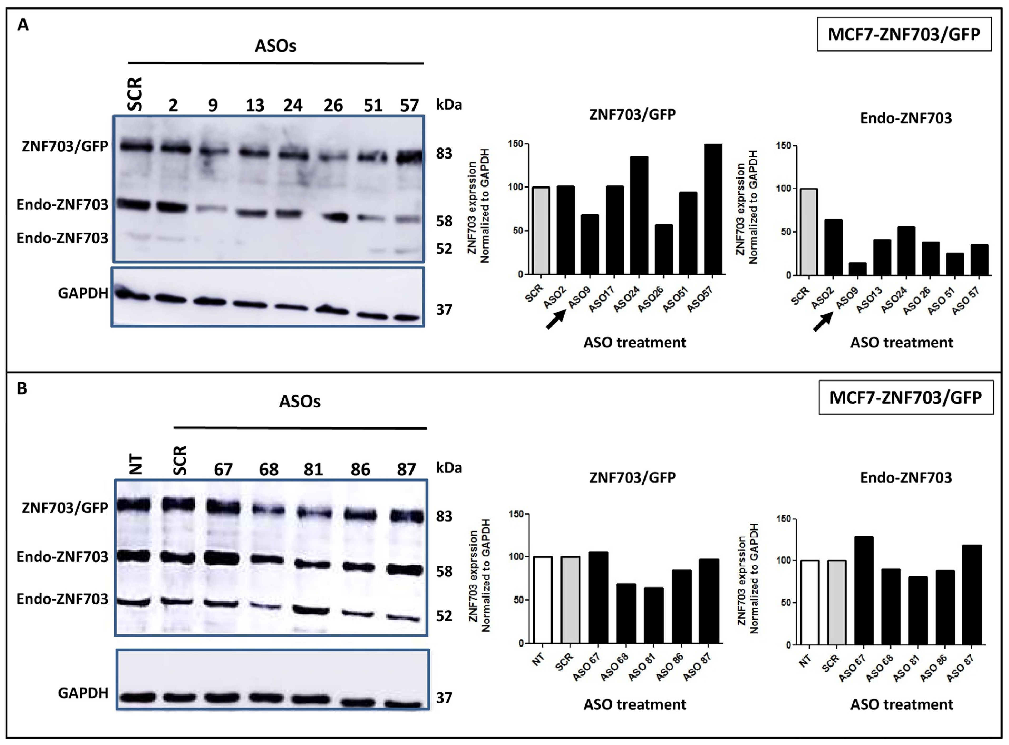
We screened, by gene walk, all ASO sequences targeting ZNF703 full-length mRNA. An initial set of 14 ZNF703-ASOs (ASO2, ASO9, ASO13, ASO17, ASO24, ASO26, ASO37, ASO51, ASO67, ASO68, ASO81, ASO83, ASO86 and ASO87) against ZNF703 mRNA was then designed (Figure S1A). The ZNF703-ASOs were evaluated by WB for their capacity to repress both endogenous and exogenous ZNF703 (ZNF703 and ZNF703/GFP) protein expression in MCF7-ZNF703/GFP BC cells at 100 nM concentration (Figure S1B and Figure 2). MCF7-ZNF703/GFP was chosen because it is a luminal breast cancer cell line in which ZNF703 was artificially overexpressed leading to various consequences including cell proliferation and resistance to tamoxifen previously reported and known to reflect luminal B phenotype [34]. As shown in Figure S1B and Figure 2A, ZNF703-ASO9 was the most efficient ASO for silencing ZNF703 and achieved more than 70% inhibition of endogenous ZNF703 protein expression. ZNF703-ASO9 was used for further analyses.

3.3. ZNF703-ASO9 Targeting ZNF703 mRNA Downregulates ZNF703 Protein Expression in BC Cell Lines

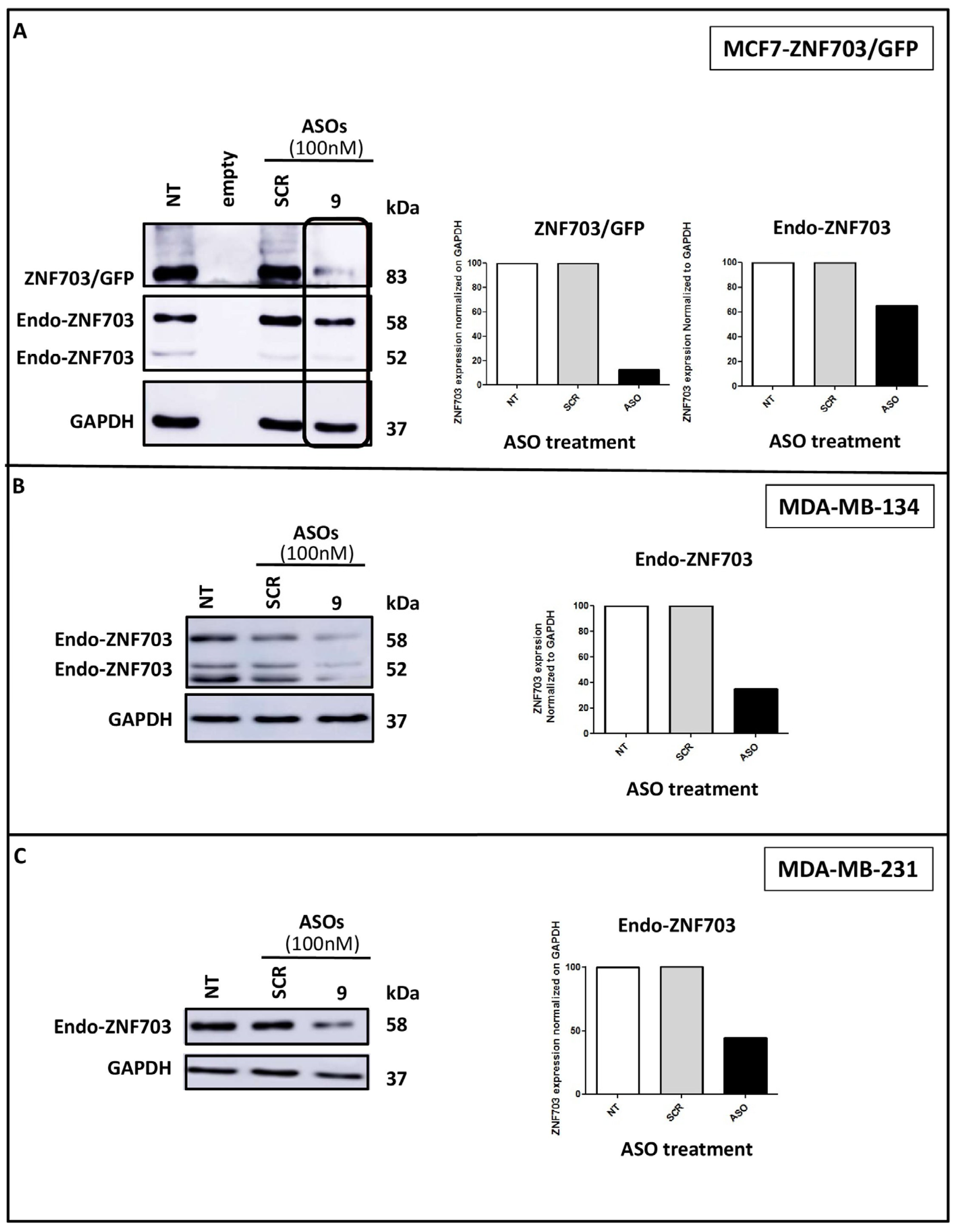
We assessed the inhibitory activity of ZNF703-ASO9 on ZNF703 expression in two other BC cell lines (MDA-MB-134, luminal, and MDA-MB-231, basal) to establish its consistency in effectively inhibiting its target gene (Figure 3). MDA-MB-134 is a luminal B breast cancer cell line with the highest ZNF703 mRNA expression (Figure 1A). MDA-MB-231 is a triple negative breast cancer cell line and could be used for comparison.
Treatment with ASO9 (100 nM) caused a significant (>70%) inhibition of ZNF703/GFP but only 40% inhibition of the endo-ZNF703 in MCF7-ZNF703/GFP probably due to the competition with exogenous ZNF703/GFP mRNA (showed as a control in Figure 3A). Strong inhibition of ZNF703 (>70%) was also observed in MDA-MB-134 (Figure 3B), while the ZNF703-ASO9-treated MDA-MB-231 cell line exhibited lower ZNF703 expression inhibition but still greater than 50% (Figure 3C).

3.4. ZNF703-ASO9 Has Antiproliferative Effect in BC Cell Lines

To determine the effect of ZNF703-ASO9 on both endogenous and exogenous ZNF703 mRNA, MCF7-ZNF703/GFP cells were treated with increasing concentrations of ZNF703-ASO9 (Figure 4A,C). A dose-dependent inhibition of endogenous ZNF703 expression in MCF7-ZNF703/GFP was observed with the highest inhibition efficiency identified at 300 nM. ZNF703/GFP (83 kDa band) expression was inhibited at 100 nM of ZNF703-ASO9 (Figure 4A,B). The expression of the endogenous ZNF703 band at 58 kDa (expressed in all tested BC cell lines as well as in the engineered MCF7 cell lines) was inhibited by ZNF703-ASO9 following a dose-dependent manner (Figure 4A,C).
In the same experiment, we observed, in the presence of increasing concentrations of ZNF703-ASO9, a dose-dependent decrease in MCF7-ZNF703/GFP cell viability was measured with the MTT test (Figure 4D). After 72 h of incubation post-second treatment, increasing concentrations of ZNF703-ASO9 (50–200 nM) led to a gradual decrease in the fraction of viable cells. Cells either treated with control-ASO (SCR) or not treated showed a higher fraction of viable cells than ZNF703-ASO9-treated cells.
The antiproliferative activity of ZNF703-ASO9 was also evaluated in MCF7-GFP, MDA-MB-134, MDA-MB-231, and HCC1954 cell lines, and the corresponding IC50 was calculated (Figure S2A–E). HCC1954 cell line was used as a negative control due to its very low ZNF703 expression (see Figure 1). While MCF7-ZNF703/GFP exhibited the most sensitive response to ZNF703-ASO9 (112.7 nM), IC50 values of 122.6, 116.1, and 123 nM were established for MCF7-GFP, MDA-MB-134, and MDA-MB-231 (Figure S2A–D), respectively.
Cell viability assessment of MCF7-ZNF703/GFP cells after treatment with 11 various ZNF703-ASOs (200 nM) against ZNF703 mRNA showed that all exhibited a significant decrease in cell viability (Student’s t-test, *** p ≤ 0.001). ZNF703-ASO9 was the most efficient ASO (Figure S3). SCR treatment exhibited only weak toxicity.
Several reasons could explain various effects on cell viability and ZNF703 protein level of different designed ASOs [68]. Among them: (i) the primary sequence, chemical nature, and structure of the ASO can have impacts on the interaction of PS-ASOs with specific proteins; (ii) the activity of PS-ASOs is strongly influenced by the association with both inter- and intracellular proteins; (iii) PS ASO protein interactions can affect many aspects of their performance, including distribution and tissue delivery, cellular uptake, intracellular trafficking, potency and toxicity.
These results show that ZNF703 inhibition by ZNF703-ASO9 has significant antiproliferative activity in BC cell lines (>80% of cell proliferation inhibition).

3.5. ASO Inhibition of ZNF703 Induces Cell Death

We next wanted to determine if ZNF703 inhibition affected cell viability by inducing cell death.
We first studied the impact of ZNF703 inhibition on cell cycle progression using flow cytometry in MCF7-ZNF703/GFP, MDA-MB-134, and MDA-MB-231 cells, after treatment with or without ZNF703-ASO9 at 200 nM. A significant increase in cell death was observed in MCF7-ZNF703/GFP and MDA-MB-231 cells treated with ZNF703-ASO9 compared to cells treated with control-ASO.
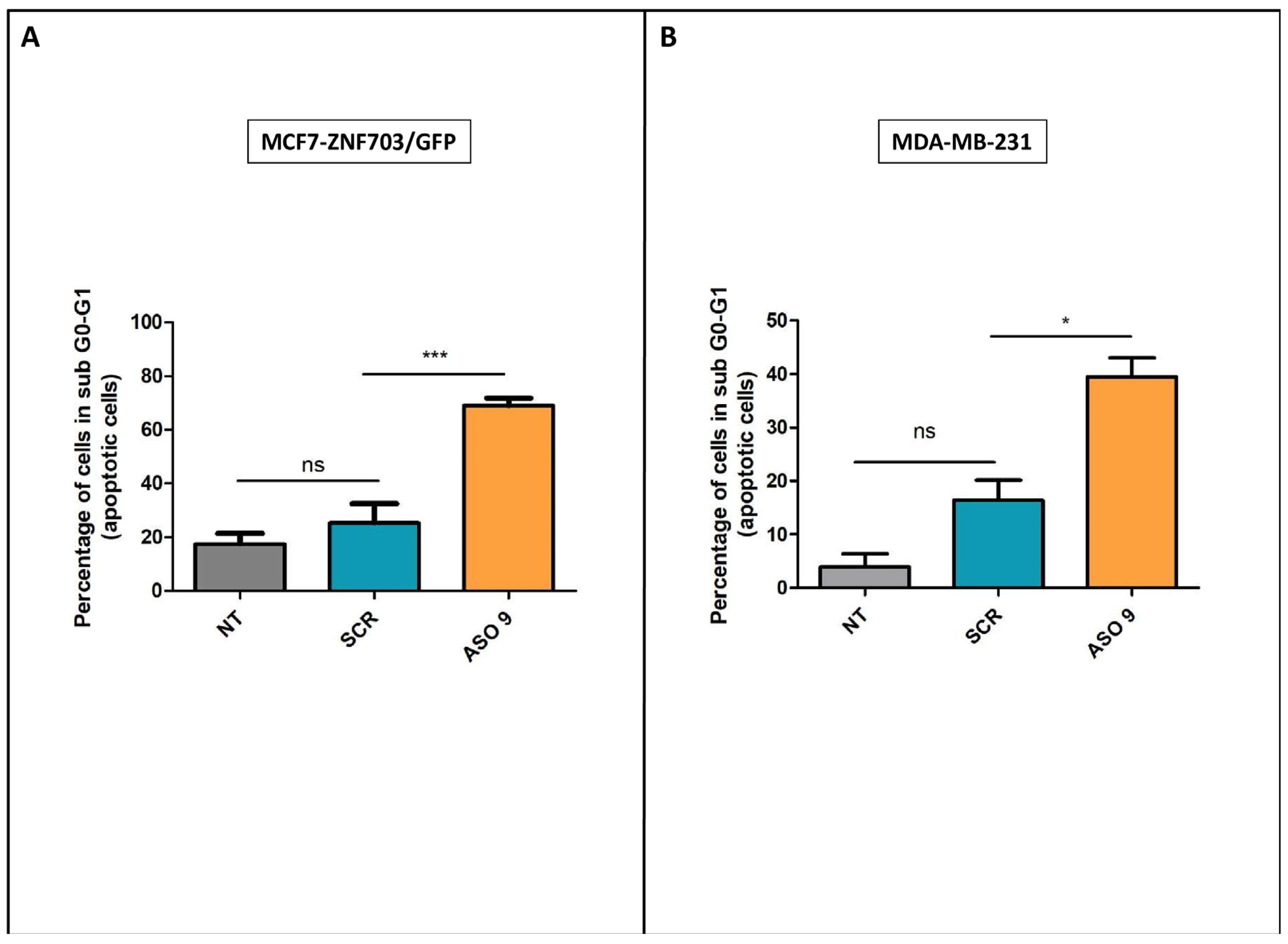
We next analyzed whether the antiproliferative activity and cell death induction by ZNF703-ASO9 was due to apoptosis by measuring annexin V binding in MCF7-ZNF703/GFP and MDA-MB-231 cells. Flow cytometry was used to quantify the apoptotic rates (Figure 5). Flow cytometry analysis of annexin V expression showed that treatment with ZNF703-ASO9 significantly increased apoptosis of MCF7-ZNF703/GFP cells (*** p ≤ 0.001) and MDA-MB-231 cells (* p ≤ 0.05) compared to control-ASO (Figure 5A,B, respectively). MCF7-ZNF703/GFP and MDA-MB-231 cells not treated (NT) and treated with control-ASO showed a non-significant difference in the percentage of annexin V positive cells, suggesting low toxicity of control-ASO.
These results suggest that ZNF703 plays a role in the maintenance of cell survival by apoptosis blockade and suggest that ZNF703 inhibition using ASO could trigger apoptosis.

3.6. The Anti-Cancer Effects of ASO9 Are Improved When Combined with Cisplatin in MCF7-ZNF703/GFP BC Cell Line

We next evaluated the efficacy of cisplatin either alone or in combination with ASO9 treatment in MCF7-ZNF703/GFP BC cells. In other terms, because ZNF703 is an oncogene originally identified in luminal BC [34,38], we wanted to see if inhibition of ZNF703 expression combined with cisplatin could improve the therapeutic efficacy of ZNF703-ASO9 in our luminal B model (MCF7-ZNF703/GFP) [34].
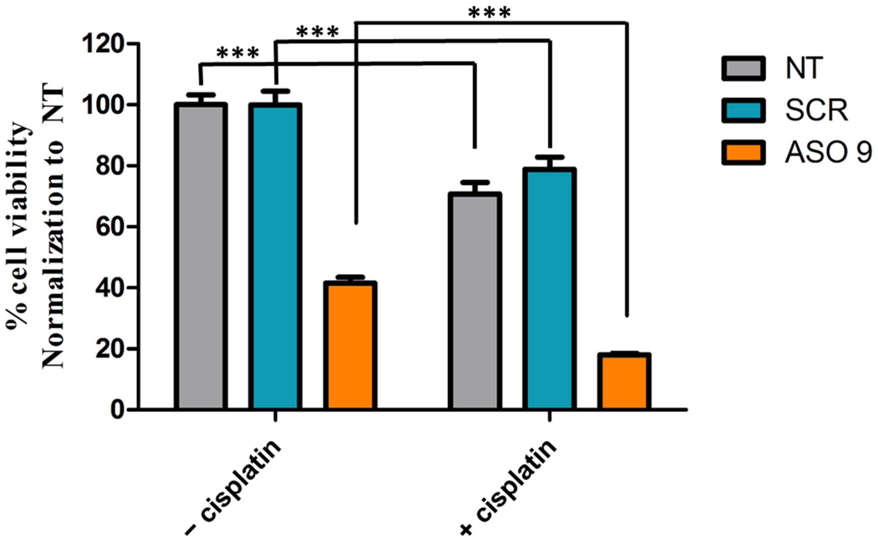
We inhibited ZNF703 mRNA expression with ASO9 (100 nM) and treated MCF7-ZNF703/GFP cell line with cisplatin (100 nM) 48h post ZNF703-ASO9 transfection. Cell viability was evaluated by the MTT test (Figure 6). Cisplatin alone or in combination with SCR reduced cell viability more efficiently than the absence of treatment (NT) or treatment with SCR alone (Student’s t-test, *** p ≤ 0.001) suggesting that MCF7-ZNF703/GFP cell line is cisplatin sensitive. It also showed that the anti-cancer ability of ZNF703-ASO9 was improved when combined with cisplatin (Student’s t-test, *** p ≤ 0.001) (Figure 6).
Thus, ZNF703-ASO9 could be used as a targeted therapy in combination with cisplatin to improve therapeutic approaches of luminal advanced breast cancer patients.

4. Discussion

In this study, we report the first steps in the development of an effective gene-based strategy (ASO) targeting the ZNF703 mRNA in BC. From an initial set of 14 ASOs, we identified ASO9 as the most efficient for silencing ZNF703 expression in an engineered breast cancer cell line. We show that ZNF703 inhibition decreases cell proliferation and induces apoptosis in BC cell lines. We also report that the combination with cisplatin improved ASO9′s anti-cancer effects in MCF 7-ZNF703/GFP luminal BC cell line.

4.1. ZNF703 Oncogene Is a Potential Therapeutic Target in Breast and Other Advanced Cancers

ZNF703 is a transcriptional corepressor that regulates many genes involved in multiple facets of the cancer phenotype, including proliferation, invasion, and regulation of stem cells. Commonly found amplified in luminal B breast cancer, ZNF703 gene overexpression influences several aspects of the pathology of BC, including ER signaling and the phenotypes of progenitor cells [34,38,40]. ZNF703 overexpression is associated with poor prognosis in luminal BC, colorectal cancers, head and neck squamous carcinomas, cholangiocarcinomas, and ovarian cancers [34,38,41,42,43,44,45,47,50]. ZNF703 overexpression was also reported in other cancers such as gastric cancers, non-small cell lung, PTC, MTC, and HCC [44,46,47,48,49,51].
ZNF703 overexpression increases cell proliferation, stimulates tumor migration/invasion, and is involved in endocrine and chemoresistance in luminal B BCs. ZNF703 is a target gene of the ER transcription and suppresses ER expression in a negative feedback loop [69]. It also suppresses cell cycle inhibitors p27 and p15, which leads to upregulation of E2F1 and increased cell proliferation [34,38]. An indirect effect on proliferation is observed in cells expressing ZNF703 by interference with the inhibitory functions of TGFβ signaling [38]. ZNF703 might also play a role in tumor metastasis by promoting EMT through the repression of E-cadherin expression [40].
Thus, ZNF703 overexpression facilitates tumorigenesis, metastatic invasion, and predicts poor prognosis in various advanced cancers. It may be a potential therapeutic target for various cancers.

4.2. ASO Targeting ZNF703 mRNA May Be an Effective Strategy for Advanced BCs

There is no known direct or indirect inhibitor of ZNF703. ZNF703 is neither an enzyme nor a surface receptor and thus not easily targetable. Moreover, as a nuclear protein, it has poor bio-accessibility. We thus developed an ASO strategy to downregulate ZNF703 expression.
The use of nucleic acid-based technologies for gene silencing is increasingly taking center stage in therapy. Recently, over ten nucleic acid-based drugs have received FDA approval for the treatment of various diseases with several others currently at various stages of clinical trials [70,71]. ASOs have a long history of clinical development with eight approved ASOs since 1998 [72,73].
Several works have validated the therapeutic efficacy of ASOs in different cancer types including prostate cancer [54,55,56,57,59,60], ovarian cancer [74], and breast cancer [58].
We designed and developed an ASO against different ZNF703 sequences and screened their efficacies to inhibit ZNF703 at the mRNA and protein levels. ASO9 reduced ZNF703 by 70% at 100 nM. An increase in the dose of ASO resulted in an increased inhibitory effect on ZNF703 expression. ZNF703-ASO9 did not only inhibit the intrinsic expression of its target but also the engineered expression of ZNF703. ZNF703 protein and mRNA expression correlated well, which was good news for ASO use.
ZNF703 has been identified as a driver of the 8p12 amplification in luminal B BC. High expression of ZNF703 luminal B tumor patients is associated with poor clinical outcome [34,41].
We found that a dose-dependent inhibition of exogenous ZNF703 in our MCF7 model overexpressing ZNF703 was directly correlated with a dose-dependent decrease in cell viability. Proliferation inhibition may be explained by cell cycle blockade and cell death induction via an apoptotic pathway. Such results are in agreement with previous studies using small interfering RNA (ZNF703-siRNA) [42,43,48,49]. ZNF703 inhibition suppressed cell proliferation and blocked the cell cycle in BT-549 and MDA-MB-468 basal BC (TNBC) cells [42]. A G1-phase arrest was induced by ZNF703 inhibition in BT-549 and MDA-MB-468. We found a similar result in MDA-MB-231 basal cell line treated with ZNF703-ASO9.
Other studies using siRNA showed that knockdown of ZNF703 expression inhibited papillary thyroid carcinoma, medullary thyroid carcinoma, and colorectal cancer cell proliferation and migration [43,48,49] comforting ZNF703 as an oncogene and a potential therapeutic target for other advanced cancers.

4.3. ZNF703 mRNA-Targeting May Be Used in Combination with Chemo- and Hormone-Therapy of Advanced Breast Cancers

After BC treatment, surviving cancer cells cause metastasis, which remains the main cause of cancer-related mortality.
Clinical trials have shown that cisplatin treatment (alone or in combination with other anticancer drugs) is efficient in inducing BC cell death and decreasing tumor volume [75,76]. These findings have renewed interest in cisplatin and other chemotherapies sharing ancestry with cisplatin as a therapy for BC. However, several mechanisms may explain BC resistance against cisplatin [77].
Here, we report that the anti-cancer ability of ZNF703-ASO9 was improved when it is combined with cisplatin in the luminal B model MCF7-ZNF703/GFP.
Because luminal B BC patients after several lines of treatment without success present a poor chance of survival, the smallest improvement may be appreciated. Strategies such as ASO9/cisplatin combination could be considered as a targeted therapy and may thus constitute a real hope for luminal advanced BC patients.
For high-risk, ER+, HER2− BC, standard adjuvant anthracycline-taxane regimens are appropriate when neoadjuvant chemotherapy is chosen. However, optimal hormone therapy is the standard of care in the adjuvant setting, whether or not a pathological complete response is obtained [78]. Tamoxifen is commonly used in the treatment of luminal BC. However, half of patients treated with tamoxifen are insensitive. Luminal BC cell lines overexpressing ZNF703 were reported resistant to tamoxifen through activation of AKT/MTOR signaling [79]. Overexpression of ZNF703 in MCF7 luminal BC cells induced activation of the AKT/MTOR signaling pathway, downregulation of ERα, and reduction of the tamoxifen’s antitumor effect. While low-dose tamoxifen stimulated the growth of cells overexpressing ZNF703, treatments of tamoxifen-treated MDA-MB-134 and HCC1500 luminal B BC cell lines with (i) ZNF703-siRNA alone significantly reduced survival rates whereas the combination ZNF703-siRNA with the MTOR inhibitor rapamycin enhanced the antitumor effect of tamoxifen [79]. This information suggests that the next step to evaluate the ability of ZNF703-ASO9 treatment to restore hormone sensitivity in luminal BC could be to use tamoxifen combined with an MTOR inhibitor. Recently, an ASO-targeting circPVT1 was reported to inhibit ER+ cell and tumor growth, re-sensitizing tamoxifen-resistant ER+ BC cells to tamoxifen treatment [80].
Cyclin-dependent kinase 4 and 6 (CDK4/6) inhibitors (e.g., palbociclib, ribociclib) have shown significant efficacy in ER+ BC [24,25]. Therapy designs combining ZNF703-ASO9 and these inhibitors should also be considered.

5. Conclusions

ZNF703 overexpression facilitates tumorigenesis, metastatic invasion, and predicts poor prognosis in luminal B BCs but also in other advanced cancers. ZNF703 inhibition may be a potential targeted treatment for such advanced cancers. Our work shows that ASO technology is a way to efficiently inhibit ZNF703 in luminal B BC cells. This inhibition decreased cell proliferation, provoked apoptosis and the combination with cisplatin improved the anti-cancer ability of ZNF703-ASO9 in the luminal B model MCF7-ZNF703/GFP. ASO-based inhibition of ZNF703 may be useful in the treatment of luminal B BCs as well as other advanced cancers either as a monotherapy or in combination with other therapies.

Supplementary Materials

The following supporting information can be downloaded at: https://www.mdpi.com/article/10.3390/pharmaceutics15071930/s1, Figure S1: ASO design for ZNF703 mRNA targeting and selection ASOs by WB analysis; Figure S2: IC50 calculation for ASO9 in five breast cancer cells; Figure S3: Cell viability tests on MCF7-ZNF703/GFP after treatment with 11 various ASOs against ZNF703 mRNA.

Author Contributions

Conceptualization, methodology, and M.C.; validation, D.B., P.R. and M.C.; formal analysis, S.U.-I., T.K.L., K.O., A.G., F.B., P.R., D.B. and M.C.; molecular experiments: S.U.-I., J.A., C.P. and T.K.L.; bioinformatic and statistical analyses: A.G., T.K.L. and P.F.; writing—original draft preparation, M.C.; writing—review and editing, all authors; supervision, D.B., P.R. and M.C. All authors have read and agreed to the published version of the manuscript.

Funding

This work was supported by grants from the Ligue Nationale Contre Le Cancer (labels EL2019 and EL2022 [F.B.]), and R22030AA [P.R.]), ITMO cancer (C21033AS, [P.R.]), Association Ruban Rose (2020 [F.B.]), Fondation Groupe EDF [D.B.], and Canceropole-PACA (2021 [M.C.]). S.U.-I. was supported by a grant from the TETFUND-Tertiary Education Trust Fund/AEFUNAI.

Institutional Review Board Statement

Not applicable.

Informed Consent Statement

Not applicable.

Data Availability Statement

All data are contained in the article.

Acknowledgments

The authors wish to thank computing facilities DISC (Datacenter IT and Scientific Computing, CRCM) and DSIO (Institut Paoli Calmettes) for their technical support.

Conflicts of Interest

The authors declare no conflict of interest.

References

  1. Sung, H.; Ferlay, J.; Siegel, R.L.; Laversanne, M.; Soerjomataram, I.; Jemal, A.; Bray, F. Global Cancer Statistics 2020: GLOBOCAN Estimates of Incidence and Mortality Worldwide for 36 Cancers in 185 Countries. CA Cancer J. Clin. 2021, 71, 209–249. [Google Scholar] [CrossRef] [PubMed]
  2. Sotiriou, C.; Pusztai, L. Gene-expression signatures in breast cancer. N. Engl. J. Med. 2009, 360, 790–800. [Google Scholar] [CrossRef] [PubMed] [Green Version]
  3. Perou, C.M.; Børresen-Dale, A.-L. Systems biology and genomics of breast cancer. Cold Spring Harb. Perspect. Biol. 2011, 3, a003293. [Google Scholar] [CrossRef] [Green Version]
  4. Early Breast Cancer Trialists’ Collaborative Group (EBCTCG). Effects of chemotherapy and hormonal therapy for early breast cancer on recurrence and 15-year survival: An overview of the randomised trials. Lancet 2005, 365, 1687–1717. [Google Scholar] [CrossRef]
  5. Kohler, B.A.; Sherman, R.L.; Howlader, N.; Jemal, A.; Ryerson, A.B.; Henry, K.A.; Boscoe, F.P.; Cronin, K.A.; Lake, A.; Noone, A.-M.; et al. Annual Report to the Nation on the Status of Cancer, 1975–2011, Featuring Incidence of Breast Cancer Subtypes by Race/Ethnicity, Poverty, and State. J. Natl. Cancer Inst. 2015, 107, djv048. [Google Scholar] [CrossRef]
  6. Robinson, D.R.; Wu, Y.-M.; Vats, P.; Su, F.; Lonigro, R.J.; Cao, X.; Kalyana-Sundaram, S.; Wang, R.; Ning, Y.; Hodges, L.; et al. Activating ESR1 mutations in hormone-resistant metastatic breast cancer. Nat. Genet. 2013, 45, 1446–1451. [Google Scholar] [CrossRef] [PubMed] [Green Version]
  7. Toy, W.; Shen, Y.; Won, H.; Green, B.; Sakr, R.A.; Will, M.; Li, Z.; Gala, K.; Fanning, S.; King, T.A.; et al. ESR1 ligand-binding domain mutations in hormone-resistant breast cancer. Nat. Genet. 2013, 45, 1439–1445. [Google Scholar] [CrossRef] [PubMed] [Green Version]
  8. Ignatiadis, M.; Sotiriou, C. Luminal breast cancer: From biology to treatment. Nat. Rev. Clin. Oncol. 2013, 10, 494–506. [Google Scholar] [CrossRef]
  9. Fleurot, E.; Goudin, C.; Hanoux, V.; Bonnamy, P.-J.; Levallet, J. Estrogen receptor α regulates the expression of syndecan-1 in human breast carcinoma cells. Endocr. Relat. Cancer 2019, 26, 615–628. [Google Scholar] [CrossRef]
  10. Meijer, D.; Sieuwerts, A.M.; Look, M.P.; van Agthoven, T.; Foekens, J.A.; Dorssers, L.C.J. Fibroblast growth factor receptor 4 predicts failure on tamoxifen therapy in patients with recurrent breast cancer. Endocr. Relat. Cancer 2008, 15, 101–111. [Google Scholar] [CrossRef]
  11. van Agthoven, T.; Sieuwerts, A.M.; Veldscholte, J.; Meijer-van Gelder, M.E.; Smid, M.; Brinkman, A.; den Dekker, A.T.; Leroy, I.M.; van Ijcken, W.F.J.; Sleijfer, S.; et al. CITED2 and NCOR2 in anti-oestrogen resistance and progression of breast cancer. Br. J. Cancer 2009, 101, 1824–1832. [Google Scholar] [CrossRef] [Green Version]
  12. Tran, B.; Bedard, P.L. Luminal-B breast cancer and novel therapeutic targets. Breast Cancer Res. 2011, 13, 221. [Google Scholar] [CrossRef] [PubMed]
  13. van Agthoven, T.; Godinho, M.F.E.; Wulfkuhle, J.D.; Petricoin, E.F.; Dorssers, L.C.J. Protein pathway activation mapping reveals molecular networks associated with antiestrogen resistance in breast cancer cell lines. Int. J. Cancer 2012, 131, 1998–2007. [Google Scholar] [CrossRef]
  14. Salmans, M.L.; Zhao, F.; Andersen, B. The estrogen-regulated anterior gradient 2 (AGR2) protein in breast cancer: A potential drug target and biomarker. Breast Cancer Res. 2013, 15, 204. [Google Scholar] [CrossRef] [PubMed]
  15. Levine, K.M.; Priedigkeit, N.; Basudan, A.; Tasdemir, N.; Sikora, M.J.; Sokol, E.S.; Hartmaier, R.J.; Ding, K.; Ahmad, N.Z.; Watters, R.J.; et al. FGFR4 overexpression and hotspot mutations in metastatic ER+ breast cancer is enriched in the lobular subtype. NPJ Breast Cancer 2019, 5, 19. [Google Scholar] [CrossRef] [PubMed] [Green Version]
  16. Arruabarrena-Aristorena, A.; Maag, J.L.V.; Kittane, S.; Cai, Y.; Karthaus, W.R.; Ladewig, E.; Park, J.; Kannan, S.; Ferrando, L.; Cocco, E.; et al. FOXA1 Mutations Reveal Distinct Chromatin Profiles and Influence Therapeutic Response in Breast Cancer. Cancer Cell 2020, 38, 534–550.e9. [Google Scholar] [CrossRef]
  17. Garcia-Recio, S.; Thennavan, A.; East, M.P.; Parker, J.S.; Cejalvo, J.M.; Garay, J.P.; Hollern, D.P.; He, X.; Mott, K.R.; Galván, P.; et al. FGFR4 regulates tumor subtype differentiation in luminal breast cancer and metastatic disease. J. Clin. Investig. 2020, 130, 4871–4887. [Google Scholar] [CrossRef]
  18. Jin, X.; Ge, L.-P.; Li, D.-Q.; Shao, Z.-M.; Di, G.-H.; Xu, X.-E.; Jiang, Y.-Z. LncRNA TROJAN promotes proliferation and resistance to CDK4/6 inhibitor via CDK2 transcriptional activation in ER+ breast cancer. Mol. Cancer 2020, 19, 87. [Google Scholar] [CrossRef]
  19. Augereau, P.; Patsouris, A.; Bourbouloux, E.; Gourmelon, C.; Abadie Lacourtoisie, S.; Berton Rigaud, D.; Soulié, P.; Frenel, J.S.; Campone, M. Hormonoresistance in advanced breast cancer: A new revolution in endocrine therapy. Ther. Adv. Med. Oncol. 2017, 9, 335–346. [Google Scholar] [CrossRef] [Green Version]
  20. Tong, C.W.S.; Wu, M.; Cho, W.C.S.; To, K.K.W. Recent Advances in the Treatment of Breast Cancer. Front. Oncol. 2018, 8, 227. [Google Scholar] [CrossRef] [Green Version]
  21. Ellis, M.J.; Suman, V.J.; Hoog, J.; Lin, L.; Snider, J.; Prat, A.; Parker, J.S.; Luo, J.; DeSchryver, K.; Allred, D.C.; et al. Randomized phase II neoadjuvant comparison between letrozole, anastrozole, and exemestane for postmenopausal women with estrogen receptor-rich stage 2 to 3 breast cancer: Clinical and biomarker outcomes and predictive value of the baseline PAM50-based intrinsic subtype--ACOSOG Z1031. J. Clin. Oncol. 2011, 29, 2342–2349. [Google Scholar] [CrossRef] [Green Version]
  22. Ellis, M.J.; Suman, V.J.; Hoog, J.; Goncalves, R.; Sanati, S.; Creighton, C.J.; DeSchryver, K.; Crouch, E.; Brink, A.; Watson, M.; et al. Ki67 Proliferation Index as a Tool for Chemotherapy Decisions During and After Neoadjuvant Aromatase Inhibitor Treatment of Breast Cancer: Results from the American College of Surgeons Oncology Group Z1031 Trial (Alliance). J. Clin. Oncol. 2017, 35, 1061–1069. [Google Scholar] [CrossRef] [PubMed]
  23. Harbeck, N.; Gluz, O.; Christgen, M.; Kates, R.E.; Braun, M.; Küemmel, S.; Schumacher, C.; Potenberg, J.; Kraemer, S.; Kleine-Tebbe, A.; et al. De-Escalation Strategies in Human Epidermal Growth Factor Receptor 2 (HER2)–Positive Early Breast Cancer (BC): Final Analysis of the West German Study Group Adjuvant Dynamic Marker-Adjusted Personalized Therapy Trial Optimizing Risk Assessment and Therapy Response Prediction in Early BC HER2- and Hormone Receptor–Positive Phase II Randomized Trial—Efficacy, Safety, and Predictive Markers for 12 Weeks of Neoadjuvant Trastuzumab Emtansine With or Without Endocrine Therapy (ET) Versus Trastuzumab Plus ET. J. Clin. Oncol. 2017, 35, 3046–3054. [Google Scholar] [CrossRef] [PubMed]
  24. Turner, N.C.; Ro, J.; André, F.; Loi, S.; Verma, S.; Iwata, H.; Harbeck, N.; Loibl, S.; Huang Bartlett, C.; Zhang, K.; et al. Palbociclib in Hormone-Receptor–Positive Advanced Breast Cancer. N. Engl. J. Med. 2015, 373, 209–219. [Google Scholar] [CrossRef] [PubMed] [Green Version]
  25. Hortobagyi, G.N.; Stemmer, S.M.; Burris, H.A.; Yap, Y.-S.; Sonke, G.S.; Paluch-Shimon, S.; Campone, M.; Blackwell, K.L.; André, F.; Winer, E.P.; et al. Ribociclib as First-Line Therapy for HR-Positive, Advanced Breast Cancer. N. Engl. J. Med. 2016, 375, 1738–1748. [Google Scholar] [CrossRef]
  26. Formisano, L.; Lu, Y.; Servetto, A.; Hanker, A.B.; Jansen, V.M.; Bauer, J.A.; Sudhan, D.R.; Guerrero-Zotano, A.L.; Croessmann, S.; Guo, Y.; et al. Aberrant FGFR signaling mediates resistance to CDK4/6 inhibitors in ER+ breast cancer. Nat. Commun. 2019, 10, 1373. [Google Scholar] [CrossRef] [Green Version]
  27. Ono, M.; Oba, T.; Shibata, T.; Ito, K.-I. The mechanisms involved in the resistance of estrogen receptor-positive breast cancer cells to palbociclib are multiple and change over time. J. Cancer Res. Clin. Oncol. 2021, 147, 3211–3224. [Google Scholar] [CrossRef]
  28. Metzger-Filho, O.; Procter, M.; de Azambuja, E.; Leyland-Jones, B.; Gelber, R.D.; Dowsett, M.; Loi, S.; Saini, K.S.; Cameron, D.; Untch, M.; et al. Magnitude of trastuzumab benefit in patients with HER2-positive, invasive lobular breast carcinoma: Results from the HERA trial. J. Clin. Oncol. 2013, 31, 1954–1960. [Google Scholar] [CrossRef]
  29. Ades, F.; Zardavas, D.; Bozovic-Spasojevic, I.; Pugliano, L.; Fumagalli, D.; de Azambuja, E.; Viale, G.; Sotiriou, C.; Piccart, M. Luminal B breast cancer: Molecular characterization, clinical management, and future perspectives. J. Clin. Oncol. 2014, 32, 2794–2803. [Google Scholar] [CrossRef] [Green Version]
  30. Cornen, S.; Guille, A.; Adélaïde, J.; Addou-Klouche, L.; Finetti, P.; Saade, M.-R.; Manai, M.; Carbuccia, N.; Bekhouche, I.; Letessier, A.; et al. Candidate luminal B breast cancer genes identified by genome, gene expression and DNA methylation profiling. PLoS ONE 2014, 9, e81843. [Google Scholar] [CrossRef] [Green Version]
  31. Cancer Genome Atlas Network. Comprehensive molecular portraits of human breast tumours. Nature 2012, 490, 61–70. [Google Scholar] [CrossRef] [PubMed] [Green Version]
  32. Ha, S.M.; Chae, E.Y.; Cha, J.H.; Kim, H.H.; Shin, H.J.; Choi, W.J. Association of BRCA Mutation Types, Imaging Features, and Pathologic Findings in Patients with Breast Cancer with BRCA1 and BRCA2 Mutations. Am. J. Roentgenol. 2017, 209, 920–928. [Google Scholar] [CrossRef] [PubMed]
  33. Addou-Klouche, L.; Adélaïde, J.; Finetti, P.; Cervera, N.; Ferrari, A.; Bekhouche, I.; Sircoulomb, F.; Sotiriou, C.; Viens, P.; Moulessehoul, S.; et al. Loss, mutation and deregulation of L3MBTL4 in breast cancers. Mol. Cancer 2010, 9, 213. [Google Scholar] [CrossRef] [PubMed] [Green Version]
  34. Sircoulomb, F.; Nicolas, N.; Ferrari, A.; Finetti, P.; Bekhouche, I.; Rousselet, E.; Lonigro, A.; Adélaïde, J.; Baudelet, E.; Esteyriès, S.; et al. ZNF703 gene amplification at 8p12 specifies luminal B breast cancer. EMBO Mol. Med. 2011, 3, 153–166. [Google Scholar] [CrossRef]
  35. Finetti, P.; Guille, A.; Adelaide, J.; Birnbaum, D.; Chaffanet, M.; Bertucci, F. ESPL1 is a candidate oncogene of luminal B breast cancers. Breast Cancer Res. Treat. 2014, 147, 51–59. [Google Scholar] [CrossRef]
  36. Gelsi-Boyer, V.; Orsetti, B.; Cervera, N.; Finetti, P.; Sircoulomb, F.; Rougé, C.; Lasorsa, L.; Letessier, A.; Ginestier, C.; Monville, F.; et al. Comprehensive profiling of 8p11-12 amplification in breast cancer. Mol. Cancer Res. 2005, 3, 655–667. [Google Scholar] [CrossRef] [Green Version]
  37. Adélaïde, J.; Finetti, P.; Bekhouche, I.; Repellini, L.; Geneix, J.; Sircoulomb, F.; Charafe-Jauffret, E.; Cervera, N.; Desplans, J.; Parzy, D.; et al. Integrated profiling of basal and luminal breast cancers. Cancer Res. 2007, 67, 11565–11575. [Google Scholar] [CrossRef] [Green Version]
  38. Holland, D.G.; Burleigh, A.; Git, A.; Goldgraben, M.A.; Perez-Mancera, P.A.; Chin, S.-F.; Hurtado, A.; Bruna, A.; Ali, H.R.; Greenwood, W.; et al. ZNF703 is a common Luminal B breast cancer oncogene that differentially regulates luminal and basal progenitors in human mammary epithelium. EMBO Mol. Med. 2011, 3, 167–180. [Google Scholar] [CrossRef]
  39. Song, X.; Ji, J.; Rothstein, J.H.; Alexeeff, S.E.; Sakoda, L.C.; Sistig, A.; Achacoso, N.; Jorgenson, E.; Whittemore, A.S.; Klein, R.J.; et al. MiXcan: A framework for cell-type-aware transcriptome-wide association studies with an application to breast cancer. Nat. Commun. 2023, 14, 377. [Google Scholar] [CrossRef]
  40. Slorach, E.M.; Chou, J.; Werb, Z. Zeppo1 is a novel metastasis promoter that represses E-cadherin expression and regulates p120-catenin isoform expression and localization. Genes Dev. 2011, 25, 471–484. [Google Scholar] [CrossRef] [Green Version]
  41. Reynisdottir, I.; Arason, A.; Einarsdottir, B.O.; Gunnarsson, H.; Staaf, J.; Vallon-Christersson, J.; Jonsson, G.; Ringnér, M.; Agnarsson, B.A.; Olafsdottir, K.; et al. High expression of ZNF703 independent of amplification indicates worse prognosis in patients with luminal B breast cancer. Cancer Med. 2013, 2, 437–446. [Google Scholar] [CrossRef] [PubMed] [Green Version]
  42. Zhang, X.; Mu, X.; Huang, O.; Wang, Z.; Chen, J.; Chen, D.; Wang, G. ZNF703 promotes triple-negative breast cancer cells through cell-cycle signaling and associated with poor prognosis. BMC Cancer 2022, 22, 226. [Google Scholar] [CrossRef] [PubMed]
  43. Ma, F.; Bi, L.; Yang, G.; Zhang, M.; Liu, C.; Zhao, Y.; Wang, Y.; Wang, J.; Bai, Y.; Zhang, Y. ZNF703 promotes tumor cell proliferation and invasion and predicts poor prognosis in patients with colorectal cancer. Oncol. Rep. 2014, 32, 1071–1077. [Google Scholar] [CrossRef] [Green Version]
  44. Yang, G.; Ma, F.; Zhong, M.; Fang, L.; Peng, Y.; Xin, X.; Zhong, J.; Yuan, F.; Gu, H.; Zhu, W.; et al. ZNF703 acts as an oncogene that promotes progression in gastric cancer. Oncol. Rep. 2014, 31, 1877–1882. [Google Scholar] [CrossRef] [PubMed] [Green Version]
  45. Yang, H.; Jiang, W.Q.; Cao, Y.; Sun, Y.A.; Wei, J.; An, X.; Zhang, Y.C.; Song, M.; Wang, S.S.; Yuan, Z.Y.; et al. Elevated ZNF703 Protein Expression Is an Independent Unfavorable Prognostic Factor for Survival of the Patients with Head and Neck Squamous Cell Carcinoma. Dis. Markers 2015, 2015, 640263. [Google Scholar] [CrossRef] [Green Version]
  46. Baykara, O.; Dalay, N.; Kaynak, K.; Buyru, N. ZNF703 Overexpression may act as an oncogene in non-small cell lung cancer. Cancer Med. 2016, 5, 2873–2878. [Google Scholar] [CrossRef]
  47. Li, K.; Wang, J.; Han, J.; Lan, Y.; Xie, C.; Pan, S.; Liu, L. Overexpression of ZNF703 facilitates tumorigenesis and predicts unfavorable prognosis in patients with cholangiocarcinoma. Oncotarget 2016, 7, 76108–76117. [Google Scholar] [CrossRef] [Green Version]
  48. Yang, X.; Liu, G.; Zang, L.; Li, D.; Yu, F.; Xiang, X.; Li, W. ZNF703 is Overexpressed in Papillary Thyroid Carcinoma Tissues and Mediates K1 Cell Proliferation. Pathol. Oncol. Res. 2020, 26, 355–364. [Google Scholar] [CrossRef]
  49. Yang, X.; Liu, G.; Li, W.; Zang, L.; Li, D.; Wang, Q.; Yu, F.; Xiang, X. Silencing of zinc finger protein 703 inhibits medullary thyroid carcinoma cell proliferation in. vitro and in vivo. Oncol. Lett. 2020, 19, 943–951. [Google Scholar] [CrossRef]
  50. Wang, S.; Wang, C.; Hu, Y.; Li, X.; Jin, S.; Liu, O.; Gou, R.; Zhuang, Y.; Guo, Q.; Nie, X.; et al. ZNF703 promotes tumor progression in ovarian cancer by interacting with HE4 and epigenetically regulating PEA15. J. Exp. Clin. Cancer Res. 2020, 39, 264. [Google Scholar] [CrossRef]
  51. Wang, H.; Xu, H.; Ma, F.; Zhan, M.; Yang, X.; Hua, S.; Li, W.; Li, Y.; Lu, L. Zinc finger protein 703 induces EMT and sorafenib resistance in hepatocellular carcinoma by transactivating CLDN4 expression. Cell Death Dis. 2020, 11, 225. [Google Scholar] [CrossRef] [Green Version]
  52. Crooke, S.T. Molecular Mechanisms of Antisense Oligonucleotides. Nucleic Acid Ther. 2017, 27, 70–77. [Google Scholar] [CrossRef]
  53. Dhuri, K.; Bechtold, C.; Quijano, E.; Pham, H.; Gupta, A.; Vikram, A.; Bahal, R. Antisense Oligonucleotides: An Emerging Area in Drug Discovery and Development. J. Clin. Med. 2020, 9, 2004. [Google Scholar] [CrossRef] [PubMed]
  54. Rocchi, P.; So, A.; Kojima, S.; Signaevsky, M.; Beraldi, E.; Fazli, L.; Hurtado-Coll, A.; Yamanaka, K.; Gleave, M. Heat shock protein 27 increases after androgen ablation and plays a cytoprotective role in hormone-refractory prostate cancer. Cancer Res. 2004, 64, 6595–6602. [Google Scholar] [CrossRef] [Green Version]
  55. Rocchi, P.; Beraldi, E.; Ettinger, S.; Fazli, L.; Vessella, R.L.; Nelson, C.; Gleave, M. Increased Hsp27 after androgen ablation facilitates androgen-independent progression in prostate cancer via signal transducers and activators of transcription 3-mediated suppression of apoptosis. Cancer Res. 2005, 65, 11083–11093. [Google Scholar] [CrossRef] [PubMed] [Green Version]
  56. Baylot, V.; Katsogiannou, M.; Andrieu, C.; Taieb, D.; Acunzo, J.; Giusiano, S.; Fazli, L.; Gleave, M.; Garrido, C.; Rocchi, P. Targeting TCTP as a new therapeutic strategy in castration-resistant prostate cancer. Mol. Ther. 2012, 20, 2244–2256. [Google Scholar] [CrossRef] [Green Version]
  57. Karaki, S.; Benizri, S.; Mejías, R.; Baylot, V.; Branger, N.; Nguyen, T.; Vialet, B.; Oumzil, K.; Barthélémy, P.; Rocchi, P. Lipid-oligonucleotide conjugates improve cellular uptake and efficiency of TCTP-antisense in castration-resistant prostate cancer. J. Control Release 2017, 258, 1–9. [Google Scholar] [CrossRef] [PubMed]
  58. Nguyen, D.T.; Le, T.K.; Paris, C.; Cherif, C.; Audebert, S.; Udu-Ituma, S.O.; Benizri, S.; Barthélémy, P.; Bertucci, F.; Taïeb, D.; et al. Antisense Oligonucleotide-Based Therapeutic against Menin for Triple-Negative Breast Cancer Treatment. Biomedicines 2021, 9, 795. [Google Scholar] [CrossRef]
  59. Cherif, C.; Nguyen, D.T.; Paris, C.; Le, T.K.; Sefiane, T.; Carbuccia, N.; Finetti, P.; Chaffanet, M.; Kaoutari, A.E.; Vernerey, J.; et al. Menin inhibition suppresses castration-resistant prostate cancer and enhances chemosensitivity. Oncogene 2022, 41, 125–137. [Google Scholar] [CrossRef]
  60. Le, T.K.; Cherif, C.; Omabe, K.; Paris, C.; Lannes, F.; Audebert, S.; Baudelet, E.; Hamimed, M.; Barbolosi, D.; Finetti, P.; et al. DDX5 mRNA-targeting antisense oligonucleotide as a new promising therapeutic in combating castration-resistant prostate cancer. Mol. Ther. 2023, 31, 471–486. [Google Scholar] [CrossRef]
  61. Forozan, F.; Veldman, R.; Ammerman, C.A.; Parsa, N.Z.; Kallioniemi, A.; Kallioniemi, O.-P.; Ethier, S.P. Molecular cytogenetic analysis of 11 new breast cancer cell lines. Br. J. Cancer 1999, 81, 1328–1334. [Google Scholar] [CrossRef] [PubMed] [Green Version]
  62. Parker, J.S.; Mullins, M.; Cheang, M.C.; Leung, S.; Voduc, D.; Vickery, T.; Davies, S.; Fauron, C.; He, X.; Hu, Z.; et al. Supervised risk predictor of breast cancer based on intrinsic subtypes. J. Clin. Oncol. 2009, 27, 1160–1167. [Google Scholar] [CrossRef]
  63. Charafe-Jauffret, E.; Ginestier, C.; Monville, F.; Finetti, P.; Adélaïde, J.; Cervera, N.; Fekairi, S.; Xerri, L.; Jacquemier, J.; Birnbaum, D.; et al. Gene expression profiling of breast cell lines identifies potential new basal markers. Oncogene 2006, 25, 2273–2284. [Google Scholar] [CrossRef] [Green Version]
  64. Irizarry, R.A.; Wu, Z.; Jaffee, H.A. Comparison of Affymetrix GeneChip expression measures. Bioinformatics 2006, 22, 789–794. [Google Scholar] [CrossRef] [Green Version]
  65. Perou, C.M.; Sørlie, T.; Eisen, M.B.; van de Rijn, M.; Jeffrey, S.S.; Rees, C.A.; Pollack, J.R.; Ross, D.T.; Johnsen, H.; Akslen, L.A.; et al. Molecular portraits of human breast tumours. Nature 2000, 406, 747–752. [Google Scholar] [CrossRef] [Green Version]
  66. Hu, Z.; Fan, C.; Oh, D.S.; Marron, J.S.; He, X.; Qaqish, B.F.; Livasy, C.; Carey, L.A.; Reynolds, E.; Dressler, L.; et al. The molecular portraits of breast tumors are conserved across microarray platforms. BMC Genom. 2006, 7, 96. [Google Scholar] [CrossRef] [Green Version]
  67. Ghandi, M.; Huang, F.W.; Jané-Valbuena, J.; Kryukov, G.V.; Lo, C.C.; McDonald, E.R., 3rd; Barretina, J.; Gelfand, E.T.; Bielski, C.M.; Li, H.; et al. Next-generation characterization of the Cancer Cell Line Encyclopedia. Nature 2019, 569, 503–508. [Google Scholar] [CrossRef]
  68. Crooke, S.T.; Vickers, T.A.; Liang, X.H. Phosphorothioate modified oligonucleotide-protein interactions. Nucleic Acids Res. 2020, 48, 5235–5253. [Google Scholar] [CrossRef] [PubMed]
  69. Kristensen, V. Divide and conquer: The genetic basis of molecular subclassification of breast cancer. EMBO Mol. Med. 2011, 3, 183–185. [Google Scholar] [CrossRef]
  70. Sharma, V.K.; Watts, J.K. Oligonucleotide therapeutics: Chemistry, delivery and clinical progress. Future Med. Chem. 2015, 7, 2221–2242. [Google Scholar] [CrossRef] [PubMed]
  71. Roberts, T.C.; Langer, R.; Wood, M.J.A. Advances in oligonucleotide drug delivery. Nat. Rev. Drug Discov. 2020, 19, 673–694. [Google Scholar] [CrossRef]
  72. Bennett, C.F.; Swayze, E.E. RNA targeting therapeutics: Molecular mechanisms of antisense oligonucleotides as a therapeutic platform. Annu. Rev. Pharmacol. Toxicol. 2010, 50, 259–293. [Google Scholar] [CrossRef] [PubMed]
  73. Chery, J. RNA therapeutics: RNAi and antisense mechanisms and clinical applications. Postdoc. J. 2016, 4, 35. [Google Scholar] [CrossRef] [PubMed] [Green Version]
  74. Yamashita, T.; Tazawa, S.; Yawei, Z.; Katayama, H.; Kato, Y.; Nishiwaki, K.; Yokohama, Y.; Ishikawa, M. Suppression of invasive characteristics by antisense introduction of overexpressed HOX genes in ovarian cancer cells. Int. J. Oncol. 2006, 28, 931–938. [Google Scholar] [CrossRef] [Green Version]
  75. Silver, D.P.; Richardson, A.L.; Eklund, A.C.; Wang, Z.C.; Szallasi, Z.; Li, Q.; Juul, N.; Leong, C.-O.; Calogrias, D.; Buraimoh, A.; et al. Efficacy of Neoadjuvant Cisplatin in Triple-Negative Breast Cancer. J. Clin. Oncol. 2010, 28, 1145–1153. [Google Scholar] [CrossRef]
  76. Prabhakaran, P.; Hassiotou, F.; Blancafort, P.; Filgueira, L. Cisplatin Induces Differentiation of Breast Cancer Cells. Front. Oncol. 2013, 3, 134. [Google Scholar] [CrossRef] [Green Version]
  77. Chen, S.H.; Chang, J.Y. New Insights into Mechanisms of Cisplatin Resistance: From Tumor Cell to Microenvironment. Int. J. Mol. Sci. 2019, 20, 4136. [Google Scholar] [CrossRef] [PubMed] [Green Version]
  78. Lucas, M.W.; Kelly, C.M. Optimal Choice of Neoadjuvant Chemotherapy for HER2-Negative Breast Cancer: Clinical Insights. Cancer Manag. Res. 2022, 14, 2493–2506. [Google Scholar] [CrossRef]
  79. Zhang, X.; Mu, X.; Huang, O.; Xie, Z.; Jiang, M.; Geng, M.; Shen, K. Luminal breast cancer cell lines overexpressing ZNF703 are resistant to tamoxifen through activation of Akt/mTOR signaling. PLoS ONE 2013, 8, e72053. [Google Scholar] [CrossRef] [Green Version]
  80. Yi, J.; Wang, L.; Hu, G.S.; Zhang, Y.Y.; Du, J.; Ding, J.C.; Ji, X.; Shen, H.F.; Huang, H.H.; Ye, F.; et al. CircPVT1 promotes ER-positive breast tumorigenesis and drug resistance by targeting ESR1 and MAVS. EMBO J. 2023, 42, e112408. [Google Scholar] [CrossRef]
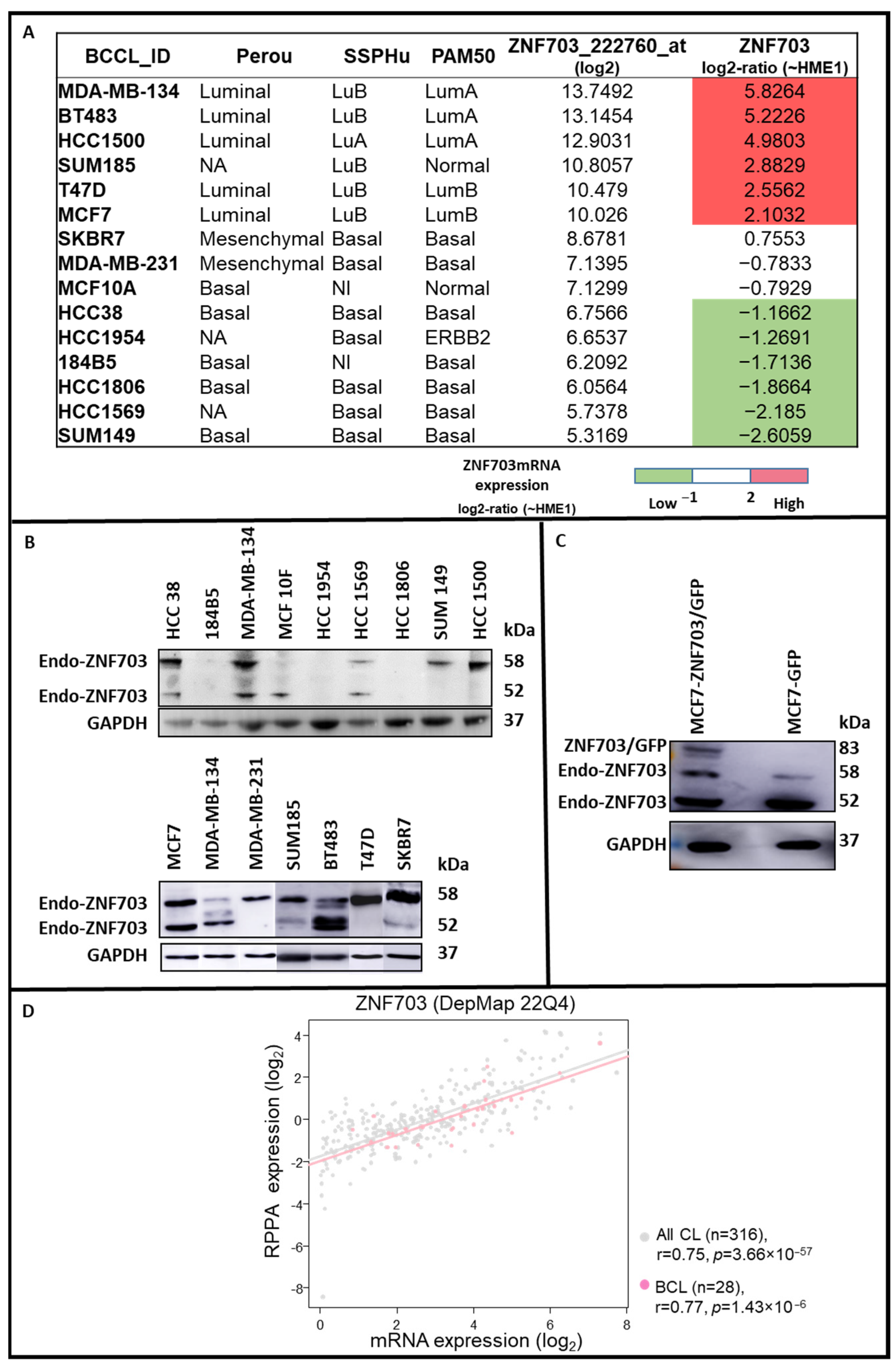
Figure 1. ZNF703 mRNA and ZNF703 protein expression in breast cancer cell lines. 15 BC cell lines were chosen for their ZNF703 mRNA expression centered on that observed in the HME-1 cell line and defined as follows: (i) top 6 of upregulated (High); (ii) 3 medium (Med) and (iii) top 6 of down-regulated (Low). For each of them, a molecular subtype was established with Perou, SSP, and PAM50 classifications [62,65,66] (A). The ZNF703 protein expression was assessed on lysates of the 15 breast tumor cell lines and flow cytometry sorted MCF7-ZNF703/GFP and MCF7-GFP cells (used as controls) by western blotting (WB) ((B,C), respectively). The two endogenous ZNF703 protein isoforms (58 and 52kDa) (Endo-ZNF703) were variably observed in cell lines. Only the full-length 58kDa ZNF703 isoform was approved by the CCDS database (https://www.ncbi.nlm.nih.gov/CCDS/CcdsBrowse.cgi?REQUEST=CCDS&DATA=CCDS6094 (accessed on 15 May 2022); Protein identity/Ref Sequence: NP_079345.1) and considered as previously reported [41,42,43]. An additional band corresponding to ZNF703/GFP fusion protein (83 kDa) was only observed in MCF7-ZNF703/GFP. High ZNF703 expression was observed in BT483, HCC1500, MCF7, SKBR7, and T47D. The ZNF703 mRNA and ZNF703 protein expression data collected from 316 cancer cell lines, including 28 breast cancer cell lines, [67] were analyzed and compared. A significant positive correlation between the mRNA and protein expression levels of ZNF703 was established in all cell lines (Pearson r = 0.75, p = 3.66 × 10−57) and breast cell lines (Pearson r = 0.77, p = 1.43 × 10⁻⁶) (D).
Pharmaceutics 15 01930 g001
Figure 2. ZNF703 ASOs screening for inhibitory activity on ZNF703 mRNA expression. (A,B). ZNF703 expression is assessed by WB on protein lysates extracted from engineered MCF7-ZNF703/GFP BC cells treated with 100 nM concentration of various designed ASOs. WB images showing ZNF703 band signals intensities were respectively quantified for ZNF703/GFP (83 kDa) and endo-ZNF703 (58 kDa) by Image J, normalized to GAPDH, and represented as bars. ASO9 was identified to show higher downregulation efficiency and effectively represses both endogenous and exogenous ZNF703 (B).
Pharmaceutics 15 01930 g002
Figure 3. ASO9 downregulates ZNF703 protein in BC cell lines. Western blot analysis showed ZNF703 inhibition with ASO9 in engineered MCF7-ZNF703/GFP and MCF7-GFP, MDA-MB-134, and MDA-MB-231 BC cell lines ((A,B,C), respectively). WB images showing ZNF703 band signal intensities were respectively quantified for ZNF703/GFP (83 kDa) and endo-ZNF703 (58 kDa) by Image J, normalized to GAPDH, and represented as bars. While ASO9 downregulated both endogenous and exogenous ZNF703 expression in MCF7-ZNF703/GFP (A), a lower expression inhibition was observed in MDA-MB-134 and MDA-MB-231 cell lines ((B,C), respectively).
Pharmaceutics 15 01930 g003
Figure 4. ASO9 reduces ZNF703 expression and viability in MCF7-ZNF703/GFP BC cells. MCF7- ZNF703/GFP cells were treated with ASO9 or control-ASO (scrambled, SCR) at various concentrations. ZNF703 expression inhibition was analyzed by WB (A). WB images showing ZNF703 band signals intensities were respectively quantified for ZNF703/GFP (83kDa) and endo-ZNF703 (58 kDa) by Image J, normalized to GAPDH, and represented as bars. The ZNF703/GFP (83kDa band) expression was quickly inhibited from 100nM dose of ASO9 (A,B) while the endogenous ZNF703 band at 58 kDa, was inhibited by ASO9 following a dose-dependent manner (A,C). The cellular toxicity effect of ZNF703 knockdown induced by ASO9 in MCF7-ZNF703/GFP cells was measured with MTT assay. ASO9 decreased the number of viable cells in a dose-dependent manner, with the cell viability rate of control-ASO and ASO9 reduced to 62.247 ± 5.508 and 21.913 ± 0.090 at 200 nM, respectively (Student’s t-test, *** p < 0.001, and **** p < 0.0001) (D).
Pharmaceutics 15 01930 g004
Figure 5. ASO9 induces apoptosis in MCF7-ZNF703/GFP and MDA-MB-231 BC cell lines. A cell apoptosis test by annexin V binding was done in MCF7-ZNF703/GFP and MDA-MB-231 BC cell lines ((A,B), respectively). Flow cytometry analysis of annexin V expression showed that treatment with ZNF-ASO9 significantly increased apoptosis of MCF7-ZNF703/GFP cells (*** p ≤ 0.001) and MDA-MB-231 cells (* p ≤ 0.05) compared to control-ASO. MCF7-ZNF703/GFP and MDA-MB-231 cells not treated (NT) and treated with control-ASO showed a non-significant difference in the percentage of annexin V positive cells, suggesting low toxicity of control-ASO.
Pharmaceutics 15 01930 g005
Figure 6. The combination with cisplatin improves the anti-cancer ability of ZNF703-ASO9 in MCF 7-ZNF703/GFP BC cell line. To test the efficacy of cisplatin combination with ASO9 treatment, we inhibited ZNF703 with ASO9 (100 nM) and treated MCF7-ZNF703/GFP BC cell line with cisplatin (100 nM) 48hrs post ASO9 transfection. The experiment was done in triplicate and cell viability was evaluated by the MTT test. The results showed that: (i) cisplatin alone or in combination with SCR reduced cell viability more efficiently than the absence of treatment (NT) or with SCR alone (Student’s t-test, *** p ≤ 0.001); (ii) ZNF703 inhibition by ASO9 in combination with cisplatin reduced cell viability more efficiently than treatment with ASO9 alone or cisplatin alone (Student’s t-test, *** p ≤ 0.001).
Pharmaceutics 15 01930 g006
Disclaimer/Publisher’s Note: The statements, opinions and data contained in all publications are solely those of the individual author(s) and contributor(s) and not of MDPI and/or the editor(s). MDPI and/or the editor(s) disclaim responsibility for any injury to people or property resulting from any ideas, methods, instructions or products referred to in the content.