Review

Bacillus licheniformis: A Producer of Antimicrobial Substances, including Antimycobacterials, Which Are Feasible for Medical Applications

by
Margarita O. Shleeva
*,
Daria A. Kondratieva
and
Arseny S. Kaprelyants
A.N. Bach Institute of Biochemistry, Federal Research Centre ‘Fundamentals of Biotechnology’, Russian Academy of Sciences, 119071 Moscow, Russia
*
Author to whom correspondence should be addressed.
Pharmaceutics 2023, 15(7), 1893; https://doi.org/10.3390/pharmaceutics15071893
Submission received: 26 May 2023 / Revised: 2 July 2023 / Accepted: 3 July 2023 / Published: 5 July 2023

Abstract

:
Bacillus licheniformis produces several classes of antimicrobial substances, including bacteriocins, which are peptides or proteins with different structural composition and molecular mass: ribosomally synthesized by bacteria (1.4–20 kDa), non-ribosomally synthesized peptides and cyclic lipopeptides (0.8–42 kDa) and exopolysaccharides (>1000 kDa). Different bacteriocins act against Gram-positive or Gram-negative bacteria, fungal pathogens and amoeba cells. The main mechanisms of bacteriocin lytic activity include interaction of peptides with membranes of target cells resulting in structural alterations, pore-forming, and inhibition of cell wall biosynthesis. DNase and RNase activity for some bacteriocines are also postulated. Non-ribosomal peptides are synthesized by special non-ribosomal multimodular peptide synthetases and contain unnatural amino acids or fatty acids. Their harmful effect is due to their ability to form pores in biological membranes, destabilize lipid packaging, and disrupt the peptidoglycan layer. Lipopeptides, as biosurfactants, are able to destroy bacterial biofilms. Secreted polysaccharides are high molecular weight compounds, composed of repeated units of sugar moieties attached to a carrier lipid. Their antagonistic action was revealed in relation to bacteria, viruses, and fungi. Exopolysaccharides also inhibit the formation of biofilms by pathogenic bacteria and prevent their colonization on various surfaces. However, mechanism of the harmful effect for many secreted antibacterial substances remains unknown. The antimicrobial activity for most substances has been studied in vitro only, but some substances have been characterized in vivo and they have found practical applications in medicine and veterinary. The cyclic lipopeptides that have surfactant properties are used in some industries. In this review, special attention is paid to the antimycobacterials produced by B. licheniformis as a possible approach to combat multidrug-resistant and latent tuberculosis. In particular, licheniformins and bacitracins have shown strong antimycobacterial activity. However, the medical application of some antibacterials with promising in vitro antimycobacterial activity has been limited by their toxicity to animals and humans. As such, similar to the enhancement in the antimycobacterial activity of natural bacteriocins achieved using genetic engineering, the reduction in toxicity using the same approach appears feasible. The unique capability of B. licheniformis to synthesize and produce a range of different antibacterial compounds means that this organism can act as a natural universal vehicle for antibiotic substances in the form of probiotic cultures and strains to combat various types of pathogens, including mycobacteria.

1. Introduction

The spread of bacterial strains that cause severe infectious diseases but are now resistant to known antibiotics necessitates the search for and development of new approaches to combat these diseases [1]. The most known and medically important example that illustrates this problem is the growing number of cases of multidrug-resistant strains of Mycobacterium tuberculosis (Mtb), which is the causative agent of tuberculosis. In addition to drug resistance, Mtb is able to asymptomatically persist in the host organism for many years, causing latent forms of tuberculosis. In this dormant state, Mtb cells are also resistant to known antibiotics [2,3,4].
The search for and study of substances that have bactericidal or bacteriostatic properties against human and animal pathogens are also required for the development of new antibiotic therapy or disinfectants for objects and surfaces that have been in close contact with patients and therefore may carry pathogenic bacteria. Currently, in addition to the synthesis of new chemical substances, considerable attention has focused on the analysis of the potential of natural products from different origins as antimicrobials. The discovery of antibiotics with activity against human pathogens is often based on the observation of the interaction between microorganisms, called antagonism. This antagonism manifests through the synthesis and release of substances that inhibit or completely suppress the growth of other species. Under natural conditions, a microorganism-secreted substance(s) that inhibits the growth of another organism gains a competitive advantage in the struggle for environmental resources. Most of the antibiotics used for medical applications are secreted products or derivatives of microorganisms belonging to the order Actinomycetalis (among them, the most well-known are Streptomyces). The bacterial world represents a huge reservoir of not-yet-discovered and used substances that have antibacterial potential. In this regard, representatives of the genus Bacillus are known as producers of many enzymes and antimicrobial compounds. For example, Bacillus amyloliquefaciens is a source of the natural antibiotic barnase (ribonuclease), alpha-amylase, which is used in starch hydrolysis; protease subtilisin, which is used in combination with detergents; and the restriction enzyme, BamH1, used in DNA research [5]. Bacillus subtilis produces 66 derived antimicrobials, and Bacillus brevis produces 23 peptide antibiotics [6]. There is a growing interest in considering these substances, including bacteriocins, as alternative antimicrobials for the treatment of human and animal infections [7,8,9,10,11].
Currently, the use of bacterial probiotic strains and their metabolic products is considered a new approach for the control and prevention of various infectious diseases [12]. Animal studies have demonstrated that probiotics from the Bacillus genus have antimicrobial properties. This conclusion also applies to humans [13,14]. The use of bacteriocins and antimicrobial peptides produced by probiotic strains is a suitable alternative to antibiotics because their production is inexpensive and resistance to them is rare [15]. They exhibit a broad spectrum of activity against many Gram-positive and Gram-negative bacteria and fungi. Owing to the efficacy and cost-effectiveness of many of these compounds, they are attractive for clinical use [16]. A few natural peptides have shown potential because of their desirable therapeutic properties, including antimicrobial, antiviral, anticancer, and contraceptive activities. Additionally, they have been shown to protect against topical and systemic infections in combination with conventional antibiotics [17].
Among the organisms belonging to the Bacillus genus, Bacillus licheniformis is a unique specie that which produces wide variety of antimicrobial substances. This bacterium shows promise for use as a probiotic in the treatment of dysbacteriosis, which is caused by various diseases [13]. The effectiveness of B. licheniformis as a probiotic is associated with its ability to produce a large amount of substances with antimicrobial, antioxidant and immunomodulatory activities [13], for example, a phosphorus-containing triene antibiotic called proticin [18,19]. B. licheniformis shows a protective effect in zebrafish (Danio rerio) against Vibrio parahaemolyticus infection. Due to the antagonistic activity of this probiotic, the complete survival of infected fish was observed in contrast with untreated fish [20]. This probiotic, in combination with Bifidobacterium breve, significantly inhibited the adhesion of the pathogen Kocuria rhizophila in vitro [21] and showed antivibrio activity against Vibrio parahaemolyticus [22]. The use of a crude extract from B. licheniformis resulted in marked antiviral activity against porcine epidemic diarrhea virus in Vero cells and reduced virus shedding in piglets [23]. After the administration of fermented B. licheniformis products, the number of pathogenic bacteria including Clostridium perfringens significantly decreased in cats with chronic diarrhea [24]. In piglets, B. licheniformis treatment showed positive effects against Salmonella [25]. Probiotic B. licheniformis produces antimicrobial substances and has a strong ability to auto- and coaggregate against pathogenic bacteria [26]. Approaches are being developed to combat bacterial biofilms using silver nanoparticles and the probiotic B. licheniformis [27].
The bacteriocins from B. licheniformis are being considered as natural preparations as a preservative in the food industry [28,29].
In general, bacteriocins are a group of antimicrobial peptides that represent a potential alternative to classical antibiotics in the fight against antimicrobial resistance in pathogenic microorganisms. Several reports have been published in the literature about numerous bacteriocins, many of which currently remain undiscovered due to the wide variety of their natural sources; hence, further research in this area is required [11].
Considering the medical and industrial application of Bacillus licheniformis, a thorough description and characterization of the variety of antimicrobial compounds produced and their use against resistant pathogens, such as mycobacteria, are required.
Therefore, this review focused on the current state of knowledge about the classes of antibiotic substances produced by B. licheniformis and their structure and properties, which may allow a more comprehensive perspective of their antimicrobial substances, including their antimycobacterial properties.

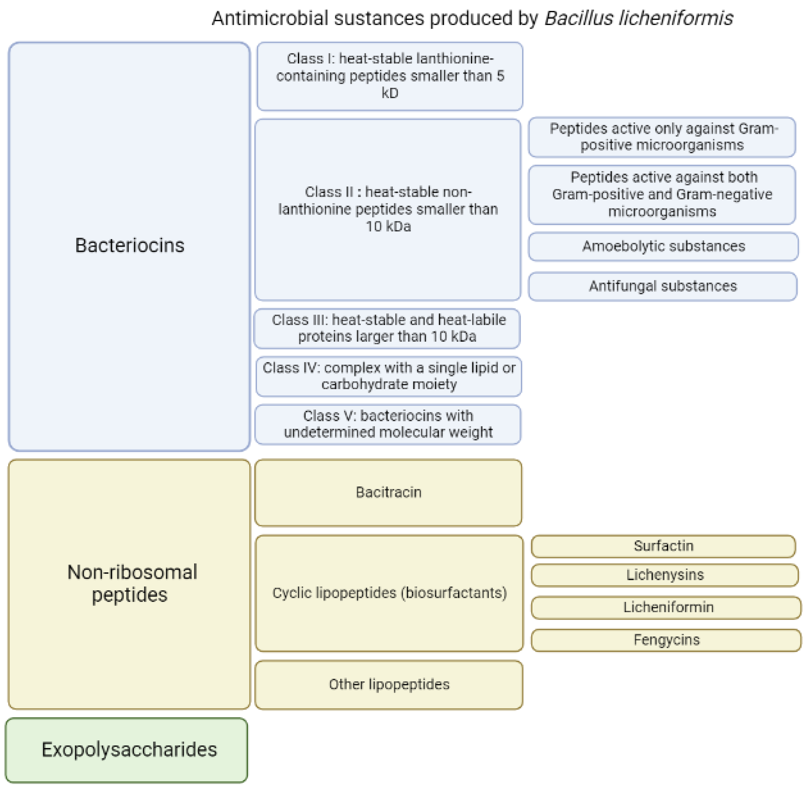
2. Antibacterial Substances Secreted by Bacillus licheniformis

The endospore-forming bacterium Bacillus licheniformis is capable of producing a large amount of substances with different structures and different antibacterial activities [30] (Figure 1). When grown on identical media, different strains of B. licheniformis produce different sets of substances with antibacterial activities [31]. It is plausible that all strains of B. licheniformis potentially are able to produce variety of antimicrobial substances, however, the synthesis and production of particular substance can be differently regulated on the transcriptional or translational level for certain strains grown in identical media. As a result, the amount of secreted antimicrobial compounds may substantially vary from strain to strain allowing to consider them as unique producers of defined set of antimicrobials. The secreted antimicrobial substances have molecular masses ranging from 1.4 to 20 kDa [28,29,32,33,34,35,36,37,38].
Changing the composition of the medium used for B. licheniformis growth results in the alterations in the secreted substances. Thus, on media containing iron, B. licheniformis can synthesize the red pigment pulcherrimin [39]. When grown on a medium with lactate and a high ratio of nitrogen and carbon, B. licheniformis strain N.C.T.C. 7072 produces licheniformins; when grown on a medium with glucose and a low nitrogen/carbon ratio, this strain produces bacitracins [37]. Several substances synthesized by B. licheniformis have been investigated as antibiotics against various types of bacteria. They are listed and characterized below. Some of them (bacitracin) are used in combined antibacterial preparations intended for topical use. Others are used as oral antibiotics, but only in animals due to their toxic effects.
Among the antimicrobial components (Figure 1) produced by various strains of B. licheniformis in a nutrient medium, several groups differ in properties and structure.

2.1. Bacteriocins

Bacteriocins are substances represented by an amino acid sequence (peptides or proteins) that act against other strains of bacteria or closely related species. They demonstrate both bactericidal and bacteriostatic effects. Bacteriocins are natural antimicrobial peptides that are ribosomally synthesized by bacteria [10,11,40]. Genes whose expression leads to the synthesis of bacteriocins are organized into clusters of operons and can be located in the genome, plasmids, or other mobile genetic elements. These genes are inducible; peptide secretion and accumulation outside the cell are required for their induction. More details regarding bacteriocins biosynthesis are described in the review by Nishie et al. [9]. Bacteriocins are heterogeneous substances that demonstrate various biochemical properties, molecular weights, inhibitory spectra and mechanisms of action [10,41]. Due to the wide spectrum of antagonistic activity inherent to the bacteriocins of some strains of microorganisms, they have the potential to be used as a component of antibacterial drugs. The resistance to enzyme activity of the antimicrobial peptides produced by Bacillus spp. differs with stability over a wide range of pH and temperature. Most of these peptides have high specificity against microbial pathogens and low cytotoxicity against human cells [42]. The related bactericidal mechanisms include the pore-forming type, nuclease type with DNase and RNase functions, and the peptidoglycanase type [10] (Figure 2).
As a result, pores are formed, which leads to the rapid removal of small cytoplasmic molecules, ions from target cells, and the collapse of the protonmotive force, leading to the death of bacterial cells [9,43]. However, other antimicrobial mechanisms of bacteriocins were also identified [11]. Despite the recent popularity of research on the properties of bacteriocins and their use in medicine, veterinary sciences, and food industry [10,11], many bacteriocins have not yet been studied.
The production of several bacteriocin-like substances (Table 1) with different characteristics and a wide spectrum of activity against pathogenic bacteria was recorded in the strains of B. licheniformis [44]. For example, B. licheniformis SMIA-2, a thermophilic and thermostable enzyme-producing strain, is active against some strains of Staphylococcus aureus and Bacillus sp. The genome annotation of this strain detected gene clusters responsible for antimicrobial component production (lichenysin, fengycin, lichenicidin and bacillibactin biosynthetic gene clusters) [45].
B. licheniformis produces various bacteriocins ranging in molecular weight from 1.4 to 55 kDa, but the expression of a particular antimicrobial agent may depend on environmental conditions, growth period, and the specific bacterial strain [28,32,33,36,46].
In general, based on their thermostability, size and chemical moieties, bacteriocins are classified into four major groups [47]: class I, heat-stable lanthionine-containing peptides smaller than 5 kDa; class II, heat-stable non-lanthionine peptides smaller than 10 kDa; class III, heat-labile proteins larger than 30 kDa; and class IV, complex with a single lipid or carbohydrate moiety [48]. In this review, to describe the antimicrobial substances produced by different strains of B. licheniformis, Cotter’s classification is used, with a slight modification: heat-stable and heat-labile proteins larger than 10 kDa are assigned to class III, and class V was added, which includes proteins with undetermined molecular weight (Table 1, Figure 1).
Table 1. Antimicrobial bacteriocins produced by Bacillus licheniformis.
2.1.1. Class I: Heat-Stable Lanthionine-Containing Peptides Smaller Than 5 kDA
Substance(s) Specific/Unspecific NameProducing StrainMolecular MassActivity AssayReference
SublicheninB. licheniformis MCC 25123.348 kDaKocuria rhizophila ATCC 9341
Pediococcus lolii MCC 2972
Enterococcus durans B20G1
Enterococcus faecalis MF3
E. faecalis MM2
E. faecalis CHL1
E. faecalis CHL3
E. faecalis CHL
E. faecalis MCC 3063
E. faecalis MCC 2773
Enterococcus faecium MCC 2763
Entercoccus avium CS32
Enterococcus cecorum 1-40a
Lactobacillus plantarum MCC 2774
Listeria monocytogenes
Staphylococcus aureus
Staphylococcus aureus (MRSA)
[49]
Escherichia coli
Klebsiella pneumoniae
LichenicidinBacillus licheniformis DSM 13
(also produced by ATCC 14580, VK21,WIT 562, 564 and 566 strains, IMF20, IMF66, IMF69 and IMF80, I89)
3 kDa and 3.25 kDaBacillus cereus DSM 31
Bacillus halodurans DSM 18197
Bacillus megaterium KM (ATCC 13632)
Bacillus subtilis 168 (DSM 402)
Bacillus spec. HIL Y-85,54728
Enterococcus faecium BM 4147–1
Enterococcus faecium L4001
Lactobacillus sake 790 E2
Lactococcus lactis NCTC 497
Micrococcus luteus DSM 1790
Micrococcus luteus ATCC 4698
Staphylococcus aureus ATCC 33592 (MRSA)
S. aureus ATCC 29213 (MSSA)
S. aureus 1450/94
S. aureus Cowan (ATCC 12598)
S. aureus Newman (NCTC 8178)
S. aureus SG511
S. aureus Wood 46 (ATCC 10832)
Staphylococcus carnosus TM300
Staphylococcus gallinarum Tü 928
Staphylococcus saprophyticus DSM 20229
Staphylococcus simulans 22
S. aureus LT440/09 (community acquired MRSA)
S. aureus LT420/09 (MRSA)
S. aureus LT819/09 (MRSA, Rhine-Hessen epidemic strain)
Enterococcus faecalis
Streptococcus agalactiae
B. subtilis L1
Rhodococcus sp. strain SS2
M. luteus B1314
B. megaterium VKM41
B. pumilus 2001
B. globigii I
B. amyloliquefaciens I
M. smegmatis 1171
M. phlei 1291
[50,51,52,53,54,55]
2.1.2. Class II: heat-stable non-lanthionine peptides smaller than 10 kDa
2.1.2.1. Peptides active only against Gram-positive microorganisms
Bacillocin 490B. licheniformis 490/52 kDaBacillus licheniformis 5 A2
Listeria innocua
Staphylococcus epidermidis
Bacillus anthracis 7700
Bacillus subtilis AZ56
Bacillus cereus 6A2
Bacillus stearothermophilus 9A19
Bacillus smithii PRO/S
[28]
Bacteriocin-like substanceBacillus licheniformis H13.5 kDaE. faecalis ATCC 19433
L. monocytogenes ATCC 19111
B. cereus ATCC 14579
B. subtilis ATCC 6633
Lactobacillus species ATCC 33198
Lactobacillus fermentum
[56]
P. fluorescens
Bacteriocin-like antibacterial peptidesB. licheniformis AnBa9<10 kDaStaphylococcus aureus GCS1
Bacillus cereus GCS2
Staphylococcus epidermidis GCS4
Kurthia gibsonii GCS6
Micrococcus luteus GCS7
Streptococcus mitis GCS9
Bacillus subtilis B-4219
L. lactis B-1821
Staphylococcus epidermidis B-4268
Bacilus smithii NRS-173
Lactobacillus acidophilus B-4495
Micrococcus luteus B-287
Pediococcus acidilactici B-14958
Leuconostoc mesenteriodes
[35]
LicheninB. licheniformis 26L10/3RA1.4 kDaStreptococcus bovis SB3
Streptococcus bovis 26
Ruminococcus avefaciens OF-2
Ruminococcus avefaciens C94
Ruminococcus albus B119
Ruminococcus albus A-6
Butyrivibrio fibrisolvens OR 12
Eubacterium ruminantium GA-195
Lactobacillus casei ED-108
[32]
Bacteriocin BL8B. licheniformis BTHT81.4 kDaClostridium perfringens
Staphylococcus aureus
Bacillus cereus
Bacillus circulans
Bacillus pumilus
[57]
BSCY2B.licheniformis CY26.5 kDaB.subtilis 6633[58]
Licheniocin
50.2
B. licheniformis VPS50.23.25 kDaBacillus subtilis ATCC 6633
B. subtilis 168
B. subtilis W23
Enterococcus faecalis ATCC 29212
Enterococcus saccharolyticus ATCC 43076
Lactobacillus plantarum LMG92088
Lactobacillus zeae
Lactococcus lactis IL1403
Listeria monocytogenes ATCC 19111
Micrococcus luteus ATCC 7468
Staphylococcus aureus ATCC 25923
Staph. aureus ATCC 33591
Streptococcus agalactiae ATCC 12386
[59]
2.1.2.2. Peptides active against both Gram-positive and Gram-negative microorganisms
Bacteriocin like inhibitory substance (BLIS)Bacillus licheniformis IITRHR2 (FJ447354)1.2 kDaBacillus cereus MTCC 1305
Bacillus subtilis MTCC 736
Bifidobacterium bifidum NCDC 235
Enterococcus faecalis MTCC 439
Enterococcus faecalis NCDC1 14
Lactobacillus casei NCDC017
Lactobacillus lactis NCDC094
Leuconostoc mesenteroides NCDC 219
Listeria monocytogenes MTCC 387
Listeria monocytogenes MTCC1 143
Pediococcus pentosaceus NCDC 273
Staphylococcus thermophilus NCDC 074
[60]
Escherichia coli MTCC 1687
Pseudomonas aeruginosa MTCC 9027
Shigella flexneri MTCC 1457
Shigella sonnei MTCC 2957
Bacteriocin MKU3B. licheniformis MKU31.5 kDaBacillus subtilis B4219
Bacillus smithii NRS173
Lactobacillus acidophilus B4495
Lactobacillus fermentum B1840
Lactobacillus lactis B1821
Staphylococcus epidermidis B4268
Micrococcus luteus B287
Leuconostoc mesenteriodes B1118
Pediococcus acidilactici B14958
Staphylococcus aureus GCS1
Bacillus cereus GCS2
Bacillus cereus GCS3
Staphylococcus epidermidis GCS4
Staphylococcus epidermidis GCS5
Kurthia gibsonii GCS6
Micrococcus luteus GCS7
Bacillus subtilis GCS8
Streptococcus fecalis GCS9
Bacillus cereus GCS10
Bacillus cereus GCS11
Lactobacillus acidophilus GCS12
[34]
Escherichia coli DH5a
Bacteriocin-like substanceB. licheniformis B1164 kDaB. cereus CGMCC1.230
Listeria monocytogenes CVCC1599
Micrococcus luteus CMCC28001
S. aureus CMCC26003
S. aureus CICC21601
S. aureus CVCC1885
Streptococcus equi subsp. zooepidemicus CVCC1903
[61]
E. coli CVCC245
E. coli CICC21525
E. coli CVCC195
E. coli CVCC249
S. enterica ser. Pullorum CVCC79301
S. enterica ser. typhimurium CVCC541d
ppABPB. licheniformis Me1 (MCC 2016)Between 3 and 3.5 kDaL. innocua FB 21
L. murrayi FB 69
M. luteus ATCC 9341
L. monocytogenes Scott A
Staph. aureus FRI 722
B. cereus F 4433
K. rhizophila ATCC 9341
[62,63,64]
Salm. typhimurium MTCC 1251, FB 231
Salm. paratyphi FB 254
E. coli CFR 02
Y. enterocolitica MTCC 859
Shigella flexineri (clinical isolate)
Licheniformins A,B,CB. licheniformis
NCTC 7072
3.8–4.8 kDaMycobacterium phlei
Staphylococcus aureus
[37,65]
E. coli
Antimicrobial compoundBacillus licheniformis MCC25146.5 kDaM. luteus ATCC9341
S. aureus FRI722
[31]
Klebsiella sp.
A. hydrophila NRRL B445
2.1.3. Class III: heat-stable and heat-labile proteins larger than 10 kDa
BLIS_SXAU06B. licheniformis SXAU0614 kDaS. aureus
S. epidermidis
M. luteus
L. monocytogenes
[66]
BL-DZ1 (BL00275)B. licheniformis strain D114 kDaBacillus pumilus TiO1
Candida albicans BH
[67]
Pseudomonas aeruginosa PAO1 (biofouling)
YbdN ProteinB. licheniformis (seaweed isolate)30.7 kDaMRSA 9551
MRSA J2407
VRE 788
VRE 1349
L. monocytogenes NCTC 7973, NCTC 10357
[68]
ChitinaseB. licheniformis MY75
(also produced by Mb-2, TP–1, S213, SSCL-10, B307 strains)
55 (67,68/62/60,65,66) kDaG. saubinetii
A. niger
(Phoma medicaginis)
[69,70,71,72,73,74,75]
Antifungal Protein F2B. licheniformis BS-331 kDaAspergillus niger
Magnaporthe oryzae
Rhizoctonia solani
Fusarium oxysporum
[76]
Antimicrobials proteinB. licheniformis JS16 kDaBacillus cereus
Bacillus subtilis
[77]
Shigella dysenteriae
Salmonella typhimurium
AMSB. licheniformis T6-520 kDaDesulfovibrio alaskensis NCIMB 1349[36,78]
AMSB. licheniformis
H2O-1
Between 90 and 120 kDaDesulfovibrio alaskensis NCIMB 1349
SRB-containing consortium T6-lab
[36,78]
2.1.4. Class IV: complex with a single lipid or carbohydrate moiety
F4, F5 and F6B. licheniformis BFP011Less than 45 kDaB. amyloliquefaciens TISTR 1045
B. licheniformis TISTR 1010
B. subtilis ATCC 6633
B. subtilis TISTR 008
B. pumilus TISTR 905
B. cereus ATCC 11778
B. megaterium (clinical isolate)
S. aureus ATCC 25923
[79]
E. coli O157: H7
V. cholerae (clinical isolate)
K. pneumonia ATCC 17736
S. typhi ATCC 5784
Ieodoglucomides A and BB. licheniformis 09IDYM23NDS. aureus
B. subtilis
B. cereus
[80]
E. coli
P. aeruginosa
S. typhi
Ieodoglucomide C
and
ieodoglycolipid
B. licheniformis 09IDYM23NDStaphylococcus aureus
Bacillus subtilis
Bacillus cereus
[81]
Escherichia coli
Pseudomonas aeruginosa
Salmonella typhi
2.1.5. Class V: bacteriocins with undetermined molecular weight
Antipathogenic MetabolitesBacillus licheniformis (Upper arm skin isolate)NDStaph. aureus ATCC 6538[82]
Kl. Pneumoniae subsp.
pneumonia CMSOGH
Antipathogenic MetabolitesBacillus licheniformis (Upper arm skin isolate)NDKl. Pneumoniae subsp.
pneumoniae
[83]
Antimicrobial substanceB. licheniformis A-1-5B-APNDPrevotella intermedia 1/P
Streptococcus mutans ATCC 35668
Micrococcus luteus DSM 1790
[84]
Porphyromonas gulae 3/H
BacteriocinB. licheniformis HJ2020 MT192715.1NDStaphylococcus aureus
Bacillus cereus
Bacillus subtilis
[85]
Escherichia coli 0157:H7
Salmonella typhi
Pseudomonas aeruginosa
ND—no data. Gram- bacteria are below the line, and Gram+ bacteria are above the line.

2.1.1. Class I: Heat-Stable Lanthionine-Containing Peptides Smaller Than 5 kDa

Lantibiotics are antimicrobial peptides that undergo post-translational modification. They contain non-standard amino acids: lanthionine, β-methyl lanthionine and dehydrated residues (dehydrated amino acids) [86]. Their molecular weight does not exceed 5 kDa. Lantibiotics are active at low concentrations and are therefore attractive antimicrobials. They mainly target lipid II. A number of lantibiotics interact with the cell wall precursor lipid II (undecaprenyl-pyrophosphoryl-MurNAc-(pentapeptide)-GlcNAc), which prevents cell wall biosynthesis and contributes to the destruction of the bacterial membrane [87]. Thus, the most well-studied lantibiotic nisin interacts with the pyrophosphate fragment of lipid II. Critical to this binding are the two N-terminal rings of the lantibiotic [88]. The formation of the pore complex results in cell membrane permeabilization and dissipation of the proton motive force [87].
In general, lantibiotics are synthesized and secreted by Gram-positive microorganisms, and their activity is most often manifested in connection with closely related Gram-positive bacteria. For Gram-negative bacteria, their activity is rather limited [86] because the cell wall of Gram-negative bacteria is an effective permeability barrier due to the presence of an outer membrane, which prevents access to the peptidoglycan layer (localization of lipid II) and the cytoplasmic membrane (Figure 2). Moreover, the anionic cell surface of Gram-negative bacteria promotes the binding of cationic lantibiotics, where such an interaction potentially increases the stability of the outer membrane through electrostatic interactions [89].
B. licheniformis produces two lantibiotics that may be important for applications in various industries (Table 1).
The first one is sublichenin, which is a subtilin-like lantibiotic of probiotic bacterium Bacillus licheniformis MCC 2512 that has a molecular weight of 3.348 kDa and the succinylated form has a molecular weight of 3.448 kDa [31,49]. Subtilin is a natural variant of nisin that refers to linear pentacyclic class I antibiotics [90]. The sublichenins from B. licheniformis are almost identical to the JS-4 subtilin from B. subtilis. Subtilin JS-4 retained >90% and 86.1% of its antibacterial activity even after a 30 min exposure to 80–100 °C and 121 °C, respectively, indicating considerable thermostability. Subtilin JS-4 was also rapidly inactivated by proteolytic enzymes including proteinase K, trypsin, papain and pepsin [91]. It also showed a broad antimicrobial spectrum against Gram-positive bacteria. Subtilin JS-4 inhibited the growth of foodborne bacterium L. monocytogenes by increasing cell membrane permeability, triggering pore formation and K+ leakage, and damaging cell membrane integrity, which eventually disrupted the membrane and cellular structure [91] (Figure 2).
The second antibiotic is lichenicidin, a dipeptide lantibiotic consisting of a synergetic lantibiotic pair, Licα (3.251kDa) and Licβ (3.021 kDa), which was described for B. licheniformis DSM 13. This substance demonstrated activity against the growth of Gram-positive bacteria, such as Bacillus subtilis, Micrococcus luteus, Staphylococcus aureus, Streptococcus pyogenes, Staphylococcus simulans and enterococci but neither caused hemolysis nor inhibited the growth of Gram-negative bacteria. Lichenicidin is associated with the cell surface and shows stability against trypsin, chymotrypsin, and proteases [50]. Moreover, lichenicidin can be produced by other strains of B. licheniformis, and the structure of its peptides may differ depending on the strain. Lichenicidin was not cytotoxic to human erythrocytes and fibroblasts [92]. B licheniformis strain ATCC 14580 produced lichenicidin with activity against a range of pathogenic microorganisms including Listeria monocytogenes, Staphylococcus aureus, vancomycin-resistant enterococci, Bacillus cereus, Streptococcus pneumoniae and Streptococcus mutants [51]. Lichenicidin has been produced by B. licheniformis strain VK21 [52], and WIT 562, 564 and 566 [53]. Additionally, lichenicidin production was found in B. licheniformis isolates (isolated from retail infant milk formulae): strains IMF20, IMF66, IMF69 and IMF80. These strains demonstrated antimicrobial activity against target Gram-positive organisms. No activity was observed against the Gram-negative bacteria E. coli or S. typhimurium [54]. For Lchα and Lchβ, lichenicidin subunits produced by VK21 strain, described tertiary structures and details of mode of action [52].
Another variant of lichenicidin comprises two mature peptides, Bliα and Bliβ, produced by the I89 strain; their synergistic activity is required for full activity [55,93].
Lichenicidin acts through a dual mode of action that involves the α subunit recognition of lipid II, providing specificity and stability for the interaction of β subunit, which induces leakage of the intracellular contents of bacteria [94,95] (Figure 2).

2.1.2. Class II: Heat-Stable Non-Lanthionine Peptides Smaller Than 10 kDa

This class includes unmodified peptides with a molecular weight up to 10 kDa. The vast majority of them are thermostable membrane-active peptides. Among them, peptides that are active only against Gram-positive microorganisms and active against both Gram-positive and Gram-negative microorganisms can be distinguished (Table 1). Peptides with antifungal (Table 2) and amoebolytic (Table 3) activities have also been identified.

B. licheniformis Secreted Peptides Active Only against Gram-Positive Microorganisms

Because antagonism provides a survival advantage in the suppression of related species of microorganisms, most bacteriocins secreted by different strains of B. licheniformis are active only against Gram-positive bacteria (Table 1). Peptides may be insensitive or sensitive to the action of proteolytic enzymes. However, the vast majority of the identified bacteriocins that are active only against Gram-positive microorganisms are sensitive to the action of proteinases.
Bacillocin 490, a bacteriocin with a low molecular mass (2 kDa) and produced by a thermophilic strain (B. licheniformis 490/5) isolated from dairy foods, shows high thermal stability, with 46.4% residual activity after 1 h of exposure to 100 °C. This bacteriocin was inactivated by pronase E and proteinase K. Bactericidal activity was maintained during storage at 4 °C and was remarkably stable over a wide pH range. The activity range of bacillocin 490 was limited to some Gram-positive bacteria. The highest antimicrobial activity was observed against Bacillus stearothermophilus, B. smithii, B. subtilis and B. anthracis. Moderate inhibition of B. cereus, very low inhibition of Listeria innocua and S. aureus, and no inhibition of B. thuringensis and Streptococcus thermophilus were noted. This activity spectrum shows that bacillocin 490 is principally active against species phylogenetically related to the producer strain. The incubation of B. smithii in the presence of bacillocin 490 resulted in 96% death in 30 min, indicating that bacteriocin has a bactericidal effect [28].
The supernatant of thermophilic strain B. licheniformis H1 exhibited antagonistic activity against various species of Gram-positive bacteria, such as Listeria monocytogenes but not against Gram-negative bacteria, except for Pseudomonas fluorescens. The inactivation of this bacteriocin-like activity by a-chymotrypsin, trypsin, and papain was highly significant. No significant decrease was found in antimicrobial activity after the incubation of a bacteriocin-containing supernatant from B. licheniformis H1with pepsin or lipase. The bacteriocin-like substance was stable at temperatures up to 75 °C for 60 min, but activity was lost after being autoclaved at 121 °C for 15 min. Concentrated antimicrobial activity was found in the protein fraction obtained with 60% ammonium sulfate saturation. The results of sodium dodecyl sulfate–polyacrylamide electrophoresis analysis of concentrated partially purified supernatants collected after resting bacterial cells at 55 °C revealed a bacteriocin-like protein with a molecular mass of approximately 3.5 kDa [56].
B. licheniformis AnBa9 produced antibacterial bacteriocin-type peptides with a molecular mass of <10 kDa. The production of these peptides was 25-fold higher under optimized conditions for producer growth compared with nonoptimized condition. The level of bacteriocin production and its specific activity gradually decreased with increasing concentrations of lactose and NH4NO3. F high concentration of yeast extract, an alkaline pH and an elevated temperature improved the production of antibacterial peptides by B. licheniformis AnBa9. B. licheniformis AnBa9 inhibited several Gram-positive bacteria, including Staphylococcus aureus, Bacillus cereus, Staphylococcus epidermidis, Kurthia gibsonii, Micrococcus luteus, Streptococcus mitis, Bacillus subtilis, L. lactis, Bacillus smithii, Lactobacillus acidophilus, Pediococcus acidilactici, and Leuconostoc mesenteriodes. However, these bacteriocins did not inhibit Listeria strains or Gram-negative bacteria. The loss of antibacterial activity of the permeate after treatment with proteinase K, pronase E, and trypsin, suggested that these bacteriocins are sensitive to proteolytic enzymes. They were resistant to temperature up to 100 °C for 30 min and over a wide range of pH from 4 to 12 [35].
Under anaerobic conditions, B. licheniformis 26L10/3RA produced an inhibitory bacteriocin-like component called lichenin. This peptide was purified to homogeneity, and the molecular mass was estimated at approximately 1.4 kDa. Lichenin was found to be hydrophobic, was sensitive to atmospheric oxygen, retained biological activity even after boiling for 10 min, and was active over a pH range of 4.0–9.0. It was active against Streptococcus bovis, Ruminococcus albus, Ruminococcus avefaciens, and Eubacterium ruminantium. The biological activity of this peptide was completely inactivated by proteinase K treatment, but this peptide was resistant to trypsin. Heat treatment at 80 °C and boiling for 10 min at pH 4.0 and 9.0 resulted in a significant reduction in biological activity. Lichenin production was observed only upon B. licheniformis anaerobic growth, the antibacterial activity of which was also demonstrated for the reference strains grown under anaerobic conditions only. The inability of lichenin to inhibit aerobically grown bacteria was explained either by its inactivation by atmospheric oxygen or the target bacteria due to oxidative respiration. No N-terminal block was observed in the sequence and the peptide did not show any characteristics of cyclicity. However, the seventh amino acid residue could not be identified and it did not belong to any of the natural amino acids [32].
Strain BTHT8, identified as B. licheniformis, inhibited the growth of Gram-positive test organisms. The active component, labeled as bacteriocin BL8, was purified from the supernatant of B. licheniformis strain BTHT8. The molecular mass was determined as 1.4 kDa. The results of N-terminal amino acid sequencing of BL8 identified a 13 amino acid sequence stretch. Bacteriocin BL8 was stable even after boiling at 100 °C for 30 min and over a wide pH range of 1–12 [57].
A bacteriocin from B. licheniformis cy2, named BSCY2, was stable at pH 2.5–9.5, showing activity against B. subtilis. BSCY2 was stable below 40 °C and retained its antimicrobial activity during long tern storage at −20 °C and −70 °C. BSCY2 was inactivated after 15 min exposure to temperatures over 80 °C and lost 50% of its antimicrobial activity within 2 h at 70 °C. BSCY2 was inactivated by proteinase K treatment, which indicated its proteinous nature. The direct detection of the BSCY2 band showing antimicrobial activity on Tricine–SDS-PAGE suggested an apparent molecular mass of about 6.5 kDa [58].
In contrast to the above-mentioned bacteriocins of this group, some bacteriocins retain their activity after treatment with proteolytic enzymes.
B. licheniformis strain VPS50.2 produced bacteriocin licheniocin 50.2 (molecular mass of approximately 3.25 kDa) and was effective against Gram-positive bacteria, including Listeria monocytogenes, methicillin-resistant Staphylococcus aureus, and b-haemolytic streptococci. Bacteriocin activity was insensitive to lysozyme and proteinase K, being heat stable after incubation at 100 °C for 30 min and over a wide range of pH (2–12). The inhibitory spectrum considered in this study was limited to Gram-positive bacteria only. The maximum antagonistic activity was found in the precipitate with 60% saturation of ammonium sulfate [59].
Despite the varying degrees of sensitivity to the action of proteolytic enzymes, bacteriocins of this group are resistant to elevated temperatures and wide pH values, so they are especially suitable for medical applications.

B. licheniformis Secreted Peptides Active against Both Gram-Positive and Gram-Negative Microorganisms

The bacteriocins secreted by B. licheniformis that show activity against both Gram-positive and Gram-negative microorganisms are common and are sensitive to the action of proteolytic enzymes but resistant to elevated temperatures. They show different sensitivities to a wide range of pH values. All bacteriocins of this sub-group (Table 1) are sensitive to the action of proteinases.
B. licheniformis strain IITRHR2 produced a bacteriocin-like inhibitory substance (~1.2 kDa), which was thermostable (up to 80 °C but showed decreased activity at higher temperatures) and pH-resistant but lost activity when subjected to proteinase treatment (proteinase K and pronase E). This bacteriocin inhibited various Gram-positive bacterial strains, such as B. subtilis, B. cereus, Streptococcus thermophilus, Pediococcus pentosaceus, Leuconostoc mesenteroides, L. monocytogenes, Bifidobacterium bifidum, and Enterococcus faecalis. The growth of Gram-negative bacteria Shigella flexneri, Shigella sonnei and Pseudomonas aeruginosa was also inhibited by this compound [60].
A culture supernatant of B. licheniformis MKU3 exhibited bacteriocin-like activity against several type strains of Gram-positive bacteria, such as Bacillus subtilis, Bacillus smithii, Staphylococcus epidermidis, Micrococcus luteus, Leuconostoc mesenteriodes and Pediococus acidilactici, B. cereus, B. megaterium, K. gibsonii, Staphyloccus sp., Streptococcus sp., and Micrococcus caseolyticus but not Listeria sp. However, Gram-negative bacteria, such as Serratia marcescens and Pseudomonas fluorescens B10 were not inhibited by this bacteriocin, excluding Escherichia coli. The extract showed strong activity against different fungi including Aspergillus niger, A. versicolor, A. fischeri, and A. fumigatus and the yeast Candida albicans. The active substance apparently is a bacteriocin-like protein with a molecular mass of 1.5 kDa. The activity of this bacteriocin was stable at pH 3.0–10.0 and temperatures up to 100 °C for 60 min but was inactivated by proteinase K, trypsin or pronase E. The bacteriocin lost its activity after incubation at 121 °C for 15 min. The composition of the medium affected the production of this bacteriocin [34].
B. licheniformis strain B116 showed strong antimicrobial activity against Staphylococcus aureus and Salmonella enterica ser. Pullorum. Bacteriocin was precipitated by ammonium sulfate, and its molecular mass was determined as ~4 kDa. The culture supernatant of this strain exhibited antimicrobial activity against both Gram-positive and Gram-negative bacteria, including Bacillus cereus, Staphylococcus aureus, Listeria monocytogenes, Micrococcus luteus, Escherichia coli, Streptococcus equi, and Salmonella spp. The bacteriocin was resistant to heat, acid, and alkaline treatments. The activity of this bacteriocin was totally lost after digestion by pronase and activity was partially lost after digestion by papain and lipase. The inactivation by lipase indicated that this bacteriocin may have contained a lipid moiety [61].
B. licheniformis MCC 2016 (strain was also named Me1) produced the antibacterial peptide ppABP that was completely abolished by proteinase K. The culture, which was isolated from milk, was able to produce a proteinaceous antibacterial peptide with a low molecular weight between 3.0 and 3.5 kDa. It exhibited a broad spectrum of inhibitory activity and was stable over a wide range of temperature and pH. ppABPs were found to be thermally stable for 15 min at 80 °C. The SN of this strain exhibited inhibitory activity against both Gram-positive and Gram-negative food-borne and human pathogens [63,64]. Films activated with ppABP from B. licheniformis Me1 showed a zone of inhibition that was not confined to the film area, indicating that the ppABP diffused from the films into the medium [62].
B. licheniformis strain N.C.T.C. 7072 produced licheniformins, which are antibacterial agents, with in vitro bacteriostatic activity against many organisms, including Mycobacterium tuberculosis. In addition to inhibiting the growth of mycobacteria, they showed efficacy against Staphylococcus aureus and Escherichia coli [65]. The peptides had a molecular mass of 3.8, 4.4 and 4.8 kDa, respectively [37].
B. licheniformis strains MCC2512 and MCC2514 exhibited inhibitory activity against Micrococcus luteus, Staphylococcus aureus, Klebsiella sp., and Aeromonas hydrophila. In addition to these pathogenic strains, B. licheniformis strain MCC2512 also showed inhibitory activity against Listeria monocytogenes and Salmonella typhimurium. The activities of the bacteriocins from both cultures were completely lost upon exposure to proteinase K, indicating the proteinaceous nature of the compounds. After treating the sample with trypsin and pepsin, 100% of the activity was retained; however, with a-amylase, 50% of the activity was lost. The isolated bacteriocins varied in their mechanisms of action and stability. The molecular weights of the inhibitor components from MCC2514 and MCC2512 were 6.5 and 3.5 kDa, respectively. B. licheniformis MCC2512 produced a subtilin-type antimicrobial compound that acted on cell wall synthesis. Strain MCC2514 inhibited RNA synthesis [31]. The active substance produced by B. licheniformis MCC2512 was identified as sublichenin [49] (Figure 2).

B. licheniformis Peptide Activity against Fungal Pathogens

An important characteristic of some bacteriocins is antifungal activity (Table 2), which substantially expands the horizons of their application in medicine, agriculture, and the food industry.
The cell-free supernatant of B. licheniformis, ZJU12, isolated from soil exhibited pronounced antibacterial (for Gram-positive bacteria) activity. The bacteriocin-like peptides produced by B. licheniformis ZJU12 showed no activity against Gram-negative bacteria, but exhibited activity against fungi (Xanthomonas oryzae pv.oryzae, Alternaria brassicae, Fusarium oxysporum, and others). After treatment with proteinase K and trypsin, the antagonistic activity was completely lost. The molecular mass estimated via Tricine–SDS-PAGE of the antagonistic compound was approximately 3 kDa. These characteristics indicated that the antagonistic substances produced by this strain had the property of bacteriocin. The activity was stable following exposure up to 100 °C for 30 min but was completely lost at 121 °C for 15 min. The maximum antagonistic activity was found in the resolved precipitate of supernatant with 60% saturation of ammonium sulfate. The toxicity of the substance was low because no adverse effects on mice were detected at a dose of up to 0.8 mg/20 g in acute toxicity tests [33].
B. licheniformis strain MGrP1 produced antibiotics in liquid media containing soyabean meal and mannitol that inhibited the growth of plant fungal pathogens of agricultural importance, namely Colletotrichum lindemuthianum (bean anthracnose), Colletotrichum kahawae (coffee berry disease), Fusarium oxysporum f.sp. phaseoli (fusarium yellow), and Alternaria solani (early blight). The results of paper chromatography combined with bioautography revealed two thermostable active compounds whose activity was optimal at pH 6. Low pH ranges and autoclaving temperatures significantly reduced the activities of the antibiotics [96].
Fungicin M-4, produced by B. licheniformis M-4, is composed of 34 amino acid residues of 7 different amino acids, including 4 residues of ornithine per molecule. The same strain showed inhibitory activity against the human pathogenic amoeba Naegleria fowleri. Purified fungicin M-4 demonstrated antifungal activity against the pathogenic fungi Sporothrix schenckii and Microsporum canis. Fungicin M-4 was resistant to proteolytic enzymes and lipase. Its antifungal activity was fairly resistant to heat, although incubation at 80 °C for 30 min caused 30% inactivation. The activity was stable in the pH range from 2.5 to 9.0. Its molecular weight was 3.6 kDa. Attempts to deduce an amino acid sequence were unsuccessful, suggesting that fungicin may be a cyclic peptide or it is blocked at its amino-terminal end [97].
Peptide A12-C from B. licheniformis A12 showed a pronounced antifungal effect, being an acidic hydrophilic peptide with a mass of 0.77 kDa, containing only six different amino acids. Peptide A12-C was resistant to proteolytic enzymes, such as trypsin, pronase, and proteinase K, as well as to carboxypeptidase A, alkaline phosphatase, lipase, lysozyme, β-glucosidase, and β-glucuronidase. Peptide A12-C showed resistance to heat (100 °C for 30 min at pH 7.0) and incubation at room temperature under acidic conditions (pH 2.5), but 75% of the activity was lost after incubation at pH 9.5 for 30 min at room temperature. Peptide A12-C was active against several fungi (Microsporum canis, Mucor mucedo, M. plumbeus, Sporothrix schenckii, and Trichophyton mentagrophytes) and bacteria (Bacillus megaterium, Corynebacteriurn glutamicum, Sarcina and Mycobacterium phlei) [46].
B. licheniformis NCIMB 8874 produced peptide ComX with antifungal activity against the fungal leaf pathogen Alternaria alternata. ComX comprises 13 amino acid residues: Glu-Ala-Gly-Trp-Gly-Pro-Tyr-Pro-Asn-Leu-Trp-Phe-Lys [99].

Amoebolytic Substances from B. licheniformis

Bacteriocins with amoebolytic activity (Table 3) have been identified, all of which showed resistance to the action of proteolytic enzymes and elevated temperatures.
B. licheniformis A12 produced two amoebolytic substances (amoebicins A12-A and A12-B) in liquid media during sporulation. Both substances were heat- and protease-resistant peptides containing aspartic acid, glutamic acid, serine, proline, and tyrosine in a molar ratio of 5:2:2:2:2. No fatty acids or carbohydrates were detected. Both amoebicins retained 100% of their activity after being heated at 100 °C for 30 min at pH 7.0. They were also resistant to incubation at room temperature under acidic conditions (pH 2.5) but lost 75% of their activity upon incubation at pH 9.5 for 30 min. The crude supernatants, as well as the purified substances, retained 100% of their activity after storage for 1 month at 4 °C or for 6 months at −20 °C. Amoebicins A12-A and A12-B were resistant to the enzymes trypsin, pronase, proteinase K, alkaline phosphatase, lipase, lysozyme, α-glucosidase, and 3-glucuronidase. They were also resistant to carboxy peptidase A, suggesting that a free carboxyl terminus was not present. Their molecular weight was found to be 1.43–1.60 kDa. Purified amoebicins A12-A and A12-B exhibited amoebolytic action against Naegleria fowleri. They also exhibited antibiotic action against yeasts (Saccharomyces heterogenicus and Cryptococcus neoformans) and several fungal species (Aspergillus niger, Microsporum canis, Mucor plumbeus, and Trychophyton mentagrophytes). Their antibacterial spectrum appears to be restricted to Bacillus megaterium, Corynebacterium glutamicum, and Sarcina sp. The amoebolytic effect was studied via electron microscopy. At 10 min after addition, the characteristic shape of the cells changed. First, they developed abnormal globular pseudopodia, and then they became rounded. After 30 min of incubation, the cell membrane ruptured, with the release of the cytoplasmic material. All of this was followed by complete cellular destruction within 1 h [107].
B. licheniformis M-4 produced three antibiotic peptides (m4-A, m4-B, and m4-C) with amoebolytic activity. They were active against human pathogenic and non-pathogenic strains of Naegleria fowleri, which is the causative agent of primary amoebic meningoencephalitis. The amoebicidal activity of these peptides was resistant to the actions of trypsin, proteinase K, and carboxypeptidase A. They were cyclic peptides with molecular weights ranging from 3.0 to 3.2 kDa. These peptides were composed of six different amino acids (Asp, Glu, Ser, Thr, Pro, and Tyr), and they differed only in the number of Asp residues. The three amoebicins had a broad antifungal spectrum, although peptide m4-C showed a two-fold higher specific activity against a variety of fungi and yeasts than others. The three peptides showed a narrow antibacterial spectrum, but Bacillus megaterium (not spores) was highly sensitive [98]. The amoebicins from B. licheniformis M-4 differ from those produced by strain A12 in molecular weight, in their amino acid composition (A12-A and A12-B contained threonine), in the number of residues per molecule, and in their solubility in water (A12-A and A12-B are not water-soluble) [98,107].
B. licheniformis D-13 produced three hydrophobic peptides (amoebicins d13-A, d13-B, and d13-C) that showed anti-amoebic activity against human-pathogenic and non-pathogenic species of Naegleria and had a broad spectrum of antibacterial activity. The three amoebicins showed the same amino acid composition and molecular weight of 1.87 kDa. The three amoebicins were stable at pH 2.5 to 9.5, and they retained 100% of their activity after being heated at 100 °C for 30 min and after being stored at −20 °C for 6 months. Because the purified amoebicins were not soluble in aqueous buffers, a mixture of partially purified amoebicins in 20 mM Tris-HCl (pH 7.2) was tested for sensitivity to various enzymes. The mixture retained 100% of its activity after being treated for 1 h with proteases (trypsin, pronase, and proteinase K), lipase, or β-glucuronidase. Amoebicin d13-B caused the lysis of amoebae through the disorganization of the cell membrane (Figure 2). No amino acid residues were detected after the N-terminal sequence of amoebicin d13-B, suggesting that this peptide is cyclic or blocked at its amino terminus [108].

2.1.3. Class III: Heat-Stable and Heat-Labile Proteins Larger Than 10 kDa

This class includes unmodified peptides with a molecular weight larger than 10 kDa (Table 1). In most cases, these are thermostable membrane-active peptides that are sensitive to proteinase treatment.
B. licheniformis SXAU06 produced a bacteriocin-like substance (BLIS) with an approximate molecular weight of 14 kDa designated as BLIS_SXAU06. It was active against Escherichia coli, Salmonella enterica, Staphylococcus aureus, Staphylococcus epidermidis, Micrococcus luteus, and Listeria monocytogenes. BLIS_SXAU06 exhibited high resistance to treatment at high temperature, high acidity and alkalinity, and proteinase K, but it was fully inactivated by pronase E and partially inactivated by trypsin and pepsin. BLIS_SXAU06 was heterologously expressed in E. coli, and the recombinant BLIS_SXAU06 exhibited effective antibacterial activity against S. aureus, S. epidermidis, M. luteus, and L. monocytogenes [66].
When a tropical marine strain of B. licheniformis D1 was grown in Luria–Bertani (LB) broth-containing tryptone medium, it produced a 14 kDa protein BL-DZ1 (BL00275) with antimicrobial activity against pathogenic Candida albicans BH, Pseudomonas aeruginosa PAO1, and biofouling Bacillus pumilus TiO1 cultures. The antimicrobial activity was lost after treatment with trypsin and proteinase K. The protein was stable at 75 °C for 30 min and over a pH range of 3.0 to 11.0. The BL-DZ1 protein was able to inhibit both biofilm growth and disrupt pre-formed biofilms of C. albicans, P. aeruginosa, and B. pumilus [67].
B. licheniformis HS10 produced an antifungal protein with a molecular weight of approximately 55 kDa, identified as carboxypeptidase. It showed significant inhibition activity of eight different kinds of plant pathogenic fungi, and it was stable with strong biological activity at as high as 100 °C for 30 min and in pH values ranging from 6 to 10. The biological activity was negatively affected by protease K. The protein had broad spectrum antifungal activity against seven kinds of plant pathogenic fungi [100].
Isolated from seaweed, B. licheniformis produced a protein with antibacterial activity against methicillin-resistant Staphylococcus aureus, vancomycin-resistant enterococci, and Listeria monocytogenes. The antibacterial activity was strongest in cultures grown under shaking at 210 to 230 rpm. No antibacterial activity was found in cultures grown statically or at other rotary shaking speeds. The antibacterial compound was sensitive to proteinase K, pronase, and trypsin, but was not affected by Tween-20, -40, -60, or -80, or a- or b-amylase. Its activity was not adversely affected by heating up to 40 °C or treatment at pH from 5 to 14. The bioactive compound was determined to be associated with a 30.7 kDa protein, that showed homology to the secreted YbdN protein of B. licheniformis ATCC 14580 [68].
B. licheniformis MY75 secreted high levels of extracellular chitinase with a molecular weight of 55 kDa and inhibited the growth of pathogenic fungi Gibberella saubinetii and Aspergillus niger. The secretion of this protein was induced by chitin powder [69]. Chitinase proteins were present in the culture supernatant of B. licheniformis Mb-2 [74], B. licheniformis TP–1 [75], B. licheniformis S213 [70], B. licheniformis SSCL-10 [71], B. licheniformis B307 [72].
B. licheniformis BS-3 produced an antifungal 31 kDa protein, F2, that inhibited the growth of Aspergillus niger, Magnaporthe oryzae, and Rhizoctonia solani. The F2 protein was moderately resistant to hydrolysis by trypsin and proteinase K. Higher F2 antifungal activity was observed from pH 6.0 to pH 10.0 and at a temperature below 70 °C for 30 min [76].
As in the other cases, this group of bacteriocins contains some proteinase-resistant members. Owing to this property, these proteins may be applicable for administration through the digestive system.
B. licheniformis strain JS produced 16 kDa antimicrobial protein (AMP), which demonstrated more activity against Gram-positive bacteria Bacillus cereus and less activity against Gram-negative bacteria (S. dysenteriae and S. typhimurium). The purified peptide also increased the effectiveness of antibiotics, such as kanamycin, neomycin, and streptomycin. Hence, it could be important because the AMPs produced by B. licheniformis may facilitate the entry of these antibiotics inside the pathogens and increase their efficiency. The antimicrobial activity was 100% after AMP incubation between 10 and 90 °C. The trypsin digestion study revealed that AMP retained 100% of its activity [77].
B. licheniformis T6-5 inhibited more than 65% of the 40 Bacillus strains and of the sulfate-reducing bacteria Desulfovibrio alaskensis. The treatment of the supernatant with organic solvents led to total (acetone, ethanol, and methanol) or partial (chloroform) inactivation of the inhibitor component. The inhibitor probably contained a lipidic portion as a part of its structure. This substance was heat-stable after incubation at 100 °C for 1 h and maintained its activity after being autoclaved at 121 °C for 15 min. It was active in a wide range of pH (3.5–9.5). The inhibitory component was resistant to the action of pronase E, proteinase K, trypsin, RNase, chitinase, b-galactosidase, a-galactosidase, and manosidase. The substance produced by strain T6-5 was estimated via dialysis to be bigger than 12 kDa. According to the results of SDS-PAGE analysis, strain T6-5 showed an inhibitory zone of ca. 20 kDa, corresponding to the molecular weight suggested by the dialysis membrane approach [36]. The substance inhibitory zones of B. licheniformis H2O-1 antimicrobial were related to a region of high molecular mass (90–120 kDa) [36]. B. licheniformis strains T6-5 and H2O-1 prevented the formation of B. pumilus LF4 biofilm and eliminated pre-established LF4 biofilm [78]. The nature and precise structure of the above inhibitory substances are still unclear.

2.1.4. Class IV: Complex with a Lipid Moiety or Carbohydrate Moiety

B. licheniformis BFP011 isolated from papaya (Thailand), produced extracellular antimicrobial substances that were active against some important phytopathogens, pathogenic and spoilage microorganisms, such as Colletotrichum capsici, and Escherichia coli O157: H7, and Salmonella typhi ATCC 5784. The three types of antimicrobial substances (F4, F5, and F6) produced by B. licheniformis BFP011 were not sensitive to pronase as revealed in stationary phase cultures. The antimicrobial substances of this bacterium were stable at 37 and 70 °C and partly resistant at 121 °C. Most of the antimicrobial protein substances from the culture supernatant were extracellular compounds having low molecular weights of less than 45 kDa. The antimicrobial substances of B. licheniformis BFP011 contained peptides and unsaturated fatty acids; however, the precise structural organizations of these compounds are not known. They exhibited a broad spectrum of antimicrobial activity against both Gram-positive and Gram-negative bacteria and the fungus C. capsici. These substances differed from iturin A (commercial), bacitracin (commercial), and a bacteriocin-like substance of B. licheniformis P40 [79] (Table 1).
Two glycolipopeptides, ieodoglucomides A and B, were isolated from marine-derived Bacillus licheniformis 09IDYM23. They consisted of an amino acid, a new fatty acid, a succinic acid, and a sugar. These glycolipopeptides showed moderate antimicrobial activity when tested against both Gram-positive and Gram-negative bacteria and fungi, such as S. aureus, P. aeruginosa, E. coli, B. cereus, and A. niger. The molecular formula of ieodoglucomides A and B were assigned as C30H53NO12 and –C29H51NO12, respectively [80].
The same strain, 09IDYM23, produced a glycolipopeptide, ieodoglucomide C, and a new monoacyldiglycosylglycerolipid, ieodoglycolipid. These compounds showed antimicrobial activity against fungi C. albicans, A. niger, R. solani, C. acutatum, and B. cenerea and bacteria S. aureus, B. subtilis, B. cereus, S. typhi, E. coli, and P. aeruginosa. The molecular formulae of each isolated component were determined to be C29H51NO12 and C30H56O14, respectively [81].

2.1.5. Class V: Bacteriocins with Undetermined Molecular Weight

A skin isolate of B. licheniformis showed the most potent antibacterial activity at pH 7, with an incubation period of 48 h, at an incubation temperature of 25 °C. The antipathogenic metabolites were then detected as bacteriocin-like substances, which demonstrated heat stability up to 80 °C for 30 min. The papain-treated cell-free supernatant did not show any bacteriocin activity, suggesting that the substances were antimicrobial peptides. This bacteriocin inhibited the growth of Staph. aureus and Kl. pneumoniae subsp. Pneumonia [109] (Table 1).
A skin isolate B. licheniformis UpA was observed to produce antimicrobial metabolite that was effective against Klebsiella pneumoniae subsp. pneumoniae. It was detected as a bacteriocin-like substance and was further confirmed as an antimicrobial peptide through papain treatment. The produced bacteriocin was stable with heat treatment up to 80 °C for 30 min and up to pH 7 [83].
The supernatant of B. licheniformis A-1-5B-AP significantly reduced the growth of oral pathogenic strains Porphyromonas gulae 3/H, Prevotella intermedia 1/P, and Streptococcus mutans ATCC 35668. However, B. licheniformis A-2-11B-AP only significantly inhibited the growth of P. intermedia 1/P and S. mutans ATCC 35668. The enzyme-treated SN of B. licheniformis A-1-5B-AP did not lose its antimicrobial effect and significantly inhibited the growth of Micrococcus luteus DSM 1790. Proteinase K, lipase, or α-amylase did not affect the antimicrobial activity present in the SN of strain of B. licheniformis A-1-5B-AP. The presence of genes associated with the synthesis of lichenysin was detected, although their presence in the medium was not confirmed [84].
B. licheniformis HJ2020 MT192715.1 produced a bacteriocin active against many food spoilage microorganisms. The residual inhibition activity of bacteriocin varied according to the incubation conditions and treatment type. The inhibitory activity was 220 and 360 U mL−1 against the clinical isolates of pathogenic strains Escherichia coli and Salmonella typhi, respectively; the activity was 42, 60, and 80 U/mL against to B. subtilis, B. cereus and Candida albican, respectively [85]. No activity was detected against Lactobacillus or Bifidobacterium. These results are similar to those reported for B. licheniformis P40 [29]. The bacteriocin lost approximately 25–40% of its activity when incubated in acidic pH (between 3 and 5), it lost approximately 80% of its activity at pH 10, and no activity was observed at pH 12. The heat stability of the bacteriocin also was tested, and the results showed that it retained all activity when incubated at 5–35 °C for 30 min. It lost approximately 25–50% of its activity after incubation at 50–80 °C and lost all activity when incubated at 100 °C for 30 min or autoclaved at 121 °C for 15 min at 15 psi. The reduction in bacteriocin activity and the loss of all of its activity at high temperature were attributed to denaturation, indicating the proteinaceous nature of bacteriocin. The results also revealed that the bacteriocin was stable when treated with α-amylase and lipase, pointing to the absence of glycosidic or lipidic residuals [85].
The bacteriocins produced by B. licheniformis are characterized by resistance to various pH ranges, thermal stability, and, in some cases, sensitivity to proteolytic enzymes. However, they differ in the spectrum of antibacterial activity for different strains of B. licheniformis. For example, a bacteriocin produced by B. licheniformis MKU3 isolated from slaughterhouse sediments did not inhibit L. monocytogenes, P. fluorescens or S. marcescens, but inhibited E. coli [34]. A bacteriocin-like peptide produced by B. licheniformis ZJU12 isolated from soil exhibited antagonistic activity against S. aureus [33]; B. licheniformis P40 inhibited E. aerogenes but did not inhibit P. fluorescens [29]. Anaerobiosis specifically expressing lichenin demonstrated a narrow spectrum of activity against ruminal anaerobes [32].
Bacteriocins have different structures and different mechanisms of action against bacteria. Their ability to reach their targets is crucial for their effectiveness. Due to variations in cell wall structure, certain bacteriocins, particularly those designed to target intracellular components, may encounter challenges in penetrating the cell walls of mycobacteria or Gram-negative bacteria. Conversely, pore-forming bacteriocins have demonstrated a broader spectrum of activity against various types of bacteria.

2.2. Non-Ribosomal Biosynthesized Peptides

Non-ribosomal peptides (Table 4) are synthesized via the sequential condensation of amino acids by special non-ribosomal multimodular peptide synthetases, which are mainly found in bacteria and fungi. Many peptides, not produced by ribosomes, contain unnatural amino acids and other molecules that are not found in the peptides synthesized by ribosomes [110]. Such peptides include many well-known substances, such as antibacterial drugs (penicillin and vancomycin), antitumor compounds (bleomycin) and immunosuppressants (cyclosporine) [111].

2.2.1. Bacitracin

Bacitracin, the first non-ribosomal peptide antibiotic isolated from B. licheniformis cultures [130], is actively used in human and veterinary medicine and shows sufficient safety [131]. It is a topical medicine used for disinfection of wound. Bacitracin is a polypeptide of approximately 1.42 kDa, and a non-ribosomally synthesized dodecapeptide antibiotic produced by certain strains of B. subtilis and B. licheniformis [114]. Bacitracin contains 12 amino acids, 4 of which are the D-isomers of glutamic acid, aspartic acid, phenylalanine, and ornithine [132]. The synthesis of this peptide is rare in other species of the genus Bacillus, which indicates the importance of its discovery in B. licheniformis. This antibiotic inhibits the cell wall synthesis of many Gram-positive and some Gram-negative bacteria [133]. In addition, due to its fast elimination rate and low absorption, it can be used as an additive in animal feed [134]. The bacitracin from B. licheniformis is also known as ayficin [113] (Table 4). This antibiotic is a mixture of at least five polypeptides and consists of three separate compounds: bacitracin A, B, and C [135]. This antibiotic is released from bacteria only under cultural conditions that eventually support spore formation [132]. Bacitracin begins to be synthesized in the early exponential phase of vegetative growth, reaching a constant rate in the stationary phase of growth in synthetic media without glucose. The addition of glucose inhibited the synthesis of bacitracin; however, this inhibition was not the result of catabolite repression but occurred owing to a decrease in the pH of the growth medium, presumably due to the accumulation of pyruvate and acetate [115].
Bacitracin showed a potent antibiotic activity against Gram-positive cocci, staphylococci, streptococci, corynebacteria, Treponema pallidum, T. vincenti, Actinomyces israeli, anaerobic cocci, clostridia, neisseria, most gonococci, and meningococci, but it was relatively ineffective against most other Gram-negative bacteria [115]. It influenced the transport of metal ions, the synthesis of peptidoglycan, the permeability of membranes, and the biosynthesis of enzymes in the cell; it could also inhibit biofilm formation in cariogenic Streptococcus mutans [136]. It is not used as an antibiotic in humans owing to its toxic effect [137]. Bacitracin A shows activity against rice pathogen Pantoea ananatis [114].
Bacitracin is able to inhibit the activity of subtilisin-like serine endopeptidases, porcine glutamyl and neutral aminopeptidases [138], and protein disulfide isomerase [139]. Bacitracin inhibits the activity of a highly glycosylated cell surface membrane serine aminopeptidase (porcine dipeptidyl peptidase-IV), which plays a role in tumor progression and glucose metabolism [140]. In addition, bacitracin showed dual specificity: as a metal-ion-independent RNase and as a magnesium-dependent DNase. It was able to degrade nucleic acids, being especially active against RNA molecules [141] (Figure 2). A disruption of peptidoglycan by prevention of the lipid II formation by bacitracin is described in [142] (Figure 3).
Six isolates of B. licheniformis from retail infant milk formulae (IMF1, IMF2, IMF5, IMF6, IMF 22, and IMF78) demonstrated higher antimicrobial potency than lichenicidin-producing strains. The result of further analyses identified a peptide of 1.422 kDa. This peptide showed high homology to the non-ribosomal peptides bacitracin and subpeptin, known to be produced by Bacillus spp. Strains IMF20, IMF66, IMF69, and IMF80 were also able to produce the two-peptide antibiotic lichenicidin [54].
Two antimicrobial peptides, subpeptin JM4-A and subpeptin JM4-B, with molecular masses of 1.42271 kDa and 1.42265 kDa, respectively, were produced by the soil isolate Bacillus subtilis JM4 [143,144].
Bacillus licheniformis strain EI-34-6 was isolated from the surfaces of the seaweed P. palmata and grown in an air-membrane surface (AMS) bioreactor. The cells produced antimicrobial compounds that were not previously produced when grown in shake flask cultures. The inhibitory compounds were active against Staphylococcus aureus strains MRSA9551 and MRSA14986 and vancomycin-resistant Enterococcus strains VRE788 and VRE1349. Glycerol and ferric iron were important for the production of antimicrobial compounds and red pigment, similar to pulcherrimin. The release of these secondary metabolites and bacitracin was not due to the onset of sporulation. The spent cell-free medium recovered from beneath the reactor membrane induced the production of antimicrobial compounds and red pigment in shake flask cultures. The antimicrobial compound was purified, and, on the basis of its chemical structure, it was determined to be bacitracin [145]. The supernatant produced by the bacteria was also capable of dispersing bacterial biofilms. The source of this activity was extracellular DNase (NucB), an enzyme that rapidly breaks up the biofilms of both Gram-positive and Gram-negative bacteria. The produced ribonuclease (barnase) may have an important role in dispersal efficacy [146].

2.2.2. Cyclic Lipopeptides (Biosurfactants)

Biosurfactants include the amphiphilic compounds produced by microorganisms that show strong surface and emulsifying activity. These are microbial surfactants: chemically active compounds with an amphiphilic structure with hydrophilic (peptides or amino acids and polysaccharides) and hydrophobic (fatty acids) fragments. They are able to localize between liquids with different polarities, thereby reducing the surface and interfacial tension. They have a very low critical micelle concentration, no toxicity, high biodegradability and resistance to extreme conditions, such as high temperatures, extreme pH and high salinity [147]. Surfactants are used as cleaning agents, detergents, dispersants, moisturizers, and emulsifiers, and in the bioremediation of oil-contaminated sites [112]. Due to their antimicrobial and antiviral activities, they have been used to combat microbial and viral infections of plants [148]. The use of surfactants in the composition of antitumor drugs is effective [149,150,151]. Microbial surfactants have a number of advantages, such as biodegradability, operation in a wide range of pH and temperature, resistance to high concentrations of NaCl, higher selectivity and stability, and antibacterial and antifungal activities [152].
Several lipopeptide biosurfactants produced by B. licheniformis have antimicrobial activity [102,124,152,153] (Table 4). B. licheniformis is able to secrete biosurfactants (Table 1), such as lipopeptides, under various growth conditions: in the presence or absence of oxygen and under high-salinity and -temperature conditions [154]. They can be a useful tool to combat biofilm-forming bacteria. Lipopeptides are of particular interest because of their high surface tension and antibiotic potential [155].
A lipopeptide biosurfactant generally consists of a fatty acid chain and a peptide chain with several amino acids [155]. In lipopeptides a fatty acid residue is covalently linked to a peptide chain. This family includes surfactin, lichenysin, iturin and fengycin [156]. The relationship between the structure and functions of lipopeptides is expressed by varying degrees of antagonistic action depending on the pathogen, although in general they all cause the appearance of pores in cell membranes. B. licheniformis is capable of producing cyclic lipopeptides related to biosurfactants [157]. The results of an analysis of B. licheniformis isolated in seven different geographic areas showed that they differed in their lipopeptides content depending on the locality [158].

Surfactin Homologues

Surfactin is a well-characterized cyclic lipoprotein isolated from Bacillus subtilis and one of the most effective and powerful biosurfactant [159]. The surfactin family is a mixture of cyclic lipopeptides containing variants of a heptapeptide and a β-hydroxy fatty acid with chain length of 13–18 carbon atoms. A lactone bridge between the β-hydroxyl function of the acid and the carboxy-terminal function of the peptide confers a cyclic structure to the molecule [118]. When this lipopeptide interacts with Gram-positive bacteria, cell lysis is observed [160]. Surfactin is able to form pores in biological membranes and destabilize lipid packaging (Figure 2). Due to hydrophobic interactions, it binds to membranes and affects the ordering of hydrocarbon chains, which affects the thickness of the membrane [161]. Surfactin biosynthesis is catalyzed non-ribosomally via the action of a large multienzyme complex consisting of four modular building blocks, called surfactin synthetase [162].
B. licheniformis HSN221 produced nine variants of surfactin and lichenysin lipopeptides. The medium containing glucose, ammonium chloride, and yeast extract was especially suitable for the production of surfactin homologues [116,157]. The molecular masses of the two produced surfactin monomethyl esters and one lichenysin monomethyl esters detected via ESI-MS were 1.048, 1.049, and 1.063 kDa, respectively [163].
B. licheniformis BC98 inhibited the growth of phytopathogens, such as Magnaporthe grisea, Curvularia lunata, and Rhizoctonia bataticola. The active component had a molecular mass of 1.035 kDa. The active lipopeptide was identified as surfactin. The activity of the antagonistic lipopeptide was highly stable at extreme pH and temperatures; the antagonistic lipopeptide was also resistant to protease treatment. The result of the microscopic analysis of the effect of the antagonist on M. grisea revealed bulbous hyphae showing patchy and vacuolated cytoplasm. This lipopeptide was highly potent in its antagonistic activity; it completely inhibited the growth of M. grisea at a low concentration of 1 µg mL–1 [101].
The lipopeptides isolated from B. licheniformis supernatant [102] showed the highest structural analogy with the surfactin produced by B. subtilis [159]. The lipophilic part consisting of branched C14 or C15 hydroxy saturated fatty acids was linked to the hydrophilic peptide moiety, which contained seven amino acids (Glu, Asp, Val, three Leu, and Ile) via a lactone linkage. Antibiotic activity was demonstrated against Gram-negative bacteria (Pseudomonas aeruginosa and Escherichia coli), yeasts, and some fungi (Trichoderma reesei and Penicillium oxalicum). Two molecular weights, 1.022 and 1.036 kDa, were determined. The mass difference of 14 units characterized the lipopeptide as a mixture of closely related molecules varying in their fatty acid residues [102].
The lipopeptides produced by B. licheniformis MB01 were determined to be cyclic surfactin homologs with molecular weights 0.994, 1.008, 1.022, and 1.036 kDa. The lipopeptides demonstrated resistance to UV light and changes in pH and temperature. These surfactins were active against the Gram-positive and -negative bacteria (Escherichia coli, Vibrio cholerae, Vibrio parahaemolyticus, Vibrio harveyi, Pseudomonas aeruginosa, Staphylococcus aureus, and Proteus species) [117].
B. licheniformis V9T14 produced C13, C14 and C15 surfactin homologues, whose structures were confirmed by the product ion spectra of the sodiated molecules at m/z 1.030, 1.044 and 1.058 kDa, respectively [118]. The V9T14 biosurfactant was active against Escherichia coli CFT073 biofilm formation [119].
B. licheniformis B6 produced surfactin among other lipopeptides [127].
B. licheniformis ATCC 12713 produced surfactin that showed a strong antibacterial activity against C. perfringens and Brachyspira hyodysenteriae, the pathogens causing necrotic enteritis and swine dysentery, respectively. The major isoform of surfactin in B. licheniformis was found to be surfactin C [120]. The fermented products obtained from the same strain were able to inhibit the growth of Staphylococcus aureus in vitro; adding them to dietary feed could ameliorate Clostridium perfringens induced intestinal necrotic lesions in broilers [121,164]. This surfactin showed stronger bacterial killing activity against C. perfringens but not against the causative agent of swine dysentery, Brachyspira hyodysenteriae, unlike surfactin from Bacillus subtilis [120]. Furthermore, B. licheniformis ATCC 12713-derived surfactin exhibited anti-coccidial activity by inhibiting the life cycle of Eimeria species. This surfactin directly inhibited E. tenella oocyst growth in vivo, thereby preventing coccidiosis in broilers [165].
B. licheniformis 86 produced a mixture of lipopeptides with the major components ranging in size from 0.979 to 1.091 kDa and varying in increments of 14 Da. The most abundant components were of 1.021, 1.035 and 1.049 kDa in size. The data on the structure of this surfactant indicate its surfactin-like nature [122,166].
B. licheniformis F2.2 produced a non-lipopeptide type biosurfactant BL1193 together with lipopeptides, plipastatin, and surfactin in an amino acid-depleting medium. Plipastatin inhibited the growth of Gram-positive bacteria (B. subtilis), Gram-negative bacteria (Pseudomonas aeruginosa and Escherichia coli), and Eumycetes (Aspergillus niger, Penicillium sp., Fusarium sp., and Cladosporium sp.). Plipastatin and surfactin were abundantly produced in nutrient-rich medium. In addition, a non-lipopeptide-type biosurfactant BL1193 was produced upon the growth of the producer in a synthetic medium but not in a rich medium [103].

Lichenysins

A surface-active substance known as lichenysin is produced by B. licheniformis (Table 4) as a secondary metabolite, and its biosynthesis is catalyzed by non-ribosomal peptide synthetases. Its structure is similar to that of surfactin. Both compounds can be produced under aerobic or anaerobic conditions [167]. Lichenysin has a higher surfactant power and a much higher hemolytic activity than surfactin [124,168]. The main difference between lichenysin and surfactin is the presence of glutamine residue (Gln) at position 1 of the lichenysin peptide sequence in place of the glutamic acid (Glu) of surfactin, which results in changes in its physicochemical properties. Lichenysin is a better chelating agent toward Ca2+ than surfactin [168]. Some strains of B. licheniformis produced lichenysins and were mostly detected as sodium adducts at m/z 1.029 and with a size of 1.057 kDa [169].
B. licheniformis NBRC 104464 produced a cyclic lipopeptide different from surfactin, lichenysin with m/z 1.0295, 1.0435, and 1.0575. The association constant of this lichenysin with Ca2+ was four-fold higher than that of surfactin [123].
Both aerobically and anaerobically, B. licheniformis BAS50 produced lichenysin A, with the major components ranging in size from 1.006 to 1.034 kDa. Lichenysin A has an isoleucine as the C-terminal amino acid instead of the leucine of surfactin and lichenysin B and an asparagine residue instead of the aspartic acid residue of surfactin, lichenysin B, and lichenysin C. Glucose and sucrose, but not arabinose, fructose, or maltose, enabled the best surfactant production. The inhibitory activity was observed against Acinetobacter calcoaceticus, Alcaligenes eutrophus, Bacillus cereus, Bacillus sp. strain ATCC 39307, Escherichia coli, Enterobacter sp. strain 306, Pseudomonas fluorescens, Pseudomonas proteofaciens, Staphylococcus aureus. No growth inhibition by lichenysin A was detected for B. licheniformis BAS50, B. subtilis, or Rhodococcus globerulus [124].
The eight types of lichenysin commonly produced by B. licheniformis are lichenysin A, lichenysin B, lichenysin C, lichenysin D, lichenysin G, [Val7] lichenysin G, [Ile4] lichenysin G, and [Ile2,4] lichenysin G [124,170,171,172]. The lichenysin B-producing strain JF-2 was re-identified as Bacillus mojavensis strain JF-2 [124,173].
The lichenysin types differ due to the type and sequence of amino acids in the lactone ring [172].
In 1999, a series of nine lactone lipopeptide biosurfactants, representatives of the lichenisins group, was isolated from B. licheniformis strain IM 1307. According to the authors, they were at least 10 times more active than surfactins [171].
Later, nine lipopeptides (surfactins and lichenysins) produced by B. licheniformis HSN221 were identified via chromatography and mass spectrometry. By varying the composition of the nutrient medium, the strain produced either surfactins or lichenysins. The types of lipopeptides produced from natural substrates were the same, which contained lichenysin C13, lichenysin C14, and lichenysin C15. The lipopeptides produced from synthesized media were homologues of surfactin C13 and lichenysin C12. According to the structure of lichenysin A, the molecular masses of lichenysin C12, lichenysin C13, lichenysin C14, lichenysin C15 and lichenysin C16 are 0.992, 1.006, 1.020, 1.034, and 1.048 kDa, respectively [157].
Lichenysin showed toxic effects in pig ileum organoids and human epithelial CaCO2 cells. The concentration of lichenysin needed to reduce cell viability by 50% (IC50) was 16.6 µg/mL for Caco-2 human intestinal epithelial cells and 16.8 µg/mL for pig ileum organoids. For surfactin, the IC50 value was 23.5 µg/mL for Caco2 cells, whereas no toxicity was observed for the ileum organoids at the highest levels tested (>200 µg/mL). This indicated that lichenysin is more toxic to these cell types than surfactin [167].
B. licheniformis strain P40 produced an antibacterial cyclic peptide (BLS) that contains fatty acids, such as surfactin and lichenysin, but with a lower molecular weight of 0.8 kDa. It was resistant to temperatures up to 100 °C and pH ranging from 3 to 10, but lost its activity when treated with pronase E. However, it was resistant to papain, trypsin, proteinase K, and trichloroacetic acid. This peptide already demonstrated a wide-spectrum action, presenting bactericidal activity toward pathogenic and spoilage bacteria, such as B. cereus, L. monocytogenes, E. carotovora, and Streptococcus spp. However, Staphylococcus aureus and Escherichia coli were resistant to the action of this substance. The precipitation at a low saturation of ammonium sulfate and the elution at a void volume of gel filtration indicated that the BLS was secreted in the form of large aggregates [29,126].

Licheniformins

The physical properties and chemical structure of the licheniformin lipopeptide produced by B. licheniformis MS3 were studied [125]. The molecular weight of licheniformins is 1.438 kDa. This lipopeptide has a lactone ring consisting of four amino acid residues (Asp, Ser, Gly, and Tyr), which is additionally linked by an amide bond to the remaining amino acids (Gly, Ala, and Val). Hence, its peptide ring is not directly linked to the fatty acid moiety [125]. The structure of licheniformins is similar to that of the lipopeptide biosurfactant (kurstakin) produced by Bacillus thuringiensis, which shows antifungal activity against Stachybotrys charatum [174].

Fengycins

The fengycin family consists of a β-hydroxy fatty acid connected to the N-terminus of a decapeptide. The C-terminal residue of the peptide moiety is linked to the tyrosine residue at position 3, forming the branching point of the acyl-peptide and the eight-membered cyclic lactone. The length of the β-hydroxy fatty acid tail varies and links the amino group of its N-terminal amino acid Glu [118]. Fengycins exhibit antibacterial activity against both Gram-positive and Gram-negative microorganisms. In addition, these substances have been shown to be active against filamentous fungi [175,176,177]. Being a surfactant, fengycins interact with biological membranes and form pores in them, which leads to a change in the permeability of the membrane [178] (Figure 2). Its action is associated with a modification of the alignment of the phospholipid acyl chain and a global decrease in the cooperativity of the lipid–lipid and lipid–fengycin interaction in the bilayer membrane [179]. This effect may be related to the ability of fengycins to change the hydrophobicity of the bacterial surface, influence the development of biofilms and flagella, and prevent the attachment of bacterial cells to various surfaces, including plastic, glass, and tissues [117,180,181].
B. licheniformis B6 produced lipopeptides (LPs) that demonstrated antibacterial activity against clinical pathogenic strains Staphylococcus aureus, Escherichia coli, and Klebsiella sp. In the presence of LPs, biofilm structures were destabilized, turning these strains into weak biofilm-formers. Kurstakin and iturin were identified via MALDI TOF. The mass spectra revealed mass peaks assigned to fengycins and bacitracins ranging from m/z 0.850 to m/z 1.200 kDa; and assigned to the isoforms of kurstakins, surfactins, and iturins ranging from m/z 1.300 and m/z 1.650 kDa. Surfactin was detected, rather than lichenysin, which is the lipopeptide expected to be produced by B. licheniformis species. Signals of bacitracin and fengycins were also found, the latter with a higher number of homologues and relative intensity than the other lipopeptides. These results showed that the lipopeptides synthesized by B. licheniformis B6 have both antibacterial and antibiofilm activity against pathogenic bacteria of health importance [127].
The lipopeptide biosurfactants produced by the B. licheniformis strain V9T14 showed an anti-adhesion activity against the biofilm formation of human pathogenic bacterial strains. The presence of two main fengycin isoforms was found, with protonated molecules at m/z 1.478 and 1.506 kDa, corresponding to C17 fengycin A and C17 fengycin B, respectively. Other homologues (C14 to C16) were revealed and confirmed as belonging to fengycin A or B [118]. The biosurfactants produced by V9T14 inhibited E.coli and those produced by V19T21 inhibited S. aureus biofilm formation [119] Moreover, the V9T14 biosurfactant was able to increase the biofilm eradication efficacy of different antibiotics against an uropathogenic Escherichia coli strain [182].

2.2.3. Other Lipopeptides

B. licheniformis strain M104, when grown on whey, produced a lipopeptide biosurfactant with activity against Gram-positive bacteria (Bacillus subtilis, Bacillus thuringiensis, Bacillus cereus, and Staphylococcus aureus) and Gram- negative bacteria (Pseudomonas aeruginosa, Escherichia coli, Salmonella typhimurium, and Proteous vulgaris), as well as yeast (Candida albicans). Listeria monocytogenes and Klebsiella pneumoniae were resistant to the action of this biosurfactant [153]. Its chemical structure was not established.
B. licheniformis 603, isolated from a mixture of drilling fluid and subsurface thermal water, produced a cyclic lipopeptide with growth-inhibiting activity against Corynebacterium variabilis and Acinetobacter sp. Additionally, this lipopeptide prevented the adhesion of bacterial cells to a glass surface. This compound was a heptapeptide containing L-Asp, L-Leu, L-Leu, L-Val, L-Val, L-Glu, and L-Leu, with N-acylated to the N-terminal amino acid (L-Asp) by a 3-hydroxy fatty acid, the 3-OH group of which was esterified by the C-terminal amino acid, L-Leu [128].
CB-1 is a unique chitin-binding antifungal including peptides and fatty acids. It is an aggregation product of four peptides of 1.035, 1.504, 4.018, and 5.024 kDa. According to the results of gel filtration column chromatography, the molecular mass was estimated as 42 kDa. It showed inhibitory activity against some phytopathogenic fungi, including Pyricularia oryzae and Rhizoctonia solani, and less activity against bacteria and yeast [104].
A lipopeptide surfactant from the marine sponge-associated Bacillus licheniformis NIOT-AMKV06 showed antimicrobial activity against life-threatening clinical pathogens, such as Enterococcus faecalis, Bacillus subtilis, Salmonella typhi, Vibrio cholera, and Klebsiella pneumoniae and some other bacteria [129].
Thus, the surfactants synthesized by B. licheniformis have the potential to inhibit the growth and biofilm formation of human and animal pathogenic bacteria, mainly Gram-positive ones, such as Staphylococcus aureus, Listeria monocytogenes, and B. cereus, and some Gram-negative bacteria, including Escherichia coli, Salmonella Typhimurium, and Aeromonas sp. [29,31,177,183,184]. However, due to their toxicity towards animal and human cells, their application in medical and veterinary practice is limited to topical usage and in the form of disinfectants.
Many antimicrobial and antifungal peptides and proteins produced by B. licheniformis are resistant to the action of proteinases. Perhaps the stability of these proteins can be explained by the presence of a cyclic peptide structure in these bacteriocins containing unusual amino acids [185].

2.3. Exopolysaccharides

Exopolysaccharides (EPSs) are high molecular weight compounds composed of repeated units of sugar moieties, attached to a carrier lipid, and associated with proteins, lipids, organic and inorganic compounds (acetate, glycerol, pyruvate, sulfate, carbo xylate, succinate, and phosphates), metal ions, and DNA [186]. In some cases, EPSs demonstrated antimicrobial activity against bacterial pathogens (Table 5), both Gram-positive and Gram-negative. Their antagonistic action was revealed in relation to bacteria, viruses, and fungi. EPSs also inhibit the formation of biofilms by pathogenic bacteria and prevent their colonization on various surfaces [187].
B. licheniformis can synthesize EPSs with various biological activities (Table 1), including those with antibacterial and antioxidant effects [189]. A typical example is levan (fructan), which includes fructose polymers linked with β-2,6-fructofuranosidic bonds. Levan is synthesized by an enzyme, levansucrase. It has antioxidant and antibacterial activities against Staphylococcus aureus, E. coli, and Pseudomonas aeruginosa [188]. B. licheniformis RN and B. licheniformis SVD1 produced levans that showed potential as substances with antibacterial, antibiofilm, antiviral, and anticarcinogenic effects [192,193].
B. licheniformis 24 produced EPSs consisting of galactose, glucose, and mannose and showed antioxidant activity. Additionally, these EPSs possessed antibacterial activity against Vibrio cholera [189].
B. licheniformis Dahb1 produced EPS with antioxidant and antibiofilm/antibacterial activity against Gram-negative (Pseudomonas aeruginosa and Proteus vulgaris) and Gram-positive species (Bacillus subtilis and Bacillus pumilus) as well as the fungus Candida albicans. The contents of carbohydrates, proteins, and uronic acid in EPSs were 680.43, 386.15, and 56.72/mg, respectively. The results of the hemolytic assay showed low cytotoxicity of this EPS at 5 mg/mL [106].
B. licheniformis T14 produced EPS-T14 (molecular weight of 1000 kDa) with antibiofilm activity. It contained fructose and fucose as the major monosaccharides. EPS-T14 reduced the biofilm formation of both Gram-negative and Gram-positive bacteria (multiresistant clinical strains of Escherichia coli, Klebsiella pneumoniae, Pseudomonas aeruginosa, and Staphylococcus aureus) [190].
The exopolysaccharide (1800 kDa) purified from the culture supernatant of sponge-associated B. licheniformis was able to inhibit biofilm formation of E. coli and Pseudomonas fluorescens but not able to reduce the growth of these bacteria. This EPS was composed of a-D-galactopyranosyl-(1→2)-glycerol-phosphate monomeric units [191].

3. Antimicrobial Substances of B. licheniformis Active against Mycobacteria

Most of the antibacterial components produced by different strains of B. licheniformis are active only against Gram-positive microorganisms. Some are also active against Gram-negative microorganisms. Few substances have been reported to be active against mycobacteria (Table 6), whose cell wall differs from that of Gram-positive and Gram-negative bacteria and effectively functions as a permeability barrier [194].
Mycobacterium tuberculosis causes a respiratory tract infection known as tuberculosis. On average, 10 million people worldwide are infected with this disease each year, and the mortality rate is between 11 and 15%. Cases of multidrug and extensive resistance in M. tuberculosis have limited the efforts to control its spread, especially in developing countries [195]. In addition to drug resistance, the causative agent of tuberculosis (Mycobacterium tuberculosis (Mtb)) is able to asymptomatically persist in the host organism for many years, causing latent forms of tuberculosis. In this dormant state, Mtb cells are also resistant to known antibiotics [2,3]. Due to its unique metabolic plasticity, the mycobacterium survives under the stressful conditions of the host organism and under antibiotic therapy. In these cases, the mycobacteria can gradually move into a state of reduced metabolic activity, a dormancy associated with the ineffective treatment of latent tuberculosis infection [196].
Infection with COVID-19 often results in the transition of latent tuberculosis to an active form, which, in a significant percentage of cases, is drug-resistant [197]. According to the WHO, every one in four inhabitants of the planet is an asymptomatic carrier of tuberculosis; thus, a permanent reservoir exists of tuberculosis infection, from which a pandemic can develop at any moment. The lockdowns and restrictions imposed during COVID-19 may lead to an additional 1.4 million TB deaths between 2020 and 2025, according to the WHO [195]. Thus, the search for new substances capable of killing mycobacteria is an important task in medical microbiology and chemistry.
The unique structure of the mycobacterial cell wall and the characteristic slow growth of M. tuberculosis may presumably interfere with the action of lantibiotics (Figure 2). Lantibiotics can bind to the lipid II of mycobacteria, making them candidates as anti-tuberculosis drugs. The lipid II structure of mycobacteria is different from that of other bacteria, including modifications of both N-acetylmuramic acid (MurNAc) and the side chain of the peptide [198].
The nisin produced by lactococci showed activity against mycobacteria M. smegmatis and M. bovis, with intracellular ATP leakage and proton motive force dissipation. Nisin and lacticin were also active against clinical isolates of mycobacteria in vitro, including M. tuberculosis [199,200]. B. licheniformis MCC 2512T produced a natural variant of nisin, subtilin [90,91], which was active against M. tuberculosis [201] (Figure 2).
In 1946, it was demonstrated that B. licheniformis produced several antibacterial substances that inhibit the growth of mycobacteria, including the causative agent of tuberculosis, M. tuberculosis [202]. One of these substances was named licheniformin. Later, this compound was found, however, to be toxic, causing damage to the kidneys after prolonged administration [65]. In the following study, it was revealed that B. licheniformis produced three similar components, designated as licheniformins A, B, and C. These peptides have similar molecular weights and amino acid compositions, possessing both antibacterial activity and toxicity, although to somewhat different degrees. All three peptides have similar molecular weight (3.8–4.8 kDa), optical rotation, and elemental compositions. Purified licheniformin C was less active against mycobacteria than the original crude preparation and caused more pronounced kidney damage. Licheniformin B was slightly more active in vitro than the parent substance but also caused extensive renal damage. Licheniformin A was much less toxic than either of the other fractions but still caused some kidney damage and was less effective than streptomycin in controlling tuberculosis in mice. Licheniformins A and B were more active against Mycobacterium phlei than licheniformin C and less toxic to mice than licheniformin C [37].
Different species of laboratory animals are not equally susceptible to the nephrotoxic action of licheniformin A5. Compared with mice, rabbits are resistant, and rats are relatively sensitive [203]. However, the nephrotoxicity suspended further work with these compounds despite their high effectivity as anti-TB substances in vitro. In addition to inhibiting the growth of mycobacteria, licheniformins showed efficacy against Staphylococcus aureus and Escherichia coli [65].
Bacitracin, at concentrations of 6.5–13.0 µg/mL, inhibited the growth of Mycobacterium smegmatis. For the inhibition of M. tuberculosis BCG, the concentration of bacitracin was 10 times higher. The main target of the action of bacitracin on mycobacteria is presumably the membrane system (Figure 2). Bacitracin caused marked alterations in mycobacterial membranous structures. Bacitracin is highly bactericidal to mycobacteria during the middle or late exponential growth phase [204].The structure of bacitracin is shown in Figure 4 and Figure 5.
The strain B. licheniformis var. mesentericus produced proticin, which is especially active against a number of Gram-positive and Gram-negative bacteria, including mycobacteria (Mycobacterium tuberculosis). The median lethal dose of proticin for mice was >150 mg/kg intravenously and 1000 mg/kg subcutaneously [38]. Proticin is a phosphorus-containing, strongly unsaturated, amorphous compound with a conjugated triene and a molecular weight 0.560 kDa. On the basis of this derivative and several degradation products, the molecular formula of proticin was found to be C31H44O7PNa. The functional groups of proticin include one OH capable of acetylation, one lactone group, and one monoester of phosphoric acid as enol ester. Proticin contains a conjugated triene [205].
Three hydrophobic peptides (amoebicins d13-A, d13-B, and d13-C) and peptide A12-C from B. licheniformis are able to inhibit Mycobacterium phlei [46,108].
Activity against M. smegmatis and M. phlei is known for lichenicidin VK21 [52]. AlpaFold prediction of lichenicidin VK21 structure is shown in Figure 6 below.
According to our unpublished observations, a laboratory strain of B. licheniformis LBSM secreted anti-M. tuberculosis 14 kDa substance(s), which inhibited the growth of multiple cells and destroyed dormant M. tuberculosis forms. This substance was resistant to proteinase action.
Although these bacteriocins have potential, in vivo studies are still required, and an appropriate delivery system still needs to be developed to reach the M. tuberculosis residing within tissues. For example, in the context of M. tuberculosis-infected macrophages in the distal lung, promising results have been reported regarding the in vivo efficacy of class IIa bacteriocins complexed with phosphatidylcholine–cardiolipin liposomes. As a complex with liposomes, bacteriocins inhibited the intracellular growth of M. tuberculosis to prolong the survival of mice in an acute TB model [206].

4. Prospects for Using Natural Substance in the Treatment of Tuberculosis

Natural producers of antimicrobial compounds are attractive starting points for finding new and better anti-tuberculosis drugs because they are rich in chemical diversity and have strong antimicrobial activity [207]. Traditionally, natural products have been used as the prototype of the various drugs that are currently actively used in medicine. These include pyrans, flavones, chalcones, coumarins, pyrimidones, and oxzolidines, which are used as anti-cancer, anti-inflammatory, antimicrobial, antiviral, and anti-tuberculosis medicines [207].
The above-mentioned examples demonstrated that the bacteriocins of different bacteria exhibit stronger in vitro antimycobacterial activity than equal concentrations of rifampicin, which is a widely used anti-TB antibiotic. They can be considered alternatives for the development of methods to combat the antibiotic-resistant strains of mycobacteria that cause tuberculosis.
Antimicrobial peptides are capable of disrupting the normal function of the mycobacterial cell wall through various approaches and then interacting with different intracellular targets (including nucleic acids and enzymes) [208]. Importantly, the likelihood of developing resistance to antimicrobial natural peptides is rather low. This is first due to their non-specific mode of action and, second, because the same molecule has different mechanisms of destruction. In addition, mutations that make bacteria resistant to bacteriocins are energy-intensive and harmful [209]. Usually, these peptides have a positive charge and can interact with a negatively charged mycobacterial cell wall [210]. As a result of this interaction, peptides enter the cytoplasm, where they can interact with intracellular targets. Due to their amphipathic nature, antimicrobial peptides can be active in both aqueous and lipid environments [211]. The interaction of bacteriocins with the mammalian cell membrane is weaker than that with the bacterial membrane. This is due to the different compositions and structures of lipids. Mammalian phospholipids are mostly zwitterionic, resulting in a neutral charge, whereas bacterial membranes have a negatively charged outer surface [212]. In mammalian membranes, zwitterionic phospholipids are found in the outer leaflet, whereas negatively charged phospholipids are found closer to the cytoplasm in the inner leaflet. The interaction of antimicrobial peptides and mammalian cell membranes is possible because hydrophobic contacts, which are weaker than the electrostatic interactions between bacteriocins and bacterial membranes. The presence of cholesterol, which stabilizes the phospholipid bilayer of mammalian membranes, reduces the activity of antimicrobial peptides [213]. Thus, because of structural differences between mammalian and bacterial membranes, peptides selectively act on bacterial cells rather than mammalian cells, indicating their suitability as therapeutic agents against pathogenic bacteria [214]. In summary, natural bacteriocins possess evident advantages in comparison with traditional antibiotics.
Many antibacterial peptides are resistant to proteases, which makes them suitable for intravenous or per os administration. Nevertheless, the medical application of some bacteriocins with promising in vitro antimycobacterial activity is limited by their toxicity toward animals and humans.
The recent technological advances have allowed the production of new antimicrobials through the structural modification of natural peptides to overcome resistance to antibiotics [215].
To enhance the antimycobacterial activity of natural bacteriocins, as well as to reduce their toxicity, biotechnological approaches are used. Through the use of these approaches, the activity of the biotechnological derivatives of nisin were enhanced against mycobacteria compared with that of the prototypical substance [216]. We might expect that a similar approach could be used in medical studies and eventually in the application of the licheniformins (see above) that were efficient in vitro which were discovered in the last century. The generation of mycobacterial species-specific bacteriocins would be an exciting step forward in the development of novel antimycobacterial drugs.
Because many bacteriocins are synthesized on ribosomes, genes that encode the structural (though as yet inactive) peptide, bacteriocins are probably more convenient for bioengineering than classical antibiotics. The latter are usually generated from small building blocks through multienzyme complexes and are not ribosomal in nature. Various strategies have been developed to modify the properties of natural bacteriocins [217,218].
Natural biosurfactants with antimicrobial, antibiofilm, and antiviral properties may be applied for the production of disinfectants, handwashing, and cleaning products that are active against mycobacterial contaminations as well. They exhibit higher biodegradability, lower toxicity, and better environmental compatibility than synthetic surfactants [219].
The synergistic effect of natural bacteriocins and traditional antibiotics may allow more successful treatment of patients with fewer side effects [220].

5. Conclusions

B. licheniformis is an organism in the bacterial world that has an effective bacterial antagonism system based on the production (secretion) of antimicrobial and antifungal substances with different structures targeting many bacterial and fungal representatives, including those that are pathogenic to animal and humans. These substances are represented by peptides, proteins, lipopeptides, and polysaccharides. Peptides and proteins are classified on the mode of their synthesis by the producer strain: bacteriocins, peptides or proteins of different structural composition and molecular mass 1.5–20 kDa synthesized by bacteria ribosomally; non-ribosomally synthesized peptides and cyclic lipopeptides; and exopolysaccharides. Based on thermostability, size and chemical moieties, bacteriocins are classified into several groups. Among them, peptides that are active only against Gram-positive microorganisms and active against both Gram-positive and Gram-negative microorganisms can be distinguished. Peptides with antifungal and amoebolytic activity were also identified. The main mechanisms of their lytic activity include interaction with membranes of target cells resulting in pore-forming and inhibition of cell wall biosynthesis. DNase and RNase activity for some bacteriocines are also postulated.
Non-ribosomal peptides are synthesized by special non-ribosomal multimodular peptide synthetases and contain unnatural amino acids. Bacitracin (a mixture of cyclic peptides of ca. 1.5 kDa) is a widely known antibiotic of this group which is active in respect to Gram-positive microorganisms and is actively used in medicine and veterinary medicine. However, in contrast to bacteriocines, bacitracin blocks cell wall formation. Cyclic lipopeptides (biosurfactants) are non-ribosomal peptides which are chemically active compounds of amphiphilic structure with hydrophilic (peptides or amino acids, and polysaccharides) and hydrophobic (fatty acids) fragments. They are active against the Gram- positive and Gram-negative bacteria and phytopathogens. Harmful effect of lipopeptides arises due to their ability to form pores in biological membranes and destabilize lipid packaging. They can be a useful tool to combat biofilm-forming bacteria.
Secreted polysaccharides are high molecular weight compounds and composed of repeated units of sugar moieties attached to a carrier lipid. They demonstrate antimicrobial activity against bacterial pathogens, both Gram-positive and Gram-negative. Their antagonistic action was revealed in relation to bacteria, viruses, and fungi. EPSs also inhibit the formation of biofilms by pathogenic bacteria and prevent their colonization on various surfaces [172].
B. licheniformis possess remarkable plasticity as a secretion vehicle. Thus, the repertoire of substances secreted by B. licheniformis depends on the growth medium composition for the particular strain and differs for different strains, which allows to modulate the type of secreted substances depending on the particular need.
Some of them are currently in use in medical and veterinary practice (e.g., bacitracin or surfactins). However, many of them have been studied in vitro only and are awaiting in vivo experiments.
A number of peptides and proteins secreted by B. licheniformis revealed strong antimycobacterial activity. However, medical application of some substances with promising in vitro antimycobacterial activity (like licheniformins or bacitracin) is limited by their toxicity in animals and humans.
Studying the relationship between the peptide structure, function, toxicity, and molecular mechanism of action can provide a more complete understanding of peptides and the development of strategies required to modify them. This information will be useful in the development of new molecules with desired properties by applying modern biotechnological approaches.
On the other hand, particular strains of B. licheniformis could be used as a natural vehicle for antibiotic substance in the form of true probiotic cultures strains to combat various types of pathogens, including mycobacteria. Moreover, current technologies allow to construct B. licheniformis strains producing multiply antibacterial peptides or proteins or their combinations directed against particular pathogen. In this case, antibacterials would be continuously produced for a long time until extinction of the producer strain from the intestinal tract. In addition, intestinal localization of multiplying B. licheniformis will protect secreted active substances from aggressive action of the stomach environment. However, more studies are needed for the exploration and development of prospective capabilities of B. licheniformis to synthesize and produce a bouquet of different antibacterial compounds for application in medicine and veterinary fields.

Author Contributions

M.O.S., D.A.K. and A.S.K. wrote the manuscript and provided a critical review. All authors have read and agreed to the published version of the manuscript.

Funding

The work was carried out within the framework of the State Task for the Federal Research Centre “Fundamentals of Biotechnology” of the Russian Academy of Sciences.

Institutional Review Board Statement

Not applicable.

Informed Consent Statement

Not applicable.

Data Availability Statement

Data sharing not applicable. No new data were created or analyzed in this study.

Conflicts of Interest

The authors declare no conflict of interest.

References

  1. Lerminiaux, N.A.; Cameron, A.D.S. Horizontal Transfer of Antibiotic Resistance Genes in Clinical Environments. Can. J. Microbiol. 2019, 65, 34–44. [Google Scholar] [CrossRef]
  2. Shleeva, M.O.; Kudykina, Y.K.; Vostroknutova, G.N.; Suzina, N.E.; Mulyukin, A.L.; Kaprelyants, A.S. Dormant Ovoid Cells of Mycobacterium Tuberculosis Are Formed in Response to Gradual External Acidification. Tuberculosis 2011, 91, 146–154. [Google Scholar] [CrossRef] [PubMed]
  3. Trutneva, K.A.; Shleeva, M.O.; Demina, G.R.; Vostroknutova, G.N.; Kaprelyans, A.S. One-Year Old Dormant, “Non-Culturable” Mycobacterium Tuberculosis Preserves Significantly Diverse Protein Profile. Front. Cell Infect. Microbiol. 2020, 10, 26. [Google Scholar] [CrossRef] [Green Version]
  4. Kaprelyants, A.; Salina, E.; Makarov, V. How to Kill Dormant Mycobacterium Tuberculosis. Int. J. Mycobacteriol. 2018, 7, 399–400. [Google Scholar]
  5. Schallmey, M.; Singh, A.; Ward, O.P. Developments in the Use of Bacillus Species for Industrial Production. Can. J. Microbiol. 2004, 50, 1–17. [Google Scholar] [CrossRef]
  6. Stoica, R.-M.; Moscovici, M.; Tomulescu, C.; Casarica, A.; Babeanu, N.; Popa, O.; Kahraman, H.A. Antimicrobial Compounds of the Genus Bacillus: A Review. Rom. Biotechnol. Lett. 2019, 24, 1111–1119. [Google Scholar] [CrossRef]
  7. Lawton, E.M.; Ross, R.P.; Hill, C.; Cotter, P.D. Two-Peptide Lantibiotics: A Medical Perspective. Mini-Rev. Med. Chem. 2007, 7, 1236–1247. [Google Scholar] [CrossRef]
  8. Cotter, P.D.; Ross, R.P.; Hill, C. Bacteriocins—A Viable Alternative to Antibiotics? Nat. Rev. Microbiol. 2013, 11, 95–105. [Google Scholar] [CrossRef] [PubMed]
  9. Nishie, M.; Nagao, J.I.; Sonomoto, K. Antibacterial Peptides “Bacteriocins”: An Overview of Their Diverse Characteristics and Applications. Biocontrol Sci. 2012, 17, 1–16. [Google Scholar] [CrossRef] [Green Version]
  10. Yang, S.C.; Lin, C.H.; Sung, C.T.; Fang, J.Y. Antibacterial Activities of Bacteriocins: Application in Foods and Pharmaceuticals. Front. Microbiol. 2014, 5, 241. [Google Scholar] [CrossRef] [Green Version]
  11. Magashi, A.M.; Bukar, A.; Omola, E.M.; Halima, B.A.; Hadiza, M.S. Bacteriocin and its application—A review. Int. J. Adv. Acad. Res. Sci. Technol. Eng. 2019, 5, 242–256. [Google Scholar]
  12. Yang, H.; Sun, Y.; Cai, R.; Chen, Y.; Gu, B. The Impact of Dietary Fiber and Probiotics in Infectious Diseases. Microb. Pathog. 2020, 140, 103931. [Google Scholar] [CrossRef] [PubMed]
  13. Ramirez-Olea, H.; Reyes-Ballesteros, B.; Chavez-Santoscoy, R.A. Potential Application of the Probiotic Bacillus Licheniformis as an Adjuvant in the Treatment of Diseases in Humans and Animals: A Systematic Review. Front. Microbiol. 2022, 13, 993451. [Google Scholar] [CrossRef]
  14. Hallaj-Nezhadi, S.; Hamdipour, R.; Shahrvirani, M.; Zare tin, R.; Chapeland-leclerc, F.; Ruprich-Robert, G.; Esnaashari, S.; Elyasi Far, B.; Dilmaghani, A. Antimicrobial Activity of Bacillus sp. Isolated Strains of Wild Honey. BMC Complement. Med. Ther. 2022, 22, 78. [Google Scholar] [CrossRef]
  15. Joerger, R.D. Alternatives to Antibiotics: Bacteriocins, Antimicrobial Peptides and Bacteriophages. Poult. Sci. 2003, 82, 640–647. [Google Scholar] [CrossRef]
  16. Seal, B.S.; Drider, D.; Oakley, B.B.; Brüssow, H.; Bikard, D.; Rich, J.O.; Miller, S.; Devillard, E.; Kwan, J.; Bertin, G.; et al. Microbial—Derived Products as Potential New Antimicrobials. Vet. Res. 2018, 49, 66. [Google Scholar] [CrossRef] [PubMed] [Green Version]
  17. Reddy, K.V.R.; Yedery, R.D.; Aranha, C. Antimicrobial Peptides: Premises and Promises. Int. J. Antimicrob. Agents 2004, 24, 536–547. [Google Scholar] [CrossRef]
  18. Todorov, S.D.; Ivanova, I.V.; Popov, I.; Weeks, R.; Chikindas, M.L. Bacillus Spore-Forming Probiotics: Benefits with Concerns? Crit. Rev. Microbiol. 2022, 48, 513–530. [Google Scholar] [CrossRef]
  19. Nesemann, G.; Präve, P.; Sukatsch, D.; Vértesy, L. Ein Polyen-Antibiotikum Aus Bakterien [A Polyene Antibiotic from Bacteria]. Naturwissenschaften 1972, 59, 81–82. [Google Scholar] [CrossRef]
  20. Girija, V.; Malaikozhundan, B.; Vaseeharan, B.; Vijayakumar, S.; Gopi, N.; Del, M.; Herrera, V.; Chen, J.; Santhanam, P. In Vitro Antagonistic Activity and the Protective Effect of Probiotic Bacillus Licheniformis Dahb1 in Zebrafish Challenged with GFP Tagged Vibrio Parahaemolyticus Dahv2. Microb. Pathog. 2018, 114, 274–280. [Google Scholar] [CrossRef]
  21. Rohith, H.S.; Halami, M.P. In Vitro Validation Studies for Adhesion Factor and Adhesion Efficiency of Probiotic Bacillus Licheniformis MCC 2514 and Bifidobacterium Breve NCIM 5671 on HT—29 Cell Lines. Arch. Microbiol. 2021, 203, 2989–2998. [Google Scholar] [CrossRef] [PubMed]
  22. Sekar, A.; Kim, M.; Jeon, H.; Kim, K. Screening and Selection of Bacteria Inhibiting White Spot Syndrome Virus Infection to Litopenaeus Vannamei. Biochem. Biophys. Rep. 2019, 19, 100663. [Google Scholar] [CrossRef] [PubMed]
  23. Peng, J.-Y.; Horng, Y.-B.; Wu, C.-H.; Chang, C.-Y.; Chang, Y.-C.; Tsai, P.-S.; Jeng, C.-R.; Cheng, Y.-H.; Chang, H.-W. Evaluation of Antiviral Activity of Bacillus Licheniformis—Fermented Products against Porcine Epidemic Diarrhea Virus. AMB Express 2019, 9, 191. [Google Scholar] [CrossRef] [PubMed]
  24. Lee, T.-W.; Chao, T.-Y.; Chang, H.-W.; Cheng, Y.-H.; Wu, C.-H.; Chang, Y.-C. The Effects of Bacillus Licheniformis—Fermented Products on the Microbiota and Clinical Presentation of Cats with Chronic Diarrhea. Animals 2022, 12, 2187. [Google Scholar] [CrossRef] [PubMed]
  25. Barba-Vidal, E.; Roll, V.F.B.; Castillejos, L.; Guerra-Ordaz, A.A.; Manteca, X.; Mallo, J.J.; Martin-Orúe, S.M. Response to a Salmonella Typhimurium Challenge in Piglets Supplemented with Protected Sodium Butyrate or Bacillus Licheniformis: Effects on Performance, Intestinal Health and Behavior. Transl. Anim. Sci. 2017, 1, 186–200. [Google Scholar] [CrossRef] [Green Version]
  26. Pahumunto, N.; Dahlen, G.; Teanpaisan, R. Evaluation of Potential Probiotic Properties of Lactobacillus and Bacillus Strains Derived from Various Sources for Their Potential Use in Swine Feeding. Probiotics Antimicrob. Proteins 2021, 15, 479–490. [Google Scholar] [CrossRef]
  27. Shanthi, S.; Jayaseelan, B.D.; Velusamy, P.; Vijayakumar, S.; Chih, C.T.; Vaseeharan, B. Biosynthesis of Silver Nanoparticles Using a Probiotic Bacillus Licheniformis Dahb1 and Their Antibiofilm Activity and Toxicity Effects in Ceriodaphnia Cornuta. Microb. Pathog. 2016, 93, 70–77. [Google Scholar] [CrossRef]
  28. Luca, M.; Mario, V.; Gino, N.; Felice, M. De Purification and Partial Characterization of Bacillocin 490, a Novel Bacteriocin Produced by a Thermophilic Strain of Bacillus Licheniformis. Microb. Cell Fact. 2002, 91, 1–5. [Google Scholar]
  29. Cladera-Olivera, F.; Caron, G.R.; Brandelli, A. Bacteriocin-like Substance Production by Bacillus Licheniformis Strain P40. Lett. Appl. Microbiol. 2004, 38, 251–256. [Google Scholar] [CrossRef]
  30. Muras, A.; Romero, M.; Mayer, C.; Otero, A. Biotechnological Applications of Bacillus Licheniformis. Crit. Rev. Biotechnol. 2021, 41, 609–627. [Google Scholar] [CrossRef]
  31. Shobharani, P.; Padmaja, R.J.; Halami, P.M. Diversity in the Antibacterial Potential of Probiotic Cultures Bacillus Licheniformis MCC2514 and Bacillus Licheniformis MCC2512. Res. Microbiol. 2015, 166, 546–554. [Google Scholar] [CrossRef] [PubMed]
  32. Pattnaik, P.; Kaushik, J.K.; Grover, S.; Batish, V.K. Purification and Characterization of a Bacteriocin-like Compound (Lichenin) Produced Anaerobically by Bacillus Licheniformis Isolated from Water Buffalo. J. Appl. Microbiol. 2001, 91, 636–645. [Google Scholar] [CrossRef]
  33. He, L.; Chen, W.L.; Liu, Y. Production and Partial Characterization of Bacteriocin-like Pepitdes by Bacillus Licheniformis ZJU12. Microbiol. Res. 2006, 161, 321–326. [Google Scholar] [CrossRef]
  34. Kayalvizhi, N.; Gunasekaran, P. Production and Characterization of a Low-Molecular-Weight Bacteriocin from Bacillus Licheniformis MKU3. Lett. Appl. Microbiol. 2008, 47, 600–607. [Google Scholar] [CrossRef] [PubMed]
  35. Anthony, T.; Rajesh, T.; Kayalvizhi, N.; Gunasekaran, P. Influence of Medium Components and Fermentation Conditions on the Production of Bacteriocin(s) by Bacillus Licheniformis AnBa9. Bioresour. Technol. 2009, 100, 872–877. [Google Scholar] [CrossRef]
  36. Korenblum, E.; Rosado, A.S.; Sebastia, G.V.; De Paiva, M.M.; Seldin, L. Production of Antimicrobial Substances by Bacillus Subtilis LFE-1, B. Firmus H 2 O-1 and B. Licheniformis T6-5 Isolated from an Oil Reservoir in Brazil. J. Appl. Microbiol. 2005, 98, 667–675. [Google Scholar] [CrossRef]
  37. Callow, R.K.; Work, T.S. Antibiotic Peptides from Bacillus Licheniformis; Licheniformins A, B and C. Biochem. J. 1952, 51, 558–568. [Google Scholar] [CrossRef] [Green Version]
  38. Präve, P.; Sukatsch, D.; Vértesy, L. Proticin, a New Phosphorus-Containing Antibiotic. I. Taxonomy, Fermentation, Isolation, and Biological Properties. J Antibiot 1972, 25, 1–3. [Google Scholar] [CrossRef] [PubMed]
  39. Li, X.; Wang, D.; Cai, D.; Zhan, Y.; Wang, Q.; Chen, S. Identification and High-Level Production of Pulcherrimin in Bacillus Licheniformis DW2. Appl. Biochem. Biotechnol. 2017, 183, 1323–1335. [Google Scholar] [CrossRef]
  40. Cleveland, J.; Montville, T.J.; Nes, I.F.; Chikindas, M.L. Bacteriocins: Safe, Natural Antimicrobials for Food Preservation. Int. J. Food Microbiol. 2001, 71, 1–20. [Google Scholar] [CrossRef]
  41. O’Sullivan, L.; Ross, R.P.; Hill, C. Potential of Bacteriocin-Producing Lactic Acid Bacteria for Improvements in Food Safety and Quality. Biochimie 2002, 84, 593–604. [Google Scholar] [CrossRef]
  42. Mercado, V.; Olmos, J. Bacteriocin Production by Bacillus Species: Isolation, Characterization, and Application. Probiotics Antimicrob. Proteins 2022, 14, 1151–1169. [Google Scholar] [CrossRef]
  43. Jack, R.W.; Tagg, J.R.; Ray, B. Bacteriocins of Gram-Positive Bacteria. Microbiol. Rev. 1995, 59, 171–200. [Google Scholar] [CrossRef] [PubMed]
  44. Abriouel, H.; Franz, C.M.A.P.; Omar, N.B.; Galvez, A. Diversity Andapplications of Bacillus Bacteriocins. FEMS Microbiol. Rev. 2011, 35, 201–232. [Google Scholar] [CrossRef] [Green Version]
  45. Bernardo, S.P.C.; Rosana, A.R.R.; de Souza, A.N.; Chiorean, S.; Martins, M.L.L.; Vederas, J.C. Draft Genome Sequence of the Thermophilic Bacterium Bacillus Licheniformis SMIA-2, an Antimicrobial- and Thermostable Enzyme-Producing Isolate from Brazilian Soil. Microbiol. Resour. Announc. 2020, 9, e00106-20. [Google Scholar] [CrossRef]
  46. Gálvez, A.; Maqueda, M.; Martínez-Bueno, M.; Lebbadi, M.; Valdivia, E. Isolation and Physico-Chemical Characterization of an Antifungal and Antibacterial Peptide Produced by Bacillus Licheniformis A12. Appl. Microbiol. Biotechnol. 1993, 39, 438–442. [Google Scholar] [CrossRef] [PubMed]
  47. Cotter, P.D.; Hill, C.; Ross, R.P. Food Microbiology: Bacteriocins: Developing Innate Immunity for Food. Nat. Rev. Microbiol. 2005, 3, 777–788. [Google Scholar] [CrossRef]
  48. Arnison, P.G.; Bibb, M.J.; Bierbaum, G.; Bowers, A.A.; Bugni, T.S.; Bulaj, G.; Camarero, J.A.; Campopiano, D.J.; Challis, G.L.; Clardy, J.; et al. Ribosomally Synthesized and Post-Translationally Modified Peptide Natural Products: Overview and Recommendations for a Universal Nomenclature. Nat. Prod. Rep. 2013, 30, 108–160. [Google Scholar] [CrossRef] [PubMed]
  49. Halami, P.M. Sublichenin, a New Subtilin-like Lantibiotics of Probiotic Bacterium Bacillus Licheniformis MCC 2512 T with Antibacterial Activity. Microb. Pathog. 2019, 128, 139–146. [Google Scholar] [CrossRef]
  50. Dischinger, J.; Josten, M.; Szekat, C.; Sahl, H.G.; Bierbaum, G. Production of the Novel Two-Peptide Lantibiotic Lichenicidin by Bacillus Licheniformis DSM 13. PLoS ONE 2009, 4, e0006788. [Google Scholar] [CrossRef] [PubMed]
  51. Begley, M.; Cotter, P.D.; Hill, C.; Ross, R.P. Identification of a Novel Two-Peptide Lantibiotic, Lichenicidin, Following Rational Genome Mining for LanM Proteins. Appl. Environ. Microbiol. 2009, 75, 5451–5460. [Google Scholar] [CrossRef] [Green Version]
  52. Shenkarev, Z.O.; Finkina, E.I.; Nurmukhamedova, E.K.; Balandin, S.V.; Mineev, K.S.; Nadezhdin, K.D.; Yakimenko, Z.A.; Tagaev, A.A.; Temirov, Y.V.; Arseniev, A.S.; et al. Isolation, Structure Elucidation, and Synergistic Antibacterial Activity of a Novel Two-Component Lantibiotic Lichenicidin from Bacillus Licheniformis VK21. Biochemistry 2010, 49, 6462–6472. [Google Scholar] [CrossRef]
  53. Prieto, M.L.; O’Sullivan, L.; Tan, S.P.; McLoughlin, P.; Hughes, H.; O’Connor, P.M.; Cotter, P.D.; Lawlor, P.G.; Gardiner, G.E. Assessment of the Bacteriocinogenic Potential of Marine Bacteria Reveals Lichenicidin Production by Seaweed-Derived Bacillus spp. Mar. Drugs 2012, 10, 2280–2299. [Google Scholar] [CrossRef]
  54. Alvarez-Ordóñez, A.; Begley, M.; Clifford, T.; Deasy, T.; Considine, K.; O’Connor, P.; Paul Ross, R.; Hill, C. Investigation of the Antimicrobial Activity of Bacillus Licheniformis Strains Isolated from Retail Powdered Infant Milk Formulae. Probiotics Antimicrob. Proteins 2014, 6, 32–40. [Google Scholar] [CrossRef]
  55. Mendo, S.; Faustino, N.A.; Sarmento, A.C.; Amado, F.; Moir, A.J.G. Purification and Characterization of a New Peptide Antibiotic Produced by a Thermotolerant Bacillus Licheniformis Strain. Biotechnol. Lett. 2004, 26, 115–119. [Google Scholar] [CrossRef]
  56. Abdel-Mohsein, H.S.; Sasaki, T.; Tada, C.; Nakai, Y. Characterization and Partial Purification of a Bacteriocin-like Substance Produced by Thermophilic Bacillus Licheniformis H1 Isolated from Cow Manure Compost. Anim. Sci. J. 2011, 82, 340–351. [Google Scholar] [CrossRef]
  57. Smitha, S.; Bhat, S.G. Thermostable Bacteriocin BL8 from Bacillus Licheniformis Isolated from Marine Sediment. J. Appl. Microbiol. 2013, 114, 688–694. [Google Scholar] [CrossRef]
  58. Chang, J.Y.; Lee, H.H.; Kim, I.C.; Chang, H.C. Characterization of bacteriocin produced by Bacillus licheniformis cy2. J. Korean Soc. Food Sci. Nutr. 2001, 30, 410–414. [Google Scholar]
  59. Berić, T.; Stanković, S.; Draganić, V.; Kojić, M.; Lozo, J.; Fira, D. Novel Antilisterial Bacteriocin Licheniocin 50.2 from Bacillus Licheniformis VPS50.2 Isolated from Soil Sample. J. Appl. Microbiol. 2014, 116, 502–510. [Google Scholar] [CrossRef]
  60. Sharma, S.; Singh, R.L.; Kakkar, P. Bacillus Licheniformis IITRHR2: A Novel Source of Antimicrobial Proteinaceous Food Substance. J. Microbiol. Antimicrob. 2010, 2, 127–133. [Google Scholar]
  61. Guo, Y.; Yu, Z.; Xie, J.; Zhang, R. Identification of a New Bacillus Licheniformis Strain Producing a Bacteriocin-like Substance. J. Microbiol. 2012, 50, 452–458. [Google Scholar] [CrossRef]
  62. Nithya, V.; Murthy, P.S.K.; Halami, P.M. Development and Application of Active Films for Food Packaging Using Antibacterial Peptide of Bacillus Licheniformis Me1. J. Appl. Microbiol. 2013, 115, 475–483. [Google Scholar] [CrossRef]
  63. Nithya, V.; Halami, P.M. Antibacterial Peptides, Probiotic Properties and Biopreservative Efficacy of Native Bacillus Species Isolated from Different Food Sources. Probiotics Antimicrob. Proteins 2012, 4, 279–290. [Google Scholar] [CrossRef]
  64. Vadakedath, N.; Halami, P.M. Characterization and Mode of Action of a Potent Bio-Preservative from Food-Grade Bacillus Licheniformis MCC 2016. Prep. Biochem. Biotechnol. 2019, 49, 334–343. [Google Scholar] [CrossRef]
  65. Callow, R.; Glover, R.; Hart, P.D.; Hills, G.M. Licheniformin, an Antibiotic Substance from Bacillus Licheniformis, Active against Mycobacterium Tuberculosis. Br. J. Exp. Pathol. 1947, 28, 418–440. [Google Scholar]
  66. Yu, X.; Han, X.; Li, Y.; Sun, Z.; Dong, C. Isolation, Identification and Prokaryotic Expression of a Bacteriocin-like Substance from Bacillus Licheniformis. Sheng Wu Gong Cheng Xue Bao 2021, 37, 2453–2462. [Google Scholar] [CrossRef]
  67. Dusane, D.H.; Damare, S.R.; Nancharaiah, Y.V.; Ramaiah, N.; Venugopalan, V.P.; Kumar, A.R.; Zinjarde, S.S. Disruption of Microbial Biofilms by an Extracellular Protein Isolated from Epibiotic Tropical Marine Strain of Bacillus Licheniformis. PLoS ONE 2013, 8, e0064501. [Google Scholar] [CrossRef] [Green Version]
  68. Jamal, M.T.; Morris, P.C.; Hansen, R.; Jamieson, D.J.; Burgess, J.G.; Austin, B. Recovery and Characterization of a 30.7-KDa Protein from Bacillus Licheniformis Associated with Inhibitory Activity against Methicillin-Resistant Staphylococcus Aureus, Vancomycin-Resistant Enterococci, and Listeria Monocytogenes. Mar. Biotechnol. 2006, 8, 587–592. [Google Scholar] [CrossRef]
  69. Xiao, L.; Xie, C.C.; Cai, J.; Lin, Z.J.; Chen, Y.H. Identification and Characterization of a Chitinase-Produced Bacillus Showing Significant Antifungal Activity. Curr. Microbiol. 2009, 58, 528–533. [Google Scholar] [CrossRef]
  70. Slimene, I.B.; Tabbene, O.; Gharbi, D.; Mnasri, B.; Schmitter, J.M.; Urdaci, M.C.; Limam, F. Isolation of a Chitinolytic Bacillus Licheniformis S213 Strain Exerting a Biological Control Against Phoma Medicaginis Infection. Appl. Biochem. Biotechnol. 2015, 175, 3494–3506. [Google Scholar] [CrossRef]
  71. Sasi, A.; Duraipandiyan, N.; Marikani, K.; Dhanasekaran, S.; Al-Dayan, N.; Venugopal, D. Identification and Characterization of a Newly Isolated Chitinase-Producing Strain Bacillus Licheniformis SSCL-10 for Chitin Degradation. Archaea 2020, 2020, 8844811. [Google Scholar] [CrossRef]
  72. Akeed, Y.; Atrash, F.; Naffaa, W. Partial Purification and Characterization of Chitinase Produced by Bacillus Licheniformis B307. Heliyon 2020, 6, e03858. [Google Scholar] [CrossRef]
  73. Kudan, S.; Pichyangkura, R. Purification and Characterization of Thermostable Chitinase from Bacillus Licheniformis SK-1. Appl. Biochem. Biotechnol. 2009, 157, 23–35. [Google Scholar] [CrossRef]
  74. Toharisman, A.; Suhartono, M.T.; Spindler-Barth, M.; Hwang, J.K.; Pyun, Y.R. Purification and Characterization of a Thermostable Chitinase from Bacillus Licheniformis Mb-2. World J. Microbiol. Biotechnol. 2005, 21, 733–738. [Google Scholar] [CrossRef]
  75. Tantimavanich, S.; Pantuwatana, S.; Bhumiratana, A.; Panbangred, W. Multiple Chitinase Enzymes from a Single Gene of Bacillus Licheniformis TP-1. J. Ferment. Bioeng. 1998, 85, 259–265. [Google Scholar] [CrossRef]
  76. Cui, T.B.; Chai, H.Y.; Jiang, L.X. Isolation and Partial Characterization of an Antifungal Protein Produced by Bacillus Licheniformis BS-3. Molecules 2012, 17, 7336–7347. [Google Scholar] [CrossRef]
  77. Waghmare, S.R.; Randive, S.A.; Jadhav, D.B.; Nadaf, N.H.; Parulekar, R.S.; Sonawane, K.D. Production of Novel Antimicrobial Protein from Bacillus Licheniformis Strain JS and Its Application against Antibiotic-Resistant Pathogens. J. Proteins Proteomics 2019, 10, 17–22. [Google Scholar] [CrossRef]
  78. Korenblum, E.; Sebastián, G.V.; Paiva, M.M.; Coutinho, C.M.L.M.; Magalhães, F.C.M.; Peyton, B.M.; Seldin, L. Action of Antimicrobial Substances Produced by Different Oil Reservoir Bacillus Strains against Biofilm Formation. Appl. Microbiol. Biotechnol. 2008, 79, 97–103. [Google Scholar] [CrossRef]
  79. Arbsuwan, N.; Sirithorn, P.; Daduang, S.; Dhiravisit, A.; Thammasirirak, S. Purification and Characterization of Antimicrobial Substances from Bacillus Licheniformis BFP011. Appl. Biochem. Microbiol. 2014, 50, 580–587. [Google Scholar] [CrossRef]
  80. Tareq, F.S.; Kim, J.H.; Lee, M.A.; Lee, H.S.; Lee, Y.J.; Lee, J.S.; Shin, H.J. Erratum: Ieodoglucomides A and B from a Marine-Derived Bacterium Bacillus Licheniformis (Organic Letters (1466)). Org. Lett. 2013, 15, 2071. [Google Scholar] [CrossRef]
  81. Tareq, F.S.; Lee, H.S.; Lee, Y.J.; Lee, J.S.; Shin, H.J. Ieodoglucomide C and Ieodoglycolipid, New Glycolipids from a Marine-Derived Bacterium Bacillus Licheniformis 09IDYM23. Lipids 2015, 50, 513–519. [Google Scholar] [CrossRef]
  82. Karim, R.; Mahmud, M.N.; Hakim, M.A. Production of Bacteriocin Like Substances as Antipathogenic Metabolites by Staphylococcus Warneri Isolated from Healthy Human Skin. Univers. J. Microbiol. Res. 2017, 5, 40–48. [Google Scholar] [CrossRef]
  83. Karim, R.; Nuruddin Mahmud, M.A.H. Detection of Bacteriocin like Substances from Normal Skin Microflora as Alternative to Conventional Antibiotics. Asian J. Agric. Biol. 2019, 7, 531–537. [Google Scholar]
  84. Šurín Hudáková, N.; Kačírová, J.; Sondorová, M.; Šelianová, S.; Mucha, R.; Maďar, M. Inhibitory Effect of Bacillus Licheniformis Strains Isolated from Canine Oral Cavity. Life 2022, 12, 1238. [Google Scholar] [CrossRef]
  85. Jebur, H.A.; Auda, J.M. Evalution of Antimicrobial Activity of Partial Purified Bacteriocin from Local Isolate of Bacillus Licheniforims HJ2020 MT192715.1. Iraqi J. Agric. Sci. 2020, 51, 1644–1652. [Google Scholar] [CrossRef]
  86. Du, A.; Staden, P.V.; van Zyl, W.F.; Trindade, M.; Dicks, L.M.T.; Smith, C. Therapeutic Application of Lantibiotics and Other Lanthipeptides: Old and New Findings. Appl. Environ. Microbiol. 2021, 87, e00186-21. [Google Scholar]
  87. Wiedemann, I.; Breukink, E.; van Kraaij, C.; Kuipers, O.P.; Bierbaum, G.; de Kruijff, B.; Sahl, H.-G. Specific Binding of Nisin to the Peptidoglycan Precursor Lipid II Combines Pore Formation and Inhibition of Cell Wall Biosynthesis for Potent Antibiotic Activity. J. Biol. Chem. 2001, 276, 1772–1779. [Google Scholar] [CrossRef] [Green Version]
  88. Hsu, S.D.; Breukink, E.; Tischenko, E.; Lutters, M.A.G.; Kruijff, B.D.; Kaptein, R.; Bonvin, A.M.J.J.; van Nuland, N.A.J. The Nisin—Lipid II Complex Reveals a Pyrophosphate Cage That Provides a Blueprint for Novel Antibiotics. Nat. Struct. Mol. Biol. 2004, 11, 963–967. [Google Scholar] [CrossRef] [Green Version]
  89. Helander, I.M.; Mattila-Sandholm, T. Permeability Barrier of the Gram-Negative Bacterial Outer Membrane with Special Reference to Nisin. Int. J. Food Microbiol. 2000, 60, 153–161. [Google Scholar] [CrossRef]
  90. Banerjee, S.; Hansen, J.N. Structure and Expression of a Gene Encoding the Precursor of Subtilin, a Small Protein Antibiotic. J. Biol. Chem. 1988, 263, 9508–9514. [Google Scholar] [CrossRef]
  91. Wei, Z.; Shan, C.; Zhang, L.; Ge, D.; Wang, Y.; Xia, X.; Liu, X.; Zhou, J. A Novel Subtilin-like Lantibiotics Subtilin JS-4 Produced by Bacillus Subtilis JS-4, and Its Antibacterial Mechanism against Listeria Monocytogenes. LWT-Food Sci. Technol. 2021, 142, 110993. [Google Scholar] [CrossRef]
  92. Barbosa, J.C.; Silva, Í.C.; Caetano, T.; Mösker, E.; Seidel, M.; Lourenço, J.; Süssmuth, R.D.; Santos, N.C.; Gonçalves, S.; Mendo, S. Assessing the Potential of the Two-Peptide Lantibiotic Lichenicidin as a New Generation Antimicrobial. World J. Microbiol. Biotechnol. 2022, 38, 18. [Google Scholar] [CrossRef]
  93. Caetano, T.; Krawczyk, J.M.; Mösker, E.; Süssmuth, R.D.; Mendo, S. Heterologous Expression, Biosynthesis, and Mutagenesis of Type II Lantibiotics from Bacillus Licheniformis in Escherichia Coli. Chem. Biol. 2011, 18, 90–100. [Google Scholar] [CrossRef] [Green Version]
  94. Barbosa, J.C.; Goncalves, S.; Makowski, M.; Silva, I.C.; Caetano, T.; Schneider, T.; Mosker, E.; Süssmuth, R.D.; Santos, N.C.; Mendo, S.; et al. Insights into the Mode of Action of the Two-Peptide Lantibiotic Lichenicidin. Colloids Surfaces B Biointerfaces 2022, 211, 112308. [Google Scholar] [CrossRef]
  95. Panina, I.S.; Balandin, S.V.; Tsarev, A.V.; Chugunov, A.O.; Tagaev, A.A.; Finkina, E.I.; Antoshina, D.V.; Sheremeteva, E.V.; Paramonov, A.S.; Rickmeyer, J.; et al. Specific Binding of the α-Component of the Lantibiotic Lichenicidin to the Peptidoglycan Precursor Lipid II Predetermines Its Antimicrobial Activity. Int. J. Mol. Sci. 2023, 24, 1332. [Google Scholar] [CrossRef]
  96. Makumba, B.A.N.; Mwaura, F.B.; Mutitu, E.W. In Vitro and in Vivo Tests of Bacillus Licheniformis MGrP1 Antibiotics Culture Filtrate as a Potential Biocontrol Agent against Bean Anthracnose. E. Afr. J. Pure Appl. Sci. 2009, 2, 1–16. [Google Scholar]
  97. Lebbadi, M.; Gálvez, A.; Maqueda, M.; Martínez-Bueno, M.; Valdivia, E. Fungicin M4: A Narrow Spectrum Peptide Antibiotic from Bacillus Licheniformis M-4. J. Appl. Bacteriol. 1994, 77, 49–53. [Google Scholar] [CrossRef]
  98. Lebbadi, M.; Gálvez, A.; Valdivia, E.; Martínez-Blueno, M.; Maqueda, M. Purification of Amoebolytic Substances from Bacillus Licheniformis M-4. Arch. Microbiol. 1994, 162, 98–102. [Google Scholar] [CrossRef]
  99. Esmaeilishirazifard, E.; Dariush, A.; Moschos, S.A.; Keshavarz, T. A Novel Antifungal Property for the Bacillus Licheniformis ComX Pheromone and Its Possible Role in Inter-Kingdom Cross-Talk. Appl. Microbiol. Biotechnol. 2018, 102, 5197–5208. [Google Scholar] [CrossRef]
  100. Wang, Z.; Wang, Y.; Zheng, L.; Yang, X.; Liu, H.; Guo, J. Isolation and Characterization of an Antifungal Protein from Bacillus Licheniformis HS10. Biochem. Biophys. Res. Commun. 2014, 454, 48–52. [Google Scholar] [CrossRef]
  101. Tendulkar, S.R.; Saikumari, Y.K.; Patel, V.; Raghotama, S.; Munshi, T.K.; Balaram, P.; Chattoo, B.B. Isolation, Purification and Characterization of an Antifungal Molecule Produced by Bacillus Licheniformis BC98, and Its Effect on Phytopathogen Magnaporthe Grisea. J. Appl. Microbiol. 2007, 103, 2331–2339. [Google Scholar] [CrossRef]
  102. Jenny, K.; Kiippeli, O.; Fiechter, A. Applied Microbiology Biotechnology Biosurfactants from Bacillus Licheniformis: Structural Analysis and Characterization. Appl. Microbiol. Biotechnol. 1991, 36, 5–13. [Google Scholar] [CrossRef]
  103. Thaniyavarn, J.; Roongsawang, N.; Kameyama, T.; Haruki, M.; Imanaka, T.; Morikawa, M.; Kanaya, S. Production and Characterization of Biosurfactants from Bacillus Licheniformis F2.2. Biosci. Biotechnol. Biochem. 2003, 67, 1239–1244. [Google Scholar] [CrossRef]
  104. Oita, S.; Horita, M.; Yanagi, S.O. Purification and Properties of a New Chitin-Binding Antifungal CB-1 from Bacillus Licheniformis. Biosci. Biotechnol. Biochem. 1996, 60, 481–483. [Google Scholar] [CrossRef] [PubMed]
  105. Gomaa, E.Z. Antimicrobial Activity of a Biosurfactant Produced by Bacillus Licheniformis Strain M104 Grown on Whey. Braz. Arch. Biol. Technol. 2013, 56, 259–268. [Google Scholar] [CrossRef] [Green Version]
  106. Abinaya, M.; Vaseeharan, B.; Divya, M.; Vijayakumar, S.; Govindarajan, M.; Alharbi, N.S.; Khaled, J.M.; Al-anbr, M.N.; Benelli, G. Structural Characterization of Bacillus Licheniformis Dahb1 Exopolysaccharide—Antimicrobial Potential and Larvicidal Activity on Malaria and Zika Virus Mosquito Vectors. Environ. Sci. Pollut. Res. 2018, 25, 18604–18619. [Google Scholar] [CrossRef]
  107. Galvez, A.; Valdivia, E.; Gonzalez-segura, A.; Lebbadi, M.; Martinez-Bueno, M.; Maqueda, M. Purification, Characterization, and Lytic Activity against Naegleria Fowleri of Two Amoebicins Produced by Bacillus Licheniformis A12. Appl. Environ. Microbiol. 1993, 59, 1480–1486. [Google Scholar] [CrossRef] [Green Version]
  108. Galvez, A.; Maqueda, M.; Cordovilla, P.; Martinez-Bueno, M.; Lebbadi, M.; Valdivia, E. Characterization and Biological Activity against Naegleria Fowleri of Amoebicins Produced by Bacillus Licheniformis D-13. Antimicrob. Agents Chemother. 1994, 38, 1314–1319. [Google Scholar] [CrossRef] [PubMed] [Green Version]
  109. Karim, R.; Mahmud, N.; Sharifuzzaman, M.; Islam, H. Production of Bacteriocin Like Substances as Antipathogenic Metabolites by Bacillus Licheniformis Isolated from Healthy Human Skin. Int. J. Sci. Basic Appl. Res. 2017, 36, 48–60. [Google Scholar]
  110. Finking, R.; Marahiel, M.A. Biosynthesis of Nonribosomal Peptides. Annu. Rev. Microbiol. 2004, 58, 453–488. [Google Scholar] [CrossRef]
  111. Süssmuth, R.D.; Mainz, A. Nonribosomal Peptide Synthesis—Principles and Prospects Reviews. Angew. Chem. Int. Ed. Engl. 2017, 56, 3770–3821. [Google Scholar] [CrossRef]
  112. Mnif, I.; Ghribi, D. Lipopeptides Biosurfactants: Mean Classes and New Insights for Industrial, Biomedical, and Environmental Applications. Biopolymers 2015, 104, 129–147. [Google Scholar] [CrossRef] [PubMed]
  113. Hills, G.M.; Belton, F.C.; Blatchley, E.D. Ayfivin: Production in chemically defined media and comparison with licheniformin. Br. J. Exp. Pathol. 1949, 30, 427–443. [Google Scholar]
  114. Jin, P.; Tan, Z.; Wang, H.; Liu, W.; Miao, W. Antimicrobial Effect of Bacillus Licheniformis HN-5 Bacitracin A on Rice Pathogen Pantoea Ananatis. BioControl 2020, 66, 249–257. [Google Scholar] [CrossRef]
  115. Toscano, W.A.; Storm, D.R. Bacitracin. Pharmacol. Ther. 1982, 16, 199–210. [Google Scholar] [CrossRef]
  116. Li, Y.; Yang, S.; Mu, B. The Surfactin and Lichenysin Isoforms Produced by Bacillus Licheniformis HSN 221. Anal. Lett. 2010, 43, 929–940. [Google Scholar] [CrossRef]
  117. Chen, Y.; Liu, S.A.; Mou, H.; Ma, Y.; Li, M.; Hu, X. Characterization of Lipopeptide Biosurfactants Produced by Bacillus Licheniformis MB01 from Marine Sediments. Front. Microbiol. 2017, 8, 871. [Google Scholar] [CrossRef] [Green Version]
  118. Pecci, Y.; Rivardo, F.; Martinotti, M.G.; Allegrone, G. LC/ESI-MS/MS Characterisation of Lipopeptide Biosurfactants Produced by the Bacillus Licheniformis V9T14 Strain. J. Mass Spectrom. 2010, 45, 772–778. [Google Scholar] [CrossRef]
  119. Rivardo, F.; Turner, R.J.; Allegrone, G.; Ceri, H.; Martinotti, M.G. Anti-Adhesion Activity of Two Biosurfactants Produced by Bacillus Spp. Prevents Biofilm Formation of Human Bacterial Pathogens. Appl. Microbiol. Biotechnol. 2009, 83, 541–553. [Google Scholar] [CrossRef]
  120. Horng, Y.B.; Yu, Y.H.; Dybus, A.; Hsiao, F.S.H.; Cheng, Y.H. Antibacterial Activity of Bacillus Species-Derived Surfactin on Brachyspira Hyodysenteriae and Clostridium Perfringens. AMB Express 2019, 9, 188. [Google Scholar] [CrossRef] [PubMed] [Green Version]
  121. Lin, E.R.; Cheng, Y.H.; Hsiao, F.S.H.; Proskura, W.S.; Dybus, A.; Yu, Y.H. Optimization of Solid-State Fermentation Conditions of Bacillus Licheniformis and Its Effects on Clostridium Perfringens-Induced Necrotic Enteritis in Broilers. Rev. Bras. Zootec. 2019, 48, e170298. [Google Scholar] [CrossRef] [Green Version]
  122. Horowitz, S.; Gilbert, J.N.; Griffin, W.M. Isolation and Characterization of a Surfactant Produced by Bacillus Licheniformis 86. J. Ind. Microbiol. 1990, 6, 243–248. [Google Scholar] [CrossRef]
  123. Habe, H.; Taira, T.; Imura, T. Surface Activity and Ca 2 + -Dependent Aggregation Property of Lichenysin Produced by Bacillus Licheniformis NBRC 104464. J. Oleo Sci. 2018, 67, 1307–1313. [Google Scholar] [CrossRef] [Green Version]
  124. Yakimov, M.M.; Timmis, K.N.; Wray, V.; Fredrickson, H.L. Characterization of a New Lipopeptide Surfactant Produced by Thermotolerant and Halotolerant Subsurface Bacillus Licheniformis BAS50. Appl. Environ. Microbiol. 1995, 61, 1706–1713. [Google Scholar] [CrossRef] [Green Version]
  125. Biria, D.; Maghsoudi, E.; Roostaazad, R.; Dadafarin, H.; Lotfi, S.S.; Amoozegar, M.A. Purification and Characterization of a Novel Biosurfactant Produced by Bacillus Licheniformis MS3. World J. Microbiol. Biotechnol. 2010, 26, 871–878. [Google Scholar] [CrossRef]
  126. Teixeira, M.L.; Cladera-Olivera, F.; dos Santos, J.; Brandelli, A. Purification and Characterization of a Peptide from Bacillus Licheniformis Showing Dual Antimicrobial and Emulsifying Activities. Food Res. Int. 2009, 42, 63–68. [Google Scholar] [CrossRef]
  127. Díaz, P.R.; Torres, M.J.; Petroselli, G.; Erra-Balsells, R.; Audisio, M.C. Antibacterial Activity of Bacillus Licheniformis B6 against Viability and Biofilm Formation of Foodborne Pathogens of Health Importance. World J. Microbiol. Biotechnol. 2022, 38, 181. [Google Scholar] [CrossRef]
  128. Batrakov, S.G.; Rodionova, T.A.; Esipov, S.E.; Polyakov, N.B.; Sheichenko, V.I.; Shekhovtsova, N.V.; Lukin, S.M.; Panikov, N.S.; Nikolaev, Y.A. A Novel Lipopeptide, an Inhibitor of Bacterial Adhesion, from the Thermophilic and Halotolerant Subsurface Bacillus Licheniformis Strain 603. Biochim. Biophys. Acta-Mol. Cell Biol. Lipids 2003, 1634, 107–115. [Google Scholar] [CrossRef] [PubMed]
  129. Lawrance, A.; Balakrishnan, M.; Joseph, T.C.; Palaiya Sukumaran, D.; Nambali Valsalan, V.; Gopal, D.; Ramalingam, K. Functional and Molecular Characterization of a Lipopeptide Surfactant from the Marine Sponge-Associated Eubacteria Bacillus Licheniformis NIOT-AMKV06 of Andaman and Nicobar Islands, India. Mar. Pollut. Bull. 2014, 82, 76–85. [Google Scholar] [CrossRef]
  130. Johnson, B.A.; Anker, H.; Meleney, F.L. Bacitracin: A new antibiotic produced by a member of the b. subtilis group. Science 1945, 102, 376–377. [Google Scholar] [CrossRef]
  131. Logan, N.A. Bacillus Species of Medical and Veterinary Importance. J. Med. Microbiol. 1988, 25, 157–165. [Google Scholar] [CrossRef] [Green Version]
  132. Bernlohrl, R.W.; Novell, G.D. Some Characteristics of Bacitracin Producrion by Bacillus Licheniformis. Arch. Biochem. Biophys. 1960, 87, 232–238. [Google Scholar] [CrossRef]
  133. Wang, Y.; Luo, Q.; Xiao, T.; Zhu, Y.; Xiao, Y. Impact of Polymyxin Resistance on Virulence and Fitness among Clinically Important Gram-Negative Bacteria. Engineering 2022, 13, 178–185. [Google Scholar] [CrossRef]
  134. Cai, D.; Zhang, B.; Zhu, J.; Xu, H.; Liu, P.; Wang, Z.; Li, J.; Yang, Z.; Ma, X.; Chen, S. Enhanced Bacitracin Production by Systematically Engineering S-Adenosylmethionine Supply Modules in Bacillus Licheniformis. Front. Bioeng. Biotechnol. 2020, 8, 305. [Google Scholar] [CrossRef] [PubMed] [Green Version]
  135. Caulier, S.; Nannan, C.; Gillis, A.; Licciardi, F.; Bragard, C.; Mahillon, J. Overview of the Antimicrobial Compounds Produced by Members of the Bacillus Subtilis Group. Front. Microbiol. 2019, 10, 302. [Google Scholar] [CrossRef] [Green Version]
  136. Tran, C.; Cock, I.E.; Chen, X.; Feng, Y. Antimicrobial Bacillus: Metabolites and Their Mode of Action. Antibiot. MDPI 2022, 11, 88. [Google Scholar] [CrossRef]
  137. ZINTEL, H.A.; MA, R.A. The Absorption, Distribution, Excretion and Toxicity of Bacitracin In. Am. J. Med. Sci. 1949, 218, 439–445. [Google Scholar] [CrossRef] [PubMed]
  138. Arrebola, Y.; Rivera, L.; Pedroso, A.; Mcguire, R.; Mario, E.; Tresanco, V.; Bergado, G.; Charli, J.; Sánchez, B.; Arrebola, Y.; et al. Bacitracin Is a Non-Competitive Inhibitor of Porcine M1 Family Neutral and Glutamyl Aminopeptidases. Nat. Prod. Res. 2019, 35, 2958–2962. [Google Scholar] [CrossRef] [PubMed]
  139. Xu, S.; Sankar, S.; Neamati, N. Protein Disulfide Isomeras: A Promising Target for Cancer Therapy. Drug Discov. Today 2014, 19, 222–240. [Google Scholar] [CrossRef]
  140. Mendez, L.R.; Arrebola, Y.; Valdés-Tresanco, M.E.; Díaz-Guevara, L.; Bergado, G.; Sánchez, B.; Charli, J.; Alonso, I.P. Macromolecules Bestatin and Bacitracin Inhibit Porcine Kidney Cortex Dipeptidyl Peptidase IV Activity and Reduce Human Melanoma MeWo Cell Viability. Int. J. Biol. Macromol. 2020, 164, 2944–2952. [Google Scholar] [CrossRef]
  141. Ciesio, J.; Wrzesi, J.; Stokowa-so, K.; Nagaj, J.; Kasprowicz, A.; Leszek, B.; Szczepanik, W. Antibiotic Bacitracin Induces Hydrolytic Degradation of Nucleic Acids. Biochim. Biophys. Acta J. 2014, 1840, 1782–1789. [Google Scholar] [CrossRef]
  142. Siewert, G.; Strominger, J.L. Bacitracin: An Inhibitor of the Dephosphorylation of Lipid Pyrophosphate, an Intermediate in Biosynthesis of the Peptidoglycan of Bacterial Cell Walls. Proc. Natl. Acad. Sci. USA 1967, 57, 767–773. [Google Scholar] [CrossRef]
  143. Wu, S.; Jia, S.; Sun, D.; Chen, M.; Chen, X.; Zhong, J.; Huan, L. Purification and Characterization of Two Novel Antimicrobial Peptides Subpeptin JM4-A and Subpeptin JM4-B Produced by Bacillus Subtilis. Curr. Microbiol. 2005, 51, 292–296. [Google Scholar] [CrossRef]
  144. Wu, S.; Zhong, J.; Huan, L. Genetics of Subpeptin JM4-A and Subpeptin JM4-B Production by Bacillus Subtilis JM4. Biochem. Biophys. Res. Commun. 2006, 344, 1147–1154. [Google Scholar] [CrossRef]
  145. Yan, L. Cross-Species Induction of Antimicrobial Compounds in Bacilli; Boyd, K.G.; Adams, D.R.; Burgess, J.G. Biofilm-Specific Cross-Species Induction of Antimicrobial Compounds in Bacilli. Appl. Environ. Microbiol. 2003, 69, 3719–3727. [Google Scholar] [CrossRef] [Green Version]
  146. Nijland, R.; Hall, M.J.; Grant Burgess, J. Dispersal of Biofilms by Secreted, Matrix Degrading, Bacterial DNase. PLoS ONE 2010, 5, e0015668. [Google Scholar] [CrossRef] [Green Version]
  147. Mnif, I.; Ghribi, D. Microbial Derived Surface Active Compounds: Properties and Screening Concept. World J. Microbiol. Biotechnol. 2015, 31, 1001–1020. [Google Scholar] [CrossRef]
  148. Ongena, M.; Jacques, P. Bacillus Lipopeptides: Versatile Weapons for Plant Disease Biocontrol. Trends Microbiol. 2008, 16, 115–125. [Google Scholar] [CrossRef]
  149. Chen, C.; Hu, J.; Zhang, S.; Zhou, P.; Zhao, X.; Xu, H.; Zhao, X.; Yaseen, M.; Lu, J.R. Molecular Mechanisms of Antibacterial and Antitumor Actions of Designed Surfactant-like Peptides. Biomaterials 2012, 33, 592–603. [Google Scholar] [CrossRef]
  150. Wang, D.; Richter, C.; Rühling, A.; Hüwel, S.; Glorius, F.; Galla, H.J. Anti-Tumor Activity and Cytotoxicity in Vitro of Novel 4,5-Dialkylimidazolium Surfactants. Biochem. Biophys. Res. Commun. 2015, 467, 1033–1038. [Google Scholar] [CrossRef]
  151. Bakr, S.A. Surface, Biological and Antitumor Activity of Some Thio-Based Cationic Surfactants. J. Am. Sci. 2017, 13, 106–120. [Google Scholar] [CrossRef]
  152. Ron, E.Z.; Rosenberg, E. Natural Roles of Biosurfactants. Environ. Microbiol. 2001, 3, 229–236. [Google Scholar] [CrossRef]
  153. Eman Zakaria Gomaa Antimicrobial Activity of a Biosurfactant Produced by Bacillus Licheniformis Strain M104 Grown on Whey. Afr. J. Microbiol. Res. 2012, 6, 463. [CrossRef]
  154. Lin, L.; Chyau, C.; Hsu, W. Production and Properties of a Raw-Starch-Degrading Amylase from the Thermophilic and Alkaliphilic bacillus Sp. TS-23. Biotechnol. Appl. Biochem. 1998, 28, 61–68. [Google Scholar]
  155. Bonmatin, J.; Laprévote, O.; Peypoux, F. Diversity Among Microbial Cyclic Lipopeptides: Iturins and Surfactins. Activity-Structure Relationships to Design New Bioactive Agents. Comb. Chem. High Throughput Screen. 2003, 6, 541–556. [Google Scholar] [CrossRef]
  156. Baruzzi, F.; National, I.; Quintieri, L.; National, I.; Morea, M.; National, I.; Caputo, L. Antimicrobial Compounds Produced by Bacillus spp. and Applications in Food. In Science against Microbial Pathogens: Communicating Current Research and Technological Advances; World Scientific: Singapore, 2011. [Google Scholar]
  157. Li, Y.-M.; Namir, Y.L.; Haddad, N.I.A.; Yang, S.-Z.; Mu, B.-Z. Variants of Lipopeptides Produced by Bacillus Licheniformis HSN221 in Different Medium Components Evaluated by a Rapid Method ESI-MS. Int. J. Pept. Res. Ther. 2008, 14, 229–235. [Google Scholar] [CrossRef]
  158. Price, N.P.J.; Rooney, A.P.; Swezey, J.L.; Perry, E.; Cohan, F.M. Mass Spectrometric Analysis of Lipopeptides from Bacillus Strains Isolated from Diverse Geographical Locations. FEMS Microbiol. Lett. 2007, 271, 83–89. [Google Scholar] [CrossRef] [Green Version]
  159. Arima, K.; Kakinuma, A.; Tamura, G. Surfactin, a Crystalline Peptidelipid Surfactant Produced by Bacillus Subtilis: Isolation, Characterization and Its Inhibition of Fibrin Clot Formation. Biochem. Biophys. Res. Commun. 1968, 31, 488–494. [Google Scholar] [CrossRef]
  160. He, L.; Chen, W. Synergetic Activity of Nisin with Cell-Free Supernatant of Bacillus Licheniformis ZJU12 against Food-Borne Bacteria. Food Res. Int. 2006, 39, 905–909. [Google Scholar] [CrossRef]
  161. Carrillo, C.; Teruel, J.A.; Aranda, F.J.; Ortiz, A. Molecular Mechanism of Membrane Permeabilization by the Peptide Antibiotic Surfactin. Biochim. Biophys. Acta 2003, 1611, 91–97. [Google Scholar] [CrossRef] [Green Version]
  162. Peypoux, F.; Bonmatin, J.M.; Wallach, J. Recent Trends in the Biochemistry of Surfactin. Appl. Microbiol. Biotechnol. 1999, 51, 553–563. [Google Scholar] [CrossRef]
  163. Li, Y.; Yang, S.; Mu, B. Structural Characterization of Lipopeptide Methyl Esters Produced by Bacillus Licheniformis HSN 221. Chem. Biodivers. 2010, 7, 2065–2075. [Google Scholar] [CrossRef]
  164. Cheng, Y.; Horng, Y.; Dybus, A.; Yu, Y. Bacillus Licheniformis -Fermented Products Improve Growth Performance and Intestinal Gut Morphology in Broilers under Clostridium Perfringens Challenge. J. Poult. Sci. 2021, 58, 30–39. [Google Scholar] [CrossRef]
  165. Yu, Y.; Wu, C.; Chen, W.; Hua, K.; Liu, J.; Cheng, Y. Effectiveness of Bacillus Licheniformis-Fermented Products and Their Derived Antimicrobial Lipopeptides in Controlling Coccidiosis in Broilers. Animals 2021, 11, 3576. [Google Scholar] [CrossRef]
  166. Horowitz, S.; Griffin, W.M. Structural Analysis of Bacillus Licheniformis 86 Surfactant. J. Ind. Microbiol. 1991, 7, 45–52. [Google Scholar] [CrossRef]
  167. Yeak, K.Y.C.; Perko, M.; Staring, G.; Fernandez-Ciruelos, B.M.; Wells, J.M.; Abee, T.; Wells-Bennik, M.H.J. Lichenysin Production by Bacillus Licheniformis Food Isolates and Toxicity to Human Cells. Front. Microbiol. 2022, 13, 831033. [Google Scholar] [CrossRef]
  168. Grangemard, I.; Wallach, J.; Maget-Dana, R.; Peypoux, F. Lichenysin: A More Efficient Cation Chelator Than Surfactin. Appl. Biochem. Biotechnol. 2001, 90, 199–210. [Google Scholar] [CrossRef]
  169. Joshi, S.J.; Geetha, S.J.; Desai, A.J. Characterization and Application of Biosurfactant Produced by Bacillus Licheniformis R2. Appl. Biochem. Biotechnol. 2015, 177, 346–361. [Google Scholar] [CrossRef]
  170. Konz, D.; Doekel, S.; Marahiel, M.A. Molecular and Biochemical Characterization of the Protein Template Controlling Biosynthesis of the Lipopeptide Lichenysin. J. Bacteriol. 1999, 181, 133–140. [Google Scholar] [CrossRef] [Green Version]
  171. Grangemard, I.; Bonmatin, J.M.; Bernillon, J.; Das, B.C.; Peypoux, F. Lichenysins G, a Novel Family of Lipopeptide Biosurfactants from Bacillus Licheniformis IM 1307: Production, Isolation and Structural Evaluation by NMR and Mass Spectrometry. J. Antibiot. 1999, 52, 363–373. [Google Scholar] [CrossRef] [Green Version]
  172. Nerurkar, A.S. Structural and Molecular Characteristics of Lichenysin and Its Relationship with Surface Activity. Biosurfactants 2010, 672, 304–315. [Google Scholar]
  173. Folmsbee, M.; Duncan, K.; Han, S.O.; Nagle, D.; Jennings, E.; McInerney, M. Re-Identification of the Halotolerant, Biosurfactant-Producing Bacillus Licheniformis Strain JF-2 as Bacillus Mojavensis Strain JF-2. Syst. Appl. Microbiol. 2006, 29, 645–649. [Google Scholar] [CrossRef]
  174. Hathout, Y.; Ho, Y.; Ryzhov, V.; Demirev, P.; Fenselau, C. Kurstakins: A New Class of Lipopeptides Isolated from Bacillus Thuringiensis. J. Nat. Prod. 2000, 63, 1492–1496. [Google Scholar] [CrossRef]
  175. Hu, L.B.; Shi, Z.Q.; Zhang, T.; Yang, Z.M. Fengycin Antibiotics Isolated from B-FS01culture Inhibit the Growth of Fusarium Moniliforme Sheldon ATCC 38932. FEMS Microbiol. Lett. 2007, 272, 91–98. [Google Scholar] [CrossRef] [Green Version]
  176. Torres, M.J.; Brandan, C.P.; Petroselli, G.; Erra-balsells, R.; Audisio, M.C. Antagonistic Effects of Bacillus Subtilis Subsp. Subtilis and B. Amyloliquefaciens against Macrophomina Phaseolina: SEM Study of Fungal Changes and UV-MALDI-TOF MS Analysis of Their Bioactive Compounds. Microbiol. Res. 2016, 182, 31–39. [Google Scholar] [CrossRef]
  177. Lin, L.-Z.; Zheng, Q.-W.; Wei, T.; Zhang, Z.-Q.; Zhao, C.-F.; Zhong, H.; Xu, Q.-Y.; Lin, J.-F.; Guo, L.-Q. Isolation and Characterization of Fengycins Produced by Bacillus Amyloliquefaciens JFL21 and Its Broad-Spectrum Antimicrobial Potential Against Multidrug-Resistant Foodborne Pathogens. Front. Microbiol. 2020, 11, 579621. [Google Scholar] [CrossRef] [PubMed]
  178. Deleu, M.; Paquot, M.; Nylander, T. Fengycin Interaction with Lipid Monolayers at the Air—Aqueous Interface—Implications for the Effect of Fengycin on Biological Membranes. J. Colloid Interface Sci. 2005, 283, 358–365. [Google Scholar] [CrossRef]
  179. Deleu, M.; Paquot, M.; Nylander, T. Effect of Fengycin, a Lipopeptide Produced by Bacillus Subtilis, on Model Biomembranes. Biophys. J. 2008, 94, 2667–2679. [Google Scholar] [CrossRef] [PubMed] [Green Version]
  180. Rivardo, F.; Giovanna, M.; Joseph, R.; Ceri, H. Synergistic Effect of Lipopeptide Biosurfactant with Antibiotics against Escherichia Coli CFT073 Biofilm. Int. J. Antimicrob. Agents 2011, 37, 324–331. [Google Scholar] [CrossRef] [PubMed]
  181. Moryl, M.; Spętana, M.; Dziubek, K.; Paraszkiewicz, K.; Różalska, S.; Płaza, G.A.; Różalski, A. Antimicrobial, Antiadhesive and Antibiofilm Potential of Lipopeptides Synthesised by Bacillus Subtilis, on Uropathogenic Bacteria. Acta Biochim Pol. 2015, 62, 725–732. [Google Scholar] [CrossRef]
  182. Martinotti, G.; Vanderhye, P.C. Biosurfactant Composition Produced by a New Bacillus Licheniformis Strain, Uses and Products Thereof. CA2744172A1, 17 June 2010. [Google Scholar]
  183. Giri, S.S.; Ryu, E.C.; Sukumaran, V.; Park, S.C. Antioxidant, Antibacterial, and Anti-Adhesive Activities of Biosurfactants Isolated from Bacillus Strains. Microb. Pathog. 2019, 132, 66–72. [Google Scholar] [CrossRef]
  184. Giri, S.S.; Sen, S.S.; Jun, J.W.; Sukumaran, V.; Park, S.C. Role of Bacillus Licheniformis VS16-Derived Biosurfactant in Mediating Immune Responses in Carp Rohu and Its Application to the Food Industry. Front. Microbiol. 2017, 8, 514. [Google Scholar] [CrossRef] [Green Version]
  185. Betzel, C.; Bellemann, M.; Pal, G.P.; Bajorath, J.; Saenger, W.; Wilson, K.S. X-ray and Model-building Studies on the Specificity of the Active Site of Proteinase K. Proteins Struct. Funct. Bioinform. 1988, 4, 157–164. [Google Scholar] [CrossRef]
  186. Angelin, J.; Kavitha, M. Exopolysaccharides from Probiotic Bacteria and Their Health Potential. Int. J. Biol. Macromol. 2020, 162, 853–865. [Google Scholar] [CrossRef]
  187. Abdalla, A.K.; Ayyash, M.M.; Olaimat, A.N.; Osaili, T.M. Exopolysaccharides as Antimicrobial Agents: Mechanism and Spectrum of Activity. Front. Microbiol. 2021, 12, 664395. [Google Scholar] [CrossRef]
  188. Hertadi, R.; Permatasari, N.U.; Ratnaningsih, E. Box-Wilson Design for Optimization of in Vitro Levan Production and Levan Application as Antioxidant and Antibacterial Agents. Iran. Biomed. J. 2021, 25, 202–212. [Google Scholar] [CrossRef]
  189. Petrova, P.; Arsov, A.; Ivanov, I.; Tsigoriyna, L.; Petrov, K. New Exopolysaccharides Produced by Bacillus Licheniformis 24 Display Substrate-Dependent Content and Antioxidant Activity. Microorganisms 2021, 9, 2127. [Google Scholar] [CrossRef] [PubMed]
  190. Spanò, A.; Laganà, P.; Visalli, G.; Maugeri, T.L.; Gugliandolo, C. In Vitro Antibiofilm Activity of an Exopolysaccharide from the Marine Thermophilic Bacillus Licheniformis T14. Curr. Microbiol. 2016, 72, 518–528. [Google Scholar] [CrossRef]
  191. Sayem, S.M.A.; Manzo, E.; Ciavatta, L.; Tramice, A.; Cordone, A.; Zanfardino, A.; De Felice, M.; Varcamonti, M. Anti-Biofilm Activity of an Exopolysaccharide from a Sponge-Associated Strain of Bacillus Licheniformis. Microb. Cell Fact. 2011, 10, 74. [Google Scholar] [CrossRef] [PubMed] [Green Version]
  192. Nakapong, S.; Pichyangkura, R.; Ito, K.; Iizuka, M.; Pongsawasdi, P. High Expression Level of Levansucrase from Bacillus Licheniformis RN-01 and Synthesis of Levan Nanoparticles. Int. J. Biol. Macromol. 2013, 54, 30–36. [Google Scholar] [CrossRef] [PubMed]
  193. van Dyk, J.S.; Kee, N.L.A.; Frost, C.L.; Pletschke, B.I. Extracellular Polysaccharide Production in Bacillus Licheniformis Svd1 and Its Immunomodulatory Effect. BioResources 2012, 7, 4976–4993. [Google Scholar] [CrossRef] [Green Version]
  194. Lambert, P.A. Cellular Impermeability and Uptake of Biocides and Antibiotics in Gram-Positive Bacteria and Mycobacteria. J. Appl. Microbiol. Symp. Suppl. 2002, 92, 46S–54S. [Google Scholar] [CrossRef]
  195. Global Tuberculosis Report 2020; World Health Organization: Geneva, Switzerland, 2020; ISBN 9789240013131.
  196. Zhang, Y. Persistent and Dormant Tubercle Bacilli and Latent Tuberculosis. Front. Biosci. 2004, 9, 1136–1156. [Google Scholar] [CrossRef] [Green Version]
  197. Patra, K.; Batabyal, S.; Mandal, K.; Ghose, D.; Sarkar, J. Tuberculosis and COVID-19: A Combined Global Threat to Human Civilization. Clin. Epidemiol. Glob. Health 2022, 15, 101031. [Google Scholar] [CrossRef]
  198. Mahapatra, S.; Yagi, T.; Belisle, J.T.; Espinosa, B.J.; Hill, P.J.; McNeil, M.R.; Brennan, P.J.; Crick, D.C. Mycobacterial Lipid II Is Composed of a Complex Mixture of Modified Muramyl and Peptide Moieties Linked to Decaprenyl Phosphate. J. Bacteriol. 2005, 187, 2747–2757. [Google Scholar] [CrossRef] [Green Version]
  199. Karczewski, J.; Krasucki, S.P.; Asare-Okai, P.N.; Diehl, C.; Friedman, A.; Brown, C.M.; Maezato, Y.; Streatfield, S.J. Isolation, Characterization and Structure Elucidation of a Novel Lantibiotic from Paenibacillus sp. Front. Microbiol. 2020, 11, 598789. [Google Scholar] [CrossRef]
  200. Carroll, J.; Draper, L.A.; Connor, P.M.O.; Coffey, A.; Hill, C.; Ross, R.P.; Cotter, P.D.; Mahony, J.O. Comparison of the Activities of the Lantibiotics Nisin and Lacticin 3147 against Clinically Significant Mycobacteria. Int. J. Antimicrob. Agents 2010, 36, 132–136. [Google Scholar] [CrossRef] [Green Version]
  201. Knights, V.; Tompsett, R. Relationship of Type of Growth of M. Tuberculosis to Antituberculous Activity of Subtilin. Exp. Biol. Med. 1950, 73, 55–60. [Google Scholar] [CrossRef]
  202. Callow, R.; Hart, P. Antibiotic Material from Bacillus Licheniformis (Weigmann, Emend. Gibson) Active against Species of Mycobacteria. Nature 1946, 157, 334–335. [Google Scholar] [CrossRef]
  203. Keppie, J.; Ross, J.M.; Day, J.O.R. The Toxicity and Pharmacology of Licheniformin A5. Br. J. Pharmacol. 1950, 5, 474–484. [Google Scholar] [CrossRef] [Green Version]
  204. Rieber, M.; Imaeda, T.; Cesari, I.M. Bacitracin Action on Membranes of Mycobacteria. J. Gen. Microbiol. 1969, 55, 155–159. [Google Scholar] [CrossRef] [Green Version]
  205. Vértesy, L. Proticin, a New Phosphorus-Containing Antibiotic. II. Characterization and Chemical Studies. J. Antibiot. 1972, 25, 4–10. [Google Scholar] [CrossRef] [Green Version]
  206. Sosunov, V.; Mischenko, V.; Eruslanov, B.; Svetoch, E.; Shakina, Y.; Stern, N.; Majorov, K.; Sorokoumova, G.; Selishcheva, A.; Apt, A. Antimycobacterial Activity of Bacteriocins and Their Complexes with Liposomes. J. Antimicrob. Chemother. 2007, 59, 919–925. [Google Scholar] [CrossRef] [Green Version]
  207. Kaur, T.; Sharma, P.; Gupta, G.K.; Ntie-Kang, F.; Kumar, D. Treatment of Tuberculosis by Natural Drugs: A Review. Plant Arch. 2019, 19, 2168–2176. [Google Scholar]
  208. Almatar, M.; Makky, E.A.; Yakıcı, G.; Var, I.; Kayar, B.; Köksal, F. Antimicrobial Peptides as an Alternative to Anti-Tuberculosis Drugs. Pharmacol. Res. 2018, 128, 288–305. [Google Scholar] [CrossRef]
  209. Silva, J.P.; Appelberg, R.; Miguel, F.M. Antimicrobial Peptides as Novel Anti-Tuberculosis Therapeutics. Biotechnol. Adv. 2016, 34, 924–940. [Google Scholar] [CrossRef] [Green Version]
  210. Abedinzadeh, M.; Gaeini, M.; Sardari, S. Natural Antimicrobial Peptides against Mycobacterium Tuberculosis. J. Antimicrob. Chemother. 2015, 70, 1285–1289. [Google Scholar] [CrossRef] [Green Version]
  211. Giuliani, A.; Pirri, G.; Nicoletto, S.F. Antimicrobial Peptides: An Overview of a Promising Class of Therapeutics; De Gruyter: Berlin, Germany, 2007; Volume 2, ISBN 1153500700105. [Google Scholar]
  212. Ebenhan, T.; Gheysens, O.; Kruger, H.G.; Zeevaart, J.R.; Sathekge, M.M. Antimicrobial Peptides: Their Role as Infection-Selective Tracers for Molecular Imaging. Biomed Res. Int. 2014, 2014, 867381. [Google Scholar] [CrossRef] [Green Version]
  213. Lai, Y.; Gallo, R.L. AMPed up Immunity: How Antimicrobial Peptides Have Multiple Roles in Immune Defense. Trends Immunol. 2009, 30, 131–141. [Google Scholar] [CrossRef] [Green Version]
  214. Matsuzaki, K. Control of Cell Selectivity of Antimicrobial Peptides. Biochim. Biophys. Acta-Biomembr. 2009, 1788, 1687–1692. [Google Scholar] [CrossRef] [Green Version]
  215. Danquah, C.A.; Minkah, P.A.B.; Junior, I.O.D.; Amankwah, K.B.; Somuah, S.O. Antimicrobial Compounds from Microorganisms. Antibiotics 2022, 11, 285. [Google Scholar] [CrossRef]
  216. Carroll, J.; Field, D.; Connor, P.M.O.; Cotter, P.D.; Coffey, A.; Hill, C.; Mahony, J.O. Gene Encoded Antimicrobial Peptides, a Template for the Design of Novel Anti-Mycobacterial Drugs. Bioeng. Bugs 2010, 1, 408–412. [Google Scholar] [CrossRef] [Green Version]
  217. Cotter, P.D. Bioengineering: A Bacteriocin Perspective. Bioengineered 2015, 3, 313–319. [Google Scholar] [CrossRef] [Green Version]
  218. Liu, W.; Hansen, N. Enhancement of the Chemical and Antimicrobial Properties of Subtilin by Site-Directed Mutagenesis. J. Biol. Chem. 1992, 267, 25078–25085. [Google Scholar] [CrossRef]
  219. Moutinho, L.F.; Moura, F.R.; Silvestre, R.C.; Romão-Dumaresq, A.S. Microbial Biosurfactants: A Broad Analysis of Properties, Applications, Biosynthesis and Techno-Economical Assessment of Rhamnolipid Production. Biotechnol. Prog. 2020, 37, e3093. [Google Scholar] [CrossRef]
  220. Mathur, H.; Field, D.; Rea, M.C.; Cotter, P.D.; Hill, C.; Ross, R.P. Bacteriocin-Antimicrobial Synergy: A Medical and Food Perspective. Front. Microbiol. 2017, 8, 1205. [Google Scholar] [CrossRef] [Green Version]
Figure 1. Antibacterial substances produced by Bacillus licheniformis.
Pharmaceutics 15 01893 g001
Figure 2. Mechanisms of antibacterial action of substances produced by Bacillus licheniformis.
Pharmaceutics 15 01893 g002
Figure 3. Disruption of peptidoglycan by prevention of the lipid II formation by bacitracin.
Pharmaceutics 15 01893 g003
Figure 4. Chemical structure of bacitracin A (PubChem).
Pharmaceutics 15 01893 g004
Figure 5. Tertiary structure of bacitracin (predicted by AlphaFold).
Pharmaceutics 15 01893 g005
Figure 6. AlphaFold predicted structures of lichenicidin subunits.
Pharmaceutics 15 01893 g006
Table 2. Antifungal substances produced by Bacillus licheniformis.
Substance(s) Specific/Unspecific NameProducing StrainMolecular MassActivity AssayReference
Bacteriocin-like peptidesBacillus licheniformis ZJU123 kDaXanthomonas oryzae pv. oryzae Zhe 173
Alternaria brassicae (cabbage isolate)
Fusarium oxysporum (cotton isolate)
Guignardia sp. (shihu isolate)
Pyricularia grisea (rice isolate)
Rhizoctonia solani (rice isolate)
[33]
Antibiotics culture filtrateBacillus licheniformis strain MGrP1NDColletotrichum lindemuthianum
C. kahawae
Fusarium oxysporum f.sp. phaseoli
Alternaria solani
[96]
Fungicin M-4Bacillus licheniformis M-43.6 kDaMicrosporum canis CECT 2797
Mucor mucedo CECT 2653
Mucor plumbeus CCM F 443
Sporothrix schenckii CECT 2799
[97]
Amoebicins M4—a, b, cBacillus licheniformis M-43–3.2 kDaAspergillus niger CECT 2089
Candida albicans CECT 1394
Cryptococcus neoformans CECT 1075
Microsporum canis CECT 2797
Mucor mucedo CECT 2653
Mucor plumbeus CCM F443
Penicillium sp.
Rhizopus oryzae CECT 2340
Saccharomyces cerevisiae
Sporothrix schenckii CECT 2799
Trychophyton mentagrophytes CECT 2793
[98]
Peptide A12-CB. licheniformis A120.77 kDaMicrosporum canis CECT 2797
Mucor mucedo CECT 2653
M. plumbeus CCM F 443
Sporothrix schenckii CECT 2799
Trychophyton mentagrophytes CECT 2793
[46]
QSM (ComX pheromone)B. licheniformis NCIMB 8874NDA. flavus NRL 3375, ESP 15[99]
Bacteriocin MKU3B. licheniformis MKU31.5 kDaCandida albicans MTCC 183
Aspergillus niger MKU1
Aspergillus fischeri FXN1
Aspergillus fumigatus MKU3
[34]
Antifungal proteinB. licheniformis HS1055 kDaPhytophthora capsici
Botrytis cinerea
Sclerotinia sclerotiorum
Bipolaris maydis
Fusarium graminearum
Bipolaris sorokinianum
Gaeumannomyces graminis
[100]
ChitinaseB. licheniformis MY75
(also produced by Mb-2,TP–1, S213, SSCL-10, B307 strains)
55 (67,68/62/60,65,66) kDaG. saubinetii
A. niger
(Phoma medicaginis)
[69,70,71,72,73,74,75]
Antifungal Protein F2B. licheniformis BS-331 kDaAspergillus niger
Magnaporthe oryzae
Rhizoctonia solani
Fusarium oxysporum (schl.)f.sp. momordicae
[76]
Ieodoglucomides A and BB. licheniformis 09IDYM23NDC. albicans
A. niger
[80]
Ieodoglucomide C
and
ieodoglycolipid
B. licheniformis 09IDYM23NDC. albicans
A. niger
R. solani
C. acutatum
B. cenerea
[81]
F4, F5 and F6B. licheniformis BFP011Less than 45 kDaC. capsici[79]
BacteriocinB. licheniformis HJ2020 MT192715.1NDCandida albicans[85]
SurfactinB. licheniformis BC981.035 kDaSclerotium sclerotinii
Phomopsis phyllanthi
Rhizoctonia bataticola
Aspergillus niger N 573
Curvularia lunata
Magnaporthe grisea
Helminthosporium sp.
Chaetomium sp.
Fusarium verticillioides
Pestaliopsis magnifera
Gleosporium magnefera
[101]
LipopeptidesB. licheniformis (soil isolate)1.022 and 1.036 kDaCandida utilis
C. tropicalis
Trichosporon cutaneum
Saccharomyces cerevisiae
Trichoderma reesei
Penicillium oxalicum
[102]
BL1193 (non-lipopeptide-type biosurfactant together with lipopeptides, plipastatin, and surfactin)B. licheniformis F2.21.193 kDaAspergillus niger
Penicillium sp.
Fusarium sp.
Cladosporium sp.
(inhibited by plipastatin)
[103]
CB-1Bacillus licheniformis42 kDaPyriculariz oryzae MAFF 101002
Rhizoctonia solani CF-1
Corticium rolfsii MAFF 712043
Tyromyces palustris MAFF 420001
Botrytis cinerca MAFF 712057
Coriolus versicolor CF-2
Fusarium oxysporum NFRI 1011
Saccharomyces cerevisiae Y02587
[104]
BiosurfactantBacillus licheniformis M104NDCandida albicans ATCC 70014[105]
Bl-EPSB. licheniformis Dahb1NDC. albicans[106]
Peptide A12-A, A12-BBacillus licheniformis A121.43–1.6 kDaCandida albicans CECT 1394
Cryptococcus neoformans CECT
Saccharomyces heterogenicus
Aspergillus niger CECT 2089
Microsporum canis CECT 2797
Mucor mucedo CECI 2653
Mucorplumbeus CCM F443
Sporothrix schenchii CECT 2799
Ttychophyton mentagrophytes CECT 2793
B. megaterium
Cotynebactenum glutamicum CECT 78
Sarcina sp.
[107]
BL-DZ1 (BL00275)B. licheniformis strain D114 kDaCandida albicans BH[67]
Table 3. Amoebolytic substances produced by Bacillus licheniformis.
Substance(s) Specific/Unspecific NameProducing StrainMolecular MassActivity AssayReference
Peptide A12-A и A12-BBacillus licheniformis A121.43–1.6 kDaN. fowlen S-3 (=ATCC 30809)
N. fowlen HB-1 (=ATCC 30174)
N. lovaniensis Aq/9/1/45D
N. gruberi CCAP 1516/le
B. megaterium
Cotynebactenum glutamicum CECT 78
Sarcina sp.
B. megaterium
Cotynebactenum glutamicum CECT 78
Sarcina sp.
[107]
Amoebicins M4—a, b, cBacillus licheniformis M-43–3.2 kDaAcanthamoeba sp. Gr-1
Naegleria fowleri S-3 (=ATCC 30809)
N. fowleri HB-1 (=ATCC 30174)
Naegleria lovaniensis Aq/9/1/45D
Naegleria gruberi CCAP 1516/le
[98]
Amoebicins d13-A, d13-B and
d13-C
B. licheniformis strain D-131.87 kDaAcanthamoeba sp. strain Gr-1
Naegleria lovaniensis Aq/9/1/45D
Naegleria gruberi CCAP 1516/le
N. fowleri S-3 (=ATCC 30809)
N. fowleri HB-1 (=ATCC 30174)
Alcaligenes facecalis
B. licheniformis M-4, A12
Bacillus megaterium
Corynebacterium glutamicum CECT 78
Enterococcus faecalis S-13, S-14, S-48, S-86
Micrococcus luteus
Mycobacterium phlei
Pseudomonas reptilovora N5
[108]
Table 4. Antimicrobial non-ribosomal biosynthesized peptides produced by Bacillus licheniformis.
Substance(s) Specific/Unspecific NameProducing StrainMolecular MassActivity AssayReference
2.2.1. Bacitracin
Bacitracin/AyfivinBacillus licheniformis strain EI-34-6, HN-51.42 kDaM. tuberculosis
M. smegmatis
Actinomyces israeli
Pantoea ananatis
gram-positive cocci
staphylococci
streptococci
corynebacteria
anaerobic cocci
clostridia
[112,113,114,115]
Treponema pallidum
T. vincenti
neisseria
gonococci
meningococci
Antimicrobial compound (a variant of subpeptin and bacitracin)B. licheniformis IMF1, IMF2, IMF5, IMF6, IMF22 and IMF781.42 kDaL. lactis HP
L. bulgaricus LMG 6901
S. aureus ST528
S. agalactiae ATCC 13813
L. innocua FH2333
L. monocytogenes LO28
[54]
2.2.2. Cyclic lipopeptides (biosurfactants)
2.2.2.1. Surfactinhomologues
Surfactin and lichenysin isoformsBacillus licheniformis HSN 2211.048–1.063 kDaND[116]
SurfactinB. licheniformis BC981.035 kDaSclerotium sclerotinii
Phomopsis phyllanthi
Rhizoctonia bataticola
Aspergillus niger N 573
Curvularia lunata
Magnaporthe grisea
Helminthosporium sp.
Chaetomium sp.
Fusarium verticillioides
Pestaliopsis magnifera
Gleosporium magnefera
[101]
LipopeptidesB. licheniformis (soil isolate)1.022 and 1.036 kDaPseudomonas aeruginosa
Escherichia coli
[102]
Lipopeptide biosurfactantsB. licheniformis MB010.994, 1.008, 1.022, and 1.036 kDaStaphylococcus aureus[117]
Escherichia coli
Vibrio cholerae
Vibrio parahaemolyticus
Vibrio harveyi
Pseudomonas aeruginosa
Proteus species
Lipopeptide biosurfactants
(surfactin homologues and fengycin A, B)
B. licheniformis V9T14 (DSM 21038)NDE. coli CFT073
(biofilm formation)
[118,119]
Surfactin (major isoform—surfactin C)B. licheniformis ATCC 12713NDC. perfringens ATCC13124
Staphylococcus aureus BCRC10780
[120,121]
B. hyodysenteriae ATCC 27164
Surfactant BL86Bacillus licheniformis BL86from 0.979 to 1.091 kDa and varying in increments of 14 DaND[122]
BL1193 (non-lipopeptide type biosurfactant together with lipopeptides, plipastatin, and surfactin)B. licheniformis F2.21.193 kDaB. subtilis[103]
Pseudomonas aeruginosa
Escherichia coli
(inhibited by plipastatin)
2.2.2.2. Lichenysins
LichenysinB. licheniformis NBRC 104464ND [123]
Lichenysins AB. licheniformis BAS501.006–1.034 kDaBacillus sp. Strain ATCC 39307
Bacillus subtilis
Staphylococcus aureus
[124]
Acinetobacter calcoaceticus
Alcaligenes eutrophus
Pseudomonas fluorescens
Pseudomonas proteofaciens
Escherichia coli
Enterobacter sp. strain 306
2.2.2.3. Biosurfactant licheniformin
LicheniforminB. licheniformis MS31.438 kDaND[125]
BLSB. licheniformis P400.800 kDaBacillus cereus ATCC 14579
Bacillus cereus (food isolate)
Bacillus subtilis (food isolate)
Corynebacterium fimi NCTC 7547
Enterococcus faecalis (clinical isolate)
Lactobacillus acidophilus ATCC 4356
Listeria monocytogenes ATCC 7644
Listeria inoccua (food isolate)
Rhodococcus sp.
Staphylococcus intermedius (clinical isolate)
Streptococcus sp. (b-haemolytic)
Streptococcus sp. (clinical isolate)
[29,126]
Aeromonas hydrophila (clinical isolate)
Aeromonas sp. (clinical isolate)
Enterobacter aerogenes (food isolate)
Erwinia carotorovora (food isolate)
Erwinia carotorovora 309 (food isolate)
Erwinia carotorovora A325 (food isolate)
Pasteurella haemolytica (clinical isolate)
Salmonella gallinarium (clinical isolate)
2.2.2.4. Fengycins
Fengycins A,B (and surfactin homologues)B. licheniformis V9T14 (DSM 21038)
(also produced by Bacillus licheniformis B6)
NDE. coli CFT073
(biofilm formation)
[118,119,127]
Lipopeptides (fengycins A and B, iturin, kurstakin, surfactin isophorms)Bacillus licheniformis B61.2–1.65 kDaStaphylococcus aureus[127]
Escherichia coli
Klebsiella sp.
2.2.3. Other lipopeptides
BiosurfactantBacillus licheniformis M104NDBacillus subtilis ATCC 6633
Bacillus thuringiensis var. kurstaki ATCC 19266
Bacillus thuringiensis ATCC 10792
Bacillus cereus ATCC 9634
Staphylococcus aureus ATCC 25928
Methicillin-resistant Staphylococcus aureus ATCC 25928
[105]
Proteus vulgaris ATCC 13315
Pseudomonas aeruginosa ATCC 10145
Escherichia coli ATCC 11775
Escherichia coli ATCC 11246
Salmonella typhimurium ATCC 14028
Antiadhesin (I)B. licheniformis 603NDCorynebacterium variabilis Ac1122[128]
Acinetobacter sp.
CB-1Bacillus licheniformis42 kDaBacillus cereus NFRI 8004[104]
Escherichia coli K-12
NIOT-AMKV06Bacillus licheniformis NIOT-AMKV06NDEnterococcus faecalis MTCC439
Bacillus subtilis MTCC441
Staphylococcus aureus MTCC96
Micrococcus luteus MTCC1541
[129]
Salmonella typhi MTCC734
Proteus mirabilis MTCC142
Vibrio cholerae MTCC3904
Klebsiella pneumoniae MTCC109
Shigella flexineri MTCC1457
ND—no data. Gram- bacteria are lower the line, and Gram+ bacteria are above the line.
Table 5. Antimicrobial exopolysaccharides produced by Bacillus licheniformis.
Substance(s) Specific/Unspecific NameProducing StrainMolecular MassActivity AssayReference
Levan (fructan)B. licheniformis BK1, BK2~2–100 × 103 kDaStaphylococcus aureus[188]
E. coli
Pseudomonas aeruginosa
EPS1B. licheniformis 24NDVibrio cholerae non-O1[189]
Bl-EPSB. licheniformis Dahb1NDB. subtilis KT763078.1
B. pumilus Dahb3 HQ693273.1
[106]
P. aeruginosa Dahp1 (HQ400663.1)
P. vulgaris Dahp1 (HQ116441.1)
EPS-T14B. licheniformis T141000 kDamultiresistant clinical strains:
Staphylococcus aureus
[190]
Escherichia coli
Klebsiella pneumonia
Pseudomonas aeruginosa
ExopolysaccharideB. licheniformis SP11800 kDaEscherichia coli PHL628
Pseudomonas fluorescens
(only biofilms formation)
[191]
ND—no data. Gram- bacteria are lower the line, and Gram+ bacteria are above the line.
Table 6. Antimycobacterial substances produced by Bacillus licheniformis.
Substance NameMolecular MassSensitivity to EnzymesSensitivity to TemperatureReference
Bacitracin/Ayfivin1.42 kDaNDResistant to temperature under 60 °C.[113,130]
Proticin0.56 kDaNDND[38]
Peptide A12-C0.77 kDaResistant to trypsin, pronase and proteinase K, carboxypeptidase A, alkaline phosphatase, lipase, lysozyme, β-glucosidase and β-glucuronidaseresistant to heat (100 °C for 30 min at pH 7.0)[46]
Licheniformins3.8–4.8 kDaNDND[37]
Amoebicins d13-A, d13-B, and d13-C1.87 kDaResistant to trypsin, pronase, proteinase, lipase and β-glucuronidaseretained 100% of the activity after being heated at 100 °C for 30 min and also after being stored at −20 °C for 6 months[108]
Lichenicidin3 kDa and 3.25 kDaNo loss of antimicrobial activity after treatment with trypsin and chymotrypsin, inactivation by proteinase K and pronase Esupernatant was inactivated within two hours by 80 °C and within 30 min by boiling[50,52]
ND—no data.
Disclaimer/Publisher’s Note: The statements, opinions and data contained in all publications are solely those of the individual author(s) and contributor(s) and not of MDPI and/or the editor(s). MDPI and/or the editor(s) disclaim responsibility for any injury to people or property resulting from any ideas, methods, instructions or products referred to in the content.

