Article

Isolation of Secondary Metabolites from Achillea grandifolia Friv. (Asteraceae) and Main Compounds’ Effects on a Glioblastoma Cellular Model

by
Olga S. Tsiftsoglou
1,
Nikos Krigas
2,*,
Christos Gounaris
1,
Christina Papitsa
1,
Maria Nanouli
1,
Evrysthenis Vartholomatos
3,
Georgios S. Markopoulos
3,4,
Rafaela Isyhou
3,
George Alexiou
3,5 and
Diamanto Lazari
1,*
1
Laboratory of Pharmacognosy, School of Pharmacy, Faculty of Health Sciences, Aristotle University of Thessaloniki, 54124 Thessaloniki, Greece
2
Institute of Plant Breeding and Genetic Resources, Hellenic Agricultural Organization—Demeter, 57001 Thermi, Greece
3
Neurosurgical Institute, University of Ioannina School of Medicine, 45110 Ioannina, Greece
4
Haematology Laboratory—Unit of Molecular Biology and Translational Flow Cytometry, University Hospital of Ioannina, 45500 Ioannina, Greece
5
Department of Neurosurgery, University Hospital of Ioannina, 45500 Ioannina, Greece
*
Authors to whom correspondence should be addressed.
Pharmaceutics 2023, 15(5), 1383; https://doi.org/10.3390/pharmaceutics15051383
Submission received: 28 February 2023 / Revised: 27 April 2023 / Accepted: 27 April 2023 / Published: 30 April 2023
(This article belongs to the Special Issue Biomedical Applications of Natural Plant Extract)

Abstract

:
This study aims at the isolation and structural determination of the secondary metabolites of the herbaceous perennial plant Achillea grandifolia Friv. (Asteraceae). The examination of the non-volatile content of the leaves and flowers of A. grandifolia afforded the isolation of sixteen secondary metabolites. On the basis of NMR spectra, the identified compounds included ten sesquiterpene lactones; three guaianolides—rupicolin A (1), rupicolin B (2), and (4S,6aS,9R,9aS,9bS)-4,6a,9-trihydroxy-9-methyl-3,6-dimethylene-3a,4,5,6,6a,9,9a,9b-octahydro-3H-azuleno [4,5-b]furan-2-one (3); two eudesmanolides—artecalin (4) and ridentin B (5); two sesquiterpene methyl esters—(1S,2S,4αR,5R,8R,8αS)-decahydro-1,5,8-trihydroxy-4α,8-dimethyl–methylene-2-naphthaleneacetic acid methylester (6) and 1β, 3β, 6α-trihydroxycostic acid methyl ester (7); three secoguaianolides—acrifolide (8), arteludovicinolide A (9), and lingustolide A (10); and an iridoid—loliolide (11). Moreover, five known flavonoids, i.e., apigenin, luteolin, eupatolitin, apigenin 7-O-glucoside, and luteolin 7-O-glucoside (12-16) were also purified from the aerial parts of the plant material. We also investigated the effect of rupicolin A (1) and B (2) (main compounds) on U87MG and T98G glioblastoma cell lines. An MTT assay was performed to define cytotoxic effects and to calculate the IC50, while flow cytometry was employed to analyze the cell cycle. The IC50 values of reduced viability during the 48 h treatment for compound (1) and (2) were 38 μM and 64 μM for the U87MG cells and 15 μM and 26 μM for the T98G cells, respectively. Both rupicolin A and B induced a G2/M cell cycle arrest.

1. Introduction

The genus Achillea has a great pharmacological importance and belongs to the Asteraceae family; this genus includes more than 130 wild-growing species worldwide, which are mainly distributed in the northern hemisphere, and almost 40% occur in the Balkan peninsula [1]. Due to numerous medicinal properties (anti-microbial, antioxidant, anti-inflammatory, wound healing activity, anti-diabetic activity), the aerial parts of several members of this genus have been used extensively in folk medicine according to many ethnopharmacological studies [2,3]. Numerous phytochemical studies have reported that members of the genus Achillea are usually terpenoid- rich (mainly sesquiterpene lactones) and phenolic-rich plants with phenolic acids, flavonoids, and coumarins. Moreover, many investigations have proved the cytotoxic effects of natural products (extracts, essential oils, and isolated compounds) from different Achillea species such as A. millefolium L., A. clavennae L., A. talagonica Boiss., A. wilhelmsii C. Koch, A. fragrantissima (Forssk.) Sch. Bip., A. teretifolia Willd., and A. coarctata Poir. [4,5,6,7,8].
Achillea grandifolia Friv. is an herbaceous perennial plant, which is subendemic to the Balkan Peninsula with populations in adjacent Anatolia in Turkey. Previous studies have examined infusions of A. grandifolia with a focus on the antioxidant activity, total phenol, and total flavonoid contents of the extracts, as well as on its essential oil composition [9,10]. The methanol extract from A. grandifolia collected from the Balkan Peninsula is reported to have antibacterial and antioxidant properties [11]. The first research aiming to detect secondary metabolites in A. grandifolia [12] have led to the isolation of 6-hydroxyluteolin 6-methyl ether (nepetin), coumarin esculetin, and a flavonol that was assumed to be quercetin dimethyl ether; all were characterized using chromatographic technics (TLC) compared to standards and were coupled with mass and UV spectroscopy. However, no NMR spectra of these compounds have ever been presented [12]. In this line, another older study examining the roots of A. grandifolia have revealed the presence of two piperidides [13].
Several phytochemical investigations in other Achillea species indicate that many secondary metabolites isolated from members of this genus are highly bioactive [14]. For example, the anti-spasmodic flavonoids cynaroside, cosmosiin, 3β-methoxy-iso-seco-tanapartholide, tanaphillin have all been isolated from A. millefolium; iso-seco-tanapartholide and 8-hydroxy-3-methoxy-iso-seco-tanaparatholide have been isolated from A. falcata L. and are associated with the significant ability to inhibit HaCaT-cell growth by significantly decreasing the viability of keratinocyte cells [14]. Other examples refer to ligustolide-A and arteludovicinolide-A that exhibit anti-inflammatory properties or 1α,6α,8α-trihydroxy-5α,7β H-guaia-3,10,14,11,13-trien-12-oic acid and 1α,6α,8α-trihydroxy-5α,7βH-guaia-3,9,11,13-trien-12-oic acid, which may significantly enhance the proliferation of beneficial macrophages [14]. Moreover, in a recent study, the protective effect of rupicolin A and acrifolide has been investigated against iodixanol-induced cytotoxicity in cultured renal tubular cells (LLC-PK1), although none of them showed any toxic effect at 25μΜ and 50μΜ, respectively [15].
To date, there are no reports in the literature concerning the isolation of non-volatile terpenoid compounds from A. grandifolia; to fill the existing gap, we report herein the findings of the investigation performed separately on its flowers (compound head inflorescences) and compound leaves. Moreover, the potential anti-proliferative role of rupicoline A (1) and B (2) in cancer cells such as glioblastoma (GBM) has never been studied. Therefore, we investigated the effect of these two main compounds in two GBM cell lines as well.

2. Materials and Methods

2.1. General Experimental Procedures

Column chromatography (CC) was carried out on silica gel 60 (Merck Art. 9385, Darmstadt, Germany), gradient elution, with the solvent mixtures indicated in each case. Vacuum liquid chromatography (VLC) was carried out on silica gel 60 H (Merck Art. 7736), gradient elution, with the solvent mixtures indicated in each case. Thin liquid chromatography (TLC) was carried out on silica gel plates (Kieselgel F254, Merck, Art. 5554); detection on TLC plates: UV light (absorbance: 254 and 366 nm). For the visualization of the chromatograms on silica gel, vanillin–H2SO4 spray reagent was used. For the high-performance liquid chromatography (HPLC), a Lab Alliance Series III pump equipped with Clarity software and a Shodex RI Detector was used using a C18 ODS1 Spherisorb with a 10μm column that measured 250 mm × 10 mm (Waters).
Spectroscopic NMR data: The 1H-NMR and 13C-NMR spectra were recorded in CD3OD using AGILENT DD2 500 (500.1 MHz for 1H-NMR and 125.5 MHz for 13C-NMR) spectrometers. The chemical shifts are provided in δ (ppm) values relative to TMS (3.31 ppm for 1H-NMR and 49.05 ppm for 13C-NMR).
Plant material: The above-ground part of 10–15 wild-growing plants was collected in June 2019 from Mt. Pelion (Southeastern Thessaly, Greece; 39°26′18.84″ N, 23°02′46.57″ E). The leaf material was detached from stems, and the inflorescences were cut-off from stems before drying. A taxonomically identified voucher specimen (No Lazari D. 7347) has been deposited at the School of Pharmacy of the Aristotle University of Thessaloniki (Greece).

2.2. Extraction and Isolation

The air-dried flowers (328.17 g) and air-dried leaves (268.47 g) of A. grandifollia were successively extracted at room temperature with a mixture of solvents (petroleum ether: ether: MeOH/1:1:1) first and with methanol right after the extracts provided with the mixture of solvents were washed with brine. Then, the aqueous layer was re-extracted with ethyl acetate. The extraction procedure was the same as described previously [16]. The organic layer was dried over Na2SO4 and concentrated under reduced pressure to obtain a viscous mass (~8.23 g for the inflorescences and ~4.25 g for the leaves).

2.3. Compound Isolation

The residue of the organic phase from the inflorescences (8.23 g) was subjected to VLC over silica gel (10 × 7 cm) being used as eluent mixtures of increasing polarity (Hexane-Ethyl acetate-Acetone-Methanol) to finally yield eleven fractions (A–L) (Figure 1, Table 1). The fraction D (770.3 mg) was submitted to CC on silica gel using CH2Cl2-MeOH mixtures of increasing polarity as eluents to provide thirteen fractions (DA–DN). The fraction DL (62.2 mg) was submitted to Sephadex LH-20 using MeOH (100%) as eluent to give three fractions (DLA-DLC). From these, fractions DLB (2.5 mg) was identified as compound 12 (Apigenin). The fraction E (509.1 mg) was submitted to CC on silica gel using CH2Cl2-MeOH mixtures of increasing polarity as eluents to give eleven fractions (EA–EL). A quantity (52.1 mg) of the fraction EG (176.4 mg) was further fractionated by semi-preparative HPLC (MeOH:H2O, 1:1, 1.50 mL/min), which allowed the isolation of compound 2 (rupicolin B) (tR = 18.61 min, 10.9mg), compound 1 (rupicolin A) (tR = 23.05 min, 5.8 mg), compound 13 (Luteolin) (tR = 42.62 min, 0.9 mg), and compound 12 (apigenin) (tR = 50.95 min, 2.1 mg). The fraction EK (24.7 mg) was submitted to C.C. on Sephadex LH-20 with MeOH as eluent to yield seven fractions (EKA–EKG). One of these fractions, EKF (7.2 mg) was identified as a specific compound 14 (eupatolitin). The fraction F (829.3 mg) was submitted to CC on silica gel using CH2Cl2-MeOH mixtures of increasing polarity as eluents to provide eleven fractions (FA–FL). The fraction FF (85.6 mg) was further fractionated by semipreparative HPLC (MeOH:H2O, 1:1, 1.50 mL/min), which allowed the isolation of compound 1 (rupicolin A) (tR = 21.41 min, 6.8 mg). Also, 33.5 mg of the fraction FG (265.2 mg) were further fractionated by semi-preparative HPLC (MeOH:H2O, 1:1, 1.50 mL/min), which allowed the isolation of compound 2 (rupicolin B) (tR = 19.04 min, 12.2 mg) and compound 1 (rupicolin A) (tR = 23.25 min, 4.1 mg). The fraction FI (107.2 mg) was submitted to Sephadex LH-20 using MeOH (100%) as eluent to provide eight fractions (FIA–FIH). From these, the fraction FIH (4.8 mg) was identified as compound 13 (Luteolin). The fraction G (613.8 mg) was submitted to CC on silica gel using CH2Cl2-MeOH mixtures of increasing polarity as eluents to provide eight fractions (GA–GH). The fraction GD (65.0 mg) was further fractionated by semi-preparative HPLC (MeOH:H2O, 1:1, 1.50 mL/min), which allowed the isolation of compound 6 [(1S,2S,4αR,5R,8R,8αS)-decahydro-1,5,8-trihydroxy-4α,8-dimethyl–methylene-2-naphthaleneacetic acid methyl ester] (tR = 18.12 min, 4.7 mg). A quantity (54.1 mg) of the fraction GG (144.8mg) was further fractionated by semi-preparative HPLC (MeOH:H2O, 1:1, 1.50 mL/min), which allowed the isolation of compound 3 (4S,6aS,9R,9aS,9bS)-4,6a,9-Trihydroxy-9-methyl-3,6-dimethylene-3a,4,5,6,6a,9, 9a,9b-octahydro-3H-azuleno[4,5-b]furan-2-one) (tR = 12.28 min, 12.5 mg) and compound 7 (1β,3β,6α-Trihydroxycostic acid methyl ester) (tR = 14.78 min, 3.5 mg). Fraction H (575.5 mg) was submitted to CC on silica gel using CH2Cl2-MeOH mixtures of increasing polarity as eluents to give nine fractions (HA–HI). From the fraction HF (125.1 mg), 42.5 mg were further fractionated by semi-preparative HPLC (MeOH:H2O, 1:1, 1.50 mL/min), which allowed the isolation of compound 5 (ridentin B) (tR = 16.00 min, 4.8 mg).
The methanol extract was concentrated, and the residue (9.27 g) was dissolved in boiling water (H2O). The water-soluble fraction was filtered and extracted successively with diethyl-ether (Et2O), ethyl acetate (EtOAc), and n-butanol (n-BuOH). The ethyl acetate residue (1.35 g) was subjected to column chromatography on a Sephadex LH-20 using MeOH (100%) as eluent to provide several fractions (A–M). The fraction EF was submitted to CC on a Sephadex LH-20 with MeOH as eluent to yield twelve fractions (EFA–EFH) (Figure 1, Table 1). The fraction EFF (17.1 mg) was further purified on cellulose pTLC, which led to the isolation of compound 16 (luteolin7-O-glucopyranoside) (Rf = 0.28 on 30% acetic acid). The fraction EH (62.2 mg) was further purified on silica pTLC, which led to the isolation of compound 12 (apigenin) (Rf = 0.40 for CH2Cl2:MeOH:H2O / 90:10:1). The fraction EL (21.4 mg) was further purified on cellulose pTLC, which lead to the isolation of compound 13 (luteolin) (Rf = 0.18 for 30% acetic acid) and compound 15 (apigenin 7-O-glucopyranoside) (Rf = 0.67 for 30% acetic acid).
The residue of the organic phase from the leaves (4.25 g) was subjected to VLC over silica gel (10 × 7cm) using eluent mixtures of increasing polarity (Hexane-Ethyl acetate-Acetone-Methanol) to finally yield eleven fractions (A–L) (Figure 1, Table 1). The fraction D (241.7 mg) was submitted to CC on silica gel using CH2Cl2-MeOH mixtures of increasing polarity as eluents to provide sixteen fractions (DA–DQ). The fraction DI (58.0 mg) was further fractionated by semi-preparative HPLC (MeOH:H2O, 1:1, 1.20 mL/min), which allowed the isolation of compound 3 (4S,6aS,9R,9aS,9bS)-4,6a,9-Trihydroxy-9-methyl-3,6-dimethylene-3a,4,5,6,6a,9,9a,9b-octahydro-3H-azuleno[4,5-b]furan-2-one) (tR=11.85 min, 3.1 mg), compound 2 (rupicolin B) (tR = 18.58 min, 10.8 mg), and compound 1 (rupicolin A) (tR = 25.40 min, 5.4 mg). The fraction E (130.1 mg) was submitted to CC on silica gel using CH2Cl2-MeOH mixtures of increasing polarity as eluents to give twenty-five fractions (EA–EZ). The fraction EI (9.2 mg) was further fractionated by semi-preparative HPLC (MeOH:H2O, 1:1, 1.20 mL/min), which allowed the isolation of compound 11 (loliolide) (tR = 20.46 min, 0.8 mg). The fraction EL (18.3 mg) was further fractionated by semi-preparative H.P.L.C. (MeOH:H2O, 1:1, 1.20mL/min), which allowed the isolation of compound 4 (artecalin) (tR = 22.85 min, 2.2 mg). The fraction EO (24.4 mg) was further fractionated by semi-preparative HPLC (MeOH:H2O, 1:1, 1.20 mL/min), which allowed the isolation of compound 2 (rupicolin B) (tR = 20.11min, 9.5mg) and compound 1 (rupicolin A) (tR = 26.37 min, 4.3 mg). The fraction F (299.0 mg) was submitted to CC on silica gel using CH2Cl2-MeOH mixtures of increasing polarity as eluents to provide twenty-one fractions (FA–FV). The fraction FF (10.8 mg) was further fractionated by semi-preparative HPLC (MeOH:H2O, 1:1, 1.20 mL/min), which allowed the isolation of compound 10 (lingustolide A) (tR = 13.71 min, 1.5 mg). The fraction FH (9.5 mg) was further fractionated by semi-preparative HPLC (MeOH:H2O, 1:1, 1.20 mL/min), which allowed the isolation of compound 8 (acrifolide) (tR = 12.44 min, 1.3 mg). The fraction FL (44.3 mg) was further fractionated by semi-preparative HPLC (MeOH:H2O, 1:1, 1.20 mL/min), which allowed the isolation of compound 2 (rupicolin B) (tR = 22.04 min, 6.0 mg). The fraction G was submitted to CC on silica gel using CH2Cl2-MeOH mixtures of increasing polarity as eluents to provide twenty-one fractions (GA–GV). From these, the fraction GH (8.3 mg) was identified as compound 9 (arteludovicinolide A). The fraction GQ (21.4 mg) was further fractionated by semi-preparative HPLC (MeOH:H2O, 1:1, 1.20 mL/min), which allowed the isolation of compound 3 (4S,6aS,9R,9aS,9bS)-4,6a,9-trihydroxy-9-methyl-3,6-dimethylene-3a,4,5,6,6a,9, 9a,9b-octahydro-3H-azuleno[4,5-b]furan-2-one) (tR = 13.87 min, 4.3 mg).

2.4. Viability Assay

The glioma cell lines employed were the U87MG (glioma cell line, used as a reference in neuro-oncology) and T98G (glioblastoma multiforme cell line, also popular in neuro-oncology studies). The cell lines were exposed to rupicolin A and B at increased concentrations for the viability experiments (0–100 μΜ). Both compounds were dissolved in dimethyl sulfoxide (DMSO) before incubation. The final concentration of DMSO was below 0.1% in all cases (maximal concentration 0.05% DMSO). Viability calculation was performed as previously described [17]. Briefly, the 3-(4,5-dimethylthiazol-2-yl)-2,5-diphenyltetrazolium bromide (MTT, Sigma Life Sciences, Darmstadt, Germany) assay was used to evaluate cell viability. A total of 5000 cells were placed in 96-well plates, and, after 24 h, they were treated with rupicolin A and B at increasing concentrations for additional 48 h without medium change. After incubation, MTT was added. Following the manufacturer’s protocol, we performed colorimetric analysis of absorbance which is proportional to cell viability.

2.5. Cell Cycle Analysis

Cell cycle analysis was performed as described previously [18]. Cells (104) were treated with rupicolin A and B at the IC50 value for 48 h. Equal amounts of cells were treated with plain culture media as a negative control. After being treated with trypsin, the cells were harvested, centrifuged, and then rinsed with phosphate-buffered saline solution (PBS) before being exposed to propidium iodide (PI) working solution (50 g/mL PI, 20 mg/mL RNase A, and 0.1% Triton X-100) for 20 min at 37 °C in the dark. Using a flow cytometer, information from the PI fluorescence was gathered to a total count of 10,000 nuclei (FACScalibur, BD Biosciences, San Jose, CA, USA). The cell cycle fractions G0/G1, S, G2/M, and G1/S were calculated using the CellQuest software from BD Biosciences.

3. Results

3.1. Isolated Compounds

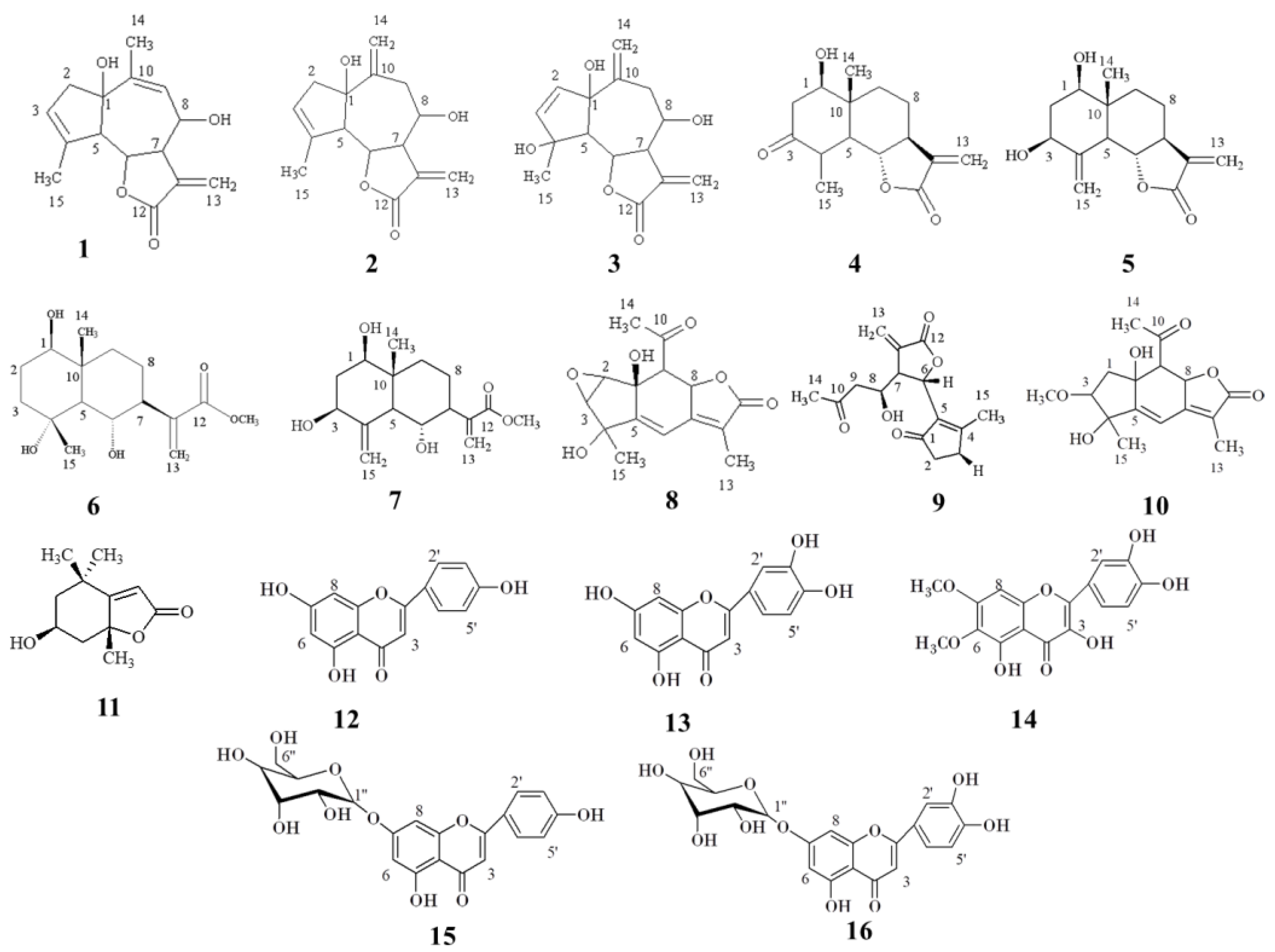
The column chromatography and semi-preparative HPLC of the methanol extract from the leaves and flowers of Achillea grandifolia led to the isolation of sixteen compounds. Eight compounds were isolated from the leaves (1, 2, 3, 4, 8, 9, 10, and 11) and twelve from the inflorences (1, 2, 3, 5, 6, 7, 9, 12, 13, 14, 15, and 16). The compounds were identified as rupicolin A (1), rupicolin B (2) [19], 4(4S,6aS,9R,9aS,9bS)-4,6a,9-Trihydroxy-9-methyl-3,6-dimethylene 3a,4,5,6,6a,9,9a, 9b-octahydro-3H-azuleno[4,5-b]furan-2-one (3) [20], artecalin (4) [21], ridentin B (5) [22], (1S,2S,4αR,5R,8R,8αS)-decahydro-1,5,8-trihydroxy-4α,8-dimethyl–methylene-2-naphthaleneacetic acid methyl ester (6) [23], 1β,3β,6α trihydroxycostic acid methyl ester (7) [24], acrifolide (8) [25], arteludovicinolide A (9) [26], lingustolide A (10) [21], loliolide (11) [27], apigenin (12) [28], luteolin (13) [29], eupatolitin (14) [30], apigenin-7-O-glucoside (15) [28], and luteolin 7-O-glucoside (16) [29]. The structures of the isolates are given in Figure 2 and were elucidated based on 1D and 2D NMR spectral analyses and by comparison of them with those found in the literature (see Tables S1–S16 and Figures S1–S52 in the supplementary materials).

3.2. Biological Activity

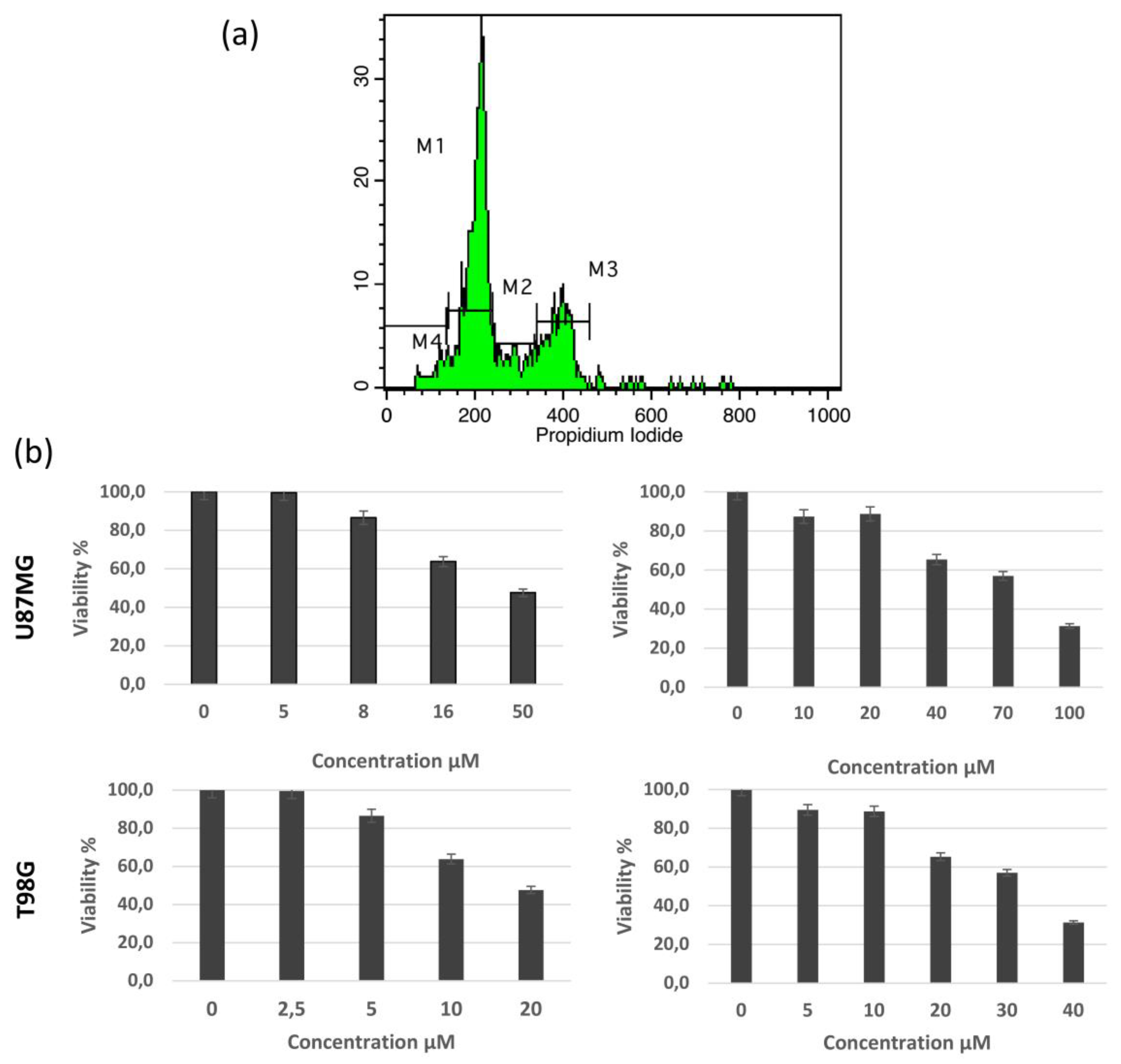
We next analyzed the possible effects of the main compounds (1) and (2) on glioblastoma. The quantification of IC50 values were based on the quantification of MTT fluorescence, which is proportional to viable cell population. The concentration leading to a decrease in 50% of the standard MTT colorimetric absorbance for each cell line corresponds to the IC50. The concentrations used and the respective results are presented in Figure 3, panel B. The IC50 value of reduced viability during the 48-h treatment for rupicolin A (1) and rupicolin B (2) was 38 μM and 64 μM for the U87MG cells and 15 μM and 26 μM for the T98G cells, respectively. Both rupicolin A and B induced a G2/M cell cycle arrest. In the U87MG cells, the G2/M fraction was increased from 20.87% to 54.40% and 37.00% and in the T98G cells from 25.73% to 50.67% and 42.93% following rupicolin A and B treatment, respectively (Table 1). An increase in the S phase was also observed, which was more prominent in the case of the U87 line. Rupicolin A (1) and rupicolin B (2) in higher concentrations induced cytotoxic phenomena, thereby slightly inducing the subG1 cell population (Table 2 and Figure 3). A flow cytometric analysis of the DNA content with a guide for cell cycle fractions quantification is presented in Figure 3.

4. Discussion

The genus Achillea is well known for its richness in flavonoids. Many studies reveal that apigenin, luteolin, and their derivatives are the main flavonoids in polar extracts of the aerial parts of the plants, since they are detected in many species such as Achillea millefolium [31], A. collina [32], A. sivasica Çelik & Akpulat [33]. These results are in accordance with the current research on the isolation of compounds 12, 13, 14, 15, and 16.
All compounds referred herein were isolated from A. grandifolia for the first time. Moreover, according to our knowledge, this is the first report regarding the occurrence of compounds 3, 6, 11, and 14 in any Achillea species. Further studies are needed to verify whether these compounds are species-specific or are to be found in other Achillea species as well.
Rupicolin A and B (1 and 2) have been isolated for the first time from the plant Artemisia tripartita Rydb. subsp. rupicola Beetle (Asteraceae) [19]. It has been suggested that the occurrence of rupicolins in many species of the genus Achillea can serve as a good chemotaxonomical marker [34]. In many cases, there is a co-occurrence of both rupicolin A and B (compounds 1 and 2) in different Achillea species such as in A. biebersteinii Kotschy [35,36], A. setacea Waldst. & Kit. [37], A. crithmifolia Waldst. & Kit. [38], A. clypeolata Sm. [39], A. chrysocoma Friv., A. coarctata [40], and A. clavennae [34].
The name of compound 9 (arteludovicinolide-A) has been given after Artemisia luboviciana Nutt. (Asteraceae) was found to be the plant from which this component was isolated for the very first time [24]. Until now, this compound had also been isolated from Achillea chrysocoma [40], A. coarctata [41], and A. millefolium [26].
Acrifolide is a 1,2-seco-guaianoIide hemiacetal that has only been reported, to date, in Achillea species (compound 8). The first report of this compound was in 2000 from the aerial parts of A. crithmifollia [42]. According to our knowledge, acrifolide has only been isolated from plants belonging to the genus Achillea such as A. chrysocoma [40] and A. pseudopectinata Janka [43].
Another seco-guaianolide derivative, namely, lingustolide-A (compound 10), has been isolated from the leaves of the examined plant. This is a very rare compound, and its first report originates from Achillea ligustica All. (Asteraceae) [21]. Later, it has been isolated from Achillea coarctata [41] and Artemisia argyi H.Lév. & Vaniot [44].
Previous studies [45] have firstly described NMR data about the compound 7, namely, 1β,3β,6α-Trihydroxycostic acid methyl ester, and the triacetate derivative of this natural product was isolated from Artemisia rutifolia Stephan ex Spreng. [46]. It should be noted, however, that this study provides the first report for this compound in a member of the genus Achillea i.e., A. grandifolia.
The compound 6, namely, (1S,2S,4aR,5R,8R,8aS)-decahydro-1,5,8-trihydroxy-4a,8-dimethyl-methylene-2-naphthaleneacetic acid methyl ester, is also a very rare natural product, and this study is only the second to isolate this compound from plants, while the first report belongs to Laurus nobilis L. [23].
The first isolation of the eudesmanolide ridentin B (5) was performed from Artemisia tripartita subsp. rupicola [19], but it has also been reported from other plants of the same genus [47] such as A. asiatica Nakai ex Pump. [22], as well as from Achillea coarctata and A. chrysocoma [40].
Artecalin (4) is an eudesmanolide which has also been reported in other plants of the genus Achillea, such as A. biebersteinii [35], A. coarctata and A. chrysocoma [40], and A. ligustica [21]. Artecalin is a widely distributed sesquiterpene lactone in members of the Asteraceae family, since this compound has been isolated also from Artemisia californica Less., A. tripartite subsp. rupicola [48], and Tanacetum santolina C. Winkl [49], among others.
Loliolide (11) is a monoterpenoid hydroxylactone that is abundant in Asteraceae such as Mantisalca salmantica (L.) Briq. & Cavill. [50], Artemisia integrifolia L. [51], Xanthium spinosum L. [27], and Gynura bicolor (Roxb. ex Willd.) DC. [52], among others. Moreover, loliolide has been detected in the non-polar extracts (volatile oil and hexane extract) of Achillea millefolium [53] and Achillea biebersteinii [54].
Both rupicolin A and B are guaianolides. According to the literature, these types of sesquiterpene lactones exhibit various medicinal properties such as anthelmintic, antimicrobial, high anti-tumor, antifeedant, root-growth, and germination-inhibiting actions [55]. This report acted as a motive, and, for this reason, the isolated compounds 1 and 2 were also subjected to an anti-proliferative assay on two different glioblastoma cell lines, U87MG and T98G. It is known that natural substances have anti-glioma activity [18,56]. However, this is the first research that examined the anti-proliferative activity of rupicolin B. Notably, a G2/M cell cycle arrest coupled with an induction of the S phase fraction, especially in the case of U87MG cells, is known to be associated with the anti-glioma activity of natural compounds such as deglucohellebrin [18], thereby supporting a similar mechanism of action that may be associated with an anti-inflammatory effect and the action of NF-kB transcription factor. Previous studies have revealed that rupicolin A had a protective effect on iodixanol-induced cytotoxicity in LLC-PK1 cells at concentrations of 10 µM, with a cell survival rate of 75.1 ± 1.9% [15]. The current research offered evidence that natural substances exert anti-cancer effects, thereby suggesting that rupicolin A and B are promising candidates for further research, given the continuous need for novel therapies for the second cause of human mortality [57]. To our knowledge, there are only few reports related to the anti-tumour activities of sesquiterpene lactones [58,59,60], thus also suggesting that guaianolides such as rupicolin A and rupicolin B may exhibit potential antitumor activities against various tumor cell lines. Our research confirmed this potential activity by illustrating the anti-proliferative and cytotoxic action of these compounds in glioma cell lines. To date, there is only one study concerning the anti-tumor activity of rupicolin A in cultured renal tubular cells (LLC-PK1) [15], while there is no study for rupicolin B; the results herein confirmed the anti-tumor activity of rupicolin B for the first time. Furthermore, the results of the current study expand our knowledge on the action of both rupicolin A and rupicolin B on the cell cycle distribution of cancer cells.

5. Conclusions

The phytochemical investigation of Achillea grandifolia proved that this species is a rich source of sesquiterpene lactones and flavonoids. We extracted and characterized sixteen known compounds from the inflorescences and leaves of A grandifolia. This is the first study leading to the isolation of non-volatile terpenoid compounds from the aerial part of this species of the genus Achillea. The most abundant compounds were rupicolin A (1) and B (2). These compounds were isolated from both the inflorescences and leaves of the species under study. Furthermore, these two main compounds of A. grandifolia were also examined for their anti-proliferative effect on U87MG and T98G glioblastoma cell lines. The preliminary results of the treatment of glioblastoma cell lines with A and B indicate that they have the potential to support novel anti-cancer therapies. Our current studies focus on the validation of the findings reported herein and on the investigation of the mechanism of their biological action with the aim to facilitate their establishment as possible anti-glioma agents.

Supplementary Materials

The following supporting information can be downloaded at: https://www.mdpi.com/article/10.3390/pharmaceutics15051383/s1, Supplementary material Note S1: Eluents used in the vacuum liquid chromatography (VLC) experiment; Figure S1: 1H-NMR spectrum of compound 1 (CDCl3, 500MHz); Figure S2: 1H-NMR full spectrum (A) and parts thereof (B) compound 1 (CD3OD, 500MHz); Figure S3: gDQCOSY spectrum of compound 1 (CD3OD, 500 MHz); Figure S4: 13C-NMR spectrum of compound 1 (CD3OD, 125MHz); Figure S5: gHSQCAD spectrum of compound 1 (CD3OD, 500 MHz); Figure S6: 1H-NMR full spectrum (A) and parts thereof (B) of compound 2 (CD3OD, 500MHz); Figure S7: gDQCOSY spectrum of compound 2 (CD3OD, 500 MHz); Figure S8: 1H-NMR full spectrum (A) and parts thereof (B) of compound 3 (CD3OD, 500MHz); Figure S9: gDQCOSY spectrum of compound 3 (CD3OD, 500 MHz); Figure S10: 13C-NMR spectrum of compound 3 (CD3OD, 125MHz); Figure S11: gHSQCAD spectrum of compound 3 (CD3OD, 500 MHz); Figure S12: gHMBCAD spectrum of compound 3 (CD3OD, 500 MHz); Figure S13: 1H-NMR full spectrum (A) and parts thereof (B) of compound 4 (CD3OD, 500MHz); Figure S14: 13C-NMR spectrum of compound 4 (CD3OD, 125MHz); Figure S15: gDQCOSY spectrum of compound 4 (CD3OD, 500 MHz); Figure S16: gHSQCAD spectrum of compound 4 (CD3OD, 500 MHz); Figure S17: gHMBCAD spectrum of compound 4 (CD3OD, 500 MHz); Figure S18: 1H-NMR full spectrum (A) and parts thereof (B) of compound 5 (CD3OD, 500MHz); Figure S19: gDQCOSY spectrum of compound 5 (CD3OD, 500 MHz); Figure S20: 13C-NMR spectrum of compound 5 (CD3OD, 125MHz); Figure S21: 1H-NMR full spectrum (A) and parts thereof (B) of compound 6 (CD3OD, 500MHz); Figure S22: gDQCOSY spectrum of compound 6 (CD3OD, 500 MHz); Figure S23: 13C-NMR spectrum of compound 6 (CD3OD, 125MHz); Figure S24: gHSQCAD spectrum of compound 6 (CD3OD, 500 MHz); Figure S25: gHMBCAD spectrum of compound 6 (CD3OD, 500 MHz); Figure S26: 1H-NMR full spectrum (A) and parts thereof (B) of compound 7 (CD3OD, 500MHz); Figure S27: gDQCOSY spectrum of compound 7 (CD3OD, 500 MHz); Figure S28: 13C-NMR spectrum of compound 7 (CD3OD, 125MHz); Figure S29: gHSQCAD spectrum of compound 7 (CD3OD, 500 MHz); Figure S30: gHMBCAD spectrum of compound 7 (CD3OD, 500 MHz); Figure S31: 1H-NMR spectrum of compound 8 (CD3OD, 500MHz); Figure S32: gDQCOSY spectrum of compound 8 (CD3OD, 500 MHz); Figure S33: 13C-NMR spectrum of compound 8 (CD3OD, 125MHz); Figure S34: gHSQCAD spectrum of compound 8 (CD3OD, 500 MHz); Figure S35: gHMBCAD spectrum of compound 8 (CD3OD, 500 MHz); Figure S36: 1H-NMR full spectrum (A) and parts thereof (B) of compound 9 (CD3OD, 500MHz); Figure S37: gDQCOSY spectrum of compound 9 (CD3OD, 500 MHz); Figure S38: 13C-NMR spectrum of compound 9 (CD3OD, 125MHz); Figure S39: gHSQCAD spectrum of compound 9 (CD3OD, 500 MHz); Figure S40: 1H-NMR spectrum of compound 10 (CD3OD, 500MHz); Figure S41: gDQCOSY spectrum of compound 10 (CD3OD, 500 MHz); Figure S42: 1H-NMR spectrum of compound 11 (CD3OD, 500MHz); Figure S43: gDQCOSY spectrum of compound 11 (CD3OD, 500 MHz); Figure S44: 13C-NMR spectrum of compound 11 (CD3OD, 125MHz); Figure S45: gHSQCAD spectrum of compound 11 (CD3OD, 500 MHz); Figure S46: gHMBCAD spectrum of compound 11 (CD3OD, 500 MHz); Figure S47: 1H-NMR spectrum of compound 12 (CD3OD, 500MHz); Figure S48: 1H-NMR spectrum of compound 13 (CD3OD, 500MHz); Figure S49: 1H-NMR spectrum of compound 14 (CD3OD, 500MHz); Figure S50: 1H-NMR spectrum of compound 15 (CD3OD, 500MHz); Figure S51: gDQCOSY spectrum of compound 15 (CD3OD, 500 MHz); Figure S52: 1H-NMR spectrum of compound 16 (CD3OD, 500MHz); Table S1: 1H and 13C NMR of compound 1 (CD3OD, 500 MHz); Table S2: 1H and 13C NMR of compound 2 (CD3OD, 500 MHz); Table S3: 1H and 13C NMR of compound 3 (CD3OD, 500 MHz); Table S4: 1H and 13C NMR of compound 4 (CD3OD, 500 MHz); Table S5: 1H and 13C NMR of compound 5 (CD3OD, 500 MHz); Table S6: 1H and 13C NMR of compound 6 (CD3OD, 500 MHz); Table S7: 1H and 13C NMR of compound 7 (CD3OD, 500 MHz); Table S8: 1H and 13C NMR of compound 8 (CD3OD, 500 MHz); Table S9: 1H and 13C NMR of compound 9 (CD3OD, 500 MHz); Table S10: 1H and 13C NMR of compound 10 (CD3OD, 500 MHz); Table S11: 1H and 13C NMR of compound 11 (CD3OD, 500 MHz); Table S12: 1H NMR of compound 12 (CD3OD, 500 MHz); Table S13: 1H NMR of compound 13 (CD3OD, 500 MHz); Table S14: 1H NMR of compound 14 (CD3OD, 500 MHz); Table S15: 1H NMR of compound 15 (CD3OD, 500 MHz); Table S16: 1H NMR of compound 16 (CD3OD, 500 MHz).

Author Contributions

Conceptualization, D.L., O.S.T., G.A., and N.K.; methodology, O.S.T., G.S.M., G.A., E.V., D.L. and N.K.; validation, G.S.M., G.A. and D.L.; formal analysis, O.S.T., C.G., C.P., M.N., E.V. and R.I.; investigation, O.S.T., C.G., C.P., M.N., E.V., G.S.M., R.I., G.A., D.L. and N.K.; resources, D.L., G.A., G.S.M. and N.K.; data curation, O.S.T., N.K., G.S.M., G.A. and D.L.; writing—original draft preparation, O.S.T., D.L. and N.K; writing—review and editing, O.S.T., N.K., G.S.M., G.A. and D.L.; visualization, O.S.T., N.K., E.V., G.S.M. and G.A.; supervision, D.L., G.A. and G.S.M.; project administration, D.L., G.S.M., G.A. and N.K. All authors have read and agreed to the published version of the manuscript.”

Funding

We acknowledge support of this work by the project “Upgrading the Plant Capital (PlantUp)” (MIS 5002803), which was implemented under the action “Reinforcement of the Research and Innovation Infrastructure”, which was funded by the operational program “Competitiveness, Entrepreneurship and Innovation” (NSRF 2014-2020) and co-financed by Greece and the European Union (European Regional Development Fund).

Institutional Review Board Statement

Not applicable.

Informed Consent Statement

Not applicable.

Data Availability Statement

All data referred to or generated in this study are included in tables or figures and are available upon request.

Acknowledgments

The authors would like to thank the anonymous reviewers for their valuable comments and suggestions, as well as Efthimia Antonopoulou for assistance during field work.

Conflicts of Interest

The authors declare no conflict of interest.

References

  1. Radulović, N.; Zlatković, B.; Palić, R.; Stojanović, G. Chemotaxonomic significance of the Balkan Achillea volatiles. Nat. Prod. Commun. 2007, 2, 453–474. [Google Scholar] [CrossRef]
  2. Stanković, N.; Mihajilov-Krstev, T.; Zlatković, B.; Stankov-Jovanović, V.; Mitić, V.; Jović, J.; Čomić, L.; Kocić, B.; Bernstein, N. Antibacterial and antioxidant activity of traditional medicinal plants from the Balkan peninsula. NJAS Wagen. J. Life Sc. 2016, 78, 21–28. [Google Scholar] [CrossRef]
  3. Mohammadhosseini, M.; Sarker, S.D.; Akbarzadeh, A. Chemical composition of the essential oils and extracts of Achillea species and their biological activities: A review. J. Ethnopharmacol. 2017, 199, 257–315. [Google Scholar] [CrossRef] [PubMed]
  4. Bali, E.B.; Açık, L.; Elçi, P.; Sarper, M.; Avcu, F.; Vural, M. In vitro anti-oxidant, cytotoxic and pro-apoptotic effects of Achillea teretifolia Willd. extracts on human prostate cancer cell lines. Pharmacogn. Mag. 2015, 11 (Suppl. S2), S308–S315. [Google Scholar] [CrossRef] [PubMed]
  5. Choucry, M.A. Chemical composition and anticancer activity of Achillea fragrantissima (Forssk.) Sch. Bip. (Asteraceae) essential oil from Egypt. J. Pharmacogn. Phytother. 2017, 9, 1–5. [Google Scholar] [CrossRef]
  6. Mouhid, L.; Gómez de Cedrón, M.; Vargas, T.; García-Carrascosa, E.; Herranz, N.; García-Risco, M.; Reglero, G.; Fornari, T.; Ramírez de Molina, A. Identification of antitumoral agents against human pancreatic cancer cells from Asteraceae and Lamiaceae plant extracts. BMC Complement. Altern. Med. 2018, 18, 254. [Google Scholar] [CrossRef]
  7. Abou Baker, D.H. Achillea millefolium L. ethyl acetate fraction induces apoptosis and cell cycle arrest in human cervical cancer (HeLa) cells. Ann. Agric. Sci. 2020, 65, 42–48. [Google Scholar] [CrossRef]
  8. Papakosta, K.; Grafakou, M.E.; Barda, C.; Kostopoulos, I.V.; Tsitsilonis, O.; Skaltsa, H. Cytotoxicity and anti-cancer activity of the genus Achillea L. Curr. Med. Chem. 2020, 27, 6910–6925. [Google Scholar] [CrossRef]
  9. Taşkın, D.; Alkaya, D.B.; Dölen, E. Evaluation of antioxidant capacity and analysis of major phenolic compounds in Achillea grandifolia by HPLC-DAD with Q-TOF LC/MS Confirmation. Chiang Mai J. Sci. 2018, 45, 287–298. Available online: https://www.thaiscience.info/Journals/Article/CMJS/10989292.pdf (accessed on 29 April 2023).
  10. Tsiftsoglou, O.S.; Atskakani, M.-E.; Krigas, N.; Stefanakis, M.K.; Gounaris, C.; Hadjipavlou-Litina, D.; Lazari, D. Exploring the medicinal potential of Achillea grandifolia in Greek wild-growing populations: Characterization of volatile compounds, anti-inflammatory and antioxidant activities of leaves and inflorescences. Plants 2023, 12, 613. [Google Scholar] [CrossRef]
  11. Özek, G. Phytochemical characterization of Achillea grandifolia Friv. essential oil and its potent against oxidative damage, acetylcholinesterase and α-amylase. Eskişehir Tech. Univ. J. Sci. Tech. Appl. Sci. Eng. 2018, 19, 671–684. [Google Scholar] [CrossRef]
  12. Wollenweber, E.; Valant-Vetxhera, K.M.; Ivancheva, S.; Kumanov, B. Flavonoid aglycones from the leaf surfaces of some Achlllea species. Phytochemistry 1987, 26, 181–182. [Google Scholar] [CrossRef]
  13. Greger, H.; Grenz, M.; Bohlman, F. Piperidides and other amides from Achillea species. Phytochemistry 1982, 21, 1071–1074. [Google Scholar] [CrossRef]
  14. Saeidnia, S.; Gohari, A.; Mokhber-Dezfuli, N.; Kiuchi, F. A review on phytochemistry and medicinal properties of the genus Achillea. DARU J. Pharm. Sci. 2011, 19, 173–186. Available online: https://www.ncbi.nlm.nih.gov/pmc/articles/PMC3232110 (accessed on 29 April 2023).
  15. Lee, D.; Kim, K.O.; Lee, D.; Kang, K.S. Anti-Apoptotic and antioxidant effects of 3-Epi-Iso-Seco-Tanapartholide isolated from Artemisia argyi against iodixanol-induced kidney epithelial cell death. Biomolecules 2020, 10, 867. [Google Scholar] [CrossRef]
  16. Skaltsa, H.; Lazari, D.; Georgiadou, E.; Kakavas, S.; Constantinidis, T. Sesquiterpene lactones from Centaurea Species: C. thessala subsp. drakiensis and C. attica subsp. attica. Planta Med. 1999, 65, 393. [Google Scholar] [CrossRef]
  17. Alexiou, G.A.; Lazari, D.; Markopoulos, G.; Vartholomatos, E.; Hodaj, E.; Galani, V.; Kyritsis, A.P. Moschamine inhibits proliferation of glioblastoma cells via cell cycle arrest and apoptosis. Tumor Biol. 2017, 39, 1–12. [Google Scholar] [CrossRef]
  18. Vartholomatos, E.; Alexiou, G.A.; Markopoulos, G.S.; Lazari, D.; Tsiftsoglou, O.; Chousidis, I.; Leonardos, I.; Kyritsis, A.P. Deglucohellebrin: A Potent Agent for Glioblastoma Treatment. Anticancer Agents Med. Chem. 2020, 20, 103–110. [Google Scholar] [CrossRef]
  19. Irwin, M.A.; Geissman, T.A. Ridentin-β: An eudesmanolide from Artemisia tripartita ssp. rupicola. Phytochemistry 1973, 12, 871–873. [Google Scholar] [CrossRef]
  20. Jakupovic, J.; Klemeyer, H.; Bohlmann, F.; Graven, E.H. Glaucolides and guaianolides from Artemisia afra. Phytochemistry 1988, 27, 1129–1133. [Google Scholar] [CrossRef]
  21. Ahmed, A.A.; Gáti, T.; Taha, A.H.; Ali, A.T.; Tzakou, O.A.; Couladis, M.A.; Mabry, T.J.; Tóth, G. Ligustolide A and B, two novel sesquiterpenes with rare skeletons and three 1,10-seco-guaianolide derivatives from Achillea ligustica. Tetrahedron 2003, 59, 3729–3735. [Google Scholar] [CrossRef]
  22. Hajdú, Z.; Hohmann, J.; Forgo, P.; Máthé, I.; Molnár, J.; Zupkó, I. Antiproliferative activity of Artemisia asiatica extract and its constituents on human tumor cell lines. Planta Med. 2014, 80, 1692–1697. [Google Scholar] [CrossRef] [PubMed]
  23. Julianti, E.; Jang, K.H.; Lee, S.; Lee, D.; Mar, W.; Oh, K.B.; Shin, J. Sesquiterpenes from the leaves of Laurus nobilis L. Phytochemistry 2012, 80, 70–76. [Google Scholar] [CrossRef] [PubMed]
  24. Jakupovic, J.; Tan, R.X.; Bohlmann, F.; Boldt, P.E.; Jia, Z.J. Sesquiterpene lactones from Artemisia ludoviciana. Phytochemistry 1991, 30, 1573–1577. [Google Scholar] [CrossRef]
  25. Todorova, M.N.; Vogler, B.; Tsankova, E.T. Acrifolide, a novel sesquiterpene lactone from Achillea crithmifolia. Nat. Prod. Lett. 2000, 14, 463–468. [Google Scholar] [CrossRef]
  26. Li, Y.; Ni, Z.Y.; Zhu, M.C.; Zhang, K.; Wu, Y.B.; Dong, M.; Shi, Q.W.; Huo, C.H.; Sauriol, F.; Kiyota, H.; et al. New 1, 10-Seco-guaianolides from the flowers of Achillea millefolium. Z. Naturforsch. B 2012, 67, 438–446. [Google Scholar] [CrossRef]
  27. Yuan, Z.; Zheng, X.; Zhao, Y.; Liu, Y.; Zhou, S.; Wei, C.; Hu, Y.; Shao, H. Phytotoxic compounds isolated from leaves of the invasive weed Xanthium spinosum. Molecules 2018, 23, 2840. [Google Scholar] [CrossRef]
  28. Ersöz, T.; Harput, Ü.Ş.; Saracoğlu, I.; Çaliş, I. Phenolic compounds from Scutellaria pontica. Turk. J. Chem. 2002, 26, 581–588. Available online: https://journals.tubitak.gov.tr/chem/vol26/iss4/16 (accessed on 29 April 2023).
  29. Patora, J.; Klimek, B. Flavonoids from lemon balm (Melissa officinalis L., Lamiaceae). Acta Pol. Pharm. 2002, 59, 139–143. Available online: https://europepmc.org/article/med/12365606 (accessed on 29 April 2023).
  30. Hussien, T.A.; El-Toumy, S.A.; Hassan, H.M.; Hetta, M.H. Cytotoxic and antioxidant activities of secondary metabolites from Pulicaria undulata. Int. J. Pharm. Pharm. Sci. 2016, 8, 150–155. [Google Scholar] [CrossRef]
  31. Benetis, R.; Radušienė, J.; Januli, V. Variability of phenolic compounds in flowers of Achillea millefolium wild populations in Lithuania. Medicina 2008, 44, 775–781. [Google Scholar] [CrossRef]
  32. Karlová, K. Accumulation of flavonoid compounds in flowering shoots of Achillea collina Becker ex. Rchb. alba during flower development. Hortic. Sci. 2006, 33, 158–162. [Google Scholar] [CrossRef]
  33. Haliloglu, Y.; Ozek, T.; Tekin, M.; Goger, F.; Baser, K.H.C.; Ozek, G. Phytochemicals, antioxidant, and antityrosinase activities of Achillea sivasica Çelik and Akpulat. Int. J. Food Prop. 2017, 20, S693–S706. [Google Scholar] [CrossRef]
  34. Stojanović, G.; Radulović, N.; Hashimoto, T.; Palić, R. In vitro antimicrobial activity of extracts of four Achillea species: The composition of Achillea clavennae L. (Asteraceae) extract. J. Ethnopharmacol. 2005, 101, 185–190. [Google Scholar] [CrossRef]
  35. Yusupov, M.I.; Kasymov, S.Z.; Abdullaev, N.D.; Sidyakin, G.P.; Yagudaev, M.R. A new lactone, isoridentin, from Achillea biebersteinii. Chem. Nat. Compd. 1977, 13, 674–676. [Google Scholar] [CrossRef]
  36. Mohamed, A.E.H.H.; Mohamed, N.S.; Hamed, A.R.; Hegazy, M.E.F. Anti-inflammatory activity of highly oxygenated terpenoids from Achillea biebersteinii Afan. Z. Naturforsch. 2016, 71, 429–432. [Google Scholar] [CrossRef]
  37. Zitterl-Eglseer, K.; Jurenitsch, J.; Korhammer, S.; Haslinger, E.; Sosa, S.; Della Loggia, R.; Kubelka, W.; Franz, C. Sesquiterpene lactones of Achillea setacea with antiphlogistic activity. Planta Med. 1991, 57, 444–446. [Google Scholar] [CrossRef]
  38. Milosavljević, S.; Aljančić, I.; Macura, S.; Milinković, D.; Stefanović, M. Sesquiterpene lactones from Achillea crithmifolia. Phytochemistry 1991, 30, 3464–3466. [Google Scholar] [CrossRef]
  39. Todorova, M.; Krasteva, M.; Markova, M.; Tsankova, E.; Taskova, R.; Peev, D.R. Terpenoids from Achillea clypeolata. Phytochemistry 1998, 49, 2371–2374. [Google Scholar] [CrossRef]
  40. Todorova, M.; Tsankova, E. Sesquiterpene lactones from Achillea chrysocoma and Achillea coarctata. Z. Naturforsch. C 2001, 56, 957–960. [Google Scholar] [CrossRef]
  41. Hegazy, M.E.F.; Abdel-Lateff, A.; Gamal-Eldeen, A.M.; Turky, F.; Hirata, T.; Paré, P.W.; Karchesy, J.; Kamel, M.S.; Ahmed, A.A. Anti-inflammatory activity of new guaiane acid derivatives from Achillea coarctata. Nat. Prod. Commun. 2008, 3, 851–856. [Google Scholar] [CrossRef]
  42. Todorova, M.N.; Markova, M.; Tsankova, E.T. Crithmifolide: A sesquiterpene lactone from Achillea crithmifolia. Phytochemistry 1998, 49, 2429–2432. [Google Scholar] [CrossRef]
  43. Trifunović, S.; Aljančić, Ι.; Vajs, V.; Macura, S.; Milosavljević, S. Sesquiterpene lactones and flavonoids of Achillea depressa. Biochem. Syst. Ecol. 2005, 33, 317–322. [Google Scholar] [CrossRef]
  44. Zhang, S.J.; Ma, Y.L.; Wang, J.L.; Li, J.; Zhao, M.; Bai, L.M. Chemical constituents of Artemisia argyi. Zhong Cao Yao 2019, 50, 1906–1914. Available online: https://pesquisa.bvsalud.org/portal/resource/pt/wpr-851198 (accessed on 29 April 2023).
  45. Zhang, L.B.; Lv, J.L. Sesquiterpenoids from Artemisia argyi and their COXs inhibitory activities. Fitoterapia 2019, 139, 104372. [Google Scholar] [CrossRef]
  46. Tan, R.X.; Jia, Z.J.; Jakupovic, J.; Bohlmann, F.; Huneck, S. Sesquiterpene lactones from Artemisia rutifolia. Phytochemistry 1991, 30, 3033–3035. [Google Scholar] [CrossRef]
  47. Kelsey, R.G.; Shafizadeh, F. Sesquiterpene lactones and systematics of the genus Artemisia. Phytochemistry 1979, 18, 1591–1611. [Google Scholar] [CrossRef]
  48. Geissman, T.A.; Griffin, T.S.; Irwin, M.A. Sesquiterpene lactones of Artemisia. Artecalin from A. californica and A. tripartita ssp. rupicola. Phytochemistry 1969, 8, 1297–1300. [Google Scholar] [CrossRef]
  49. Abduazimov, B.K.; Yunusov, A.I.; Sidyakin, G.P. Sesquiterpene lactones of Tanacetum santolina. Chem. Nat. Compd. 1980, 16, 452–454. [Google Scholar] [CrossRef]
  50. Fernández, I.; Pedro, J.R.; Vidal, R. Norisoprenoids from Centaurea aspera and C. salmantica. Phytochemistry 1993, 34, 733–736. [Google Scholar] [CrossRef]
  51. Wang, J.; Yi, Z.; Ma, Y.; Zhao, M.; Li, J.; Zhang, S. Study on chemical constituents of Artemisia integrifolia. Zhong Cao Yao 2016, 47, 2241–2245. [Google Scholar] [CrossRef]
  52. Chen, J.; Mangelinckx, S.; Adams, A.; Li, W.; Wang, Z.; De Kimpe, N. Chemical constituents from the aerial parts of Gynura bicolor. Nat. Prod. Commun. 2012, 7, 1563–1564. Available online: https://journals.sagepub.com/doi/pdf/10.1177/1934578X1200701203 (accessed on 29 April 2023). [CrossRef]
  53. Ni, Z.; Wu, Y.; Zhang, K.; Dong, M.; Sauriol, F.; Huo, C.; Gu, Y.; Shi, Q. A monoterpene and two sesquiterpenoids from the flowers of Achillea millefolium. Chem. Nat. Compd. 2013, 49, 450–453. [Google Scholar] [CrossRef]
  54. Akkol, E.; Küpeli Koca, U.; Pesin, I.; Yilmazer, D. Evaluation of the wound healing potential of Achillea biebersteinii Afan. (Asteraceae) by in vivo excision and incision Models. Evid. Based Complementary Altern. Med. 2011, 2011, 474026. [Google Scholar] [CrossRef]
  55. Dhyani, P.; Sati, P.; Sharma, E.; Attri, D.C.; Bahukhandi, A.; Tynybekov, B.; Szopa, A.; Sharifi-Rad, J.; Calina, D.; Suleria, H.A.R.; et al. Sesquiterpenoid lactones as potential anti-cancer agents: An update on molecular mechanisms and recent studies. Cancer Cell Int. 2022, 22, 305. [Google Scholar] [CrossRef]
  56. Vartholomatos, E.; Mantziou, S.; Alexiou, G.A.; Lazari, D.; Sioka, C.; Kyritsis, A.; Markopoulos, G.S. An NF-κB- and Therapy-Related Regulatory Network in Glioma: A Potential Mechanism of Action for Natural Antiglioma Agents. Biomedicines 2022, 10, 935. [Google Scholar] [CrossRef]
  57. Sung, H.; Ferlay, J.; Siegel, R.L.; Laversanne, M.; Soerjomataram, I.; Jemal, A.; Bray, F. Global cancer statistics 2020: GLOBOCAN estimates of incidence and mortality worldwide for 36 cancers in 185 countries. CA Cancer J. Clin. 2021, 71, 209–249. [Google Scholar] [CrossRef]
  58. Li, Y.; Ni, Z.; Zhu, M.; Dong, M.; Wang, S.; Shi, Q.; Zhang, M.; Wang, Y.; Huo, C.; Kiyota, H.; et al. Antitumour activities of sesquiterpene lactones from Inula helenium and Inula japonica. Z. Naturforsch. C. 2012, 67, 375–380. [Google Scholar] [CrossRef]
  59. Surowiak, A.K.; Balcerzak, L.; Lochyński, S.; Strub, D.J. Biological activity of selected natural and synthetic terpenoid lactones. Int. J. Mol. Sci. 2021, 22, 5036. [Google Scholar] [CrossRef]
  60. Tohme, R.; Al Aaraj, L.; Ghaddar, T.; Gali-Muhtasib, H.; Saliba, N.A.; Darwiche, N. Differential growth inhibitory effects of highly oxygenated guaianolides isolated from the Middle Eastern indigenous plant Achillea falcata in HCT-116 colorectal cancer cells. Molecules 2013, 18, 8275–8288. [Google Scholar] [CrossRef]
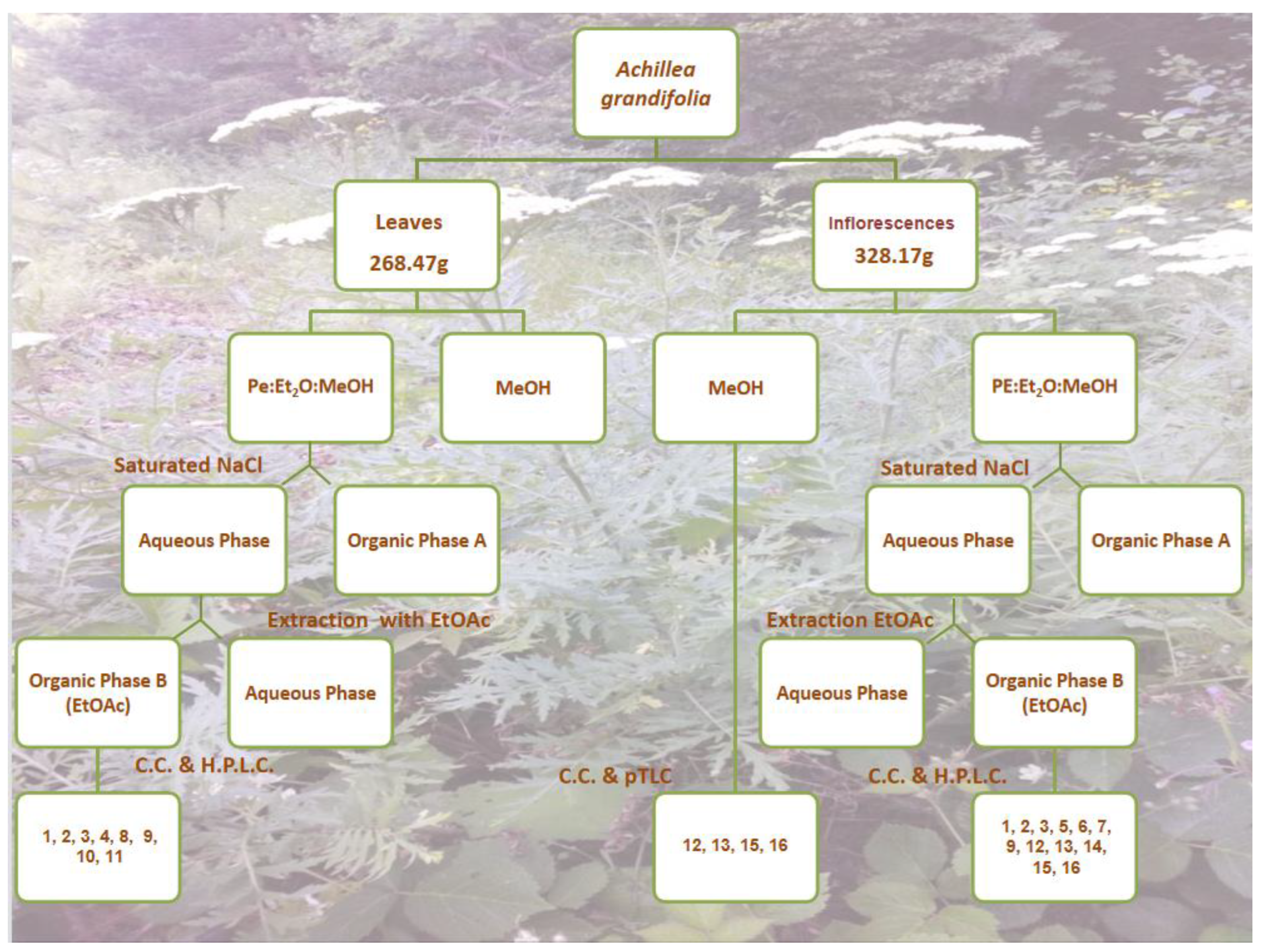
Figure 1. Schematic process of isolation of secondary metabolites from Achillea grandifolia with individual numbers referring to the isolated compounds. (L): Leaves; (I): Inflorescences.
Pharmaceutics 15 01383 g001
Figure 2. Isolated compounds from the leaves and inflorescences of Achillea grandifolia.
Pharmaceutics 15 01383 g002
Figure 3. (a): Cell-cycle distribution assessed by flow cytometry in U87 and T98 glioblastoma cells. An indicative fluorescence distribution following PI staining is depicted (T98G cells treated with rupicolin B). The markers M1, M2, M3, and M4 correspond to the fraction of cells in G0, G1, S, and G2/M cell-cycle phases and subG1 cells, respectively. (b): Effect of different concentrations of rupicoline A (A) and rupicoline Β (B) on U87 and T98 cells. Different concentrations are presented in X-axis, while the difference in viability based on MTT colorimetric absorbance is presented in Y-axis.
Pharmaceutics 15 01383 g003
Table 1. Different compounds isolated from fractions of leaves (L) and inflorescences (I) of Achillea grandifolia.
Isolated in FractionsCompound NumberCompound Name
DI(L), EG(F), EO(I), FF(I), FG(I)1Rupicolin A
DI(L), EG(I), EO(L), FL(L), FG(I)2Rupicolin B
DI(L), GG(I), GQ(L)34S,6aS,9R,9aS,9bS)-4,6a,9-Trihydroxy-9-methyl-3,6-dimethylene-3a,4,5,6,6a,9,9a,9b-octahydro-3H-azuleno[4,5-b]furan-2-one)
EL(L)4Artecalin
FH(I)5Ridentin B
GD(I)6(1S,2S,4αR,5R,8R,8αS)-decahydro-1,5,8-trihydroxy-4α,8-dimethyl–methylene-2-naphthaleneacetic acid methyl ester
GG(I)71β,3β,6α-Trihydroxycostic acid methyl ester
FH(L)8Acrifolide
GH(L)9Arteludovicinolide A
FF(L)10Lingustolide A
EI(L)11Loliolide
DLB(I), EG(I), EH(L)12Apigenin
EG(I), EL(L), FIH(I)13Luteolin
EFK(F)14Eupatolitin
EL(L)15Apigenin 7-O-glucopyranoside
EFF(L)16Luteolin 7-O-glucopyranoside
Table 2. Cell-cycle distribution assessed by flow cytometry in U87 and T98 glioblastoma cells. In each case, IC50 values were used for incubation.
Cell LineU87MGT98G
Cell Cycle
Phases
ControlRupicolin ARupicolin BControlRupicolin ARupicolin B
G164.7315.8723.6058.2719.2732.60
S9.2024.2737.6013.6724.1317.20
G2/M20.8754.4037.0025.7350.6742.93
subG13.133.203.400.802.404.07
Disclaimer/Publisher’s Note: The statements, opinions and data contained in all publications are solely those of the individual author(s) and contributor(s) and not of MDPI and/or the editor(s). MDPI and/or the editor(s) disclaim responsibility for any injury to people or property resulting from any ideas, methods, instructions or products referred to in the content.