Article

A Wall Fragment of Cutibacterium acnes Preserves Junctional Integrity Altered by Staphylococcus aureus in an Ex Vivo Porcine Skin Model

1
Department of Medicine and Health Science “V. Tiberio”, Università degli Studi del Molise, 8600 Campobasso, Italy
2
Department of Agricultural, Environmental and Food Sciences (DiAAA), Università degli Studi del Molise, 8600 Campobasso, Italy
*
Author to whom correspondence should be addressed.
Pharmaceutics 2023, 15(4), 1224; https://doi.org/10.3390/pharmaceutics15041224
Submission received: 27 February 2023 / Revised: 7 April 2023 / Accepted: 7 April 2023 / Published: 12 April 2023

Abstract

:
(1) Background alteration of the skin microbiota, dysbiosis, causes skin barrier impairment resulting in disease development. Staphylococcus aureus, the main pathogen associated with dysbiosis, secretes several virulence factors, including α-toxin that damages tight junctions and compromises the integrity of the skin barrier. The use of members of the resident microbiota to restore the skin barrier, bacteriotherapy, represents a safe treatment for skin conditions among innovative options. The aim of this study is the evaluation of a wall fragment derived from a patented strain of Cutibacterium acnes DSM28251 (c40) alone and conjugated to a mucopolysaccharide carrier (HAc40) in counteracting S. aureus pathogenic action on two tight junction proteins (Claudin-1 and ZO-1) in an ex vivo porcine skin infection model. Methods: skin biopsies were infected with live S. aureus strains ATCC29213 and DSM20491. Tissue was pre-incubated or co-incubated with c40 and HAc40. (3) Results: c40 and HAc40 prevent and counteract Claudin-1 and Zo-1 damage (4) Conclusions: c40 and the functional ingredient HAc40 represent a potential non-pharmacological treatment of skin diseases associated with cutaneous dysbiosis of S. aureus. These findings offer numerous avenues for new research.

1. Introduction

The skin’s primary function is to provide a physical barrier that protects the underlying tissues from the external ambient. Due to a dense cellular network (the corneocytes) and a rich lipid matrix, the stratum corneum, the most superficial portion of the epidermis, guarantees this function [1].
Crucial components of skin defensive mechanisms are the tight junctions (TJs). They are barrier proteins that counteract pathogens, chemicals, and other external substances from body damage [2]. Several TJ proteins with diverse epidermal localisation patterns have been identified in human, porcine, murine, and canine skin [3,4,5,6,7]. For example, occludin (Ocln) or cingulin is a cell–cell junction restricted to the stratum granulosum. ZO-1 and Claudin-4 are found in the stratum granulosum and the upper stratum spinosum, whereas Claudin-1 is found in all the layers, including the stratum corneum [8].
A factor that critically modulates the skin’s barrier function and homeostasis is the colonising microbiota [9]. Under physiological conditions, the skin microbiota has a mutualistic relationship with the host. It contributes to cutaneous homeostasis by inflammatory response modulation and protection against disease-causing pathogens, regulating the skin’s pH balance. Moreover, this microbiota is also involved in producing vitamins, enzymes, and other biochemicals essential for skin health [10,11,12].
Among the most studied species belonging to the skin microbiota, Malassezia, Cutibacterium, Staphylococcus, and Corynebacterium spp. contribute to skin homeostasis through several mechanisms [13,14]. Indeed, their secretion of protease and lipase enzymes and free fatty acids are involved in skin lipidic-film surface breakdown [15]. Moreover, bacteriocin production prevents skin pathogen colonisation [16]. Furthermore, the quorum sensing importance in moulds and yeasts inhibition by indole-based mediators for Malassezia and other commensal bacteria has been demonstrated.
In this scenario, an imbalance of these delicate relationships might influence the skin ecosystem, causing dysbiosis [12]. Dysbiosis is often driven by pathogenic microorganisms’ overgrowth and/or a decrease in the variety of microbiota composition [17], representing one of the main triggering factors in numerous skin disease pathogenesis [12]. Indeed, evidence shows that skin microbiota diversity is reduced in individuals with certain skin conditions [18]. For example, in atopic dermatitis patients, it has often been observed that the pathogen Staphylococcus aureus (S. aureus) increases in correspondence with a decrease of certain skin commensals levels, such as S. epidermidis, S. hominis, and C. acnes [19].
S. aureus is an opportunistic pathogen that, in eubiosis conditions, colonises the skin of healthy individuals. However, this pathogen can proliferate in inflammatory skin conditions, releasing virulence factors that promote bacterial adhesion and toxin synthesis, directly damaging tissue [20]. Besides being a skin commensal, 30% of individuals harbour S. aureus in the nose. Methicillin-resistant S. aureus (MRSA), which accounts for most of the antibiotic-resistant bacteria’s invasive infections, is carried by 2% of the population. Many severe and fatal diseases, such as bacteremia, endocarditis, pneumonia, osteomyelitis, skin infections, and sepsis, are sustained by S. aureus [21].
The principal S. aureus toxin involved in skin pathologies is the α-toxin, which causes the formation of heptameric pores on target cells, epithelial and endothelial breaches by adherens junctions breaking, and cytoskeleton impairment. In addition, δ-toxin or similar cytolytic peptides called phenol-soluble modulins (PSMs) may also activate mast cells. Lastly, exfoliative toxins can induce damage to desmosomes by cleaving desmoglein 1, leading to Staphylococcus scalded skin syndrome [22].
The lack of curative treatment for skin conditions (i.e., dysbiosis cutaneous disorders) has led to an increased interest in alternative and complementary therapies [23].
Recent insights into skin commensals have been applied to a new therapeutic strategy using live bacteria: bacteriotherapy. This therapy may hold the key to developing highly compliant treatments and medical cures for patients [24]. Therefore, as mentioned above, skin microorganisms work with their metabolites to maintain skin stability and interfere with pathogenic bacteria growth regulating the microbiota balance [25,26,27].
This therapeutic approach’s safety, effectiveness, and sustainability need to be further investigated. In particular, the administration of specific bacterial strains has been investigated as an exclusionary treatment strategy against pathogens associated with chronic skin disorders [28]. Besides using probiotic strains, the possibility of restoring skin eubiosis by applying viable or lysate-heat-killed or derived substances from commensal strains is gaining ground in treating skin disorders [29]. These findings encourage the potential use of specific cutaneous strains in restoring skin microbiome eubiosis.
In addition to bacteriotherapy, other therapeutic strategies targeting α-toxin have already been investigated [30,31]. One antivirulence strategy for counteracting toxins is the use of antibodies. Passive immunisation would provide affected patients with immediate treatment. In contrast, active immunisation would require several boosters and a long period to achieve effective immune responses, ultimately lessening the severity of S. aureus infections [30].
In addition to anti-toxin antibodies, studies have shown the efficacy of synthetic nanoparticles resembling cell membranes, such as liposomes, in sequestering bacterial toxins and regulating alpha-hemolysin expression [32,33].
The antibacterial, antifungal, antiparasitic, and antiviral activities of Antimicrobial Peptides (AMPs) are highly diversified. In addition to a wide range of antibacterial properties, AMPs also have anti-toxin properties [34,35,36,37].
Compounds derived from natural products with anti-toxin capabilities represent another therapeutic strategy for treating S. aureus infections. While isorhamnetin and puerarin demonstrated inhibition of alfa-hemolysin expression, baicalin effectively disrupted α-toxin activity by preventing the formation of the toxin pore complex on the surface of the host cell [38,39,40,41].
However, these strategies have been tested in vitro or in vivo alone or in combination with antibiotics to attenuate α-toxin as a virulence factor in S. aureus infections and its resistant strains (MRSA) [30,31]. On the other hand, given the multifactorial etiopathogenesis of many skin disorders (i.e., atopic dermatitis), bacteriotherapy may provide an advantage over previous strategies. Indeed, in addition to direct action on virulence factors (i.e., α-toxin), it can ensure the restoration of cutaneous eubiosis [23,42,43].
Thus, the aim of the study is the evaluation of a wall fragment derived from a patented strain of C. acnes DSM28251 (donated by Aileens Pharma Srl) alone and conjugated to a mucopolysaccharide carrier (HAc40) in counteracting S. aureus pathogenic action on two tight junction proteins (Claudin-1 and ZO-1) in an ex vivo porcine skin infection model used as in vivo alternative for medical devices testing, drug skin permeation, and cosmetics studies [44].

2. Materials and Methods

2.1. Chemicals and Reagents

Tryptic soy broth was purchased from Biolife and prepared according to manufacturer instructions (30 g/L, pH 7.2).
SuperFrost Plus slides used for histology and immunofluorescence assays were purchased from Thermo Scientific (Waltham, MA, USA).
Anti-Claudin-1 Rabbit Polyclonal (13050-1-1AP) was purchased from PROTEINTECHTM (Chicago, IL, USA). Anti-ZO-1 Monoclonal (ZO-1-1A12) was purchased from INVITROGENTM (Waltham, MA, USA). All antibodies were used at a concentration of 1:100 in 1% NGS in PBS (1X). Goxms Alexa fluor plusTM 488 and goat anti-rabbit Alexa fluorTM 568 purchased from Thermo Scientific were used at a dilution of 1:1000 in 1% NGS in PBS (1X).
Hyaluronic acid (HA) with medium molecular weight (0.50 × 10⁶ DA) was provided by Xi’an Rongsheng Biotechnology Co., Ltd. (Xi’an, China), HA was dissolved in distilled water at 0.5 mg/mL. c40 purified bacterial fragments of C. acnes DSM28251 and HAc40 were used at 25 µg/mL and 0.5 mg/mL, respectively.

2.2. Bacterial Growth, Bacterial Suspensions, and Free Cells Supernatant (FCS) Preparation

Cultivation of Staphylococcus aureus strains (ATCC29213 and DSM20491) was performed aerobically on a rotary shaker (120 rpm) at 37 °C overnight in tryptic soy broth.
Bacterial suspensions were obtained by centrifugation at 7000× g for 10 min at 4 °C. The culture medium was removed, and the bacterial pellets were washed 2 times in PBS pH 7.4 and titrated to a final concentration of 108 CFU/mL (OD600).
Free Cells Supernatant (FCS) from S. aureus DSM20491 was prepared as follows: The supernatant fraction was collected by centrifugation (6000× g, 15 min, 4 °C; and 10,000× g, 15 min, 4 °C). Then the supernatant was filtered through a 0.45 µm filter (to remove any remaining cells).

2.3. FCS Qualitative Analysis

Qualitative analysis of the FCSs’ content obtained from S. aureus DSM20491, ATCC29213, and culture medium alone was performed according to Lind et al. with some modifications [45]. Briefly, S. aureus strains were grown aerobically in 100 mL of tryptic soy broth, pH 7.2, on a rotary shaker (120 rpm) at 37 °C using a 4% inoculum from an exponentially developing culture. The bacteria were collected by centrifugation at 4 °C after 18 h (hours) (20 min at 16,000× g). The supernatants were filtered with 0.45 µm filters to remove all bacterial cells, mixed with solid ammonium sulfate (75% saturated), and left in a cold chamber for 2 h [31].
Samples were denatured in a 2X loading sample buffer (4% SDS, 50 mM Tris pH 6.8, 50 mM Tricine, 0.0005% Coomassie Brilliant Blue R250, 0.075% dithiothreitol, 12% glycerol) at 98 °C for 5 min, then separated on 12,5-10% Tris-Tricine SDS-polyacrylamide gel. The gel was stained in a staining solution (0.1% Coomassie Brilliant Blue R-250, 45% methanol, and 45% glacial acetic acid) for 1 h with gentle agitation. Subsequently, the gel was destained in a destaining solution. This solution was replenished several times until the background of the gel was fully destained. The molecular weight of the protein bands was determined by analysis with GelAnalyzer 19.1 software [46].
The FCS protein content was determined by spectrophotometric reading at 595 nm with ready-to-use Bradford reagents.
For further experiments, S. aureus DSM20491 FCS was used at a final concentration of 15 µg/µL.

2.4. Explant Preparation

Porcine ear skin explant was performed according to the protocol provided by Hwang et al. Porcine ears were taken from animal carcasses immediately after sacrifice and kept at 4 °C until use (within 1 h after removal). The tissues were degreased, cut with a dermatome, cleaned with 70% ethanol, washed in PBS, and dissected to obtain about 6 mm wide biopsies. Tissues presenting with abnormalities, such as oedema, abrasion, or heavy streaks, were discarded [27]. Each biopsy was then positioned in 35 mm culture plates by placing the dermis into contact with the medium nutrients and the epidermis in the air–liquid interface. The maintenance DMEM was supplemented with 5% FBS, 2% P/S, gentamycin (50 mg/mL), amphotericin 1×, and dexamethasone (35 µg/mL). After 24 h, the tissue was cleaned, washed twice with PBS, and resuspended in a fresh medium for 24 h to remove contaminants [16]. Following 48 h, the tissue was prepared for infection. The tissue was washed thrice with PBS and then resuspended in 1 ml of DMEM supplemented with 5% FBS without antibiotics for 2 h.

2.5. Experimental Design

2.5.1. Preincubation

The biopsies were pre-incubated with the test substances: c40, HA, and HAc40 at the concentration previously mentioned. The plates were incubated for 2 h at 37 °C with 10% CO2. After two hours, tissues were infected with S. aureus suspensions (106 CFU/mL) and S. aureus DSM20491 FCS.

2.5.2. Co-Incubation

Bacterial suspensions and FCSs were concomitantly administered with tested substances (HA, HAc40, and c40) on explanted tissues at the previously described concentrations for the co-incubation assay. Tissues were incubated for 24 or 48 h, washed thrice with PBS, and fixed in 10% buffered formalin until inclusion.

2.6. Hematoxylin and Eosin Stain

Briefly, sections embedded in paraffin from each specimen were cut at 5 mm, mounted on glass, and dried overnight at 37 °C. All sections were then deparaffinised in xylene, rehydrated through a graded series of alcohol, and washed in phosphate-buffered saline (PBS). Successively, the slides were stained for 1 min with hematoxylin. The staining tray was placed under a rather weak tap water jet for 10 min to wash away the excess hematoxylin. The slides were stained for 1 min with eosin solution and rinsed with water to remove the extra dye. The samples were dehydrated with ethanol solutions at decreasing concentrations, and successively, the slides were mounted with a xylene-based mounting medium and visualised with the Leica TM 5000p scanning system. Prevention of Staphylococcus aureus penetration into the stratum corneum was used as a benchmark to evaluate treatment effects with the test suspensions (HA, c40, and HAc40).

2.7. Immunofluorescence

Briefly, sections embedded in paraffin from each specimen were cut at 5 mm, mounted on glass, and dried overnight at 37 °C. All sections were then deparaffinised in xylene, rehydrated through a graded series of alcohol, and washed in phosphate-buffered waline (PBS). PBS was used for all subsequent washes and antiserum dilution. Antigen unmasking was carried out through 4 washes of 2 minutes each with sodium citrate (pH 6.0) solution. Tissue sections were blocked with a solution of 10% NGS in PBS 1X for 1 h. Slides were then incubated at 4 °C overnight with anti-Claudin-1 Rabbit Polyclonal antibody (13050-1-1AP PROTEINTECHTM) or anti-Occludin monoclonal antibody (OC-3F10 INVITROGENTM) or anti-ZO-1 Monoclonal antibody (ZO-1-1A12 INVITROGENTM) all at a concentration of 1:100 1% NGS in PBS. After several washes (3 × 5 min) to remove excess antibody, the slides were incubated for 1 h with the secondary antibodies (Goxms Alexa fluor plusTM 488 antibody and goat anti-rabbit Alexa fluorTM 568 antibody) diluted 1:1000 in 1% NGS in PBS. After several washes (3 × 5 min), sections were incubated with 4′,6-diamidino-2-phenylindole (DAPI) for 5 min and washed again (3 × 5 min) before being incubated for 10 min with a Sudan black solution 70% ethanol. Slides were mounted and observed under a fluorescence microscope (C2/C2si confocal microscope—Nikon, Japan). Negative controls for each tissue section were prepared by substituting the primary antiserum with non-immune IgG. All slides were stained in a single batch for each experiment, receiving equal staining. As described by Selam et al., the immunofluorescence staining intensity was evaluated and classified as absent, weak, moderate, or intense [47].

2.8. Microscopic Observation

All images were captured with the DS-Qi2 camera (Nikon, Japan) of a C2/C2si confocal microscope (Nikon, Japan) mounted on a Ti2-U base with 20× optical zoom. NIS-Elements (Nikon, Japan) v.5.01 software was used for the analysis. All staining images were acquired and processed with the same settings, and representative areas were scanned.

3. Results

3.1. FCS Qualitative Analysis

A band at 34 kDa corresponding to the molecular weight of the α-toxin monomer suggests the presence of α-toxin in S. aureus DSM20491 FCS (Figure 1A). As expected, qualitative analysis of the non-toxin-producing S. aureus strain ATCC29213 FCS confirmed the absence of the same band (Figure 1B). GelAnalyzer software analysis achieved the bands’ molecular weight [45].

3.2. Hyaluronic Acid, c40 Fragment Alone and Conjugated with Hyaluronic Acid on Hematoxylin-Eosin-Stained Sections of Animal Explants

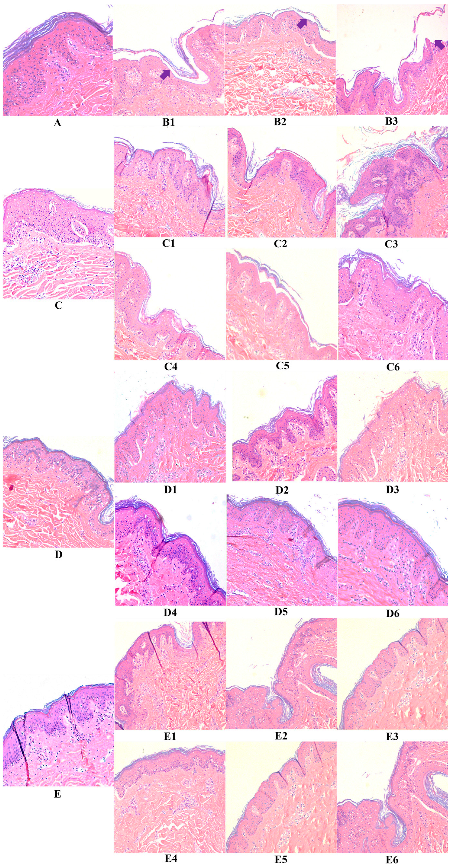
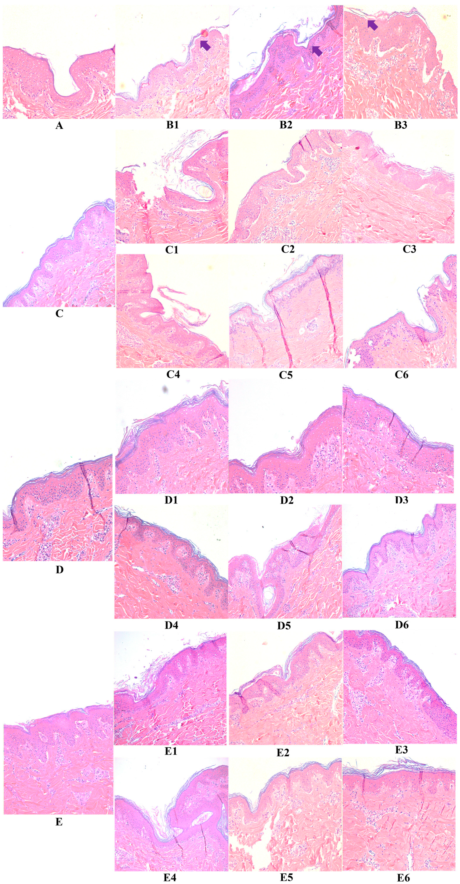
Control tissues, i.e., “untreated” sample A, “hyaluronic acid” sample C, “fragment c40” sample D, and “HAc40” sample E, stained with eosin-hematoxylin (E&E) (Figure 2 and Figure 3) show a normal skin structure, free of morphological changes and with epidermis covered by cytokeratin. Conversely, 24 h (Figure 2) and 48 h (Figure 3) after S. aureus strains infection (ATCC29213 sample B1 and DSM20491 sample B2) along with FCS (sample B3), degeneration of keratinocytes as well as evident detachment of the stratum corneum were observed (full arrow).
Similarly, pre-incubated (samples C1, C2, and C3) or co-incubated (samples C4, C5, and C6) tissues with HA and infected with the S. aureus strains or the FCS show marked signs of stratum corneum detachment, although intact. These findings suggested that HA alone could not protect from the pathogen- and FCS-induced damage. Similar results were obtained after 24 h (Figure 2) and 48 h of incubation (Figure 3).
In contrast, the morphology of c40 pre-incubated or co-incubated tissues (samples D1, D2, D3 and D4, D5, D6) and infected with the two bacterial strains or with the FCS shows a normal skin structure free of significant signs of degeneration (Figure 2). The same results were observed after the 48-h incubation period (Figure 3).
Similar findings were recorded for HAc40 pre-incubated (samples E1, E2, and E3) or co-incubated (E4, E5, and E6) tissues after 24 h (Figure 2) and 48 h of incubation (Figure 3).

3.3. c40, and HAc40 Activity on Animal Explant Sections Subjected to Immunofluorescence

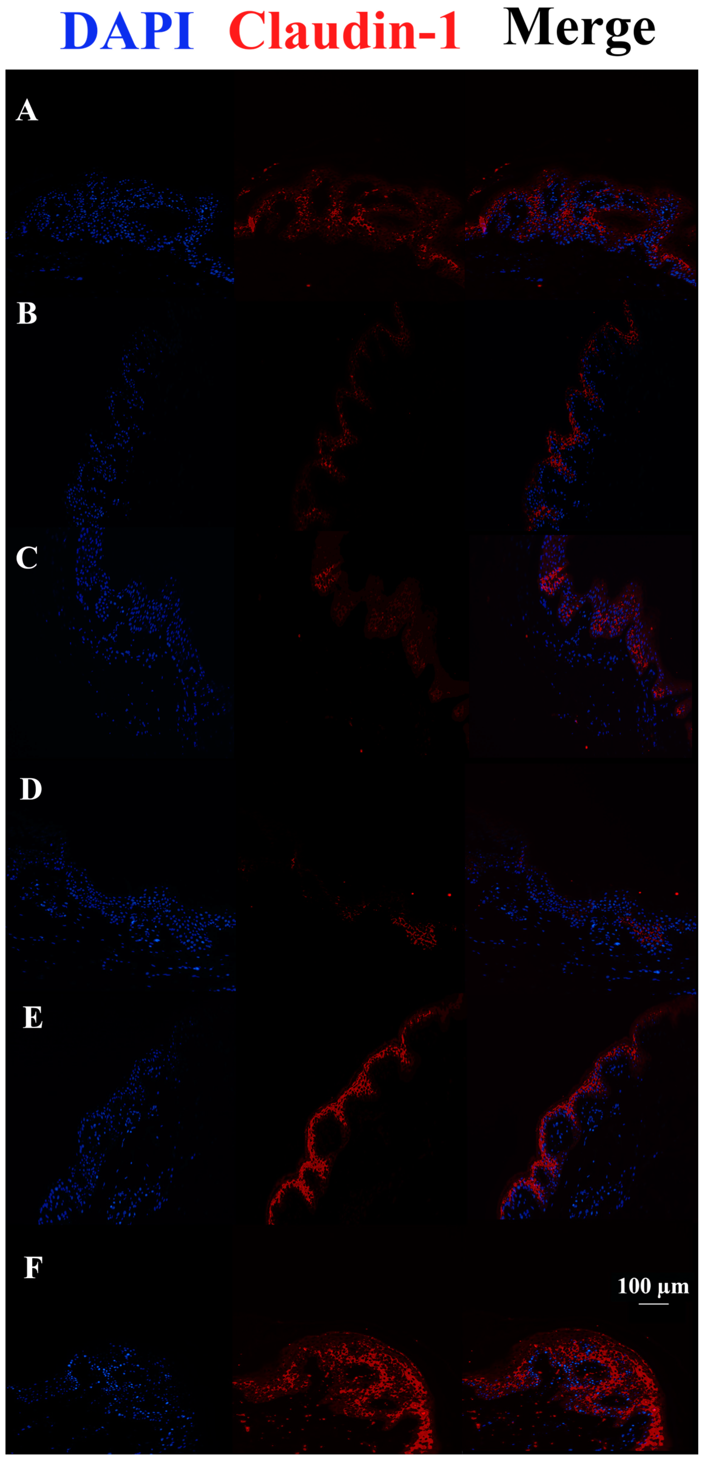
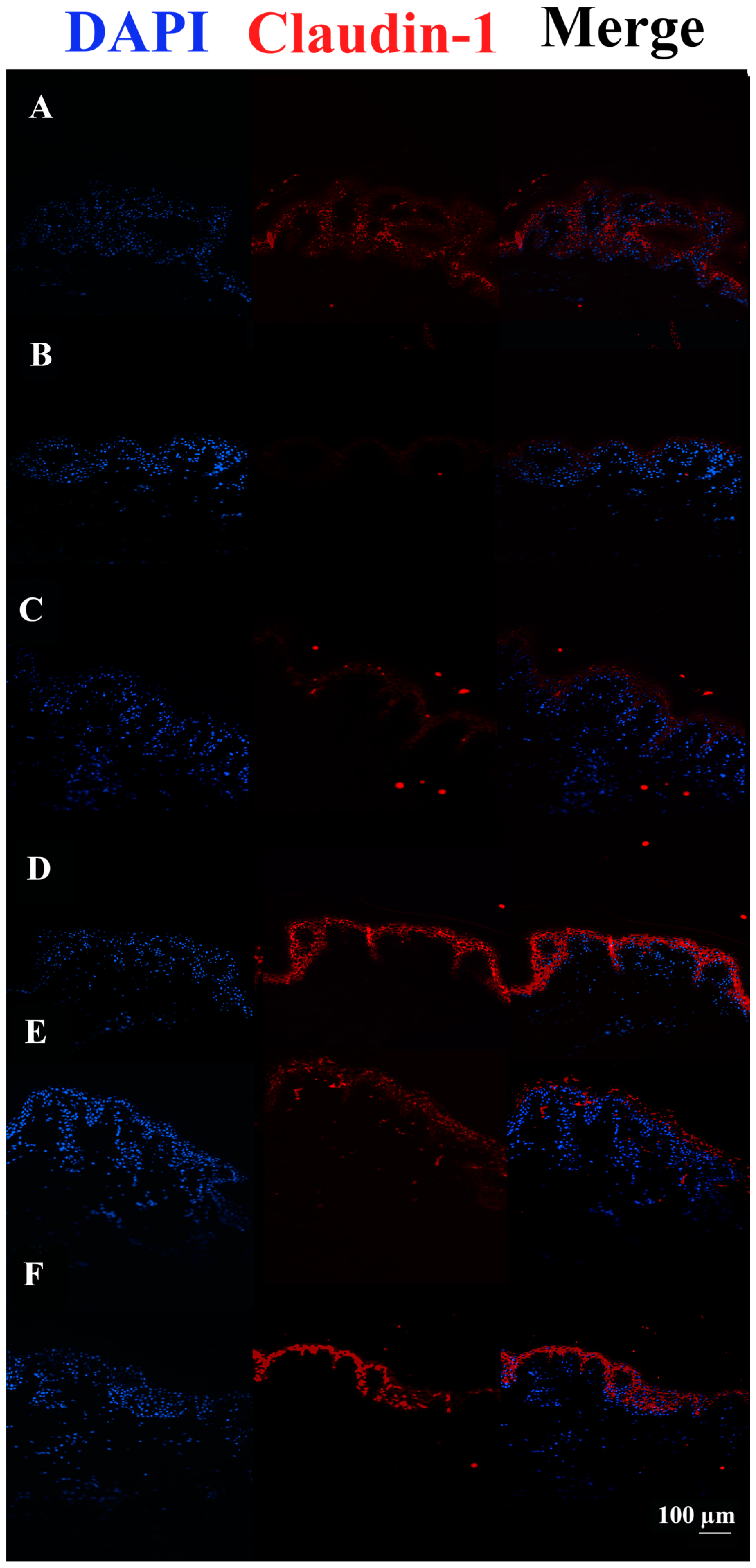
After 48 h, Claudin-1 appears down-regulated in tissues infected with S. aureus ATCC29213 compared to control tissue. On the other hand, c40 and HAc40 treatments but not HAc40 preincubation were shown to have a more pronounced fluorescence signal than the control tissue (Figure 4).
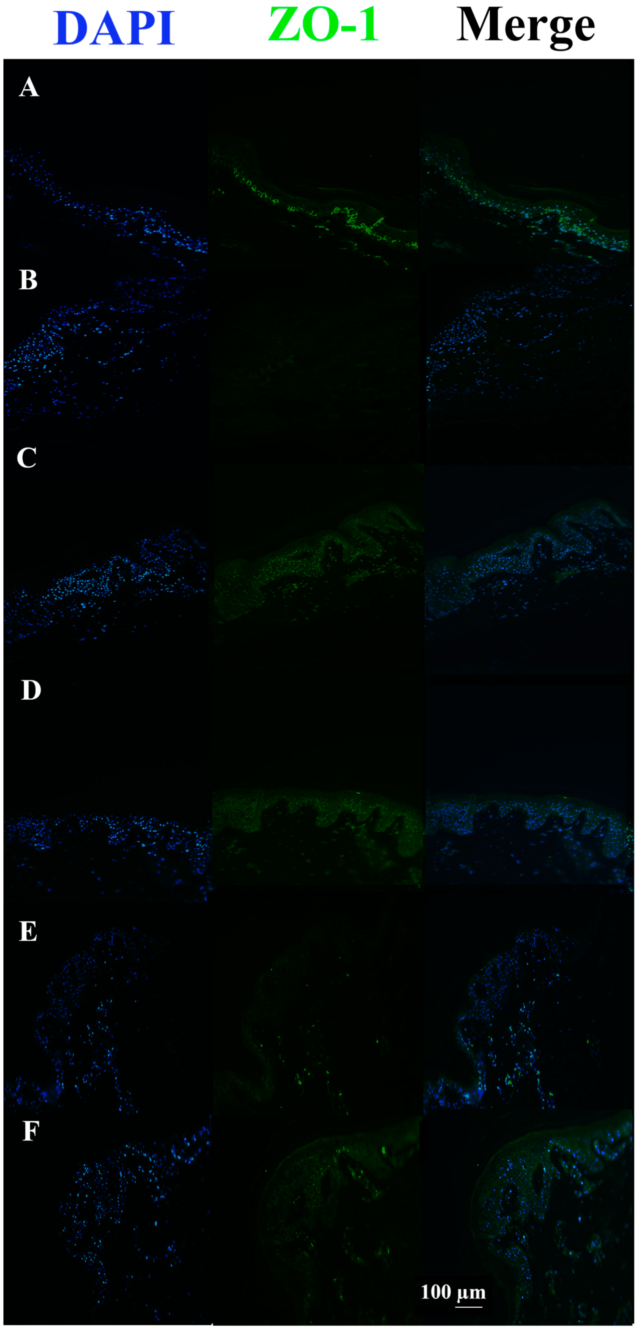
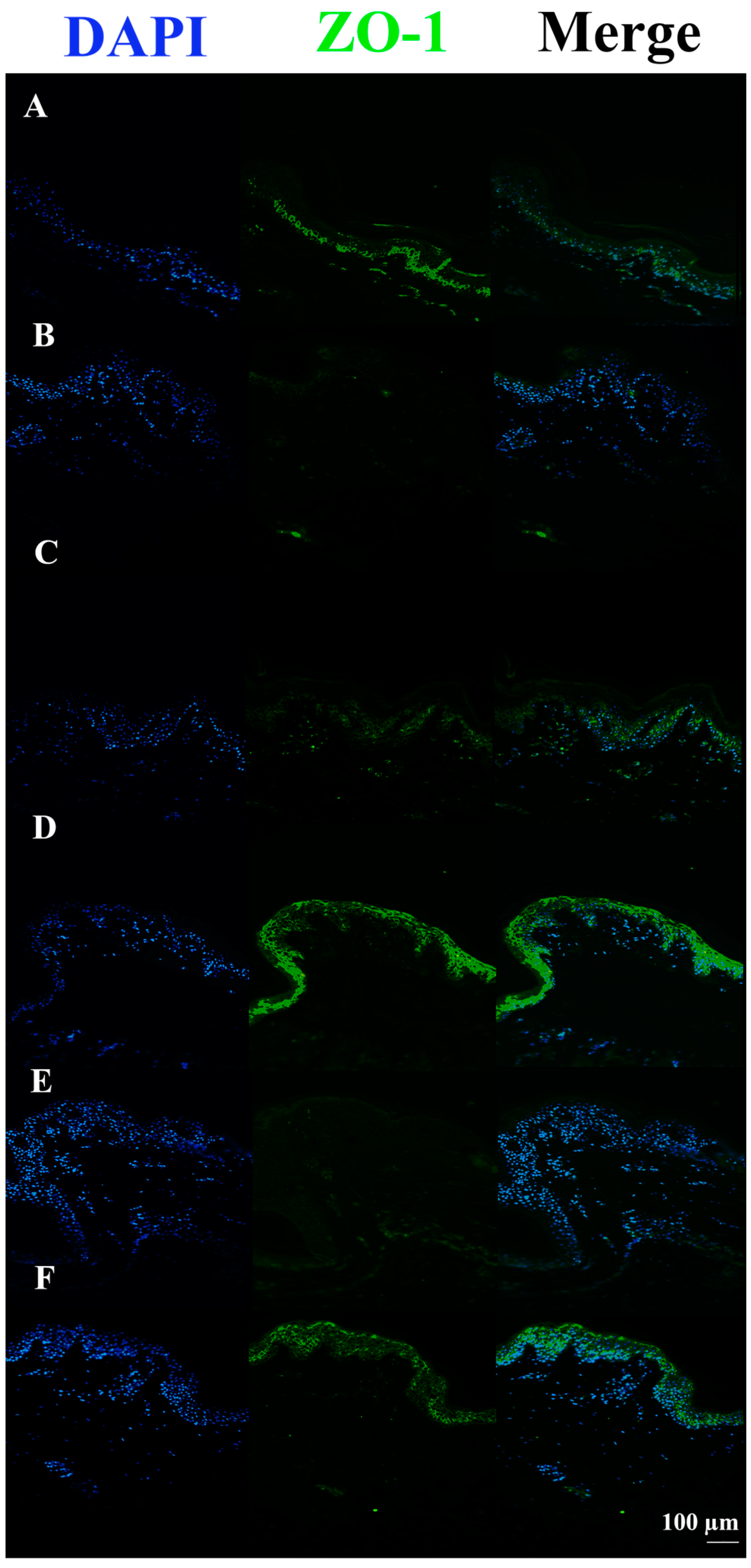
In S. aureus ATCCC29213 infected tissues, the ZO-1 signal was almost completely absent compared to the untreated control. Conversely, all infected tissues pre-incubated or co-incubated with c40 and Hac40 showed ZO-1 up-regulation, with a signal almost comparable to the control (Figure 5).
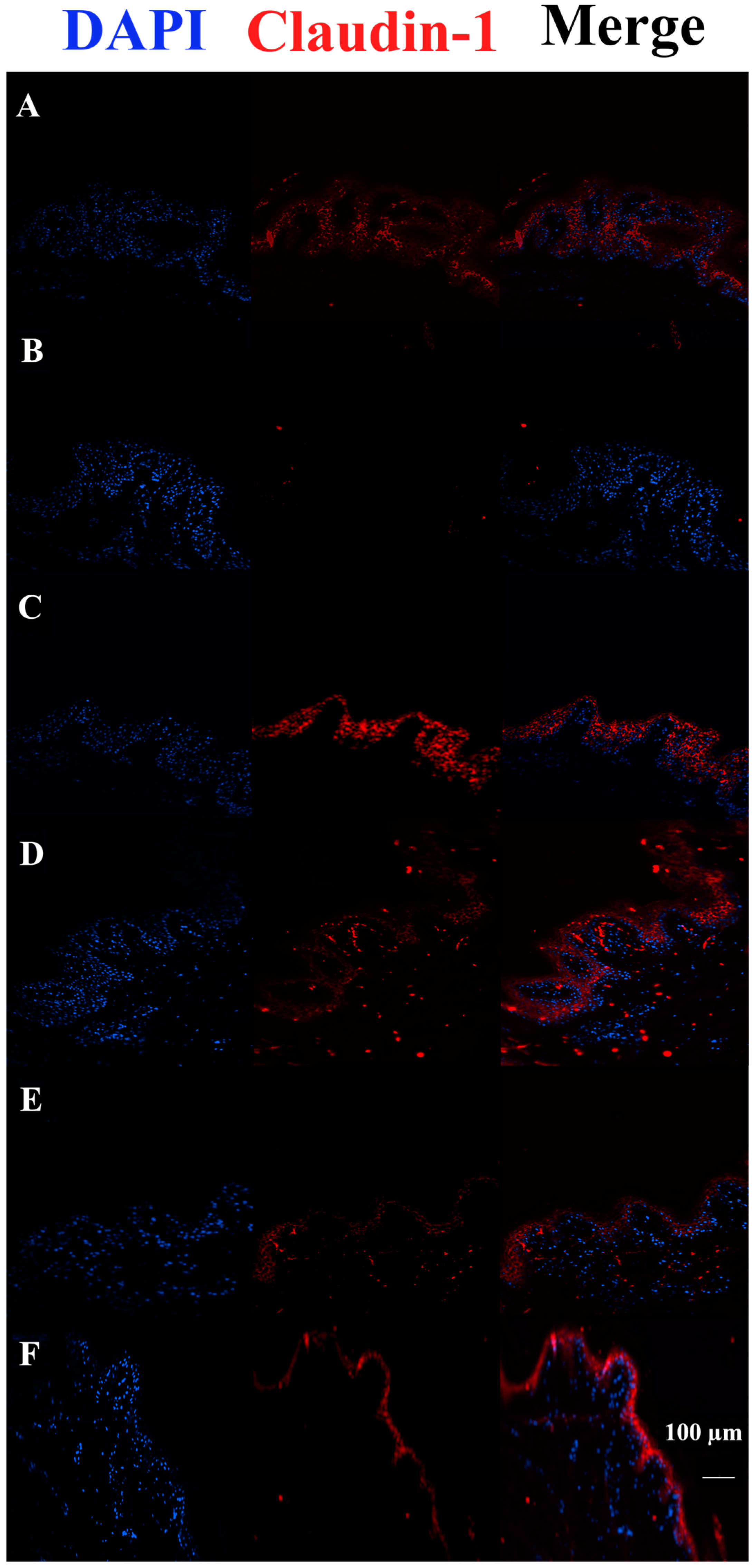
S. aureus DSM20491 live bacterial infection revealed a decrease in the Claudin-1 signal compared to not treated infected tissues. Conversely, all treatments increased the Claudin-1 signal, especially the Hac40 pre-incubation (Figure 6).
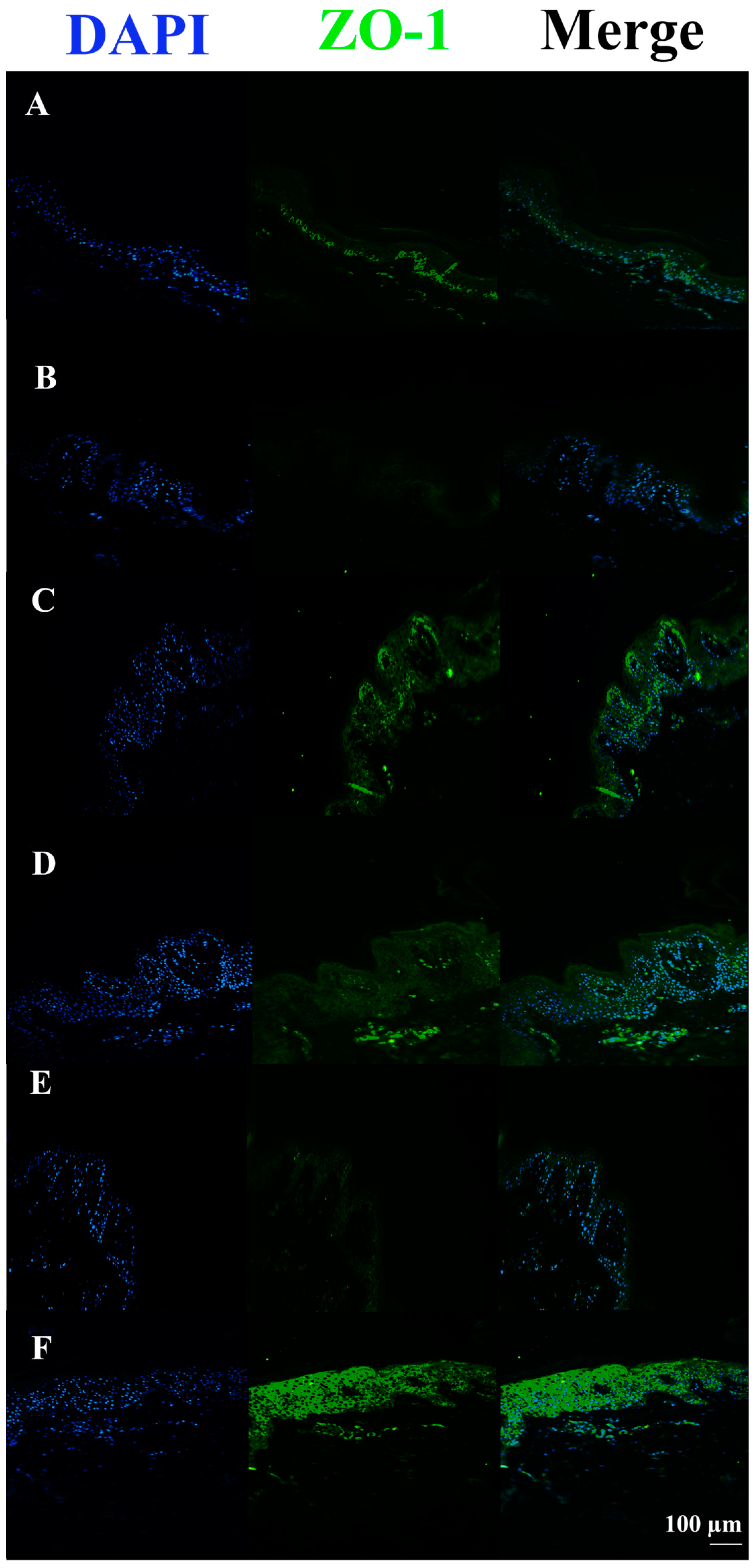
In S. aureus DSM20491 infected tissues, the ZO-1 signal was almost completely absent compared to untreated control tissue. Vice versa, all tissues infected with the pathogen but also pre-incubated or co-incubated with c40 and HAc40 (HAc40 co-incubated tissue excluded) showed an up-regulation in the ZO-1 signal, with an expression level comparable to the control tissue (Figure 7).
The Claudin-1 signal was completely absent in FCS-treated tissues. On the contrary, all treated tissues pre-incubated with HAc40 showed a stronger signal than controls. Remarkably, the Claudin-1 signal in c40 pre-incubated sections was comparable to the control (Figure 8).
Moreover, the ZO-1 signal was almost completely absent in tissues treated with FCS compared to the untreated control. All FCS-treated pre-incubated tissues showed stronger signals, almost comparable to the controls. Among these, HAc40-treated tissue revealed a ZO-1 signal comparable to the control (Figure 9).

4. Discussion

The investigation of bacteria’s therapeutic use in alleviating certain human diseases has gained popularity in recent years. Probiotic bacteria are known to bring numerous benefits to the human gut, such as epithelium repair, intestinal barrier function improvement, and immune response regulation [48].
The use of topical bacteriotherapy was first proposed for acne and seborrhea treatment back in 1912 when the efficacy of L. bulgaricus in improving symptomatology was demonstrated [49]. Recent studies have shown that probiotics and their derivatives can positively impact skin diseases [50]. For instance, experiments with lysates of L. rhamnosus GG (LGG) have shown that these products strengthen tight junctions and promote keratinocyte proliferation and migration [51].
In addition to well-known probiotic bacteria, studies on the possible therapeutic applications of the microorganisms belonging to the skin microbiota have been increasing in recent years [18,52].
Several pieces of evidence have proven that these microorganisms contribute to skin homeostasis by defending host cells against the proliferation of pathogenic microorganisms (such as S. aureus) and promoting skin barrier function [25,53].
Although the role of C. acnes species in skin health and disease is still being elucidated, it is now understood that there are substantial variations among strains, some of which have been demonstrated to have protective or even symbiotic properties [54]. This microorganism can regulate skin homeostasis by inhibiting pathogenic species through the secretion of specific bacteriocins or competition for nutrients [55].
In addition, C. acnes is able to secrete short-chain fatty acids (SCFAs) that are metabolised by the gut microbiome and have been shown to contribute to maintaining the colonic epithelium barrier by modulating tight epithelial junctions [29]. Furthermore, it colonises specific niches preventing the proliferation of pathogens. It has also been observed to modulate the immune response and oxidative stress mitigation [56]. The safety of topical treatment with a mixture of probiotic strains from the skin commensal C. acnes in patients with acne has been demonstrated recently by Karoglan et al. [25].
The main S. aureus virulence factors implicated in AD are a series of superantigens (adhesins and exotoxins) that mediate bacterial invasion and spread. These superantigens can alter skin barrier function, the microbiome composition, and the host immune response [57,58,59]. Furthermore, recent in vitro investigations have shown the down-regulation of key TJ proteins, including ZO-1, occludin, and claudin-1 [60].
In this scenario, the therapeutic potential of c40, a wall fragment derived from a patented and safe-deposit-protected strain of C. acnes, alone and conjugated to hyaluronic acid (HAc40) on one of the main pathogenetic mechanisms triggered by S. aureus and the down-regulation of tight junctions have been investigated.
The ex vivo experimental model selection based on pig skin biopsies leans on a well-established pertinence to human skin from both histological and physiological viewpoints. From an ethical perspective, pig biopsies were collected as a by-product from slaughterhouses and, in this particular context, do not contradict the Cosmetics Directive. Furthermore, this collection approach can control the animal skin age and the sampling body site [61].
Our results demonstrate that tissue infected with both strains of S. aureus and the supernatant containing the α-toxin showed clear damage to the stratum corneum by eosin-hematoxylin staining, confirming the pathogenic action on the epidermis. Evaluation by immunohistochemistry of the expression levels of the tight junction proteins Claudin-1 and ZO-1 deepened the pathogenetic mechanism of S. aureus just discussed by revealing a reduced signal in infected tissue compared to control tissue. Surprisingly, all tissues infected with the two S. aureus strains and the supernatant containing the toxin but pre-incubated and/or co-incubated with c40 and HAc40 showed a stronger signal comparable to that of the control, suggesting that the wall fragment can interfere with the degradation of tight junction proteins exerted by S. aureus.

5. Conclusions

To sum up, this study demonstrated that c40 and HAc40 could protect the tissues’ horny layer and tight junction proteins from S. aureus infection damage. Indeed, c40 and its functional ingredient HAc40 can be a potential non-pharmacological treatment, acting by counteracting the degradation of the skin barrier caused by S. aureus and consequently restoring the natural composition of the skin microbiome. These findings offer numerous avenues for research. Other virulence factors produced by S. aureus seem to play a crucial role in the inflammatory process driven by the bacterium (lipoproteins that activate TLR-2 on keratinocytes or the δ-toxin that activates mast cell degranulation) could be taken into account. Finally, interactions between S. aureus and the host cell surface, including protein A, clumping factor B, and proteins responsible for fibronectin binding, should be studied in detail.

Author Contributions

Conceptualization, R.D.M., G.P.P. and I.M.; methodology, I.M. and A.P.; validation, G.P.P. and R.D.M.; formal analysis, A.P., A.G. and N.V.; investigation, I.M., E.F., A.M. and M.A.C.; data curation, L.P., D.P., N.V. and F.V.; writing—original draft preparation, L.P., M.A.C. and A.G.; writing—review and editing, G.P.P., A.M. and D.P.; visualisation, I.M., E.F. and F.V.; supervision, R.D.M.; funding acquisition, R.D.M. All authors have read and agreed to the published version of the manuscript.

Funding

This research was partially supported by a donation from the company “Aileens Pharma srl” to Prof. Roberto Di Marco, at the University of Molise, Italy. The funding sustained the publication costs. The sponsor had no role in any other step of the manuscript elaboration.

Data Availability Statement

The data presented in this study are available on request from the corresponding author.

Conflicts of Interest

The authors declare no conflict of interest. The funders had no role in the study’s design; in the collection, analyses, or interpretation of data; in the writing of the manuscript; or in the decision to publish the results.

References

  1. Brandner, J.M. Importance of tight junctions in relation to skin barrier function. Curr. Probl. Dermatol. 2016, 49, 27–37. [Google Scholar] [PubMed]
  2. Svoboda, M.; Bílková, Z.; Muthný, T. Could tight junctions regulate the barrier function of the aged skin? J. Dermatol. Sci. 2016, 81, 147–152. [Google Scholar] [CrossRef]
  3. Brandner, J.M.; Schulzke, J.D. Hereditary barrier-related diseases involving the tight junction: Lessons from skin and intestine. Cell Tissue Res. 2015, 360, 723–748. [Google Scholar] [CrossRef] [PubMed]
  4. Herbig, M.E.; Houdek, P.; Gorissen, S.; Zorn-Kruppa, M.; Wladykowski, E.; Volksdorf, T.; Grzybowski, S.; Kolios, G.; Willers, C.; Mallwitz, H.; et al. A custom tailored model to investigate skin penetration in porcine skin and its comparison with human skin. Eur. J. Pharm. Biopharm. 2015, 95, 99–109. [Google Scholar] [CrossRef]
  5. Ohnemus, U.; Kohrmeyer, K.; Houdek, P.; Rohde, H.; Wladykowski, E.; Vidal, S.; Horstkotte, M.A.; Aepfelbacher, M.; Kirschner, N.; Behne, M.J. Regulation of epidermal tight-junctions (TJ) during infection with exfoliative toxin-negative Staphylococcus strains. J. Investig. Dermatol. 2008, 128, 906–916. [Google Scholar] [CrossRef]
  6. Olivry, T.; Dunston, S.M. Expression patterns of superficial epidermal adhesion molecules in an experimental dog model of acute atopic dermatitis skin lesions. Vet. Dermatol. 2015, 26, 53–e18. [Google Scholar] [CrossRef]
  7. Roussel, A.; Knol, A.; Bourdeau, P.; Bruet, V. Optimization of an immunohistochemical method to assess distribution of tight junction proteins in canine epidermis and adnexae. J. Comp. Pathol. 2014, 150, 35–46. [Google Scholar] [CrossRef]
  8. Yuki, T.; Haratake, A.; Koishikawa, H.; Morita, K.; Miyachi, Y.; Inoue, S. Tight junction proteins in keratinocytes: Localisation and contribution to barrier function. Exp. Derm. 2007, 16, 324–330. [Google Scholar] [CrossRef] [PubMed]
  9. Harris-Tryon, T.A.; Grice, E.A. Microbiota and maintenance of skin barrier function. Science 2022, 376, 940–945. [Google Scholar] [CrossRef]
  10. Grice, E.A.; Segre, J.A. The skin microbiome. Nat. Rev. Microbiol. 2011, 9, 244–253. [Google Scholar] [CrossRef]
  11. Luna, P.C. Skin microbiome as years go by. Am. J. Clin. Dermatol. 2020, 21, 12–17. [Google Scholar] [CrossRef] [PubMed]
  12. Chen, P.; He, G.; Qian, J.; Zhan, Y.; Xiao, R. Potential role of the skin microbiota in Inflammatory skin diseases. J. Cosmet. Dermatol. 2021, 20, 400–409. [Google Scholar] [CrossRef]
  13. Flowers, L.; Grice, E.A. The skin microbiota: Balancing risk and reward. Cell Host Microbe 2020, 28, 190–200. [Google Scholar] [CrossRef] [PubMed]
  14. Williams, H.; Flohr, C. How epidemiology has challenged 3 prevailing concepts about atopic dermatitis. J. Allergy Clin. Immunol. 2006, 118, 209–213. [Google Scholar] [CrossRef]
  15. Meisel, J.S.; Hannigan, G.D.; Tyldsley, A.S.; SanMiguel, A.J.; Hodkinson, B.P.; Zheng, Q.; Grice, E.A. Skin microbiome surveys are strongly influenced by experimental design. J. Investig. Dermatol. 2016, 136, 947–956. [Google Scholar] [CrossRef] [PubMed]
  16. Baldwin, H.E.; Bhatia, N.; Friedman, A.; Prunty, T.; Martin, R.; Seite, S. The role of cutaneous microbiota harmony in maintaining a functional skin barrier. SKIN J. Cutan. Med. 2017, 1, s139. [Google Scholar] [CrossRef]
  17. Foster, T.J.; Geoghegan, J.A.; Ganesh, V.K.; Höök, M. Adhesion, invasion and evasion: The many functions of the surface proteins of Staphylococcus aureus. Nat. Rev. Microbiol. 2014, 12, 49–62. [Google Scholar] [CrossRef]
  18. Sanford, J.A.; Gallo, R.L. Functions of the skin microbiota in health and disease. Semin. Immunol. 2013, 25, 370–377. [Google Scholar] [CrossRef]
  19. Paller, A.S.; Kong, H.H.; Seed, P.; Naik, S.; Scharschmidt, T.C.; Gallo, R.L.; Luger, T.; Irvine, A.D. The microbiome in patients with atopic dermatitis. J. Allergy Clin. Immunol. 2019, 143, 26–35. [Google Scholar] [CrossRef]
  20. Iwatsuki, K.; Yamasaki, O.; Morizane, S.; Oono, T. Staphylococcal cutaneous infections: Invasion, evasion and aggression. J. Dermatol. Sci. 2006, 42, 203–214. [Google Scholar] [CrossRef]
  21. Alam, M.A. Antibacterial pyrazoles: Tackling resistant bacteria. Future Med. Chem. 2022, 14, 343–362. [Google Scholar] [CrossRef]
  22. Bukowski, M.; Wladyka, B.; Dubin, G. Exfoliative toxins of Staphylococcus aureus. Toxins 2010, 2, 1148–1165. [Google Scholar] [CrossRef]
  23. Magnifico, I.; Petronio Petronio, G.; Venditti, N.; Cutuli, M.A.; Pietrangelo, L.; Vergalito, F.; Mangano, K.; Zella, D.; Di Marco, R. Atopic dermatitis as a multifactorial skin disorder. Can the analysis of pathophysiological targets represent the winning therapeutic strategy? Pharmaceuticals 2020, 13, 411. [Google Scholar] [CrossRef] [PubMed]
  24. Ito, Y.; Amagai, M. Controlling skin microbiome as a new bacteriotherapy for inflammatory skin diseases. Inflamm. Regen. 2022, 42, 1–13. [Google Scholar] [CrossRef] [PubMed]
  25. Lee, H.-J.; Kim, M. Skin Barrier Function and the Microbiome. Int. J. Mol. Sci. 2022, 23, 13071. [Google Scholar] [CrossRef]
  26. Nakatsuji, T.; Hata, T.R.; Tong, Y.; Cheng, J.Y.; Shafiq, F.; Butcher, A.M.; Salem, S.S.; Brinton, S.L.; Rudman Spergel, A.K.; Johnson, K. Development of a human skin commensal microbe for bacteriotherapy of atopic dermatitis and use in a phase 1 randomised clinical trial. Nat. Med. 2021, 27, 700–709. [Google Scholar] [CrossRef]
  27. Zhou, H.; Shi, L.; Ren, Y.; Tan, X.; Liu, W.; Liu, Z. Applications of Human Skin Microbiota in the Cutaneous Disorders for Ecology-Based Therapy. Front. Cell. Infect. Microbiol. 2020, 10, 570261. [Google Scholar] [CrossRef]
  28. Junca, H.; Pieper, D.H.; Medina, E. The emerging potential of microbiome transplantation on human health interventions. Comput. Struct. Biotechnol. J. 2022, 20, 615–627. [Google Scholar] [CrossRef]
  29. Rozas, M.; Hart de Ruijter, A.; Fabrega, M.J.; Zorgani, A.; Guell, M.; Paetzold, B.; Brillet, F. From dysbiosis to healthy skin: Major contributions of Cutibacterium acnes to skin homeostasis. Microorganisms 2021, 9, 628. [Google Scholar] [CrossRef]
  30. Ahmad-Mansour, N.; Loubet, P.; Pouget, C.; Dunyach-Remy, C.; Sotto, A.; Lavigne, J.P.; Molle, V. Staphylococcus aureus Toxins: An Update on Their Pathogenic Properties and Potential Treatments. Toxins 2021, 13, 677. [Google Scholar] [CrossRef] [PubMed]
  31. Kane, T.L.; Carothers, K.E.; Lee, S.W. Virulence factor targeting of the bacterial pathogen Staphylococcus aureus for vaccine and therapeutics. Curr. Drug Targets 2018, 19, 111–127. [Google Scholar] [CrossRef]
  32. Wolfmeier, H.; Mansour, S.C.; Liu, L.T.; Pletzer, D.; Draeger, A.; Babiychuk, E.B.; Hancock, R.E.W. Liposomal therapy attenuates dermonecrosis induced by community-associated methicillin-resistant Staphylococcus aureus by targeting α-type phenol-soluble modulins and α-hemolysin. EBioMedicine 2018, 33, 211–217. [Google Scholar] [CrossRef]
  33. Chen, Y.; Chen, M.; Zhang, Y.; Lee, J.H.; Escajadillo, T.; Gong, H.; Fang, R.H.; Gao, W.; Nizet, V.; Zhang, L. Broad-spectrum neutralisation of pore-forming toxins with human erythrocyte membrane-coated nanosponges. Adv. Healthc. Mater. 2018, 7, 1701366. [Google Scholar] [CrossRef]
  34. Karginov, V.A.; Nestorovich, E.M.; Schmidtmann, F.; Robinson, T.M.; Yohannes, A.; Fahmi, N.E.; Bezrukov, S.M.; Hecht, S.M. Inhibition of S. aureus α-hemolysin and B. anthracis lethal toxin by β-cyclodextrin derivatives. Bioorg. Med. Chem. 2007, 15, 5424–5431. [Google Scholar] [CrossRef]
  35. Dong, J.; Qiu, J.; Wang, J.; Li, H.; Dai, X.; Zhang, Y.; Wang, X.; Tan, W.; Niu, X.; Deng, X. Apigenin alleviates the symptoms of Staphylococcus aureus pneumonia by inhibiting the production of alpha-hemolysin. FEMS Microbiol. Lett. 2013, 338, 124–131. [Google Scholar] [CrossRef]
  36. Jiang, L.; Yi, T.; Shen, Z.; Teng, Z.; Wang, J. Aloe-emodin Attenuates Staphylococcus aureus Pathogenicity by Interfering with the Oligomerization of α-Toxin. Front. Cell. Infect. Microbiol. 2019, 9, 157. [Google Scholar] [CrossRef]
  37. Wang, J.; Zhou, X.; Liu, S.; Li, G.; Shi, L.; Dong, J.; Li, W.; Deng, X.; Niu, X. Morin hydrate attenuates Staphylococcus aureus virulence by inhibiting the self-assembly of α-hemolysin. J. Appl. Microbiol. 2015, 118, 753–763. [Google Scholar] [CrossRef]
  38. Qiu, J.; Niu, X.; Dong, J.; Wang, D.; Wang, J.; Li, H.; Luo, M.; Li, S.; Feng, H.; Deng, X. Baicalin protects mice from Staphylococcus aureus pneumonia via inhibition of the cytolytic activity of α-hemolysin. J. Infect. Dis. 2012, 206, 292–301. [Google Scholar] [CrossRef] [PubMed]
  39. Jiang, L.; Li, H.; Wang, L.; Song, Z.; Shi, L.; Li, W.; Deng, X.; Wang, J. Isorhamnetin Attenuates Staphylococcus aureus-Induced Lung Cell Injury by Inhibiting Alpha-Hemolysin Expression. J. Microbiol. Biotechnol. 2016, 26, 596–602. [Google Scholar] [CrossRef]
  40. Nabavi, S.F.; Braidy, N.; Habtemariam, S.; Orhan, I.E.; Daglia, M.; Manayi, A.; Gortzi, O.; Nabavi, S.M. Neuroprotective effects of chrysin: From chemistry to medicine. Neurochem. Int. 2015, 90, 224–231. [Google Scholar] [CrossRef]
  41. Zhou, Y.X.; Zhang, H.; Peng, C. Puerarin: A review of pharmacological effects. Phytother. Res. PTR 2014, 28, 961–975. [Google Scholar] [CrossRef] [PubMed]
  42. Pietrangelo, L.; Magnifico, I.; Guerrera, A.; Cutuli, M.A.; Petronio, G.P.; Venditti, N.; Covelli, M.; Buccieri, N.; Garofalo, S.; Di Marco, R.; et al. LimpiAD foam and the potential control of the pressure ulcers onset. Biomed. Pharmacother. 2021, 144, 112327. [Google Scholar] [CrossRef] [PubMed]
  43. Pietrangelo, L.; Dattola, A.; Magnifico, I.; Petronio Petronio, G.; Cutuli, M.A.; Venditti, N.; Guarnieri, A.; Wollenberg, A.; Pellacani, G.; Di Marco, R. Efficacy and Microbiota Modulation Induced by LimpiAL 2.5%, a New Medical Device for the Inverse Psoriasis Treatment. Int. J. Mol. Sci. 2023, 24, 6339. [Google Scholar] [CrossRef]
  44. Hwang, J.H.; Jeong, H.; Lee, N.; Hur, S.; Lee, N.; Han, J.J.; Jang, H.W.; Choi, W.K.; Nam, K.T.; Lim, K.M. Ex Vivo Live Full-Thickness Porcine Skin Model as a Versatile In Vitro Testing Method for Skin Barrier Research. Int. J. Mol. Sci. 2021, 22, 657. [Google Scholar] [CrossRef] [PubMed]
  45. Lind, I.; Ahnert-Hilger, G.; Fuchs, G.; Gratzl, M. Purification of alpha-toxin from Staphylococcus aureus and application to cell permeabilisation. Anal. Biochem. 1987, 164, 84–89. [Google Scholar] [CrossRef]
  46. Lazar, I., Jr.; Lazar, I., Sr. GelAnalyzer 19.1. 2023. Available online: www.gelanalyzer.com (accessed on 1 February 2023).
  47. Selam, B.; Kayisli, U.A.; Mulayim, N.; Arici, A. Regulation of Fas ligand expression by estradiol and progesterone in human endometrium. Biol. Reprod. 2001, 65, 979–985. [Google Scholar] [CrossRef]
  48. Tegegne, B.A.; Kebede, B. Probiotics, their prophylactic and therapeutic applications in human health development: A review of the literature. Heliyon 2022, 8, e09725. [Google Scholar] [CrossRef]
  49. Bowe, W.P.; Logan, A.C. Acne vulgaris, probiotics and the gut-brain-skin axis-back to the future? Gut. Pathog. 2011, 3, 1–11. [Google Scholar] [CrossRef]
  50. Roudsari, M.R.; Karimi, R.; Sohrabvandi, S.; Mortazavian, A. Health effects of probiotics on the skin. Crit. Rev. Food Sci. Nutr. 2015, 55, 1219–1240. [Google Scholar] [CrossRef]
  51. Mohammedsaeed, W.; Cruickshank, S.; McBain, A.J.; O’Neill, C.A. Lactobacillus rhamnosus GG Lysate Increases Re-Epithelialization of Keratinocyte Scratch Assays by Promoting Migration. Sci. Rep. 2015, 5, 16147. [Google Scholar] [CrossRef]
  52. Yang, Y.; Qu, L.; Mijakovic, I.; Wei, Y. Advances in the human skin microbiota and its roles in cutaneous diseases. Microb. Cell Factories 2022, 21, 176. [Google Scholar] [CrossRef] [PubMed]
  53. Swaney, M.H.; Kalan, L.R. Living in Your Skin: Microbes, Molecules, and Mechanisms. Infect. Immun. 2021, 89, e00695-20. [Google Scholar] [CrossRef] [PubMed]
  54. Rhee, M.S.; Alqam, M.L.; Jones, B.C.; Phadungpojna, S.; Day, D.; Hitchcock, T.M. Characterization of a live Cutibacterium acnes subspecies defendens strain XYCM42 and clinical assessment as a topical regimen for general skin health and cosmesis. J. Cosmet. Dermatol. 2022, 22, 1031–1045. [Google Scholar] [CrossRef] [PubMed]
  55. Brüggemann, H.; Salar-Vidal, L.; Gollnick, H.P.M.; Lood, R. A Janus-Faced Bacterium: Host-Beneficial and -Detrimental Roles of Cutibacterium acnes. Front. Microbiol. 2021, 12, 673845. [Google Scholar] [CrossRef] [PubMed]
  56. Andersson, T.; Ertürk Bergdahl, G.; Saleh, K.; Magnúsdóttir, H.; Stødkilde, K.; Andersen, C.B.F.; Lundqvist, K.; Jensen, A.; Brüggemann, H.; Lood, R. Common skin bacteria protect their host from oxidative stress through secreted antioxidant RoxP. Sci. Rep. 2019, 9, 3596. [Google Scholar] [CrossRef]
  57. Kim, J.; Kim, B.E.; Leung, D.Y. Pathophysiology of atopic dermatitis: Clinical implications. Allergy Asthma Proc. 2019, 40, 84–92. [Google Scholar] [CrossRef]
  58. Tsilochristou, O.; du Toit, G.; Sayre, P.H.; Roberts, G.; Lawson, K.; Sever, M.L.; Bahnson, H.T.; Radulovic, S.; Basting, M.; Plaut, M.; et al. Association of Staphylococcus aureus colonisation with food allergy occurs independently of eczema severity. Clin. Immunol. 2019, 144, 494–503. [Google Scholar]
  59. Blicharz, L.; Rudnicka, L.; Samochocki, Z.; Alergologii, A. Staphylococcus aureus: An underestimated factor in the pathogenesis of atopic dermatitis? Postep. Derm. Alergol. 2019, 36, 11. [Google Scholar] [CrossRef]
  60. Seite, S.; Bieber, T. Barrier function and microbiotic dysbiosis in atopic dermatitis. Clin. Cosmet. Investig. Dermatol. 2015, 8, 479. [Google Scholar] [CrossRef]
  61. Richert, S.; Schrader, A.; Schrader, K. Transdermal delivery of two antioxidants from different cosmetic formulations. Int. J. Cosmet. Sci. 2003, 25, 5–13. [Google Scholar] [CrossRef]
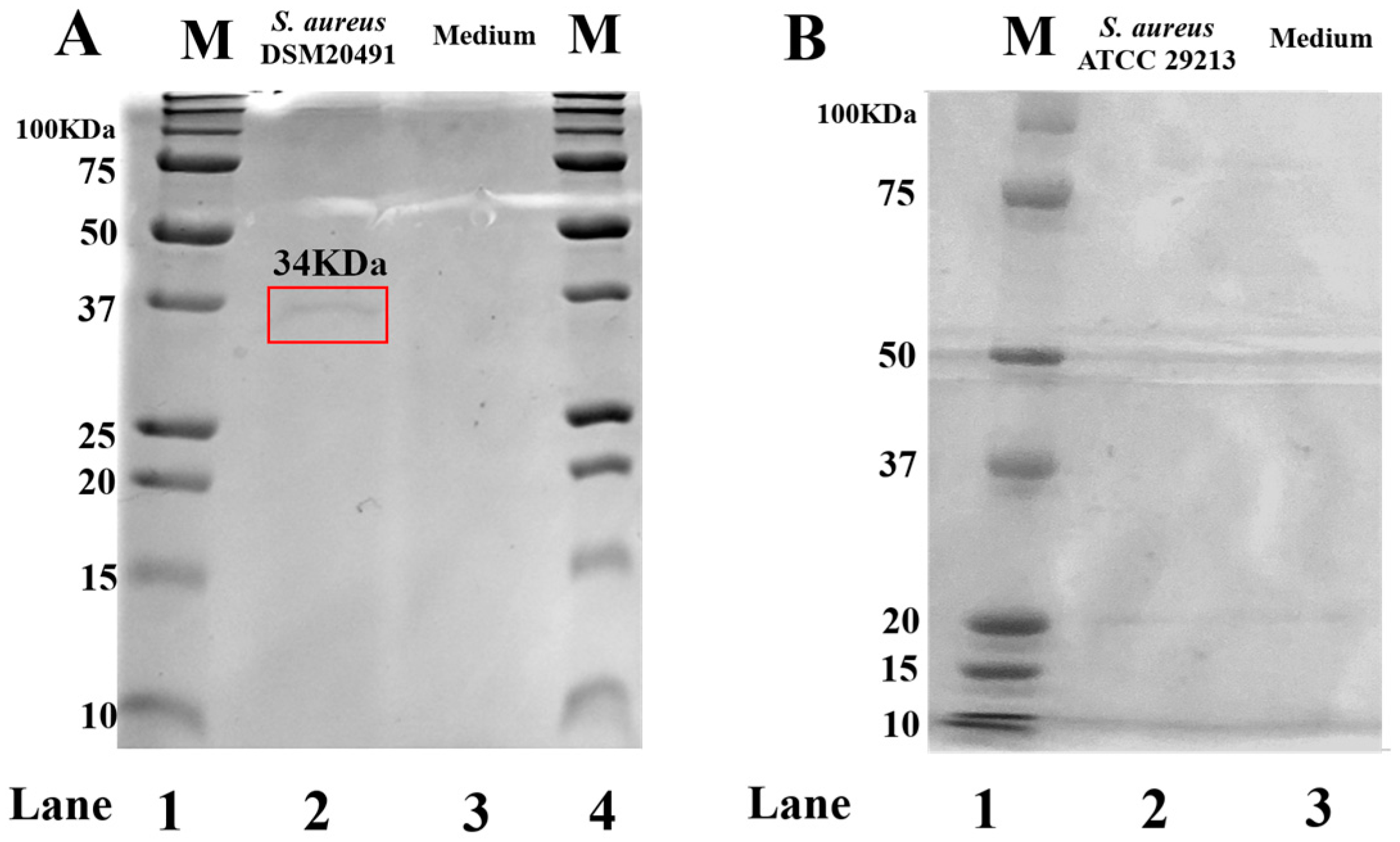
Figure 1. FCS qualitative analysis by SDS-PAGE. (A): Ladder (lane 1) Precision Plus Protein™ Unstained Protein Standards (BIORAD cat.no 1610363); S. aureus DSM4020491 (lane 2). (B): Ladder (lane 1) Precision Plus Protein™ Kaleidoscope™ Prestained Protein Standards (BIORAD cat.no 1610395) S. aureus ATCC29213 (lane 2). Samples were separated into 12% acrylamide gels. The bands’ molecular weight analysis was performed by GelAnalyzer 19.1 [46]. The culture medium (lane 3, Figure 1A,B) was used as a control. The gel was stained in Coomassie blue.
Pharmaceutics 15 01224 g001
Figure 2. 10× magnification eosin-hematoxylin stained tissues after 24 h infection with Staphylococcus aureus ATCC29213 and DSM20491 (panels B1,B2), FCS (panel B3), untreated (panel A) and treated (pre-incubated and coincubated) tissues with HA (panels C1C6), c40 (panels D1D6) and HAc40 (panels E1E6).
Pharmaceutics 15 01224 g002
Figure 3. 10× magnification eosin-hematoxylin stained tissues after 48 h infection with Staphylococcus aureus ATCC29213 and DSM20491 (panels B1,B2), FCS (panel B3), untreated (panel A) and treated (pre-incubated and coincubated) tissues with HA (panels C1C6), c40 (panels D1D6) and HAc40 (panels E1E6).
Pharmaceutics 15 01224 g003
Figure 4. Claudin-1 immunofluorescence on animal explant sections infected with S. aureus ATCC29213 live bacteria (20× magnification). DAPI channel on the left side (461 nm blue); Anti-Claudin-1 1 antibody Alexa568-conjugated in the middle; images are shown on the right. Lane description. (A) Not infected tissues; (B) not treated infected tissues; (C) c40 pre-incubated infected tissues; (D) HAc40 pre-incubated infected tissues; (E) c40 co-incubated infected tissues; (F) Hac40 co-incubated infected tissues. The Claudin-1 signal is stronger in tissues treated with c40 and Hac40 than in infected tissues alone. Scale bar 100 µm.
Pharmaceutics 15 01224 g004
Figure 5. ZO-1 immunofluorescence on animal explant sections infected with S. aureus ATCC29213 live bacteria (20× magnification). DAPI channel (461 nm, blue) is shown on the left side; Anti-ZO-1 antibody Alexa488 nm-conjugated is in the middle; merge images are shown on the right. Lane description. (A) Not infected tissues; (B) not treated infected tissues; (C) c40 pre-incubated infected tissues; (D) Hac40 pre-incubated infected tissues; (E): c40 co-incubated infected tissues; (F) Hac40 co-incubated infected tissues. The ZO-1 signal in Hac40 co-incubated tissues was almost comparable to the untreated control. Scale bar 100 µm.
Pharmaceutics 15 01224 g005
Figure 6. Claudin-1 immunofluorescence on animal explant sections infected with S. aureus DSM20491 live bacteria (20× magnification). DAPI channel (461 nm, blue) is shown on the left side; Anti-Claudin-1 antibody Alexa568-conjugated is in the middle; merge images are shown on the right. Lane description. (A) Not infected tissues; (B) not treated infected tissues; (C) c40 pre-incubated infected tissues; (D) Hac40 pre-incubated infected tissues; (E) c40 co-incubated infected tissues; (F) HAc40 co-incubated infected tissues. Tissues pretreated with c40 and HAc40 or co-incubated with HAc40 showed Claudin-1 up-regulation compared to untreated tissues. Scale bar 100 µm.
Pharmaceutics 15 01224 g006
Figure 7. ZO-1 immunofluorescence on animal explant sections infected with S. aureus DSM20491 live bacteria (20× magnification). DAPI (461 nm, blue) is shown on the left side; Anti-ZO-1 antibody Alexa488 nm-conjugated is shown in the middle; merge images are shown on the right. Lane description. (A) Not infected tissues; (B) not treated infected tissues; (C) c40 pre-incubated infected tissues; (D) HAc40 pre-incubated infected tissues; (E) c40 co-incubated infected tissues; (F) HAc40 co-incubated infected tissues. c40 and HAc40 pre-incubated and HAc40 co-incubated tissues revealed a ZO-1 signal (green) almost comparable to untreated control. Scale bar 100 µm.
Pharmaceutics 15 01224 g007
Figure 8. Claudin-1 immunofluorescence on animal explant sections infected with FCS processing (20× magnification). DAPI channel is shown on the left side (461 nm, blue); Anti-Claudin-1 antibody Alexa568-conjugated is shown in the middle; merge images are shown on the right. Lane description. (A) Not infected tissues; (B) not treated infected tissues; (C) c40 pre-incubated infected tissues; (D) HAc40 pre-incubated infected tissues; (E) c40 co-incubated infected tissues; (F) HAc40 co-incubated infected tissues. HAc40 pre-incubated and co-incubated tissues showed a Claudin-1 signal stronger compared to the FCS tissue alone. Scale bar 100 µm.
Pharmaceutics 15 01224 g008
Figure 9. ZO-1 immunofluorescence on animal explant sections infected with FCS processing (20× magnification). DAPI (461 nm, blue) is shown on the left side; Anti-ZO-1 antibody Alexa488 nm-conjugated is shown in the middle; merge images are shown on the right. Lane description. (A) Not infected tissues; (B) not treated infected tissues; (C) c40 pre-incubated infected tissues; (D) HAc40 pre-incubated infected tissues; (E) c40 co-incubated infected tissues; (F): HAc40 co-incubated infected tissues. The ZO-1 (FITC) signal in Hac40 pre-incubated and c40 co-incubated tissues was almost comparable to the untreated control. Scale bar 100 µm.
Pharmaceutics 15 01224 g009
Disclaimer/Publisher’s Note: The statements, opinions and data contained in all publications are solely those of the individual author(s) and contributor(s) and not of MDPI and/or the editor(s). MDPI and/or the editor(s) disclaim responsibility for any injury to people or property resulting from any ideas, methods, instructions or products referred to in the content.