
Nanotechnology in Cancer Diagnosis and Treatment


 , and
, and 
Abstract
1. Introduction
2. Conventional Methods of Cancer Diagnosis via Imaging Technology
3. Significance of Nanoparticles in Cancer Diagnosis
4. Synthesis of Nanomaterials
5. Nanoparticle Function in Cancer Image Enhancement and Contrasting Agents
6. Major Advantages of Nanomaterials
7. Commonly Used Nanoparticles in Cancer Diagnosis
7.1. Metallic Nanoparticles
7.1.1. Platinum Nanoparticles
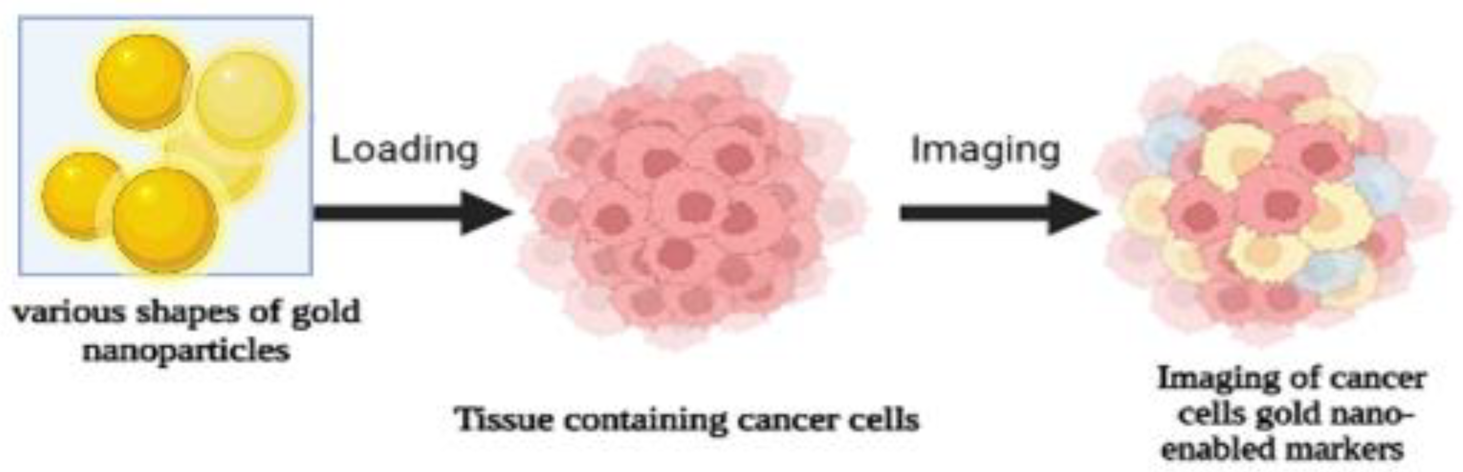
7.1.2. Gold Nanoparticles
7.2. Magnetic Nanoparticles

| Type of Nanoparticle | Cancer Cells | Applications | Citations |
|---|---|---|---|
| Magnetic gold nanoparticles | Breast cancer checks | ELISA-based detection of breast cancer, specifically for HER2 breast cancer patients. | [43] |
| Magnetic nanoparticles | Liver cancer cells | Enhanced detection of liver cancer cells (in vitro) | [22] |
| Magnetic nanoparticles | Brain cancer cells | Magnetic nanoparticles as contrast agents in the diagnosis and treatment of cancer (in vivo) | [44] |
| Surface-modified magnetic nanoparticles | Colon cancer cells | For colon cancer cell theranostics (in vitro) | [45] |
| Superparamagnetic iron oxide nanoparticles | Pancreatic cancer cells | Pancreatic cancer diagnosis using MRI and potential for early diagnosis through targeted strategies | [46] |
7.3. Polymeric-Based Nanoparticles
7.4. Metal Oxide Nanoparticles
7.5. Quantum Dots
7.6. Graphene
7.7. Fullerene
7.8. Carbon Nanotubes
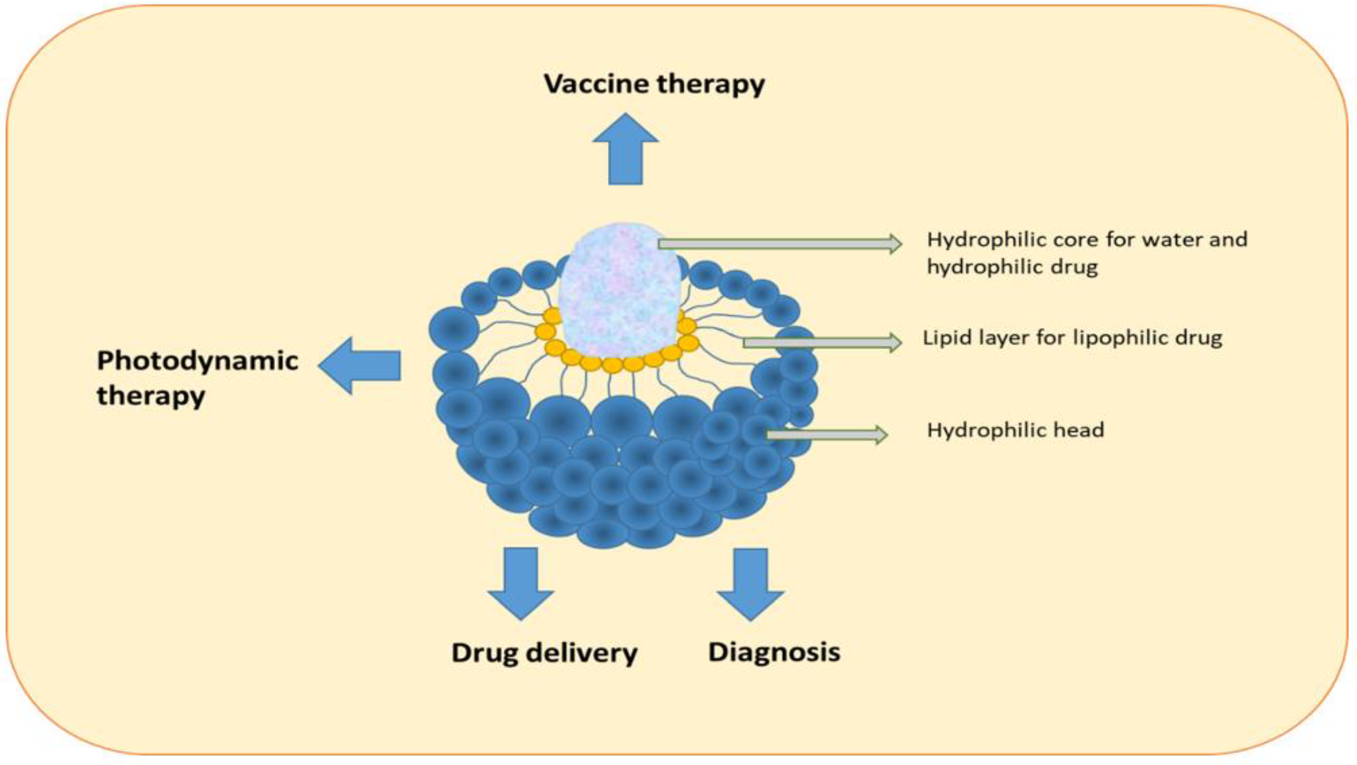
7.9. Liposomes
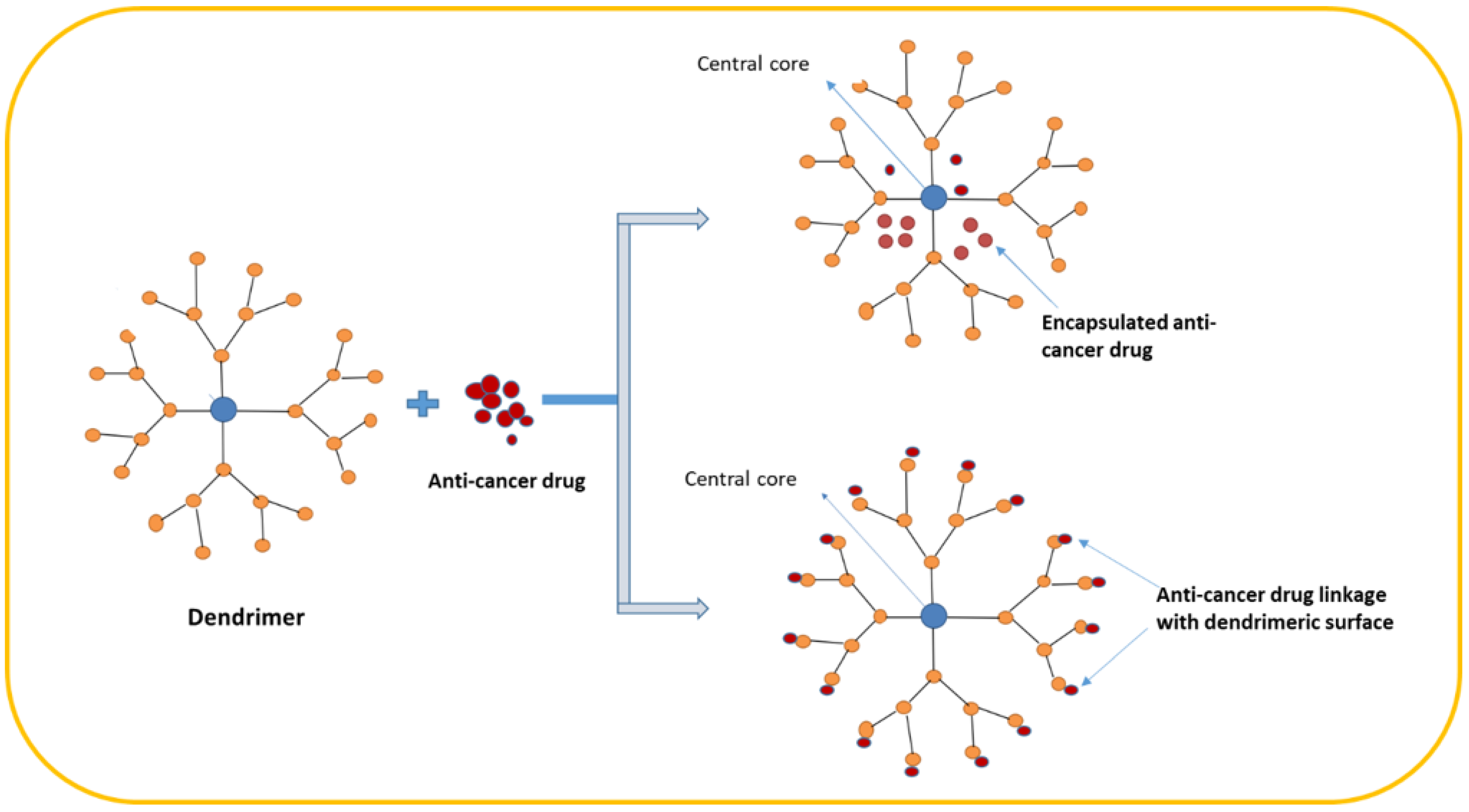
7.10. Dendrimers
7.11. Nanostructure Lipid Carriers (NLCs)
7.12. Other Nanoparticles and Their Application in Various Cancer Cells
7.13. Formulation of Nanomaterials for Drug Delivery
7.14. Clinical Application of Nanomaterials in Cancer Treatment
7.15. Current Challenges and Future Prospects
Author Contributions
Funding
Data Availability Statement
Conflicts of Interest
References
- Jarvie, H.; Stephen, K.; Dobson, P. Nanoparticle. Encyclopedia Britannica. 2019. Available online: https://www.britannica.com/science/nanoparticle (accessed on 10 March 2023).
- Swain, S.; Sahu, P.K.; Beg, S.; Manohar Babu, S. Nanoparticles for Cancer Targeting: Current and Future Directions. Curr. Drug Deliv. 2016, 13, 1290–1302. [Google Scholar] [CrossRef]
- Zhang, W.; Lu, Y.; Zang, Y.; Han, J.; Xiong, Q.; Xiong, J. Photodynamic Therapy and Multi-Modality Imaging of Up-Conversion Nanomaterial Doped with AuNPs. Int. J. Mol. Sci. 2022, 23, 1227. [Google Scholar] [CrossRef]
- Abed, A.; Derakhshan, M.; Karimi, M.; Shirazinia, M.; Mahjoubin-Tehran, M.; Homayonfal, M.; Hamblin, M.R.; Mirzaei, S.A.; Soleimanpour, H.; Dehghani, S.; et al. Platinum Nanoparticles in Biomedicine: Preparation, Anti-Cancer Activity, and Drug Delivery Vehicles. Front. Pharmacol. 2022, 13, 797804. [Google Scholar] [CrossRef]
- Khan, I.; Saeed, K.; Khan, I. Nanoparticles: Properties, applications and toxicities. Arab. J. Chem. 2017, 12, 908–931. [Google Scholar] [CrossRef]
- Chapman, S.; Dobrovolskaia, M.; Farahani, K.; Goodwin, A.; Joshi, A.; Lee, H.; Meade, T.; Pomper, M.; Ptak, K.; Rao, J.; et al. Nanoparticles for cancer imaging: The good, the bad, and the promise. Nano Today 2013, 8, 454–460. [Google Scholar] [CrossRef]
- Baetke, S.C.; Lammers, T.; Kiessling, F. Applications of nanoparticles for diagnosis and therapy of cancer. Br. J. Radiol. 2015, 88, 20150207. [Google Scholar] [CrossRef]
- Ramanathan, A. Toxicity of nanoparticles challenges and opportunities. Appl. Microsc. 2019, 49, 1–11. [Google Scholar] [CrossRef]
- Patel, P.; Patel, V.; Patel, P.M. Synthetic strategy of dendrimers: A review. J. Indian Chem. Soc. 2022, 99, 100514. [Google Scholar] [CrossRef]
- Wang, X.; Li, J.; Zhang, W.; Li, P.; Zhang, W.; Wang, H.; Tang, B. Evaluating diabetic ketoacidosis via a MOF sensor for fluorescence imaging of phosphate and pH. Chem. Commun. 2022, 58, 3023–3026. [Google Scholar] [CrossRef]
- Zhang, W.; Wang, X.; Li, P.; Zhang, W.; Wang, H.; Tang, B. Evaluating Hyperthyroidism-Induced Liver Injury Based on In Situ Fluorescence Imaging of Glutathione and Phosphate via Nano-MOFs Sensor. Anal. Chem. 2020, 92, 8952–8958. [Google Scholar] [CrossRef]
- Gao, L.; Zhang, Y.; Zhao, L.; Niu, W.; Tang, Y.; Gao, F.; Cai, P.; Yuan, Q.; Wang, X.; Jiang, H.; et al. An artificial metalloenzyme for catalytic cancer-specific DNA cleavage and operando imaging. Sci. Adv. 2020, 6, eabb1421. [Google Scholar] [CrossRef]
- The Past, Present, and Future of Medical Imaging. 2021. Available online: https://chanzuckerberg.com/blog/the-past-present-and-future-of-medical-imaging/ (accessed on 10 March 2023).
- Schiffman, J.D.; Fisher, P.G.; Gibbs, P. Early Detection of Cancer: Past, Present, and Future. In American Society of Clinical Oncology Educational Book; American Society of Clinical Oncology: Alexandria, VA, USA, 2015; pp. 57–65. [Google Scholar] [CrossRef]
- American Cancer Society. MRI for Cancer. Available online: https://www.cancer.org/treatment/understanding-your-diagnosis/tests/mri-for-cancer.html (accessed on 16 May 2019).
- Yang, H.; Wen, L.; Wang, X.; Zhao, J.; Dong, J.; Yin, X.; Xu, F.; Yang, M.; Huo, D.; Hou, C. A test strip electrochemical disposable by 3D MXA/AuNPs DNA-circuit for the detection of miRNAs. Microchim. Acta 2022, 189, 1–10. [Google Scholar] [CrossRef] [PubMed]
- Leja, M.; Linē, A. Early detection of gastric cancer beyond endoscopy—New methods. Best Pract. Res. Clin. Gastroenterol. 2021, 50–51, 101731. [Google Scholar] [CrossRef]
- Haghighi, F.H.; Binaymotlagh, R.; Mirahmadi-Zare, S.Z.; Hadadzadeh, H. Aptamer/magnetic nanoparticles decorated with fluorescent gold nanoclusters for selective detection and collection of human promyelocytic leukemia (HL-60) cells from a mixture. Nanotechnology 2019, 31, 025605. [Google Scholar] [CrossRef]
- Chen, H.; Zhen, Z.; Todd, T.; Chu, P.K.; Xie, J. Nanoparticles for improving cancer diagnosis. Mater. Sci. Eng. R Rep. 2013, 74, 35–69. [Google Scholar] [CrossRef]
- Pavitra, E.; Dariya, B.; Srivani, G.; Kang, S.-M.; Alam, A.; Sudhir, P.-R.; Kamal, M.A.; Raju, G.S.R.; Han, Y.-K.; Lakkakula, B.V.K.S.; et al. Engineered nanoparticles for imaging and drug delivery in colorectal cancer. Semin. Cancer Biol. 2019, 69, 293–306. [Google Scholar] [CrossRef]
- Teodor, E.D.; Gatea, F.; Ficai, A.; Radu, G.L. Functionalized magnetic nanostructures for anticancer therapy. Curr. Drug Targets 2018, 19, 239–247. [Google Scholar] [CrossRef]
- Zhai, R.; Gong, X.; Xie, J.; Yuan, Y.; Xu, F.; Jiang, Y.; Huang, Z.; Dai, X.; Zhang, Y.; Qian, X.; et al. Ultrasensitive analysis of heat shock protein 90α with antibodies orderly arrayed on a novel type of immunoprobe based on magnetic COFs. Talanta 2018, 191, 553–560. [Google Scholar] [CrossRef]
- Huyan, T.; Li, H.; Peng, H.; Chen, J.; Yang, R.; Zhang, W.; Li, Q. Extracellular Vesicles—Advanced Nanocarriers in Cancer Therapy: Progress and Achievements. Int. J. Nanomed. 2020, ume 15, 6485–6502. [Google Scholar] [CrossRef]
- Jaishree, V.; Gupta, P.D. Nanotechnology: A Revolution in Cancer Diagnosis. Indian J. Clin. Biochem. 2012, 27, 214–220. [Google Scholar] [CrossRef]
- Wang, J.; Cao, F.; He, S.; Xia, Y.; Liu, X.; Jiang, W.; Yu, Y.; Zhang, H.; Chen, W. FRET on lateral flow test strip to enhance sensitivity for detecting cancer biomarker. Talanta 2018, 176, 444–449. [Google Scholar] [CrossRef]
- Wang, L.; Song, X.; Yu, M.; Niu, L.; Zhao, Y.; Tang, Y.; Zheng, B.; Song, X.; Xie, L. Serum exosomal miR-377-3p and miR-381-3p as diagnostic biomarkers in colorectal cancer. Future Oncol. 2022, 18, 793–805. [Google Scholar] [CrossRef]
- Gao, T.; Zhi, J.; Mu, C.; Gu, S.; Xiao, J.; Yang, J.; Wang, Z.; Xiang, Y. One-step detection for two serological biomarker species to improve the diagnostic accuracy of hepatocellular carcinoma. Talanta 2018, 178, 89–93. [Google Scholar] [CrossRef]
- Czubacka, E.; Czerczak, S. Are platinum nanoparticles safe to human health? Med. Pract. 2019, 70, 487–495. [Google Scholar] [CrossRef]
- Ruiz, A.L.; Garcia, C.B.; Gallon, S.N.; Webster, T.J. Novel Silver-Platinum Nanoparticles for Anticancer and Antimicrobial Applications. Int. J. Nanomed. 2020, ume 15, 169–179. [Google Scholar] [CrossRef]
- Depciuch, J.; Stec, M.; Klebowski, B.; Maximenko, A.; Drzymała, E.; Baran, J.; Parlinska-Wojtan, M. Size effect of platinum nanoparticles in simulated anticancer photothermal therapy. Photodiagnosis Photodyn. Ther. 2020, 29, 101594. [Google Scholar] [CrossRef]
- Gurunathan, S.; Jeyaraj, M.; Kang, M.-H.; Kim, J.-H. Anticancer Properties of Platinum Nanoparticles and Retinoic Acid: Combination Therapy for the Treatment of Human Neuroblastoma Cancer. Int. J. Mol. Sci. 2020, 21, 6792. [Google Scholar] [CrossRef]
- Aygun, A.; Gülbagca, F.; Ozer, L.Y.; Ustaoglu, B.; Altunoglu, Y.C.; Baloglu, M.C.; Atalar, M.N.; Alma, M.H.; Sen, F. Biogenic platinum nanoparticles using black cumin seed and their potential usage as antimicrobial and anticancer agent. J. Pharm. Biomed. Anal. 2019, 179, 112961. [Google Scholar] [CrossRef]
- Klebowski, B.; Stec, M.; Depciuch, J.; Gałuszka, A.; Pajor-Swierzy, A.; Baran, J.; Parlinska-Wojtan, M. Gold-Decorated Platinum and Palladium Nanoparticles as Modern Nanocomplexes to Improve the Effectiveness of Simulated Anticancer Proton Therapy. Pharmaceutics 2021, 13, 1726. [Google Scholar] [CrossRef]
- Al-Fahdawi, M.Q.; Al-Doghachi, F.A.; Abdullah, Q.K.; Hammad, R.T.; Rasedee, A.; Ibrahim, W.N.; Alshwyeh, H.A.; Alosaimi, A.A.; Aldosary, S.K.; Eid, E.E.; et al. Oxidative stress cytotoxicity induced by platinum-doped magnesia nanoparticles in cancer cells. Biomed. Pharmacother. 2021, 138, 111483. [Google Scholar] [CrossRef]
- Mieszawska, A.J.; Mulder, W.J.M.; Fayad, Z.A.; Cormode, D.P. Multifunctional Gold Nanoparticles for Diagnosis and Therapy of Disease. Mol. Pharm. 2013, 10, 831–847. [Google Scholar] [CrossRef]
- Singh, P.; Pandit, S.; Mokkapati, V.; Garg, A.; Ravikumar, V.; Mijakovic, I. Gold Nanoparticles in Diagnostics and Therapeutics for Human Cancer. Int. J. Mol. Sci. 2018, 19, 1979. [Google Scholar] [CrossRef]
- Daraee, H.; Pourhassan-Moghaddam, M.; Akbarzadeh, A.; Zarghami, N.; Rahmati-Yamchi, M. Gold nanoparticle–oligonucleotide conjugate to detect the sequence of lung cancer biomarker. Artif. Cells Nanomed. Biotechnol. 2015, 44, 1417–1423. [Google Scholar] [CrossRef]
- Yang, Y.; Yan, Q.; Liu, Q.; Li, Y.; Liu, H.; Wang, P.; Chen, L.; Zhang, D.; Li, Y.; Dong, Y. An ultrasensitive sandwich-type electrochemical immunosensor based on the signal amplification strategy of echinoidea-shaped Au@Ag-Cu2O nanoparticles for prostate specific antigen detection. Biosens. Bioelectron. 2018, 99, 450–457. [Google Scholar] [CrossRef]
- Chakraborty, D.; Viveka, T.S.; Arvind, K.; Shyamsundar, V.; Kanchan, M.; Alex, S.A.; Chandrasekaran, N.; Vijayalakshmi, R.; Mukherjee, A. A facile gold nanoparticle–based ELISA system for detection of osteopontin in saliva: Towards oral cancer diagnostics. Clin. Chim. Acta 2018, 477, 166–172. [Google Scholar] [CrossRef]
- Zeng, Y.; Bao, J.; Zhao, Y.; Huo, D.; Chen, M.; Yang, M.; Fa, H.; Hou, C. A sensitive label-free electrochemical immunosensor for detection of cytokeratin 19 fragment antigen 21-1 based on 3D graphene with gold nanopaticle modified electrode. Talanta 2018, 178, 122–128. [Google Scholar] [CrossRef]
- Sharma, M. Chapter 18—Transdermal and Intravenous Nano Drug Delivery Systems: Present and Future. In Applications of Targeted Nano Drugs and Delivery Systems; Mohapatra, S.S., Ranjan, S., Dasgupta, N., Mishra, R.K., Thomas, S., Eds.; Elsevier: Amsterdam, The Netherlands, 2019; pp. 499–550. [Google Scholar]
- Sankaranarayanan, S.; Hariram, M.; Vivekanandhan, S.; Ngamcharussrivichai, C. Chapter 15—Biosynthesized transition metal oxide nanostructures for photocatalytic degradation of organic dyes. In Green Functionalized Nanomaterials for Environmental Applications; Shanker, U., Hussain, C.M., Rani, M., Eds.; Elsevier: Amsterdam, The Netherlands, 2022; pp. 417–460. [Google Scholar]
- Shamsipur, M.; Emami, M.; Farzin, L.; Saber, R. A sandwich-type electrochemical immunosensor based on in situ silver deposition for determination of serum level of HER2 in breast cancer patients. Biosens. Bioelectron. 2018, 103, 54–61. [Google Scholar] [CrossRef]
- Sardoiwala, M.N.; Kaundal, B.; Roy Choudhury, S. Chapter 37—Development of Engineered Nanoparticles Expediting Diagnostic and Therapeutic Applications Across Blood–Brain Barrier. In Handbook of Nanomaterials for Industrial Applications; Mustansar Hussain, C., Ed.; Elsevier: Amsterdam, The Netherlands, 2018; pp. 696–709. [Google Scholar]
- Immanuel, S.; Aparna, T.K.; Sivasubramanian, R. Chapter 5—Graphene–Metal Oxide Nanocomposite Modified Electrochemical Sensors. In Graphene-Based Electrochemical Sensors for Biomolecules; Pandikumar, A., Rameshkumar, P., Eds.; Elsevier: Amsterdam, The Netherlands, 2019; pp. 113–138. [Google Scholar]
- Pan, S.; Jeevanandam, J.; Acquah, C.; Tan, K.X.; Udenigwe, C.C.; Danquah, M.K. Chapter 25—Drug delivery systems for cardiovascular ailments. In Drug Delivery Devices and Therapeutic Systems; Chappel, E., Ed.; Academic Press: Cambridge, MA, USA, 2021; pp. 567–599. [Google Scholar]
- Orel, V.E.; Tselepi, M.; Mitrelias, T.; Rykhalskyi, A.; Romanov, A.; Shevchenko, A.; Burlaka, A.; Lukin, S.; Barnes, C.H. Nanomagnetic Modulation of Tumor Redox State. Nanomed. Nanotechnol. Biol. Med. 2018, 14, 1249–1256. [Google Scholar] [CrossRef] [PubMed]
- Zhou, J.; Meli, V.S.; Chen, E.Y.-T.; Kapre, R.; Nagalla, R.; Xiao, W.; Borowsky, A.D.; Lam, K.S.; Liu, W.F.; Louie, A.Y. Magnetic resonance imaging of tumor-associated-macrophages (TAMs) with a nanoparticle contrast agent. RSC Adv. 2022, 12, 7742–7756. [Google Scholar] [CrossRef]
- Vilímová, I.; Chourpa, I.; David, S.; Soucé, M.; Hervé-Aubert, K. Two-step formulation of magnetic nanoprobes for microRNA capture. RSC Adv. 2022, 12, 7179–7188. [Google Scholar] [CrossRef]
- Moore, A.; Marecos, E.; Bogdanov, A.; Weissleder, R. Tumoral Distribution of Long-circulating Dextran-coated Iron Oxide Nanoparticles in a Rodent Model. Radiology 2000, 214, 568–574. [Google Scholar] [CrossRef] [PubMed]
- Ren, L.; Wang, L.; Rehberg, M.; Stoeger, T.; Zhang, J.; Chen, S. Applications and Immunological Effects of Quantum Dots on Respiratory System. Front. Immunol. 2022, 12, 5665. [Google Scholar] [CrossRef] [PubMed]
- Xu, Y.-M.; Tan, H.W.; Zheng, W.; Liang, Z.-L.; Yu, F.-Y.; Wu, D.-D.; Yao, Y.; Zhong, Q.-H.; Yan, R.; Lau, A.T.Y. Cadmium telluride quantum dot-exposed human bronchial epithelial cells: A further study of the cellular response by proteomics. Toxicol. Res. 2019, 8, 994–1001. [Google Scholar] [CrossRef] [PubMed]
- Abbasi, E.; Akbarzadeh, A.; Kouhi, M.; Milani, M. Graphene: Synthesis, bio-applications, and properties. Artif. Cells Nanomed. Biotechnol. 2014, 44, 150–156. [Google Scholar] [CrossRef] [PubMed]
- Lee, X.J.; Lim, H.N.; Abdul Rahman, M.B.; Che Abdullah, C.A.; Muthoosamy, K. Chapter 7—Functionalization of Graphene for Nanodelivery of Drugs. In Synthesis, Technology and Applications of Carbon Nanomaterials; Rashid, S.A., Raja Othman, R.N.I., Hussein, M.Z., Eds.; Elsevier: Amsterdam, The Netherlands, 2019; pp. 157–176. [Google Scholar]
- Zou, Y.; Huang, S.; Liao, Y.; Zhu, X.; Chen, Y.; Chen, L.; Liu, F.; Hu, X.; Tu, H.; Zhang, L.; et al. Isotopic graphene–isolated-Au-nanocrystals with cellular Raman-silent signals for cancer cell pattern recognition. Chem. Sci. 2018, 9, 2842–2849. [Google Scholar] [CrossRef]
- Javanbakht, S.; Namazi, H. Doxorubicin loaded carboxymethyl cellulose/graphene quantum dot nanocomposite hydrogel films as a potential anticancer drug delivery system. Mater. Sci. Eng. C 2018, 87, 50–59. [Google Scholar] [CrossRef]
- Pooresmaeil, M.; Namazi, H. Surface modification of graphene oxide with stimuli-responsive polymer brush containing β-cyclodextrin as a pendant group: Preparation, characterization, and evaluation as controlled drug delivery agent. Colloids Surf. B Biointerfaces 2018, 172, 17–25. [Google Scholar] [CrossRef] [PubMed]
- Karimi, S.; Namazi, H. Simple preparation of maltose-functionalized dendrimer/graphene quantum dots as a pH-sensitive biocompatible carrier for targeted delivery of doxorubicin. Int. J. Biol. Macromol. 2020, 156, 648–659. [Google Scholar] [CrossRef] [PubMed]
- Kroto, H.W.; Walton, D.R.M. Fullerene. Encyclopedia Britannica. 2022. Available online: https://www.britannica.com/science/fullerene (accessed on 23 January 2023).
- Mao, C.-C. Nanomaterials and Aging. Curr. Stem Cell Res. Ther. 2021, 16, 57–65. [Google Scholar] [CrossRef] [PubMed]
- Afreen, S.; Muthoosamy, K.; Manickam, S.; Hashim, U. Functionalized fullerene (C60) as a potential nanomediator in the fabrication of highly sensitive biosensors. Biosens. Bioelectron. 2015, 63, 354–364. [Google Scholar] [CrossRef]
- Bilobrov, V.; Sokolova, V.; Prylutska, S.; Panchuk, R.; Litsis, O.; Osetskyi, V.; Evstigneev, M.; Prylutskyy, Y.; Epple, M.; Ritter, U.; et al. A Novel Nanoconjugate of Landomycin A with C60 Fullerene for Cancer Targeted Therapy: In Vitro Studies. Cell. Mol. Bioeng. 2018, 12, 41–51. [Google Scholar] [CrossRef]
- Lin, H.-S.; Lin, T.-S.; Lai, R.-S.; D’Rosario, T.; Luh, T.-Y. Fullerenes as a new class of radioprotectors. Int. J. Radiat. Biol. 2001, 77, 235–239. [Google Scholar] [CrossRef] [PubMed]
- Amreddy, N.; Ahmed, R.A.; Munshi, A.; Ramesh, R. Tumor-Targeted Dendrimer Nanoparticles for Combinatorial Delivery of siRNA and Chemotherapy for Cancer Treatment. Drug Deliv. Syst. 2019, 2059, 167–189. [Google Scholar] [CrossRef]
- Dhull, V. A Nafion/AChE-cSWCNT/MWCNT/Au-based amperometric biosensor for the determination of organophosphorous compounds. Environ. Technol. 2018, 41, 566–576. [Google Scholar] [CrossRef]
- Lotfipanah, S.; Zeinali, M.; Yaghmaei, P. Induction of caspase-2 gene expression in carboxyl-functionalized carbon nanotube-treated human T-cell leukemia (Jurkat) cell line. Drug Chem. Toxicol. 2019, 44, 394–399. [Google Scholar] [CrossRef] [PubMed]
- Hasanzade, Z.; Raissi, H. Carbon and boron nanotubes as a template material for adsorption of 6-Thioguanine chemotherapeutic: A molecular dynamics and density functional approach. J. Biomol. Struct. Dyn. 2019, 38, 697–707. [Google Scholar] [CrossRef] [PubMed]
- Shaki, H.; Raissi, H.; Mollania, F.; Hashemzadeh, H. Modeling the interaction between anti-cancer drug penicillamine and pristine and functionalized carbon nanotubes for medical applications: Density functional theory investigation and a molecular dynamics simulation. J. Biomol. Struct. Dyn. 2019, 38, 1322–1334. [Google Scholar] [CrossRef] [PubMed]
- Dong, Y.-D.; Tchung, E.; Nowell, C.; Kaga, S.; Leong, N.; Mehta, D.; Kaminskas, L.M.; Boyd, B.J. Microfluidic preparation of drug-loaded PEGylated liposomes, and the impact of liposome size on tumour retention and penetration. J. Liposome Res. 2017, 29, 1–9. [Google Scholar] [CrossRef] [PubMed]
- Samson, A.A.S.; Park, S.; Kim, S.-Y.; Min, D.-H.; Jeon, N.L.; Song, J.M. Liposomal co-delivery-based quantitative evaluation of chemosensitivity enhancement in breast cancer stem cells by knockdown of GRP78/CLU. J. Liposome Res. 2018, 29, 44–52. [Google Scholar] [CrossRef]
- Dendrimers. Available online: https://www.cd-bioparticles.net/biodegradable-polymers/dendrimers?page=2 (accessed on 1 January 2023).
- Zhang, M.; June, S.M.; Long, T.E. Principles of Step-Growth Polymerization (Polycondensation and Polyaddition). In Polymer Science: A Comprehensive Reference, 10 Volume Set; Elsevier: Amsterdam, The Netherlands, 2012; Volume 5, pp. 7–47. [Google Scholar] [CrossRef]
- Bulbake, U.; Kommineni, N.; Ionov, M.; Bryszewska, M.; Khan, W. Comparison of cationic liposome and PAMAM dendrimer for delivery of anti-Plk1 siRNA in breast cancer treatment. Pharm. Dev. Technol. 2019, 25, 9–19. [Google Scholar] [CrossRef]
- Harish, V.; Ansari, M.; Tewari, D.; Gaur, M.; Yadav, A.B.; García-Betancourt, M.-L.; Abdel-Haleem, F.M.; Bechelany, M.; Barhoum, A. Nanoparticle and Nanostructure Synthesis and Controlled Growth Methods. Nanomaterials 2022, 12, 3226. [Google Scholar] [CrossRef] [PubMed]
- Singla, R.; Sharma, C.; Shukla, A.K.; Acharya, A. Toxicity Concerns of Therapeutic Nanomaterials. J. Nanosci. Nanotechnol. 2019, 19, 1889–1907. [Google Scholar] [CrossRef] [PubMed]
- Teow, Y.; Asharani, P.V.; Hande, M.P.; Valiyaveettil, S. Health impact and safety of engineered nanomaterials. Chem. Commun. 2011, 47, 7025–7038. [Google Scholar] [CrossRef] [PubMed]
- Cho, H.-Y.; Hossain, K.; Lee, J.-H.; Han, J.; Lee, H.J.; Kim, K.-J.; Kim, J.-H.; Lee, K.-B.; Choi, J.-W. Selective isolation and noninvasive analysis of circulating cancer stem cells through Raman imaging. Biosens. Bioelectron. 2018, 102, 372–382. [Google Scholar] [CrossRef] [PubMed]
- Duarte, M.; Subedi, P.; Yilmaz, E.; Marcus, K.; Laurell, T.; Ekström, S. Molecularly imprinted polymers synthesized via template immobilization on fumed silica nanoparticles for the enrichment of phosphopeptides. J. Mol. Recognit. 2017, 31, e2677. [Google Scholar] [CrossRef]
- Odiba, A.; Ottah, V.; Ottah, C.; Anunobi, O.; Ukegbu, C.; Edeke, A.; Uroko, R.; Omeje, K. Therapeutic nanomedicine surmounts the limitations of pharmacotherapy. Open Med. 2017, 12, 271–287. [Google Scholar] [CrossRef]
- De Jong, W.H.; Borm, P.J. Drug delivery and nanoparticles:applications and hazards. Int. J. Nanomed. 2008, 3, 133–149. [Google Scholar] [CrossRef]
- Arms, L.; Smith, D.W.; Flynn, J.; Palmer, W.; Martin, A.; Woldu, A.; Hua, S. Advantages and Limitations of Current Techniques for Analyzing the Biodistribution of Nanoparticles. Front. Pharmacol. 2018, 9, 802. [Google Scholar] [CrossRef]
- Lane, M.E. Nanoparticles and the skin—Applications and limitations. J. Microencapsul. 2011, 28, 709–716. [Google Scholar] [CrossRef]
- Hlaing, C.B.; Chariyakornkul, A.; Pilapong, C.; Punvittayagul, C.; Srichairatanakool, S.; Wongpoomchai, R. Assessment of Systemic Toxicity, Genotoxicity, and Early Phase Hepatocarcinogenicity of Iron (III)-Tannic Acid Nanoparticles in Rats. Nanomaterials 2022, 12, 1040. [Google Scholar] [CrossRef]
- Kim, I.Y.; Kwak, M.; Kim, J.; Lee, T.G.; Heo, M.B. Comparative Study on Nanotoxicity in Human Primary and Cancer Cells. Nanomaterials 2022, 12, 993. [Google Scholar] [CrossRef] [PubMed]
- Dikshit, P.; Kumar, J.; Das, A.; Sadhu, S.; Sharma, S.; Singh, S.; Gupta, P.; Kim, B. Green Synthesis of Metallic Nanoparticles: Applications and Limitations. Catalysts 2021, 11, 902. [Google Scholar] [CrossRef]
- Liu, Y.; Na Peng, N.; Yao, Y.; Zhang, X.; Peng, X.; Zhao, L.; Wang, J.; Peng, L.; Wang, Z.; Mochizuki, K.; et al. Breaking the nanoparticle’s dispersible limit via rotatable surface ligands. Nat. Commun. 2022, 13, 1–10. [Google Scholar] [CrossRef] [PubMed]
- Arami, H.; Teeman, E.; Troksa, A.; Bradshaw, H.; Saatchi, K.; Tomitaka, A.; Gambhir, S.S.; Häfeli, U.O.; Liggitt, D.; Krishnan, K.M. Tomographic magnetic particle imaging of cancer targeted nanoparticles. Nanoscale 2017, 9, 18723–18730. [Google Scholar] [CrossRef] [PubMed]
- Moskvin, M.; Babič, M.; Reis, S.; Cruz, M.M.; Ferreira, L.P.; Carvalho, M.D.; Lima, S.A.C.; Horák, D. Biological evaluation of surface-modified magnetic nanoparticles as a platform for colon cancer cell theranostics. Colloids Surf. B Biointerfaces 2018, 161, 35–41. [Google Scholar] [CrossRef] [PubMed]
- Zhang, C.; Yan, Y.; Zou, Q.; Chen, J.; Li, C. Superparamagnetic iron oxide nanoparticles for MR imaging of pancreatic cancer: Potential for early diagnosis through targeted strategies. Asia-Pac. J. Clin. Oncol. 2015, 12, 13–21. [Google Scholar] [CrossRef]
- François, A.; Battah, S.; MacRobert, A.J.; Bezdetnaya, L.; Guillemin, F.; D’Hallewin, M.-A. Fluorescence diagnosis of bladder cancer: A novel in vivo approach using 5-aminolevulinic acid (ALA) dendrimers. BJU Int. 2012, 110, E1155–E1162. [Google Scholar] [CrossRef]
- Oddone, N.; Lecot, N.; Fernández, M.; Rodriguez-Haralambides, A.; Cabral, P.; Cerecetto, H.; Benech, J.C. In vitro and in vivo uptake studies of PAMAM G4.5 dendrimers in breast cancer. J. Nanobiotechnol. 2016, 14, 1–12. [Google Scholar] [CrossRef]
- Yildiz, T.; Gu, R.; Zauscher, S.; Betancourt, T. Doxorubicin-loaded protease-activated near-infrared fluorescent polymeric nanoparticles for imaging and therapy of cancer. Int. J. Nanomed. 2018, ume 13, 6961–6986. [Google Scholar] [CrossRef]
- Ekinci, M.; Santos-Oliveira, R.; Ilem-Ozdemir, D. Biodistribution of 99mTc-PLA/PVA/Atezolizumab nanoparticles for non-small cell lung cancer diagnosis. Eur. J. Pharm. Biopharm. 2022, 176, 21–31. [Google Scholar] [CrossRef] [PubMed]
- Guo, Y.; Zhang, X.; Wu, F.-G. A graphene oxide-based switch-on fluorescent probe for glutathione detection and cancer diagnosis. J. Colloid Interface Sci. 2018, 530, 511–520. [Google Scholar] [CrossRef] [PubMed]
- Ansari, M.T.; Ramlan, T.A.; Jamaluddin, N.N.; Zamri, N.; Salfi, R.; Khan, A.; Sami, F.; Majeed, S.; Hasnain, M.S. Lipid-based Nanocarriers for Cancer and Tumor Treatment. Curr. Pharm. Des. 2020, 26, 4272–4276. [Google Scholar] [CrossRef] [PubMed]
- Chattha, G.M.; Arshad, S.; Kamal, Y.; Chattha, M.A.; Asim, M.H.; Raza, S.A.; Mahmood, A.; Manzoor, M.; Dar, U.I.; Arshad, A. Nanorobots: An innovative approach for DNA-based cancer treatment. J. Drug Deliv. Sci. Technol. 2023, 80, 104173. [Google Scholar] [CrossRef]
- Arshad, S.; Rehman, M.U.; Asim, M.H.; Mahmood, A.; Ijaz, M.; Alamgeer; Irfan, H.M.; Anwar, F.; Ali, M.Y. Calycosin-loaded nanostructured lipid carriers: In-vitro and in-vivo evaluation for enhanced anti-cancer potential. J. Drug Deliv. Sci. Technol. 2021, 67, 102957. [Google Scholar] [CrossRef]
- Li, R.; Ji, Z.; Chang, C.H.; Dunphy, D.R.; Cai, X.; Meng, H.; Zhang, H.; Sun, B.; Wang, X.; Dong, J.; et al. Surface interactions with compartmentalized cellular phosphates explain rare earth oxide nanoparticle hazard and provide opportunities for safer design. ACS Nano. 2014, 8, 1771–1783. [Google Scholar] [CrossRef]
- Liu, Y.; Zhang, H.; Cui, H.; Zhang, F.; Zhao, L.; Liu, Y.; Meng, Q. Combined and targeted drugs delivery system for colorectal cancer treatment: Conatumumab decorated, reactive oxygen species sensitive irinotecan prodrug and quercetin co-loaded nanostructured lipid carriers. Drug Deliv. 2022, 29, 342–350. [Google Scholar] [CrossRef]
- Fenton, O.S.; Olafson, K.N.; Pillai, P.S.; Mitchell, M.; Langer, R. Advances in Biomaterials for Drug Delivery. Adv. Mater. 2018, 30, e1705328. [Google Scholar] [CrossRef]
- Chen, X.; Tong, R.; Shi, Z.; Yang, B.; Liu, H.; Ding, S.; Wang, X.; Lei, Q.; Wu, J.; Fang, W. MOF Nanoparticles with Encapsulated Autophagy Inhibitor in Controlled Drug Delivery System for Antitumor. ACS Appl. Mater. Interfaces 2018, 10, 2328–2337. [Google Scholar] [CrossRef]
- Lv, S.; Wu, Y.; Cai, K.; He, H.; Li, Y.; Lan, M.; Chen, X.; Cheng, J.; Yin, L. High Drug Loading and Sub-Quantitative Loading Efficiency of Polymeric Micelles Driven by Donor–Receptor Coordination Interactions. J. Am. Chem. Soc. 2018, 140, 1235–1238. [Google Scholar] [CrossRef]
- Culver, H.R.; Clegg, J.R.; Peppas, N.A. Analyte-Responsive Hydrogels: Intelligent Materials for Biosensing and Drug Delivery. Acc. Chem. Res. 2017, 50, 170–178. [Google Scholar] [CrossRef] [PubMed]
- Wang, H.; Li, M.; Hu, J.; Wang, C.; Xu, S.; Han, C.C. Multiple Targeted Drugs Carrying Biodegradable Membrane Barrier: Anti-Adhesion, Hemostasis, and Anti-Infection. Biomacromolecules 2013, 14, 954–961. [Google Scholar] [CrossRef]
- Wu, J.; Zhang, Z.; Gu, J.; Zhou, W.; Liang, X.; Zhou, G.; Han, C.C.; Xu, S.; Liu, Y. Mechanism of a long-term controlled drug release system based on simple blended electrospun fibers. J. Control. Release 2020, 320, 337–346. [Google Scholar] [CrossRef]
- Li, X.; He, Y.; Hou, J.; Yang, G.; Zhou, S. A Time-Programmed Release of Dual Drugs from an Implantable Trilayer Structured Fiber Device for Synergistic Treatment of Breast Cancer. Small 2019, 16, 1902262. [Google Scholar] [CrossRef] [PubMed]
- Xu, L.; Li, W.; Sadeghi-Soureh, S.; Amirsaadat, S.; Pourpirali, R.; Alijani, S. Dual drug release mechanisms through mesoporous silica nanoparticle/electrospun nanofiber for enhanced anticancer efficiency of curcumin. J. Biomed. Mater. Res. Part A 2021, 110, 316–330. [Google Scholar] [CrossRef] [PubMed]
- Sanoff, H.K.; Moon, D.H.; Moore, D.T.; Boles, J.; Bui, C.; Blackstock, W.; O’Neil, B.H.; Subramaniam, S.; McRee, A.J.; Carlson, C.; et al. Phase I/II trial of nano-camptothecin CRLX101 with capecitabine and radiotherapy as neoadjuvant treatment for locally advanced rectal cancer. Nanomed. Nanotechnol. Biol. Med. 2019, 18, 189–195. [Google Scholar] [CrossRef] [PubMed]


| Type of Nanoparticles | Application of Nanoparticles | Properties | Example of Cancer Diagnosis |
|---|---|---|---|
| 1. Carbon-based nanoparticles | Used for cancer detection and diagnosis | Excellent physio-chemical properties, including high-level penetration into the cell membrane, high surface area, and high capacity for drug loading | Both in vivo and in vitro studies show that nanodroplets are an effective contrast material for both photoacoustic and ultrasound imaging |
| 2. Ceramic nanoparticles | For better drug delivery and cancer imaging | High biocompatibility | Clinical studies on gold NS-based photothermal therapy are under consideration for ablating repetitive head and neck tumors, as well as cancer imaging |
| 3. Metallic nanoparticles | Detection and imaging of cancer cells/tissues | Magnetic nanoparticles are crucial for metastatic breast cancer detection and protection | Gold nanoparticles for Raman imaging |
| 4. Polymeric nanoparticles | Drug delivery and diagnostics | Surrounded by a polymer shell | Block copolymer-coated nanoparticles (TPIONPs) connected with RGD peptides and dye molecules to target tumors |
| 5. Lipid-based nanoparticles | Use as a drug carrier and drug delivery system for cancer diagnosis | Better biocompatibility and low toxicity in comparison with inorganic nanoparticles | Conjugation of anti-HER2 antibodies on phospholipid-coated QDs revealed the ability to target HER2-positive tumors |
| Cancer | Nanoparticle Type | Application | Results | Citation |
|---|---|---|---|---|
| Glioblastoma and melanoma cells | Ag-Pt nanoparticles | Treating cancer cell glioblastoma and melanoma | No cytotoxic effects in healthy cells (in vitro) | [26] |
| Colon cancer cell lines | Platinum nanoparticles (Pt NPs) | Microscopy images of colon cancer cells for better imaging | Cancer cell mortality increased to 62% (in vitro) | [27] |
| Neuroblastoma cancer | PtNPs and RA nanoparticles | Induction of cancer cell death | PtNPs and RA nanoparticles induced cancer cell death due to apoptosis, as well as oxidative DNA damage (in vitro) | [28] |
| Breast and HeLa cervical cancer lines | Biogenic-platinum nanoparticles | Application as an antimicrobial and anticancer agent | Development of a potential antibacterial and anticancer agent (in vitro) | [29] |
| Colon cancer cells | Gold-decorated platinum and palladium nanoparticles | Improved the effectiveness of simulated anticancer proton therapy | Induced cancer cell death due to apoptosis (in vitro) | [30] |
| Colon cancer cells | Pt/MgO nanoparticles | Induced cancer cell death in colon cancer cells | Downregulation of Bcl2 in colon cancer cells. Upregulation of Bax and p53 in colon cancer cells (in vitro) | [31] |
| Nanoparticles | Nanoparticle Type | Imaging Application | Advantage | Citation |
|---|---|---|---|---|
| Gold nanoparticles | Spheres | X-ray imaging | High payload delivery | [32] |
| rods, shells, labeled spheres | Fluorescence imaging | |||
| Spheres, stars | Surface-enhanced Raman spectroscopy imaging | Generates a strong electromagnetic field | ||
| Spheres, clusters, rods, shells | Photoacoustic imaging | Requires strong absorption in the NIR window | ||
| Primarily spheres | Optical imaging | The light used is in the NIR window |
| Type of Cell | Application | Results | Citation |
|---|---|---|---|
| microRNA biology | Detection of miRNA-155 | It can detect cancer under optimum experimental conditions (in vitro, in vivo) | [13] |
| Tumor | Drug delivery for cancer imaging | Inert nanoparticle surfaces enable better imaging by reducing protein absorption (in vitro) | [33] |
| Lung cancer biomarker | GNP crosslinked with hnRNPB1, thiol as crosslinker | hnRNPB1 biomarker for cancer diagnosis (in vitro) | [34] |
| Prostate-specific antigen (PSA) | Immunosensor-based nanomaterial | Immunosensor with high sensitivity, selectivity, and long-term stability for cancer bioassay analysis (in vitro) | [35] |
| Oral squamous cell carcinoma | Nano-ELISA associated with gold nanorod assay | ELISA improved the sensitivity of cancer analyses (in vitro, in vivo) | [36] |
| Cell lung cancer (NSCLC) | Development of immuno-sensor | Better diagnosis of CYFRA21-1 cancer detection (in vitro, in vivo) | [37] |
| Detection of cancer biomarker CEA | Fluorescence of FITC via FRET | Fluorescence FITC using the FRET technique can detect cancer cells with better selectivity (in vitro, in vivo) | [21] |
| Type of Nanomaterial | Use | Results | Citation |
|---|---|---|---|
| Graphene quantum dots (doxorubicin) (DOX) | Drugs against blood cancer cells | In vitro results showed no significant toxicity against blood cancer cells (in vitro) | [56] |
| Graphene oxide (GO) (doxorubicin, DOX) and hydrophobic (Methotrexate MTX) | MTT assay for cytotoxicity of GCANBN | Nanomaterials can successfully deliver drugs as nanocarriers (in vitro) | [57] |
| Graphene quantum dots (GQDs) | Graphene quantum dot (GQDs)-Fe3O4@C@TDGQDs microspheres for drug delivery | In vitro results showed that Fe3O4@C@TDGQDs microspheres are safer materials for drug delivery of cancer drugs (in vitro) | [58] |
| Fullerene Type | Application | Function | Citation |
|---|---|---|---|
| Fullerene (C60) | Biosensor fabrication | Successful development of biosensors to detect glucose levels in blood serum (in vitro, in vivo) | [61] |
| Fullerene (C60) | In vitro cytotoxic activity of C60 + LA nanocomplex by MTT assay | C60 + LA nanocomplex showed higher cytotoxicity toward cancer cells (in vitro) | [62] |
| C3 | In vitro testing of C3 for clonogenic cancer detection | C3 protected GM-CFC in a concentration-dependent manner (in vitro) | [63] |
| Nanoparticle | Application | Results | Citation |
|---|---|---|---|
| Carbon nanotubes and boron nitride nanotubes (BNNT) | Drug carbon nanotube and boron nitride nanotube (BNNT)-based carriers | Carbon nanotubes are superior to CNT as nanocarriers of the 6-TG drug (in vitro) | [67] |
| SWCNT + B3LYP and M06-2X | Drug delivery and as a drug carrier | The functionalization of SWCNT has increased the drug solubility in an aqueous solution (in vitro) | [68] |
| Complex | Function | Application | Results | Citation |
|---|---|---|---|---|
| Dendrimer generation CLs and (PG4) | Breast cancer treatment | Cell cycle analysis | Dendrimers can be used for carrier delivery in breast cancer therapy (in vitro) | [73] |
| Cisplatin (CDDP) and human antigen R (HuR)- | Encapsulating chemotherapeutic drugs | Drug delivery | Developing multifunctional dendrimer-based nanoparticles (in vitro) | [74] |
| 5-aminolevulinic acid (ALA) dendrimers | Bladder cancer | Diagnosis | Fluorescence diagnosis of bladder cancer: a novel in vivo approach (in vivo) | [75] |
| PAMAM G4.5 dendrimers | Breast cancer | Diagnosis | In vitro and in vivo uptake studies | [76] |
| Nanoparticle | Cancer Type | Application | Citation |
|---|---|---|---|
| miRNA | Colorectal cancer (CRC) | It can be used as a circulating biomarker for the early diagnosis of CRC (in vitro) | [84] |
| Raman-active nanoprobe (RAN | Circulating cancer stem cells (CCSCs) | Better imaging using the Raman imaging method to detect cancer cells (in vitro, in vivo) | [85] |
| Fumed silica nanoparticles | Detecting cancer pathways | Nanoparticles can successfully bind to multi-site phosphorylated peptides for better cancer detection (in vitro, in vivo) | [86] |
Disclaimer/Publisher’s Note: The statements, opinions and data contained in all publications are solely those of the individual author(s) and contributor(s) and not of MDPI and/or the editor(s). MDPI and/or the editor(s) disclaim responsibility for any injury to people or property resulting from any ideas, methods, instructions or products referred to in the content. |
© 2023 by the authors. Licensee MDPI, Basel, Switzerland. This article is an open access article distributed under the terms and conditions of the Creative Commons Attribution (CC BY) license (https://creativecommons.org/licenses/by/4.0/).
Share and Cite
Alrushaid, N.; Khan, F.A.; Al-Suhaimi, E.A.; Elaissari, A. Nanotechnology in Cancer Diagnosis and Treatment. Pharmaceutics 2023, 15, 1025. https://doi.org/10.3390/pharmaceutics15031025
Alrushaid N, Khan FA, Al-Suhaimi EA, Elaissari A. Nanotechnology in Cancer Diagnosis and Treatment. Pharmaceutics. 2023; 15(3):1025. https://doi.org/10.3390/pharmaceutics15031025
Chicago/Turabian StyleAlrushaid, Noor, Firdos Alam Khan, Ebtesam Abdullah Al-Suhaimi, and Abdelhamid Elaissari. 2023. "Nanotechnology in Cancer Diagnosis and Treatment" Pharmaceutics 15, no. 3: 1025. https://doi.org/10.3390/pharmaceutics15031025
APA StyleAlrushaid, N., Khan, F. A., Al-Suhaimi, E. A., & Elaissari, A. (2023). Nanotechnology in Cancer Diagnosis and Treatment. Pharmaceutics, 15(3), 1025. https://doi.org/10.3390/pharmaceutics15031025