
Natural Biopolymers as Smart Coating Materials of Mesoporous Silica Nanoparticles for Drug Delivery


 and
and
Abstract
1. Introduction
2. Mesoporous Silica Nanoparticles
2.1. Brief Description about the Origin, Development, and Classification of MSNs
2.2. MSNs Properties for Drug Delivery Applications
2.3. MSNs for Drug Delivery Applications
3. Engineering of MSNs as Stimuli-Responsive Drug Delivery Systems
3.1. Types of MSNs Gatekeepers
3.2. External Stimuli and Internal Stimuli
3.2.1. External Stimuli
- TemperatureThe overexpressed inflammatory markers in the infection or inflammation processes and tumors tissues can provoke moderate temperature increases of up to 4 or 5 °C as an immune system response by the leukocytes of the organism. Thermosensitive gatekeepers in nature grafted to MSNs have been used to block the pores entrances and avoid the diffusion of payload in an unspecific site as well as for displaying a lower critical solution temperature (LCST) at 37 °C. In this sense, the water-soluble polymers are ideal candidates to apply this kind of external stimuli because they are able to respond to temperature changes since they suffer a reversible conformational change in response to temperature variations over LCST. Another benefit is to enhance the colloidal stability of the MSNs. The most commonly employed are poly(N-isopropylacrylamide) (pNIPAM) and several analogs, as well as polyethylene glycol acrylates (PEG acrylates) or natural biopolymers agar and agarose, since they suffer a degradation or alteration of the polymer network that provokes drug releases as a consequence of induced localized hyperthermia [72,73,74,75,76,77,78].
- Magnetic stimuliIt has been employed for triggering drug release from MSNs through the application of permanent or alternating magnetic fields that provoke an increase of temperature, exploited to generate hyperthermia-mediated cell death in different biomedical applications [79,80,81,82]. In this context, superparamagnetic iron oxide nanoparticles (SPIONs) are the most widely applied for magnetic stimuli-responsive drug delivery due to their capacity to transform magnetic energy into heat through Brownian or Nells fluctuations [83]. Normally these nanoparticles, for example Fe3O4 NPs, are encapsulated into MSNs using aerosol techniques or sol-gel processes [79,84,85].
- LightThis type of exogenous stimulus can include and select different regions of the wavelengths as ultraviolet, visible and near-infrared light [86,87] and it has been explored by many researchers as a non-invasive method and spatiotemporal control for triggering drug release from MSNs [88,89,90,91]. Its easy application, low toxicity, and precise focalization constitute some of the advantages, while low tissue penetration is the major drawback.
- UltrasoundThis external stimulus presents an easy regulation of tissue penetration depth by tuning some basic parameters, absence of ionizing radiations and non-invasiveness. For these reasons, it is an efficient element for carrying out cargo delivery at the target site with spatiotemporal control from responsive MSNs without any damage of the healthy tissues [59,63,92,93,94].
3.2.2. Internal Stimuli
- pHpH has become the focus of numerous investigations in oncology and the engineering of MSNs with gatekeepers responsive to the lower extracellular pH of tumor and inflamed tissues has been a common strategy for the targeted release of anticancer therapeutic agents. The cancerous or malignant cells produce acidic byproducts during their altered metabolic behavior that are transported to the extracellular environment producing a tumor microenvironment with pH between 6.0 and 7.0, while extracellular pH of normal tissues is 7.4 (standard physiological pH). In a similar way, during the endocytosis process, internalized nanoparticles are exposed to a pH-gradient depending on the cell compartment or organelle. Intracellular organelles, such as endosomes (pH = 5.5) and lysosomes (pH < 5.5) also have an acidic pH [95,96,97,98]. Both phenomena have been widely exploited by researchers to design novel nanocarriers based on biopolymer coated-MSNs able to deliver the therapeutic cargo entrapped in the mesopores by the chemical shift in the selected pH-sensitive gatekeeper molecules employed. This review will describe in detail some examples of nanosystems with sensitive-pH proteinaceous and polysaccharides biopolymers as coating and capping agents.
- EnzymesNumerous pathological states or diseased tissues provide the dysregulation of certain enzymes and/or specific antibodies, both hypo- or overexpressed. In fact, proliferative or metastatic behavior are often stimulated by enzymes overexpressed from malignant cells. These enzymes can be found in either an extracellular or intracellular environment, giving rise to additional ways for gatekeeper application. As a result, this mechanism can also be used for triggering the drug release on-demand from MSNs with very high specificity, accuracy, and efficiency, since enzymes have the ability to cleave very specific peptidic sequences [99,100,101,102].It is important to note the role of the matrix metalloproteinases (MMPs), especially MMP2, as one of the important internal pathological changes of the tumor microenvironment. They are overexpressed in almost every type of human cancer and associated with tumor invasiveness, metastasis, and angiogenesis [103]. Matrix metalloproteinases are enzymes responsible for remodeling the extracellular matrix (ECM) and for that reason they are capable of degrading all kinds of ECM proteins, including many biopolymers, playing a key role in angiogenesis and metastasis. Thus, their efficient catalytic capability is especially appealing to prepare tailor-made nanodevices with enzymatic response. This revision provides representative examples of smart nanodevices consisting of MSNs end-capped via enzyme-degradable biopolymers.
- Redox potentialDistinct concentration of certain reductive species, such as glutathione (GSH) and reductive oxygen species (ROS), between the intra-cellular and the extra-cellular space, and also between healthy and tumor tissues, represent another interesting approach to develop smart drug delivery nanocarriers [99,104,105]. For instance, the overexpression of redox species such as GSH, which can be found four times higher in tumors than healthy tissues, is employed to design redox-responsive MSNs [106]. This kind of molecule is able to cleave different capping agents such as biopolymers grafted to MSNs via disulfide (S–S) bonds. Examples of this methods can be found bellow.
- Small moleculesSimilarly, certain chemical species are produced or accumulated in unbalanced amounts by diseased tissues. Gatekeepers sensitive to these types of molecules have been employed to cap MSNs and control the drug release outpointing glucose, antigens, and adenosine triphosphate (ATP)-aptamers, among others, as the most important biogenic biomolecules applied as triggers in the literature [107,108,109,110].
3.3. Advantages of Natural Biopolymers as Smart Coating Material
4. Functionalization of MSNs with Biopolymers
4.1. MSNs Coated with Polysaccharides
4.1.1. Chitosan
4.1.2. Alginate
4.1.3. Hyaluronic Acid
4.1.4. Cellulose and Starch-Derived Dextrin and β-Cyclodextrin
4.2. MSNs Coated with Protein-Based Biopolymers
4.2.1. Silk Fibroin
4.2.2. Gelatin
4.2.3. Albumin
4.3. MSNs Coated with Other Biopolymers
4.3.1. Polydopamine
4.3.2. Poly(L-histidine)
4.3.3. Curcumin and Poly(curcumin) Derivatives
5. Conclusions and Future Perspectives
Author Contributions
Funding
Institutional Review Board Statement
Informed Consent Statement
Data Availability Statement
Conflicts of Interest
References
- Lammers, T.; Ferrari, M. The Success of Nanomedicine. Nano Today 2020, 31, 100853. [Google Scholar] [CrossRef]
- Mitchell, M.J.; Billingsley, M.M.; Haley, R.M.; Wechsler, M.E.; Peppas, N.A.; Langer, R. Engineering Precision Nanoparticles for Drug Delivery. Nat. Rev. Drug Discov. 2021, 20, 101–124. [Google Scholar] [CrossRef] [PubMed]
- Blanco, E.; Shen, H.; Ferrari, M. Principles of Nanoparticle Design for Overcoming Biological Barriers to Drug Delivery. Nat. Biotechnol. 2015, 33, 941–951. [Google Scholar] [CrossRef] [PubMed]
- Barenholz, Y. (Chezy) Doxil®—The First FDA-Approved Nano-Drug: Lessons Learned. J. Control. Release 2012, 160, 117–134. [Google Scholar] [CrossRef]
- Li, H.; Jin, H.; Wan, W.; Wu, C.; Wei, L. Cancer Nanomedicine: Mechanisms, Obstacles and Strategies. Nanomedicine 2018, 13, 1639–1656. [Google Scholar] [CrossRef] [PubMed]
- Vallet-Regí, M.; Colilla, M.; Izquierdo-Barba, I.; Manzano, M. Mesoporous Silica Nanoparticles for Drug Delivery: Current Insights. Molecules 2018, 23, 47. [Google Scholar] [CrossRef]
- Castillo, R.R.; Lozano, D.; González, B.; Manzano, M.; Izquierdo-Barba, I.; Vallet-Regí, M. Advances in Mesoporous Silica Nanoparticles for Targeted Stimuli-Responsive Drug Delivery: An Update. Expert Opin. Drug Deliv. 2019, 16, 415–439. [Google Scholar] [CrossRef]
- Salve, R.; Kumar, P.; Ngamcherdtrakul, W.; Gajbhiye, V.; Yantasee, W. Stimuli-Responsive Mesoporous Silica Nanoparticles: A Custom-Tailored next Generation Approach in Cargo Delivery. Mater. Sci. Eng. C Mater. Biol. Appl. 2021, 124, 112084. [Google Scholar] [CrossRef]
- Yanagisawa, T.; Shimizu, T.; Kuroda, K.; Kato, C. The Preparation of Alkyltriinethylaininonium–Kaneinite Complexes and Their Conversion to Microporous Materials. Bull. Chem. Soc. Jpn. 1990, 63, 988–992. [Google Scholar] [CrossRef]
- Kresge, C.T.; Leonowicz, M.E.; Roth, W.J.; Vartuli, J.C.; Beck, J.S. Ordered Mesoporous Molecular Sieves Synthesized by a Liquid-Crystal Template Mechanism. Nature 1992, 359, 710–712. [Google Scholar] [CrossRef]
- Vallet-Regi, M.; Rámila, A.; del Real, R.P.; Pérez-Pariente, J. A New Property of MCM-41: Drug Delivery System. Chem. Mater. 2001, 13, 308–311. [Google Scholar] [CrossRef]
- Vallet-Regí, M.; Schüth, F.; Lozano, D.; Colilla, M.; Manzano, M. Engineering Mesoporous Silica Nanoparticles for Drug Delivery: Where Are We after Two Decades? Chem. Soc. Rev. 2022, 51, 5365–5451. [Google Scholar] [CrossRef] [PubMed]
- Vallet-Regí, M.; Balas, F.; Arcos, D. Mesoporous Materials for Drug Delivery. Angew. Chem. Int. Ed. 2007, 46, 7548–7558. [Google Scholar] [CrossRef] [PubMed]
- Manzano, M.; Vallet-Regí, M. Mesoporous Silica Nanoparticles for Drug Delivery. Adv. Funct. Mater. 2020, 30, 1902634. [Google Scholar] [CrossRef]
- Croissant, J.G.; Fatieiev, Y.; Almalik, A.; Khashab, N.M. Mesoporous Silica and Organosilica Nanoparticles: Physical Chemistry, Biosafety, Delivery Strategies, and Biomedical Applications. Adv. Healthc. Mater. 2018, 7, 1700831. [Google Scholar] [CrossRef]
- Grün, M.; Lauer, I.; Unger, K.K. The Synthesis of Micrometer- and Submicrometer-Size Spheres of Ordered Mesoporous Oxide MCM-41. Adv. Mater. 1997, 9, 254–257. [Google Scholar] [CrossRef]
- Cai, Q.; Luo, Z.-S.; Pang, W.-Q.; Fan, Y.-W.; Chen, X.-H.; Cui, F.-Z. Dilute Solution Routes to Various Controllable Morphologies of MCM-41 Silica with a Basic Medium. Chem. Mater. 2001, 13, 258–263. [Google Scholar] [CrossRef]
- Fowler, C.E.; Khushalani, D.; Lebeau, B.; Mann, S. Nanoscale Materials with Mesostructured Interiors. Adv. Mater. 2001, 13, 649–652. [Google Scholar] [CrossRef]
- Nooney, R.I.; Thirunavukkarasu, D.; Chen, Y.; Josephs, R.; Ostafin, A.E. Synthesis of Nanoscale Mesoporous Silica Spheres with Controlled Particle Size. Chem. Mater. 2002, 14, 4721–4728. [Google Scholar] [CrossRef]
- Wu, S.-H.; Mou, C.-Y.; Lin, H.-P. Synthesis of Mesoporous Silica Nanoparticles. Chem. Soc. Rev. 2013, 42, 3862–3875. [Google Scholar] [CrossRef]
- Tang, F.; Li, L.; Chen, D. Mesoporous Silica Nanoparticles: Synthesis, Biocompatibility and Drug Delivery. Adv. Mater. 2012, 24, 1504–1534. [Google Scholar] [CrossRef] [PubMed]
- Manzano, M.; Vallet-Regí, M. New Developments in Ordered Mesoporous Materials for Drug Delivery. J. Mater. Chem. 2010, 20, 5593. [Google Scholar] [CrossRef]
- Sorrenti, A.; Illa, O.; Ortuño, R.M. Amphiphiles in Aqueous Solution: Well beyond a Soap Bubble. Chem. Soc. Rev. 2013, 42, 8200–8219. [Google Scholar] [CrossRef] [PubMed]
- Manzano, M. Chronology of Global Success: 20 Years of Prof Vallet-Regí Solving Questions. Pharmaceutics 2021, 13, 2179. [Google Scholar] [PubMed]
- Manzano, M.; Vallet-Regí, M. Mesoporous Silica Nanoparticles in Nanomedicine Applications. J. Mater. Sci. Mater. Med. 2018, 29, 65. [Google Scholar] [CrossRef] [PubMed]
- Vallet-Regí, M. Ordered Mesoporous Materials in the Context of Drug Delivery Systems and Bone Tissue Engineering. Chem. A Eur. J. 2006, 12, 5934–5943. [Google Scholar] [CrossRef]
- Colilla, M.; Balas, F.; Manzano, M.; Vallet-Regí, M. Novel Method to Synthesize Ordered Mesoporous Silica with High Surface Areas. Solid State Sci. 2008, 10, 408–415. [Google Scholar] [CrossRef]
- Song, S.-W.; Hidajat, K.; Kawi, S. Functionalized SBA-15 Materials as Carriers for Controlled Drug Delivery: Influence of Surface Properties on Matrix−Drug Interactions. Langmuir 2005, 21, 9568–9575. [Google Scholar] [CrossRef]
- Bourquin, J.; Milosevic, A.; Hauser, D.; Lehner, R.; Blank, F.; Petri-Fink, A.; Rothen-Rutishauser, B. Biodistribution, Clearance, and Long-Term Fate of Clinically Relevant Nanomaterials. Adv. Mater. 2018, 30, 1704307. [Google Scholar] [CrossRef]
- Croissant, J.G.; Fatieiev, Y.; Khashab, N.M. Degradability and Clearance of Silicon, Organosilica, Silsesquioxane, Silica Mixed Oxide, and Mesoporous Silica Nanoparticles. Adv. Mater. 2017, 29, 1604634. [Google Scholar] [CrossRef]
- Paris, J.L.; Colilla, M.; Izquierdo-Barba, I.; Manzano, M.; Vallet-Regí, M. Tuning Mesoporous Silica Dissolution in Physiological Environments: A Review. J. Mater. Sci. 2017, 52, 8761. [Google Scholar] [CrossRef]
- Bindini, E.; Chehadi, Z.; Faustini, M.; Albouy, P.-A.; Grosso, D.; Cattoni, A.; Chanéac, C.; Azzaroni, O.; Sanchez, C.; Boissière, C. Following in Situ the Degradation of Mesoporous Silica in Biorelevant Conditions: At Last, a Good Comprehension of the Structure Influence. ACS Appl. Mater. Interfaces 2020, 12, 13598–13612. [Google Scholar] [CrossRef] [PubMed]
- Sun, X.; Kong, B.; Wang, W.; Chandran, P.; Selomulya, C.; Zhang, H.; Zhu, K.; Liu, Y.; Yang, W.; Guo, C.; et al. Mesoporous Silica Nanoparticles for Glutathione-Triggered Long-Range and Stable Release of Hydrogen Sulfide. J. Mater. Chem. B 2015, 3, 4451–4457. [Google Scholar] [CrossRef] [PubMed]
- Souris, J.S.; Lee, C.-H.; Cheng, S.-H.; Chen, C.-T.; Yang, C.-S.; Ho, J.A.; Mou, C.-Y.; Lo, L.-W. Surface Charge-Mediated Rapid Hepatobiliary Excretion of Mesoporous Silica Nanoparticles. Biomaterials 2010, 31, 5564–5574. [Google Scholar] [CrossRef]
- Miller, L.; Winter, G.; Baur, B.; Witulla, B.; Solbach, C.; Reske, S.; Lindén, M. Synthesis, Characterization, and Biodistribution of Multiple 89Zr-Labeled Pore-Expanded Mesoporous Silica Nanoparticles for PET. Nanoscale 2014, 6, 4928–4935. [Google Scholar] [CrossRef]
- Yu, T.; Hubbard, D.; Ray, A.; Ghandehari, H. In Vivo Biodistribution and Pharmacokinetics of Silica Nanoparticles as a Function of Geometry, Porosity and Surface Characteristics. J. Control. Release 2012, 163, 46–54. [Google Scholar] [CrossRef]
- Zhang, Y.-N.; Poon, W.; Tavares, A.J.; McGilvray, I.D.; Chan, W.C.W. Nanoparticle–Liver Interactions: Cellular Uptake and Hepatobiliary Elimination. J. Control. Release 2016, 240, 332–348. [Google Scholar] [CrossRef]
- Beck, M.; Mandal, T.; Buske, C.; Lindén, M. Serum Protein Adsorption Enhances Active Leukemia Stem Cell Targeting of Mesoporous Silica Nanoparticles. ACS Appl. Mater. Interfaces 2017, 9, 18566–18574. [Google Scholar] [CrossRef]
- Chen, F.; Hong, H.; Zhang, Y.; Valdovinos, H.F.; Shi, S.; Kwon, G.S.; Theuer, C.P.; Barnhart, T.E.; Cai, W. In Vivo Tumor Targeting and Image-Guided Drug Delivery with Antibody-Conjugated, Radiolabeled Mesoporous Silica Nanoparticles. ACS Nano 2013, 7, 9027–9039. [Google Scholar] [CrossRef]
- Goel, S.; Chen, F.; Hong, H.; Valdovinos, H.F.; Hernandez, R.; Shi, S.; Barnhart, T.E.; Cai, W. VEGF121-Conjugated Mesoporous Silica Nanoparticle: A Tumor Targeted Drug Delivery System. ACS Appl. Mater. Interfaces 2014, 6, 21677–21685. [Google Scholar] [CrossRef]
- Goel, S.; Chen, F.; Luan, S.; Valdovinos, H.F.; Shi, S.; Graves, S.A.; Ai, F.; Barnhart, T.E.; Theuer, C.P.; Cai, W. Engineering Intrinsically Zirconium-89 Radiolabeled Self-Destructing Mesoporous Silica Nanostructures for In Vivo Biodistribution and Tumor Targeting Studies. Adv. Sci. Weinh. Baden Wurtt. Ger. 2016, 3, 1600122. [Google Scholar] [CrossRef]
- Chen, F.; Valdovinos, H.F.; Hernandez, R.; Goel, S.; Barnhart, T.E.; Cai, W. Intrinsic Radiolabeling of Titanium-45 Using Mesoporous Silica Nanoparticles. Acta Pharmacol. Sin. 2017, 38, 907. [Google Scholar] [CrossRef] [PubMed]
- He, Q.; Zhang, Z.; Gao, F.; Li, Y.; Shi, J. In Vivo Biodistribution and Urinary Excretion of Mesoporous Silica Nanoparticles: Effects of Particle Size and PEGylation. Small 2011, 7, 271–280. [Google Scholar] [CrossRef] [PubMed]
- Kramer, L.; Winter, G.; Baur, B.; Kuntz, A.J.; Kull, T.; Solbach, C.; Beer, A.J.; Lindén, M. Quantitative and Correlative Biodistribution Analysis of 89Zr-Labeled Mesoporous Silica Nanoparticles Intravenously Injected into Tumor-Bearing Mice. Nanoscale 2017, 9, 9743–9753. [Google Scholar] [CrossRef] [PubMed]
- Huang, X.; Li, L.; Liu, T.; Hao, N.; Liu, H.; Chen, D.; Tang, F. The Shape Effect of Mesoporous Silica Nanoparticles on Biodistribution, Clearance, and Biocompatibility in Vivo. ACS Nano 2011, 5, 5390–5399. [Google Scholar] [CrossRef]
- Shao, D.; Lu, M.; Zhao, Y.; Zhang, F.; Tan, Y.; Zheng, X.; Pan, Y.; Xiao, X.; Wang, Z.; Dong, W.; et al. The Shape Effect of Magnetic Mesoporous Silica Nanoparticles on Endocytosis, Biocompatibility and Biodistribution. Acta Biomater. 2017, 49, 531–540. [Google Scholar] [CrossRef]
- Frickenstein, A.N.; Hagood, J.M.; Britten, C.N.; Abbott, B.S.; McNally, M.W.; Vopat, C.A.; Patterson, E.G.; MacCuaig, W.M.; Jain, A.; Walters, K.B.; et al. Mesoporous Silica Nanoparticles: Properties and Strategies for Enhancing Clinical Effect. Pharmaceutics 2021, 13, 570. [Google Scholar] [CrossRef]
- Mal, N.K.; Fujiwara, M.; Tanaka, Y. Photocontrolled Reversible Release of Guest Molecules from Coumarin-Modified Mesoporous Silica. Nature 2003, 421, 350–353. [Google Scholar] [CrossRef]
- Wen, J.; Yang, K.; Liu, F.; Li, H.; Xu, Y.; Sun, S. Diverse Gatekeepers for Mesoporous Silica Nanoparticle Based Drug Delivery Systems. Chem. Soc. Rev. 2017, 46, 6024–6045. [Google Scholar] [CrossRef]
- Huang, P.; Lian, D.; Ma, H.; Gao, N.; Zhao, L.; Luan, P.; Zeng, X. New Advances in Gated Materials of Mesoporous Silica for Drug Controlled Release. Chin. Chem. Lett. 2021, 32, 3696–3704. [Google Scholar] [CrossRef]
- Wu, S.; Huang, X.; Du, X. PH- and Redox-Triggered Synergistic Controlled Release of a ZnO-Gated Hollow Mesoporous Silica Drug Delivery System. J. Mater. Chem. B 2015, 3, 1426–1432. [Google Scholar] [CrossRef]
- Eskandari, P.; Bigdeli, B.; Porgham Daryasari, M.; Baharifar, H.; Bazri, B.; Shourian, M.; Amani, A.; Sadighi, A.; Goliaei, B.; Khoobi, M.; et al. Gold-Capped Mesoporous Silica Nanoparticles as an Excellent Enzyme-Responsive Nanocarrier for Controlled Doxorubicin Delivery. J. Drug Target. 2019, 27, 1084–1093. [Google Scholar] [CrossRef] [PubMed]
- Muhammad, F.; Guo, M.; Qi, W.; Sun, F.; Wang, A.; Guo, Y.; Zhu, G. PH-Triggered Controlled Drug Release from Mesoporous Silica Nanoparticles via Intracelluar Dissolution of ZnO Nanolids. J. Am. Chem. Soc. 2011, 133, 8778–8781. [Google Scholar] [CrossRef] [PubMed]
- Knežević, N.Ž.; Lin, V.S.Y. A Magnetic Mesoporous Silica Nanoparticle-Based Drug Delivery System for Photosensitive Cooperative Treatment of Cancer with a Mesopore-Capping Agent and Mesopore-Loaded Drug. Nanoscale 2013, 5, 1544–1551. [Google Scholar] [CrossRef] [PubMed]
- Chen, T.; Yu, H.; Yang, N.; Wang, M.; Ding, C.; Fu, J. Graphene Quantum Dot-Capped Mesoporous Silica Nanoparticles through an Acid-Cleavable Acetal Bond for Intracellular Drug Delivery and Imaging. J. Mater. Chem. B 2014, 2, 4979–4982. [Google Scholar] [CrossRef]
- Kim, J.; Cho, H.R.; Jeon, H.; Kim, D.; Song, C.; Lee, N.; Choi, S.H.; Hyeon, T. Continuous O2-Evolving MnFe2O4 Nanoparticle-Anchored Mesoporous Silica Nanoparticles for Efficient Photodynamic Therapy in Hypoxic Cancer. J. Am. Chem. Soc. 2017, 139, 10992–10995. [Google Scholar] [CrossRef]
- Chen, Y.; Ai, K.; Liu, J.; Sun, G.; Yin, Q.; Lu, L. Multifunctional Envelope-Type Mesoporous Silica Nanoparticles for PH-Responsive Drug Delivery and Magnetic Resonance Imaging. Biomaterials 2015, 60, 111–120. [Google Scholar] [CrossRef]
- Xing, L.; Zheng, H.; Cao, Y.; Che, S. Coordination Polymer Coated Mesoporous Silica Nanoparticles for PH-Responsive Drug Release. Adv. Mater. 2012, 24, 6433–6437. [Google Scholar] [CrossRef]
- Paris, J.L.; Cabañas, M.V.; Manzano, M.; Vallet-Regí, M. Polymer-Grafted Mesoporous Silica Nanoparticles as Ultrasound-Responsive Drug Carriers. ACS Nano 2015, 9, 11023–11033. [Google Scholar] [CrossRef]
- Gisbert-Garzarán, M.; Vallet-Regí, M. Redox-Responsive Mesoporous Silica Nanoparticles for Cancer Treatment: Recent Updates. Nanomaterials 2021, 11, 2222. [Google Scholar] [CrossRef]
- Cheng, C.-A.; Deng, T.; Lin, F.-C.; Cai, Y.; Zink, J.I. Supramolecular Nanomachines as Stimuli-Responsive Gatekeepers on Mesoporous Silica Nanoparticles for Antibiotic and Cancer Drug Delivery. Theranostics 2019, 9, 3341–3364. [Google Scholar] [CrossRef] [PubMed]
- Bafkary, R.; Ahmadi, S.; Fayazi, F.; Karimi, M.; Fatahi, Y.; Ebrahimi, S.M.; Atyabi, F.; Dinarvand, R. Amphiphilic Hyperbranched Polyester Coated Rod Mesoporous Silica Nanoparticles for PH-Responsive Doxorubicin Delivery. DARU J. Pharm. Sci. 2020, 28, 171–180. [Google Scholar] [CrossRef]
- Paris, J.L.; Manzano, M.; Cabañas, M.V.; Vallet-Regí, M. Mesoporous Silica Nanoparticles Engineered for Ultrasound-Induced Uptake by Cancer Cells. Nanoscale 2018, 10, 6402–6408. [Google Scholar] [CrossRef]
- Chen, T.; Wu, W.; Xiao, H.; Chen, Y.; Chen, M.; Li, J. Intelligent Drug Delivery System Based on Mesoporous Silica Nanoparticles Coated with an Ultra-PH-Sensitive Gatekeeper and Poly(Ethylene Glycol). ACS Macro Lett. 2016, 5, 55–58. [Google Scholar] [CrossRef] [PubMed]
- Agostini, A.; Mondragón, L.; Pascual, L.; Aznar, E.; Coll, C.; Martínez-Máñez, R.; Sancenón, F.; Soto, J.; Marcos, M.D.; Amorós, P.; et al. Design of Enzyme-Mediated Controlled Release Systems Based on Silica Mesoporous Supports Capped with Ester-Glycol Groups. Langmuir 2012, 28, 14766–14776. [Google Scholar] [CrossRef]
- Upadhye, M.; Kuchekar, M.; Pujari, R.; Sable, N. Biopolymers: A Comprehensive Review. Open Access Res. J. Sci. Technol. 2022, 4, 13–18. [Google Scholar] [CrossRef]
- Jacob, J.; Haponiuk, J.T.; Thomas, S.; Gopi, S. Biopolymer Based Nanomaterials in Drug Delivery Systems: A Review. Mater. Today Chem. 2018, 9, 43–55. [Google Scholar] [CrossRef]
- Bansal, K.K.; Mishra, D.K.; Rosling, A.; Rosenholm, J.M. Therapeutic Potential of Polymer-Coated Mesoporous Silica Nanoparticles. Appl. Sci. 2020, 10, 289. [Google Scholar] [CrossRef]
- Liu, H.-J.; Xu, P. Smart Mesoporous Silica Nanoparticles for Protein Delivery. Nanomaterials 2019, 9, 511. [Google Scholar] [CrossRef] [PubMed]
- Doroudian, M.; Azhdari, M.H.; Goodarzi, N.; O’Sullivan, D.; Donnelly, S.C. Smart Nanotherapeutics and Lung Cancer. Pharmaceutics 2021, 13, 1972. [Google Scholar] [CrossRef]
- Farjadian, F.; Ghasemi, S.; Akbarian, M.; Hoseini-Ghahfarokhi, M.; Moghoofei, M.; Doroudian, M. Physically Stimulus-Responsive Nanoparticles for Therapy and Diagnosis. Front. Chem. 2022, 10, 2675. [Google Scholar] [CrossRef] [PubMed]
- Karesoja, M.; McKee, J.; Karjalainen, E.; Hietala, S.; Bergman, L.; Linden, M.; Tenhu, H. Mesoporous Silica Particles Grafted with Poly(Ethyleneoxide-Block-N-Vinylcaprolactam). J. Polym. Sci. Part A Polym. Chem. 2013, 51, 5012–5020. [Google Scholar] [CrossRef]
- Ribeiro, T.; Coutinho, E.; Rodrigues, A.S.; Baleizão, C.; Farinha, J.P.S. Hybrid Mesoporous Silica Nanocarriers with Thermovalve-Regulated Controlled Release. Nanoscale 2017, 9, 13485–13494. [Google Scholar] [CrossRef]
- Martelli, G.; Zope, H.R.; Bròvia Capell, M.; Kros, A. Coiled-Coil Peptide Motifs as Thermoresponsive Valves for Mesoporous Silica Nanoparticles. Chem. Commun. 2013, 49, 9932–9934. [Google Scholar] [CrossRef] [PubMed]
- Hou, T.; Zhang, N.; Yan, C.; Ding, M.; Niu, H.; Guan, P.; Hu, X. Curcumin-Loaded Protein Imprinted Mesoporous Nanosphere for Inhibiting Amyloid Aggregation. Int. J. Biol. Macromol. 2022, 221, 334–345. [Google Scholar] [CrossRef] [PubMed]
- Sun, R.; Wang, W.; Wen, Y.; Zhang, X. Recent Advance on Mesoporous Silica Nanoparticles-Based Controlled Release System: Intelligent Switches Open up New Horizon. Nanomaterials 2015, 5, 2019–2053. [Google Scholar] [CrossRef]
- Choi, Y.; Kim, J.; Yu, S.; Hong, S. PH- and Temperature-Responsive Radially Porous Silica Nanoparticles with High-Capacity Drug Loading for Controlled Drug Delivery. Nanotechnology 2020, 31, 335103. [Google Scholar] [CrossRef]
- Khodadadi Yazdi, M.; Taghizadeh, A.; Taghizadeh, M.; Stadler, F.J.; Farokhi, M.; Mottaghitalab, F.; Zarrintaj, P.; Ramsey, J.D.; Seidi, F.; Saeb, M.R.; et al. Agarose-Based Biomaterials for Advanced Drug Delivery. J. Control. Release 2020, 326, 523–543. [Google Scholar] [CrossRef]
- Guisasola, E.; Baeza, A.; Talelli, M.; Arcos, D.; Moros, M.; de la Fuente, J.M.; Vallet-Regí, M. Magnetic-Responsive Release Controlled by Hot Spot Effect. Langmuir 2015, 31, 12777–12782. [Google Scholar] [CrossRef]
- Guisasola, E.; Asín, L.; Beola, L.; de la Fuente, J.M.; Baeza, A.; Vallet-Regí, M. Beyond Traditional Hyperthermia: In Vivo Cancer Treatment with Magnetic-Responsive Mesoporous Silica Nanocarriers. ACS Appl. Mater. Interfaces 2018, 10, 12518–12525. [Google Scholar] [CrossRef]
- Che, E.; Gao, Y.; Wan, L.; Zhang, Y.; Han, N.; Bai, J.; Li, J.; Sha, Z.; Wang, S. Paclitaxel/Gelatin Coated Magnetic Mesoporous Silica Nanoparticles: Preparation and Antitumor Efficacy in Vivo. Microporous Mesoporous Mater. 2015, 204, 226–234. [Google Scholar] [CrossRef]
- Shi, M.; Zhang, J.; Li, J.; Fan, Y.; Wang, J.; Sun, W.; Yang, H.; Peng, C.; Shen, M.; Shi, X. Polydopamine-Coated Magnetic Mesoporous Silica Nanoparticles for Multimodal Cancer Theranostics. J. Mater. Chem. B 2019, 7, 368–372. [Google Scholar] [CrossRef] [PubMed]
- Laurent, S.; Dutz, S.; Häfeli, U.O.; Mahmoudi, M. Magnetic Fluid Hyperthermia: Focus on Superparamagnetic Iron Oxide Nanoparticles. Adv. Colloid Interface Sci. 2011, 166, 8–23. [Google Scholar] [CrossRef] [PubMed]
- Boissiere, C.; Grosso, D.; Chaumonnot, A.; Nicole, L.; Sanchez, C. Aerosol Route to Functional Nanostructured Inorganic and Hybrid Porous Materials. Adv. Mater. 2011, 23, 599–623. [Google Scholar] [CrossRef] [PubMed]
- Guisasola, E.; Baeza, A.; Talelli, M.; Arcos, D.; Vallet-Regí, M. Design of Thermoresponsive Polymeric Gates with Opposite Controlled Release Behaviors. RSC Adv. 2016, 6, 42510–42516. [Google Scholar] [CrossRef]
- Deng, K.; Li, C.; Huang, S.; Xing, B.; Jin, D.; Zeng, Q.; Hou, Z.; Lin, J. Recent Progress in Near Infrared Light Triggered Photodynamic Therapy. Small 2017, 13, 1702299. [Google Scholar] [CrossRef]
- Martínez-Carmona, M.; Lozano, D.; Baeza, A.; Colilla, M.; Vallet-Regí, M. A Novel Visible Light Responsive Nanosystem for Cancer Treatment. Nanoscale 2017, 9, 15967–15973. [Google Scholar] [CrossRef]
- Lei, W.; Sun, C.; Jiang, T.; Gao, Y.; Yang, Y.; Zhao, Q.; Wang, S. Polydopamine-Coated Mesoporous Silica Nanoparticles for Multi-Responsive Drug Delivery and Combined Chemo-Photothermal Therapy. Mater. Sci. Eng. C 2019, 105, 110103. [Google Scholar] [CrossRef]
- Paris, J.L.; Villaverde, G.; Gómez-Graña, S.; Vallet-Regí, M. Nanoparticles for Multimodal Antivascular Therapeutics: Dual Drug Release, Photothermal and Photodynamic Therapy. Acta Biomater. 2020, 101, 459–468. [Google Scholar] [CrossRef]
- Martínez-Carmona, M.; Baeza, A.; Rodriguez-Milla, M.A.; García-Castro, J.; Vallet-Regí, M. Mesoporous Silica Nanoparticles Grafted with a Light-Responsive Protein Shell for Highly Cytotoxic Antitumoral Therapy. J. Mater. Chem. B 2015, 3, 5746–5752. [Google Scholar] [CrossRef]
- Duan, C.; Liang, L.; Li, L.; Zhang, R.; Xu, Z.P. Recent Progress in Upconversion Luminescence Nanomaterials for Biomedical Applications. J. Mater. Chem. B 2018, 6, 192–209. [Google Scholar] [CrossRef] [PubMed]
- Manzano, M.; Vallet-Regí, M. Ultrasound Responsive Mesoporous Silica Nanoparticles for Biomedical Applications. Chem. Commun. 2019, 55, 2731–2740. [Google Scholar] [CrossRef] [PubMed]
- Paris, J.L.; Villaverde, G.; Cabañas, M.V.; Manzano, M.; Vallet-Regí, M. From Proof-of-Concept Material to PEGylated and Modularly Targeted Ultrasound-Responsive Mesoporous Silica Nanoparticles. J. Mater. Chem. B 2018, 6, 2785–2794. [Google Scholar] [CrossRef] [PubMed]
- Paris, J.L.; Mannaris, C.; Cabañas, M.V.; Carlisle, R.; Manzano, M.; Vallet-Regí, M.; Coussios, C.C. Ultrasound-Mediated Cavitation-Enhanced Extravasation of Mesoporous Silica Nanoparticles for Controlled-Release Drug Delivery. Chem. Eng. J. 2018, 340, 2–8. [Google Scholar] [CrossRef]
- Martínez-Carmona, M.; Lozano, D.; Colilla, M.; Vallet-Regí, M. Lectin-Conjugated PH-Responsive Mesoporous Silica Nanoparticles for Targeted Bone Cancer Treatment. Acta Biomater. 2018, 65, 393–404. [Google Scholar] [CrossRef]
- Santha Moorthy, M.; Bharathiraja, S.; Manivasagan, P.; Lee, K.D.; Oh, J. Synthesis of Surface Capped Mesoporous Silica Nanoparticles for PH-Stimuli Responsive Drug Delivery Applications. Medchemcomm 2017, 8, 1797–1805. [Google Scholar] [CrossRef]
- Gisbert-Garzaran, M.; Lozano, D.; Vallet-Regí, M.; Manzano, M. Self-Immolative Polymers as Novel PH-Responsive Gate Keepers for Drug Delivery. RSC Adv. 2017, 7, 132–136. [Google Scholar] [CrossRef]
- Boffito, M.; Torchio, A.; Tonda-Turo, C.; Laurano, R.; Gisbert-Garzarán, M.; Berkmann, J.C.; Cassino, C.; Manzano, M.; Duda, G.N.; Vallet-Regí, M.; et al. Hybrid Injectable Sol-Gel Systems Based on Thermo-Sensitive Polyurethane Hydrogels Carrying PH-Sensitive Mesoporous Silica Nanoparticles for the Controlled and Triggered Release of Therapeutic Agents. Front. Bioeng. Biotechnol. 2020, 8, 384. [Google Scholar] [CrossRef]
- Martínez-Carmona, M.; Colilla, M.; Vallet-Regí, M. Smart Mesoporous Nanomaterials for Antitumor Therapy. Nanomaterials 2015, 5, 1906–1937. [Google Scholar] [CrossRef]
- Cheng, Y.-J.; Luo, G.-F.; Zhu, J.-Y.; Xu, X.-D.; Zeng, X.; Cheng, D.-B.; Li, Y.-M.; Wu, Y.; Zhang, X.-Z.; Zhuo, R.-X.; et al. Enzyme-Induced and Tumor-Targeted Drug Delivery System Based on Multifunctional Mesoporous Silica Nanoparticles. ACS Appl. Mater. Interfaces 2015, 7, 9078–9087. [Google Scholar] [CrossRef]
- Liu, Y.; Ding, X.; Li, J.; Luo, Z.; Hu, Y.; Liu, J.; Dai, L.; Zhou, J.; Hou, C.; Cai, K. Enzyme Responsive Drug Delivery System Based on Mesoporous Silica Nanoparticles for Tumor Therapy in Vivo. Nanotechnology 2015, 26, 145102. [Google Scholar] [CrossRef] [PubMed]
- Baeza, A.; Guisasola, E.; Torres-Pardo, A.; González-Calbet, J.M.; Melen, G.J.; Ramirez, M.; Vallet-Regí, M. Hybrid Enzyme-Polymeric Capsules/Mesoporous Silica Nanodevice for In Situ Cytotoxic Agent Generation. Adv. Funct. Mater. 2014, 24, 4625–4633. [Google Scholar] [CrossRef]
- Zou, Z.; He, X.; He, D.; Wang, K.; Qing, Z.; Yang, X.; Wen, L.; Xiong, J.; Li, L.; Cai, L. Programmed Packaging of Mesoporous Silica Nanocarriers for Matrix Metalloprotease 2-Triggered Tumor Targeting and Release. Biomaterials 2015, 58, 35–45. [Google Scholar] [CrossRef]
- Gisbert-Garzarán, M.; Lozano, D.; Matsumoto, K.; Komatsu, A.; Manzano, M.; Tamanoi, F.; Vallet-Regí, M. Designing Mesoporous Silica Nanoparticles to Overcome Biological Barriers by Incorporating Targeting and Endosomal Escape. ACS Appl. Mater. Interfaces 2021, 13, 9656–9666. [Google Scholar] [CrossRef] [PubMed]
- Gisbert-Garzarán, M.; Manzano, M.; Vallet-Regí, M. Mesoporous Silica Nanoparticles for the Treatment of Complex Bone Diseases: Bone Cancer, Bone Infection and Osteoporosis. Pharmaceutics 2020, 12, 83. [Google Scholar] [CrossRef] [PubMed]
- Shen, L.; Pan, S.; Niu, D.; He, J.; Jia, X.; Hao, J.; Gu, J.; Zhao, W.; Li, P.; Li, Y. Facile Synthesis of Organosilica-Capped Mesoporous Silica Nanocarriers with Selective Redox-Triggered Drug Release Properties for Safe Tumor Chemotherapy. Biomater. Sci. 2019, 7, 1825–1832. [Google Scholar] [CrossRef]
- Xu, B.; Jiang, G.; Yu, W.; Liu, D.; Zhang, Y.; Zhou, J.; Sun, S.; Liu, Y. H2O2-Responsive Mesoporous Silica Nanoparticles Integrated with Microneedle Patches for the Glucose-Monitored Transdermal Delivery of Insulin. J. Mater. Chem. B 2017, 5, 8200–8208. [Google Scholar] [CrossRef]
- Shen, Y.; Cao, B.; Snyder, N.R.; Woeppel, K.M.; Eles, J.R.; Cui, X.T. ROS Responsive Resveratrol Delivery from LDLR Peptide Conjugated PLA-Coated Mesoporous Silica Nanoparticles across the Blood–Brain Barrier. J. Nanobiotechnology 2018, 16, 13. [Google Scholar] [CrossRef]
- Mo, R.; Jiang, T.; DiSanto, R.; Tai, W.; Gu, Z. ATP-Triggered Anticancer Drug Delivery. Nat. Commun. 2014, 5, 3364. [Google Scholar] [CrossRef]
- Lai, J.; Shah, B.P.; Zhang, Y.; Yang, L.; Lee, K.-B. Real-Time Monitoring of ATP-Responsive Drug Release Using Mesoporous-Silica-Coated Multicolor Upconversion Nanoparticles. ACS Nano 2015, 9, 5234–5245. [Google Scholar] [CrossRef] [PubMed]
- Jiménez-Jiménez, C.; Manzano, M.; Vallet-Regí, M. Nanoparticles Coated with Cell Membranes for Biomedical Applications. Biology 2020, 9, 406. [Google Scholar] [CrossRef] [PubMed]
- García, M.C. 12—Drug delivery systems based on nonimmunogenic biopolymers. In Engineering of Biomaterials for Drug Delivery Systems; Parambath, A., Ed.; Woodhead Publishing: Cambridge, UK, 2018; pp. 317–344. ISBN 978-0-08-101750-0. [Google Scholar]
- Sundar, S.; Kundu, J.; Kundu, S.C. Biopolymeric Nanoparticles. Sci. Technol. Adv. Mater. 2010, 11, 14104. [Google Scholar] [CrossRef] [PubMed]
- MacCuaig, W.M.; Samykutty, A.; Foote, J.; Luo, W.; Filatenkov, A.; Li, M.; Houchen, C.; Grizzle, W.E.; McNally, L.R. Toxicity Assessment of Mesoporous Silica Nanoparticles upon Intravenous Injection in Mice: Implications for Drug Delivery. Pharmaceutics 2022, 14, 969. [Google Scholar] [CrossRef]
- Hong, S.; Kim, K.Y.; Wook, H.J.; Park, S.Y.; Lee, K.D.; Lee, D.Y.; Lee, H. Attenuation of the in Vivo Toxicity of Biomaterials by Polydopamine Surface Modification. Nanomedicine 2011, 6, 793–801. [Google Scholar] [CrossRef] [PubMed]
- Byrne, S.J.; Williams, Y.; Davies, A.; Corr, S.A.; Rakovich, A.; Gun’ko, Y.K.; Rakovich, Y.P.; Donegan, J.F.; Volkov, Y. “Jelly Dots”: Synthesis and Cytotoxicity Studies of CdTe Quantum Dot-Gelatin Nanocomposites. Small 2007, 3, 1152–1156. [Google Scholar] [CrossRef] [PubMed]
- Abedini, F.; Ebrahimi, M.; Roozbehani, A.H.; Domb, A.J.; Hosseinkhani, H. Overview on Natural Hydrophilic Polysaccharide Polymers in Drug Delivery. Polym. Adv. Technol. 2018, 29, 2564–2573. [Google Scholar] [CrossRef]
- Gulati, N.M.; Stewart, P.L.; Steinmetz, N.F. Bioinspired Shielding Strategies for Nanoparticle Drug Delivery Applications. Mol. Pharm. 2018, 15, 2900–2909. [Google Scholar] [CrossRef]
- Amoozgar, Z.; Yeo, Y. Recent Advances in Stealth Coating of Nanoparticle Drug Delivery Systems. Wiley Interdiscip. Rev. Nanomed. Nanobiotechnol. 2012, 4, 219–233. [Google Scholar] [CrossRef]
- Xie, M.; Lei, H.; Zhang, Y.; Xu, Y.; Shen, S.; Ge, Y.; Li, H.; Xie, J. Non-Covalent Modification of Graphene Oxide Nanocomposites with Chitosan/Dextran and Its Application in Drug Delivery. RSC Adv. 2016, 6, 9328–9337. [Google Scholar] [CrossRef]
- Visan, A.I.; Popescu-Pelin, G.; Socol, G. Degradation Behavior of Polymers Used as Coating Materials for Drug Delivery—A Basic Review. Polymers 2021, 13, 1272. [Google Scholar] [CrossRef]
- Gopinath, V.; Saravanan, S.; Al-Maleki, A.R.; Ramesh, M.; Vadivelu, J. A Review of Natural Polysaccharides for Drug Delivery Applications: Special Focus on Cellulose, Starch and Glycogen. Biomed. Pharmacother. 2018, 107, 96–108. [Google Scholar] [CrossRef] [PubMed]
- Sonia, T.A.; Sharma, C.P. Chitosan and Its Derivatives for Drug Delivery Perspective BT. In Chitosan for Biomaterials I; Jayakumar, R., Prabaharan, M., Muzzarelli, R.A.A., Eds.; Springer: Berlin/Heidelberg, Germnay, 2011; pp. 23–53. ISBN 978-3-642-23114-8. [Google Scholar]
- Pritchard, E.M.; Kaplan, D.L. Silk Fibroin Biomaterials for Controlled Release Drug Delivery. Expert Opin. Drug Deliv. 2011, 8, 797–811. [Google Scholar] [CrossRef] [PubMed]
- Liu, Y.; Feng, L.; Liu, T.; Zhang, L.; Yao, Y.; Yu, D.; Wang, L.; Zhang, N. Multifunctional PH-Sensitive Polymeric Nanoparticles for Theranostics Evaluated Experimentally in Cancer. Nanoscale 2014, 6, 3231–3242. [Google Scholar] [CrossRef] [PubMed]
- Birlik Demirel, G.; Aygul, E.; Dag, A.; Atasoy, S.; Cimen, Z.; Cetin, B. Folic Acid-Conjugated PH and Redox-Sensitive Ellipsoidal Hybrid Magnetic Nanoparticles for Dual-Triggered Drug Release. ACS Appl. Bio Mater. 2020, 3, 4949–4961. [Google Scholar] [CrossRef]
- Zou, Z.; He, D.; He, X.; Wang, K.; Yang, X.; Qing, Z.; Zhou, Q. Natural Gelatin Capped Mesoporous Silica Nanoparticles for Intracellular Acid-Triggered Drug Delivery. Langmuir 2013, 29, 12804–12810. [Google Scholar] [CrossRef]
- Johnson, R.P.; John, J.V.; Kim, I. Poly(l-Histidine)-Containing Polymer Bioconjugate Hybrid Materials as Stimuli-Responsive Theranostic Systems. J. Appl. Polym. Sci. 2014, 131, 796. [Google Scholar] [CrossRef]
- Song, J.; Winkeljann, B.; Lieleg, O. Biopolymer-Based Coatings: Promising Strategies to Improve the Biocompatibility and Functionality of Materials Used in Biomedical Engineering. Adv. Mater. Interfaces 2020, 7, 2000850. [Google Scholar] [CrossRef]
- Xu, Y.; Xiao, L.; Chang, Y.; Cao, Y.; Chen, C.; Wang, D. PH and Redox Dual-Responsive MSN-S-S-CS as a Drug Delivery System in Cancer Therapy. Materials 2020, 13, 1279. [Google Scholar] [CrossRef]
- Zhou, S.; Sha, H.; Ke, X.; Liu, B.; Wang, X.; Du, X. Combination Drug Release of Smart Cyclodextrin-Gated Mesoporous Silica Nanovehicles. Chem. Commun. 2015, 51, 7203–7206. [Google Scholar] [CrossRef]
- Luo, W.; Xu, X.; Zhou, B.; He, P.; Li, Y.; Liu, C. Formation of Enzymatic/Redox-Switching Nanogates on Mesoporous Silica Nanoparticles for Anticancer Drug Delivery. Mater. Sci. Eng. C 2019, 100, 855–861. [Google Scholar] [CrossRef]
- Baneshi, M.; Dadfarnia, S.; Shabani, A.M.H.; Sabbagh, S.K.; Haghgoo, S.; Bardania, H. A Novel Theranostic System of AS1411 Aptamer-Functionalized Albumin Nanoparticles Loaded on Iron Oxide and Gold Nanoparticles for Doxorubicin Delivery. Int. J. Pharm. 2019, 564, 145–152. [Google Scholar] [CrossRef]
- Daryasari, M.P.; Akhgar, M.R.; Mamashli, F.; Bigdeli, B.; Khoobi, M. Chitosan-Folate Coated Mesoporous Silica Nanoparticles as a Smart and PH-Sensitive System for Curcumin Delivery. RSC Adv. 2016, 6, 105578–105588. [Google Scholar] [CrossRef]
- Lohiya, G.; Katti, D.S. Carboxylated Chitosan-Mediated Improved Efficacy of Mesoporous Silica Nanoparticle-Based Targeted Drug Delivery System for Breast Cancer Therapy. Carbohydr. Polym. 2022, 277, 118822. [Google Scholar] [CrossRef]
- Zahiri, M.; Babaei, M.; Abnous, K.; Taghdisi, S.M.; Ramezani, M.; Alibolandi, M. Hybrid Nanoreservoirs Based on Dextran-Capped Dendritic Mesoporous Silica Nanoparticles for CD133-Targeted Drug Delivery. J. Cell. Physiol. 2020, 235, 1036–1050. [Google Scholar] [CrossRef] [PubMed]
- Liu, Q.; Wang, J.; Yang, L.; Xia, X.; Wang, M.; Chen, S.; Zhu, R.; Wang, Q.; Wu, X.; Wang, S. Facile Synthesis by a Covalent Binding Reaction for PH-Responsive Drug Release of Carboxylated Chitosan Coated Hollow Mesoporous Silica Nanoparticles. IET Nanobiotechnol. 2018, 12, 446–452. [Google Scholar] [CrossRef] [PubMed]
- Ahmadi Nasab, N.; Hassani Kumleh, H.; Beygzadeh, M.; Teimourian, S.; Kazemzad, M. Delivery of Curcumin by a PH-Responsive Chitosan Mesoporous Silica Nanoparticles for Cancer Treatment. Artif. Cells Nanomed. Biotechnol. 2018, 46, 75–81. [Google Scholar] [CrossRef]
- Khatami, F.; Matin, M.M.; Danesh, N.M.; Bahrami, A.R.; Abnous, K.; Taghdisi, S.M. Targeted Delivery System Using Silica Nanoparticles Coated with Chitosan and AS1411 for Combination Therapy of Doxorubicin and AntimiR-21. Carbohydr. Polym. 2021, 266, 118111. [Google Scholar] [CrossRef] [PubMed]
- Joseph, M.M.; Ramya, A.N.; Vijayan, V.M.; Nair, J.B.; Bastian, B.T.; Pillai, R.K.; Therakathinal, S.T.; Maiti, K.K. Targeted Theranostic Nano Vehicle Endorsed with Self-Destruction and Immunostimulatory Features to Circumvent Drug Resistance and Wipe-Out Tumor Reinitiating Cancer Stem Cells. Small 2020, 16, 1–17. [Google Scholar] [CrossRef]
- Cai, D.; Han, C.; Liu, C.; Ma, X.; Qian, J.; Zhou, J.; Li, Y.; Sun, Y.; Zhang, C.; Zhu, W. Chitosan-Capped Enzyme-Responsive Hollow Mesoporous Silica Nanoplatforms for Colon-Specific Drug Delivery. Nanoscale Res. Lett. 2020, 15, 1–13. [Google Scholar] [CrossRef]
- Gan, Q.; Zhu, J.; Yuan, Y.; Liu, H.; Qian, J.; Li, Y.; Liu, C. A Dual-Delivery System of PH-Responsive Chitosan-Functionalized Mesoporous Silica Nanoparticles Bearing BMP-2 and Dexamethasone for Enhanced Bone Regeneration. J. Mater. Chem. B 2015, 3, 2056–2066. [Google Scholar] [CrossRef]
- Yuan, N.; Li, S.; Li, G. Sodium Alginate Coated Mesoporous Silica for Dual Bio-Responsive Controlled Drug Delivery. J. Drug Deliv. Sci. Technol. 2018, 46, 348–353. [Google Scholar] [CrossRef]
- Feng, W.; Nie, W.; He, C.; Zhou, X.; Chen, L.; Qiu, K.; Wang, W.; Yin, Z. Effect of PH-Responsive Alginate/Chitosan Multilayers Coating on Delivery Efficiency, Cellular Uptake and Biodistribution of Mesoporous Silica Nanoparticles Based Nanocarriers. ACS Appl. Mater. Interfaces 2014, 6, 8447–8460. [Google Scholar] [CrossRef] [PubMed]
- Fang, Z.; Li, X.; Xu, Z.; Du, F.; Wang, W.; Shi, R.; Gao, D. Hyaluronic Acid-Modified Mesoporous Silica-Coated Superparamagnetic Fe(3)O(4) Nanoparticles for Targeted Drug Delivery. Int. J. Nanomed. 2019, 14, 5785–5797. [Google Scholar] [CrossRef] [PubMed]
- Jiang, H.; Shi, X.; Yu, X.; He, X.; An, Y.; Lu, H. Hyaluronidase Enzyme-Responsive Targeted Nanoparticles for Effective Delivery of 5-Fluorouracil in Colon Cancer. Pharm. Res. 2018, 35, 1–9. [Google Scholar] [CrossRef] [PubMed]
- Song, K.; Tang, Z.; Song, Z.; Meng, S.; Yang, X.; Guo, H.; Zhu, Y.; Wang, X. Hyaluronic Acid-Functionalized Mesoporous Silica Nanoparticles Loading Simvastatin for Targeted Therapy of Atherosclerosis. Pharmaceutics 2022, 14, 1265. [Google Scholar] [CrossRef]
- Pham, L.M.; Kim, E.C.; Ou, W.; Phung, C.D.; Nguyen, T.T.; Pham, T.T.; Poudel, K.; Gautam, M.; Nguyen, H.T.; Jeong, J.H.; et al. Targeting and Clearance of Senescent Foamy Macrophages and Senescent Endothelial Cells by Antibody-Functionalized Mesoporous Silica Nanoparticles for Alleviating Aorta Atherosclerosis. Biomaterials 2021, 269, 120677. [Google Scholar] [CrossRef]
- Wang, B.B.; Yan, L.X.; Chen, L.J.; Zhao, X.; Yan, X.P. Responsive Nanoplatform for Persistent Luminescence “Turn-on” Imaging and “on-Demand” Synergistic Therapy of Bacterial Infection. J. Colloid Interface Sci. 2022, 610, 687–697. [Google Scholar] [CrossRef]
- Hakeem, A.; Zahid, F.; Duan, R.; Asif, M.; Zhang, T.; Zhang, Z.; Cheng, Y.; Lou, X.; Xia, F. Cellulose Conjugated FITC-Labelled Mesoporous Silica Nanoparticles: Intracellular Accumulation and Stimuli Responsive Doxorubicin Release. Nanoscale 2016, 8, 5089–5097. [Google Scholar] [CrossRef]
- Nejabat, M.; Mohammadi, M.; Abnous, K.; Taghdisi, S.M.; Ramezani, M.; Alibolandi, M. Fabrication of Acetylated Carboxymethylcellulose Coated Hollow Mesoporous Silica Hybrid Nanoparticles for Nucleolin Targeted Delivery to Colon Adenocarcinoma. Carbohydr. Polym. 2018, 197, 157–166. [Google Scholar] [CrossRef]
- Wang, Y.; Wang, J.; Gou, K.; Kang, W.; Guo, X.; Zhu, K.; Li, S.; Li, H. PH/H2O2Dual-Responsive Chiral Mesoporous Silica Nanorods Coated with a Biocompatible Active Targeting Ligand for Cancer Therapy. ACS Appl. Mater. Interfaces 2021, 13, 35397–35409. [Google Scholar] [CrossRef]
- Du, Q.; Liu, Q. ROS-Responsive Hollow Mesoporous Silica Nanoparticles Loaded with Glabridin for Anti-Pigmentation Properties. Microporous Mesoporous Mater. 2021, 327, 111429. [Google Scholar] [CrossRef]
- Altememy, D.; Khoobi, M.; Javar, H.A.; Alsamarrai, S.; Khosravian, P. Synthesis and Characterization of Silk Fibroin-Coated Mesoporous Silica Nanoparticles for Tioguanine Targeting to Leukemia. Int. J. Pharm. Res. 2020, 12, 1181–1192. [Google Scholar] [CrossRef]
- Martínez-Carmona, M.; Lozano, D.; Colilla, M.; Vallet-Regí, M. Selective Topotecan Delivery to Cancer Cells by Targeted PH-Sensitive Mesoporous Silica Nanoparticles. RSC Adv. 2016, 6, 50923–50932. [Google Scholar] [CrossRef]
- Zhang, Y.; Xu, J. Mesoporous Silica Nanoparticle-Based Intelligent Drug Delivery System for Bienzyme-Responsive Tumour Targeting and Controlled Release. R. Soc. Open Sci. 2018, 5, 170986. [Google Scholar] [CrossRef]
- Zhang, Y.; Xing, Y.; Xian, M.; Shuang, S.; Dong, C. Folate-Targeting and Bovine Serum Albumin-Gated Mesoporous Silica Nanoparticles as a Redox-Responsive Carrier for Epirubicin Release. New J. Chem. 2019, 43, 2694–2701. [Google Scholar] [CrossRef]
- Pourjavadi, A.; Tehrani, Z.M. Mesoporous Silica Nanoparticles with Bilayer Coating of Poly(Acrylic Acid-Co-Itaconic Acid) and Human Serum Albumin (HSA): A PH-Sensitive Carrier for Gemcitabine Delivery. Mater. Sci. Eng. C Mater. Biol. Appl. 2016, 61, 782–790. [Google Scholar] [CrossRef]
- Ji, F.; Sun, H.; Qin, Z.; Zhang, E.; Cui, J.; Wang, J.; Li, S.; Yao, F. Engineering Polyzwitterion and Polydopamine Decorated Doxorubicin-Loaded Mesoporous Silica Nanoparticles as a PH-Sensitive Drug Delivery. Polymers 2018, 10, 326. [Google Scholar] [CrossRef]
- Ramezani-Aliakbari, M.; Varshosaz, J.; Mirian, M.; Khodarahmi, G.; Rostami, M. PH-Responsive Glucosamine Anchored Polydopamine Coated Mesoporous Silica Nanoparticles for Delivery of Anderson-Type Polyoxomolybdate in Breast Cancer. J. Microencapsul. 2022, 39, 433–451. [Google Scholar] [CrossRef]
- Chang, D.; Gao, Y.; Wang, L.; Liu, G.; Chen, Y.; Wang, T.; Tao, W.; Mei, L.; Huang, L.; Zeng, X. Polydopamine-Based Surface Modification of Mesoporous Silica Nanoparticles as PH-Sensitive Drug Delivery Vehicles for Cancer Therapy. J. Colloid Interface Sci. 2016, 463, 279–287. [Google Scholar] [CrossRef]
- Mu, S.; Liu, Y.; Wang, T.; Zhang, J.; Jiang, D.; Yu, X.; Zhang, N. Unsaturated Nitrogen-Rich Polymer Poly(l-Histidine) Gated Reversibly Switchable Mesoporous Silica Nanoparticles Using “Graft to” Strategy for Drug Controlled Release. Acta Biomater. 2017, 63, 150–162. [Google Scholar] [CrossRef]
- Xu, X.; Lü, S.; Wu, C.; Wang, Z.; Feng, C.; Wen, N.; Liu, M.; Zhang, X.; Liu, Z.; Liu, Y.; et al. Curcumin Polymer Coated, Self-Fluorescent and Stimuli-Responsive Multifunctional Mesoporous Silica Nanoparticles for Drug Delivery. Microporous Mesoporous Mater. 2018, 271, 234–242. [Google Scholar] [CrossRef]
- Xu, X.; Lü, S.; Gao, C.; Wang, X.; Bai, X.; Duan, H.; Gao, N.; Feng, C.; Liu, M. Polymeric Micelle-Coated Mesoporous Silica Nanoparticle for Enhanced Fluorescent Imaging and PH-Responsive Drug Delivery. Chem. Eng. J. 2015, 279, 851–860. [Google Scholar] [CrossRef]
- Younes, I.; Rinaudo, M. Chitin and Chitosan Preparation from Marine Sources. Structure, Properties and Applications. Mar. Drugs 2015, 13, 1133–1174. [Google Scholar] [CrossRef]
- Aranaz, I.; Alcántara, A.R.; Civera, M.C.; Arias, C.; Elorza, B.; Heras Caballero, A.; Acosta, N. Chitosan: An Overview of Its Properties and Applications. Polymers 2021, 13, 3256. [Google Scholar] [CrossRef]
- Aranaz, I.; Mengibar, M.; Harris, R.; Panos, I.; Miralles, B.; Acosta, N.; Galed, G.; Heras, A. Functional Characterization of Chitin and Chitosan. Curr. Chem. Biol. 2009, 3, 203–230. [Google Scholar]
- Aibani, N.; Rai, R.; Patel, P.; Cuddihy, G.; Wasan, E.K. Chitosan Nanoparticles at the Biological Interface: Implications for Drug Delivery. Pharmaceutics 2021, 13, 1686. [Google Scholar] [CrossRef]
- Chen, Y.; Wu, D.; Zhong, W.; Kuang, S.; Luo, Q.; Song, L.; He, L.; Feng, X.; Tao, X. Evaluation of the PEG Density in the PEGylated Chitosan Nanoparticles as a Drug Carrier for Curcumin and Mitoxantrone. Nanomaterials 2018, 8, 486. [Google Scholar] [CrossRef]
- Yu, S.; Xu, X.; Feng, J.; Liu, M.; Hu, K. Chitosan and Chitosan Coating Nanoparticles for the Treatment of Brain Disease. Int. J. Pharm. 2019, 560, 282–293. [Google Scholar] [CrossRef]
- Pourjavadi, A.; Tehrani, Z.M. Mesoporous Silica Nanoparticles (MCM-41) Coated PEGylated Chitosan as a PH-Responsive Nanocarrier for Triggered Release of Erythromycin. Int. J. Polym. Mater. Polym. Biomater. 2014, 63, 692–697. [Google Scholar] [CrossRef]
- Shahidi, M.; Abazari, O.; Dayati, P.; Bakhshi, A.; Rasti, A.; Haghiralsadat, F.; Naghib, S.M.; Tofighi, D. Aptamer-Functionalized Chitosan-Coated Gold Nanoparticle Complex as a Suitable Targeted Drug Carrier for Improved Breast Cancer Treatment. Nanotechnol. Rev. 2022, 11, 2875–2890. [Google Scholar] [CrossRef]
- Liu, W.; Zhu, Y.; Wang, F.; Li, X.; Liu, X.; Pang, J.; Pan, W. Galactosylated Chitosan-Functionalized Mesoporous Silica Nanoparticles for Efficient Colon Cancer Cell-Targeted Drug Delivery. R. Soc. Open Sci. 2022, 5, 181027. [Google Scholar] [CrossRef] [PubMed]
- Kean, T.; Thanou, M. Biodegradation, Biodistribution and Toxicity of Chitosan. Adv. Drug Deliv. Rev. 2010, 62, 3–11. [Google Scholar] [CrossRef] [PubMed]
- Kumar, A.; Kumar, A. Chitosan as a Biomedical Material: Properties and Applications. In Biopolymers: Structure, Performance and Applications; Nova Science Publishers: New York, NY, USA, 2017; pp. 139–154. ISBN 978-1-53611-846-9. [Google Scholar]
- Marques, C.; Som, C.; Schmutz, M.; Borges, O.; Borchard, G. How the Lack of Chitosan Characterization Precludes Implementation of the Safe-by-Design Concept. Front. Bioeng. Biotechnol. 2020, 8, 165. [Google Scholar] [CrossRef] [PubMed]
- Shi, S.-F.; Jia, J.-F.; Guo, X.-K.; Zhao, Y.-P.; Chen, D.-S.; Guo, Y.-Y.; Cheng, T.; Zhang, X.-L. Biocompatibility of Chitosan-Coated Iron Oxide Nanoparticles with Osteoblast Cells. Int. J. Nanomed. 2012, 7, 5593–5602. [Google Scholar] [CrossRef]
- Cui, L.; Liu, W.; Liu, H.; Qin, Q.; Wu, S.; He, S.; Pang, X.; Zhu, C.; Shen, P. PH-Triggered Charge-Reversal Mesoporous Silica Nanoparticles Stabilized by Chitosan Oligosaccharide/Carboxymethyl Chitosan Hybrids for Effective Intracellular Delivery of Doxorubicin. ACS Appl. Bio Mater. 2019, 2, 1907–1919. [Google Scholar] [CrossRef]
- Deng, Z.; Zhen, Z.; Hu, X.; Wu, S.; Xu, Z.; Chu, P.K. Hollow Chitosan-Silica Nanospheres as PH-Sensitive Targeted Delivery Carriers in Breast Cancer Therapy. Biomaterials 2011, 32, 4976–4986. [Google Scholar] [CrossRef]
- Iraji, S.; Ganji, F.; Rashidi, L. Surface Modified Mesoporous Silica Nanoparticles as Sustained-Release Gallic Acid Nano-Carriers. J. Drug Deliv. Sci. Technol. 2018, 47, 468–476. [Google Scholar] [CrossRef]
- Shah, P.V.; Rajput, S.J. Facile Synthesis of Chitosan Capped Mesoporous Silica Nanoparticles: A PH Responsive Smart Delivery Platform for Raloxifene Hydrochloride. AAPS PharmSciTech 2018, 19, 1344–1357. [Google Scholar] [CrossRef] [PubMed]
- Hakeem, A.; Duan, R.; Zahid, F.; Dong, C.; Wang, B.; Hong, F.; Ou, X.; Jia, Y.; Lou, X.; Xia, F. Dual Stimuli-Responsive Nano-Vehicles for Controlled Drug Delivery: Mesoporous Silica Nanoparticles End-Capped with Natural Chitosan. Chem. Commun. 2014, 50, 13268–13271. [Google Scholar] [CrossRef]
- Niu, S.; Zhang, X.; Williams G., R.; Wu, J.; Gao, F.; Fu, Z.; Chen, X.; Lu, S.; Zhu, L. Hollow Mesoporous Silica Nanoparticles Gated by Chitosan-Copper Sulfide Composites as Theranostic Agents for the Treatment of Breast Cancer. Acta Biomater. 2021, 126, 408–420. [Google Scholar] [CrossRef]
- Şen Karaman, D.; Sarwar, S.; Desai, D.; Björk, E.M.; Odén, M.; Chakrabarti, P.; Rosenholm, J.M.; Chakraborti, S. Shape Engineering Boosts Antibacterial Activity of Chitosan Coated Mesoporous Silica Nanoparticle Doped with Silver: A Mechanistic Investigation. J. Mater. Chem. B 2016, 4, 3292–3304. [Google Scholar] [CrossRef] [PubMed]
- Hu, Y.; Ke, L.; Chen, H.; Zhuo, M.; Yang, X.; Zhao, D.; Zeng, S.; Xiao, X. Natural Material-Decorated Mesoporous Silica Nanoparticle Container for Multifunctional Membrane-Controlled Targeted Drug Delivery. Int. J. Nanomed. 2017, 12, 8411–8426. [Google Scholar] [CrossRef] [PubMed]
- Szegedi, Á.; Shestakova, P.; Trendafilova, I.; Mihayi, J.; Tsacheva, I.; Mitova, V.; Kyulavska, M.; Koseva, N.; Momekova, D.; Konstantinov, S.; et al. Modified Mesoporous Silica Nanoparticles Coated by Polymer Complex as Novel Curcumin Delivery Carriers. J. Drug Deliv. Sci. Technol. 2019, 49, 700–712. [Google Scholar] [CrossRef]
- Tzankov, B.; Voycheva, C.; Yordanov, Y.; Aluani, D.; Spassova, I.; Kovacheva, D.; Lambov, N.; Tzankova, V. Development and in Vitro Safety Evaluation of Pramipexole-Loaded Hollow Mesoporous Silica (HMS) Particles. Biotechnol. Biotechnol. Equip. 2019, 33, 1204–1215. [Google Scholar] [CrossRef]
- Popat, A.; Liu, J.; Lu, G.Q.; Qiao, S.Z. A PH-Responsive Drug Delivery System Based on Chitosan Coated Mesoporous Silica Nanoparticles. J. Mater. Chem. 2012, 22, 11173–11178. [Google Scholar] [CrossRef]
- Gulfam, M.; Chung, B.G. Development of PH-Responsive Chitosan-Coated Mesoporous Silica Nanoparticles. Macromol. Res. 2014, 22, 412–417. [Google Scholar] [CrossRef]
- AlSawaftah, N.M.; Awad, N.S.; Pitt, W.G.; Husseini, G.A. PH-Responsive Nanocarriers in Cancer Therapy. Polymers 2022, 14, 936. [Google Scholar] [CrossRef]
- Liu, Z.; Zhou, D.; Liao, L. PH/Redox/Lysozyme-Sensitive Hybrid Nanocarriers with Transformable Size for Multistage Drug Delivery. Front. Bioeng. Biotechnol. 2022, 10, 882308. [Google Scholar] [CrossRef]
- Wu, X.; Farooq, M.A.; Li, T.; Geng, T.; Kutoka, P.T.; Wang, B. Cationic Chitosan-Modified Silica Nanoparticles for Oral Delivery of Protein Vaccine. J. Biomed. Mater. Res. Part A 2021, 109, 2111–2119. [Google Scholar] [CrossRef]
- Fasolino, I.; Raucci, M.G.; Soriente, A.; Demitri, C.; Madaghiele, M.; Sannino, A.; Ambrosio, L. Osteoinductive and Anti-Inflammatory Properties of Chitosan-Based Scaffolds for Bone Regeneration. Mater. Sci. Eng. C 2019, 105, 110046. [Google Scholar] [CrossRef]
- Bombaldi de Souza, R.F.; Bombaldi de Souza, F.C.; Thorpe, A.; Mantovani, D.; Popat, K.C.; Moraes, Â.M. Phosphorylation of Chitosan to Improve Osteoinduction of Chitosan/Xanthan-Based Scaffolds for Periosteal Tissue Engineering. Int. J. Biol. Macromol. 2020, 143, 619–632. [Google Scholar] [CrossRef] [PubMed]
- Pereira, L.; Cotas, J. Introductory Chapter: Alginates—A General Overview. In Alginates: Recent Uses of This Natural Polymer; Pereira, L., Ed.; IntechOpen: Rijeka, Croatia, 2020; p. Ch. 1. ISBN 978-1-78985-642-2. [Google Scholar]
- Gupta, M.N.; Raghava, S. 5—Smart Systems Based on Polysaccharides. In Natural-Based Polymers for Biomedical Applications; Woodhead Publishing Series in Biomaterials; Reis, R.L., Neves, N.M., Mano, J.F., Gomes, M.E., Marques, A.P., Azevedo, H.S., Eds.; Woodhead Publishing: Cambridge, UK, 2008; pp. 129–161. ISBN 978-1-84569-264-3. [Google Scholar]
- Tønnesen, H.H.; Karlsen, J. Alginate in Drug Delivery Systems. Drug Dev. Ind. Pharm. 2002, 28, 621–630. [Google Scholar] [CrossRef]
- Lee, K.Y.; Mooney, D.J. Alginate: Properties and Biomedical Applications. Prog. Polym. Sci. 2012, 37, 106–126. [Google Scholar] [CrossRef]
- Jadach, B.; Świetlik, W.; Froelich, A. Sodium Alginate as a Pharmaceutical Excipient: Novel Applications of a Well-Known Polymer. J. Pharm. Sci. 2022, 111, 1250–1261. [Google Scholar] [CrossRef] [PubMed]
- Miao, T.; Wang, J.; Zeng, Y.; Liu, G.; Chen, X. Polysaccharide-Based Controlled Release Systems for Therapeutics Delivery and Tissue Engineering: From Bench to Bedside. Adv. Sci. 2018, 5, 1700513. [Google Scholar] [CrossRef] [PubMed]
- Raghav, S.; Jain, P.; Kumar, D. Alginates: Properties and Applications. In Polysaccharides: Properties and Applications; John Wiley & Sons: New York, NY, USA, 2021; pp. 399–422. ISBN 9781119711414. [Google Scholar]
- Agüero, L.; Zaldivar-Silva, D.; Peña, L.; Dias, M.L. Alginate Microparticles as Oral Colon Drug Delivery Device: A Review. Carbohydr. Polym. 2017, 168, 32–43. [Google Scholar] [CrossRef]
- Popova, M.; Szegedi, A.; Yoncheva, K.; Konstantinov, S.; Petrova, G.P.; Aleksandrov, H.A.; Vayssilov, G.N.; Shestakova, P. New Method for Preparation of Delivery Systems of Poorly Soluble Drugs on the Basis of Functionalized Mesoporous MCM-41 Nanoparticles. Microporous Mesoporous Mater. 2014, 198, 247–255. [Google Scholar] [CrossRef]
- Hu, L.; Sun, C.; Song, A.; Chang, D.; Zheng, X.; Gao, Y.; Jiang, T.; Wang, S. Alginate Encapsulated Mesoporous Silica Nanospheres as a Sustained Drug Delivery System for the Poorly Water-Soluble Drug Indomethacin. Asian J. Pharm. Sci. 2014, 9, 183–190. [Google Scholar] [CrossRef]
- Kuang, Y.; Zhai, J.; Xiao, Q.; Zhao, S.; Li, C. Polysaccharide/Mesoporous Silica Nanoparticle-Based Drug Delivery Systems: A Review. Int. J. Biol. Macromol. 2021, 193, 457–473. [Google Scholar] [CrossRef]
- Tzankov, B.; Tzankova, V.; Aluani, D.; Yordanov, Y.; Spassova, I.; Kovacheva, D.; Avramova, K.; Valoti, M.; Yoncheva, K. Development of MCM-41 Mesoporous Silica Nanoparticles as a Platform for Pramipexole Delivery. J. Drug Deliv. Sci. Technol. 2019, 51, 26–35. [Google Scholar] [CrossRef]
- Theodorakis, N.; Saravanou, S.-F.; Kouli, N.-P.; Iatridi, Z.; Tsitsilianis, C. PH/Thermo-Responsive Grafted Alginate-Based SiO(2) Hybrid Nanocarrier/Hydrogel Drug Delivery Systems. Polymers 2021, 13, 1228. [Google Scholar] [CrossRef] [PubMed]
- Yang, H.; Chen, Y.; Chen, Z.; Geng, Y.; Xie, X.; Shen, X.; Li, T.; Li, S.; Wu, C.; Liu, Y. Chemo-Photodynamic Combined Gene Therapy and Dual-Modal Cancer Imaging Achieved by PH-Responsive Alginate/Chitosan Multilayer-Modified Magnetic Mesoporous Silica Nanocomposites. Biomater. Sci. 2017, 5, 1001–1013. [Google Scholar] [CrossRef] [PubMed]
- Cheng, R.; Feng, F.; Meng, F.; Deng, C.; Feijen, J.; Zhong, Z. Glutathione-Responsive Nano-Vehicles as a Promising Platform for Targeted Intracellular Drug and Gene Delivery. J. Control. Release 2011, 152, 2–12. [Google Scholar] [CrossRef] [PubMed]
- Gómez-Florit, M.; Domingues, R.M.A.; Bakht, S.M.; Mendes, B.B.; Reis, R.L.; Gomes, M.E. 1.3.6—Natural materials. In Biomaterials Science: An Introduction to Materials in Medicine, 4th ed.; Wagner, W.R., Sakiyama-Elbert, S.E., Zhang, G., Yaszemski, M.J.B.T.-B.S., Eds.; Academic Press: Cambridge, MA, USA, 2020; pp. 361–375. ISBN 978-0-12-816137-1. [Google Scholar]
- Dicker, K.T.; Gurski, L.A.; Pradhan-Bhatt, S.; Witt, R.L.; Farach-Carson, M.C.; Jia, X. Hyaluronan: A Simple Polysaccharide with Diverse Biological Functions. Acta Biomater. 2014, 10, 1558–1570. [Google Scholar] [CrossRef] [PubMed]
- Boeriu, C.G.; Springer, J.; Kooy, F.K.; van den Broek, L.A.M.; Eggink, G. Production Methods for Hyaluronan. Int. J. Carbohydr. Chem. 2013, 2013, 624967. [Google Scholar] [CrossRef]
- Liu, L.; Liu, Y.; Li, J.; Du, G.; Chen, J. Microbial Production of Hyaluronic Acid: Current State, Challenges, and Perspectives. Microb. Cell Fact. 2011, 10, 99. [Google Scholar] [CrossRef] [PubMed]
- Choi, S.; Choi, W.; Kim, S.; Lee, S.-Y.; Noh, I.; Kim, C.-W. Purification and Biocompatibility of Fermented Hyaluronic Acid for Its Applications to Biomaterials. Biomater. Res. 2014, 18, 6. [Google Scholar] [CrossRef] [PubMed]
- Huang, G.; Huang, H. Application of Hyaluronic Acid as Carriers in Drug Delivery. Drug Deliv. 2018, 25, 766–772. [Google Scholar] [CrossRef]
- Kogan, G.; Soltés, L.; Stern, R.; Gemeiner, P. Hyaluronic Acid: A Natural Biopolymer with a Broad Range of Biomedical and Industrial Applications. Biotechnol. Lett. 2007, 29, 17–25. [Google Scholar] [CrossRef]
- Kim, H.; Shin, M.; Han, S.; Kwon, W.; Hahn, S.K. Hyaluronic Acid Derivatives for Translational Medicines. Biomacromolecules 2019, 20, 2889–2903. [Google Scholar] [CrossRef]
- Chen, C.; Zhao, S.; Karnad, A.; Freeman, J.W. The Biology and Role of CD44 in Cancer Progression: Therapeutic Implications. J. Hematol. Oncol. 2018, 11, 64. [Google Scholar] [CrossRef] [PubMed]
- Jia, Y.; Chen, S.; Wang, C.; Sun, T.; Yang, L. Hyaluronic Acid-Based Nano Drug Delivery Systems for Breast Cancer Treatment: Recent Advances. Front. Bioeng. Biotechnol. 2022, 10, 145. [Google Scholar] [CrossRef] [PubMed]
- Zhao, Q.; Liu, J.; Zhu, W.; Sun, C.; Di, D.; Zhang, Y.; Wang, P.; Wang, Z.; Wang, S. Dual-Stimuli Responsive Hyaluronic Acid-Conjugated Mesoporous Silica for Targeted Delivery to CD44-Overexpressing Cancer Cells. Acta Biomater. 2015, 23, 147–156. [Google Scholar] [CrossRef] [PubMed]
- Kim, J.H.; Moon, M.J.; Kim, D.Y.; Heo, S.H.; Jeong, Y.Y. Hyaluronic Acid-Based Nanomaterials for Cancer Therapy. Polymers 2018, 10, 1133. [Google Scholar] [CrossRef]
- Shi, X.L.; Li, Y.; Zhao, L.M.; Su, L.W.; Ding, G. Delivery of MTH1 Inhibitor (TH287) and MDR1 SiRNA via Hyaluronic Acid-Based Mesoporous Silica Nanoparticles for Oral Cancers Treatment. Colloids Surf. B Biointerfaces 2019, 173, 599–606. [Google Scholar] [CrossRef]
- Xu, H.; Wang, Z.; Li, Y.; Guo, Y.; Zhou, H.; Li, Y.; Wu, F.; Zhang, L.; Yang, X.; Lu, B.; et al. Preparation and Characterization of a Dual-Receptor Mesoporous Silica Nanoparticle-Hyaluronic Acid-RGD Peptide Targeting Drug Delivery System. RSC Adv. 2016, 6, 40427–40435. [Google Scholar] [CrossRef]
- Liu, Y.; Dai, R.; Wei, Q.; Li, W.; Zhu, G.; Chi, H.; Guo, Z.; Wang, L.; Cui, C.; Xu, J.; et al. Dual-Functionalized Janus Mesoporous Silica Nanoparticles with Active Targeting and Charge Reversal for Synergistic Tumor-Targeting Therapy. ACS Appl. Mater. Interfaces 2019, 11, 44582–44592. [Google Scholar] [CrossRef] [PubMed]
- Shahin, S.A.; Wang, R.; Simargi, S.I.; Contreras, A.; Parra Echavarria, L.; Qu, L.; Wen, W.; Dellinger, T.; Unternaehrer, J.; Tamanoi, F.; et al. Hyaluronic Acid Conjugated Nanoparticle Delivery of SiRNA against TWIST Reduces Tumor Burden and Enhances Sensitivity to Cisplatin in Ovarian Cancer. Nanomed. Nanotechnol. Biol. Med. 2018, 14, 1381–1394. [Google Scholar] [CrossRef]
- Zhong, S.; Liu, P.; Ding, J.; Zhou, W. Hyaluronic Acid-Coated MTX-PEI Nanoparticles for Targeted Rheumatoid Arthritis Therapy. Crystals 2021, 11, 321. [Google Scholar] [CrossRef]
- Wang, F.; Zhang, L.; Bai, X.; Cao, X.; Jiao, X.; Huang, Y.; Li, Y.; Qin, Y.; Wen, Y. Stimuli-Responsive Nanocarrier for Co-Delivery of MiR-31 and Doxorubicin to Suppress High MtEF4 Cancer. ACS Appl. Mater. Interfaces 2018, 10, 22767–22775. [Google Scholar] [CrossRef]
- Park, K.; Lee, M.-Y.; Kim, K.S.; Hahn, S.K. Target Specific Tumor Treatment by VEGF SiRNA Complexed with Reducible Polyethyleneimine–Hyaluronic Acid Conjugate. Biomaterials 2010, 31, 5258–5265. [Google Scholar] [CrossRef] [PubMed]
- Ding, J.; Liang, T.; Zhou, Y.; He, Z.; Min, Q.; Jiang, L.; Zhu, J. Hyaluronidase-Triggered Anticancer Drug and SiRNA Delivery from Cascaded Targeting Nanoparticles for Drug-Resistant Breast Cancer Therapy. Nano Res. 2017, 10, 690–703. [Google Scholar] [CrossRef]
- Ren, Y.; Bai, Y.; Zhang, Z.; Cai, W.; Del Rio Flores, A. The Preparation and Structure Analysis Methods of Natural Polysaccharides of Plants and Fungi: A Review of Recent Development. Molecules 2019, 24, 3122. [Google Scholar] [CrossRef] [PubMed]
- El-Gendi, H.; Taha, T.H.; Ray, J.B.; Saleh, A.K. Recent Advances in Bacterial Cellulose: A Low-Cost Effective Production Media, Optimization Strategies and Applications. Cellulose 2022, 29, 7495–7533. [Google Scholar] [CrossRef]
- Gopinath, V.; Kamath, S.M.; Priyadarshini, S.; Chik, Z.; Alarfaj, A.A.; Hirad, A.H. Multifunctional Applications of Natural Polysaccharide Starch and Cellulose: An Update on Recent Advances. Biomed. Pharmacother. 2022, 146, 112492. [Google Scholar] [CrossRef] [PubMed]
- Hickey, R.J.; Pelling, A.E. Cellulose Biomaterials for Tissue Engineering. Front. Bioeng. Biotechnol. 2019, 7, 45. [Google Scholar] [CrossRef]
- Islam, S.U.; Ul-Islam, M.; Ahsan, H.; Ahmed, M.B.; Shehzad, A.; Fatima, A.; Sonn, J.K.; Lee, Y.S. Potential Applications of Bacterial Cellulose and Its Composites for Cancer Treatment. Int. J. Biol. Macromol. 2021, 168, 301–309. [Google Scholar] [CrossRef]
- Tiwari, N.; Nawale, L.; Sarkar, D.; Badiger, M. V Carboxymethyl Cellulose-Grafted Mesoporous Silica Hybrid Nanogels for Enhanced Cellular Uptake and Release of Curcumin. Gels 2017, 3, 8. [Google Scholar] [CrossRef] [PubMed]
- Sivamaruthi, B.S.; kumar Nallasamy, P.; Suganthy, N.; Kesika, P.; Chaiyasut, C. Pharmaceutical and Biomedical Applications of Starch-Based Drug Delivery System: A Review. J. Drug Deliv. Sci. Technol. 2022, 77, 103890. [Google Scholar] [CrossRef]
- Díaz, A.; Dini, C.; Viña, S.Z.; García, M.A. Starch Extraction Process Coupled to Protein Recovery from Leguminous Tuberous Roots (Pachyrhizus Ahipa). Carbohydr. Polym. 2016, 152, 231–240. [Google Scholar] [CrossRef]
- Li, X.; Chen, Y.; Zhang, X.; Zhao, Y. Fabrication of Biodegradable Auto-Fluorescent Organosilica Nanoparticles with Dendritic Mesoporous Structures for PH/Redox-Responsive Drug Release. Mater. Sci. Eng. C 2020, 112, 110914. [Google Scholar] [CrossRef] [PubMed]
- Chen, C.; Sun, W.; Yao, W.; Wang, Y.; Ying, H.; Wang, P. Functional Polymeric Dialdehyde Dextrin Network Capped Mesoporous Silica Nanoparticles for PH/GSH Dual-Controlled Drug Release. RSC Adv. 2018, 8, 20862–20871. [Google Scholar] [CrossRef] [PubMed]
- Biwer, A.; Antranikian, G.; Heinzle, E. Enzymatic Production of Cyclodextrins. Appl. Microbiol. Biotechnol. 2002, 59, 609–617. [Google Scholar] [CrossRef]
- Ma, X.; Zhao, Y. Biomedical Applications of Supramolecular Systems Based on Host–Guest Interactions. Chem. Rev. 2015, 115, 7794–7839. [Google Scholar] [CrossRef]
- Yi, S.; Zheng, J.; Lv, P.; Zhang, D.; Zheng, X.; Zhang, Y.; Liao, R. Controlled Drug Release from Cyclodextrin-Gated Mesoporous Silica Nanoparticles Based on Switchable Host-Guest Interactions. Bioconjug. Chem. 2018, 29, 2884–2891. [Google Scholar] [CrossRef]
- Peng, L.; Liu, S.; Feng, A.; Yuan, J. Polymeric Nanocarriers Based on Cyclodextrins for Drug Delivery: Host–Guest Interaction as Stimuli Responsive Linker. Mol. Pharm. 2017, 14, 2475–2486. [Google Scholar] [CrossRef]
- Zhu, X.; Wang, C.Q. PH and Redox-Operated Nanovalve for Size-Selective Cargo Delivery on Hollow Mesoporous Silica Spheres. J. Colloid Interface Sci. 2016, 480, 39–48. [Google Scholar] [CrossRef]
- Yang, Y.; Shao, Z.; Chen, X.; Zhou, P. Optical Spectroscopy to Investigate the Structure of Regenerated Bombyx Mori Silk Fibroin in Solution. Biomacromolecules 2004, 5, 773–779. [Google Scholar] [CrossRef] [PubMed]
- Vepari, C.; Kaplan, D.L. Silk as a Biomaterial. Prog. Polym. Sci. 2007, 32, 991–1007. [Google Scholar] [CrossRef] [PubMed]
- Zhou, C.Z.; Confalonieri, F.; Jacquet, M.; Perasso, R.; Li, Z.G.; Janin, J. Silk Fibroin: Structural Implications of a Remarkable Amino Acid Sequence. Proteins 2001, 44, 119–122. [Google Scholar] [CrossRef]
- Sah, M.K.; Pramanik, K. Regenerated Silk Fibroin from B. Mori Silk Cocoon for Tissue Engineering Applications. Int. J. Environ. Sci. Dev. 2010, 1, 404–408. [Google Scholar] [CrossRef]
- Foox, M.; Zilberman, M. Drug Delivery from Gelatin-Based Systems. Expert Opin. Drug Deliv. 2015, 12, 1547–1563. [Google Scholar] [CrossRef]
- Coelho, J.F.; Ferreira, P.C.; Alves, P.; Cordeiro, R.; Fonseca, A.C.; Góis, J.R.; Gil, M.H. Drug Delivery Systems: Advanced Technologies Potentially Applicable in Personalized Treatments. EPMA J. 2010, 1, 164–209. [Google Scholar] [CrossRef] [PubMed]
- Baydin, T.; Aarstad, O.A.; Dille, M.J.; Hattrem, M.N.; Draget, K.I. Long-Term Storage Stability of Type A and Type B Gelatin Gels: The Effect of Bloom Strength and Co-Solutes. Food Hydrocoll. 2022, 127, 107535. [Google Scholar] [CrossRef]
- Sahoo, N.; Sahoo, R.K.; Biswas, N.; Guha, A.; Kuotsu, K. Recent Advancement of Gelatin Nanoparticles in Drug and Vaccine Delivery. Int. J. Biol. Macromol. 2015, 81, 317–331. [Google Scholar] [CrossRef] [PubMed]
- Zhang, B.; Ding, Z.; Dong, J.; Lin, F.; Xue, Z.; Xu, J. Macrophage-Mediated Degradable Gelatin-Coated Mesoporous Silica Nanoparticles Carrying Pirfenidone for the Treatment of Rat Spinal Cord Injury. Nanomed. Nanotechnol. Biol. Med. 2021, 37, 102420. [Google Scholar] [CrossRef] [PubMed]
- Yu, X.; Liu, T.; Lin, R. Development and Characterization of a Glimepiride-Loaded Gelatin-Coated Mesoporous Hollow Silica Nanoparticle Formulation and Evaluation of Its Hypoglycemic Effect on Type-2 Diabetes Model Rats. Assay Drug Dev. Technol. 2020, 18, 369–378. [Google Scholar] [CrossRef]
- Elzoghby, A.O. Gelatin-Based Nanoparticles as Drug and Gene Delivery Systems: Reviewing Three Decades of Research. J. Control. Release Off. J. Control. Release Soc. 2013, 172, 1075–1091. [Google Scholar] [CrossRef]
- Wong, C.; Stylianopoulos, T.; Cui, J.; Martin, J.; Chauhan, V.P.; Jiang, W.; Popovic, Z.; Jain, R.K.; Bawendi, M.G.; Fukumura, D. Multistage Nanoparticle Delivery System for Deep Penetration into Tumor Tissue. Proc. Natl. Acad. Sci. USA 2011, 108, 2426–2431. [Google Scholar] [CrossRef]
- Kratz, F. Albumin as a Drug Carrier: Design of Prodrugs, Drug Conjugates and Nanoparticles. J. Control. Release Off. J. Control. Release Soc. 2008, 132, 171–183. [Google Scholar] [CrossRef]
- Tada, D.; Tanabe, T.; Tachibana, A.; Yamauchi, K. Drug Release from Hydrogel Containing Albumin as Crosslinker. J. Biosci. Bioeng. 2005, 100, 551–555. [Google Scholar] [CrossRef] [PubMed]
- Liu, J.; Zhang, B.; Luo, Z.; Ding, X.; Li, J.; Dai, L.; Zhou, J.; Zhao, X.; Ye, J.; Cai, K. Enzyme Responsive Mesoporous Silica Nanoparticles for Targeted Tumor Therapy in Vitro and in Vivo. Nanoscale 2015, 7, 3614–3626. [Google Scholar] [CrossRef] [PubMed]
- Ehrenberg, M.S.; Friedman, A.E.; Finkelstein, J.N.; Oberdörster, G.; McGrath, J.L. The Influence of Protein Adsorption on Nanoparticle Association with Cultured Endothelial Cells. Biomaterials 2009, 30, 603–610. [Google Scholar] [CrossRef] [PubMed]
- Walkey, C.D.; Chan, W.C.W. Understanding and Controlling the Interaction of Nanomaterials with Proteins in a Physiological Environment. Chem. Soc. Rev. 2012, 41, 2780–2799. [Google Scholar] [CrossRef]
- Shahabi, S.; Döscher, S.; Bollhorst, T.; Treccani, L.; Maas, M.; Dringen, R.; Rezwan, K. Enhancing Cellular Uptake and Doxorubicin Delivery of Mesoporous Silica Nanoparticles via Surface Functionalization: Effects of Serum. ACS Appl. Mater. Interfaces 2015, 7, 26880–26891. [Google Scholar] [CrossRef]
- Gao, Y.; Che, X.; Zheng, C.; Hou, K.; Qu, X.; Liu, Y.; Zhu, Z. Effect of an Albumin-Coated Mesoporous Silicon Nanoparticle Platform for Paclitaxel Delivery in Human Lung Cancer Cell Line A549. J. Nanomater. 2016, 2016, 14. [Google Scholar] [CrossRef]
- Özliseli, E.; Ṣen Karaman, D.; Chakraborti, S.; Slita, A.; Parikainen, M.; Sahlgren, C.M.; Rosenholm, J.M. Rational Evaluation of Human Serum Albumin Coated Mesoporous Silica Nanoparticles for Xenogenic-Free Stem Cell Therapies. Colloids Surf. A Physicochem. Eng. Asp. 2020, 600, 124945. [Google Scholar] [CrossRef]
- Nairi, V.; Medda, S.; Piludu, M.; Casula, M.F.; Vallet-Regì, M.; Monduzzi, M.; Salis, A. Interactions between Bovine Serum Albumin and Mesoporous Silica Nanoparticles Functionalized with Biopolymers. Chem. Eng. J. 2018, 340, 42–50. [Google Scholar] [CrossRef]
- dos Santos, S.N.; Rezende Dos Reis, S.R.; Pires, L.P.; Helal-Neto, E.; Sancenón, F.; Barja-Fidalgo, T.C.; Medina de Mattos, R.; Nasciutti, L.E.; Martínez-Máñez, R.; Santos-Oliveira, R. Avoiding the Mononuclear Phagocyte System Using Human Albumin for Mesoporous Silica Nanoparticle System. Microporous Mesoporous Mater. 2017, 251, 181–189. [Google Scholar] [CrossRef]
- Fang, J.; Wang, Q.; Yang, G.; Xiao, X.; Li, L.; Yu, T. Albumin-MnO2 Gated Hollow Mesoporous Silica Nanosystem for Modulating Tumor Hypoxia and Synergetic Therapy of Cervical Carcinoma. Colloids Surf. B Biointerfaces 2019, 179, 250–259. [Google Scholar] [CrossRef]
- Pandita, D.; Munjal, A.; Poonia, N.; Awasthi, R.; Kalonia, H.; Lather, V. Albumin-Coated Mesoporous Silica Nanoparticles of Docetaxel: Preparation, Characterization, and Pharmacokinetic Evaluation. Assay Drug Dev. Technol. 2021, 19, 226–236. [Google Scholar] [CrossRef] [PubMed]
- Cheng, W.; Zeng, X.; Chen, H.; Li, Z.; Zeng, W.; Mei, L.; Zhao, Y. Versatile Polydopamine Platforms: Synthesis and Promising Applications for Surface Modification and Advanced Nanomedicine. ACS Nano 2019, 13, 8537–8565. [Google Scholar] [CrossRef] [PubMed]
- Liu, Y.; Ai, K.; Lu, L. Polydopamine and Its Derivative Materials: Synthesis and Promising Applications in Energy, Environmental, and Biomedical Fields. Chem. Rev. 2014, 114, 5057–5115. [Google Scholar] [CrossRef] [PubMed]
- Taghizadeh, A.; Taghizadeh, M.; Yazdi, M.K.; Zarrintaj, P.; Ramsey, J.D.; Seidi, F.; Stadler, F.J.; Lee, H.; Saeb, M.R.; Mozafari, M. Mussel-Inspired Biomaterials: From Chemistry to Clinic. Bioeng. Transl. Med. 2022, 7, e10385. [Google Scholar] [CrossRef]
- Dreyer, D.R.; Miller, D.J.; Freeman, B.D.; Paul, D.R.; Bielawski, C.W. Elucidating the Structure of Poly(Dopamine). Langmuir 2012, 28, 6428–6435. [Google Scholar] [CrossRef] [PubMed]
- Lee, H.; Dellatore, S.M.; Miller, W.M.; Messersmith, P.B. Mussel-Inspired Surface Chemistry for Multifunctional Coatings. Science 2007, 318, 426–430. [Google Scholar] [CrossRef]
- Sui, Y.; Gao, X.; Wang, Z.; Gao, C. Antifouling and Antibacterial Improvement of Surface-Functionalized Poly(Vinylidene Fluoride) Membrane Prepared via Dihydroxyphenylalanine-Initiated Atom Transfer Radical Graft Polymerizations. J. Memb. Sci. 2012, 394–395, 107–119. [Google Scholar] [CrossRef]
- Weng, Y.; Chen, J.; Tu, Q.; Li, Q.; Maitz, M.F.; Huang, N. Biomimetic Modification of Metallic Cardiovascular Biomaterials: From Function Mimicking to Endothelialization in Vivo. Interface Focus 2012, 2, 356–365. [Google Scholar] [CrossRef]
- Zheng, Q.; Lin, T.; Wu, H.; Guo, L.; Ye, P.; Hao, Y.; Guo, Q.; Jiang, J.; Fu, F.; Chen, G. Mussel-Inspired Polydopamine Coated Mesoporous Silica Nanoparticles as PH-Sensitive Nanocarriers for Controlled Release. Int. J. Pharm. 2014, 463, 22–26. [Google Scholar] [CrossRef]
- Duo, Y.; Li, Y.; Chen, C.; Liu, B.; Wang, X.; Zeng, X.; Chen, H. DOX-Loaded PH-Sensitive Mesoporous Silica Nanoparticles Coated with PDA and PEG Induce pro-Death Autophagy in Breast Cancer. RSC Adv. 2017, 7, 39641–39650. [Google Scholar] [CrossRef]
- Zheng, X.; Zhang, J.; Wang, J.; Qi, X.; Rosenholm, J.M.; Cai, K. Polydopamine Coatings in Confined Nanopore Space: Toward Improved Retention and Release of Hydrophilic Cargo. J. Phys. Chem. C 2015, 119, 24512–24521. [Google Scholar] [CrossRef]
- Pada, A.-K.; Desai, D.; Sun, K.; Prakirth Govardhanam, N.; Törnquist, K.; Zhang, J.; Rosenholm, J.M. Comparison of Polydopamine-Coated Mesoporous Silica Nanorods and Spheres for the Delivery of Hydrophilic and Hydrophobic Anticancer Drugs. Int. J. Mol. Sci. 2019, 20, 3408. [Google Scholar] [CrossRef] [PubMed]
- Rahoui, N.; Jiang, B.; Hegazy, M.; Taloub, N.; Wang, Y.; Yu, M.; Huang, Y.D. Gold Modified Polydopamine Coated Mesoporous Silica Nano-Structures for Synergetic Chemo-Photothermal Effect. Colloids Surf. B Biointerfaces 2018, 171, 176–185. [Google Scholar] [CrossRef] [PubMed]
- Li, Y.; Duo, Y.; Bao, S.; He, L.; Ling, K.; Luo, J.; Zhang, Y.; Huang, H.; Zhang, H.; Yu, X. EpCAM Aptamer-Functionalized Polydopamine-Coated Mesoporous Silica Nanoparticles Loaded with DM1 for Targeted Therapy in Colorectal Cancer. Int. J. Nanomed. 2017, 12, 6239–6257. [Google Scholar] [CrossRef] [PubMed]
- Chai, S.; Kan, S.; Sun, R.; Zhou, R.; Sun, Y.; Chen, W.; Yu, B. Fabricating Polydopamine-Coated MoSe(2)-Wrapped Hollow Mesoporous Silica Nanoplatform for Controlled Drug Release and Chemo-Photothermal Therapy. Int. J. Nanomed. 2018, 13, 7607–7621. [Google Scholar] [CrossRef]
- Cheng, W.; Liang, C.; Xu, L.; Liu, G.; Gao, N.; Tao, W.; Luo, L.; Zuo, Y.; Wang, X.; Zhang, X.; et al. TPGS-Functionalized Polydopamine-Modified Mesoporous Silica as Drug Nanocarriers for Enhanced Lung Cancer Chemotherapy against Multidrug Resistance. Small 2017, 13, 1700623. [Google Scholar] [CrossRef]
- Lee, E.S.; Na, K.; Bae, Y.H. Polymeric Micelle for Tumor PH and Folate-Mediated Targeting. J. Control. Release Off. J. Control. Release Soc. 2003, 91, 103–113. [Google Scholar] [CrossRef]
- Lee, E.S.; Shin, H.J.; Na, K.; Bae, Y.H. Poly(L-Histidine)-PEG Block Copolymer Micelles and PH-Induced Destabilization. J. Control. Release Off. J. Control. Release Soc. 2003, 90, 363–374. [Google Scholar] [CrossRef]
- Abdelaziz, H.M.; Abdelmoneem, M.A.; Abdelsalam, K.; Freag, M.S.; Elkhodairy, K.A.; Elzoghby, A.O. Poly(Amino Acid) Nanoparticles as a Promising Tool for Anticancer Therapeutics; Kesharwani, P., Paknikar, K.M., Gajbhiye, V.B., Eds.; Academic Press: Cambridge, MA, USA, 2019; Chapter 9; pp. 167–204. ISBN 978-0-12-816963-6. [Google Scholar]
- Gao, Z.G.; Lee, D.H.; Kim, D.I.; Bae, Y.H. Doxorubicin Loaded PH-Sensitive Micelle Targeting Acidic Extracellular PH of Human Ovarian A2780 Tumor in Mice. J. Drug Target. 2005, 13, 391–397. [Google Scholar] [CrossRef]
- Benns, J.M.; Choi, J.-S.; Mahato, R.I.; Park, J.-S.; Kim, S.W. PH-Sensitive Cationic Polymer Gene Delivery Vehicle: N-Ac-Poly(l-Histidine)-Graft-Poly(l-Lysine) Comb Shaped Polymer. Bioconjug. Chem. 2000, 11, 637–645. [Google Scholar] [CrossRef] [PubMed]
- Bilalis, P.; Tziveleka, L.-A.; Varlas, S.; Iatrou, H. PH-Sensitive Nanogates Based on Poly(l-Histidine) for Controlled Drug Release from Mesoporous Silica Nanoparticles. Polym. Chem. 2016, 7, 1475–1485. [Google Scholar] [CrossRef]
- Anand, P.; Kunnumakkara, A.B.; Newman, R.A.; Aggarwal, B.B. Bioavailability of Curcumin: Problems and Promises. Mol. Pharm. 2007, 4, 807–818. [Google Scholar] [CrossRef] [PubMed]
- Naksuriya, O.; Okonogi, S.; Schiffelers, R.M.; Hennink, W.E. Curcumin Nanoformulations: A Review of Pharmaceutical Properties and Preclinical Studies and Clinical Data Related to Cancer Treatment. Biomaterials 2014, 35, 3365–3383. [Google Scholar] [CrossRef] [PubMed]
- Sharma, R.A.; Gescher, A.J.; Steward, W.P. Curcumin: The Story so Far. Eur. J. Cancer 2005, 41, 1955–1968. [Google Scholar] [CrossRef]
- Yallapu, M.M.; Ebeling, M.C.; Khan, S.; Sundram, V.; Chauhan, N.; Gupta, B.K.; Puumala, S.E.; Jaggi, M.; Chauhan, S.C. Novel Curcumin-Loaded Magnetic Nanoparticles for Pancreatic Cancer Treatment. Mol. Cancer Ther. 2013, 12, 1471–1480. [Google Scholar] [CrossRef]
- Sahu, A.; Kasoju, N.; Bora, U. Fluorescence Study of the Curcumin-Casein Micelle Complexation and Its Application as a Drug Nanocarrier to Cancer Cells. Biomacromolecules 2008, 9, 2905–2912. [Google Scholar] [CrossRef]
- Yang, T.-F.; Chen, C.-N.; Chen, M.-C.; Lai, C.-H.; Liang, H.-F.; Sung, H.-W. Shell-Crosslinked Pluronic L121 Micelles as a Drug Delivery Vehicle. Biomaterials 2007, 28, 725–734. [Google Scholar] [CrossRef]
- Xu, X.; Lü, S.; Gao, C.; Feng, C.; Wu, C.; Bai, X.; Gao, N.; Wang, Z.; Liu, M. Self-Fluorescent and Stimuli-Responsive Mesoporous Silica Nanoparticles Using a Double-Role Curcumin Gatekeeper for Drug Delivery. Chem. Eng. J. 2016, 300, 185–192. [Google Scholar] [CrossRef]


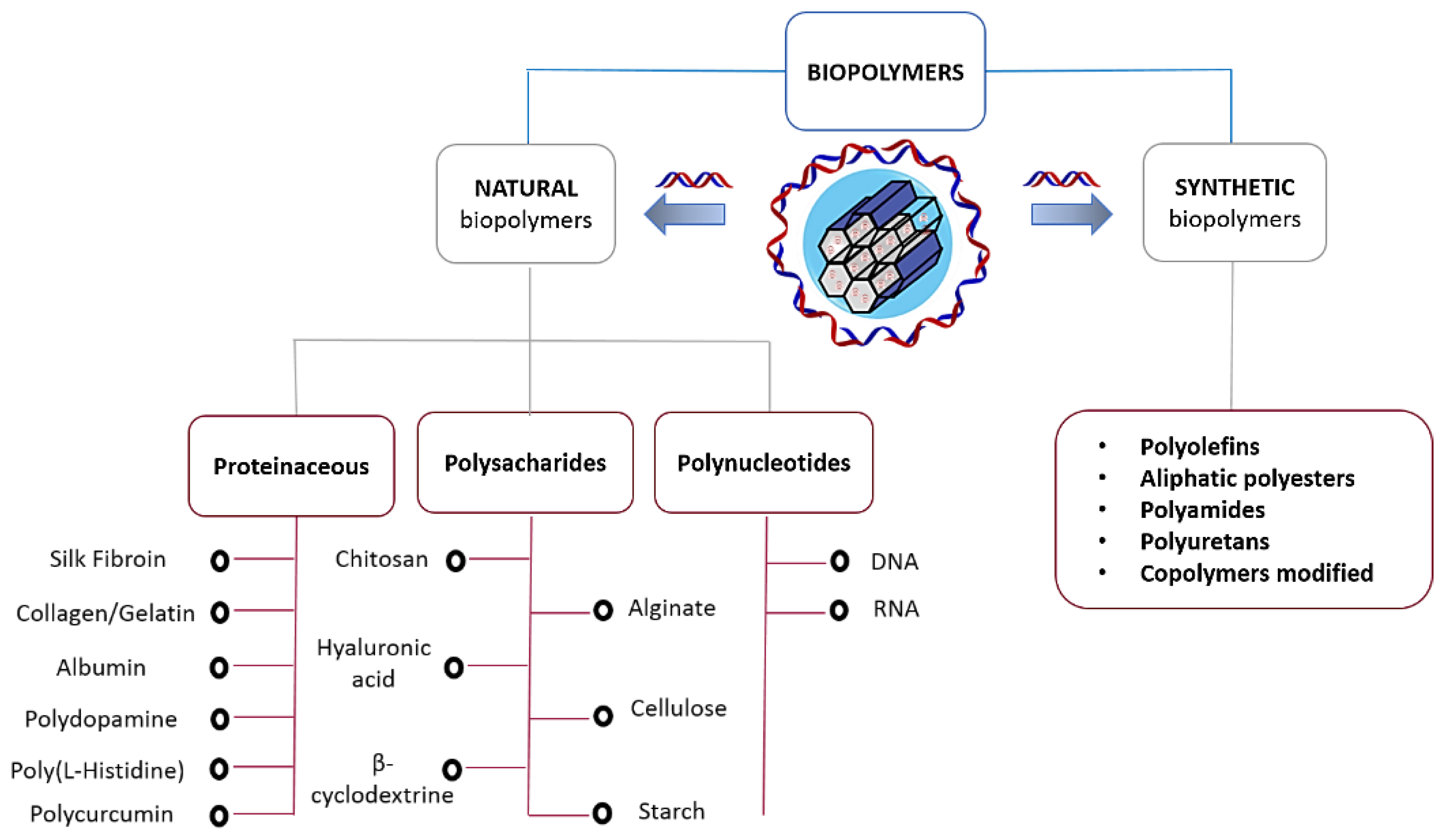
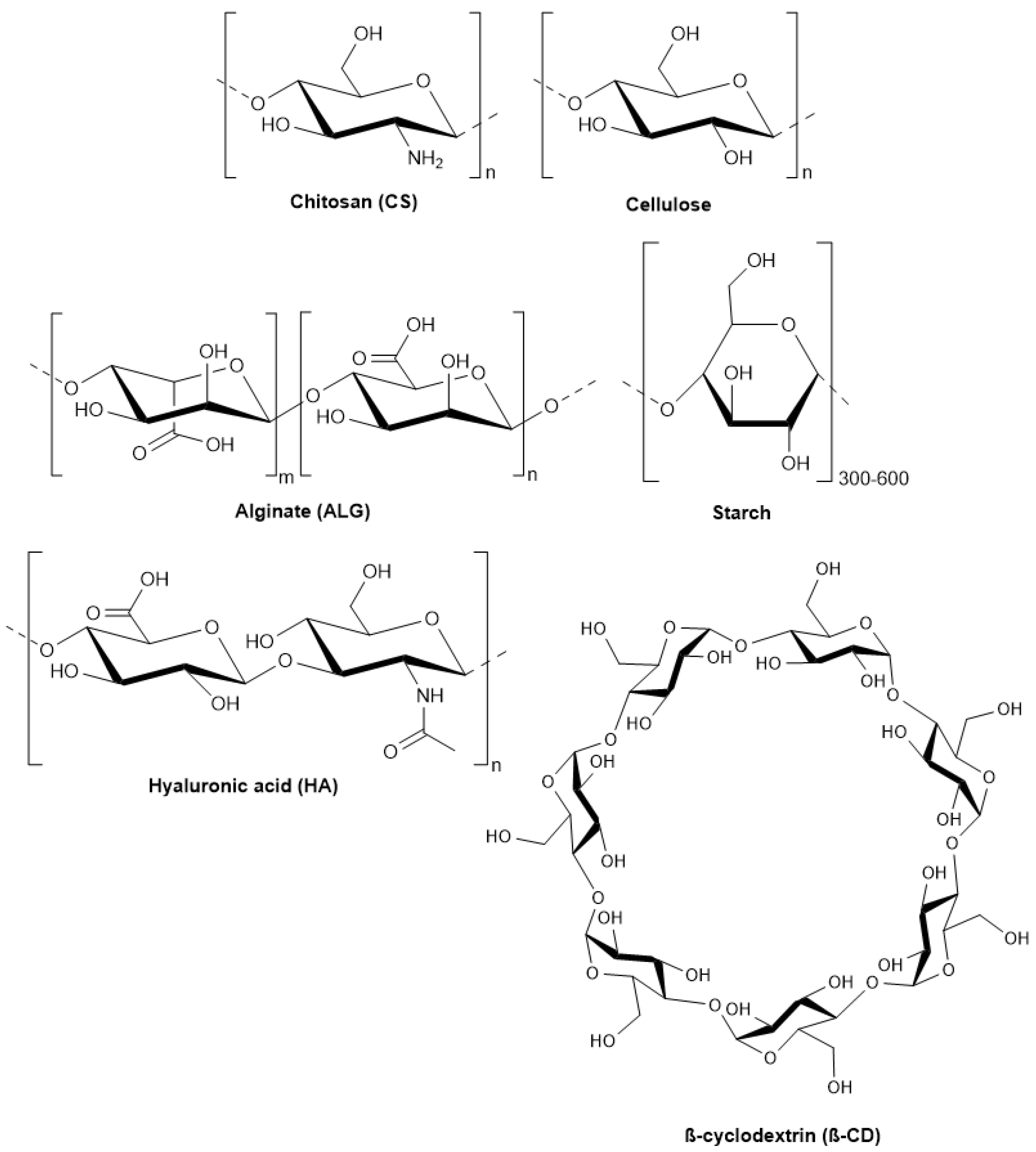
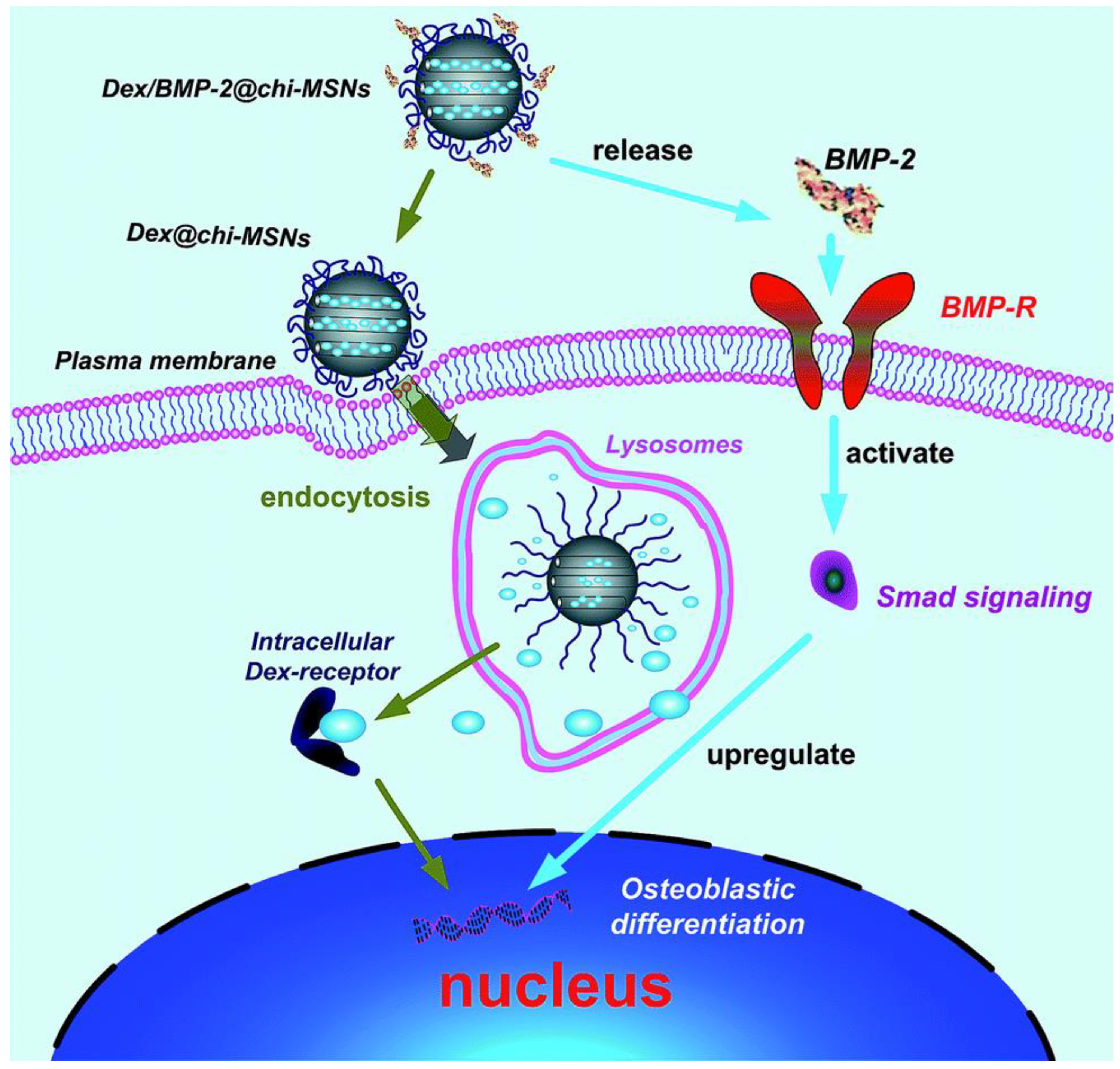
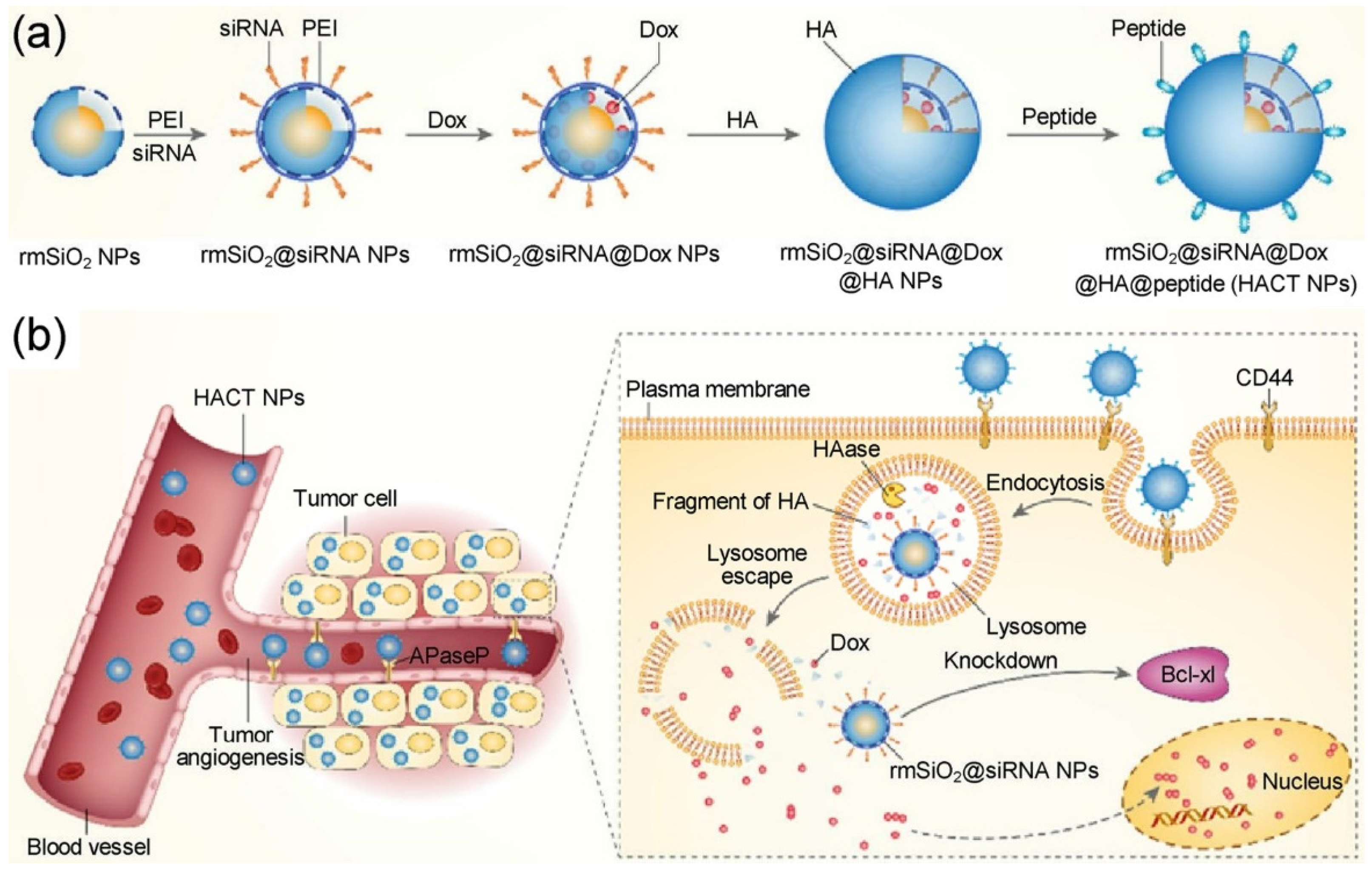
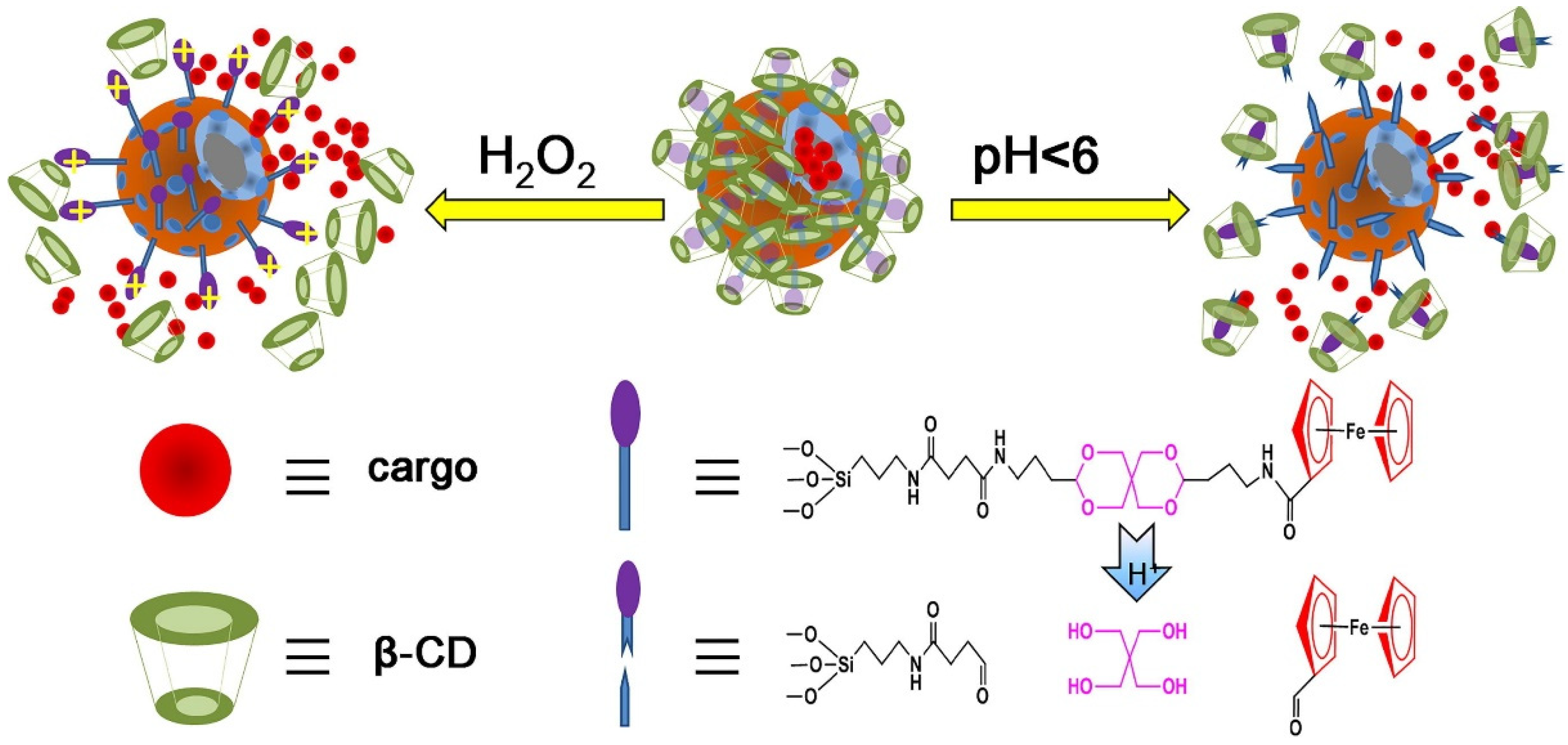
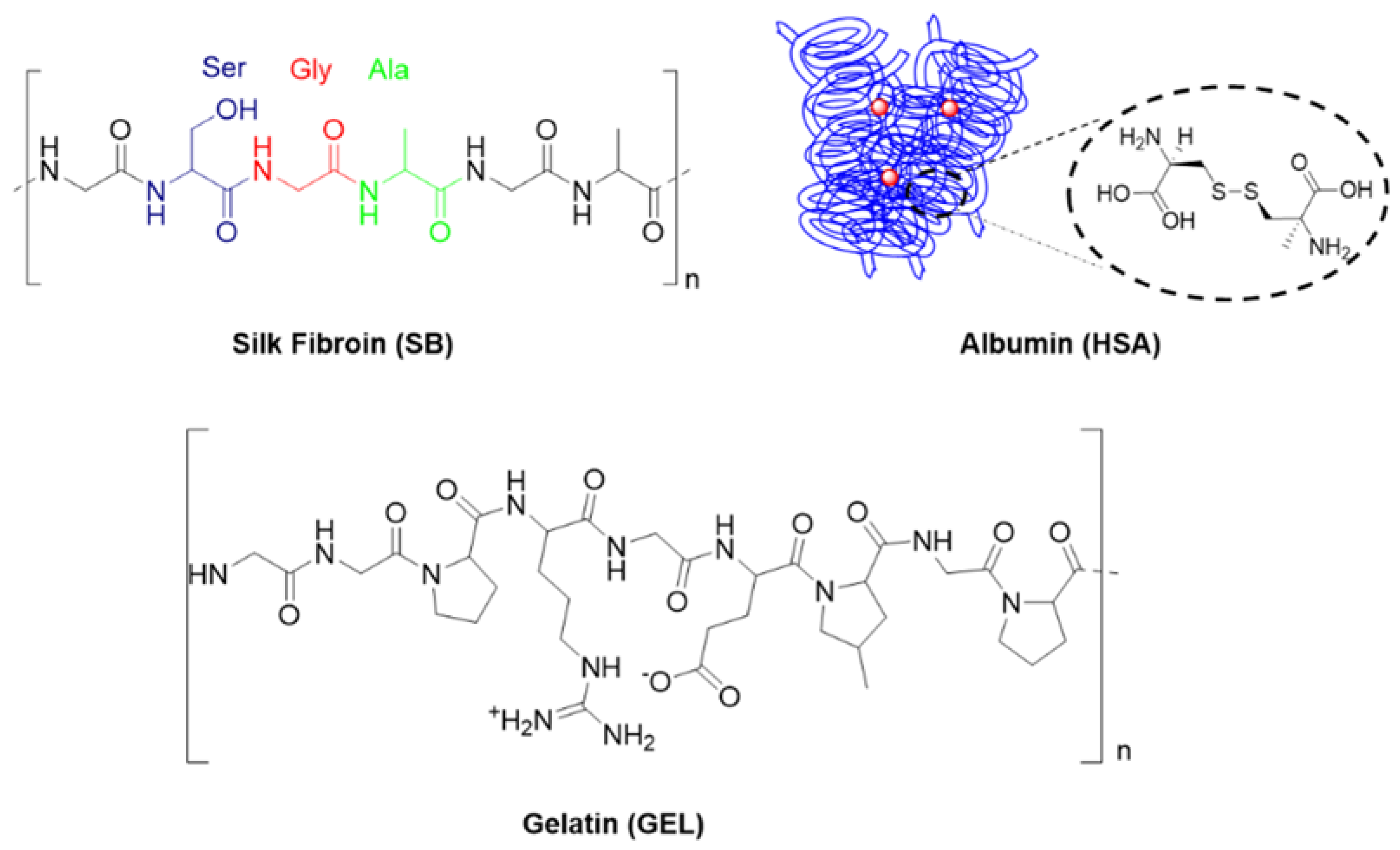
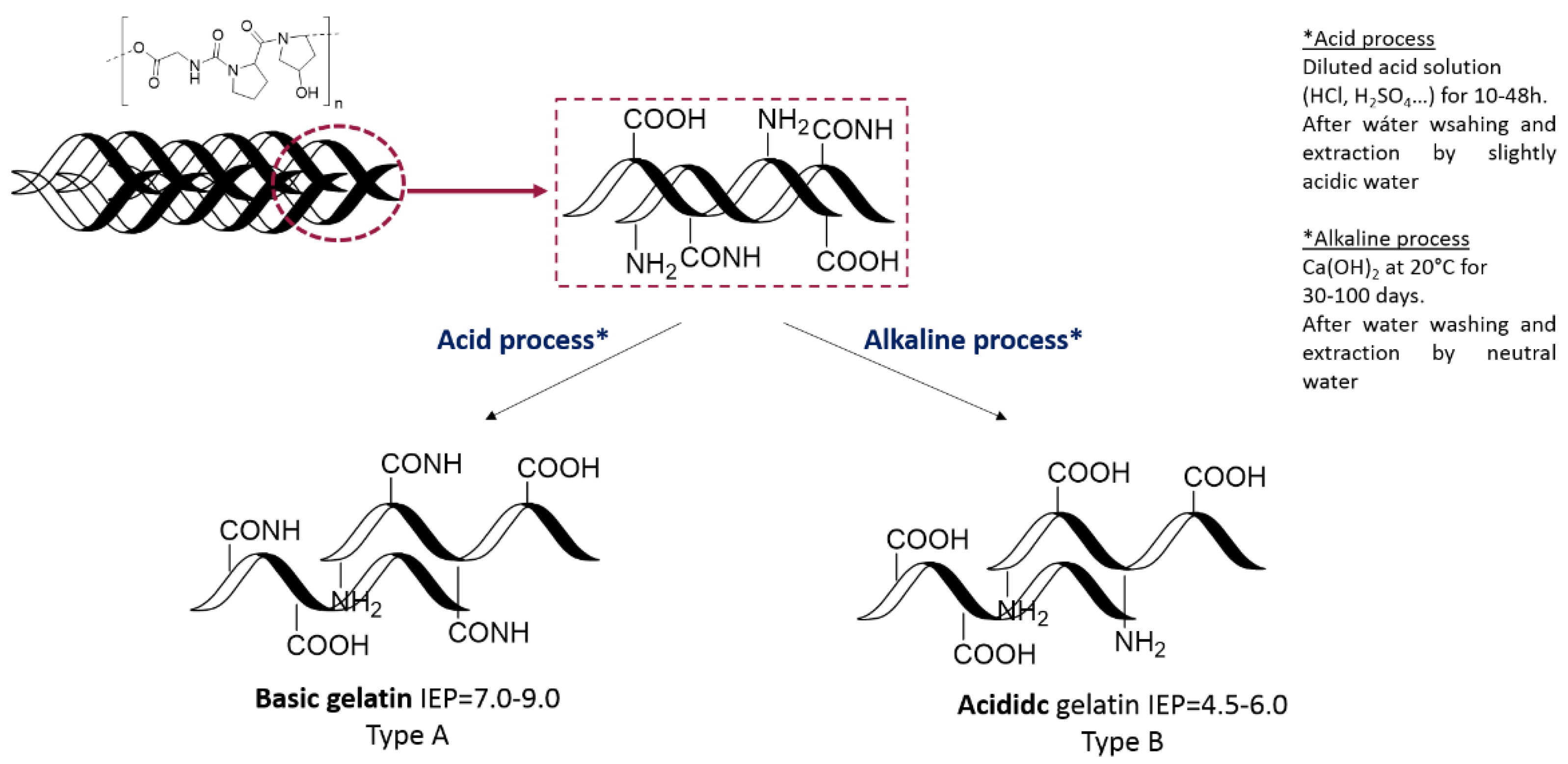
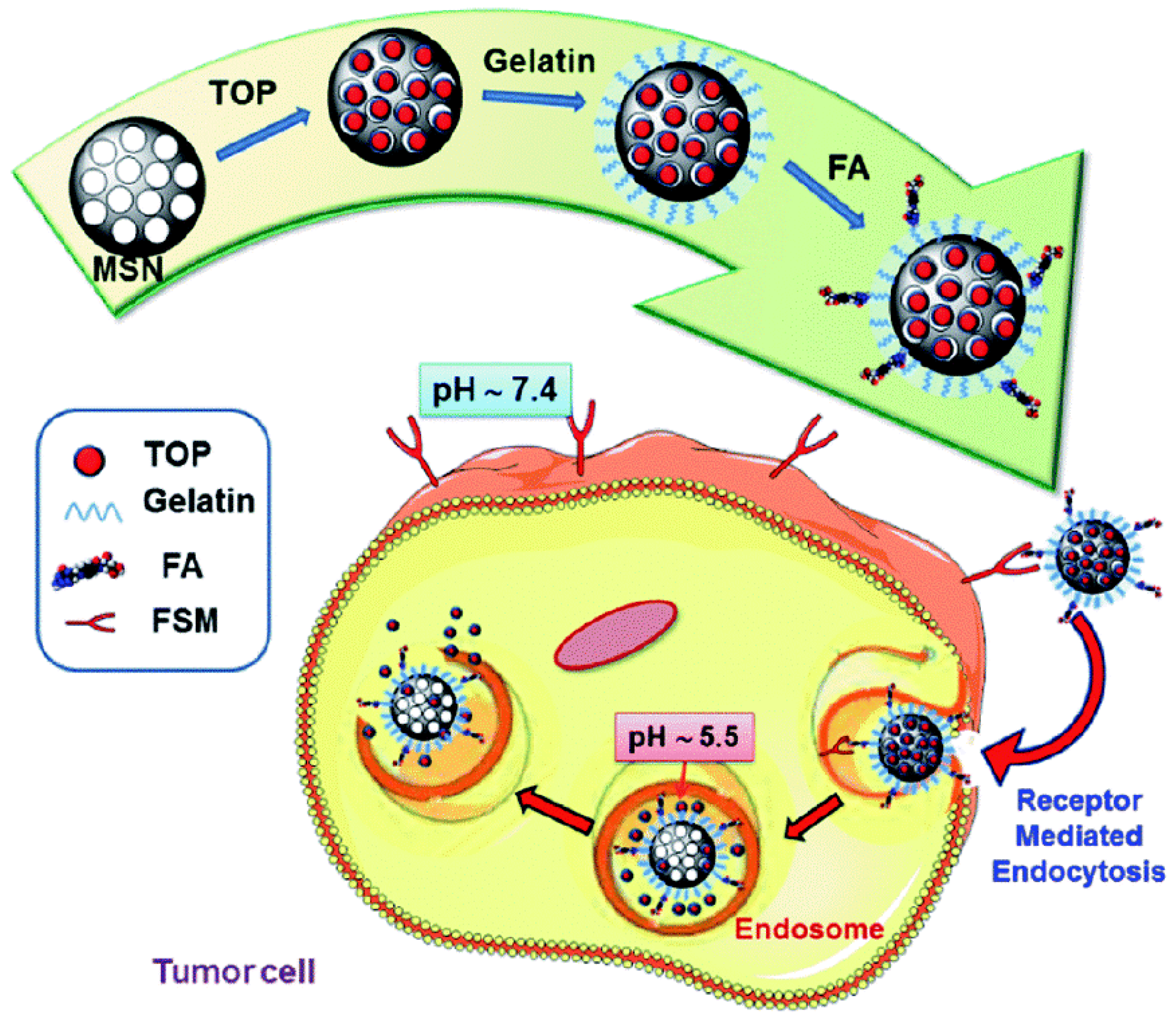
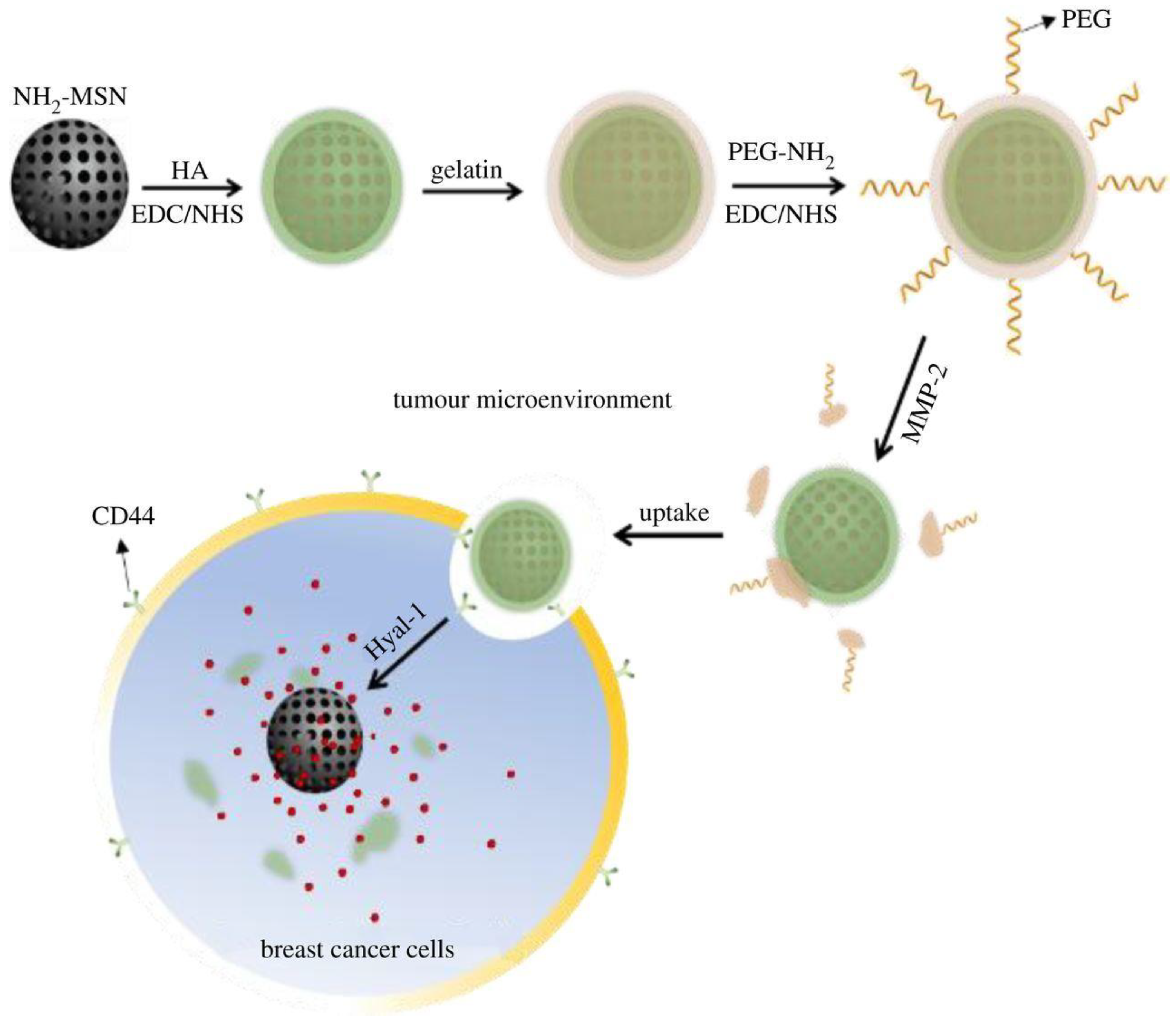
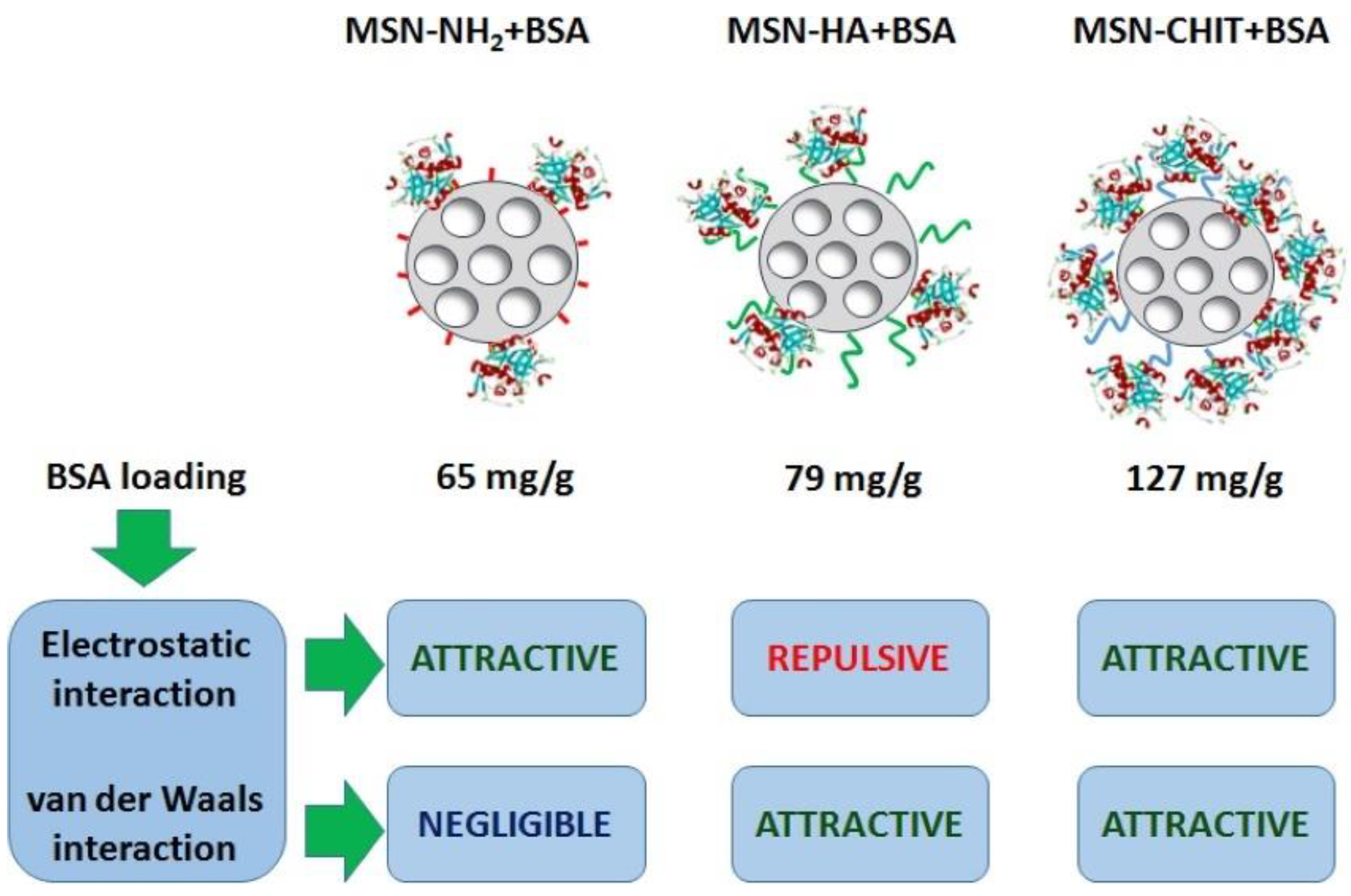
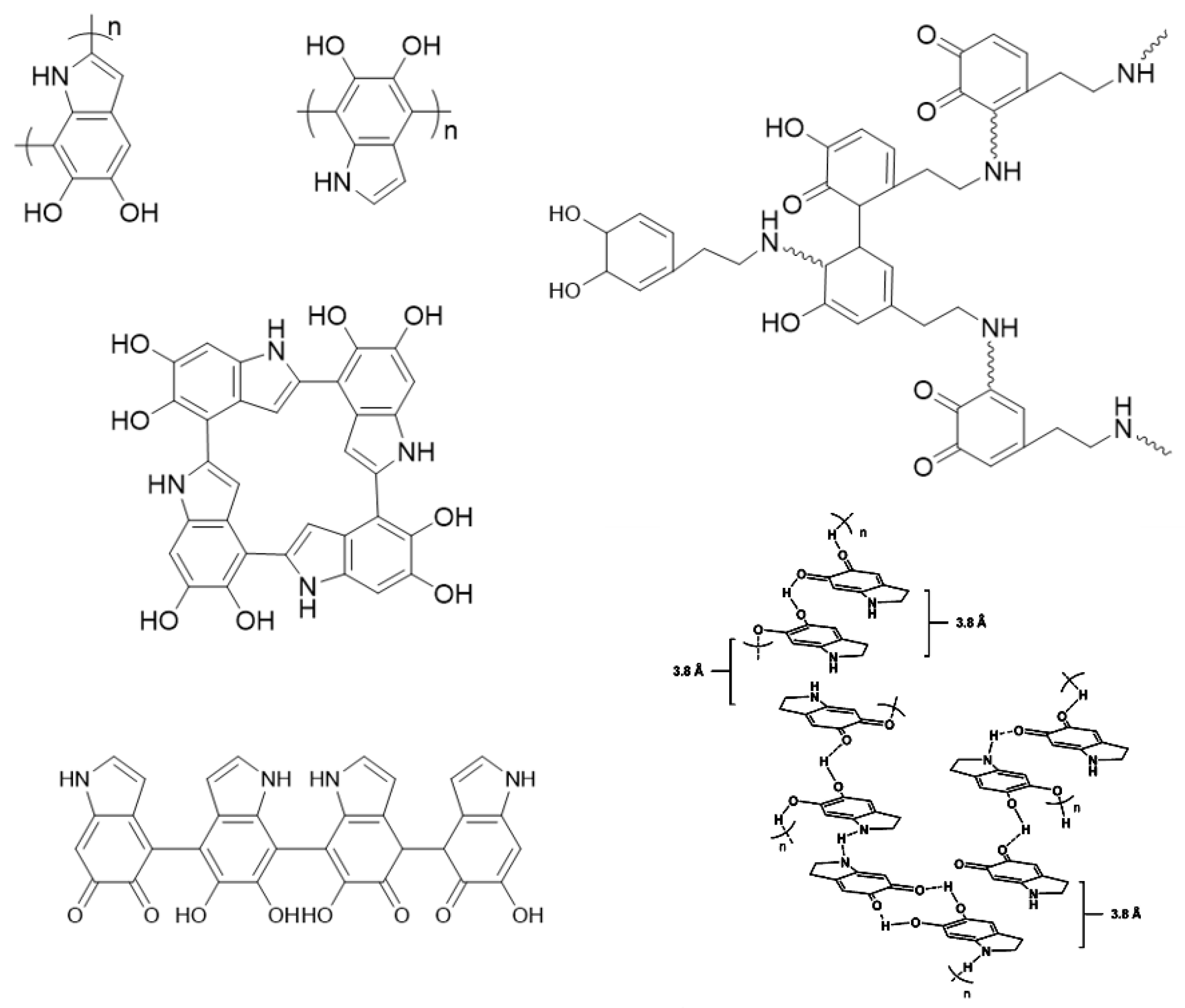
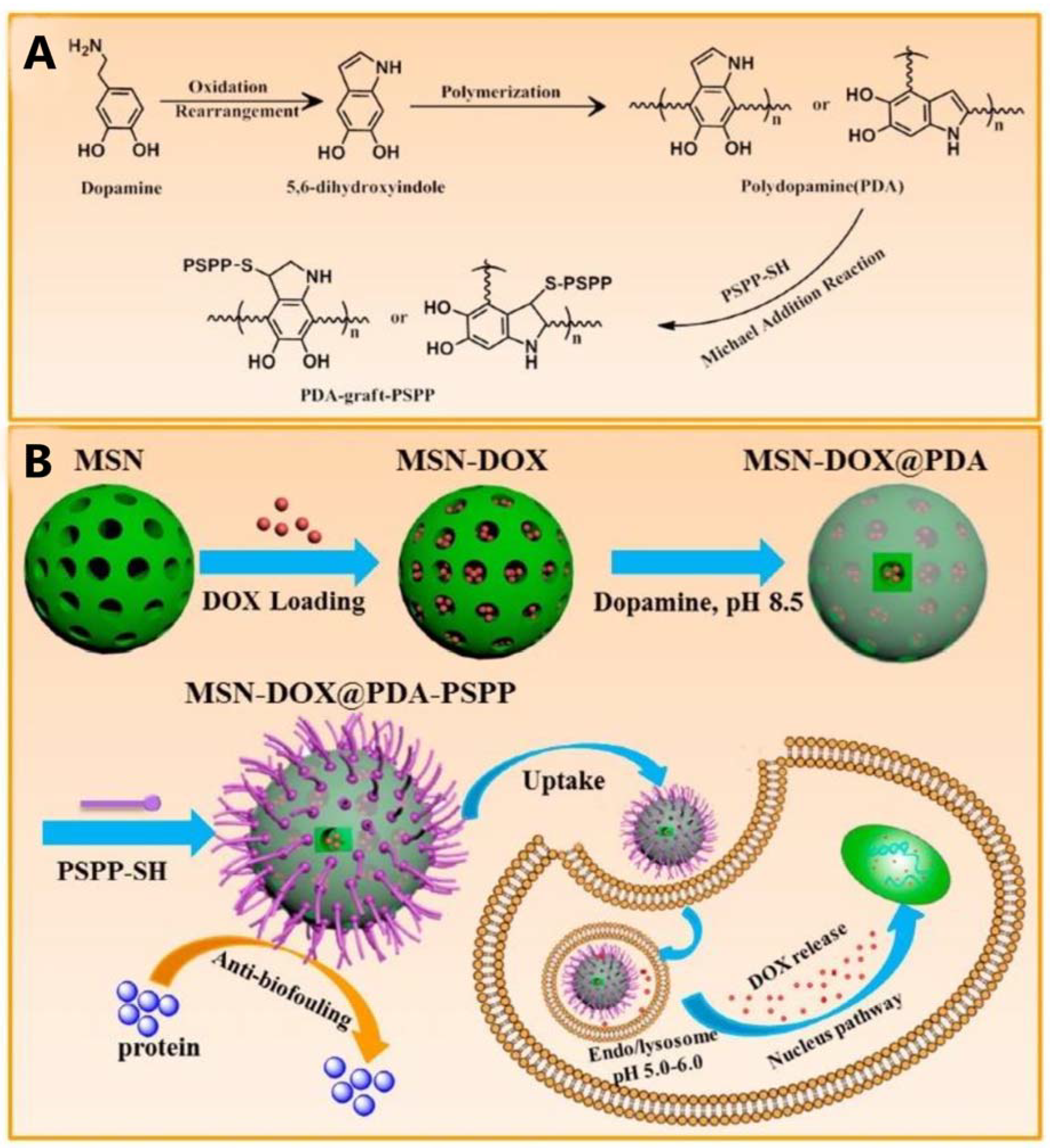
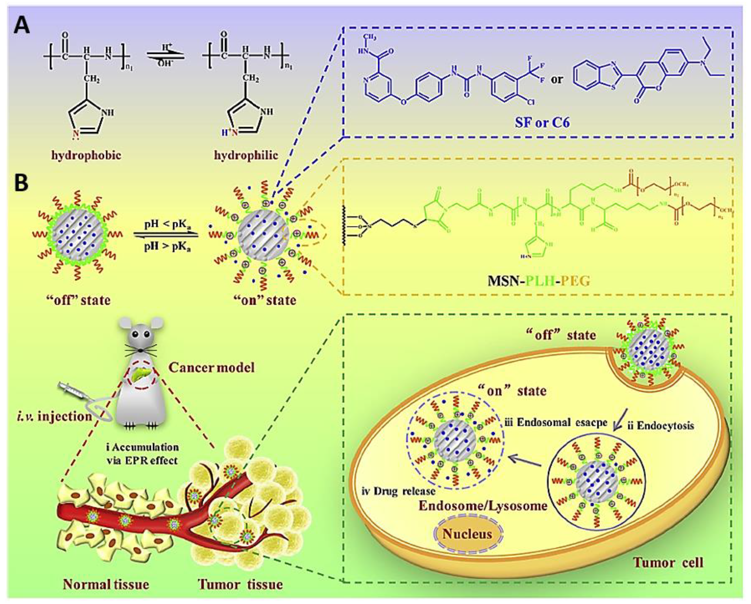
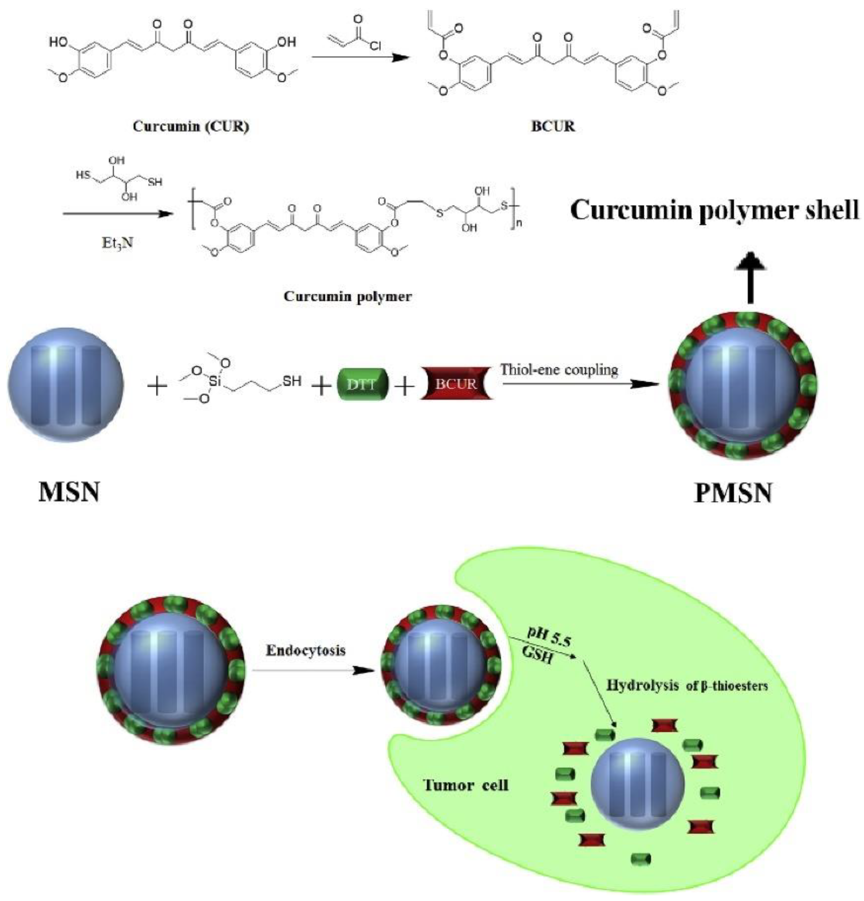
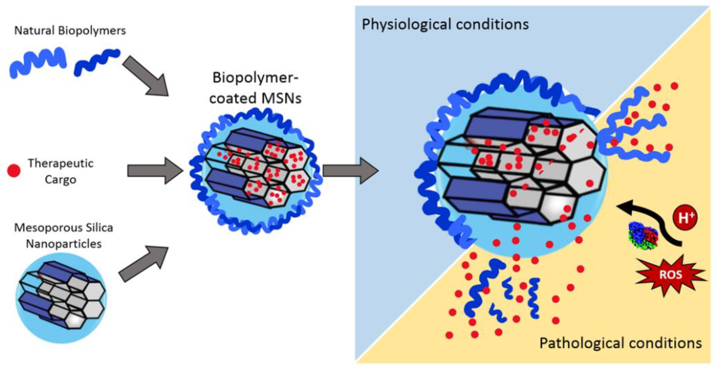
| Polysaccharides | |||||||
|---|---|---|---|---|---|---|---|
| Biopolymer | Endogenous Stimulus | Silica Core | Cargo | Targeting Ligand | Application | Ref. | |
| Chitosan | pH | Hollow MSNs | Doxorubicin | - | Cancer | [137] | |
| pH | MCM-41 | Curcumin | - | Glioblastoma | [138] | ||
| pH | MCM-41 | Doxorubicin and antimiR-21 | AS1411 aptamer | Colorectal and breast cancer | [139] | ||
| pH and redox | Mn-doped MSNs | Doxorubicin and Tariquidar | Folic acid and hyaluronic acid | Drug resistant and cancer stem cells | [140] | ||
| enzymatic | Hollow MSNs | Doxorubicin | - | Cancer | [141] | ||
| pH | MCM-41 | Dexamethasone and BMP-2 | - | In vivo bone regeneration | [142] | ||
| Alginate | pH and redox | MCM-41 | Doxorubicin | - | Cancer | [143] | |
| Alginate/ Chitosan | pH | MCM-41 | Doxorubicin | - | Cancer | [144] | |
| Hyaluronic acid | pH | Fe3O4NPs @MSNs | Doxorubicin | - | Targeted treatment of breast cancer | [145] | |
| enzymatic | MCM-41 | 5-fluorouracil | - | Targeted treatment of colon cancer | [146] | ||
| enzymatic | MCM-41 | Simvastatin | - | Atherosclerosis | [147] | ||
| Enzymatic and pH | MCM-41 | Rosuvastatin | CD9 | Atherosclerosis | [148] | ||
| enzymatic | PLNPs a @MSNs | Cinnamaldehyde | - | Multidrug resistant antibacterial infections | [149] | ||
| Cellulose | Enzymatic and pH | MCM-41 | Doxorubicin | - | Cancer | [150] | |
| Carboxy methyl cellulose | pH | Hollow MSNs | Doxorubicin | AS1411 aptamer | Colorectal and breast cancer | [151] | |
| β-cyclodextrin | pH and redox | MSNanorods | Doxorubicin | Hyaluronic acid | Cancer | [152] | |
| redox | Hollow MSNs | Glabridin | Ada-R8 | Transdermal delivery | [153] | ||
| Protein-based biopolymers | |||||||
| Biopolymer | Endogenous stimulus | Silica core | Cargo | Targeting ligand | Application | Ref. | |
| Silk Fibroin | pH | MCM-41 | Tioguanine (TG) | - | Leukemia | [154] | |
| Gelatin | pH | MCM-41 | Topotecan (TOP) | Folic Acid (FA) | Cancer | [155] | |
| Enzymatic (MMP-2) | MCM-41 | Doxorubicin | Folic Acid (FA) | Cancer | [103] | ||
| Bienzymatic (MMP-2 and Hyaluronidase) | MCM-41 | Doxorubicin | Hyaluronic Acid (HA) | Breast cancer | [156] | ||
| Albumin (HSA b and/or BSA c) | redox | MCM-41 | Epirubicin (EPI) | Folic acid | Cancer | [157] | |
| pH | MCM-41 | Gemcitabine | - | Cancer | [158] | ||
| Polydopamine | pH | MCM-41 | Doxorubicin | - | Cancer | [159] | |
| pH | MCM-41 | Anderson-type polyoxomolybdate | Glucosamine | Breast Cancer | [160] | ||
| pH | MCM-41 | Desipramine (DES) | - | Cancer | [161] | ||
| Poly(L-histidine) | pH | MCM-41 | Sorafenib | - | Cancer | [162] | |
| PCUR d | BCUR e | Enzymatic (GSH) | MCM-41 | Doxorubicin | - | Cancer-therapeutic imaging | [163] |
| CUR f | pH | MCM-41 | Doxorubicin and Curcumin | - | Cancer | [164] | |
Disclaimer/Publisher’s Note: The statements, opinions and data contained in all publications are solely those of the individual author(s) and contributor(s) and not of MDPI and/or the editor(s). MDPI and/or the editor(s) disclaim responsibility for any injury to people or property resulting from any ideas, methods, instructions or products referred to in the content. |
© 2023 by the authors. Licensee MDPI, Basel, Switzerland. This article is an open access article distributed under the terms and conditions of the Creative Commons Attribution (CC BY) license (https://creativecommons.org/licenses/by/4.0/).
Share and Cite
Dumontel, B.; Conejo-Rodríguez, V.; Vallet-Regí, M.; Manzano, M. Natural Biopolymers as Smart Coating Materials of Mesoporous Silica Nanoparticles for Drug Delivery. Pharmaceutics 2023, 15, 447. https://doi.org/10.3390/pharmaceutics15020447
Dumontel B, Conejo-Rodríguez V, Vallet-Regí M, Manzano M. Natural Biopolymers as Smart Coating Materials of Mesoporous Silica Nanoparticles for Drug Delivery. Pharmaceutics. 2023; 15(2):447. https://doi.org/10.3390/pharmaceutics15020447
Chicago/Turabian StyleDumontel, Bianca, Verónica Conejo-Rodríguez, María Vallet-Regí, and Miguel Manzano. 2023. "Natural Biopolymers as Smart Coating Materials of Mesoporous Silica Nanoparticles for Drug Delivery" Pharmaceutics 15, no. 2: 447. https://doi.org/10.3390/pharmaceutics15020447
APA StyleDumontel, B., Conejo-Rodríguez, V., Vallet-Regí, M., & Manzano, M. (2023). Natural Biopolymers as Smart Coating Materials of Mesoporous Silica Nanoparticles for Drug Delivery. Pharmaceutics, 15(2), 447. https://doi.org/10.3390/pharmaceutics15020447