Article

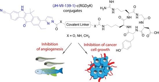
Synthesis and Anti-Angiogenic Activity of Novel c(RGDyK) Peptide-Based JH-VII-139-1 Conjugates

1
Department of Chemistry, Aristotle University of Thessaloniki, University Campus, 54124 Thessaloniki, Greece
2
Zebrafish Disease Model Laboratory, Biomedical Research Foundation Academy of Athens, 11527 Athens, Greece
*
Author to whom correspondence should be addressed.
Pharmaceutics 2023, 15(2), 381; https://doi.org/10.3390/pharmaceutics15020381
Submission received: 23 December 2022 / Revised: 18 January 2023 / Accepted: 19 January 2023 / Published: 22 January 2023

Abstract

:
Peptide–drug conjugates are delivery systems for selective delivery of cytotoxic agents to target cancer cells. In this work, the optimized synthesis of JH-VII-139-1 and its c(RGDyK) peptide conjugates is presented. The low nanomolar SRPK1 inhibitor, JH-VII-139-1, which is an analogue of Alectinib, was linked to the ανβ3 targeting oligopeptide c(RGDyK) through amide, carbamate and urea linkers. The chemostability, cytotoxic and antiangiogenic properties of the synthesized hybrids were thoroughly studied. All conjugates retained mid nanomolar-level inhibitory activity against SRPK1 kinase and two out of four conjugates, geo75 and geo77 exhibited antiproliferative effects with low micromolar IC50 values against HeLa, K562, MDA-MB231 and MCF7 cancer cells. The activities were strongly related to the stability of the linkers and the release of JH-VII-139-1. In vivo zebrafish screening assays demonstrated the ability of the synthesized conjugates to inhibit the length or width of intersegmental vessels (ISVs). Flow cytometry experiments were used to test the cellular uptake of a fluorescein tagged hybrid in MCF7 and MDA-MB231 cells that revealed a receptor-mediated endocytosis process. In conclusion, most conjugates retained the inhibitory potency against SRPK1 as JH-VII-139-1 and demonstrated antiproliferative and antiangiogenic activities. Further animal model experiments are needed to uncover the full potential of such peptide conjugates in cancer therapy and angiogenesis-related diseases.

Graphical Abstract

1. Introduction

Angiogenesis is described as the formation of new blood vessels from pre-existing vessels [1,2]. It is a vital physiological process for growth, development, and wound repair by which various materials, oxygen and nutrients are delivered to tissues and cellular wastes are removed. In cancer, abnormal angiogenesis is related to cell growth, tumorigenesis, and metastases [3,4,5]. However, recent findings implicate angiogenesis in many other diseases [6], such as diabetic retinopathy, neovascular glaucoma [7], autoimmune diseases, multiple sclerosis [8], rheumatoid arthritis [9], cardiovascular diseases [10], atherosclerosis [11], and cerebral ischemia. Angiogenesis is regulated by many pathways and signaling molecules such as the vascular endothelial growth factor (VEGF), the transforming growth factor (TGF)-β, the angiopoietin-1 and 2, the placental growth factor (PGF), αvβ3 and αvβ5 integrins [12], the Notch, Akt, Jak/STAT, ephrin/Eph signaling pathways and others [13,14]. VEGF binds to receptor VEGFR and this interaction predominantly regulates pathological and physiological angiogenesis [15,16,17]. The VEGFR signaling is monitored at multiple levels [18]. For example, serine-arginine (SR) protein kinases (SRPKs) regulate the splicing of proangiogenic VEGF165 by phosphorylation of the serine/arginine splicing factor 1 (SRSF1) [19]. Therefore, targeting SRPK1 kinase to regulate angiogenesis in angiogenic disorders has been proposed by several research groups. Bates et al. has used the SRPK1 inhibitor SRPIN340 to reduce proangiogenic VEGF165 and inhibit melanoma tumor growth in vivo [20]. Oltean et al. demonstrated that SPHINX and SRPIN340 treatment changed the expression of VEGF165 towards the anti-angiogenic splice isoform VEGF165b and decreased tumor growth in orthotopic PC3 models in mice [21].
In previous research efforts by our group, the cyclic pentapeptide c(RGDyK), which is a potent integrin αvβ3 ligand, was employed for the targeted delivery of various anticancer drugs and bioactive molecules such as gemcitabine [22], the triterpenoids cucurbitacins [23], and platinum complexes [24]. c(RGDyK) peptide potently binds αvβ3 integrins (IC50 = 3.8 ± 0.42 nM) and, less potently, αvβ5 (IC50 = 503 ± 55 nM), αvβ6 (IC50 = 86 ± 7 nM), and α5β1 integrins (IC50 = 236 ± 45 nM) [25]. In earlier studies, the SRPIN803 inhibitor which binds to SRPK1 and CK2 kinases was coupled to c(RGDyK) peptide aiming to form a new conjugate with improved antiangiogenic activities compared to both the peptide and the SRPIN803 (Figure 1) [26]. Although the antiangiogenic activities of the synthesized SRPIN803-c(RGDyK) hybrids and SRPIN803 were evident in zebrafish embryos, an unfavorable stability profile of SRPIN803 was observed as SRPIN803 and its derivatives undergo a retro-Knoevenagel reaction. As a next step, we intended to develop new anti-angiogenic compounds based on the more potent SRPK1 inhibitor, JH-VII-139-1 [27]. JH-VII-139-1 came up from the optimization of FDA-approved ALK inhibitor Alectinib [28], which previously was shown to bind to SRPK1. Gray and coworkers demonstrated that JH-VII-139-1 potently inhibits SRPK1 with an IC50 value of 1.1 nM and blocks angiogenesis in an age-related macular degeneration (AMD) animal model [27]. In this work, the synthesis of JH-VII-139-1 peptide conjugates with c(RGDyK) and the properties of the new hybrid compounds against SRPK1, cancer cell growth and angiogenesis are reported.

2. Materials and Methods

2.1. Chemistry

2.1.1. General Experimental Details

All reactions were carried out under an atmosphere of Argon unless otherwise specified. Commercial reagents were used without further purification. Reactions were monitored by TLC and using UV light as a visualizing agent and aqueous ceric sulfate/phosphomolybdic acid, ethanolic p-anisaldehyde solution, potassium permanganate solution, and heat as developing agents. The 1H and 13C NMR spectra were recorded at 500 and 126 MHz (Agilent) with tetramethylsilane as an internal standard. Chemical shifts are indicated in δ values (ppm) from internal reference peaks (TMS 1H 0.00; CDCl3 1H 7.26, 13C 77.16, (CD3)2SO 1H 2.50, 13C 39.52, (CD3)2CO 1H 2.05, 13C 29.84, 206.26). LC-MS analysis was performed on a LC-20AD Shimadzu connected to Shimadzu LCMS-2010EV (Shimadzu Kyoto, Japan) equipped with C18 analytical column (Supelco discovery C18, 5 μm 250 × 4.6 mm).

2.1.2. Synthetic Procedures

7-Methoxy-1,1-dimethyl-3,4-dihydronaphthalen-2(1H)-one, 2. To a solution of 7-methoxy-2-tetralone (5 g, 28.5 mmol) in THF (20 mL) were added tetrabutylammonium bisulfate (1.53 g, 4.5 mmol), methyl iodide (5.25 mL, 85.5 mmol), and 50% aqueous KOH solution (32.5 mL) and stirred at room temperature for 2 h. The mixture was diluted with EA and washed with water and brine. The organic layer was dried over Na2SO4 and concentrated under reduced pressure. The residue was purified by flash column chromatography (gradient elution PS–EA, 15:1 to 10:1) to afford the desired product, 2 (5.3 g, 91%) as a white solid. The spectral data were in accordance with those reported in the literature [29]. 2: 1H NMR (500 MHz, CDCl3) δ 7.09 (d, J = 8.3 Hz, 1H), 6.88 (d, J = 2.5 Hz, 1H), 6.74 (dd, J = 8.3, 2.5 Hz, 1H), 3.81 (s, 3H), 3.03 (t, J = 6.9 Hz, 2H), 2.66 (t, J = 6.9 Hz, 2H), 1.42 (s, 6H). 13C NMR (126 MHz, CDCl3) δ 214.7, 158.8, 145.0, 129.1, 127.5, 112.3, 111.4, 55.4, 48.0, 37.6, 27.8, 26.9. ESI-MS, positive mode: m/z calcd mass for C13H16O2Na [M + Na] + = 227.10, was found 227.05.
6-Bromo-7-methoxy-1,1-dimethyl-3,4-dihydronaphthalen-2(1H)-one, 3. To a solution of 2 (2.00 g, 9.8 mmol) in CH3CN (40 mL) was added N-bromosuccinimide (1.92 g, 10.8 mmol). After stirring for 2.5 h at room temperature, the reaction mixture was concentrated under reduced pressure. The residue was purified by column chromatography (gradient elution PS–EA, 15:1 to 13:1) to yield 3 (2.55 g, 92%) as a white solid. 1H-NMR data were in accordance with those reported in the literature [30]. 3: 1H NMR (500 MHz, CDCl3) δ 7.35 (s, 1H), 6.83 (s, 1H), 3.90 (s, 3H), 3.01 (t, J = 6.9 Hz, 2H), 2.65 (t, J = 6.9 Hz, 2H), 1.43 (s, 6H). 13C NMR (126 MHz, CDCl3) δ 213.8, 155.1, 144.2, 132.7, 129.0, 110.0, 109.7, 56.5, 48.0, 37.2, 27.5, 26.9.
3-Hydrazinylbenzonitrile, 5. 4-Cyanoaniline (472 mg, 4 mmol) was added to a round-bottomed flask containing 10 mL 6N HCl. The solution was vigorously stirred at 0 °C for 10 min. Sodium nitrite (280 mg, 4 mmol) dissolved in 2 mL water was added dropwise to the aniline solution, and the resulting mixture was stirred at 0 °C for an additional 1 h. To a beaker containing SnCl2 (1.9 g, 10 mmol) was added 10 mL of concentrated HCl, and the solution was sonicated and cooled to 0 °C. The SnCl2 solution was then added dropwise to the diazonium salt solution, and the mixture was stirred for additional 90 min at room temperature. Then, sodium hydroxide (7.2 g, 0.18 mol) dissolved in 20 mL water was added to quench the reaction. The solution was extracted with 20 mL of diethyl ether three times, and the organic layers were combined and concentrated under reduced pressure to give the crude product, 5 (300 mg, 56%), which was further purified by recrystallization in hot ethanol. The spectral data were in accordance with those reported in the literature [31]. 5: 1H NMR (500 MHz, DMSO-d6) δ 7.27–7.20 (m, 2H), 7.08 (d, J = 1.1 Hz, 1H), 7.01 (dd, J = 8.4, 1.1 Hz, 1H), 6.93 (d, J = 7.4 Hz, 1H), 4.12 (s, 2H). 13C NMR (126 MHz, DMSO-d6) δ 152.9, 129.7, 119.7, 119.4, 116.1, 113.2, 111.4.
9-Bromo-8-methoxy-6,6-dimethyl-6,11-dihydro-5H-benzo[b]carbazole-3-carbonitrile, 6a. A mixture of 3 (2.8 g, 9.89 mmol), 5 (1.58 g, 11.9 mmol), and trifluoroacetic acid (48 mL) was refluxed at 80 °C for 2 h. The reaction mixture was cooled and neutralized by saturated aqueous Na2CO3, then by NaHCO3 and extracted with EA. The combined organic layers were washed with brine, dried over Na2SO4, and concentrated under reduced pressure. The residue was resuspended in EA and precipitated solid (undesired regioisomer) was filtered off. The filtrate was evaporated under reduced pressure and purified by column chromatography (isocratic elution PS–EA 4:1) to yield 6a as an orange solid (1.9 g, 50%). 6a: 1H NMR (500 MHz, acetone-d6) δ 10.79 (s, 1H), 7.75 (s, 1H), 7.67 (d, J = 8.1 Hz, 1H), 7.55 (s, 1H), 7.34 (dd, J = 8.2, 1.2 Hz, 1H), 7.31 (s, 1H), 4.08 (s, 2H), 3.96 (s, 3H), 1.77 (s, 6H). 13C NMR (126 MHz, acetone-d6) δ 155.5, 145.4, 144.8, 136.6, 134.2, 131.2, 130.6, 127.7, 122.6, 119.8, 116.2, 111.1, 110.1, 106.8, 104.1, 56.7, 37.1, 31.1, 25.9. ESI-MS, negative mode: m/z calcd mass for C20H16BrN2O [M − H] = 379.04, was found 380.85.
9-Bromo-8-methoxy-6,6-dimethyl-6,11-dihydro-5H-benzo[b]carbazole-1-carbonitrile, 6b: 1H NMR (500 MHz, DMSO) δ 11.68 (s, 1H), 7.68 (d, J = 7.9 Hz, 1H), 7.54 (s, 1H), 7.46 (d, J = 7.1 Hz, 1H), 7.26 (s, 1H), 7.21 (t, J = 7.6 Hz, 1H), 4.23 (s, 2H), 3.91 (s, 3H), 1.70 (s, 6H). 13C NMR (126 MHz, DMSO) δ 154.1, 143.7, 143.6, 136.4, 133.0, 126.3, 125.7, 124.7, 120.8, 119.4, 116.2, 110.4, 108.9, 103.2, 99.8, 56.4, 35.9, 30.8, 25.3.
9-Bromo-8-methoxy-6,6-dimethyl-11-oxo-6,11-dihydro-5H-benzo[b]carbazole-3-carbonitrile, 7. To a solution of 6a (2.1 g, 4.96 mmol) in THF (21.4 mL) and water (2.14 mL) was gradually added DDQ (3.38 g, 14.9 mmol). After stirring for 3 h at 0 °C, the reaction mixture was evaporated under reduced pressure and the residue was dissolved in EA. The solution was washed with aqueous NaOH, water, and brine, dried over Na2SO4, filtered, and concentrated under reduced pressure. The residue was purified with column chromatography (gradient elution PS–EA 3:1 to 1:2) to afford 7 (1.58 g, 81%) as yellow solid. 1H-NMR data were in accordance with those reported in the literature [31]. 7: 1H NMR (500 MHz, acetone-d6) δ 8.42 (s, 1H), 8.41 (s, 1H), 7.92 (d, J = 0.6 Hz, 1H), 7.57 (dd, J = 8.1, 1.4 Hz, 1H), 7.50 (s, 1H), 4.09 (s, 3H), 1.91 (s, 6H). 13C NMR (126 MHz, acetone-d6) δ 178.6, 160.2, 159.9, 150.7, 136.9, 131.8, 128.9, 127.3, 125.7, 123.0, 120.4, 117.0, 111.4, 110.8, 110.6, 106.8, 57.2, 38.1, 30.4. ESI-MS, negative mode: m/z calcd mass for C20H14BrN2O2 [M − H] = 393.02, was found 394.85.
9-Ethyl-8-methoxy-6,6-dimethyl-11-oxo-6,11-dihydro-5H-benzo[b]carbazole-3-carbonitrile, 8. To a degassed mixture of 7 (1.52 g, 3.85 mmol), Cs2CO3 (3.76 g, 11.5 mmol), Pd(dppf)Cl2 (57.7 mg, 0.08 mmol) and DMF (55.7 mL) in a Schlenk flask under argon atmosphere was added a solution of triethylborane in THF (11.5 mL, 1M solution, 11.5 mmol) and the reaction was stirred at 86 °C overnight. The reaction mixture was then cooled, diluted with EA, washed several times with water, dried over Na2SO4, filtered, and concentrated under reduced pressure. The residue was purified by column chromatography (gradient elution PS–EA 2:1 to 1:2) to yield 8 (870 mg, 66%) as yellow crystalline solid. 8: 1H NMR (500 MHz, acetone-d6) δ 11.69 (s, 1H), 8.47 (d, J = 8.1 Hz, 1H), 8.12 (s, 1H), 7.90 (s, 1H), 7.57 (dd, J = 8.1, 0.5 Hz, 1H), 7.34 (s, 1H), 4.02 (s, 3H), 2.71 (q, J = 7.5 Hz, 2H), 1.88 (s, 6H), 1.24 (t, J = 7.5 Hz, 3H). 13C NMR (126 MHz, acetone-d6) δ 180.2, 161.9, 160.2, 149.1, 136.8, 132.2, 129.1, 127.4, 125.7, 125.5, 123.0, 120.6, 116.8, 111.1, 108.3, 106.4, 56.2, 37.8, 30.7, 23.6, 14.3. ESI-MS, positive mode: m/z calcd mass for C22H20N2O2 [M] + = 344.15, was found 344.95.
9-Ethyl-8-hydroxy-6,6-dimethyl-11-oxo-6,11-dihydro-5H-benzo[b]carbazole-3-carbonitrile, 9. Pyridine (141 μL, 1.74 mmol) was slowly added to a hydrochloric acid solution in 1,4-dioxane (436 μL, 4 M, 1.74 mmol) at 0 °C under argon. The mixture was stirred for 5 min and then concentrated under reduced pressure to form white highly hygroscopic pyridinium chloride salt. Both 8 (12 mg, 34.8 μmol) and pyridinium chloride (1.74 mmol) were quickly transferred to a sealed tube and stirred at 170 °C overnight. The reaction mixture was dissolved in water, neutralized by saturated aqueous NaHCO3, and extracted with EA three times. The combined organic layers were washed with aqueous NaHCO3 and brine, dried over Na2SO4, filtered, and concentrated under reduced pressure. The residue was purified by flash column chromatography (gradient elution of PS–EA 2:1 to 1:2) to yield 9 as a beige solid (9.5 mg, 83%). 9: 1H NMR (500 MHz, DMSO-d6) δ 12.67 (s, 1H), 10.20 (s, 1H), 8.32 (dd, J = 8.1, 0.4 Hz, 1H), 7.98 (d, J = 0.6 Hz, 1H), 7.94 (s, 1H), 7.59 (dd, J = 8.1, 1.4 Hz, 1H), 7.10 (s, 1H), 2.62 (q, J = 7.5 Hz, 2H), 1.70 (s, 6H), 1.19 (t, J = 7.5 Hz, 3H). 13C NMR (126 MHz, DMSO-d6) δ 179.2, 159.4, 159.3, 147.9, 135.6, 129.4, 127.8, 126.8, 124.7, 123.3, 121.7, 120.2, 116.4, 111.8, 109.4, 104.4, 36.2, 30.3, 22.5, 13.9. ESI-MS, positive mode: m/z calcd mass for C21H18N2O2 [M + H] + = 330.14, was found 330.90.
3-Cyano-9-ethyl-6,6-dimethyl-11-oxo-6,11-dihydro-5H-benzo[b]carbazol-8-yl trifluoromethanesulfonate, 10. A suspension of 9 (480 mg, 1.45 mmol) and K2CO3 (402 mg, 2.91 mmol) in DMF (13.7 mL) was slowly (portionwise) treated with N-phenyl-bis(trifluoromethanesulfonimide) (1.04 g, 2.91 mmol) at 0 °C. After 24 h, the reaction mixture was diluted with EA and poured into water. The layers were partitioned, and the organic phase washed with brine, dried over Na2SO4, and concentrated in vacuo. The residue was purified by silica gel chromatography (gradient elution PS–EA 3:1 to 1:1) to afford 10 (540 mg, 80% yield) as a yellow solid. 10: 1H NMR (500 MHz, acetone-d6) δ 11.87 (s, 1H), 8.44 (d, J = 8.1 Hz, 1H), 8.37 (s, 1H), 7.94 (d, J = 1.3 Hz, 1H), 7.84 (s, 1H), 7.60 (dd, J = 8.1, 1.3 Hz, 1H), 2.89 (q, J = 7.6 Hz, 2H), 1.91 (s, 6H), 1.36 (t, J = 7.6 Hz, 3H). 13C NMR (126 MHz, acetone-d6) δ 178.8, 160.3, 151.5, 149.1, 137.1, 136.3, 132.9, 129.5, 128.9, 126.1, 123.2, 120.7, 120.4, 119.7, 117.2, 111.2, 107.2, 37.9, 30.4, 23.4, 14.2. ESI-MS, positive mode: m/z calcd mass for C22H17F3N2O4S [M] + = 462.08, was found 462.95.
1-(Tetrahydro-2H-pyran-2-yl)-1H-pyrazole, 12. A mixture of pyrazole (1.43 g, 0.21 mol), 3,4-dihydro-2H-pyran (29 mL, 0.32 mol), and trifluoroacetic acid (80 μL, 1 mmol) was refluxed for 2 h. After quenching with sodium hydride (30 μg, 1.26 mmol), the mixture was subjected to column chromatography (gradient elution PS–EA 20:1 to 5:1) to yield 12 (3.2 g, 95%) as a colorless oil. The spectral data were in accordance with those reported in the literature [32]. 12: 1H NMR (500 MHz, CDCl3) δ 7.61 (d, J = 2.4 Hz, 1H), 7.56 (d, J = 1.4 Hz, 1H), 6.32–6.29 (m, 1H), 5.40 (dd, J = 9.7, 2.5 Hz, 1H), 4.06 (dd, J = 12.2, 2.6 Hz, 1H), 3.73–3.67 (m, 1H), 2.23–2.05 (m, 3H), 1.77–1.55 (m, 3H).
(1-(Tetrahydro-2H-pyran-2-yl)-1H-pyrazol-3-yl)boronic acid, S1. To a solution of 1-(tetrahydropyran-2-yl)-1H-pyrazole (7.64 g, 0.052 mol) in THF (50 mL), cooled to −78 °C, was added n-butyllithium (1.6 M in hexane, 32 mL, 0.052 mol) dropwise. Bulky precipitate formed and the mixture turned yellow. After 30 min, trimethyl borate (5.88 mL, 0.058 mol) was added over 10 min, and the reaction mixture was stirred at −78 °C for 1 h. The reaction was quenched with hydrochloric acid (2 M in water, 0.104 mol, 52 mL) and was left to return to ambient temperature in 1 h. The mixture was extracted with EA and the organic layers were dried over Na2SO4 and concentrated in vacuo. The residue was crystallized with PS–EA (12:1) to yield S1 (3.94 g, 40%) as a white solid. S1 was used to the next step without further purification.
3-Borono-1H-pyrazol-1-ium trifluoroacetate,13. To a solution of S1 (110 mg, 0.56 mmol) in dichloromethane (1 mL), trifluoroacetic acid (0.5 mL) was added at room temperature, and the reaction solution was stirred for 24 h. The reaction solution was concentrated, and the residue was crystallized with PS–DCM–dioxane (5:10:1) to afford 13 (70 mg 55%) as white powder. The spectral data were in accordance with those reported in the literature [33]. 13: 1H NMR (500 MHz, DMSO-d6) δ 8.28 (s, 2H), 7.52 (d, J = 1.6 Hz, 1H), 6.70 (d, J = 1.7 Hz, 1H).
9-Ethyl-6,6-dimethyl-11-oxo-8-(1H-pyrazol-3-yl)-6,11-dihydro-5H-benzo[b]carbazole-3-carbonitrile, JH-VII-139-1. A degassed mixture of 10 (76 mg, 0.164 mmol), K2CO3 (182 mg, 1.31 mmol), Pd(dppf)Cl2 (6 mg, 0.008 mmol), 13 (85 mg, 0.378 mmol), tBuXPhos (5.6 mg, 0.013 mmol), dioxane (5.06 mL) and H2O (1.26 mL) was stirred in a microwave tube at 70 °C in microwave for two hours. The reaction mixture was then cooled, diluted with EA, washed three times with water, dried over Na2SO4, filtered, and concentrated under reduced pressure. The residue was purified by flash column chromatography (gradient elution PS–EA 1:1, 1:2, 1:4) to yield JH-VII-139-1 (50 mg, 80%) as brown crystalline solid. JH-VII-139-1: 1H NMR (500 MHz, DMSO-d6) δ 13.08 (s, 1H), 8.35 (d, J = 8.1 Hz, 1H), 8.13 (s, 1H), 8.03 (s, 1H), 7.90 (s, 1H), 7.83 (s, 1H), 7.63 (d, J = 8.1 Hz, 1H), 6.65 (s, 1H), 2.94 (m, 2H), 1.80 (s, 6H), 1.19 (t, J = 7.5 Hz, 3H). 13C NMR (126 MHz, DMSO-d6) δ 180.3, 160.6, 149.2 146.2, 141.7, 137.5, 136.9, 132.4, 131.8, 129.1, 128.4, 127.6, 125.7, 123.1, 120.5, 117.0, 111.4, 106.7, 106.1, 37.4, 30.3, 27.2, 15.8. ESI-MS, negative mode: m/z calcd mass for C24H19N4O [M − H] = 379.15, was found 379.00.
(Ethane-1,2-diylbis(oxy))bis(ethane-2,1-diyl) bis(4-nitrophenyl) dicarbonate, 16. To a stirred solution of triethyleneglycol (62 mg, 0.412 μmol) in DCM (1.3 mL) and triethylamine (344 μL, 2.47 mmol), at 0 °C under argon, was added 4-nitrophenyl chloroformate (250 mg, 1.23 mmol). As the solution turned yellow, the mixture was stirred at room temperature for 2 h. The solvent was removed under reduced pressure, and the residue was chromatographed on silica gel with PS–EA (4:1, 3:1, 2:1, 1:1, EA) to afford 15 (198 mg, 76%) as a colorless oil. The spectral data were in accordance with those reported in the literature [34]. 16: 1H NMR (500 MHz, CDCl3) δ 8.27 (d, J = 9.0 Hz, 4H), 7.38 (d, J = 9.0 Hz, 4H), 4.45 (dd, J = 5.4, 3.8 Hz, 4H), 3.83 (dd, J = 5.3, 4.0 Hz, 4H), 3.74 (s, 4H).
2-(2-(2-(((4-Nitrophenoxy)carbonyl)oxy)ethoxy)ethoxy)ethyl 3-(3-cyano-9-ethyl-6,6-dimethyl-11-oxo-6,11-dihydro-5H-benzo[b]carbazol-8-yl)-1H-pyrazole-1-carboxylate, 17. To a stirred solution of (ethane-1,2-diylbis(oxy))bis(ethane-2,1-diyl) bis(4-nitrophenyl) dicarbonate 16 (53 mg, 0.11 mmol) and triethylamine (31 μL, 0.22 mmol) in dry DMF (1.2 mL) was added dropwise a solution of JH-VII-139-1 (12 mg, 31.5 μmol) in DMF (1.2 mL) at 0 °C under argon. The reaction mixture was stirred at room temperature for 24 h and then, the solvent was evaporated. The residue was chromatographed on silica gel with PS–EA (gradient elution 1:1, 1:2, 1:3, 1:4) to give 17 (22 mg, 55% yield) as a yellow solid. 17: 1H NMR (500 MHz, CDCl3) δ 9.52 (s, 1H), 8.54 (d, J = 8.1 Hz, 1H), 8.35 (s, 1H), 8.29–8.23 (m, 3H), 7.77 (s, 1H), 7.72 (s, 1H), 7.58 (dd, J = 8.2, 1.2 Hz, 1H), 7.40–7.35 (m, 2H), 6.65 (d, J = 2.8 Hz, 1H), 4.68–4.65 (m, 2H), 4.44–4.41 (m, 2H), 3.93–3.90 (m, 2H), 3.83–3.80 (m, 2H), 3.76–3.70 (m, 4H), 2.87 (q, J = 7.6 Hz, 2H), 1.75 (s, 6H), 1.24 (t, J = 7.6 Hz, 3H). 13C NMR (126 MHz, CDCl3) δ 180.4, 159.0, 156.2, 155.6, 152.6, 149.5, 145.6, 144.7, 142.0, 135.8, 135.5, 132.1, 131.9, 128.5, 127.7, 127.4, 125.9, 125.5, 123.1, 121.9, 120.2, 115.8, 111.5, 110.5, 106.5, 70.9, 68.9, 68.9, 68.4, 67.5, 36.5, 30.7, 26.5, 15.5. ESI-MS, negative mode: m/z calcd mass for C38H38N7O8 [M] + = 721.24, was found 719.95.
Conjugate geo75. To a stirred solution of 17 (3.48 mg, 4.83 μmol) and c(RGDyK) (2.3 mg, 3.71 μmol) in dry DMF (1.15 mL), under argon, was added DIPEA (2.59 μL, 14.8 μmol), and the mixture was stirred at room temperature for 24 h. The solvent was evaporated, and the product was purified by HPLC (method 1, Supplementary Materials) to yield geo75 as white solid (2.5 mg, 56%). geo75: 1H NMR (500 MHz, DMSO-d6) δ 13.03 (s, 1H), 9.09 (s, 1H), 8.47 (d, J = 2.7 Hz, 1H), 8.33 (d, J = 8.1 Hz, 2H), 8.27 (s, 1H), 8.20 (d, J = 9.3 Hz, 2H), 8.16 (s, 2H), 8.12 (d, J = 9.2 Hz, 1H), 8.01 (s, 1H), 7.91 (s, 1H), 7.66–7.59 (m, 1H), 7.13 (s, 3H), 7.01 (d, J = 2.7 Hz, 1H), 6.92 (d, J = 8.4 Hz, 2H), 6.58 (d, J = 8.4 Hz, 2H), 4.54 (m, 2H), 4.47 (s, 1H), 4.23 (m, 1H), 4.13 (m, 1H), 3.98 (s, 2H), 3.80–3.76 (m, 2H), 3.62–3.57 (m, 2H), 3.53 (s, 4H), 3.16 (m, 3H), 3.04 (dd, J = 12.3, 6.2 Hz, 2H), 2.95–2.85 (m, 4H), 2.70 (m, 1H), 2.50 (m, 2H)2.11 (m, 1H), 1.78 (s, 6H), 1.62 (m, 3H), 1.43 (s, 4H), 1.30 (d, J = 7.1 Hz, 2H), 1.16 (t, J = 7.5 Hz, 3H), 1.10 (m, 1H). 13C NMR (126 MHz, DMSO-d6) δ 178.9, 173.5, 172.0, 170.9, 170.8, 170.6, 167.8, 160.4, 156.8, 156.1, 155.5, 155.0, 148.8, 145.6, 140.9, 135.9, 135.0, 132.4, 131.3, 129.8, 128.0, 127.9, 127.7, 127.4, 126.3, 125.0, 121.6, 120.0, 116.6, 114.8, 110.5, 109.5, 104.8, 69.9, 69.7, 69.6, 68.9, 68.0, 67.4, 63.0, 54.9, 53.3, 50.8, 48.6, 40.6, 38.9, 36.4, 31.8, 29.9, 29.0, 28.8, 27.3, 26.0, 24.5, 22.9, 15.3. ESI-MS, positive mode: m/z calcd mass for C59H71N13O15 [M + H] + = 1202.52, was found 1202.30.
Bis(2,5-dioxopyrrolidin-1-yl) glutarate, 19 was synthesized and characterized by our group in a previous work [26].
2,5-Dioxopyrrolidin-1-yl 5-(3-(3-cyano-9-ethyl-6,6-dimethyl-11-oxo-6,11-dihydro-5H-benzo[b]carbazol-8-yl)-1H-pyrazol-1-yl)-5-oxopentanoate, 20. To a stirred solution of bis(2,5-dioxopyrrolidin-1-yl) glutarate 19 (18.9 mg, 57.8 μmol) and JH-VII-139-1 (11 mg, 28.9 μmol) in DMF (0.5 mL) was added triethylamine (16.6 μL, 116 μL) under argon. The reaction was then stirred at room temperature for 48 h. The solvent was removed under vacuum and the residue was chromatographed on silica gel with DCM–Acetone (gradient elution 4:0.1 to 4:0.3) to give 20 (8 mg, 47%) as white solid. 20: 1H NMR (500 MHz, DMSO-d6) δ 12.84 (s, 1H), 8.57 (d, J = 2.8 Hz, 1H), 8.34 (d, J = 8.1 Hz, 1H), 8.17 (s, 1H), 8.02 (s, 2H), 7.62 (d, J = 8.2 Hz, 1H), 7.14 (d, J = 2.8 Hz, 1H), 3.32–3.36 (m, 2H), 3.00 (q, J = 7.4 Hz, 2H), 2.87 (t, J = 7.4 Hz, 2H), 2.82 (s, 4H), 2.11–2.04 (m, 2H), 1.81 (s, 6H), 1.23 (t, J = 7.5 Hz, 3H). 13C NMR (126 MHz, DMSO-d6) δ 178.7, 171.1, 170.2, 168.7, 154.6, 145.7, 140.9, 134.5, 131.4, 129.4, 128.0, 126.6, 124.8, 121.6, 120.1, 119.5, 118.1, 117.9, 116.7, 110.9, 109.5, 104.5, 36.5, 32.2, 29.9, 29.4, 26.4, 25.4, 19.1, 15.4. ESI-MS, positive mode: m/z calcd mass for C33H30N5O6 [M + H] + = 592.21, was found 592.00.
Conjugate geo77. To a stirred solution of 20 (3.82 mg, 6.46 μmol) and c(RGDyK) (4 mg, 6.46 μmol) in DMF (2 mL), under argon, was added DIPEA (4.5 μL, 25.8 μmol) and the mixture was stirred at room temperature for 24 h. The solvent was evaporated and geo77 was purified by HPLC (method 1, Supplementary Materials) to yield geo77 as a white solid (3.2 mg, 45%). geo77: 1H NMR (500 MHz, DMSO-d6) δ 12.97 (s, 1H), 9.11 (s, 1H), 8.54 (d, J = 2.8 Hz, 1H), 8.40 (d, J = 8.7 Hz, 1H), 8.35 (d, J = 8.1 Hz, 1H), 8.20–8.30 (m, 3H), 8.21–8.12 (m, 3H), 8.03 (s, 1H), 8.01 (s, 1H), 7.91 (t, J = 5.6 Hz, 1H), 7.68–7.59 (m, 2H), 7.00–7.25 (m, 3H), 6.93 (d, J = 8.5 Hz, 2H), 6.59 (d, J = 8.4 Hz, 2H), 4.56–4.46 (m, 2H), 4.33–4.26 (m, 1H), 4.23–4.11 (m, 2H), 3.20–3.16 (m, 4H), 3.09–3.03 (m, 2H), 2.95–3.42 (m, 4H), 2.75–2.69 (m, 1H), 2.21 (t, J = 7.4 Hz, 2H), 2.06–2.13 (m, 1H), 1.98–1.90 (m, 2H), 1.81 (d, J = 1.7 Hz, 6H), 1.54–1.70 (m, 3H), 1.53–1.42 (m, 2H), 1.41–1.33 (m, 2H), 1.22 (t, J = 7.5 Hz, 3H), 1.20–1.11 (m, 2H). 13C NMR (126 MHz, DMSO-d6) δ 178.9, 173.5, 172.0, 171.5, 171.4, 170.9, 170.8, 170.6, 167.7, 160.4, 156.7, 155.5, 154.4, 145.6, 140.9, 135.8, 134.7, 131.2, 129.8, 129.4, 128.3 127.9, 127.6, 126.7, 125.0, 123.9, 121.7, 120.0, 116.5, 114.8, 110.8, 109.5, 104.8, 54.9, 53.3, 50.8, 48.6, 42.9, 40.6, 38.3, 36.4, 35.9, 34.3, 32.9, 31.8, 29.8, 29.0, 28.5, 27.2, 26.5, 24.5, 23.0, 20.2, 15.5. ESI-MS, positive mode: m/z calcd mass for C56H66N13O11 [M + H] + = 1096.49, was found 1096.00.
Bis(4-nitrophenyl) ((ethane-1,2-diylbis(oxy))bis(ethane-2,1-diyl))dicarbamate, 22. 2,2′-(Ethylenedioxy)bis(ethylamine) (1.00 g, 6.75 mmol) and pyridine (2.18 mL, 27.0 mmol) were dissolved in CH2Cl2 (250 mL). The reaction flask was cooled to 0 °C and then p-nitrophenyl chloroformate (5.44 g, 27.0 mmol) was added. The solution was warmed to room temperature while stirring for 24 h. The reaction was filtered, and the solvent was evaporated leaving a white solid. The crude product was dissolved in CH2Cl2 (150 mL) and washed with aqueous 1 M NaHSO4 (3 × 30 mL). The organic layer was dried with Na2SO4, and the solvent was evaporated. The resulting solid was crystallized twice from CH2Cl2–hexanes (1:1) providing 22 (2.19 g, 68%) as a white solid. 22: 1H NMR (500 MHz, acetone-d6, DMSO-d6) δ 8.27 (d, J = 9.1 Hz, 4H), 7.91 (t, J = 5.3 Hz, 2H), 7.43 (d, J = 9.2 Hz, 4H), 3.63 (s, 4H), 3.60 (t, J = 5.8 Hz, 4H), 3.34 (q, J = 5.8 Hz, 4H). 13C NMR (126 MHz, acetone-d6, DMSO-d6) δ 157.12, 153.93, 144.86, 125.54, 122.86, 70.48, 69.66, 41.28.
4-Nitrophenyl (2-(2-(2-(3-(3-cyano-9-ethyl-6,6-dimethyl-11-oxo-6,11-dihydro-5H-benzo[b]carbazol-8-yl)-1H-pyrazole-1-carboxamido)ethoxy)ethoxy)ethyl)carbamate, 23. To a stirred solution of bis(4-nitrophenyl) ((ethane-1,2-diylbis(oxy))bis(ethane-2,1-diyl))dicarbamate 22 (30.2 mg, 63.1 μmol) and triethylamine (17.6 μL, 126 μmol) in dry DMF (0.8 mL) was added dropwise a solution of JH-VII-139-1 (1eq) in dry DMF (0.8 μL) at 0 °C under Ar. The reaction was stirred at 25 °C overnight. Subsequently, the solvent was evaporated, and the residue was chromatographed on silica gel with DCM–Acetone (gradient elution 4:0.1, 4:0.2, 4:0.3, 4:0.4, 4:0.6) to give 23 (15 mg, 66%) as a beige solid. 23: 1H NMR (500 MHz, acetone-d6) δ 11.79 (s, 1H), 8.49 (d, J = 8.1 Hz, 1H), 8.39 (d, J = 2.6 Hz, 1H), 8.29 (s, 1H), 8.22 (d, J = 9.1 Hz, 2H), 8.01 (s, 1H), 7.93 (s, 1H), 7.90 (s, 1H), 7.61 (d, J = 8.1 Hz, 1H), 7.37 (d, J = 9.1 Hz, 2H), 6.99 (s, 1H), 6.88 (d, J = 2.6 Hz, 1H), 3.74 (t, J = 5.5 Hz, 2H), 3.69–3.60 (m, 8H), 3.35 (q, J = 5.6 Hz, 2H), 3.02 (q, J = 7.5 Hz, 2H), 1.91 (s, 6H), 1.26 (d, J = 7.5 Hz, 3H). 13C NMR (126 MHz, acetone-d6) δ 178.9, 159.4, 156.3, 153.2, 152.9, 149.2, 145.2, 141.0, 135.8, 135.4, 131.5, 128.9, 127.9, 127.7, 127.6, 126.5, 124.6, 124.5, 121.9, 121.7, 119.3, 115.8, 110.2, 108.7, 105.6, 69.9, 69.0, 40.6, 39.7, 36.3, 29.4, 26.1, 14.6. ESI-MS, positive mode: m/z calcd mass for C38H38N7O8 [M + H] + = 720.27, was found 720.00.
Conjugate geo85. To a stirred solution of 23 (3 mg, 4.2 μmol) and c(RGDyK) (2 mg, 3.23 μmol) in dry DMF (1 mL), under argon, was added DIPEA (2.25 uL, 12.9 μmol) and the mixture was stirred at room temperature for 24 h. The solvent was evaporated and geo85 was isolated by HPLC (method 1, Supplementary Materials) to yield geo85 (3.4 mg, 88%) as a white solid. geo85: 1H NMR (500 MHz, DMSO-d6) δ 13.05 (s, 1H), 9.12 (s, 1H), 8.44 (d, J = 2.7 Hz, 1H), 8.40–8.30 (m, 3H), 8.27 (s, 1H), 8.23 (d, J = 9.2 Hz, 2H), 8.17 (s, 1H), 8.13–8.08 (m, 3H), 8.02 (s, 1H), 7.95 (s, 1H), 7.65 (d, J = 9.9 Hz, 1H), 7.63 (dd, J = 8.2, 1.3 Hz, 1H), 7.30–6.98 (br, 3H) 6.94 (d, J = 8.5 Hz, 2H), 6.91 (d, J = 2.7 Hz, 1H), 6.60 (d, J = 8.4 Hz, 2H), 6.00 (t, J = 5.5 Hz, 1H), 5.86 (t, J = 5.7 Hz, 1H), 4.60–4.53 (m, 1H), 4.52–4.47 (m, 1H), 4.32–4.25 (m, 1H), 4.23–4.19 (m, 1H), 4.19–4.13 (m, 1H), 3.62–3.54 (m, 4H), 3.52–3.46 (m, 4H), 3.20 (dd, J = 14.2, 4.8 Hz, 2H), 3.12–3.05 (m, 4H), 2.97–2.92 (m, 2H), 2.91–2.86 (m, 2H), 2.74 (dd, J = 16.9, 2.8 Hz, 1H), 2.53–2.51 (m, 2H), 2.10 (d, J = 14.2 Hz, 1H), 1.80 (d, J = 2.5 Hz, 6H), 1.73–1.66 (m, 2H), 1.65–162 (m, 1H), 1.57–1.49 (m, 2H), 1.48–1.41 (m, 2H), 1.35–1.30 (m, 2H), 1.17 (t, J = 7.5 Hz, 3H), 1.15–1.10 (m, 1H). 13C NMR (126 MHz, DMSO-d6) δ 179.0, 173.7, 172.0, 171.0, 170.7, 170.5, 167.6, 160.4, 158.1, 156.7, 155.5, 153.0, 149.4, 145.5, 140.8, 135.9, 135.5, 131.1, 129.8, 129.6, 128.3, 128.0, 127.6, 126.0, 124.9, 121.6, 120.0, 116.6, 114.8, 109.5, 109.3, 104.8, 70.1, 69.5, 55.0, 53.2, 50.7, 48.6, 42.9, 40.6, 36.4, 35.8, 31.9, 29.9, 29.8, 29.4, 27.3, 26.0, 24.6, 23.0, 15.19; 4 peaks are missing due to overlapping. ESI-MS, positive mode: m/z calcd mass for C59H74N15O13 [M + H] + = 1200.55, was found 1199.80.
(S)-14-(4-((tert-butoxycarbonyl)amino)butyl)-1-(3-(3-cyano-9-ethyl-6,6-dimethyl-11-oxo-6,11-dihydro-5H-benzo[b]carbazol-8-yl)-1H-pyrazol-1-yl)-1,12-dioxo-5,8-dioxa-2,11,13-triazapentadecan-15-oic acid, 24. To a stirred solution of 23 (11 mg, 15.3 μmol) and Nε-Boc-L-lysine (3.8 mg, 15.3 μmol) in DMF (0.8 mL) was added triethylamine (8.5 μL, 61 μmol) at ambient temperature, under Ar. The mixture was stirred at room temperature for 24 h. Subsequently, the solvent was evaporated, and the product was purified with flash column chromatography (gradient elution DCM–MeOH 10:1, 10:2, 10:3) to yield 24 (8 mg, 63%) as white solid. 24: 1H NMR (500 MHz, DMSO-d6) δ 8.40 (d, J = 2.7 Hz, 1H), 8.33 (d, J = 8.1 Hz, 1H), 8.23 (t, J = 4.9 Hz, 1H), 8.17 (s, 1H), 8.10 (s, 1H), 7.94 (s, 1H), 7.58 (dd, J = 8.1, 1.3 Hz, 1H), 6.87 (d, J = 2.7 Hz, 1H), 6.57 (s, 1H), 6.07–6.01 (m, 2H), 3.95 (dd, J = 12.6, 7.3 Hz, 1H), 3.61 (t, J = 5.7 Hz, 2H), 3.58 (dd, J = 5.7, 4.0 Hz, 2H), 3.53 (dd, J = 6.1, 4.1 Hz, 2H), 3.51–3.46 (m, 3H), 3.38 (t, J = 5.7 Hz, 2H), 3.13–3.07 (m, 4H), 2.96–2.91 (m, 2H), 2.87 (dd, J = 12.9, 6.8 Hz, 2H), 1.80 (d, J = 3.7 Hz, 6H), 1.66–1.59 (m, 2H), 1.53–1.46 (m, 2H), 1.37–1.31 (m, 11H), 1.18 (t, J = 7.6 Hz, 3H). 13C NMR (126 MHz, DMSO-d6) δ 178.7, 174.8, 160.4, 157.6, 155.3, 152.7, 149.2, 145.4, 140.4, 136.1, 135.2, 131.1, 129.4, 127.7, 127.6, 125.9, 124.5, 121.4, 119.8, 116.6, 109.3, 109.0, 104.4, 77.1, 70.0, 69.4, 68.5, 53.1, 36.3, 32.2, 31.0, 29.8, 29.7, 28.7, 28.1, 25.7, 22.5, 21.8, 14.9. ESI-MS, positive mode: m/z calcd mass for C43H55N8O9 [M + H] + = 827.40, was found 826.95.
Compound 25. In a stirred solution of 24 (7 mg, 8.32 μmol) and NHS (1.44 mg, 12.5 μmol) in dry DMF (0.5 mL), was added EDCi (4.8 mg, 25 μmol) under argon at room temperature. After 3 h and upon starting material consumption (TLC monitoring), c(RGDyK) (3.6 mg, 5.83 μmol) was added, and the reaction was stirred for 48 h at room temperature. The mixture was then concentrated under reduced pressure and the residue purified by HPLC (method 1, Supplementary Materials) to afford conjugate 25 (3.5 mg, 30%) as white solid. 25: 1H NMR (500 MHz, DMSO) δ 13.10 (s, 1H), 9.15 (s, 1H), 8.49–8.31 (m, 6H), 8.24–8.16 (m, 6H), 8.04 (s, 1H), 7.96 (s, 1H), 7.91 (s, 2H), 7.70–7.62 (m, 2H), 7.27 (s, 2H), 6.98–6.85 (m, 3H), 6.77–6.70 (m, 1H), 6.60 (d, J = 8.2 Hz, 2H), 6.16 (d, J = 8.4 Hz, 2H), 6.10 (s, 1H), 4.59–4.46 (m, 2H), 4.32–4.26 (m, 1H), 4.22–4.10 (m, 2H), 4.07–4.00 (m, 1H), 3.62–3.54 (m, 4H), 3.53–3.45 (m, 4H), 3.20 (d, J = 9.3 Hz, 3H), 3.14–3.04 (m, 6H), 3.00–2.91 (m, 4H), 2.89–2.83 (m, 2H), 2.73 (d, J = 15.5 Hz, 1H), 2.57–2.53 (m, 2H), 2.12–2.06 (m, 1H), 1.80 (s, 6 H), 1.72–1.58 (m, 3H), 1.57–1.44 (m, 6H), 1.40–1.29 (m, 13H), 1.23–1.10 (m, 6H). 13C NMR (126 MHz, DMSO) δ 179.1, 173.7, 172.5, 172.0, 171.1, 170.1, 170.6, 167.7, 160.5, 157.6, 156.8, 155.5, 153.0, 149.5, 145.5, 140.8, 135.9, 135.5, 131.2, 129.9, 129.7, 128.4, 128.1, 127.7, 126.1, 125.0, 121.7, 120.1, 118.2, 116.7, 114.8, 109.5, 109.4, 104.8, 77.3, 70.1, 69.6, 69.6, 68.7, 55.0, 52.9, 50.8, 48.6, 40.4, 38.2, 36.4, 33.2, 29.9, 29.9, 29.3, 28.5, 28.3, 26.0, 24.6, 23.0, 22.6, 15.2; 3 peaks are missing due to overlapping. ESI-MS, negative mode: m/z calcd mass for C70H93N17O16 [M − H] = 1426.70, was found 1426.10.
Conjugate geo107. Compound 25 (1.8 mg, 1.26 μmol) was dissolved in a H2O/ACN/TESH/TFA (0.36 mL, 80/180/20/80) mixture and stirred for 2 h at room temperature. The mixture was then freeze dried and subjected to HPLC (method 1, Supplementary Materials) to yield geo107 (1.5mg, 90%) as beige solid. geo107: 1H NMR (500 MHz, DMSO-d6) δ 1H NMR (500 MHz, DMSO-d6) δ 9.02 (s, 1H), 8.46–8.36 (m, 4H), 8.34 (d, J = 8.1 Hz, 1H), 8.28 (d, J = 8.4 Hz, 1H), 8.20 (d, J = 9.7 Hz, 1H), 8.17 (s, 1H), 8.10 (d, J = 9.2 Hz, 1H), 8.07–7.89 (m, 5H), 7.66 (d, J = 9.1 Hz, 1H), 7.63 (d, J = 8.7 Hz, 1H), 7.49–7.24 (br, 3H), 6.96–6.90 (m, 3H), 6.86 (d, J = 8.4 Hz, 1H), 6.60 (d, J = 8.3 Hz, 2H), 6.26–6.14 (m, 2H), 4.58–4.46 (m, 2H), 4.35–4.28 (m, 1H), 4.20–4.11 (m, 2H) 4.08–0.2 (m, 1H), 3.60–3.54 (m, 4H), 3.53–3.44 (m, 4H), 3.26–3.21 (m, 3H), 3.14–3.03 (m, 6H), 2.98–2.80 (m, 4H), 2.74–2.68 (m, 3H), 2.10–2.04 (m, 1H), 1.80 (s, 6H), 1.62–1.55 (m, 3H), 1.54–1.42 (m, 6H), 1.27–1.20 (m, 4H), 1.19–1.13 (m, 6H). 13C NMR (126 MHz, DMSO) δ 179.0, 173.9, 172.3, 170.8, 166.6, 166.4, 166.3, 160.6, 157.6, 157.1, 157.0, 155.8, 155.5, 153.0, 149.5, 145.6, 140.8, 136.1, 135.5, 131.2, 129.9, 129.8, 129.7, 128.1, 127.7, 126.1, 125.0, 121.6, 120.1, 116.7, 115.1, 114.9, 109.5, 109.3, 104.8, 70.1, 69.6, 68.7, 55.8, 52.7, 49.8, 46.0, 40.5, 38.9, 36.5, 32.9, 29.9, 28.5, 27.6, 26.0, 25.2, 25.1, 22.2, 22.1, 15.2; 3 peaks are missing due to overlapping. ESI-MS, positive mode: m/z calcd mass for C65H86N17O14 [M + H] + = 1328.65, was found 1327.9.
3′,6′-Dihydroxy-3-oxo-3H-spiro[isobenzofuran-1,9′-xanthene]-5(6)-carboxylic acid, 28. 1,3-Dioxo-1,3-dihydroisobenzofuran-5-carboxylic acid 26 (1 g, 5.1 mmol) and resorcinol 27 (1.15 g, 10.4 mmol) in methanesulfonic acid (5 mL) were placed in a pressure tube equipped with a magnetic stirrer bar. The reaction mixture was stirred at 80 °C overnight and then poured to cold water (500 mL) with stirring and filtered. The solid residue was heated at reflux in EtOH (200 mL), H2O was added until precipitation, and the mixture cooled to room temperature, filtered, and freeze-dried for 24 h to yield 1.9 g (97%) of crude 5(6)-carboxyfluorescein as an orange powder. The crude compound was used without further purification. The spectral data were in accordance with those reported in the literature [35]. 28: 1H NMR (500 MHz, DMSO-d6) δ 10.14 (s, 2H), 8.40 (s, 1H), 8.29 (dd, J = 8.0, 1.4 Hz, 1H), 8.22 (d, J = 8.0 Hz, 0.7H), 8.11 (d, J = 8.0 Hz, 0.7H), 7.65 (s, 0.7H), 7.39 (d, J = 8.0 Hz, 1H), 6.70–6.65 (m, 2.2H), 6.66–6.58 (m, 3.2H), 6.56–6.52 (m, 4.8H), regioisomer ratio present: 60–40 (5/6).
2,5-Dioxopyrrolidin-1-yl 3′,6′-dihydroxy-3-oxo-3H-spiro[isobenzofuran-1,9′-xanthene]-5(6)-carboxylate, 29. 5(6)-Carboxyfluorescein 28 (200 mg, 0.531 mmol) was dissolved in dry DMF (2.5 mL). EDCi (306 mg, 1.59 mmol) and N-hydroxysuccinimide (122 mg, 1.06 mmol) were added to the mixture. The reaction was stirred under argon for 2 h and after completion, was washed with aqueous KH2PO4 three times (EA was used in moderation as additional organic solvent, since EA does not dissolve fluorescein derivatives). Organic layers were dried with Na2SO4 and concentrated under reduced pressure. The residue was then subjected to column chromatography (gradient elution EA, EA–MeOH, 10–1, 10–2) and yielded 29 as dark brown solid (120 mg, 48%). The spectral data were in accordance with those reported in the literature [35]. 29: 1H NMR (500 MHz, DMSO-d6) δ 10.18 (s, 2H), 8.54 (s, 1H), 8.43 (dd, J = 8.1, 1.6 Hz, 1H), 7.56 (d, J = 8.1 Hz, 1H), 6.72–6.65 (m, 4H), 6.57–6.53 (m, 2H), 2.93 (s, 4H), regioisomer ratio present: 72–28 (5/6). ESI-MS, positive mode: m/z calcd mass for C25H15NO9 [M] + = 473.07 was found 473.85.
5(6)-Carboxyfluorescein tagged conjugates geo106. To a stirred solution of geo107 (2 mg, 1.51 μmol) and geo98 (0.7 mg, 1.51 μmol) in dry DMF (1.33 mL), under argon, was added DIPEA (2.62 μL, 15.1 μmol), and the mixture was stirred at room temperature for 48 h. The solvent was evaporated, and the product was purified by HPLC (method 5, Supplementary Materials) to yield geo106 (1.6 mg, 63%) as a yellow solid. geo106: 1H NMR (500 MHz, DMSO-d6) δ 9.10 (s, 1H), 8.83 (s, 1H), 8.72 (s, 1H), 8.63 (s, 1H), 8.45 (d, J = 12.9 Hz, 1H), 8.39 (s, 1H), 8.34 (d, J = 8.0 Hz, 1H), 8.29 (s, 1H), 8.23–8.15 (m, 2H), 8.13 (d, J = 8.1 Hz, 1H), 8.04–8.02 (m, 2H), 7.96 (s, 1H), 7.88 (s, 1H), 7.83 (s, 1H), 7.63 (t, J = 6.9 Hz, 2H), 7.35–7.30 (m, 1H), 7.19 (s, 2H), 6.96–6.90 (m, 2H), 6.86 (d, J = 8.2 Hz, 1H), 6.69–6.56 (m, 4H), 6.55–6.51 (m, 1H), 6.17 (d, J = 8.0 Hz, 1H), 6.16–6.04 (m, 1H), 4.92–4.79 (m, 26H), 4.69–4.63 (m, 1H), 4.60–4.53 (m, 1H), 4.49 (dd, J = 13.8, 7.3 Hz, 1H), 4.38–4.25 (m, 2H), 4.23–4.16 (m, 1H), 4.14 (d, J = 9.3 Hz, 1H), 4.12–4.04 (m, 2H), 3.63–3.54 (m, 4H), 3.52–3.46 (m, 4H), 3.17 (s, 1H), 3.14–3.10 (m, 2H), 3.09–3.03 (m, 2H), 2.94 (q, J = 15.0, 2H), 2.80–2.73 (s, 3H), 2.09 (d, J = 9.3 Hz, 1H), 2.04–1.96 (m, 2H), 1.80 (s, 6H), 1.59–1.51 (m, 3H), 1.50–1.43 (m, 3H), 1.40–1.36 (m, 2H), 1.31–1.22 (m, 7H), 1.17 (t, J = 7.4 Hz, 4H), 1.16–1.07 (m, 2H). ESI-MS, negative mode: m/z calcd mass for C86H95N17O20 [M/2-H] = 841.84, was found 841.60.

2.2. Chemostability Assays

2.2.1. Stability in Buffer Solutions

The stability of the conjugates was examined by performing chemostability experiments at two different buffer solutions (pH = 5.2 and 7.4). c(RGDyK)-based conjugates were dissolved in 5 μL DMSO and transferred to 0.5 mL of the relevant buffer solution (acetate or phosphate aquatic buffer solutions). The mixture was then incubated at 37 °C, samples were collected at predetermined time points (0, 1, 2, 3, 4, 5, 24 and 48 h), and analyzed by LC-MS. Results are presented as the mean ± of SD after repeating the experiment three times.

2.2.2. Stability in Dulbecco’s Modified Eagle Medium

Conjugates geo75, geo77, geo85 and geo107 (200 μg in 5 μL DMSO) were diluted in 0.5 mL DMEM (+10% fetal bovine serum) mixture and incubated at 37 °C. At predetermined time intervals, 50 μL of the mixture was removed and quenched with 150 μL water/acetonitrile/formic acid solution (100/100/0.1 volume ratio) and the sample was analyzed by LC-MS. Three independent experiments were carried out and the results are presented as the mean ± standard deviation.

2.2.3. Stability in Human Plasma

First, 250 μg of (JH-VII-139-1)-c(RGDyK)-based conjugates were diluted in 5 μL DMSO and the mixture was added to 0.5 mL of human plasma. The mixtures were incubated at 37 °C. 50 μL of aliquot was removed at predetermined time points and quenched with 150 μL ice-cold acetonitrile (+0.1% formic acid). Then, the mixture was centrifuged at 10,000 rpm for 10 min. Then, 50 μL of supernatant was added to 50 μL of ultrapure water (+0.1% formic acid) and analyzed by LC-MS. Results are presented as the mean ± SD of three independent experiments.

2.3. In Vitro Kinase Assays

The pGEX-2T bacterial expression vector (Amersham Biosciences GmbH, Freiburg, Germany) was used to construct plasmids that encode human SRPK1 and a fragment of the N-terminal domain of turkey LBR comprising amino acids 62–92 (LBRNt(62–92)) [36]. The GST-fusion proteins were produced in bacteria and purified using glutathione-Sepharose (Amersham Biosciences) according to the manufacturer’s instructions. Kinase assays were carried out in a total volume of 25 μL containing 0.5 μg GST-SRPK1, 1.5 μg GST-LBRNt(62–92) as substrate, 12 mM Hepes pH 7.5, 10 mM MgCl2, 25 μM ATP, and increasing concentrations of the inhibitors as indicated. In all assays, the final concentration of dimethyl sulfoxide (DMSO) was adjusted to 4%. Phosphorylated GST-LBRNt(62–92) was detected by autoradiography using Super RX (Fuji medical X-ray film,(Fujifilm Holdings Corporation, Tokyo, Japan)). Incorporated radioactivity was quantified by excising the radioactive bands from the SDS-PAGE gel and scintillation counting.

2.4. Cell Toxicity

2.4.1. Cell Culture

HeLa, MCF7, MDA-MB-231 and K562 cancer cell lines were provided by ATCC (American Type Culture Collection). Cells were incubated at 37 °C with 5% CO2. HeLa, MCF7 and MDA-MB-231 cells were maintained in DMEM, while K562 cells were maintained in RPMI medium, both supplemented with 10% (v/v) fetal bovine serum (FBS) and antibiotics/antimycotics.

2.4.2. MTT Assays

HeLa, MCF7, MDA-MB-231 and K562 cells were seeded in 96-well plates (3 × 10³ cells per well) and grown in medium supplemented with 10% FBS as monolayers at 37 °C in a 5% CO2 incubator. One day after seeding, cells were exposed to increasing concentrations of the inhibitors for 48 h. The viability of the cells was estimated by an (3-(4,5-imethylthiazol-2-yl)-2,5-diphenyltetrazolium bromide (MTT) metabolic assay as described previously [37], and the percentage growth inhibition (GI50, TGI, IC50) was calculated according to National Cancer Institute recommendations as referred to Leonidis et al. [26]. Values shown represent the means ± SE of three independent experiments.

2.5. Fluorescence Microscopy

MCF7 and MDA-MB-231 cells were maintained in DMEM medium supplemented with 10% fetal bovine serum (FBS) at 37 °C in 5% CO2. Cells were seeded in 24-well plates with 5 × 10⁴ cells per well on coverslips, and after 24 h they were treated with JH-VII-139-1-c(RGDyK) hybrid compound, Geo106 (5 µM), for 2, 24 and 48 h. To test the effect of okadaic acid (endocytosis inhibitor), cells were preincubated, prior to addition of Geo106, with okadaic acid (100 nM) for 30 min. After the incubation period, the cells coverslips were fixed with 4% paraformaldehyde in PBS for 10 min at room temperature. Following the quenching of paraformaldehyde (100 mM Tris-HCl pH 7.5), the cells were washed with PBS and DNA was stained with DAPI. The coverslips were mounted with mounting medium (0.01% p-phenylenediamine and 50% glycerol in PBS) and visualized with Zeiss LSM 780 confocal microscope, using the Zen 2011 software.

2.6. Zebrafish Angiogenesis Assay

2.6.1. Zebrafish Screening Assays

The Tg(kdrl:gfp)s843 [38] was used to measure angiogenesis. Zebrafish breeding was carried out according to the European Directive 2010/63 and the Recommended Guidelines for Zebrafish Husbandry Conditions [39].
Zebrafish Tg(kdrl:gfp)s843 embryos were raised in E3 medium up to 24 hpf; then dechorinated and finally transferred to a 12-well plate (10 embryos per well). Each compound was dissolved in DMSO and then added in the wells at the following final concentrations: 10 μM JH-VII-139-1 and c(RGDyK), and 1.3 μM geo75, geo77, geo85, geo107. Treated embryos and control embryos (at the corresponding DMSO concentration) were kept at 28 °C and imaged at 72 hpf.

2.6.2. Zebrafish Imaging Analysis

After anesthetizing embryos with 0.4% tricaine methanesulfonate (MS222), a fluorescent image was acquired using a Hamamatsu ORCA-Flash4.0LT digital camera. We used the total length of 8 intersegmental vessels (ISVs) per embryo and the width from each of the above ISVs was measured at three different points, i.e., the top, the middle, and the bottom point of each ISV, and then the average width was estimated. We measured the 8 ISVs that flank the end of the yolk extension. Length and widths were measured using the open-source software ImageJ 1.53a (http://imagej.nih.gov/ij (accessed on 30 May 2022)). A total of 30 embryos (4–7 per group) from independent experiments were measured. Statistically significant differences in total width, or in the length of ISVs were calculated using ordinary one-way ANOVA between the average lengths or widths for each embryo in GraphPad Prism 9.

3. Results

3.1. Synthesis of Compounds

3.1.1. Optimized Synthesis of SRPK1 Inhibitor, JH-VII-139-1

The synthesis of the Alectinib core structure that eventually leads to JH-VII-139-1 is presented in Scheme 1. Most Alectinib analogues, including JH-VII-139-1 are fused polycyclic indoles that combine the indole moiety with a polysubstituted tetralone. To synthesize the polycyclic scaffold 6a, Fischer indole synthesis was implemented based on Kinoshita et al. [30]. Initially, 7-methoxy-2-tetralone 1 was emethylated with MeI to produce 2, which was brominated with NBS to give the desired tetralone 3. Then, 3-aminobenzonitrile was converted to 3-hydrazinylbenzonitrile 5, which reacted with 3 in a Fischer indole synthesis reaction. After removing the unwanted regioisomer 6b by filtration, 6a was isolated by column chromatography in satisfying yield (50%) compared to similar indole synthesis of the literature [30]. Oxidation of 6a with DDQ afforded 7, which was subsequently alkylated with Et3B, Pd(dppf)Cl2 and Cs2CO3 in a Suzuki–Miyaura reaction, resulting in 8. To modify the scaffold and insert the required pyrazole moiety that will lead to the synthesis of JH-VII-139-1, Zeisel–Prey ether cleavage with pyridinum chloride at high temperatures was performed to form phenol 9. Finally, 9 reacted with N-phenyl-bis(trifluoromethanesulfonimide) to produce the triflate 10, which was subsequently coupled to 1H-pyrazole-3-boronic acid 13 (Scheme 2).
The synthesis of 1H-pyrazole-3-boronic acid 13, as well as the synthesis of JH-VII-139-1, are outlined in Scheme 2. Pyrazole was protected with a DHP group leading to the formation of 12, which was then selectively borylated with (MeO)3B and n-BuLi [29,30]. The DHP-protected boronic acid was then deprotected under acidic conditions to give 13, which reacted with triflate 10. The handling of 10 proved to be difficult compared to the iodide analogue used in the JH-VII-139-1 synthesis proposed by Gray et al. [27], since it was easily hydrolyzed in H2O and heat. To avoid the hydrolysis of triflate 10, dioxane was used as the sole solvent, which unfortunately led to no reaction. The presence of H2O in the reaction proved to be crucial; therefore, the reaction was examined at different temperatures. The reaction was also tested under different conditions and solvents, where in general the starting material 10 was consumed, leading to the hydrolyzed byproduct 9 and the byproduct 14, as depicted in Table 1. After several attempts, microwave irradiation improved the yield of JH-VII-139-1 to 80% and reduced the formation of 9 to 12%. It should be noted that the yield 80% for JH-VII-139-1 is significantly improved compared to the 48% yield reported by Gray et al. using a similar methodology [27].

3.1.2. Synthesis of (JH-VII-139-1)-c(RGDyK) Peptide–Drug Conjugates

Our first efforts focused on the conjugation of JH-VII-139-1 with c(RGDyK) through a double carbamate linker. The pentapeptide c(RGDyK) was prepared using solid-phase peptide synthesis (SPPS) and head-to-tail cyclization based on the methodology of Davis et al. followed by semipreparative HPLC purification [40]. The synthesis of geo75 conjugate is summarized in Scheme 3.
Starting from triethylene glycol 15, the highly reactive dinitrophenyl carbonate 16 was obtained after reaction with 4-nitrophenyl chloroformate, Et3N in DCM (Scheme 3). Then, dropwise addition of JH-VII-139-1 solution was performed to a solution of 16, TEA and DMF to give the monosubstituted 17 in 55% yield. Careful handling and low concentrations of reactants reduced the unwanted double nucleophilic acyl substitution of 16 by JH-VII-139-1. Finally, 17 was coupled to the ε-amino group of lysine on c(RGDyK) under basic DIPEA conditions to afford conjugate geo75 in 56% yield. Conjugate geo75 was purified by semipreparative reversed-phase HPLC and its purity was examined by LC-MS, and it was found greater than 98%.
After achieving conjugation of JH-VII-139-1 and c(RGDyK) through a dicarbamate linker, linkers with different length or greater chemical stability were used for the preparation of new derivatives such as the diamide conjugate geo77 (Scheme 4). The synthesis started with the commercially available glutaric acid 18 which reacted with NHS and EDCi to quantitatively form the activated NHS glutarate 19, which then reacted with JH-VII-139-1 in basic conditions and afforded the JH-VII-139-1 derivative in 47% yield. The subsequent reaction of 20 with oligopeptide c(RGDyK) and DIPEA eventually produced conjugate geo77 in 45% yield, which was purified by HPLC with purity higher than 98%.
In an attempt to increase the chemical stability, triethylene glycol in geo75 synthesis was replaced by 2,2′-(ethylenedioxy)bis(ethylamine), which could lead to a bis-urea linker. The synthetic procedure is outlined in Scheme 5. In detail, highly reactive dicarbamate 22 was formed in a similar fashion by reacting 2,2′-(ethylenedioxy)bis(ethylamine) with 4-nitrophenyl chloroformate and EDCi. Then, JH-VII-139-1 reacted with 22 in basic conditions, and after careful handling, 23 was obtained in good yield (66%). Compound 23 was then coupled to c(RGDyK) by forming a second urea moiety, providing geo85 in 88% yield. Conjugate geo85 was easier to handle, and was isolated in greater overall yield compared to geo75, as urea derivatives were more stable and did not hydrolyze as readily as their carbamate counterparts. Conjugate geo85 was isolated after semipreparative HPLC with purity higher than 97%.
Afterwards, a linker that would increase the distance between JH-VII-139-1 and c(RGDyK) and could carry another bioactive moiety was employed. Lysine was a good candidate, as it could extend the triethylene glycol chain and bear both a carboxylic and an amino group. This approach that eventually led to the synthesis of conjugate geo107 is highlighted in Scheme 6. The synthesis starts with a nucleophilic attack by Nε-Boc-L-lysine to the highly reactive 4-nitrophenyl carbamate of 23 that produces Boc-protected 24 in 63% yield. The carboxyl group is then subjected to in situ NHS activation and direct amidation with c(RGDyK) oligopeptide and DIPEA to form Boc-protected 25 in 30% yield over two steps. The final conjugate geo107 was obtained after a rapid Boc-deprotection with TFA and triethylsilane (TESH) followed by semi-preparative reversed-phase HPLC purification (purity of geo107 > 97%).

3.1.3. Synthesis of Fluorescein-Tagged (JH-VII-139-1)-c(RGDyK) Conjugate

To gain insight into the cellular entry mechanism and the cellular biodistribution of (JH-VII-139-1)-c(RGDyK) conjugates, fluorescence labeling of geo107 with fluorescein was undertaken (Scheme 7). Two molecules of rescorcin reacted with trimellitic anhydride to produce a mixture of 5- and 6-carboxy-fluoresceins 28 in quantitative conversion [41]. Activation of 5(6) carboxyl group with NHS and DIPEA led to the formation of NHS ester 29 in 48% yield [42], which subsequently reacted with geo107 under basic conditions and formed the 5(6)-carboxyfluorescein-tagged conjugate geo106 in 63% yield. The crude photosensitive material was then subjected to semi-preparative HPLC and geo106 was isolated with 96% purity.

3.2. In Vitro Stability Studies

The stability of JH-VII-139-1 and its c(RGDyK) conjugates geo75, geo77, geo85 and geo107 was studied at pH = 5.2 and 7.4, in cell medium and human plasma. All experiments were carried out at 37 °C, and samples were analyzed with LC-ESI-MS at predetermined time intervals. Starting with JH-VII-139-1, the compound was found to be very stable under all conditions tested as presented in Figure 2 (Figures S77 and S78). The c(RGDyK) conjugates exhibit different stability profiles as analyzed below. Hydrolysis and JH-VII-139-1 release were the main reactions, with the half-lives of the conjugates ranging from minutes to hours.

3.2.1. Chemostability Profile of Conjugate geo75

Dicarbamate geo75 was tested for its stability to buffer solutions and it was completely stable at pH = 5.2 for more than 48 h, while after 24 h its residual concentration at pH = 7.4 retained more than 51% of its initial concentration. In addition, the conjugate had a declining stability pattern in Dulbecco’s modified eagle medium (DMEM) and human plasma, where it was almost completely hydrolyzed after 9- and 1-h respectively, exhibiting half-lives t1/2 = 2 h 36 min and t1/2 = 11 min (Figure 3, Figures S61–S64).

3.2.2. Chemostability Profile of Conjugate geo77

The stability of the conjugate geo77 was also examined in the same conditions (Figure 4, Figures S65–S68). In detail, geo77 was less stable at pH = 5.2 buffer, having a half-life t1/2 = 14 h 42 min compared to the t1/2 > 48 h of geo75. At neutral pH and in DMEM solutions, the conjugate was readily hydrolyzed leading to half-lives t1/2 = 1 h 44 min and t1/2 = 0 h 50 min, respectively. In human plasma, the conjugate was also unstable, and the half-life time was less than ten minutes.

3.2.3. Chemostability Profile of Conjugate geo85

Since both carbamate and amide conjugates, geo75 and geo77 exhibited short half-lives during their incubation in cell media and human plasma, our efforts were focused on the urea derivative geo85 (Figure 5, Figures S69–S72). Conjugate geo85 proved to be completely stable in both buffer media having half-lives that exceeded 48 h. Geo85 maintained its stability in DMEM and human plasma, although a 10% hydrolysis was observed after 24 h. Therefore, geo85 was the most stable conjugate and proved to be an excellent candidate for fluorescent tagging and mechanistic studies.

3.2.4. Chemostability Profile of geo107

The stability profile of geo107 was analogous to geo85, as it maintained half-lives t1/2 > 48 h in all stability experiments (Figure 6, Figures S73–S76). Hydrolysis and JH-VII-139-1 was also detected. Residual concentrations of the conjugate dropped no further than 80% of its initial concentration even in cell media or human plasma.

3.3. Biological Activity of the Synthesized Compounds

3.3.1. Inhibition of Kinase Activity by JH-VII-139-1-c(RGDyK) Conjugates

The inhibitory activity of JH-VII-139-1 and RGD-conjugated compounds was evaluated by in vitro kinase assay. As shown in Table 2, both JH-VII-139-1 and the conjugated compounds had a significant effect on SRPK1 activity, with IC50 values at the nM level (Figure S79). Therefore, the conjugation of JH-VII-139-1 to the peptide did not affect the inhibitory activity against SRPK1.

3.3.2. Cytotoxicity of JH-VII-139-1-c(RGDyK) Conjugates

The cytotoxic activity of JH-VII-139-1 and JH-VII-139-1-c(RGDyK) hybrid compounds was evaluated at different concentrations (0.5–50 μM) over a panel of cell lines including HeLa cervical cancer, MCF7 mammary carcinoma, the triple-negative breast cancer MDA-MB-231 and K562 lymphoblast cells. Integrin receptors are highly overexpressed on the surface of many types of cancer [43]. The metastatic breast cancer cell lines MDA-MB-435 [44,45] and MCF-7 [46,47], as well as HeLa cells [48], express high levels of αVβ3 integrins. On the other hand, K562 cells express very low levels of αVβ3 integrins [22].
The cytotoxic and cytostatic activities of the JH-VII-139-1-c(RGDyK) hybrid compounds were estimated by three concentration-dependent parameters: GI50 (concentration that results in 50% growth inhibition), TGI (concentration that results in total growth inhibition or cytostatic effect), and IC50 (concentration that results in 50% growth cytotoxic effect) (Table 3). JH-VII-139-1 and the hybrid compounds, geo75 and geo77, showed a significant cytostatic (GI50 = 4–12 μM) and cytotoxic (IC50 = 3–18 μM) effect against HeLa, MCF-7, MDA-MB-231 and K562 cancer cells. The most active compound was geo77, showing IC50 and GI50 values of 3 μM and 4 μM, respectively in the most sensitive cell line MCF-7. Geo85 exhibited a slightly cytostatic effect (GI50 = 33 μM), with a minor cytotoxicity (IC50 = 45 μM) only against MCF-7 cells.

3.3.3. Cellular Uptake of c(RGDyK) Conjugate geo106

The cellular uptake of geo106, a c(RGDyK) conjugate carrying a fluorescent label, was examined by fluorescent microscopy in MCF7 and MDA-MB-231 cancer cell lines (Figure 7). Confocal image data showed efficient cellular uptake of geo106 even after a short time of incubation (2 h) in MDA-MB-231 cells, with the most intense fluorescent signal obtained in 48 h in both cancer cell lines.
To demonstrate the role of the RGD moiety in targeting the conjugate to avβ3 integrin-rich tumor cells, MDA-MB-231 cells were preincubated with okadaic acid (an inhibitor of endocytosis) prior to addition of geo106 [49]. Confocal image data showed a decreased fluorescence signal, confirming that the cellular uptake of the conjugates was mediated by endocytosis (Figure 8).

3.3.4. In Vivo Zebrafish Angiogenesis Studies

To determine if compounds c(RGDyK), JH-VII-139-1, geo75, geo77, geo85, and geo107 affect angiogenesis, we used the transgenic zebrafish line Tg(kdrl:gfp). The compounds were added in the embryo medium at 24 h post fertilization (hpf) and angiogenesis was monitored by imaging the intersegmental vessels (ISVs) at 72 hpf. The length as well as the width of the ISVs were used as measurements to quantify angiogenesis.
Our results show that the conjugates significantly inhibit the width of ISVs (Figure 9), while exerting a milder effect on their length (Figure 10). More specifically, geo75, geo77, geo85, and geo107 significantly inhibited width angiogenesis at concentrations of 1.3 μM, while JH-VII-139-1 at 10 μΜ and c(RGDyK) showed a non-significant trend. On the other hand, geo77 also exhibited an effect on ISV length inhibition, while the other compounds showed a non-significant trend at the concentrations tested. In this study, no other additional morphological phenotypic changes were observed.

4. Discussion

In the present study, we disclose the synthesis and biological evaluation of several JH-VII-139-1-c(RGDyK) conjugates, which inhibit SRPK1 kinase. To develop novel antiangiogenic compounds JH-VII-139-1, a potent SRPK1 inhibitor, was coupled to c(RGDyK) peptide using different linkers. Four c(RGDyK) conjugates geo75, geo77, geo85 and geo107 were synthesized, which were examined for their stability at pH = 5.2 and 7.4 buffer solutions, cell medium and human plasma. Conjugates geo75 and geo77 demonstrated limited stability and were rapidly hydrolyzed releasing JH-VII-139-1. In human plasma, the same conjugates had short half-lives of 10–50 min. On the other hand, geo85 and geo107 showed lasting stability in buffer solutions, cell medium and human plasma. In vitro kinase assay against SRPK1 showed that all the tested JH-VII-139-1-c(RGDyK) conjugates retained the inhibitory activity against SRPK1 in the nanomolar range (30–42 nM). The cytotoxic activity of the synthesized compounds was tested against the cancer cell lines HeLa, MCF7, MDA-MB-231 and K562. Among them, MCF7 proved to be the most sensitive (lower IC50 values) to JH-VII-139-1 and its conjugates. Overall, the conjugates geo75 and geo77 bearing the cleavable linkers slightly improved the antiproliferative activity compared to JH-VII-139-1 against integrin αvβ3 overexpressing cells (HeLa, MDA-MB-231, MCF7). However, the observed activities are in part attributed to an early JH-VII-139-1 release as the stability studies indicate. The stable conjugate geo85 induced selective cytotoxicity towards MCF7 cancer cells in the mid-micromolar range, whereas it was significantly less active against HeLa and MDA-MB-231 cells. The cellular uptake of the conjugate geo106, which carries a fluorescent label, was examined by fluorescent microscopy in MCF7 and MDA-MB-231 cancer cell lines. Confocal image data showed efficient cellular uptake of geo106 even after a short time probably by a mechanism of integrin mediated endocytosis. Then, the effect of (JH-VII-139-1)-c(RGDyK) conjugates on angiogenesis was examined in vivo in zebrafish embryos. Notably, the new conjugates significantly inhibit zebrafish width angiogenesis and exert a milder effect in length angiogenesis. Among the tested conjugates, the stable geo85 was the most potent inhibitor of zebrafish width angiogenesis.

5. Conclusions

The synthesized (JH-VII-139-1)-c(RGDyK) conjugates retained the inhibitory activity of JH-VII-139-1 against SRPK1, while exhibiting different cytotoxicity profile against cancer cells expressing integrins to various extend. The activities were strongly related to the stability of the linkers and the early release of JH-VII-139-1. The stable conjugate geo85 induced increased targeting ability against MCF7 cancer cells; however it was mildly cytotoxic with GI50 = 33 ± 0.9 μΜ, TGI = 64 ± 0.9 μΜ, IC50 = 45 ± 1.0 μΜ. Remarkably, all (JH-VII-139-1)-c(RGDyK) conjugates displayed in vivo antiangiogenic effects, although a more significant effect was observed for geo85 against width angiogenesis in zebrafish embryos. Since all the synthesized conjugates inhibit SRPK1 with similar potency, the observed difference in biological activities related to cytotoxicity and anti-angiogenesis, depend essentially on the different drug release and endosomal escape mechanisms, as well as the stability of the conjugates. In addition, avβ3 integrin and SRPK1 signaling may defer considerably in different cancer cell lines. Further experiments, in particular in vivo, are clearly necessary to reveal the full potential of such peptide conjugates in cancer therapy and angiogenesis-related diseases.

Supplementary Materials

The following supporting information can be downloaded at: https://www.mdpi.com/article/10.3390/pharmaceutics15020381/s1, Figure S1–S79. 1H-NMR, 13C-NMR, LC/ESI-MS data.

Author Contributions

Conceptualization, T.G., E.N. and V.S.; methodology, T.G., E.N., D.B. and V.S.; investigation, G.L., A.K., I.S. and K.T.; resources, T.G., E.N., D.B. and V.S.; data curation, V.S., G.L. and I.S.; writing—original draft preparation, G.L., T.G., E.N., D.B. and V.S.; writing—review and editing, T.G., E.N. and V.S.; visualization, G.L., A.K. and V.S.; supervision, T.G., E.N., D.B. and V.S.; funding acquisition, T.G., E.N., D.B. and V.S. All authors have read and agreed to the published version of the manuscript.

Funding

This study was supported by the Hellenic Foundation for Research and Innovation (H.F.R.I.) under the ‘First Call for H.F.R.I. Research Projects to support Faculty members and Researchers and the procurement of high-cost research equipment grant’’ (Project Number:12), Title: Synthesis and biological evaluation of RGD-conjugated SRPK1 kinase inhibitors for integrin-targeted cancer therapy.

Institutional Review Board Statement

All zebrafish protocols described here were completed by day five of zebrafish embryo development. Therefore, these experiments are not considered animal experiments and do not fall under the protection guidelines of the directive 2010/63/EU revising directive 86/609/EEC on the protection of animals used for scientific purposes as adopted on 22 September 2010. BRFAA zebrafish facility operates under the approval of the Bioethics and Animal committees of BRFAA and the Veterinary department of Attica region (EL 25 BIOexp 03).

Informed Consent Statement

Not applicable.

Conflicts of Interest

The funders had no role in the design of the study; in the collection, analyses, or interpretation of data; in the writing of the manuscript; or in the decision to publish the results.

References

  1. Yoo, S.Y.; Kwon, S.M. Angiogenesis and its therapeutic opportunities. Mediators Inflamm. 2013, 2013, 127170. [Google Scholar] [CrossRef] [PubMed] [Green Version]
  2. Potente, M.; Gerhardt, H.; Carmeliet, P. Basic and therapeutic aspects of angiogenesis. Cell 2011, 146, 873–887. [Google Scholar] [CrossRef] [PubMed] [Green Version]
  3. Garcea, G.; Lloyd, T.D.; Gescher, A.; Dennison, A.R.; Steward, W.P.; Berry, D.P. Angiogenesis of gastrointestinal tumours and their metastases--a target for intervention? Eur. J. Cancer 2004, 40, 1302–1313. [Google Scholar] [CrossRef]
  4. Folkman, J. Role of angiogenesis in tumor growth and metastasis. Semin. Oncol. 2002, 29, 15–18. [Google Scholar] [CrossRef]
  5. Bielenberg, D.R.; Zetter, B.R. The Contribution of Angiogenesis to the Process of Metastasis. Cancer J. 2015, 21, 267–273. [Google Scholar] [CrossRef] [Green Version]
  6. Fallah, A.; Sadeghinia, A.; Kahroba, H.; Samadi, A.; Heidari, H.R.; Bradaran, B.; Zeinali, S.; Molavi, O. Therapeutic targeting of angiogenesis molecular pathways in angiogenesis-dependent diseases. Biomed. Pharmacother. 2019, 110, 775–785. [Google Scholar] [CrossRef]
  7. Adamis, A.P.; Aiello, L.P.; D’Amato, R.A. Angiogenesis and ophthalmic disease. Angiogenesis 1999, 3, 9–14. [Google Scholar] [CrossRef] [PubMed]
  8. Girolamo, F.; Coppola, C.; Ribatti, D.; Trojano, M. Angiogenesis in multiple sclerosis and experimental autoimmune encephalomyelitis. Acta Neuropathol. Commun. 2014, 2, 84. [Google Scholar] [CrossRef] [PubMed]
  9. Elshabrawy, H.A.; Chen, Z.; Volin, M.V.; Ravella, S.; Virupannavar, S.; Shahrara, S. The pathogenic role of angiogenesis in rheumatoid arthritis. Angiogenesis 2015, 18, 433–448. [Google Scholar] [CrossRef] [PubMed] [Green Version]
  10. Camaré, C.; Pucelle, M.; Nègre-Salvayre, A.; Salvayre, R. Angiogenesis in the atherosclerotic plaque. Redox Biol. 2017, 12, 18–34. [Google Scholar] [CrossRef]
  11. Jaipersad, A.S.; Lip, G.Y.; Silverman, S.; Shantsila, E. The role of monocytes in angiogenesis and atherosclerosis. J. Am. Coll. Cardiol. 2014, 63, 1–11. [Google Scholar] [CrossRef] [PubMed] [Green Version]
  12. Serini, G.; Valdembri, D.; Bussolino, F. Integrins and angiogenesis: A sticky business. Exp. Cell Res. 2006, 312, 651–658. [Google Scholar] [CrossRef] [PubMed]
  13. Ahmed, Z.; Bicknell, R. Angiogenic signalling pathways. Methods Mol. Biol. 2009, 467, 3–24. [Google Scholar] [PubMed]
  14. Thurston, G.; Kitajewski, J. VEGF and Delta-Notch: Interacting signalling pathways in tumour angiogenesis. Br. J. Cancer 2008, 99, 1204–1209. [Google Scholar] [CrossRef] [Green Version]
  15. Uemura, A.; Fruttiger, M.; D’Amore, P.A.; De Falco, S.; Joussen, A.M.; Sennlaub, F.; Brunck, L.R.; Johnson, K.T.; Lambrou, G.N.; Rittenhouse, K.D.; et al. VEGFR1 signaling in retinal angiogenesis and microinflammation. Prog. Retin. Eye Res. 2021, 84, 100954. [Google Scholar] [CrossRef]
  16. Shibuya, M. Role of VEGF-flt receptor system in normal and tumor angiogenesis. Adv. Cancer Res. 1995, 67, 281–316. [Google Scholar]
  17. Ferrara, N.; Kerbel, R.S. Angiogenesis as a therapeutic target. Nature 2005, 438, 967–974. [Google Scholar] [CrossRef]
  18. Simons, M.; Gordon, E.; Claesson-Welsh, L. Mechanisms and regulation of endothelial VEGF receptor signalling. Nat. Rev. Mol. Cell Biol. 2016, 17, 611–625. [Google Scholar] [CrossRef]
  19. Gammons, M.V.; Dick, A.D.; Harper, S.J.; Bates, D.O. SRPK1 inhibition modulates VEGF splicing to reduce pathological neovascularization in a rat model of retinopathy of prematurity. Investig. Ophthalmol. Vis. Sci. 2013, 54, 5797–5806. [Google Scholar] [CrossRef]
  20. Gammons, M.V.; Lucas, R.; Dean, R.; Coupland, S.E.; Oltean, S.; Bates, D.O. Targeting SRPK1 to control VEGF-mediated tumour angiogenesis in metastatic melanoma. Br. J. Cancer 2014, 111, 477–485. [Google Scholar] [CrossRef] [Green Version]
  21. Mavrou, A.; Brakspear, K.; Hamdollah-Zadeh, M.; Damodaran, G.; Babaei-Jadidi, R.; Oxley, J.; Gillatt, D.A.; Ladomery, M.R.; Harper, S.J.; Bates, D.O.; et al. Serine-arginine protein kinase 1 (SRPK1) inhibition as a potential novel targeted therapeutic strategy in prostate cancer. Oncogene 2015, 34, 4311–4319. [Google Scholar] [CrossRef] [PubMed]
  22. Chatzisideri, T.; Leonidis, G.; Karampelas, T.; Skavatsou, E.; Velentza-Almpani, A.; Bianchini, F.; Tamvakopoulos, C.; Sarli, V. Integrin-Mediated Targeted Cancer Therapy Using c(RGDyK)-Based Conjugates of Gemcitabine. J. Med. Chem. 2022, 65, 271–284. [Google Scholar] [CrossRef]
  23. Chatzisideri, T.; Dalezis, P.; Leonidis, G.; Bousis, S.; Trafalis, D.; Bianchini, F.; Sarli, V. Synthesis and biological studies of c(RGDyK) conjugates of cucurbitacins. Future Med. Chem. 2021, 13, 877–895. [Google Scholar] [CrossRef]
  24. Chatzisideri, T.; Thysiadis, S.; Katsamakas, S.; Dalezis, P.; Sigala, I.; Lazarides, T.; Nikolakaki, E.; Trafalis, D.; Gederaas, O.A.; Lindgren, M.; et al. Synthesis and biological evaluation of a Platinum(II)-c(RGDyK) conjugate for integrin-targeted photodynamic therapy. Eur. J. Med. Chem. 2017, 141, 221–231. [Google Scholar] [CrossRef]
  25. Kapp, T.G.; Rechenmacher, F.; Neubauer, S.; Maltsev, O.V.; Cavalcanti-Adam, E.A.; Zarka, R.; Reuning, U.; Notni, J.; Wester, H.J.; Mas-Moruno, C.; et al. A Comprehensive Evaluation of the Activity and Selectivity Profile of Ligands for RGD-binding Integrins. Sci. Rep. 2017, 7, 39805. [Google Scholar] [CrossRef] [Green Version]
  26. Leonidis, G.; Dalezis, P.; Trafalis, D.; Beis, D.; Giardoglou, P.; Koukiali, A.; Sigala, I.; Nikolakaki, E.; Sarli, V. Synthesis and Biological Evaluation of a c(RGDyK) Peptide Conjugate of SRPIN803. ACS Omega 2021, 6, 28379–28393. [Google Scholar] [CrossRef] [PubMed]
  27. Hatcher, J.M.; Wu, G.; Zeng, C.; Zhu, J.; Meng, F.; Patel, S.; Wang, W.; Ficarro, S.B.; Leggett, A.L.; Powell, C.E.; et al. SRPKIN-1: A Covalent SRPK1/2 Inhibitor that Potently Converts VEGF from Pro-angiogenic to Anti-angiogenic Isoform. Cell Chem. Biol. 2018, 25, 460–470. [Google Scholar] [CrossRef] [PubMed] [Green Version]
  28. Larkins, E.; Blumenthal, G.M.; Chen, H.; He, K.; Agarwal, R.; Gieser, G.; Stephens, O.; Zahalka, E.; Ringgold, K.; Helms, W.; et al. FDA Approval: Alectinib for the Treatment of Metastatic, ALK-Positive Non-Small Cell Lung Cancer Following Crizotinib. Clin. Cancer Res. 2016, 22, 5171–5176. [Google Scholar] [CrossRef] [PubMed] [Green Version]
  29. Song, D.; Lee, M.; Park, C.H.; Ahn, S.; Yun, C.S.; Lee, C.O.; Kim, H.R.; Hwang, J.Y. Novel 2,4-diaminopyrimidines bearing tetrahydronaphthalenyl moiety against anaplastic lymphoma kinase (ALK): Synthesis, in vitro, ex vivo, and in vivo efficacy studies. Bioorg. Med. Chem. Lett. 2016, 26, 1720–1725. [Google Scholar] [CrossRef]
  30. Kinoshita, K.; Kobayashi, T.; Asoh, K.; Furuichi, N.; Ito, T.; Kawada, H.; Hara, S.; Ohwada, J.; Hattori, K.; Miyagi, T.; et al. 9-substituted 6,6-dimethyl-11-oxo-6,11-dihydro-5H-benzo[b]carbazoles as highly selective and potent anaplastic lymphoma kinase inhibitors. J. Med. Chem. 2011, 54, 6286–6294. [Google Scholar] [CrossRef]
  31. John, A.A.; Ramil, C.P.; Tian, Y.; Cheng, G.; Lin, Q. Synthesis and Site-Specific Incorporation of Red-Shifted Azobenzene Amino Acids into Proteins. Org. Lett. 2015, 17, 6258–6261. [Google Scholar] [CrossRef] [PubMed]
  32. Wan, T.; Capaldo, L.; Laudadio, G.; Nyuchev, A.V.; Rincón, J.A.; García-Losada, P.; Mateos, C.; Frederick, M.O.; Nuño, M.; Noël, T. Decatungstate-Mediated C(sp3)–H Heteroarylation via Radical-Polar Crossover in Batch and Flow. Angew. Chem. Int. Ed. 2021, 60, 17893–17897. [Google Scholar] [CrossRef] [PubMed]
  33. Young, M.B.; Barrow, J.C.; Glass, K.L.; Lundell, G.F.; Newton, C.L.; Pellicore, J.M.; Rittle, K.E.; Selnick, H.G.; Stauffer, K.J.; Vacca, J.P.; et al. Discovery and evaluation of potent P1 aryl heterocycle-based thrombin inhibitors. J. Med. Chem. 2004, 47, 2995–3008. [Google Scholar] [CrossRef] [PubMed]
  34. Yeager, A.R.; Finney, N.S. The first direct evaluation of the two-active site mechanism for chitin synthase. J. Org. Chem. 2004, 69, 613–618. [Google Scholar] [CrossRef] [PubMed]
  35. Lee, M.; Grissom, C.B. Design, Synthesis, and Characterization of Fluorescent Cobalamin Analogues with High Quantum Efficiencies. Org. Lett. 2009, 11, 2499–2502. [Google Scholar] [CrossRef] [PubMed]
  36. Voukkalis, N.; Koutroumani, M.; Zarkadas, C.; Nikolakaki, E.; Vlassi, M.; Giannakouros, T. SRPK1 and Akt Protein Kinases Phosphorylate the RS Domain of Lamin B Receptor with Distinct Specificity: A Combined Biochemical and In Silico Approach. PLoS ONE 2016, 11, e0154198. [Google Scholar] [CrossRef]
  37. Sigala, I.; Tsamis, K.I.; Gousia, A.; Alexiou, G.; Voulgaris, S.; Giannakouros, T.; Kyritsis, A.P.; Nikolakaki, E. Expression of SRPK1 in gliomas and its role in glioma cell lines viability. Tumour Biol. 2016, 37, 8699–8707. [Google Scholar] [CrossRef]
  38. Jin, S.W.; Beis, D.; Mitchell, T.; Chen, J.N.; Stainier, D.Y.R. Cellular and molecular analyses of vascular tube and lumen formation in zebrafish. Development 2005, 132, 5199–5209. [Google Scholar] [CrossRef] [Green Version]
  39. Aleström, P.; D’Angelo, L.; Midtlyng, P.J.; Schorderet, D.F.; Schulte-Merker, S.; Sohm, F.; Warner, S. Zebrafish: Housing and husbandry recommendations. Lab. Anim. 2020, 54, 213–224. [Google Scholar] [CrossRef] [Green Version]
  40. Davis, R.A.; Lau, K.; Hausner, S.H.; Sutcliffe, J.L. Solid-phase synthesis and fluorine-18 radiolabeling of cycloRGDyK. Org. Biomol. Chem. 2016, 14, 8659–8663. [Google Scholar] [CrossRef] [Green Version]
  41. Hammershøj, P.; Kumar, E.K.P.; Harris, P.; Andresen, T.L.; Clausen, M.H. Facile Large-Scale Synthesis of 5- and 6-Carboxyfluoresceins: Application for the Preparation of New Fluorescent Dyes. Eur. J. Org. Chem. 2015, 2015, 7301–7309. [Google Scholar] [CrossRef]
  42. Adamczyk, M.; Fishpaugh, J.R.; Heuser, K.J. Preparation of succinimidyl and pentafluorophenyl active esters of 5- and 6-carboxyfluorescein. Bioconjug. Chem. 1997, 8, 253–255. [Google Scholar] [CrossRef]
  43. Desgrosellier, J.S.; Cheresh, D.A. Integrins in cancer: Biological implications and therapeutic opportunities. Nat. Rev. Cancer 2010, 10, 9–22. [Google Scholar] [CrossRef] [PubMed] [Green Version]
  44. Czarnomysy, R.; Surażyński, A.; Popławska, B.; Rysiak, E.; Pawłowska, N.; Czajkowska, A.; Bielawski, K.; Bielawska, A. Synergistic action of cisplatin and echistatin in MDA-MB-231 breast cancer cells. Mol. Cell. Biochem. 2017, 27, 13–22. [Google Scholar] [CrossRef] [Green Version]
  45. Kazmierczak, P.M.; Todica, A.; Gildehaus, F.J.; Hirner-Eppeneder, H.; Brendel, M.; Eschbach, R.S.; Hellmann, M.; Nikolaou, K.; Reiser, M.F.; Wester, H.J.; et al. 68Ga-TRAP-(RGD)3 Hybrid Imaging for the In Vivo Monitoring of αvß3-Integrin Expression as Biomarker of Anti-Angiogenic Therapy Effects in Experimental Breast Cancer. PLoS ONE 2016, 11, e0168248. [Google Scholar] [CrossRef] [PubMed] [Green Version]
  46. Braun, K.; Wiessler, M.; Pipkorn, R.; Ehemann, V.; Bäuerle, T.; Fleischhacker, H.; Müller, G.; Lorenz, P.; Waldeck, W. A cyclic-RGD-BioShuttle functionalized with TMZ by DARinv “Click Chemistry” targeted to αvβ3 integrin for therapy. Int. J. Med. Sci. 2010, 7, 326–339. [Google Scholar] [CrossRef] [PubMed] [Green Version]
  47. Kim, H.A.; Nam, K.; Kim, S.W. Tumor targeting RGD conjugated bio-reducible polymer for VEGF siRNA expressing plasmid delivery. Biomaterials 2014, 35, 7543–7552. [Google Scholar] [CrossRef] [Green Version]
  48. Xiong, L.; Yu, M.; Cheng, M.; Zhang, M.; Zhang, X.; Xu, C.; Li, F. A photostable fluorescent probe for targeted imaging of tumour cells possessing integrin alpha(v)beta(3). Mol. Biosyst. 2009, 5, 241–243. [Google Scholar] [CrossRef] [PubMed]
  49. Lee, M.H.; Kim, J.Y.; Han, J.H.; Bhuniya, S.; Sessler, J.L.; Kang, C.; Kim, J.S. Direct fluorescence monitoring of the delivery and cellular uptake of a cancer-targeted RGD peptide-appended naphthalimide theragnostic prodrug. J. Am. Chem. Soc. 2012, 134, 12668–12674. [Google Scholar] [CrossRef]
Figure 1. General structures of alectinib, JH-VII-139-1 and c(RGDyK) peptide conjugates that carry an SRPK1 inhibitor.
Pharmaceutics 15 00381 g001
Scheme 1. Synthesis of triflate derivative 10.
Pharmaceutics 15 00381 sch001
Scheme 2. Synthesis of SRPK1 inhibitor, JH-VII-139-1.
Pharmaceutics 15 00381 sch002
Scheme 3. Synthesis of conjugate geo75 bearing double carbamate linker.
Pharmaceutics 15 00381 sch003
Scheme 4. Synthesis of conjugate geo77 bearing bis-amide linker.
Pharmaceutics 15 00381 sch004
Scheme 5. Synthesis of conjugate geo85 bearing bis-urea linker.
Pharmaceutics 15 00381 sch005
Scheme 6. Synthesis of geo107 conjugate.
Pharmaceutics 15 00381 sch006
Scheme 7. Synthesis of multifunctional geo106 conjugates.
Pharmaceutics 15 00381 sch007
Figure 2. Chemostability of JH-VII-139-1.
Pharmaceutics 15 00381 g002
Figure 3. Chemostability of conjugate geo75.
Pharmaceutics 15 00381 g003
Figure 4. Chemostability of geo77.
Pharmaceutics 15 00381 g004
Figure 5. Chemostability of conjugate geo85.
Pharmaceutics 15 00381 g005
Figure 6. Chemostability of conjugate geo107.
Pharmaceutics 15 00381 g006
Figure 7. Confocal microscopy images of MCF7 and MDA-MB-231 cells after incubation with 5 μM of geo106 for 2, 24 and 48 h, respectively. Cells were fixed with 4% paraformaldehyde. DNA was stained with DAPI (pseudo color red). Cells were visualized with a Zeiss LSM 780 confocal microscope using the Zen 2011 software.
Pharmaceutics 15 00381 g007
Figure 8. Confocal microscopy images of MDA-MB-231 cells after incubation with 5 μM of geo106 for 2 h. Cells were preincubated, prior to addition of geo106, with okadaic acid (100 nM) for 30 min. Cells were fixed with 4% paraformaldehyde. DNA was stained with DAPI. Cells were visualized with a Zeiss LSM 780 confocal microscope using the Zen 2011 software.
Pharmaceutics 15 00381 g008
Figure 9. In vivo screening unravels inhibitors of angiogenesis. (AG) Fluorescence microscopy images of representative Tg(kdrl:gfp) embryos treated with the specified compounds from 24 hpf, and DMSO-treated control at 72 hpf. White arrows show sections of ISVs with decreased width. Scale bar, 100 μm. (H) Estimated and presented total width of 8 ISVs per embryo. Width was measured and averaged at three different points (top, middle and bottom point) of each ISV. Compounds JII-VII-139-1 (10 μM), geo75 (1.3 μM), geo77 (1.3 μM), geo85 (1.3 μM) and geo107 (1.3 μM), show statistically significant inhibition on the total width of ISVs, while compound c(RGDyK) (10 μM) shows a non-significant trend at the tested concentration. Data are expressed as mean ± SD (n = 4–7/group). ** p < 0.05.
Pharmaceutics 15 00381 g009
Figure 10. geo77 also affects the length of ISVs. (AG) Fluorescence microscopy images of representative Tg(kdrl:gfp) embryos treated from 24 hpf, and DMSO-treated control at 72 hpf. Yellow arrows indicate sections of ISVs with decreased length angiogenesis. Scale bar, 100 μm. (H) Estimated and presented total lengths of 8 ISVs per embryo. Compound geo77 (1.3 μM) shows statistically significant inhibition on the total length of ISVs, while the other tested compounds show a non-significant trend at the tested concentrations. Data are expressed as mean ± SD (n = 4–7/group). ** p < 0.05.
Pharmaceutics 15 00381 g010
Table 1. Suzuki–Miyaura coupling conditions for JH-VII-139-1. Reaction conditions: all reactions were carried out using triflate 10 (1.2 equiv), boronic acid 13 (1 equiv), Pd(dppf)Cl2 as catalyst (5%) and tBuXPhos (10%).
Temp.
(°C)
SolventBaseTime (h)Ratio (%) *
10
Ratio (%) *
14: 9: JH-VII-139-1
Isolated Yield for
JH-VII-139-1 (%)
85DME/H2OK2CO324040: 57: 3ND
120DME/H2OCs2CO324012: 80: 8ND
120 (MW)dioxaneK2CO32960: 4: 00
140 (MW)dioxaneK2CO31900: 10: 00
85DME/H2OK2CO324022: 64: 148
85 (MW)dioxane/H2OK2CO3200: 45: 5550
100 (MW)dioxane/H2OK2CO3200: 82: 1817
60 (MW)dioxane/H2OK2CO36300: 4: 6659
70 (MW)dioxane/H2OK2CO3200: 12: 8880
* Ratio % of JH-VII-139-1 to 9, 14 as determined by integration of the corresponding peaks in the LCMS chromatogram (254 nm). ND = not determined, MW = microwave irradiation.
Table 2. Half-maximal inhibitory activity (IC50) of JH-VII-139-1, geo75, geo77 and geo85 over SRPK1. Results are presented as mean ± SD of three independent experiments.
CompoundIC50 (nΜ) for SRPK1
JH-VII-139-132 ± 0.5
geo7535 ± 1.1
geo7730 ± 1.7
geo8542 ± 1.9
Table 3. Growth inhibition/cytostatic (GI50 and TGI) and cytotoxic/cytocidal (IC50) anticancer effects induced by JH-VII-139-1, c(RGDyK), and JH-VII-139-1-c(RGDyK) hybrid compounds against a panel of cancer cell lines after 48 hrs. Results are presented as mean ± SD of three independent experiments.
Compound (μΜ) HeLaK562MDA-MB-231MCF7
JH-VII-139-1GI508 ± 0.28 ± 0.511 ± 0.39 ± 0.2
TGI19 ± 0.215 ± 0.521 ± 0.419 ± 0.3
IC5014 ± 0.813 ± 0.318 ± 0.58 ± 0.6
geo75GI50
TGI
IC50
5 ± 0.4
11 ± 0.3
11 ± 0.3
5 ± 0.3
10 ± 0.3
12 ± 0.2
8 ± 0.2
16 ± 0.3
11 ± 0.4
5 ± 0.5
10 ± 0.5
4 ± 0.5
geo77GI505 ± 0.212 ± 0.98 ± 0.54 ± 0.4
TGI11 ± 0.323 ± 0.515 ± 0.79 ± 0.4
IC509 ± 0.217 ± 0.510 ± 0.33 ± 0.4
geo85GI50
TGI
IC50
>100
>100
>100
>100
>100
>100
>100
>100
>100
33 ± 0.9
64 ± 0.9
45 ± 1.0
c(RGDyK)GI5079 ± 2.2>10080 ± 1.2>100
TGI>100>100>100>100
IC50>100>100>100>100
Disclaimer/Publisher’s Note: The statements, opinions and data contained in all publications are solely those of the individual author(s) and contributor(s) and not of MDPI and/or the editor(s). MDPI and/or the editor(s) disclaim responsibility for any injury to people or property resulting from any ideas, methods, instructions or products referred to in the content.

