
Physiologically Based Pharmacokinetics Modeling in the Neonatal Population—Current Advances, Challenges, and Opportunities


Abstract
:1. Introduction
2. General Principles of Developing PBPK Models in Neonates
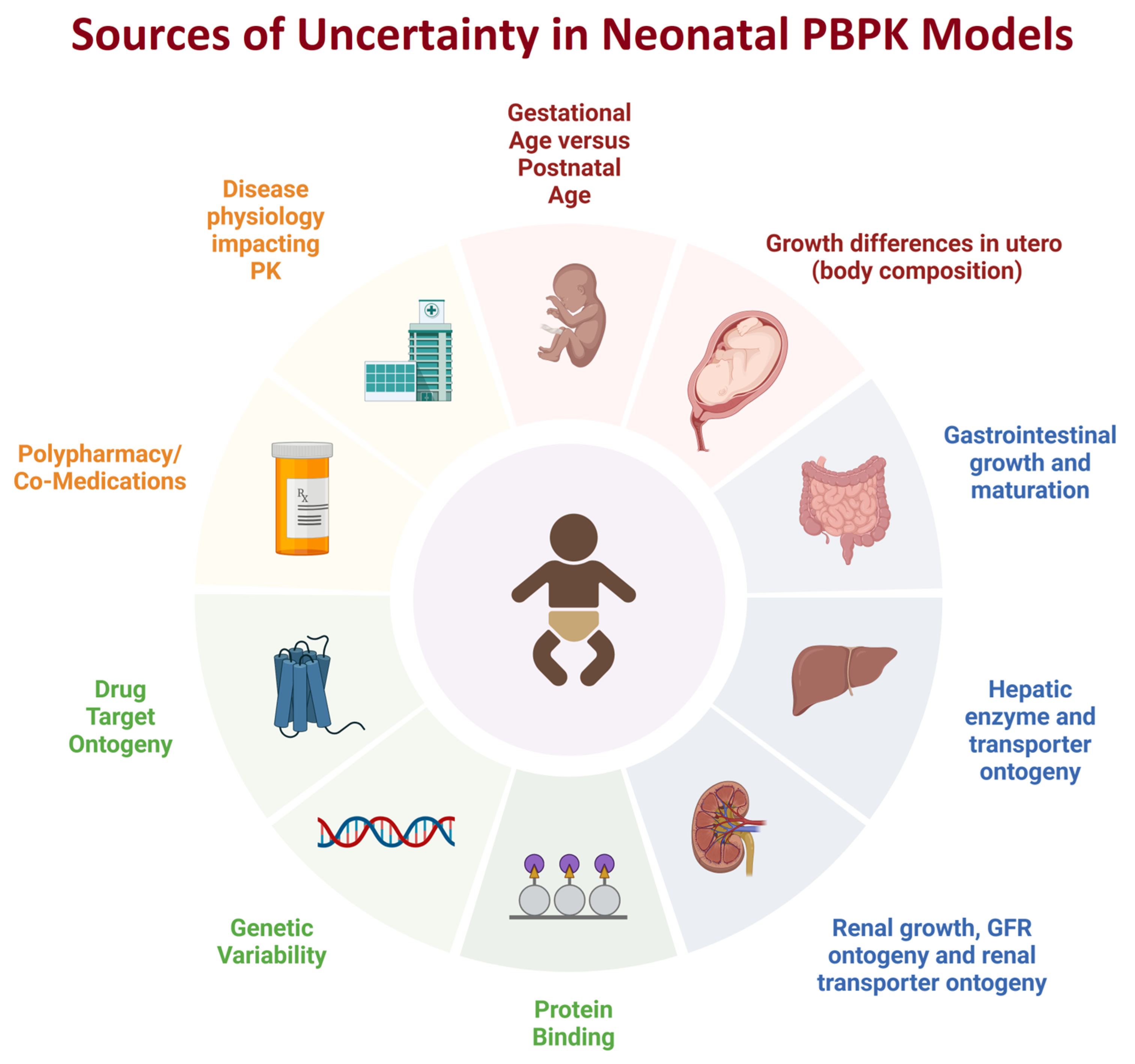
3. Advances and Unique Challenges in Developing PBPK Models in Neonates
3.1. Demographic Data
3.2. Organ Size, Blood Flow, and Composition
3.3. Ontogeny of Oral Drug Absorption
3.4. Ontogeny of Hepatic and Renal Drug Elimination Pathways
3.5. Defining Age and Maturation in a Neonatal Population
3.6. Biologics
3.7. Ontogeny of Drug Response
4. PBPK and Regulatory Application in Neonates
5. Conclusions
Author Contributions
Funding
Institutional Review Board Statement
Informed Consent Statement
Data Availability Statement
Conflicts of Interest
References
- Jamei, M. Recent Advances in Development and Application of Physiologically-Based Pharmacokinetic (PBPK) Models: A Transition from Academic Curiosity to Regulatory Acceptance. Curr. Pharmacol. Rep. 2016, 2, 161–169. [Google Scholar] [CrossRef] [PubMed]
- Abduljalil, K.; Gardner, I.; Jamei, M. Application of a Physiologically Based Pharmacokinetic Approach to Predict Theophylline Pharmacokinetics Using Virtual Non-Pregnant, Pregnant, Fetal, Breast-Feeding, and Neonatal Populations. Front. Pediatr. 2022, 10, 840710. [Google Scholar] [CrossRef] [PubMed]
- Gaohua, L.; Abduljalil, K.; Jamei, M.; Johnson, T.N.; Rostami-Hodjegan, A. A pregnancy physiologically based pharmacokinetic (p-PBPK) model for disposition of drugs metabolized by CYP1A2, CYP2D6 and CYP3A4. Br. J. Clin. Pharmacol. 2012, 74, 873–885. [Google Scholar] [CrossRef]
- Wang, K.; Jiang, K.; Wei, X.; Li, Y.; Wang, T.; Song, Y. Physiologically Based Pharmacokinetic Models Are Effective Support for Pediatric Drug Development. AAPS PharmSciTech 2021, 22, 208. [Google Scholar] [CrossRef] [PubMed]
- Nauwelaerts, N.; Macente, J.; Deferm, N.; Bonan, R.H.; Huang, M.C.; Van Neste, M.; Bibi, D.; Badee, J.; Martins, F.S.; Smits, A.; et al. Generic Workflow to Predict Medicine Concentrations in Human Milk Using Physiologically-Based Pharmacokinetic (PBPK) Modelling-A Contribution from the ConcePTION Project. Pharmaceutics 2023, 15, 1469. [Google Scholar] [CrossRef]
- Shao, W.; Shen, C.; Wang, W.; Sun, H.; Wang, X.; Geng, K.; Wang, X.; Xie, H. Development and Validation of Physiologically Based Pharmacokinetic Model of Levetiracetam to Predict Exposure and Dose Optimization in Pediatrics. J. Pharm. Sci. 2023, 112, 2667–2675. [Google Scholar] [CrossRef]
- van Hoogdalem, M.W.; Johnson, T.N.; McPhail, B.T.; Kamatkar, S.; Wexelblatt, S.L.; Ward, L.P.; Christians, U.; Akinbi, H.T.; Vinks, A.A.; Mizuno, T. Physiologically-Based Pharmacokinetic Modeling to Investigate the Effect of Maturation on Buprenorphine Pharmacokinetics in Newborns with Neonatal Opioid Withdrawal Syndrome. Clin. Pharmacol. Ther. 2022, 111, 496–508. [Google Scholar] [CrossRef]
- Engle, W.A.; American Academy of Pediatrics Committee on Fetus and Newborn. Age terminology during the perinatal period. Pediatrics 2004, 114, 1362–1364. [Google Scholar] [CrossRef]
- Quinn, J.A.; Munoz, F.M.; Gonik, B.; Frau, L.; Cutland, C.; Mallett-Moore, T.; Kissou, A.; Wittke, F.; Das, M.; Nunes, T.; et al. Preterm birth: Case definition & guidelines for data collection, analysis, and presentation of immunisation safety data. Vaccine 2016, 34, 6047–6056. [Google Scholar] [CrossRef]
- Mansoor, N.; Ahmed, M.; Czejka, M.; Sharib, S.; Hassan, S.; Hassan, A. Pharmacokinetics of Midazolam in preterm neonates with an insight in brain Tissue: A PBPK approach. Pak. J. Pharm. Sci. 2022, 35, 1459–1465. [Google Scholar]
- Abduljalil, K.; Jamei, M.; Johnson, T.N. Fetal Physiologically Based Pharmacokinetic Models: Systems Information on the Growth and Composition of Fetal Organs. Clin. Pharmacokinet. 2019, 58, 235–262. [Google Scholar] [CrossRef] [PubMed]
- Abduljalil, K.; Jamei, M.; Rostami-Hodjegan, A.; Johnson, T.N. Changes in individual drug-independent system parameters during virtual paediatric pharmacokinetic trials: Introducing time-varying physiology into a paediatric PBPK model. AAPS J. 2014, 16, 568–576. [Google Scholar] [CrossRef] [PubMed]
- Abduljalil, K.; Pan, X.; Pansari, A.; Jamei, M.; Johnson, T.N. A Preterm Physiologically Based Pharmacokinetic Model. Part I: Physiological Parameters and Model Building. Clin. Pharmacokinet. 2020, 59, 485–500. [Google Scholar] [CrossRef] [PubMed]
- Johnson, T.N.; Bonner, J.J.; Tucker, G.T.; Turner, D.B.; Jamei, M. Development and applications of a physiologically-based model of paediatric oral drug absorption. Eur. J. Pharm. Sci. 2018, 115, 57–67. [Google Scholar] [CrossRef]
- Johnson, T.N.; Rostami-Hodjegan, A.; Tucker, G.T. Prediction of the clearance of eleven drugs and associated variability in neonates, infants and children. Clin. Pharmacokinet. 2006, 45, 931–956. [Google Scholar] [CrossRef] [PubMed]
- Johnson, T.N.; Tanner, M.S.; Taylor, C.J.; Tucker, G.T. Enterocytic CYP3A4 in a paediatric population: Developmental changes and the effect of coeliac disease and cystic fibrosis. Br. J. Clin. Pharmacol. 2001, 51, 451–460. [Google Scholar] [CrossRef]
- Johnson, T.N.; Tucker, G.T.; Tanner, M.S.; Rostami-Hodjegan, A. Changes in liver volume from birth to adulthood: A meta-analysis. Liver Transpl. 2005, 11, 1481–1493. [Google Scholar] [CrossRef] [PubMed]
- Yun, Y.E.; Edginton, A.N. Model qualification of the PK-Sim(R) pediatric module for pediatric exposure assessment of CYP450 metabolized compounds. J. Toxicol. Environ. Health A 2019, 82, 789–814. [Google Scholar] [CrossRef]
- Desdicioglu, K.; Malas, M.A.; Evcil, E.H. Development of the fetal duodenum: A postmortem study. Fetal Diagn. Ther. 2009, 26, 16–23. [Google Scholar] [CrossRef]
- ICRP. Basic anatomical and physiological data for use in radiological protection: Reference values: ICRP Publication 89. Ann. ICRP 2002, 32, 1–277. [Google Scholar]
- Reiquam, C.W.; Allen, R.P.; Akers, D.R. Normal and Abnormal Small Bowel Lengths: An Analysis of 389 Autopsy Cases in Infants and Children. Am. J. Dis. Child. 1965, 109, 447–451. [Google Scholar] [CrossRef]
- Siebert, J.R. Small-intestine length in infants and children. Am. J. Dis. Child. 1980, 134, 593–595. [Google Scholar] [CrossRef] [PubMed]
- Struijs, M.C.; Diamond, I.R.; de Silva, N.; Wales, P.W. Establishing norms for intestinal length in children. J. Pediatr. Surg. 2009, 44, 933–938. [Google Scholar] [CrossRef] [PubMed]
- Avery, G.B.; Randolph, J.G.; Weaver, T. Gastric acidity in the first day of life. Pediatrics 1966, 37, 1005–1007. [Google Scholar] [CrossRef] [PubMed]
- Ebers, D.W.; Gibbs, G.E.; Smith, D.I. Gastric acidity on the first day of life. Pediatrics 1956, 18, 800–802. [Google Scholar] [CrossRef]
- Fredrikzon, B.; Hernell, O. Role of feeding on lipase activity in gastric contents. Acta Paediatr. Scand. 1977, 66, 479–484. [Google Scholar] [CrossRef]
- Van Den Abeele, J.; Rayyan, M.; Hoffman, I.; Van de Vijver, E.; Zhu, W.; Augustijns, P. Gastric fluid composition in a paediatric population: Age-dependent changes relevant for gastrointestinal drug disposition. Eur. J. Pharm. Sci. 2018, 123, 301–311. [Google Scholar] [CrossRef]
- Widstrom, A.M.; Christensson, K.; Ransjo-Arvidson, A.B.; Matthiesen, A.S.; Winberg, J.; Uvnas-Moberg, K. Gastric aspirates of newborn infants: pH, volume and levels of gastrin- and somatostatin-like immunoreactivity. Acta Paediatr. Scand. 1988, 77, 502–508. [Google Scholar] [CrossRef]
- Bonner, J.J.; Vajjah, P.; Abduljalil, K.; Jamei, M.; Rostami-Hodjegan, A.; Tucker, G.T.; Johnson, T.N. Does age affect gastric emptying time? A model-based meta-analysis of data from premature neonates through to adults. Biopharm. Drug Dispos. 2015, 36, 245–257. [Google Scholar] [CrossRef]
- Maharaj, A.R.; Edginton, A.N. Examining Small Intestinal Transit Time as a Function of Age: Is There Evidence to Support Age-Dependent Differences among Children? Drug Metab. Dispos. 2016, 44, 1080–1089. [Google Scholar] [CrossRef]
- Mooij, M.G.; Schwarz, U.I.; de Koning, B.A.; Leeder, J.S.; Gaedigk, R.; Samsom, J.N.; Spaans, E.; van Goudoever, J.B.; Tibboel, D.; Kim, R.B.; et al. Ontogeny of human hepatic and intestinal transporter gene expression during childhood: Age matters. Drug Metab. Dispos. 2014, 42, 1268–1274. [Google Scholar] [CrossRef] [PubMed]
- Konieczna, A.; Erdosova, B.; Lichnovska, R.; Jandl, M.; Cizkova, K.; Ehrmann, J. Differential expression of ABC transporters (MDR1, MRP1, BCRP) in developing human embryos. J. Mol. Histol. 2011, 42, 567–574. [Google Scholar] [CrossRef] [PubMed]
- Miki, Y.; Suzuki, T.; Tazawa, C.; Blumberg, B.; Sasano, H. Steroid and xenobiotic receptor (SXR), cytochrome P450 3A4 and multidrug resistance gene 1 in human adult and fetal tissues. Mol. Cell Endocrinol. 2005, 231, 75–85. [Google Scholar] [CrossRef]
- Mizuno, T.; Fukuda, T.; Masuda, S.; Uemoto, S.; Matsubara, K.; Inui, K.; Vinks, A.A. Developmental trajectory of intestinal MDR1/ABCB1 mRNA expression in children. Br. J. Clin. Pharmacol. 2014, 77, 910–912. [Google Scholar] [CrossRef] [PubMed]
- van Kalken, C.K.; Giaccone, G.; van der Valk, P.; Kuiper, C.M.; Hadisaputro, M.M.; Bosma, S.A.; Scheper, R.J.; Meijer, C.J.; Pinedo, H.M. Multidrug resistance gene (P-glycoprotein) expression in the human fetus. Am. J. Pathol. 1992, 141, 1063–1072. [Google Scholar]
- Baker, G.L. Human adipose tissue composition and age. Am. J. Clin. Nutr. 1969, 22, 829–835. [Google Scholar] [CrossRef]
- White, D.R.; Widdowson, E.M.; Woodard, H.Q.; Dickerson, J.W. The composition of body tissues (II). Fetus to young adult. Br. J. Radiol. 1991, 64, 149–159. [Google Scholar] [CrossRef]
- Widdowson, E.M.; Dickerson, J.W. The effect of growth and function on the chemical composition of soft tissues. Biochem. J. 1960, 77, 30–43. [Google Scholar] [CrossRef]
- Fomon, S.J.; Haschke, F.; Ziegler, E.E.; Nelson, S.E. Body composition of reference children from birth to age 10 years. Am. J. Clin. Nutr. 1982, 35, 1169–1175. [Google Scholar] [CrossRef]
- Friis-Hansen, B. Water distribution in the foetus and newborn infant. Acta Paediatr. Scand. Suppl. 1983, 305, 7–11. [Google Scholar] [CrossRef]
- Hartnoll, G.; Betremieux, P.; Modi, N. Body water content of extremely preterm infants at birth. Arch. Dis. Child. Fetal Neonatal Ed. 2000, 83, F56–F59. [Google Scholar] [CrossRef] [PubMed]
- Anblagan, D.; Deshpande, R.; Jones, N.W.; Costigan, C.; Bugg, G.; Raine-Fenning, N.; Gowland, P.A.; Mansell, P. Measurement of fetal fat in utero in normal and diabetic pregnancies using magnetic resonance imaging. Ultrasound Obstet. Gynecol. 2013, 42, 335–340. [Google Scholar] [CrossRef] [PubMed]
- Demarini, S.; Koo, W.W.; Hockman, E.M. Bone, lean and fat mass of newborn twins versus singletons. Acta Paediatr. 2006, 95, 594–599. [Google Scholar] [CrossRef] [PubMed]
- Enzi, G.; Zanardo, V.; Caretta, F.; Inelmen, E.M.; Rubaltelli, F. Intrauterine growth and adipose tissue development. Am. J. Clin. Nutr. 1981, 34, 1785–1790. [Google Scholar] [CrossRef]
- Friis, C.M.; Qvigstad, E.; Paasche Roland, M.C.; Godang, K.; Voldner, N.; Bollerslev, J.; Henriksen, T. Newborn body fat: Associations with maternal metabolic state and placental size. PLoS ONE 2013, 8, e57467. [Google Scholar] [CrossRef]
- Lampl, M.; Lee, W.; Koo, W.; Frongillo, E.A.; Barker, D.J.; Romero, R. Ethnic differences in the accumulation of fat and lean mass in late gestation. Am. J. Hum. Biol. 2012, 24, 640–647. [Google Scholar] [CrossRef]
- Ogiu, N.; Nakamura, Y.; Ijiri, I.; Hiraiwa, K.; Ogiu, T. A statistical analysis of the internal organ weights of normal Japanese people. Health Phys. 1997, 72, 368–383. [Google Scholar] [CrossRef]
- Saeki, I.; Tokunaga, S.; Matsuura, T.; Hayashida, M.; Yanagi, Y.; Taguchi, T. A formula for determining the standard liver volume in children: A special reference for neonates and infants. Pediatr. Transplant. 2012, 16, 244–249. [Google Scholar] [CrossRef]
- Knickmeyer, R.C.; Gouttard, S.; Kang, C.; Evans, D.; Wilber, K.; Smith, J.K.; Hamer, R.M.; Lin, W.; Gerig, G.; Gilmore, J.H. A structural MRI study of human brain development from birth to 2 years. J. Neurosci. 2008, 28, 12176–12182. [Google Scholar] [CrossRef]
- Sanchez, C.E.; Richards, J.E.; Almli, C.R. Neurodevelopmental MRI brain templates for children from 2 weeks to 4 years of age. Dev. Psychobiol. 2012, 54, 77–91. [Google Scholar] [CrossRef]
- Chiara, A.; Chirico, G.; Barbarini, M.; De Vecchi, E.; Rondini, G. Ultrasonic evaluation of kidney volume in term and preterm infants. Am. J. Perinatol. 1993, 10, 109–111. [Google Scholar] [CrossRef]
- Kandasamy, Y.; Rudd, D.; Lumbers, E.R.; Smith, R. An evaluation of preterm kidney size and function over the first two years of life. Pediatr. Nephrol. 2020, 35, 1477–1482. [Google Scholar] [CrossRef] [PubMed]
- Bauer, K.; Linderkamp, O.; Versmold, H.T. Systolic blood pressure and blood volume in preterm infants. Arch. Dis. Child. 1993, 69, 521–522. [Google Scholar] [CrossRef] [PubMed]
- Leipala, J.A.; Talme, M.; Viitala, J.; Turpeinen, U.; Fellman, V. Blood volume assessment with hemoglobin subtype analysis in preterm infants. Biol. Neonate 2003, 84, 41–44. [Google Scholar] [CrossRef]
- Mollison, P.L.; Veall, N.; Cutbush, M. Red cell and plasma volume in newborn infants. Arch. Dis. Child. 1950, 25, 242–253. [Google Scholar] [CrossRef] [PubMed]
- Wright, I.M.; Goodall, S.R. Blood pressure and blood volume in preterm infants. Arch. Dis. Child. Fetal Neonatal Ed. 1994, 70, F230–F231. [Google Scholar] [CrossRef] [PubMed]
- Alverson, D.C.; Eldridge, M.W.; Johnson, J.D.; Aldrich, M.; Angelus, P.; Berman, W., Jr. Noninvasive measurement of cardiac output in healthy preterm and term newborn infants. Am. J. Perinatol. 1984, 1, 148–151. [Google Scholar] [CrossRef]
- Grollmuss, O.; Gonzalez, P. Non-invasive cardiac output measurement in low and very low birth weight infants: A method comparison. Front. Pediatr. 2014, 2, 16. [Google Scholar] [CrossRef]
- Agata, Y.; Hiraishi, S.; Oguchi, K.; Misawa, H.; Horiguchi, Y.; Fujino, N.; Yashiro, K.; Shimada, N. Changes in left ventricular output from fetal to early neonatal life. J. Pediatr. 1991, 119, 441–445. [Google Scholar] [CrossRef]
- Broadhouse, K.M.; Finnemore, A.E.; Price, A.N.; Durighel, G.; Cox, D.J.; Edwards, A.D.; Hajnal, J.V.; Groves, A.M. Cardiovascular magnetic resonance of cardiac function and myocardial mass in preterm infants: A preliminary study of the impact of patent ductus arteriosus. J. Cardiovasc. Magn. Reson. 2014, 16, 54. [Google Scholar] [CrossRef]
- Ficial, B.; Finnemore, A.E.; Cox, D.J.; Broadhouse, K.M.; Price, A.N.; Durighel, G.; Ekitzidou, G.; Hajnal, J.V.; Edwards, A.D.; Groves, A.M. Validation study of the accuracy of echocardiographic measurements of systemic blood flow volume in newborn infants. J. Am. Soc. Echocardiogr. 2013, 26, 1365–1371. [Google Scholar] [CrossRef] [PubMed]
- Kessler, J.; Rasmussen, S.; Kiserud, T. The fetal portal vein: Normal blood flow development during the second half of human pregnancy. Ultrasound Obstet. Gynecol. 2007, 30, 52–60. [Google Scholar] [CrossRef] [PubMed]
- Raynaud, C.; Chiron, C.; Maziere, B.A. Follow up of regional CBF in children from birth to 18 years with Xe-133 [abstract]. J. Nucl. Med. 1990, 31S, 892. [Google Scholar]
- Wintermark, M.; Lepori, D.; Cotting, J.; Roulet, E.; van Melle, G.; Meuli, R.; Maeder, P.; Regli, L.; Verdun, F.R.; Deonna, T.; et al. Brain perfusion in children: Evolution with age assessed by quantitative perfusion computed tomography. Pediatrics 2004, 113, 1642–1652. [Google Scholar] [CrossRef] [PubMed]
- Rubin, M.I.; Bruck, E.; Rapoport, M.; Snively, M.; McKay, H.; Baumler, A. Maturation of Renal Function in Childhood: Clearance Studies. J. Clin. Investig. 1949, 28, 1144–1162. [Google Scholar] [CrossRef]
- Visser, M.O.; Leighton, J.O.; van de Bor, M.; Walther, F.J. Renal blood flow in neonates: Quantification with color flow and pulsed Doppler US. Radiology 1992, 183, 441–444. [Google Scholar] [CrossRef]
- Forestier, F.; Daffos, F.; Rainaut, M.; Bruneau, M.; Trivin, F. Blood chemistry of normal human fetuses at midtrimester of pregnancy. Pediatr. Res. 1987, 21, 579–583. [Google Scholar] [CrossRef]
- Gitlin, D.; Boesman, M. Serum alpha-fetoprotein, albumin, and gamma-G-globulin in the human conceptus. J. Clin. Investig. 1966, 45, 1826–1838. [Google Scholar] [CrossRef]
- McNamara, P.J.; Alcorn, J. Protein binding predictions in infants. AAPS PharmSci 2002, 4, E4. [Google Scholar] [CrossRef]
- Sethi, P.K.; White, C.A.; Cummings, B.S.; Hines, R.N.; Muralidhara, S.; Bruckner, J.V. Ontogeny of plasma proteins, albumin and binding of diazepam, cyclosporine, and deltamethrin. Pediatr. Res. 2016, 79, 409–415. [Google Scholar] [CrossRef]
- Maharaj, A.R.; Gonzalez, D.; Cohen-Wolkowiez, M.; Hornik, C.P.; Edginton, A.N. Improving Pediatric Protein Binding Estimates: An Evaluation of alpha1-Acid Glycoprotein Maturation in Healthy and Infected Subjects. Clin. Pharmacokinet. 2018, 57, 577–589. [Google Scholar] [CrossRef]
- Berrebi, A.; Benichou, A.C.; Sarramon, M.F.; Bessieres, M.H.; Rolland, M.; Kobuch, W.E.; Demur, C.; Solera, M.L.; Sie, P.; Amouroux, J.; et al. Biological reference values in the human fetus. 106 cord blood sampling in utero. J. Gynecol. Obstet. Biol. Reprod. 1992, 21, 355–359. [Google Scholar]
- Boulot, P.; Cattaneo, A.; Taib, J.; Peray, P.; Lefort, G.; Hedon, B.; Laffargue, F.; Viala, J.L. Hematologic values of fetal blood obtained by means of cordocentesis. Fetal Diagn. Ther. 1993, 8, 309–316. [Google Scholar] [CrossRef] [PubMed]
- Jopling, J.; Henry, E.; Wiedmeier, S.E.; Christensen, R.D. Reference ranges for hematocrit and blood hemoglobin concentration during the neonatal period: Data from a multihospital health care system. Pediatrics 2009, 123, e333–e337. [Google Scholar] [CrossRef]
- Salem, F.; Johnson, T.N.; Abduljalil, K.; Tucker, G.T.; Rostami-Hodjegan, A. A re-evaluation and validation of ontogeny functions for cytochrome P450 1A2 and 3A4 based on in vivo data. Clin. Pharmacokinet. 2014, 53, 625–636. [Google Scholar] [CrossRef] [PubMed]
- Upreti, V.V.; Wahlstrom, J.L. Meta-analysis of hepatic cytochrome P450 ontogeny to underwrite the prediction of pediatric pharmacokinetics using physiologically based pharmacokinetic modeling. J. Clin. Pharmacol. 2016, 56, 266–283. [Google Scholar] [CrossRef] [PubMed]
- Cazeneuve, C.; Pons, G.; Rey, E.; Treluyer, J.M.; Cresteil, T.; Thiroux, G.; D’Athis, P.; Olive, G. Biotransformation of caffeine in human liver microsomes from foetuses, neonates, infants and adults. Br. J. Clin. Pharmacol. 1994, 37, 405–412. [Google Scholar] [CrossRef] [PubMed]
- Sonnier, M.; Cresteil, T. Delayed ontogenesis of CYP1A2 in the human liver. Eur. J. Biochem. 1998, 251, 893–898. [Google Scholar] [CrossRef]
- Tateishi, T.; Nakura, H.; Asoh, M.; Watanabe, M.; Tanaka, M.; Kumai, T.; Takashima, S.; Imaoka, S.; Funae, Y.; Yabusaki, Y.; et al. A comparison of hepatic cytochrome P450 protein expression between infancy and postinfancy. Life Sci. 1997, 61, 2567–2574. [Google Scholar] [CrossRef]
- Hakkola, J.; Tanaka, E.; Pelkonen, O. Developmental expression of cytochrome P450 enzymes in human liver. Pharmacol. Toxicol. 1998, 82, 209–217. [Google Scholar] [CrossRef]
- Pearce, R.E.; Gaedigk, R.; Twist, G.P.; Dai, H.; Riffel, A.K.; Leeder, J.S.; Gaedigk, A. Developmental Expression of CYP2B6: A Comprehensive Analysis of mRNA Expression, Protein Content and Bupropion Hydroxylase Activity and the Impact of Genetic Variation. Drug Metab. Dispos. 2016, 44, 948–958. [Google Scholar] [CrossRef]
- Croom, E.L.; Stevens, J.C.; Hines, R.N.; Wallace, A.D.; Hodgson, E. Human hepatic CYP2B6 developmental expression: The impact of age and genotype. Biochem. Pharmacol. 2009, 78, 184–190. [Google Scholar] [CrossRef] [PubMed]
- Hines, R.N. Ontogeny of human hepatic cytochromes P450. J. Biochem. Mol. Toxicol. 2007, 21, 169–175. [Google Scholar] [CrossRef]
- Koukouritaki, S.B.; Manro, J.R.; Marsh, S.A.; Stevens, J.C.; Rettie, A.E.; McCarver, D.G.; Hines, R.N. Developmental expression of human hepatic CYP2C9 and CYP2C19. J. Pharmacol. Exp. Ther. 2004, 308, 965–974. [Google Scholar] [CrossRef] [PubMed]
- Ratanasavanh, D.; Beaune, P.; Morel, F.; Flinois, J.P.; Guengerich, F.P.; Guillouzo, A. Intralobular distribution and quantitation of cytochrome P-450 enzymes in human liver as a function of age. Hepatology 1991, 13, 1142–1151. [Google Scholar] [CrossRef]
- Treluyer, J.M.; Gueret, G.; Cheron, G.; Sonnier, M.; Cresteil, T. Developmental expression of CYP2C and CYP2C-dependent activities in the human liver: In-vivo/in-vitro correlation and inducibility. Pharmacogenetics 1997, 7, 441–452. [Google Scholar] [CrossRef]
- Stevens, J.C.; Marsh, S.A.; Zaya, M.J.; Regina, K.J.; Divakaran, K.; Le, M.; Hines, R.N. Developmental changes in human liver CYP2D6 expression. Drug Metab. Dispos. 2008, 36, 1587–1593. [Google Scholar] [CrossRef]
- Johnsrud, E.K.; Koukouritaki, S.B.; Divakaran, K.; Brunengraber, L.L.; Hines, R.N.; McCarver, D.G. Human hepatic CYP2E1 expression during development. J. Pharmacol. Exp. Ther. 2003, 307, 402–407. [Google Scholar] [CrossRef]
- Vieira, I.; Sonnier, M.; Cresteil, T. Developmental expression of CYP2E1 in the human liver. Hypermethylation control of gene expression during the neonatal period. Eur. J. Biochem. 1996, 238, 476–483. [Google Scholar] [CrossRef] [PubMed]
- Lacroix, D.; Sonnier, M.; Moncion, A.; Cheron, G.; Cresteil, T. Expression of CYP3A in the human liver—Evidence that the shift between CYP3A7 and CYP3A4 occurs immediately after birth. Eur. J. Biochem. 1997, 247, 625–634. [Google Scholar] [CrossRef] [PubMed]
- Stevens, J.C.; Hines, R.N.; Gu, C.; Koukouritaki, S.B.; Manro, J.R.; Tandler, P.J.; Zaya, M.J. Developmental expression of the major human hepatic CYP3A enzymes. J. Pharmacol. Exp. Ther. 2003, 307, 573–582. [Google Scholar] [CrossRef]
- Leeder, J.S.; Gaedigk, R.; Marcucci, K.A.; Gaedigk, A.; Vyhlidal, C.A.; Schindel, B.P.; Pearce, R.E. Variability of CYP3A7 expression in human fetal liver. J. Pharmacol. Exp. Ther. 2005, 314, 626–635. [Google Scholar] [CrossRef]
- Matlock, M.K.; Tambe, A.; Elliott-Higgins, J.; Hines, R.N.; Miller, G.P.; Swamidass, S.J. A Time-Embedding Network Models the Ontogeny of 23 Hepatic Drug Metabolizing Enzymes. Chem. Res. Toxicol. 2019, 32, 1707–1721. [Google Scholar] [CrossRef]
- Sadler, N.C.; Nandhikonda, P.; Webb-Robertson, B.J.; Ansong, C.; Anderson, L.N.; Smith, J.N.; Corley, R.A.; Wright, A.T. Hepatic Cytochrome P450 Activity, Abundance, and Expression Throughout Human Development. Drug Metab. Dispos. 2016, 44, 984–991. [Google Scholar] [CrossRef] [PubMed]
- Vyhlidal, C.A.; Pearce, R.E.; Gaedigk, R.; Calamia, J.C.; Shuster, D.L.; Thummel, K.E.; Leeder, J.S. Variability in Expression of CYP3A5 in Human Fetal Liver. Drug Metab. Dispos. 2015, 43, 1286–1293. [Google Scholar] [CrossRef] [PubMed]
- Koukouritaki, S.B.; Simpson, P.; Yeung, C.K.; Rettie, A.E.; Hines, R.N. Human hepatic flavin-containing monooxygenases 1 (FMO1) and 3 (FMO3) developmental expression. Pediatr. Res. 2002, 51, 236–243. [Google Scholar] [CrossRef] [PubMed]
- Chen, H.; Fantel, A.G.; Juchau, M.R. Catalysis of the 4-hydroxylation of retinoic acids by cyp3a7 in human fetal hepatic tissues. Drug Metab. Dispos. 2000, 28, 1051–1057. [Google Scholar]
- He, H.; Nie, Y.L.; Li, J.F.; Meng, X.G.; Yang, W.H.; Chen, Y.L.; Wang, S.J.; Ma, X.; Kan, Q.C.; Zhang, L.R. Developmental regulation of CYP3A4 and CYP3A7 in Chinese Han population. Drug Metab. Pharmacokinet. 2016, 31, 433–444. [Google Scholar] [CrossRef]
- Shuster, D.L.; Risler, L.J.; Prasad, B.; Calamia, J.C.; Voellinger, J.L.; Kelly, E.J.; Unadkat, J.D.; Hebert, M.F.; Shen, D.D.; Thummel, K.E.; et al. Identification of CYP3A7 for glyburide metabolism in human fetal livers. Biochem. Pharmacol. 2014, 92, 690–700. [Google Scholar] [CrossRef] [PubMed]
- Badee, J.; Qiu, N.; Collier, A.C.; Takahashi, R.H.; Forrest, W.F.; Parrott, N.; Schmidt, S.; Fowler, S. Characterization of the Ontogeny of Hepatic UDP-Glucuronosyltransferase Enzymes Based on Glucuronidation Activity Measured in Human Liver Microsomes. J. Clin. Pharmacol. 2019, 59 (Suppl. 1), S42–S55. [Google Scholar] [CrossRef]
- Bhatt, D.K.; Mehrotra, A.; Gaedigk, A.; Chapa, R.; Basit, A.; Zhang, H.; Choudhari, P.; Boberg, M.; Pearce, R.E.; Gaedigk, R.; et al. Age- and Genotype-Dependent Variability in the Protein Abundance and Activity of Six Major Uridine Diphosphate-Glucuronosyltransferases in Human Liver. Clin. Pharmacol. Ther. 2019, 105, 131–141. [Google Scholar] [CrossRef]
- Burchell, B.; Coughtrie, M.; Jackson, M.; Harding, D.; Fournel-Gigleux, S.; Leakey, J.; Hume, R. Development of human liver UDP-glucuronosyltransferases. Dev. Pharmacol. Ther. 1989, 13, 70–77. [Google Scholar] [CrossRef] [PubMed]
- Coughtrie, M.W.; Burchell, B.; Leakey, J.E.; Hume, R. The inadequacy of perinatal glucuronidation: Immunoblot analysis of the developmental expression of individual UDP-glucuronosyltransferase isoenzymes in rat and human liver microsomes. Mol. Pharmacol. 1988, 34, 729–735. [Google Scholar]
- Leakey, J.E.; Hume, R.; Burchell, B. Development of multiple activities of UDP-glucuronyltransferase in human liver. Biochem. J. 1987, 243, 859–861. [Google Scholar] [CrossRef] [PubMed]
- Miyagi, S.J.; Collier, A.C. The development of UDP-glucuronosyltransferases 1A1 and 1A6 in the pediatric liver. Drug Metab. Dispos. 2011, 39, 912–919. [Google Scholar] [CrossRef] [PubMed]
- Onishi, S.; Kawade, N.; Itoh, S.; Isobe, K.; Sugiyama, S. Postnatal development of uridine diphosphate glucuronyltransferase activity towards bilirubin and 2-aminophenol in human liver. Biochem. J. 1979, 184, 705–707. [Google Scholar] [CrossRef]
- Strassburg, C.P.; Strassburg, A.; Kneip, S.; Barut, A.; Tukey, R.H.; Rodeck, B.; Manns, M.P. Developmental aspects of human hepatic drug glucuronidation in young children and adults. Gut 2002, 50, 259–265. [Google Scholar] [CrossRef]
- Badee, J.; Fowler, S.; de Wildt, S.N.; Collier, A.C.; Schmidt, S.; Parrott, N. The Ontogeny of UDP-glucuronosyltransferase Enzymes, Recommendations for Future Profiling Studies and Application Through Physiologically Based Pharmacokinetic Modelling. Clin. Pharmacokinet. 2019, 58, 189–211. [Google Scholar] [CrossRef]
- Miyagi, S.J.; Collier, A.C. Pediatric development of glucuronidation: The ontogeny of hepatic UGT1A4. Drug Metab. Dispos. 2007, 35, 1587–1592. [Google Scholar] [CrossRef]
- Court, M.H.; Zhang, X.; Ding, X.; Yee, K.K.; Hesse, L.M.; Finel, M. Quantitative distribution of mRNAs encoding the 19 human UDP-glucuronosyltransferase enzymes in 26 adult and 3 fetal tissues. Xenobiotica 2012, 42, 266–277. [Google Scholar] [CrossRef]
- Miyagi, S.J.; Milne, A.M.; Coughtrie, M.W.; Collier, A.C. Neonatal development of hepatic UGT1A9: Implications of pediatric pharmacokinetics. Drug Metab. Dispos. 2012, 40, 1321–1327. [Google Scholar] [CrossRef]
- Pacifici, G.M.; Sawe, J.; Kager, L.; Rane, A. Morphine glucuronidation in human fetal and adult liver. Eur. J. Clin. Pharmacol. 1982, 22, 553–558. [Google Scholar] [CrossRef]
- Zaya, M.J.; Hines, R.N.; Stevens, J.C. Epirubicin glucuronidation and UGT2B7 developmental expression. Drug Metab. Dispos. 2006, 34, 2097–2101. [Google Scholar] [CrossRef] [PubMed]
- Boberg, M.; Vrana, M.; Mehrotra, A.; Pearce, R.E.; Gaedigk, A.; Bhatt, D.K.; Leeder, J.S.; Prasad, B. Age-Dependent Absolute Abundance of Hepatic Carboxylesterases (CES1 and CES2) by LC-MS/MS Proteomics: Application to PBPK Modeling of Oseltamivir In Vivo Pharmacokinetics in Infants. Drug Metab. Dispos. 2017, 45, 216–223. [Google Scholar] [CrossRef] [PubMed]
- Yang, D.; Pearce, R.E.; Wang, X.; Gaedigk, R.; Wan, Y.J.; Yan, B. Human carboxylesterases HCE1 and HCE2: Ontogenic expression, inter-individual variability and differential hydrolysis of oseltamivir, aspirin, deltamethrin and permethrin. Biochem. Pharmacol. 2009, 77, 238–247. [Google Scholar] [CrossRef]
- Zhu, H.J.; Appel, D.I.; Jiang, Y.; Markowitz, J.S. Age- and sex-related expression and activity of carboxylesterase 1 and 2 in mouse and human liver. Drug Metab. Dispos. 2009, 37, 1819–1825. [Google Scholar] [CrossRef]
- Chen, Y.T.; Trzoss, L.; Yang, D.; Yan, B. Ontogenic expression of human carboxylesterase-2 and cytochrome P450 3A4 in liver and duodenum: Postnatal surge and organ-dependent regulation. Toxicology 2015, 330, 55–61. [Google Scholar] [CrossRef] [PubMed]
- Prasad, B.; Gaedigk, A.; Vrana, M.; Gaedigk, R.; Leeder, J.S.; Salphati, L.; Chu, X.; Xiao, G.; Hop, C.; Evers, R.; et al. Ontogeny of Hepatic Drug Transporters as Quantified by LC-MS/MS Proteomics. Clin. Pharmacol. Ther. 2016, 100, 362–370. [Google Scholar] [CrossRef]
- Mooij, M.G.; van de Steeg, E.; van Rosmalen, J.; Windster, J.D.; de Koning, B.A.; Vaes, W.H.; van Groen, B.D.; Tibboel, D.; Wortelboer, H.M.; de Wildt, S.N. Proteomic Analysis of the Developmental Trajectory of Human Hepatic Membrane Transporter Proteins in the First Three Months of Life. Drug Metab. Dispos. 2016, 44, 1005–1013. [Google Scholar] [CrossRef]
- Leeder, J.S.; Dinh, J.C.; Gaedigk, A.; Staggs, V.S.; Prasad, B.; Pearce, R.E. Ontogeny of Scaling Factors for Pediatric Physiology-Based Pharmacokinetic Modeling and Simulation: Microsomal Protein Per Gram of Liver. Drug Metab. Dispos. 2022, 50, 24–32. [Google Scholar] [CrossRef]
- Salem, F.; Johnson, T.N.; Hodgkinson, A.B.J.; Ogungbenro, K.; Rostami-Hodjegan, A. Does “Birth” as an Event Impact Maturation Trajectory of Renal Clearance via Glomerular Filtration? Reexamining Data in Preterm and Full-Term Neonates by Avoiding the Creatinine Bias. J. Clin. Pharmacol. 2021, 61, 159–171. [Google Scholar] [CrossRef] [PubMed]
- De Cock, R.F.; Allegaert, K.; Brussee, J.M.; Sherwin, C.M.; Mulla, H.; de Hoog, M.; van den Anker, J.N.; Danhof, M.; Knibbe, C.A. Simultaneous pharmacokinetic modeling of gentamicin, tobramycin and vancomycin clearance from neonates to adults: Towards a semi-physiological function for maturation in glomerular filtration. Pharm. Res. 2014, 31, 2643–2654. [Google Scholar] [CrossRef] [PubMed]
- Ezuruike, U.; Blenkinsop, A.; Pansari, A.; Abduljalil, K. Quantification of Fetal Renal Function Using Fetal Urine Production Rate and Its Reflection on the Amniotic and Fetal Creatinine Levels During Pregnancy. Front. Pediatr. 2022, 10, 841495. [Google Scholar] [CrossRef]
- Hayton, W.L. Maturation and growth of renal function: Dosing renally cleared drugs in children. AAPS PharmSci 2000, 2, E3. [Google Scholar] [CrossRef]
- Rhodin, M.M.; Anderson, B.J.; Peters, A.M.; Coulthard, M.G.; Wilkins, B.; Cole, M.; Chatelut, E.; Grubb, A.; Veal, G.J.; Keir, M.J.; et al. Human renal function maturation: A quantitative description using weight and postmenstrual age. Pediatr. Nephrol. 2009, 24, 67–76. [Google Scholar] [CrossRef]
- Smeets, N.J.L.; IntHout, J.; van der Burgh, M.J.P.; Schwartz, G.J.; Schreuder, M.F.; de Wildt, S.N. Maturation of GFR in Term-Born Neonates: An Individual Participant Data Meta-Analysis. J. Am. Soc. Nephrol. 2022, 33, 1277–1292. [Google Scholar] [CrossRef]
- Vieux, R.; Hascoet, J.M.; Merdariu, D.; Fresson, J.; Guillemin, F. Glomerular filtration rate reference values in very preterm infants. Pediatrics 2010, 125, e1186–e1192. [Google Scholar] [CrossRef] [PubMed]
- Cheung, K.W.K.; van Groen, B.D.; Spaans, E.; van Borselen, M.D.; de Bruijn, A.; Simons-Oosterhuis, Y.; Tibboel, D.; Samsom, J.N.; Verdijk, R.M.; Smeets, B.; et al. A Comprehensive Analysis of Ontogeny of Renal Drug Transporters: mRNA Analyses, Quantitative Proteomics, and Localization. Clin. Pharmacol. Ther. 2019, 106, 1083–1092. [Google Scholar] [CrossRef] [PubMed]
- Li, S.; Xie, F. Foetal and neonatal exposure prediction and dosing evaluation for ampicillin using a physiologically-based pharmacokinetic modelling approach. Br. J. Clin. Pharmacol. 2023, 89, 1402–1412. [Google Scholar] [CrossRef] [PubMed]
- Salem, F.; Small, B.G.; Johnson, T.N. Development and application of a pediatric mechanistic kidney model. CPT Pharmacomet. Syst. Pharmacol. 2022, 11, 854–866. [Google Scholar] [CrossRef]
- Allegaert, K.; Abbasi, M.Y.; Annaert, P.; Olafuyi, O. Current and future physiologically based pharmacokinetic (PBPK) modeling approaches to optimize pharmacotherapy in preterm neonates. Expert. Opin. Drug Metab. Toxicol. 2022, 18, 301–312. [Google Scholar] [CrossRef]
- Small, B.G.; Johnson, T.N.; Rowland Yeo, K. Another Step Toward Qualification of Pediatric Physiologically Based Pharmacokinetic Models to Facilitate Inclusivity and Diversity in Pediatric Clinical Studies. Clin. Pharmacol. Ther. 2023, 113, 735–745. [Google Scholar] [CrossRef]
- Zhou, W.; Johnson, T.N.; Bui, K.H.; Cheung, S.Y.A.; Li, J.; Xu, H.; Al-Huniti, N.; Zhou, D. Predictive Performance of Physiologically Based Pharmacokinetic (PBPK) Modeling of Drugs Extensively Metabolized by Major Cytochrome P450s in Children. Clin. Pharmacol. Ther. 2018, 104, 188–200. [Google Scholar] [CrossRef]
- Mukherjee, A.; Dombi, T.; Wittke, B.; Lalonde, R. Population pharmacokinetics of sildenafil in term neonates: Evidence of rapid maturation of metabolic clearance in the early postnatal period. Clin. Pharmacol. Ther. 2009, 85, 56–63. [Google Scholar] [CrossRef]
- Claassen, K.; Thelen, K.; Coboeken, K.; Gaub, T.; Lippert, J.; Allegaert, K.; Willmann, S. Development of a Physiologically-Based Pharmacokinetic Model for Preterm Neonates: Evaluation with In Vivo Data. Curr. Pharm. Des. 2015, 21, 5688–5698. [Google Scholar] [CrossRef] [PubMed]
- Cole, T.J.; Freeman, J.V.; Preece, M.A. British 1990 growth reference centiles for weight, height, body mass index and head circumference fitted by maximum penalized likelihood. Stat. Med. 1998, 17, 407–429. [Google Scholar] [CrossRef]
- Fenton, T.R.; Kim, J.H. A systematic review and meta-analysis to revise the Fenton growth chart for preterm infants. BMC Pediatr. 2013, 13, 59. [Google Scholar] [CrossRef]
- Troutman, J.A.; Sullivan, M.C.; Carr, G.J.; Fisher, J. Development of growth equations from longitudinal studies of body weight and height in the full term and preterm neonate: From birth to four years postnatal age. Birth Defects Res. 2018, 110, 916–932. [Google Scholar] [CrossRef] [PubMed]
- Wang, X.; Qiao, Z.W.; Zhou, Z.J.; Zhuang, P.J.; Zheng, S. Postoperative morphine concentration in infants with or without biliary atresia and its association with hepatic blood flow. Anaesthesia 2014, 69, 583–590. [Google Scholar] [CrossRef] [PubMed]
- Emoto, C.; Johnson, T.N.; Neuhoff, S.; Hahn, D.; Vinks, A.A.; Fukuda, T. PBPK Model of Morphine Incorporating Developmental Changes in Hepatic OCT1 and UGT2B7 Proteins to Explain the Variability in Clearances in Neonates and Small Infants. CPT Pharmacomet. Syst. Pharmacol. 2018, 7, 464–473. [Google Scholar] [CrossRef]
- Kearns, G.L.; Abdel-Rahman, S.M.; Alander, S.W.; Blowey, D.L.; Leeder, J.S.; Kauffman, R.E. Developmental pharmacology—Drug disposition, action, and therapy in infants and children. N. Engl. J. Med. 2003, 349, 1157–1167. [Google Scholar] [CrossRef]
- Heimann, G. Enteral absorption and bioavailability in children in relation to age. Eur. J. Clin. Pharmacol. 1980, 18, 43–50. [Google Scholar] [CrossRef] [PubMed]
- Somani, A.A.; Thelen, K.; Zheng, S.; Trame, M.N.; Coboeken, K.; Meyer, M.; Schnizler, K.; Ince, I.; Willmann, S.; Schmidt, S. Evaluation of changes in oral drug absorption in preterm and term neonates for Biopharmaceutics Classification System (BCS) class I and II compounds. Br. J. Clin. Pharmacol. 2016, 81, 137–147. [Google Scholar] [CrossRef] [PubMed]
- Wollmer, E.; Ungell, A.L.; Nicolas, J.M.; Klein, S. Review of paediatric gastrointestinal physiology relevant to the absorption of orally administered medicines. Adv. Drug Deliv. Rev. 2022, 181, 114084. [Google Scholar] [CrossRef]
- Kohlmann, P.; Stillhart, C.; Kuentz, M.; Parrott, N. Investigating Oral Absorption of Carbamazepine in Pediatric Populations. AAPS J. 2017, 19, 1864–1877. [Google Scholar] [CrossRef]
- Smits, A.; Kulo, A.; de Hoon, J.N.; Allegaert, K. Pharmacokinetics of drugs in neonates: Pattern recognition beyond compound specific observations. Curr. Pharm. Des. 2012, 18, 3119–3146. [Google Scholar] [CrossRef]
- Bode, S.; Dreyer, M.; Greisen, G. Gastric emptying and small intestinal transit time in preterm infants: A scintigraphic method. J. Pediatr. Gastroenterol. Nutr. 2004, 39, 378–382. [Google Scholar] [CrossRef]
- Riezzo, G.; Indrio, F.; Raimondi, F.; Montagna, O.; Salvia, G.; Massimo, B.; Polimeno, L.; Cavallo, L.; Francavilla, R. Maturation of gastric electrical activity, gastric emptying and intestinal permeability in preterm newborns during the first month of life. Ital. J. Pediatr. 2009, 35, 6. [Google Scholar] [CrossRef]
- de Wildt, S.N.; Kearns, G.L.; Hop, W.C.; Murry, D.J.; Abdel-Rahman, S.M.; van den Anker, J.N. Pharmacokinetics and metabolism of oral midazolam in preterm infants. Br. J. Clin. Pharmacol. 2002, 53, 390–392. [Google Scholar] [CrossRef] [PubMed]
- Li, C.Y.; Hosey-Cojocari, C.; Basit, A.; Unadkat, J.D.; Leeder, J.S.; Prasad, B. Optimized Renal Transporter Quantification by Using Aquaporin 1 and Aquaporin 2 as Anatomical Markers: Application in Characterizing the Ontogeny of Renal Transporters and Its Correlation with Hepatic Transporters in Paired Human Samples. AAPS J. 2019, 21, 88. [Google Scholar] [CrossRef]
- Thomson, M.M.; Hines, R.N.; Schuetz, E.G.; Meibohm, B. Expression Patterns of Organic Anion Transporting Polypeptides 1B1 and 1B3 Protein in Human Pediatric Liver. Drug Metab. Dispos. 2016, 44, 999–1004. [Google Scholar] [CrossRef]
- Blake, M.J.; Gaedigk, A.; Pearce, R.E.; Bomgaars, L.R.; Christensen, M.L.; Stowe, C.; James, L.P.; Wilson, J.T.; Kearns, G.L.; Leeder, J.S. Ontogeny of dextromethorphan O- and N-demethylation in the first year of life. Clin. Pharmacol. Ther. 2007, 81, 510–516. [Google Scholar] [CrossRef]
- Leeder, J.S.; Kearns, G.L.; Spielberg, S.P.; van den Anker, J. Understanding the relative roles of pharmacogenetics and ontogeny in pediatric drug development and regulatory science. J. Clin. Pharmacol. 2010, 50, 1377–1387. [Google Scholar] [CrossRef] [PubMed]
- Johnson, T.N.; Howgate, E.M.; de Wildt, S.N.; Turner, M.A.; Rowland Yeo, K. Use of Developmental Midazolam and 1-Hydroxymidazolam Data with Pediatric Physiologically Based Modeling to Assess Cytochrome P450 3A4 and Uridine Diphosphate Glucuronosyl Transferase 2B4 Ontogeny In Vivo. Drug Metab. Dispos. 2023, 51, 1035–1045. [Google Scholar] [CrossRef] [PubMed]
- Salerno, S.N.; Edginton, A.; Gerhart, J.G.; Laughon, M.M.; Ambalavanan, N.; Sokol, G.M.; Hornik, C.D.; Stewart, D.; Mills, M.; Martz, K.; et al. Physiologically-Based Pharmacokinetic Modeling Characterizes the CYP3A-Mediated Drug-Drug Interaction Between Fluconazole and Sildenafil in Infants. Clin. Pharmacol. Ther. 2021, 109, 253–262. [Google Scholar] [CrossRef] [PubMed]
- Williams, J.A.; Ring, B.J.; Cantrell, V.E.; Jones, D.R.; Eckstein, J.; Ruterbories, K.; Hamman, M.A.; Hall, S.D.; Wrighton, S.A. Comparative metabolic capabilities of CYP3A4, CYP3A5, and CYP3A7. Drug Metab. Dispos. 2002, 30, 883–891. [Google Scholar] [CrossRef]
- Takahiro, R.; Nakamura, S.; Kohno, H.; Yoshimura, N.; Nakamura, T.; Ozawa, S.; Hirono, K.; Ichida, F.; Taguchi, M. Contribution of CYP3A isoforms to dealkylation of PDE5 inhibitors: A comparison between sildenafil N-demethylation and tadalafil demethylenation. Biol. Pharm. Bull. 2015, 38, 58–65. [Google Scholar] [CrossRef]
- Shum, S.; Isoherranen, N. Human Fetal Liver Metabolism of Oxycodone Is Mediated by CYP3A7. AAPS J. 2021, 23, 24. [Google Scholar] [CrossRef]
- Barter, Z.E.; Tucker, G.T.; Rowland-Yeo, K. Differences in cytochrome p450-mediated pharmacokinetics between chinese and caucasian populations predicted by mechanistic physiologically based pharmacokinetic modelling. Clin. Pharmacokinet. 2013, 52, 1085–1100. [Google Scholar] [CrossRef] [PubMed]
- Edginton, A.N.; Willmann, S. Physiology-based simulations of a pathological condition: Prediction of pharmacokinetics in patients with liver cirrhosis. Clin. Pharmacokinet. 2008, 47, 743–752. [Google Scholar] [CrossRef]
- Temrikar, Z.H.; Suryawanshi, S.; Meibohm, B. Pharmacokinetics and Clinical Pharmacology of Monoclonal Antibodies in Pediatric Patients. Paediatr. Drugs 2020, 22, 199–216. [Google Scholar] [CrossRef] [PubMed]
- Pan, X.; Stader, F.; Abduljalil, K.; Gill, K.L.; Johnson, T.N.; Gardner, I.; Jamei, M. Development and Application of a Physiologically-Based Pharmacokinetic Model to Predict the Pharmacokinetics of Therapeutic Proteins from Full-term Neonates to Adolescents. AAPS J. 2020, 22, 76. [Google Scholar] [CrossRef]
- Malik, P.R.V.; Edginton, A.N. Integration of Ontogeny Into a Physiologically Based Pharmacokinetic Model for Monoclonal Antibodies in Premature Infants. J. Clin. Pharmacol. 2020, 60, 466–476. [Google Scholar] [CrossRef]
- Basu, S.K.; Pradhan, S.; du Plessis, A.J.; Ben-Ari, Y.; Limperopoulos, C. GABA and glutamate in the preterm neonatal brain: In-vivo measurement by magnetic resonance spectroscopy. Neuroimage 2021, 238, 118215. [Google Scholar] [CrossRef]
- Kreis, R.; Hofmann, L.; Kuhlmann, B.; Boesch, C.; Bossi, E.; Huppi, P.S. Brain metabolite composition during early human brain development as measured by quantitative in vivo 1H magnetic resonance spectroscopy. Magn. Reson. Med. 2002, 48, 949–958. [Google Scholar] [CrossRef]
- European Medicines Agency (EMA). Guideline on the Investigation of Medicinal Products in the Term and Preterm Neonate (June 2009). 2009. Available online: https://www.ema.europa.eu/documents/scientific-guideline/guideline-investigation-medicinal-products-term-preterm-neonate-first-version_en.pdf (accessed on 3 June 2023).
- US Food Drug Administration. Guidance for Industry: General Clinical Pharmacology Considerations for Pediatric Studies for Drugs and Biological Products (September 2022). 2022. Available online: https://www.fda.gov/media/90358/download (accessed on 3 June 2023).
- Kuemmel, C.; Yang, Y.; Zhang, X.; Florian, J.; Zhu, H.; Tegenge, M.; Huang, S.M.; Wang, Y.; Morrison, T.; Zineh, I. Consideration of a Credibility Assessment Framework in Model-Informed Drug Development: Potential Application to Physiologically-Based Pharmacokinetic Modeling and Simulation. CPT Pharmacomet. Syst. Pharmacol. 2020, 9, 21–28. [Google Scholar] [CrossRef]
- European Medicines Agency. Guideline on the Reporting of Physiologically Based Pharmacokinetic (PBPK) Modelling and Simulation. 2018. Available online: https://www.ema.europa.eu/en/documents/scientific-guideline/guideline-reporting-physiologically-based-pharmacokinetic-pbpk-modelling-simulation_en.pdf (accessed on 30 June 2023).
- US Food Drug Administration. Guidance for Industry: Physiologically Based Pharmacokinetic Analyses—Format and Content (August 2018). Available online: https://www.fda.gov/media/101469/download (accessed on 30 June 2023).
- Yun, Y.E.; Edginton, A.N. Evaluation of models for predicting pediatric fraction unbound in plasma for human health risk assessment. J. Toxicol. Environ. Health A 2021, 84, 67–83. [Google Scholar] [CrossRef] [PubMed]
- US Food Drug Administration. Office of Clinical Pharmacology Review—Xarelto. 2021. Available online: https://www.fda.gov/media/158802/download (accessed on 30 June 2023).
- Willmann, S.; Becker, C.; Burghaus, R.; Coboeken, K.; Edginton, A.; Lippert, J.; Siegmund, H.U.; Thelen, K.; Muck, W. Development of a paediatric population-based model of the pharmacokinetics of rivaroxaban. Clin. Pharmacokinet. 2014, 53, 89–102. [Google Scholar] [CrossRef] [PubMed]
- Lutz, J.D.; Mathias, A.; German, P.; Pikora, C.; Reddy, S.; Kirby, B.J. Physiologically-Based Pharmacokinetic Modeling of Remdesivir and Its Metabolites to Support Dose Selection for the Treatment of Pediatric Patients With COVID-19. Clin. Pharmacol. Ther. 2021, 109, 1116–1124. [Google Scholar] [CrossRef]
- Chan-Tack, K.; Sampson, M.; Earp, J.; Arya, V.; Yao, L.; Alexander, J.; Hausman, E.; Belew, Y.; Birnkrant, D.; Struble, K. Considerations and Challenges in the Remdesivir COVID-19 Pediatric Development Program. J. Clin. Pharmacol. 2023, 63, 259–265. [Google Scholar] [CrossRef] [PubMed]
- US Food Drug Administration. Fact Sheet for Health Care Provider—Emergency Use Authorization (EUA) 046. VEKLURY® (Remdesivir). 2022. Available online: https://www.samc.com/assets/documents/covid19/nursing/remdesivir_eua-hcp-fact-sheet-8-2020.pdf (accessed on 30 June 2023).
- US Food Drug Administration. Combined Cross-Discipline Team Leader, Clinical, Clinical Pharmacology, and Division Director Review. VEKLURY® (Remdesivir, NDA 214787-S-11). 2022. Available online: https://cacmap.fda.gov/media/166514/download (accessed on 30 June 2023).
- Gilead Sciences. Prescribing Information—For Intravenous Use; VEKLURY® (Remdesivir) [Package Insert]; Gilead Sciences: Foster City, CA, USA, 2022; Available online: https://www.gilead.com/-/media/files/pdfs/medicines/covid-19/veklury/veklury_pi.pdf (accessed on 30 June 2023).
- Noel, G.J.; Connor, E. Commentary: “Emergency Use Authorization for Remdesivir: A Pediatric Perspective”. Pediatr. Infect. Dis. J. 2020, 39, e234. [Google Scholar] [CrossRef] [PubMed]
- US Food Drug Administration. Office of Clinical Pharmacology Review. VEKLURY® (Remdesivir, NDA 214787). 2020. Available online: https://www.accessdata.fda.gov/drugsatfda_docs/nda/2020/214787Orig1s000ClinpharmR.pdf (accessed on 30 June 2023).

| ADME Process | Parameter | Age Range Reported | Developmental Pattern Early in Life (Fetus 22 to 44 wk PMA) | Strength of Information in Neonates | Key References Containing Neonatal Data |
|---|---|---|---|---|---|
| Demographics | |||||
| Age Distribution | Fetus to adult | Uniform distribution | 3 | Health survey for England, NHANES, Health data for specific country | |
| Height/length | Fetus to adult | Increasing | 3 | Growth Charts for specific population | |
| Weight | Fetus to adult | Increasing | 3 | Growth Charts for specific population | |
| Absorption | |||||
| Small intestinal length/diameter | Fetus to adult | Increase | 3 | [19,20,21,22,23] | |
| Gastric pH | Fetus to adult | Decreasing/stable | 3 | [24,25,26,27,28] | |
| Gastric emptying | Fetus to adult | Stable | 3 | [29] meta-analysis | |
| Small intestine transit time | Neonate to adult | Stable | 1 | [30] | |
| Intestinal transporters | [31,32,33,34,35] | ||||
| Pgp | Fetus to adult | Stable | 2 | ||
| BCRP | Fetus to adult | Stable | 2 | ||
| MRP1 | Fetus to adult | Stable | 1 | ||
| OATP2B1 | Neonate to adult | Decrease | 2 | ||
| Intestinal enzymes CYP3A4 | Fetus to adult | Stable/Increasing | 1 | [16] | |
| Distribution | |||||
| Tissue composition (Individual organs) | Fetus, neonate, and adult | Changing | 1 | [36,37,38] | |
| Water Composition | [39,40,41] | ||||
| Intracellular Water | Fetus to adult | Increasing | 3 | ||
| Extracellular Water | Fetus to adult | Decreasing | 3 | ||
| Fat | Fetus to adult | Increasing | 3 | [20,42,43,44,45,46] | |
| Organ Volumes | |||||
| Liver Volume | Fetus to adult | Increasing | 3 | [11,13,17,47,48] | |
| Brain Volume | Fetus to adult | Increasing | 3 | [11,13,20,49,50] | |
| Kidney Volume | Fetus to adult | Increasing | 3 | [11,13,51,52] | |
| Fat-Free Mass Volume | Fetus to adult | Increasing | 3 | [39,43] | |
| Blood Volume | Fetus to adult | Increasing | 3 | [53,54,55,56] | |
| Organ Blood Flows | |||||
| Cardiac Output | Fetus to adult | Increasing | 3 | [57,58,59,60,61] | |
| Liver Blood Flow | Neonate to adult | Increasing with cardiac output | 1 | [62] | |
| Brain Blood Flow | Neonate to adult | Variable as fraction of cardiac output | 2 | [63,64] | |
| Kidney Blood Flow | Neonate to adult | Increasing | 2 | [65,66] | |
| Blood Proteins | |||||
| Albumin Concentration | Fetus to adult | Slowly increasing | 3 | [15,67,68,69,70] | |
| AGP Concentration | Fetus to adult | Increasing | 3 | [15,69,71] | |
| Hematocrit | Fetus to adult | Decreasing/Increasing | 3 | [72,73,74] | |
| Metabolism (Liver) | |||||
| Hepatic Enzymes | |||||
| CYP1A2 | Fetus to adult | Slowly increasing | 2 | [75,76,77,78,79] | |
| CYP2A6 | Fetus to adult | Slowly Increasing | 1 | [76,79,80] | |
| CYP2B6 | Fetus to adult | Slowly Increasing | 1 | [79,81,82] | |
| CYP2C9 | Fetus to adult | Slowly Increasing | 2 | [76,79,83,84,85,86] | |
| CYP2C19 | Fetus to adult | Slowly Increasing | 1 | [76,79,83,84,86] | |
| CYP2D6 | Fetus to adult | Slowly Increasing | 1 | [76,79,86,87] | |
| CYP2E1 | Fetus to adult | Slowly Increasing | 1 | [79,83,88,89] | |
| CYP3A4 | Fetus to adult | Stable/Slowly increasing | 2 | [76,83,90,91] | |
| CYP3A5 | Fetus to adult | Stable | 3 | [76,83,91,92,93,94,95] | |
| CYP3A7 | Fetus to adult | Decreasing | 3 | [79,83,90,91,92,96,97,98,99] | |
| UGT1A1 | Neonate to adult | Stable | 2 | [100,101,102,103,104,105,106,107] | |
| UGT1A3 | Neonate to adult | Decreasing/Increasing | 1 | [100,104,108] | |
| UGT1A4 | Neonate to adult | Increasing | 1 | [101,109] | |
| UGT1A6 | Neonate to adult | Slowly Increasing | [100,101,103,104,105] | ||
| UGT1A9 | Neonate to adult | Stable | 1 | [100,101,104,107,110,111] | |
| UGT2B4 | Fetus to adult | Stable | 1 | [100,107] | |
| UGT2B7 | Stable/increasing | [100,101,107,112,113] | |||
| UGT2B15 | Neonate to adult | Stable | 1 | [101] | |
| CES1 | Neonate to adult | Slowly Increasing | 1 | [114,115,116] | |
| CES2 | Neonate to adult | Slowly Increasing | 1 | [114,115,116,117] | |
| FMO1 | Fetus to adult | Decreasing | 1 | [96] | |
| Hepatic Transporters | |||||
| P-gp | Fetus to adult | Stable/slowly increasing | 1 | [31,118,119] | |
| BCRP | Neonate to adult | Stable | 1 | [118] | |
| OATP1B1 | Fetus to adult | Stable/slowly increasing | 1 | [31,118,119] | |
| OATP1B3 | Neonate to adult | Increasing | 1 | [31,118] | |
| OCT1 | Neonate to adult | Increasing | 1 | [118] | |
| Other | |||||
| Microsomal protein | Neonate to adult | Stable | 1 | [120] | |
| Excretion | |||||
| Glomerular filtration rate | Preterm to adult | Increasing | 3 | [121,122,123,124,125,126,127] | |
| Renal Transporters | |||||
| BCRP | Preterm to adult | Decreasing | 1 | [128] | |
| P-gp | Preterm to adult | Increasing | 1 | [128] | |
| MATE1/2 | Preterm to adult | Stable | 1 | [128] | |
| MRP4 | Preterm to adult | Stable | 1 | [128] | |
| OAT1 | Preterm to adult | Slowly Increasing | 1 | [128] | |
| OAT3 | Preterm to adult | Slowly Increasing | 1 | [128,129,130] | |
| OCT2 | Preterm to adult | Slowly Increasing | 1 | [128] | |
Disclaimer/Publisher’s Note: The statements, opinions and data contained in all publications are solely those of the individual author(s) and contributor(s) and not of MDPI and/or the editor(s). MDPI and/or the editor(s) disclaim responsibility for any injury to people or property resulting from any ideas, methods, instructions or products referred to in the content. |
© 2023 by the authors. Licensee MDPI, Basel, Switzerland. This article is an open access article distributed under the terms and conditions of the Creative Commons Attribution (CC BY) license (https://creativecommons.org/licenses/by/4.0/).
Share and Cite
Dinh, J.; Johnson, T.N.; Grimstein, M.; Lewis, T. Physiologically Based Pharmacokinetics Modeling in the Neonatal Population—Current Advances, Challenges, and Opportunities. Pharmaceutics 2023, 15, 2579. https://doi.org/10.3390/pharmaceutics15112579
Dinh J, Johnson TN, Grimstein M, Lewis T. Physiologically Based Pharmacokinetics Modeling in the Neonatal Population—Current Advances, Challenges, and Opportunities. Pharmaceutics. 2023; 15(11):2579. https://doi.org/10.3390/pharmaceutics15112579
Chicago/Turabian StyleDinh, Jean, Trevor N. Johnson, Manuela Grimstein, and Tamorah Lewis. 2023. "Physiologically Based Pharmacokinetics Modeling in the Neonatal Population—Current Advances, Challenges, and Opportunities" Pharmaceutics 15, no. 11: 2579. https://doi.org/10.3390/pharmaceutics15112579
APA StyleDinh, J., Johnson, T. N., Grimstein, M., & Lewis, T. (2023). Physiologically Based Pharmacokinetics Modeling in the Neonatal Population—Current Advances, Challenges, and Opportunities. Pharmaceutics, 15(11), 2579. https://doi.org/10.3390/pharmaceutics15112579