Development of ARPE-19-Equipped Ocular Cell Model for In Vitro Investigation on Ophthalmic Formulations
Abstract
1. Introduction
2. Materials and Methods
2.1. Materials
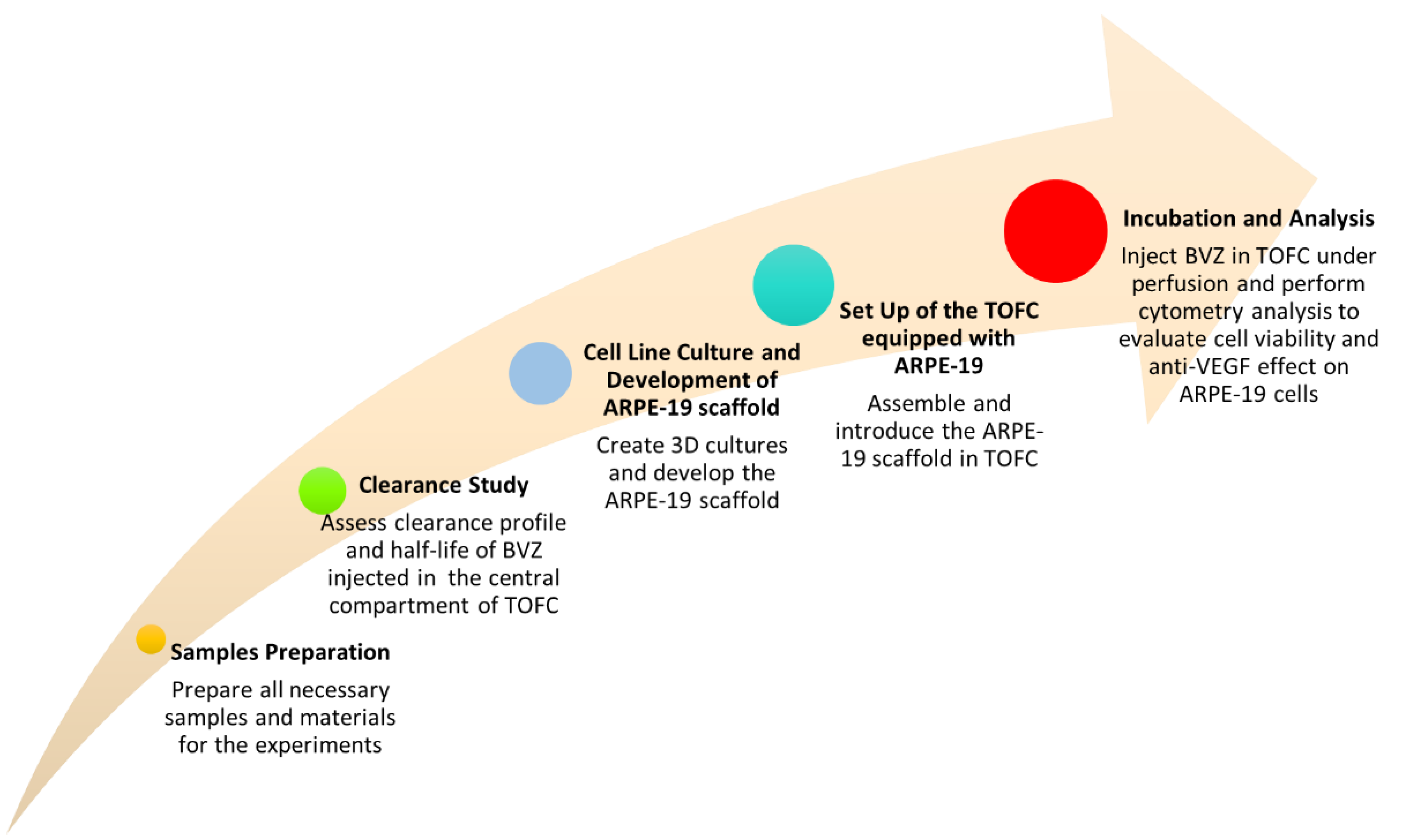
2.2. Methods
2.2.1. Samples Preparation
2.2.2. Clearance Study
2.2.3. Cell Line Culture
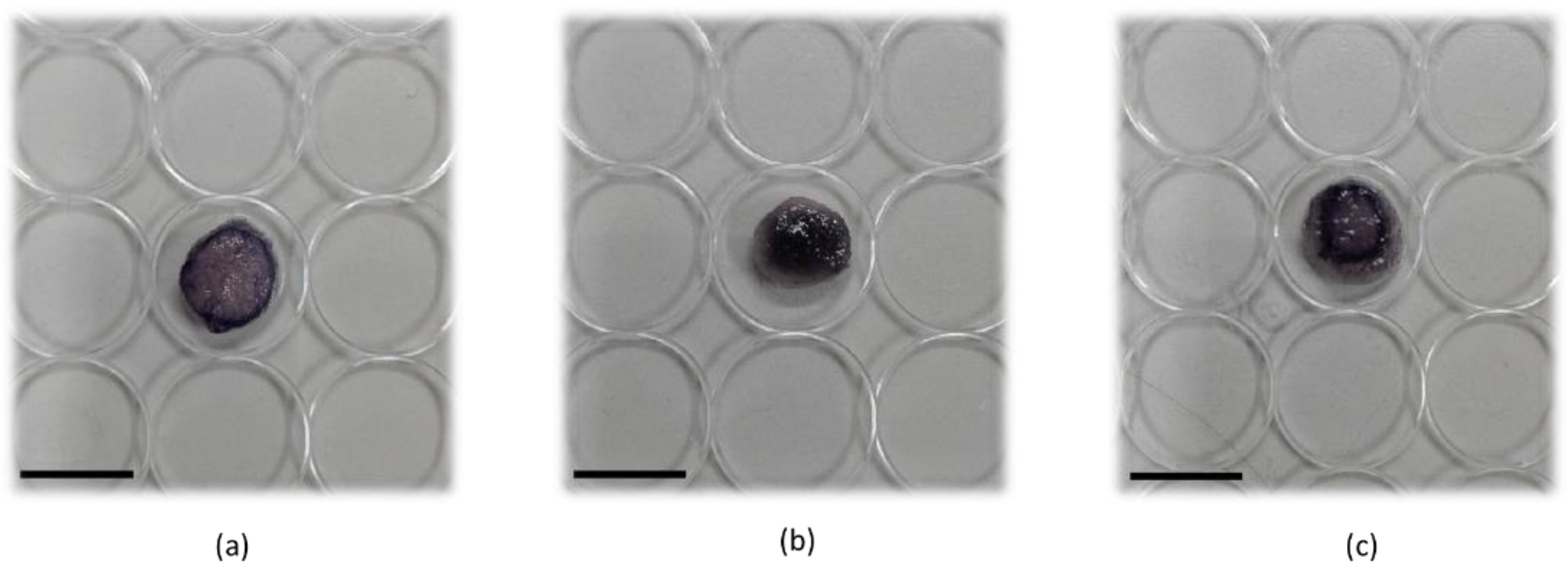
2.2.4. Generation of 3D Cultures and Development of ARPE-19 Scaffold
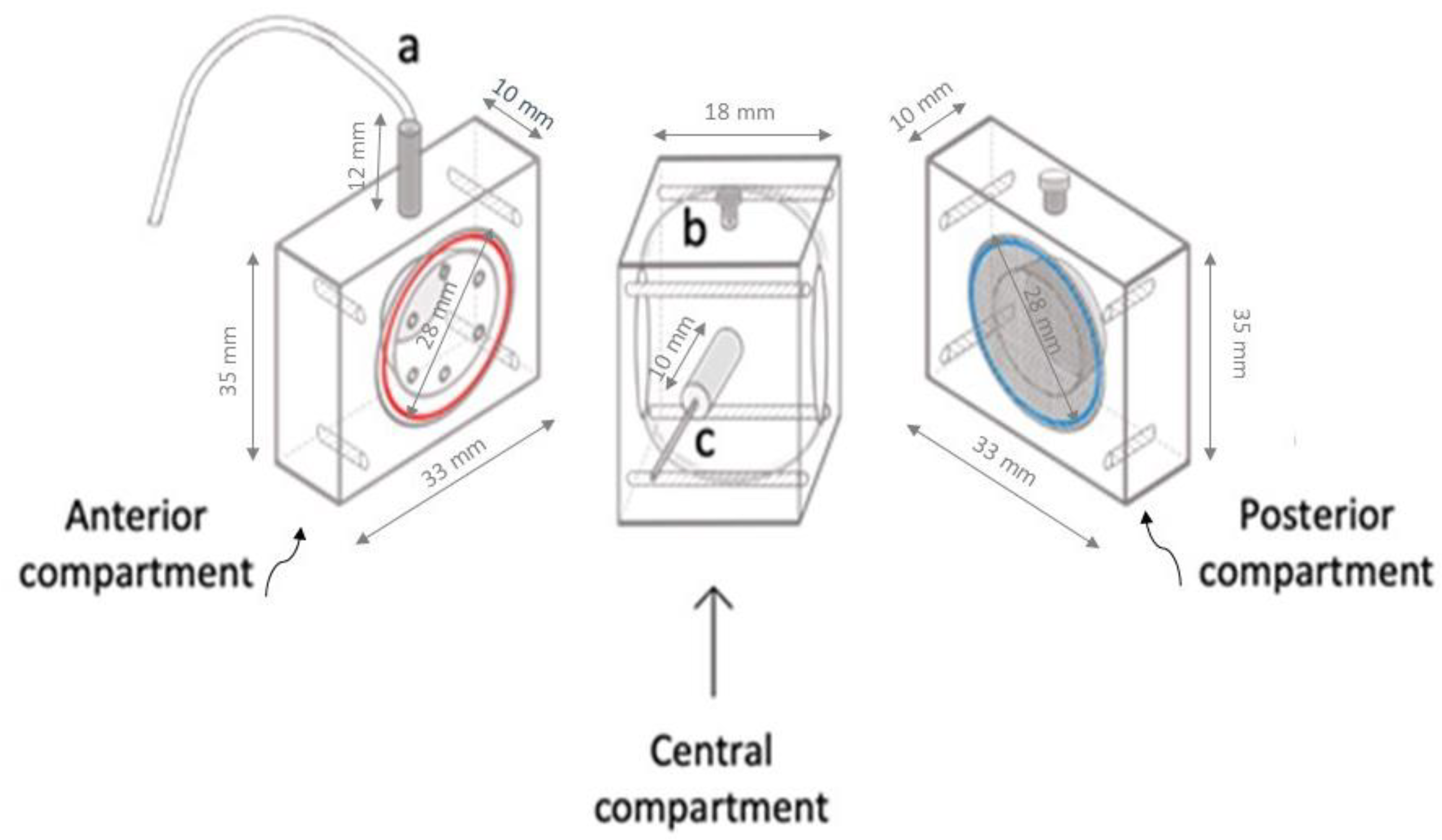
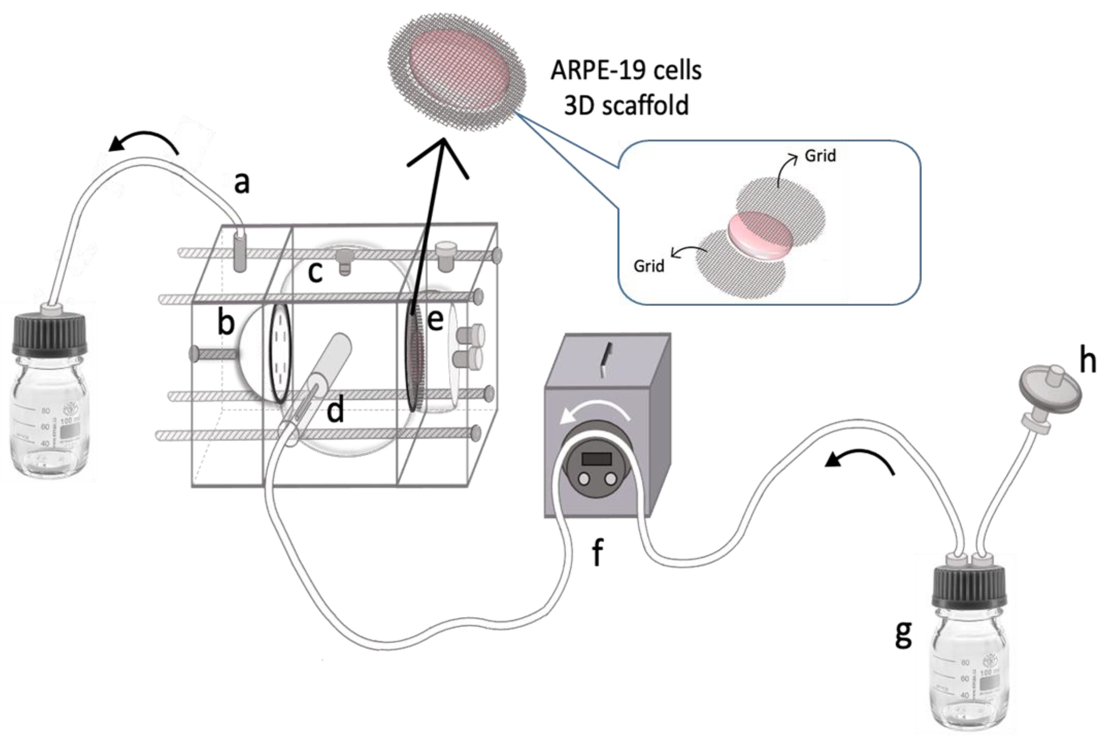
2.2.5. Setup of the TOFC Equipped with ARPE-19 Scaffold
2.2.6. Incubation of ARPE-19 Scaffold in Static Condition
2.2.7. Flow Cytometry Analysis and Evaluation of Cell Viability
2.2.8. Statistical Analysis
3. Results
3.1. BVZ Clearance Study by TOFC
3.2. Development of ARPE-19 Scaffold and Equipping within TOFC
3.3. Evaluation of Anti-VEGF Activity and Cell Viability
4. Discussion
5. Conclusions
Author Contributions
Funding
Institutional Review Board Statement
Informed Consent Statement
Data Availability Statement
Acknowledgments
Conflicts of Interest
References
- Christensen, G.; Barut, L.; Urimi, D.; Schipper, N.; Paquet-durand, F. Investigating Ex Vivo Animal Models to Test the Performance of Intravitreal Liposomal Drug Delivery Systems. Pharmaceutics 2021, 13, 1013. [Google Scholar] [CrossRef] [PubMed]
- Lehrmann, D.; Refaian, N.; Simon, M.; Rokohl, A.C.; Heind, L.M. Preclinical models in ophthalmic oncology—A narrative review. Ann. Eye Sci. 2022, 7, 14. [Google Scholar] [CrossRef]
- 2021/2784(RSP). Available online: https://oeil.secure.europarl.europa.eu/oeil/popups/ficheprocedure.do?lang=en&reference=2021/2784 (accessed on 5 June 2023).
- Gan, J.; Bolon, B.; Van Vleet, T.; Wood, C. Chapter 24—Alternative Models in Biomedical Research: In Silico, In Vitro, Ex Vivo, and Nontraditional In Vivo Approaches. In Haschek and Rousseaux’s Handbook of Toxicologic Pathology, 4th ed.; Haschek, W.M., Rousseaux, C.G., Wallig, M.A., Bolon, B., Eds.; Academic Press: Cambridge, MA, USA, 2022; pp. 925–966. [Google Scholar] [CrossRef]
- Verderio, P.; Lecchi, M.; Ciniselli, C.M.; Shishmani, B.; Apolone, G.; Manenti, G. 3Rs Principle and Legislative Decrees to Achieve High Standard of Animal Research. Animals 2023, 13, 277. [Google Scholar] [CrossRef] [PubMed]
- Mengus, C.; Muraro, M.G.; Mele, V.; Amicarella, F.; Manfredonia, C.; Foglietta, F.; Muenst, S.; Soysal, S.D.; Iezzi, G.; Spagnoli, G.C. In Vitro Modeling of Tumor−Immune System Interaction. ACS Biomater. Sci. Eng. 2018, 4, 314–323. [Google Scholar] [CrossRef]
- Foglietta, F.; Canaparo, R.; Muccioli, G.; Terreno, E.; Serpe, L. Methodological aspects and pharmacological applications of three-dimensional cancer cell cultures and organoids. Life Sci. 2020, 254, 117784–117785. [Google Scholar] [CrossRef]
- Hirt, C.; Papadimitropoulos, A.; Muraro, M.G.; Mele, V.; Panopoulos, E.; Cremonesi, E.; Ivanek, R.; Schultz-Thater, E.; Droeser, R.A.; Mengus, C.; et al. Bioreactor-engineered cancer tissue-like structures mimic phenotypes, gene expression profiles and drug resistance patterns observed “in vivo”. Biomaterials 2015, 62, 138–146. [Google Scholar] [CrossRef] [PubMed]
- Fotaki, N. Flow-through cell apparatus (USP apparatus 4): Operation and features. Dissolut. Technol. 2011, 18, 46–49. [Google Scholar] [CrossRef]
- Tojo, K. A pharmacokinetic model for ocular drug delivery. Chem. Pharm. Bull. 2004, 52, 1290–1294. [Google Scholar] [CrossRef][Green Version]
- Repetto, R.; Stocchino, A.; Cafferata, C. Experimental investigation of vitreous humour motion within a human eye model. Phys. Med. Biol. 2005, 50, 4729–4743. [Google Scholar] [CrossRef]
- Awwad, S.; Lockwood, A.; Brocchini, S.; Khaw, P.T. The PK-Eye: A Novel in Vitro Ocular Flow Model for Use in Preclinical Drug Development. J. Pharm. Sci. 2015, 104, 3330–3342. [Google Scholar] [CrossRef]
- Adrianto, M.F.; Annuryanti, F.; Wilson, C.G.; Sheshala, R.; Thakur, R.R. In vitro dissolution testing models of ocular implants for posterior segment drug delivery. Drug Deliv. Transl. Res. 2022, 12, 1355–1375. [Google Scholar] [CrossRef] [PubMed]
- Loch, C.; Nagel, S.; Guthoff, R.; Seidlitz, A.; Weitschies, W. The Vitreous Model—A new in vitro test method simulating the vitreous body Model characterization. Biomed. Eng. /Biomed. Tech. 2012, 57, 281–284. [Google Scholar] [CrossRef]
- Loch, C.; Bogdahn, M.; Stein, S.; Nagel, S.; Guthoff, R.; Weitschies, W.; Seidlitz, A. Simulation of Drug Distribution in the Vitreous Body After Local Drug Application into Intact Vitreous Body and in Progress of Posterior Vitreous Detachment. J. Pharm. Sci. 2014, 103, 517–526. [Google Scholar] [CrossRef]
- Stein, S.; Auel, T.; Kempin, W.; Bogdahn, M.; Weitschies, W.; Seidlitz, A. Influence of the test method on in vitro drug release from intravitreal model implants containing dexamethasone or fluorescein sodium in poly (D,L-lactide-co-glycolide) or polycaprolactone. Eur. J. Pharm. Biopharm. 2018, 127, 270–278. [Google Scholar] [CrossRef] [PubMed]
- Auel, T.; Großmann, L.; Schulig, L.; Weitschies, W.; Seidlitz, A. The EyeFlowCell: Development of a 3D-Printed Dissolution Test Setup for Intravitreal Dosage Forms. Pharmaceutics 2021, 13, 1394. [Google Scholar] [CrossRef]
- Yang, X.; Guo, X.; Yang, Y.; Huang, J.; Xiong, X.; Xie, X.; Tan, X. In-Vitro Eyeball Superfusion System. Patent CN101406176A, 15 April 2009. Available online: https://worldwide.espacenet.com/patent/search?q=pn%3DCN101406176A (accessed on 6 May 2023).
- Awwad, S.; Bouremel, Y.; Ibeanu, N.; Brocchini, S.J.; Khaw, P.T. Artificial Eye Assembly for Studying Ocular Pharmacokinetics. Patent WO2021186191A1, 31 August 2021. Available online: https://worldwide.espacenet.com/patent/search?q=pn%3DWO2021186191A1 (accessed on 6 May 2023).
- Juhong, H.; Yambin, P. Medical Simulation Human Eye Simulation Module. Patent CN210378044U, 21 April 2020. Available online: https://worldwide.espacenet.com/patent/search?q=pn%3DCN210378044U (accessed on 21 June 2023).
- Dongeun, H.; Jeongyun, S. Methods and Devices for Modelling the Eye. Patent US20170229043A1, 10 August 2017. Available online: https://worldwide.espacenet.com/patent/search?q=pn%3DUS2017229043A1 (accessed on 2 July 2023).
- Sapino, S.; Peira, E.; Chirio, D.; Chindamo, G.; Guglielmo, S.; Oliaro-Bosso, S.; Barbero, R.; Vercelli, C.; Re, G.; Brunella, V.; et al. Thermosensitive nanocomposite hydrogels for intravitreal delivery of cefuroxime. Nanomaterials 2019, 9, 1461. [Google Scholar] [CrossRef]
- Kummer, M.P.; Abbott, J.J.; Dinser, S.; Nelson, B.J. Artificial vitreous humor for in vitro experiments. In Proceedings of the Annual International Conference of the IEEE Engineering in Medicine and Biology, Lyon, France, 23–26 August 2007. [Google Scholar] [CrossRef]
- Bradford, M.M. A rapid and sensitive method for the quantitation of microgram quantities of protein utilizing the principle of protein-dye binding. Anal. Biochem. 1976, 72, 248–254. [Google Scholar] [CrossRef]
- Ahn, J.; Kim, H.; Woo, S.J.; Park, J.H.; Park, S.; Hwang, D.J.; Park, K.H. Pharmacokinetics of intravitreally injected bevacizumab in vitrectomized eyes. J. Ocul. Pharmacol. Ther. 2013, 29, 612–618. [Google Scholar] [CrossRef]
- Gal-Or, O.; Dotan, A.; Dachbash, M.; Tal, K.; Nisgav, Y.; Weinberger, D.; Ehrlich, R.; Livnat, T. Bevacizumab clearance through the iridocorneal angle following intravitreal injection in a rat model. Exp. Eye Res. 2016, 145, 412–416. [Google Scholar] [CrossRef]
- Nomoto, H.; Shiraga, F.; Kuno, N.; Kimura, E.; Fujii, S.; Shinomiya, K.; Nugent, A.K.; Hirooka, K.; Baba, T. Pharmacokinetics of Bevacizumab after Topical, Subconjunctival, and Intravitreal Administration in Rabbits. Investig. Ophthalmol. Vis. Sci. 2009, 50, 4807–4813. [Google Scholar] [CrossRef]
- Sinapis, C.I.; Routsias, J.G.; Sinapis, A.I.; Sinapis, D.I.; Agrogiannis, G.D.; Pantopoulou, A.; Theocharis, S.E.; Baltatzis, S.; Patsouris, E.; Perrea, D. Pharmacokinetics of intravitreal bevacizumab (Avastin®) in rabbits. Clin. Ophthalmol. 2011, 5, 697–704. [Google Scholar] [CrossRef] [PubMed]
- Moisseiev, E.; Waisbourd, M.; Ben-Artsi, E.; Levinger, E.; Barak, A.; Daniels, T.; Csaky, K.; Loewenstein, A.; Barequet, I.S. Pharmacokinetics of bevacizumab after topical and intravitreal administration in human eyes. Graefes Arch. Clin. Exp. Ophthalmol. 2014, 252, 331–337. [Google Scholar] [CrossRef] [PubMed]
- Tegtmeyer, S.; Papantoniou, I.; Müller-Goymann, C.C. Reconstruction of an in vitro cornea and its use for drug permeation studies from different formulations containing pilocarpine hydrochloride. Eur. J. Pharm. Biopharm. 2001, 51, 119–125. [Google Scholar] [CrossRef] [PubMed]
- Kutlehria, S.; Sachdeva, M.S. Role of In Vitro Models for Development of Ophthalmic Delivery Systems. Crit. Rev. Ther. Drug Carrier Syst. 2021, 38, 1–31. [Google Scholar] [CrossRef]
- Auel, T.; Scherke, L.P.; Hadlich, S.; Mouchantat, S.; Grimm, M.; Weitschies, W.; Seidlitz, A. Ex Vivo Visualization of Distribution of Intravitreal Injections in the Porcine Vitreous and Hydrogels Simulating the Vitreous. Pharmaceutics 2023, 15, 786. [Google Scholar] [CrossRef]
- Loh, Q.L.; Choong, C. Three-dimensional scaffolds for tissue engineering applications: Role of porosity and pore size. Tissue Eng. Part B Rev. 2013, 19, 485–502. [Google Scholar] [CrossRef] [PubMed]
- Merz, P.R.; Röckel, N.; Ballikaya, S.; Auffarth, G.U.; Schmack, I. Effects of ranibizumab (Lucentis®) and bevacizumab (Avastin®) on human corneal endothelial cells. BMC Ophthalmol. 2018, 18, 316–323. [Google Scholar] [CrossRef]
- Kaempf, S.; Johnen, S.; Salz, A.K.; Weinberger, A.; Walter, P.; Thumann, G. Effects of Bevacizumab (Avastin) on Retinal Cells in Organotypic Culture. Investig. Ophthalmol. Vis. Sci. 2008, 49, 3164–3171. [Google Scholar] [CrossRef][Green Version]
- Ferrara, N.; Hillan, K.J.; Gerber, H.P.; Novotny, W. Discovery and development of bevacizumab, an anti-VEGF antibody for treating cancer. Nat. Rev. Drug Discov. 2004, 3, 391–400. [Google Scholar] [CrossRef]
- Chung, M.; Lee, S.; Lee, B.J.; Son, K.; Jeon, N.L.; Kim, J.H. Wet-AMD on a Chip: Modeling Outer Blood-Retinal Barrier In Vitro. Adv. Healthc. Mater. 2018, 7, 1700028–1700034. [Google Scholar] [CrossRef]
- Ma, W.; Lee, S.E.; Guo, J.; Qu, W.; Hudson, B.I.; Schmidt, A.M.; Barile, G.R. RAGE ligand upregulation of VEGF secretion in ARPE-19 cells. Investig. Ophthalmol. Vis. Sci. 2007, 48, 1355–1361. [Google Scholar] [CrossRef] [PubMed]
- Christoforidis, J.B.; Williams, M.M.; Wang, J.; Jiang, A.; Pratt, C.; Abdel-Rasoul, M.; Hinkle, G.H.; Knopp, M.V. Anatomic and pharmacokinetic properties of intravitreal bevacizumab and ranibizumab after vitrectomy and lensectomy. Retina 2013, 33, 946–952. [Google Scholar] [CrossRef] [PubMed]


Disclaimer/Publisher’s Note: The statements, opinions and data contained in all publications are solely those of the individual author(s) and contributor(s) and not of MDPI and/or the editor(s). MDPI and/or the editor(s) disclaim responsibility for any injury to people or property resulting from any ideas, methods, instructions or products referred to in the content. |
© 2023 by the authors. Licensee MDPI, Basel, Switzerland. This article is an open access article distributed under the terms and conditions of the Creative Commons Attribution (CC BY) license (https://creativecommons.org/licenses/by/4.0/).
Share and Cite
Sapino, S.; Chindamo, G.; Peira, E.; Chirio, D.; Foglietta, F.; Serpe, L.; Vizio, B.; Gallarate, M. Development of ARPE-19-Equipped Ocular Cell Model for In Vitro Investigation on Ophthalmic Formulations. Pharmaceutics 2023, 15, 2472. https://doi.org/10.3390/pharmaceutics15102472
Sapino S, Chindamo G, Peira E, Chirio D, Foglietta F, Serpe L, Vizio B, Gallarate M. Development of ARPE-19-Equipped Ocular Cell Model for In Vitro Investigation on Ophthalmic Formulations. Pharmaceutics. 2023; 15(10):2472. https://doi.org/10.3390/pharmaceutics15102472
Chicago/Turabian StyleSapino, Simona, Giulia Chindamo, Elena Peira, Daniela Chirio, Federica Foglietta, Loredana Serpe, Barbara Vizio, and Marina Gallarate. 2023. "Development of ARPE-19-Equipped Ocular Cell Model for In Vitro Investigation on Ophthalmic Formulations" Pharmaceutics 15, no. 10: 2472. https://doi.org/10.3390/pharmaceutics15102472
APA StyleSapino, S., Chindamo, G., Peira, E., Chirio, D., Foglietta, F., Serpe, L., Vizio, B., & Gallarate, M. (2023). Development of ARPE-19-Equipped Ocular Cell Model for In Vitro Investigation on Ophthalmic Formulations. Pharmaceutics, 15(10), 2472. https://doi.org/10.3390/pharmaceutics15102472


