Article

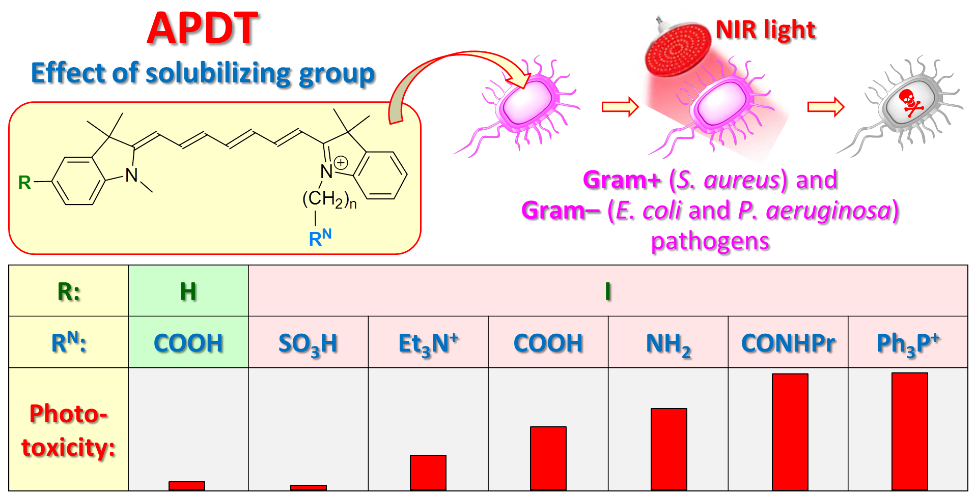
Effect of Solubilizing Group on the Antibacterial Activity of Heptamethine Cyanine Photosensitizers

1
Department of Chemical Sciences, Ariel University, Ariel 40700, Israel
2
Department of Chemical Engineering, Ariel University, Ariel 40700, Israel
*
Author to whom correspondence should be addressed.
Pharmaceutics 2023, 15(1), 247; https://doi.org/10.3390/pharmaceutics15010247
Submission received: 6 November 2022 / Revised: 3 January 2023 / Accepted: 9 January 2023 / Published: 11 January 2023
(This article belongs to the Special Issue Antimicrobial Sonodynamic and Photodynamic Therapies)

Abstract

:
Antibiotic resistance of pathogenic bacteria dictates the development of novel treatment modalities such as antimicrobial photodynamic therapy (APDT) utilizing organic dyes termed photosensitizers that exhibit a high cytotoxicity upon light irradiation. Most of the clinically approved photosensitizers are porphyrins that are poorly excitable in the therapeutic near-IR spectral range. In contrast, cyanine dyes function well in the near-IR region, but their phototoxicity, in general, is very low. The introduction of iodine atoms in the cyanine molecules was recently demonstrated to greatly increase their phototoxicity. Herein, we synthesized a series of the new iodinated heptamethine cyanine dyes (ICy7) containing various solubilizing moieties, i.e., negatively charged carboxylic (ICy7COOH) and sulfonic (ICy7SO3H) groups, positively charged triphenylphosphonium (ICy7PPh3), triethylammonium (ICy7NEt3) and amino (ICy7NH2) groups, and neutral amide (ICy7CONHPr) group. The effect of these substituents on the photodynamic eradication of Gram-positive (S. aureus) and Gram-negative (E. coli and P. aeruginosa) pathogens was studied. Cyanine dyes containing the amide and triphenylphosphonium groups were found to be the most efficient for eradication of the investigated bacteria. These dyes are effective at low concentrations of 0.05 µM (33 J/cm2) for S. aureus, 50 µM (200 J/cm2) for E. coli, and 5 µM (100 J/cm2) for P. aeruginosa and considered, therefore, promising photosensitizers for APDT applications. The innovation of the new photosensitizers consisted of a combination of the heavy-atom effect that increases singlet oxygen generation with the solubilizing group’s effect improving cell uptake, and with effective near-IR excitation. Such a combination helped to noticeably increase the APDT efficacy and should pave the way for the development of more advanced photosensitizers for clinical use.

Graphical Abstract

1. Introduction

Photodynamic therapy (PDT) [1] and photodynamic antimicrobial therapy (APDT) [2] are of great importance for treating cancer [3,4,5] and eradication of pathogenic bacteria [6], viruses [7], and fungi [8] due to their non-invasiveness and minimal side effects on normal tissues. Both PDT and APDT consist of the administering of an organic dye [9] or nanoparticle [10,11] referred to as a photosensitizer (PS) [12], followed by its light-irradiation at a specific wavelength to generate reactive species that kill cells in the vicinity. As a result of the light absorption, PS molecules pass to the first excited singlet state and then turn into the long-lived triplet state, which is responsible for the photochemical reactions yielding cytotoxic species such as singlet oxygen (1O2), superoxide anion (O2−•), hydroxyl radical (OH), hydrogen peroxide (H2O2), and organic radicals among others [1]. Thus, the efficient population of the triplet state plays an important role in the PS phototoxicity.
Most of the PSs approved by the Food and Drug Administration (FDA) are porphyrin-based dyes that predominantly absorb light in the short wavelength range, but insufficiently absorb in the biologically transparent and, thus, therapeutically important red and near-IR (NIR) spectral region [13,14]. In recent years, special attention is paid, therefore, to non-porphyrin-based PSs such as phthalocyanines [15,16,17] and cyanines [18,19,20] that exhibit high extinction coefficients within the NIR region. Among them, indocyanine green (ICG), the clinically approved NIR dye, has been widely explored as a potential photosensitizer in clinical applications [21,22]. The low phototoxicity of cyanine dyes including ICG is attractive for their applications as fluorescent biomedical reporters. However, a major limitation for their therapeutic utility in PDT and APDT is their unsatisfactory phototoxic effect.
The well-known approach to improve the phototoxicity of cyanine dyes is based on the introduction of heavy atoms such as iodine [23,24,25], which significantly increases the probability of intersystem crossing (ISC) from the singlet to the triplet state due to enhanced spin–orbit coupling resulting in the elevated rates of cytotoxic species generation [26]. The enhancement of PDT and APDT efficacy owing to the “heavy atom effect” has been previously reported in the example of cyanine dyes [27,28,29] among the photosensitizers of other dye classes [30,31].
Recently, we reported for the first time on the unexpected effect of iodine atoms in the heptamethine cyanine dyes (nICy7COOH, where n = 0–6, Figure 1) on the photodynamic eradication of pathogenic bacteria, namely, Staphylococcus aureus (S. aureus), Escherichia coli (E. coli) and Pseudomonas aeruginosa (P. aeruginosa): the increasing number of iodines may not only increase but also decrease phototoxicity [32]. Thus, the iodinated cyanines containing 2–4 iodine atoms exhibited the most pronounced photo-eradication efficacy on S. aureus, while mono- and, surprisingly, hexa-iodinated dyes were less effective. In contrast, the mono-iodinated cyanine turned out to be the most active against E. coli and P. aeruginosa, while the phototoxicity dropped down with the increasing number of iodines. This phenomenon was attributed to the intensification of the dye aggregation in aqueous media and, thereby, the reduction in the dye uptake by these bacteria [32]. As such, an optimal balance of hydrophobic–hydrophilic properties and the selection of the more appropriate solubilizing groups are required to enhance the uptake and phototoxicity of the dyes.
A known method to improve the uptake of organic molecules (e.g., drugs and dyes) and nanoparticles by cells consists of the introduction of positively charged groups, which facilitate penetration through the negatively charged cell membrane [33,34]. Our recent results showed, however, that this is not always true: the introduction of the positively charged triethylammonium group in the di-iodinated cyanine (Figure 1) significantly improved photo-eradication of E. coli and P. aeruginosa, while unpredictably reducing the killing effect towards S. aureus [32].
As a continuation of our recent research, this work investigated the effect of various neutrally, positively, and negatively charged solubilizing groups on the photo-eradication of S. aureus, E. coli, and P. aeruginosa. To this end, we synthesized a series of novel iodinated heptamethine cyanine dyes containing carboxylic (ICy7COOH), amide (ICy7CONHPr), sulfonic (ICy7SO3H), triphenylphosphonium (ICy7PPh3), triethylammonium (ICy7NEt3), and amino (ICy7NH2) groups (Figure 2). We studied their photophysical properties, uptake by bacteria, and photodynamic efficacy in comparison with the non-iodinated cyanine, Cy7COOH. To minimize the unwanted effects associated with possible dye aggregation, our investigation was performed using the example of the mono-iodinated cyanines, ICy7.

2. Materials and Methods

2.1. General

All starting materials and chemicals were supplied by Alfa Aesar and Sigma Aldrich. Solvents were purchased from Bio-Lab Israel.
Chemical reactions were monitored by TLC (Silica gel 60 F-254, Merck) and LCMS. LCMS analyses were performed using an Agilent Technologies 1260 Infinity (LC) 6120 quadruple (MS) instrument, column Agilent SB-C18, 1.8 mm, 2.1 × 50 mm, column temperature 50 °C, eluent water–acetonitrile (ACN) + 0.1% formic acid. HRMS analyses were performed in ESI positive mode by using an Agilent 6550 iFunnel Q-TOF LCMS instrument. 1H NMR and 13C NMR spectra were measured in DMSO-d6, CD3OD, and a mixture CDCl3–CD3OD on a Bruker Avance III HD (400 MHz and 100 MHz for 1H and 13C, respectively) spectrometer. Chemical shifts were reported in ppm using the solvent residual signal as an internal reference: δH(DMSO-d6) = 2.50 ppm, δC(DMSO-d6) = 39.52 ppm; δH(CDCl3) = 7.26 ppm, δC(CDCl3) = 77.16 ppm; δH(CD3OD) = 3.31 ppm, δC(CD3OD) = 49.0 ppm.

2.2. Synthesis

1,2,3,3-Tetramethyl-3H-indol-1-ium (1a) and 5-iodo-1,2,3,3-tetramethyl-3H-indol-1-ium (1b) were synthesized according to the procedure described in [35,36]: 2,3,3-trimethyl-3H-indole (1 g, 6.28 mmol) or 5-iodo-2,3,3-trimethyl-3H-indole (1 g, 3.51 mmol) was refluxed overnight with excess of methyl iodide (10 eq.). The precipitate was washed with diethyl ether and dried in a desiccator under vacuum.
1,2,3,3-Tetramethyl-3H-indol-1-ium (1a). Yield: 1.61 g (85%). 1H NMR (400 MHz, DMSO-d6, ppm): δ 7.91 (1H, d, J = 5.2 Hz), 7.83 (1H, d, J = 5.4 Hz), 7.62 (2H, t, J = 5.6 Hz), 3.98 (3H, s), 2.77 (3H, s), 1.53 (6H, s). MS m/z C12H16N+ calculated [M]+ 174.13, found m/z 174.10.
5-Iodo-1,2,3,3-tetramethyl-3H-indol-1-ium (1b): Yield: 0.96 g (65%). 1H NMR (400 MHz, DMSO-d6, ppm): δ 8.27 (1H, s), 7.99 (1H, d, J = 8.5 Hz), 7.71 (1H, d, J = 8.4 Hz), 3.93 (3H, s), 2.73 (3H, s), 1.51 (6H, s). MS m/z C12H15IN+ calculated [M]+ 300.02, found m/z 300.16.
1-(5-Carboxypentyl)-2,3,3-trimethyl-3H-indol-1-ium bromide (3a) was synthesized as described in [32]. 2,3,3-Trimethyl-3H-indole (1.0 g, 6.28 mmol) was mixed with 6-bromohexanoic acid (1.84 g, 9.41 mmol) and heated at 120 °C for 15 h in a sealed tube. The reaction mixture was cooled to r.t., then diluted with benzene (5 mL); then, the solvent was decanted, and the residue was triturated with benzene (3 × 5 mL), and filtered. The obtained precipitate was washed with acetone (1 mL) and dried to yield 3a (1.33 g, 60%). 1H NMR (400 MHz, DMSO-d6, ppm): δ 7.97 (1H, d, J = 5.1 Hz), 7.84 (1H, d, J = 5.4 Hz), 7.62 (2H, t, J = 5.1 Hz), 4.45 (2H, t, J = 7.8 Hz), 2.84 (3H, s), 2.23 (2H, t, J = 7.2 Hz), 1.85 (2H, m), 1.56 (2H, m), 1.54 (6H, s), 1.43 (2H, m). MS m/z C17H24NO2+ calculated [M]+ 274.18, found m/z 274.20.
3-(2,3,3-Trimethyl-3H-indol-1-ium-1-yl)propane-1-sulfonate (3b) was synthesized similarly to [37]. 2,3,3-trimethyl-3H-indole (1.5 g, 9.4 mmol) and 1,3-propanesultone (2.8 g, 22.9 mmol) were melted, at 120 °C, for 2 h. The reaction mixture became solid. 2-Propanol (5 mL) was added to the reaction mixture and heated, at 80 °C, for 1 h. Then, the reaction mixture was washed with diethyl ether (30 mL) and dried in a desiccator under vacuum. Then, the obtained viscous liquid was diluted with H2O and column purified on LiChroprep RP18 using 0–1% acetonitrile–water as eluent resulting in 3b (light-yellow viscous liquid). Yield: 68%. 1H NMR (400 MHz, DMSO-d6, ppm): δ 8.04 (1H, d, J = 6.9 Hz), 7.82 (1H, d, J = 8.0 Hz), 7.69–7.56 (2H, m), 4.65 (2H, t, J = 8.0 Hz), 2.83 (3H, s), 2.64 (2H, t, J = 6.5 Hz), 1.53 (6H, s). MS m/z C14H19NO3S calculated [M + H]+ 281.11, found m/z 282.21.
2,3,3-Trimethyl-1-(4-(triphenylphosphonio)butyl)-3H-indol-1-ium dibromide (3c) was synthesized as described in [38]. A mixture of 2,3,3-trimethylindolenine (500 mg, 3.14 mmol), 4-bromobutyltriphenylphosphonium bromide (4.5 g, 9.42 mmol) and potassium iodide (1.56 g, 9.42 mmol) in acetonitrile was refluxed for 72 h. After cooling to r.t., water (50 mL) and ethyl acetate (50 mL) were added to the reaction mixture, and the organic layer was separated. The aqueous phase containing the product was concentrated under reduced pressure, and the residue was washed three times with diethyl ether and dried in a desiccator under vacuum to give 3c. Yield: 74%. 1H NMR (400 MHz, DMSO-d6, ppm): δ 8.05–7.98 (1H, m), 7.94–7.71 (16H, m), 7.64–7.57 (2H, m), 4.56 (2H, t, J = 7.4 Hz), 3.86–3.71 (2H, m), 2.84 (3H, s), 2.16–2.05 (2H, m), 1.78–1.63 (2H, m), 1.48 (6H, s). MS m/z C33H36NP2+ calculated [M]2+ 238.63, found m/z 238.60.
2,3,3-Trimethyl-1-(3-(triethylammonio)propyl)-3H-indol-1-ium dibromide (3d) was synthesized similar to [32]. 2,3,3-trimethyl-3H-indole (0.5 g, 3.1 mmol) and 3-bromo-N,N,N-triethylpropan-1-aminium bromide (0.95 g, 3.1 mmol) were stirred in acetonitrile (14 mL) at reflux for 4 days. The reaction mixture was precipitated with diethyl ether (30 mL). Then, the solvent was decanted and the viscous solid was triturated with diethyl ether, filtered, washed with ether, and dried in a desiccator under vacuum to give 3d. Yield: (0.6 g) 62%. 1H NMR (400 MHz, DMSO-d6, ppm): δ 8.13 (1H, d, J = 7.3 Hz), 7.86 (1H, d, J = 7.8 Hz), 7.71–7.59 (2H, m), 4.58 (2H, t, J = 8.0 Hz), 3.33–3.18 (8H, m), 2.93 (3H, s), 2.29–2.13 (2H, m), 1.56 (6H, s), 1.23 (9H, t, J = 7.1 Hz). MS m/z C20H34N22+ calculated [M]2+ 151.13, found m/z 151.20.
1-(3-Aminopropyl)-2,3,3-trimethyl-3H-indol-1-ium bromide (3e) was synthesized as described in [39]: 2,3,3-trimethylindolenine (0.5 g, 3.14 mmol), 3-bromopropylamine hydrobromide (1.87 g, 8.61 mmol) and anhydrous toluene (10 mL) were added to a sealed tube. The suspension was heated, at 130 °C, for 4 days. After cooling to r.t., toluene was decanted. The residue was washed with toluene and triturated with acetonitrile, filtered, washed with diethyl ether, and dried to give 3e. Yield: 0.31 g (50%). 1H NMR (400 MHz, DMSO-d6, ppm): δ 8.06 (1H, d, J = 8.0 Hz), 7.98 (2H, br. s), 7.85 (1H, d, J = 7.9 Hz), 7.68–7.59 (2H, m), 4.59 (2H, t, J = 8.0 Hz), 3.12–3.00 (2H, m), 2.88 (3H, s), 2.17 (2H, t, J = 7.9 Hz), 1.56 (6H, s). MS m/z C14H21N2+ calculated [M]+ 217.17, found m/z 217.20.
General procedure for the synthesis of the dyes Cy7COOH, ICy7COOH ICy7SO3H, ICy7NEt3 and ICy7PPh3.
A solution of indolenine 1a or 1b (0.5 mmol) and N-[5-(phenylamino)-2,4-pentadienylidene]aniline hydrochloride (0.5 mmol) in acetic anhydride (2 mL) was heated, at 90 °C, for 20 min to form the corresponding intermediate product 2a or 2b. The reaction mixture was cooled to r.t., and a solution of the second indolenine 3a3d (0.5 mmol) in dry pyridine (2 mL) was added. The reaction mixture was heated, at 90 °C, for 10 min, and the obtained cyanine dye was precipitated with diethyl ether, filtered, and washed with diethyl ether.
6-(3,3-Dimethyl-2-(7-(1,3,3-trimethylindolin-2-ylidene)hepta-1,3,5-trien-1-yl)-3H-indol-1-ium-1-yl)hexanoate (Cy7COOH) was column purified on Silica gel 60 using 10% methanol–chloroform as eluent. Yield: 119 mg (52%). 1H NMR (400 MHz, CDCl3–CD3OD, ppm): δ 7.75 (2H, t, J = 13.1 Hz, CH), 7.46 (1H, t, J = 12.7 Hz, CH), 7.40–7.34 (2H, m, ArH), 7.36 (2H, d, J = 7.5 Hz, ArH), 7.22 (2H, t, J = 7.4 Hz, ArH), 7.12 (1H, d, J = 8.4 Hz, ArH), 7.09 (1H, d, J = 8.4 Hz, ArH), 6.51 (2H, t, J = 12.5 Hz, CH), 6.11 (1H, d, J = 13.4 Hz, CH), 6.09 (1H, d, J = 13.6 Hz, CH), 3.96 (2H, t, J = 7.4 Hz, NCH2), 3.57 (3H, s, NCH3), 2.31 (2H, t, J = 7.2 Hz, CH2COOH), 1.86–1.72 (2H, m, CH2), 1.65 (12H, s, 2 (CH3)2), 1.74–1.62 (2H, m, CH2), 1.53–1.41 (2H, m, CH2).13C NMR (100 MHz, CDCl3–CD3OD, ppm): δ 175.63, 171.14, 170.56, 155.75, 150.53, 141.67, 141.00, 139.80, 139.63, 127.84, 124.76, 124.26, 121.36, 121.23, 109.50, 109.40, 102.68, 102.42, 48.47, 43.09, 32.98, 30.23, 26.83, 26.19, 25.45, 23.58. HRMS m/z (ESI+) C34H40N2O2+ calculated [M]+ 509.3168, found m/z: 509.3162.
6-(2-(7-(5-Iodo-1,3,3-trimethylindolin-2-ylidene)hepta-1,3,5-trien-1-yl)-3,3-dimethyl-3H-indol-1-ium-1-yl)hexanoate (ICy7COOH) was column purified on Silica gel 60 using 5–10% methanol–chloroform as eluent. Yield: 74 mg (45%). 1H NMR (400 MHz, CDCl3–CD3OD, ppm): δ 7.79 (1H, t, J = 13.1 Hz, CH), 7.63 (1H, t, J = 13.1 Hz, CH), 7.62 (1H, d, J = 8.5 Hz, ArH), 7.57 (1H, s, ArH), 7.43 (1H, t, J = 12.7 Hz, CH), 7.39 (1H, t, J = 7.9 Hz, ArH), 7.38 (1H, d, J = 7.8 Hz, ArH), 7.28 (1H, t, J = 7.4 Hz, ArH), 7.18 (1H, d, J = 7.9 Hz, ArH), 6.80 (1H, d, J = 8.4 Hz, ArH), 6.56 (1H, t, J = 13.3 Hz, CH), 6.47 (1H, t, J = 13.3 Hz, CH), 6.23 (1H, d, J = 13.5 Hz, CH), 5.95 (1H, d, J = 13.3 Hz, CH), 4.02 (2H, t, J = 7.4 Hz, NCH2), 3.45 (3H, s, NCH3), 2.27 (2H, t, J = 7.2 Hz, CH2COOH), 1.86–1.74 (2H, m, CH2), 1.72–1.63 (2H, m, CH2), 1.66 (6H, s, 2 CH3), 1.62 (6H, s, 2 CH3), 1.52–1.42 (2H, m, CH2). 13C NMR (100 MHz, CDCl3–CD3OD, ppm): δ 176.41, 172.49, 168.14, 155.64, 152.05, 148.66, 141.84, 141.47, 140.63, 140.16, 136.45, 130.15, 128.07, 125.47, 125.20, 125.02, 121.42, 110.53, 110.27, 104.15, 101.49, 86.20, 48.73, 47.34, 43.55, 33.74, 29.95, 26.86, 26.68, 26.36, 25.42, 23.80. HRMS m/z (ESI+) C34H39IN2O2+ calculated [M]+ 635.2134, found m/z: 635.2127.
3-(2,7,5-Iodo-1,3,3-trimethylindolin-2-ylidene)hepta-1,3,5-trien-1-yl)-3,3-dimethyl-3H-indol-1-ium-1-yl)propane-1-sulfonate (ICy7SO3H) was column purified on Silica gel 60 using 2–12% methanol–chloroform as eluent. Yield: 18%. 1H NMR (400 MHz, CD3OD, ppm): δ 7.96 (1H, t, J = 12.9 Hz, CH), 7.80 (1H, t, J = 13.2 Hz, CH), 7.74 (1H, s, ArH), 7.66 (1H, d, J = 8.3, ArH), 7.58–7.49 (2H, m, ArH, CH), 7.49–7.38 (2H, m, ArH), 7.28 (1H, t, J = 7.2 Hz, ArH), 7.02 (1H, d, J = 8.3 Hz, ArH), 6.59 (1H, t, J = 12.7 Hz, CH), 6.52 (1H, d, J = 13.3 Hz, CH), 6.50 (1H, t, J = 12.9 Hz, CH), 6.12 (1H, d, J = 13.4 Hz, CH), 4.37 (2H, t, NCH2), 3.51 (3H, s, NCH3), 3.00 (2H, t, J = 6.8 Hz, CH2SO3), 2.30–2.18 (2H, m, CH2), 1.67 (6H, s, 2 CH3), 1.64 (6H, s, 2 CH3). 13C NMR (100 MHz, CD3OD, ppm): δ 174.86, 170.85, 157.78, 154.66, 151.43, 144.57, 144.36, 143.31, 142.76, 138.56, 132.31, 129.98, 128.02, 127.47, 126.77, 123.50, 113.04, 112.57, 106.59, 103.97, 87.69, 79.55, 54.90, 50.80, 44.05, 31.09, 27.85, 27.76, 24.27. HRMS: m/z (ESI+) C31H36IN2O3S calculated [M+H]+ 643.1491, found m/z: 643.1492.
2,7,5-Iodo-1,3,3-trimethylindolin-2-ylidene)hepta-1,3,5-trien-1-yl)-3,3-dimethyl-1-(3-(triethylammonio)propyl)-3H-indol-1-ium (ICy7NEt3) was column purified on LiChroprep RP18 using 5–50% acetonitrile–water + 0.05% acetic acid as eluent. Yield: 38%. 1H NMR (400 MHz, CD3OD, ppm): δ 8.05–7.91 (2H, m, CH), 7.86 (1H, s, ArH), 7.75 (1H, d, J = 8.3 Hz, ArH), 7.64 (1H, t, J = 12.8 Hz, CH), 7.50 (1H, d, J = 7.4 Hz, ArH), 7.42 (1H, t, J = 7.6 Hz, ArH), 7.36 (1H, d, J = 8.0 Hz, ArH), 7.24 (1H, t, J = 7.4 Hz, ArH), 7.14 (1H, d, J = 8.4 Hz, ArH), 6.70–6.55 (m, 2H, CH), 6.40 (1H, d, J = 13.4 Hz, CH), 6.35 (1H, d, J = 13.7 Hz, CH), 4.21 (2H, t, J = 7.4 Hz, NCH2), 3.61 (3H, s, NCH3), 3.36 (8H, q, J = 6.9 Hz, N(CH2)4), 2.24–2.11 (m, 2H, CH2), 1.91 (6H, s, 2 CH3COO), 1.71 (6H, s, 2 CH3), 1.69 (6H, s, 2 CH3), 1.31 (9H, t, J = 7.2 Hz, 3 CH3). 13C NMR (100 MHz, CD3OD, ppm): δ 179.45, 174.00, 172.06, 158.22, 154.11, 152.76, 144.86, 144.16, 143.48, 142.13, 138.80, 132.56, 129.91, 127.94, 127.72, 125.87, 123.63, 114.00, 111.28, 106.01, 104.45, 89.53, 79.55, 54.90, 54.77, 54.05, 50.42, 50.10, 49.00, 41.20, 31.67, 28.08, 27.56, 23.59, 20.95, 7.71. HRMS: m/z (ESI+) C37H50IN32+ calculated [M]2+ 331.6519, found m/z: 331.6523.
2,7,5-Iodo-1,3,3-trimethylindolin-2-ylidene)hepta-1,3,5-trien-1-yl)-3,3-dimethyl-1-(4-(triphenylphosphonio)butyl)-3H-indol-1-ium (ICy7PPh3) was column purified on LiChroprep RP18 using 0–60% acetonitrile–water + 0.5% acetic acid as eluent. Yield: 34%. 1H NMR (400 MHz, CD3OD, ppm): 7.93–7.84 (4H, m, 2 ArH, 2 CH), 7.82–7.67 (15H, m, 3 Ph), 7.62 (1H, t, J = 12.9 Hz, CH), 7.47 (1H, d, J = 7.4 Hz, ArH), 7.42–7.32 (2H, m, ArH), 7.25 (1H, t, J = 7.1 Hz, ArH), 7.07 (1H, d, J = 8.4 Hz, ArH), 6.67–6.52 (2H, m, 2 CH), 6.46 (1H, d, J = 13.6 Hz, CH), 6.23 (1H, d, J = 13.5 Hz, CH), 4.26 (2H, t, J = 7.0 Hz, NCH2), 3.61–3.46 (2H, m, CH2P), 3.56 (3H, s, NCH3), 2.17–2.06 (2H, m, CH2), 1.95 (6H, s, 2 CH3COO), 1.84–1.72 (2H, m, CH2), 1.66 (6H, s, 2 CH3), 1.56 (6H, s, 2 CH3). 13C NMR (100 MHz, CD3OD, ppm): 177.42, 174.09, 171.95, 157.94, 153.93, 152.39, 144.54, 144.41, 143.31, 142.54, 138.64, 136.34, 134.93, 134.83, 132.41, 131.66, 131.53, 129.84, 127.67, 127.44, 126.44, 123.53, 120.03, 119.17, 113.38, 112.43, 106.07, 104.64, 88.36, 50.49, 44.21, 31.47, 29.63, 29.45, 27.92, 27.82, 22.50, 22.27, 21.98, 21.31. HRMS: m/z (ESI+) C50H52IN2P2+ calculated [M]+ 838.2913, found m/z: 838.2936.
Synthesis of 2,7,5-iodo-1,3,3-trimethylindolin-2-ylidene)hepta-1,3,5-trien-1-yl)-3,3-dimethyl-1-(6-oxo-6-(propylamino)hexyl)-3H-indol-1-ium iodide (ICy7CONHPr).
A mixture of ICy7COOH (50 mg, 0.066 mmol), N,N,N′,N′-tetramethyl-O-(N-succinimidyl)uronium tetrafluoroborate (TSTU) (39.7 mg, 0.132 mmol) and N,N-diisopropylethylamine (DIPEA) (46 μL, 0.264 mmol) was dissolved in dry DMF (0.5 mL). The reaction mixture was stirred, at r.t., for 40 min and then propylamine (21 μL, 0.264 mmol) was added. After stirring, at r.t., for 30 min the obtained dye was precipitated with diethyl ether, filtered off and washed with diethyl ether. The raw product was column purified on Silica gel 60 using 5–10% methanol–chloroform as eluent. Yield: 33 mg (62%). 1H NMR (400 MHz, CD3OD, ppm): δ 8.02 (1H, t, J = 13.2 Hz, CH), 7.85 (1H, t, J = 13.2 Hz, CH), 7.74 (1H, s, ArH), 7.68 (1H, d, J = 8.9 Hz, ArH), 7.63 (1H, t, J = 12.7 Hz, CH), 7.52 (1H, d, J = 7.3 Hz, ArH), 7.44 (1H, t, J = 7.9 Hz, ArH), 7.36 (1H, d, J = 7.9 Hz, ArH), 7.31 (1H, t, J = 7.4 Hz, ArH), 6.99 (1H, d, J = 8.4 Hz, ArH), 6.62 (1H, t, J = 13.3 Hz, CH), 6.54 (1H, t, J = 13.3 Hz, CH), 6.43 (1H, d, J = 13.8 Hz, CH), 6.12 (1H, d, J = 13.2 Hz, CH), 4.17 (2H, t, J = 7.4 Hz, NCH2), 3.49 (3H, s, NCH3), 3.08 (2H, t, J = 7.4 Hz, CH2CONH), 2.20 (2H, t, J = 7.2 Hz, CH2CONH), 1.88–1.76 (2H, m, CH2), 1.74–1.60 (2H, m, CH2), 1.71 (6H, s, 2 CH3), 1.66 (6H, s, 2 CH3), 1.53–1.39 (4H, m, 2 CH2), 0.88 (3H, t, J = 7.4 Hz, CH3). Amino group (-NH-) has an exchangeable proton which is not recorded in 1H NMR spectrum due to the low concentration. 13C NMR (100 MHz, CD3OD, ppm): δ 175.73, 175.27, 170.96, 157.72, 154.56, 151.33, 144.61, 144.34, 143.40, 142.94, 138.59, 132.32, 129.92, 127.62, 127.68, 126.90, 123.53, 112.89, 112.66, 106.50, 103.65, 87.54, 52.93, 49.56, 49.28, 45.26, 42.17, 36.66, 31.13, 30.75, 28.44, 27.98, 27.83, 27.31, 26.55, 23.64, 11.76. HRMS m/z (ESI+) C37H47IN3O+ calculated [M]+ 676.2764, found m/z: 676.2789.
Synthesis of 1-(3-aminopropyl)-2,7,5-iodo-1,3,3-trimethylindolin-2-ylidene)hepta-1,3,5-trien-1-yl)-3,3-dimethyl-3H-indol-1-ium iodide (ICy7NH2). A mixture of indolenine 1b (200 mg, 0.47 mmol) and N-[5-(phenylamino)-2,4-pentadienylidene]aniline hydrochloride (133 mg, 0.47 mmol) was dissolved in acetic anhydride (2 mL) and heated, at 90 °C, for 15 min to form product 2b. Then, the solvent was evaporated to dryness, the residue 2b was dried in a desiccator under vacuum for 3 h. The solution of the second indolenine 3e (139 mg, 0.47 mmol) in DMF (2 mL) and pyridine (200 µL) was added to the product 2b. The reaction mixture was heated, at 90 °C, for 5 min, and cooled to r.t.; the obtained product was precipitated with diethyl ether, filtered, and washed with diethyl ether. The raw dye was column purified on Silica gel 60 using 5–18% methanol–chloroform as eluent. Yield: 37 mg (15%). 1H NMR (400 MHz, CDCl3–CD3OD, ppm): δ 7.88 (1H, t, J = 13.2 Hz, CH), 7.82 (1H, t, J = 13.2 Hz, CH), 7.69 (1H, d, J = 8.5 Hz, ArH), 7.68 (1H, s, ArH), 7.55 (1H, t, J = 12.9 Hz, CH), 7.46–7.36 (2H, m, ArH), 7.30 (1H, d, J = 8.2 Hz, ArH), 7.24 (1H, t, J = 7.3 Hz, ArH), 6.96 (1H, d, J = 7.8 Hz, ArH), 6.79 (1H, t, J = 13.2 Hz, CH), 6.58 (1H, t, J = 12.5 Hz, CH), 6.49 (1H, d, J = 13.5 Hz, CH), 6.15 (1H, d, J = 13.5 Hz, CH), 4.21 (2H, t, J = 9.1 Hz, NCH2), 3.54 (3H, s, NCH3), 3.20 (2H, t, J = 7.8 Hz, CH2), 2.24–2.10 (2H, m, CH2), 1.69 (6H, s, 2 CH3), 1.67 (6H, s, 2 CH3). Amino group (NH2) has exchangeable protons which are not recorded in 1H NMR spectrum due to the low concentration. 13C NMR (100 MHz, CDCl3–CD3OD, ppm): δ 151.53, 143.43, 142.31, 141.57, 138.14, 131.78, 131.72, 130.36, 129.49, 126.08, 122.91, 112.59, 111.42, 105.33, 104.08, 88.26, 49.50, 49.28, 42.00, 37.72, 31.43, 30.14, 29.77, 28.17, 28.06, 25.73. HRMS m/z (ESI+) C31H37IN3 calculated [M]+ 578.2032, found m/z: 578.2033.

2.3. Absorption and Fluorescence Measurements

The absorption spectra were recorded on a Jasco V-730 UV-Vis spectrophotometer and the fluorescence spectra were taken on an Edinburgh FS5 spectrofluorometer. The absorption and fluorescence spectra were measured, at 25 °C, in standard 1 cm quartz cells at ~1 µM dye concentrations in MeOH and 0.7% DMSO in aqueous saline. The excitation wavelength (λ*) was 680 nm (in methanol) and 700 nm (in aqueous saline).
To determine the fluorescence quantum yields (ΦF), the integrated relative intensities of the dyes were measured vs. the commercially available disulfonated Cy7 (SETA BioMedicals, https://www.setabiomedicals.com) in phosphate buffer pH 7.4 as the reference (ΦF = 13% [40]); in addition, the quantum yields were calculated according to Equation (1) [41].
ΦF = ΦFRef × (F/FRef) × (ARef/A) × (nD(media)2/nD(Ref)2),
where ΦFRef is the quantum yield of the reference, FRef and F are the areas (integral intensities) of the emission spectra (F = ∫I(λ)dλ) of the reference dye and the dye under examination, ARef and A, are the absorbances at the excitation wavelength of the reference dye and the dye under examination, and nD(Ref) and nD(media) are the refractive indices of the solvents used for the reference dye and the dye under examination, respectively.
The extinction coefficient and the quantum yield for each dye were independently measured three times, and the average value was taken.

2.4. Quantum Yield of Singlet Oxygen Generation

The quantum yields of the singlet oxygen generation (ΦΔ) in methanol were measured according to the procedure [42]. Solutions containing (i) 1,3-diphenylisobenzofuran (DPBF, A~0.3, c~1.3 × 10⁻⁵ M) as the singlet oxygen scavenger and (ii) a dye under investigation or reference dye (A~1.0, c~4−5 × 10⁻⁶ M) in methanol were prepared. 1,1′,3,3,3′,3′-Hexamethylindotricarbocyanine iodide (HITC) was used as the reference dye. The obtained solutions (3.0 mL) were light irradiated in a standard 1 cm quartz cells by using a 730 nm, 30 W LED equipped with a 60° lens from the distance of 55 cm, and the absorption spectra were recorded over time. The total irradiation time was in the range of 20–120 min. During this time, the absorbance of DPBF reduced to about 10% of its initial value. The corresponding plot representing the absorbance of DPBF at 410 nm vs. time was drawn and fitted by a first-order reaction rate function. Then, the singlet oxygen generation quantum yield (ΦΔ) was calculated relative to the reference dye (HITC) (ΦΔRef = 0.89% [42]) according to Equation (2).
ΦΔ = ΦΔRef × (r/rRef) × (ARef/A),
where ΦΔRef is the quantum yield of the singlet oxygen generation for the reference dye, rRef and r are the rates of singlet oxygen scavenger degradation obtained from the corresponding fitting curves of the reference dye and the dye under examination, and ARef and A are the absorbances at λ* = 730 nm of the reference dye and the dye under examination.
The quantum yields of the singlet oxygen generation (ΦΔ) in aqueous saline were measured according to the procedure [43]. In standard 1 cm quartz cells, the solutions of Singlet Oxygen Sensor Green (SOSG, c~6 × 10⁻⁶ M) [44] and a dye under investigation or reference dye (c~1 × 10⁻⁶ M) in saline were prepared. Indocyanine Green (ICG) was used as the reference dye. The obtained solutions (3 mL) were light irradiated by a 730 nm, 30 W LED equipped with a 60° lens from the distance of 26.5 cm and the emission spectra were recorded over time. The total irradiation time was in the range of 6–30 min. During this time, the emission of SOSG gradually increased. The corresponding plots representing the emission of SOSG at 530 nm vs. time were drawn and fitted by a zero-order reaction rate function [43]. Then, the singlet oxygen generation quantum yield ΦΔ was calculated relative to the reference dye (ICG) (ΦΔRef = 0.2% [45]) according to Equation (2) [43].
Each experiment on the ΦΔ measurements in methanol and aqueous saline was carried out in triplicate, and the average ΦΔ was taken. The reproducibility in the determination of ΦΔ was no worse than 5%.

2.5. Dye Uptake by Cells

Suspensions of S. aureus and E. coli in saline (103–104 cells/mL, 2.5 mL) were incubated with the investigated dye (cDye = 1 µM) in the dark for 30 min (25 °C), and the fluorescence intensity was measured at the dye emission maximum. Then, the bacteria were isolated by centrifugation (6000 rpm for 6 min), resuspended in saline (2.5 mL), and the fluorescence spectra of the suspensions of resuspended cells and supernatant were again measured. The dye uptake was quantified as the ratio of fluorescence intensities for the suspensions of resuspended cells and supernatants. Each measurement was performed in triplicate and the average value was taken.

2.6. Antimicrobial Studies

Cultures of S. aureus (ATCC 25923), E. coli (ATCC 25922) and P. aeruginosa (ATCC 25668) were grown on Brain Heart agar plates (BHA, Acumedia, Lansing, MI, USA) for 24 h, transferred into Brain Heart broth (BH, Acumedia, Lansing, MI, USA), grown at 37 ± 1 °C with shaking at 170 rpm until reaching the absorbance A = 0.10 ± 0.02 at 660 nm, which corresponded to a final concentration of 108 cells/mL, and the cells were diluted with commercially available sterile 0.9% saline solution to the final concentration of 103–104 cells/mL. This cell population was found to be appropriate for further dilutions, cell growth, and calculations, in particular, when we compare sensitizers exhibiting different phototoxicity.
All preparatory operations with photosensitizers were carried out in the dark to avoid their activation and photobleaching. The stock solutions of the dyes in DMSO (0.1–0.5 mM for S. aureus and 7.4–7.8 mM for E. coli and P. aeruginosa spectrophotometric control) were prepared, and the desirable final concentrations were achieved in up to three dilutions. Then, each dye solution in DMSO (7 µL) was added to bacterial suspensions (1 mL) in 0.9% saline (Falcon® 24-well polystyrene clear flat bottom plate was used). Thus, the amount of DMSO added to the bacterial suspensions was always 0.7%. The bacterial suspensions were incubated in the dark, at r.t., for 30 min and then exposed to light with shaking (or kept in the dark for the control) for certain periods of time (1, 3, 10, 30, 60, and 120 min for S. aureus and 30, 60 and 120 min for E. coli and P. aeruginosa). The light exposure was carried out by a 730 nm, 30 W LED equipped with a 60° lens from the distance of 8 cm (light power density 56 mW/cm2).
After the light exposure, aliquots of each sample (100 µL) were spread over BHA plates with a Drigalsky spreader, incubated at 37 ± 1 °C for 24 h, and the colony forming units (CFU) were counted using a colony counter Scan 500 (Interscience, Saint-Nom-la-Bretèche, France).
To verify the dark toxicity of the dyes, the same experiments were carried out in parallel without light exposure. As controls we utilized the samples of bacteria (i) without dye in the dark and (ii) without dye upon light irradiation.
All the experiments with bacteria were carried out in triplicate 4–5 times in different days and the average values were taken. The results are expressed as the mean ± standard deviation (SD).

3. Results and Discussion

3.1. Overview of the Dye Structures

All the investigated heptamethine cyanine dyes of ICy7 series had asymmetric structure; they contained a constant, 5-iodo-1,3,3-trimethyl indolenine at the one end, with a variable moiety at the second end—indolenine quaternized with carboxypentyl (ICy7COOH), N-propylamide (ICy7CONHPr), propanesulfonate (ICy7SO3H), butyl triphenylphosphonium (ICy7PPh3), propyl triethylammonium (ICy7NEt3) or propyl amino (ICy7NH2) group (Scheme 1). As the reference “parent” dye, we utilized the known non-iodinated cyanine, Cy7COOH [32]. While the carboxylic and sulfonic groups in Cy7COOH, ICy7COOH and ICy7SO3H can be deprotonated in aqueous media to provide a negative charge for these groups, amide group in ICy7CONHPr is neutral, amino group in ICy7NH2 can be protonated, and triethylammonium (ICy7NEt3) and triphenylphosphonium (ICy7PPh3) groups bear permanent positive charge.

3.2. Synthesis

The heptamethine cyanine dyes were synthesized similar to [32] with reasonable yields by a one-pot sequential reaction of N-[5-(phenylamino)-2,4-pentadienylidene]aniline with the first indolenine molecule 1a or 1b in acetic anhydride to form a corresponding N-phenylacetamide derivative 2a or 2b, which was then reacted with the second indolenine 3a3e in the presence of pyridine (Scheme 1).

3.3. Spectral Properties and Quantum Yields of Singlet Oxygen Generation

The absorption and emission maxima (λmaxAb and λmaxFl), extinction coefficients (ε), fluorescence quantum yields (ΦF), and quantum yields of the singlet oxygen generation (ΦΔ) of the obtained cyanines (cDye~1 μM) measured in methanol and in aqueous saline containing 0.7% DMSO are given in Table 1, while the corresponding absorption and emission spectra are shown in Figure S1 (see Supplementary Information).
All the investigated dyes absorb and emit in the near-IR region (740–777 nm). The introduction of the iodine atom in the “parent” Cy7COOH results in a slight red-shift in the absorption emission maxima (up to 10 nm) in both solvents.
As compared to the previously investigated highly aggregated nICy7COOH cyanines of a similar structure that contained two and more iodine atoms (Figure 1, n = 2–6) [32], the absorption spectra of the non-iodinated (Cy7COOH) and all the mono-iodinated ICy7 dyes show only a negligible aggregation in saline, while both types of dyes exhibit no signs of aggregation in methanol. The most aggregative dye is ICy7CONHPr that contains the neutral amide group: it shows pronounced aggregation bands at both the short- and long-wavelength slopes of the main absorption band.
The extinction coefficients of all the dyes, except sulfonated ICy7SO3H, recorded in aqueous saline (ε~109,000–187,000 M−1cm−1) are approximately 1.2–2.0-fold reduced compared to those measured in methanol (ε~215,000–230,000 M−1cm−1), while the highly aggregative dye ICy7CONHPr exhibits the most pronounced decrease. The only exception is highly hydrophilic ICy7SO3H, which shows very similar extinction coefficient in both solvents.
The fluorescence quantum yields (ΦF) in aqueous saline are as much as 1.9–3.2 times lower than those in methanol. The most pronounced decrease in the quantum yield (8% in saline vs. 25.4% in methanol) is demonstrated by ICy7CONHPr; this effect can be also explained by substantial dye aggregation.
The quantum yields of the singlet oxygen generation (Table 1, ΦΔ) were measured in methanol and saline using DPBF [42] and SOSG [43], respectively, as singlet oxygen scavengers. As anticipated, the ΦΔ values of mono-iodinated dyes (ΦΔ~1.7–2.9% in methanol and 15–65% in saline), in general, increase compared to those of non-iodinated Cy7COOHΔ~1.1% and 9.5%, respectively). The exception is only sulfonated dye ICy7SO3H, for which ΦΔ in saline is about twice lower compared to Cy7COOH and 3.3-fold lower than that for ICy7COOH. To the best of our knowledge, such an unpredictable effect of sulfonic group on the quantum yield of singlet oxygen generation has never been previously reported. Remarkably, the ΦΔ measured in methanol and saline change differently. Thus, ΦΔ in methanol increases in the order of Cy7COOH < ICy7NEt3ICy7COOH < ICy7SO3H < ICy7PPh3ICy7CONHPr < ICy7NH2, while in saline, this sequence is different: ICy7SO3H < Cy7COOH < ICy7COOH < ICy7NH2 < ICy7NEt3 < ICy7PPh3 < ICy7CONHPr. Additionally, the change in ΦΔ in saline is more pronounced (from 4.5 to 65, in 14.4 times) than in methanol (from 1.1 to 2.9, in 2.6 times). No clear correlation between the ΦΔ and the molecular structures of the dyes was revealed although ICy7PPh3 and ICy7CONHPr exhibit more pronounced singlet oxygen generation efficacy compared to almost all other dyes in both solvent systems.

3.4. Dye Uptake by Bacteria

Uptake of a dye by cells along with the ability to generate singlet oxygen are important parameters that affect the dye phototoxicity. We have quantified the dye uptake for Gram-positive (S. aureus) and Gram-negative (E. coli) bacteria by using a spectrophotometric method described in Section 2.5. The dye uptake by S. aureus has a tendency to increase in the order of ICy7NEt3 (0.57) < Cy7COOH (0.82) < ICy7NH2 (2.65) < ICy7SO3H (3.04) ≈ ICy7COOH (3.09) < ICy7CONHPr (3.94) < ICy7PPh3 (4.67), while for E. coli, it increases in the order of Cy7COOH (0.37) < ICy7NEt3 (0.59) < ICy7COOH (1.35) < ICy7PPh3 (2.20) ≈ ICy7SO3H (2.21) < ICy7NH2 (2.31) < ICy7CONHPr (2.93) (Figure S2). Thus, the best uptake by S. aureus and E. coli was found for ICy7PPh3 and ICy7CONHPr, respectively. The last dye also exhibits a very good uptake by S. aureus, while ICy7NH2 is on the second place for E. coli. Remarkably, the magnitude of the increase is about the same (~8-fold) for both types of bacteria.

3.5. Toxicity and Phototoxicity of the Dyes

All experiments with bacteria were carried out in aqueous saline solution. Commercial 0.9% saline solution, used in this work, is known to be acidic, which is mostly due to the presence of CO2 [46]. The saline used in our experiments had a pH value of 5.15. On the other side, the carboxypentyl group in Cy7COOH and ICy7COOH as well as propanesulfonate group in ICy7SO3H are more acidic (e.g., the pKa for hexanoic and ethanesulfonic acids are 4.88 [47] and 1.68 [48], respectively), thus indicating that the carboxylic and the sulfonic groups are mostly deprotonated and the dyes Cy7COOH, ICy7COOH, and ICy7SO3H exist in saline buffer in the non-charged zwitterion form. On the contrary, ICy7NEt3 and ICy7PPh3 have a charge of +1 that is localized on the triethylammonium and triphenylphosphonium groups, which results in the overall dye charge of +2. The ICy7CONHPr dye has a delocalized charge of +1, while the amino group in ICy7NH2 is protonated providing the overall charge of +2.
To investigate the effect of the ICy7 dyes on bacteria, the stock solutions of the dyes were prepared in DMSO and added to the bacterial suspension in saline in such a way that the concentration of DMSO in each sample was 0.7%. Then, the bacterial suspensions were incubated with each dye for 30 min in the dark (pre-irradiation incubation), exposed to light and grown in the dark for 24 h, at 37 °C, followed by the calculation of the number of bacterial colonies. The dye concentrations and the exposure time were varied for each dye.
To verify the dark cytotoxicity of the dyes, the same experiments were carried out in parallel without light exposure. We found that the investigated dyes have no detectable dark toxicity to S. aureus (Figure S3), E. coli (Figures S4 and S5) and P. aeruginosa (Figures S6 and S7) at least up to the dye concentrations of 0.5 µM for S. aureus and 50 µM for E. coli and P. aeruginosa.
In the next step, the effect of three main parameters on the photodynamic eradication of bacteria was investigated: (i) the structure of the solubilizing group in the dye molecules, (ii) the dye concentration, and (iii) the light exposure doses (the light power multiplied by the irradiation time).

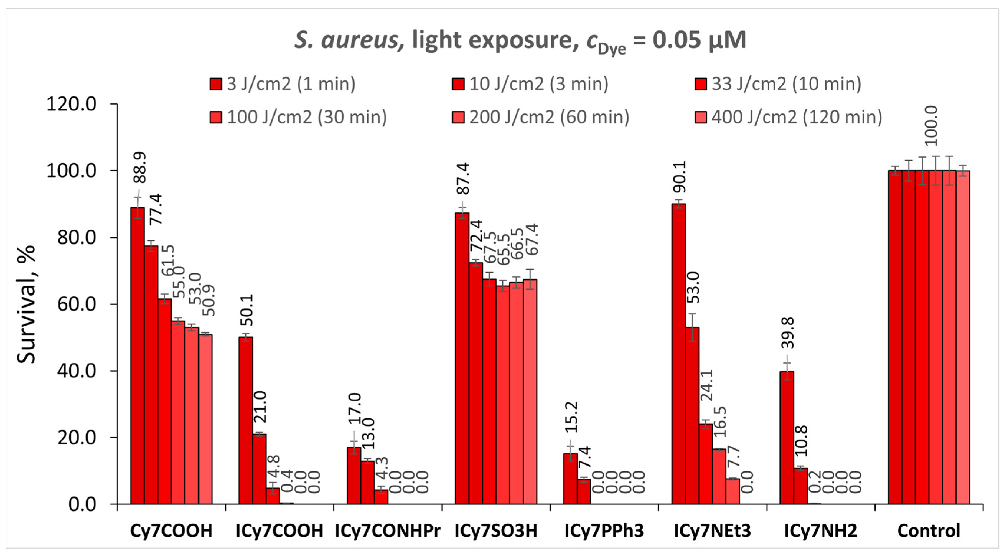
3.5.1. Photodynamic Eradication of S. aureus

The effect of the solubilizing groups in the ICy7 dyes was first investigated on Gram-positive bacteria S. aureus at constant light dose (3 J/cm2) vs. the dye concentrations (cDye = 0.01–0.5 µM). The obtained results were compared to those for the reference non-iodinated Cy7COOH. As expected, iodinated dyes ICy7COOH, ICy7CONHPr, ICy7PPh3, and ICy7NH2 demonstrate an elevated phototoxicity as compared to the non-iodinated Cy7COOH (the exceptions are ICy7SO3H and ICy7NEt3, which are discussed below). Thus, ICy7COOH at cDye = 0.5 μM eradicates S. aureus 2.9-fold more efficiently (26.8% survival) than Cy7COOH (76.6% survival).
The replacement of the negatively charged carboxylic group in ICy7COOH with the neutral N-propylamide group in ICy7CONHPr and positively charged triphenylphosphonium group in ICy7PPh3 leads to the noticeable increase in phototoxicity with only 1.6% and 1.2% survival, respectively (Figure 3, cDye = 0.5 μM; light dose 3 J/cm2). This result can be explained by the most pronounced ΦΔ (Table 1) and preferential uptake of these positively charged dyes by S. aureus (Section 3.4).
Surprisingly, the ICy7NH2 and ICy7NEt3 with an overall charge of +2 show noticeably reduced phototoxicity compared to ICy7PPh3 bearing the same charge. Moreover, ICy7NH2 produces about the same phototoxic effect as the neutral ICy7COOH, while the ICy7NEt3 has about the same phototoxicity as non-iodinated Cy7COOH. Unexpectedly, the zwitterionic dye ICy7SO3H exhibiting excellent penetration in S. aureus (Section 3.4) shows the least pronounced phototoxicity with a survival percentage of more than 80% at all the studied dye concentrations. This can be explained by the extremely low ΦΔ for this dye (4.5% in saline, Table 1).
The increase in the dye concentration results in elevating phototoxicity of all the investigated dyes although for the lower-effective Cy7COOH, ICy7COOH, ICy7SO3H, and ICy7NH2 this increase is less pronounced.
Furthermore, we studied the effect of the light dose between 3 J/cm2 and 400 J/cm2 on the phototoxicity of the dyes towards S. aureus at constant dye concentration of 0.05 µM (Figure 4). We found that the phototoxicity of ICy7COOH, ICy7CONHPr, ICy7NEt3, ICy7PPh3, and ICy7NH2, substantially increases when raising the light dose, but this increase is much less pronounced for the non-iodinated Cy7COOH and sulfonated ICy7SO3H. Noteworthy, ICy7COOH and ICy7CONHPr almost totally eradicate S. aureus at 100 J/cm2 (30 min irradiation), while ICy7PPh3 and ICy7NH2 are even more effective; they completely kill this pathogen at 33 J/cm2 (10 min). As shown in Figure 4, the iodinated dyes bearing a positive +1 (ICy7CONHPr) or +2 (ICy7PPh3 and ICy7NH2) charges cause the more pronounced eradication of these bacteria, while zwitterionic dye ICy7SO3H bearing negatively charged sulfonic group is much less phototoxic. Remarkably, both ICy7PPh3 and ICy7NEt3 bear a positive charge, but the first one is much more phototoxic compared to the second one.
To conclude, ICy7PPh3 is the most effective photosensitizer against S. aureus at a low concentration of 0.05 µM and low light dose of 33 J/cm2 (10 min). ICy7COOH, Cy7CONHPr, ICy7NEt3, and ICy7NH2 require either a larger concentration or higher light dose for complete bacteria eradication.

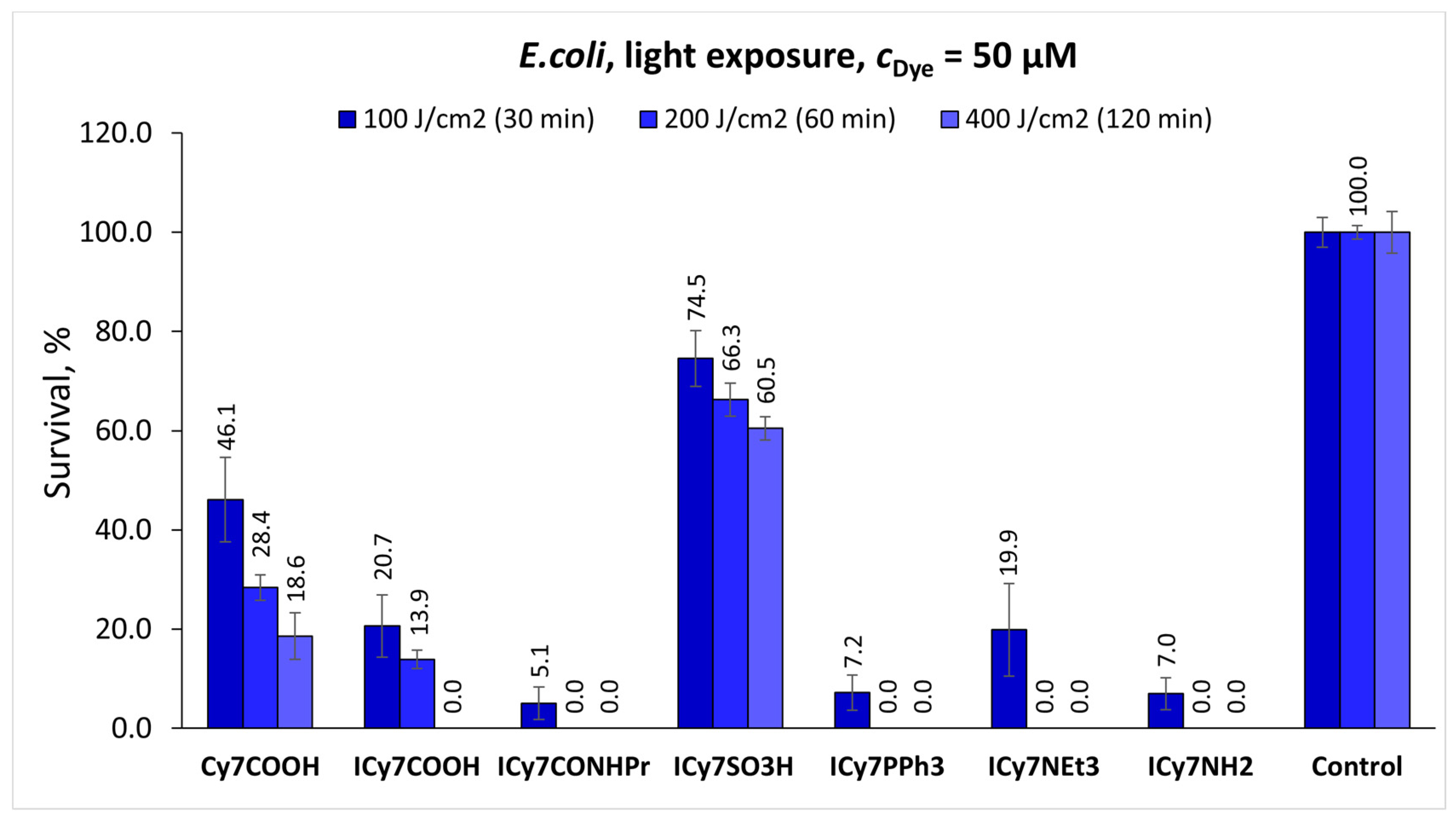
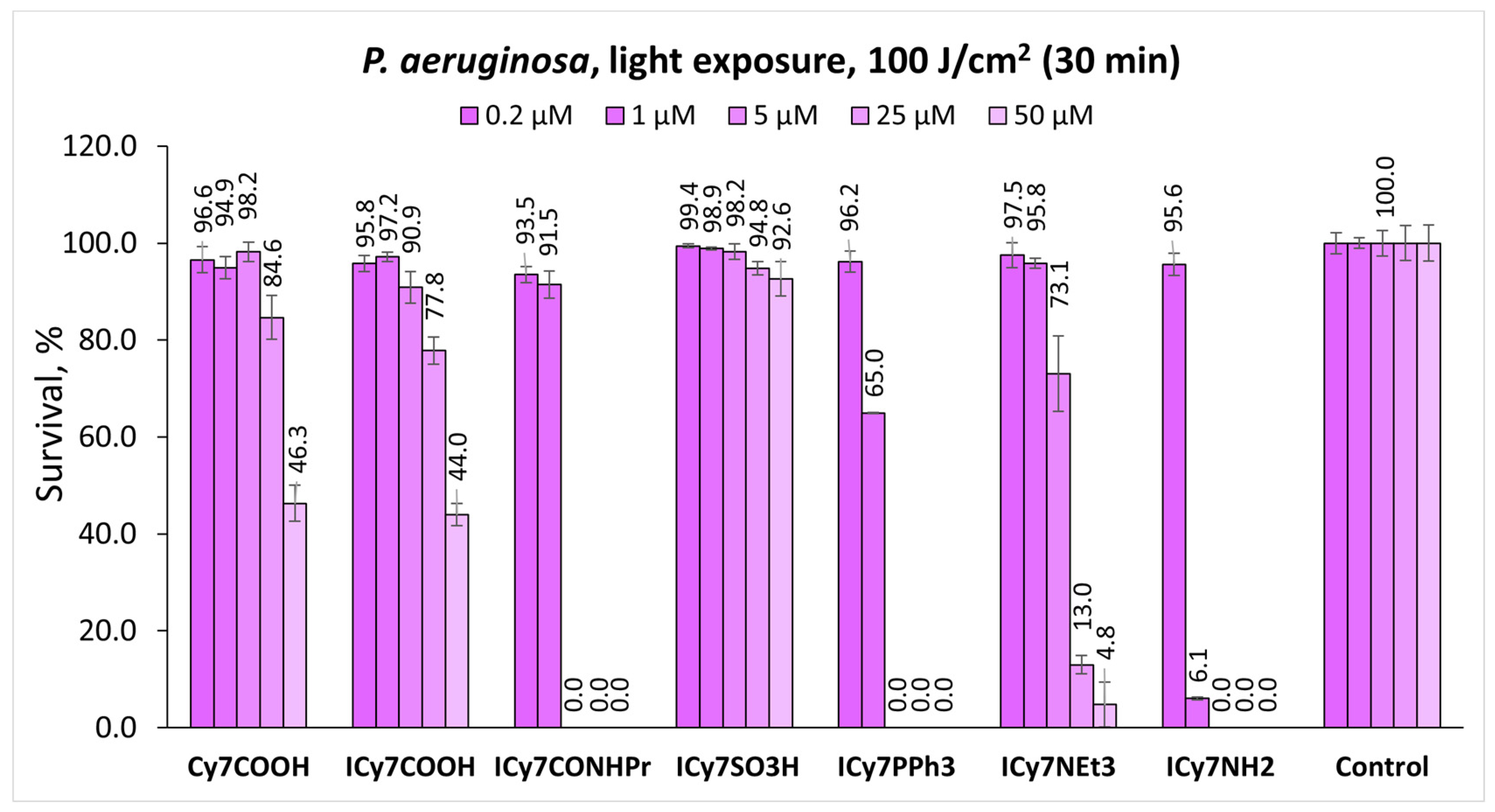
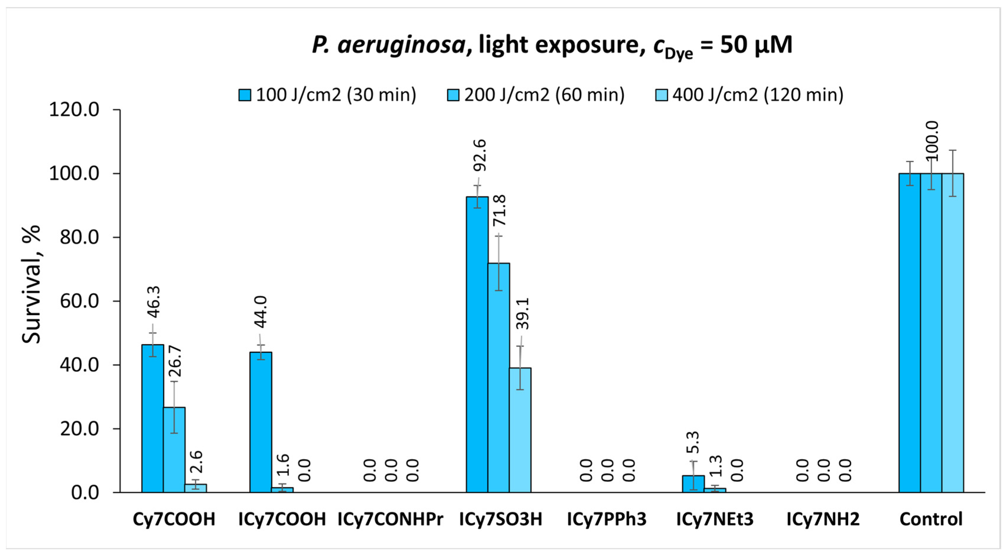
3.5.2. Photodynamic Eradication of E. coli and P. aeruginosa

In the next step, we studied phototoxicity of the dyes towards Gram-negative pathogens E. coli and P. aeruginosa (Figure 5, Figure 6, Figure 7 and Figure 8). We investigated the effect of the dye concentration at the constant light dose of 100 J/cm2 (Figure 5 and Figure 7) and the impact of the light dose at the constant dye concentration of 50 µM (Figure 6 and Figure 8) for both E. coli (Figure 5 and Figure 6) and P. aeruginosa (Figure 7 and Figure 8). These bacteria were found to be much more resistant towards photodynamic treatment compared to Gram-positive S. aureus (Figure 3 and Figure 4). Thus, a reasonable phototoxicity was observed for the dye concentration of 5–50 µM, which was two–three orders higher compared to that used for S. aureus (0.05 µM) and at the higher light dose of 100 J/cm2.
Similar to Gram-positive S. aureus, both types of Gram-negative bacteria are most resistant towards ICy7SO3H exhibiting the survival of 60.5% on E. coli and 39.1% on P. aeruginosa (light dose 400 J/cm2). Remarkably, this dye was found to be much less phototoxic than the non-iodinated Cy7COOH (survival 18.6% for E. coli and 2.6% for P. aeruginosa at 400 J/cm2). The most effective photokillers are ICy7CONHPr, ICy7PPh3 and ICy7NH2, while ICy7NEt3 and ICy7COOH are a bit less phototoxic. All these dyes are more active towards E. coli and P. aeruginosa at 100 J/cm2 (Figure 5 and Figure 7) compared to ICy7SO3H and Cy7COOH; and the difference in the phototoxicity between them is even more pronounced at 200–400 J/cm2 (Figure 6 and Figure 8). The investigated pathogens can be totally eradicated by ICy7CONHPr, ICy7PPh3, and ICy7NH2 at the dye concentration of 50 µM and light dose of 200 J/cm2 for E. coli, while 5 µM and 100 J/cm2 are sufficient for the complete eradication of P. aeruginosa.
The introduction of the positively charged triphenylphosphonium (ICy7PPh3) and triethylammonium (ICy7NEt3) groups causes a noticeable increase in the phototoxic effect on both Gram-negative pathogens, especially at higher light doses of 200–400 J/cm2. Interestingly, while ICy7NEt3 exhibited a poor phototoxicity on S. aureus (Figure 3), it performs very well on E. coli and P. aeruginosa (Figure 5, Figure 6, Figure 7 and Figure 8).
To summarize, the positively charged dyes are more effective against E. coli and P. aeruginosa, and their efficacy increases in the order of ICy7NEt3 < ICy7NH2ICy7PPh3ICy7CONHPr.

3.5.3. Phototoxicity of the Dyes vs. the Quantum Yield of the Singlet Oxygen Generation and the Uptake by Bacteria

We correlated the phototoxicity of the dyes (Phototoxicity = 100% − Cell Survival) with their quantum yields of the singlet oxygen generation at two different dye concentrations (Figure 9). It was found that the ΦΔ and phototoxicity on S. aureus (Figure 9a) and E. coli (Figure 9b) simultaneously increase in the order: ICy7SO3H < Cy7COOH < ICy7COOH < ICy7NH2ICy7PPh3ICy7CONHPr. Remarkably, ICy7CONHPr and ICy7PPh3 demonstrate the best ΦΔ and the most pronounced phototoxicity on both types of bacteria, while ICy7NEt3 shows a reduced phototoxicity compared to the anticipated value based on its ΦΔ (there is a deviation from the average curve in Figure 9).
Furthermore, we correlated the phototoxicity of the dyes with their uptake by S. aureus and E. coli (Figure 10). It can be seen that there is an excellent linear correlation between the phototoxicity and the uptake (r = 0.99 for S. aureus and r = 0.98 for E. coli) for all the investigated dyes except the sulfonated ICy7SO3H that exhibits a good uptake with a notably reduced phototoxicity.
Taking into account the obtained data on the dye uptake (Figure 10) and ΦΔ (Table 1 and Figure 9), it can be concluded that the reduced phototoxicity of Cy7SO3H is connected with its insufficient ΦΔ, while the low phototoxicity of ICy7NEt3 and Cy7COOH is due to the decreased uptake.

4. Conclusions

In summary, a series of novel iodinated heptamethine cyanine dyes containing various solubilizing groups attached to one of the quaternized indolenine moieties was synthesized. Their spectral properties, the yields of singlet oxygen generation, uptake by bacteria, dark cytotoxicity and phototoxicity against selected Gram-positive and Gram-negative pathogenic bacteria were investigated. The structure of the solubilizing group was found to have a strong effect on the dye uptake and phototoxicity. All the dyes exhibit a negligible cytotoxicity in the dark at the concentrations of at least up to 0.5 µM for S. aureus and 50 µM for E. coli and P. aeruginosa. The dyes containing neutral and positively charged groups demonstrate a high phototoxicity at nanomolar and micromolar dye concentrations against Gram-positive and Gram-negative bacteria, respectively, while the dyes with negatively charged groups have a reduced phototoxicity. The most efficient photosensitizers are positively charged iodinated heptamethine cyanines ICy7CONHPr and ICy7PPh3. These dyes completely kill S. aureus at 0.05 µM and low NIR light dose of 33 J/cm2, E. coli at 50 µM and 200 J/cm2, and P. aeruginosa at 5 µM and 100 J/cm2.
Thus, a combination of the heavy-atom effect that increased singlet oxygen generation with an appropriate solubilizing group’s effect improving cell uptake helped to noticeably increase the APDT efficacy of these new photosensitizers.
We believe that the obtained dyes ICy7CONHPr and ICy7PPh3 can be further exploited for the development of highly efficient sensitizing systems for photo-eradication of bacteria, viruses, and abnormal cells; equipping these photosensitizers with bacteria-specific carriers such as bacterial substrates or antibodies can pave the way for their clinical use.

Supplementary Materials

The following supporting information can be downloaded at: https://www.mdpi.com/article/10.3390/pharmaceutics15010247/s1.

Author Contributions

O.S.: Investigation, synthesis, spectral characterization, photodynamic experiments. D.K.: Investigation, synthesis, measurements of quantum yield of singlet oxygen generation. I.H.: Measurements of dye uptake, spectral characterization, preparation of bacteria and photodynamic experiments. M.A.: Preparation of bacteria and photodynamic experiments. F.N.: Supervision of biological experiments with bacteria. O.K.: Data analysis and presentation, writing the paper. A.B.: Purification of dyes, structure confirmation. G.G.: Reviewing the paper and experimental data, funding acquisition. L.P.: General supervision, funding acquisition, data analysis, writing the paper. All authors have read and agreed to the published version of the manuscript.

Funding

This research was funded by the Israel Scientific Foundation, grant number 810/18, Ariel University, the Ministry of Aliyah and Immigrant Absorption (Israel, KAMEA program), and the Gale Foundation.

Institutional Review Board Statement

Not applicable.

Informed Consent Statement

Not applicable.

Data Availability Statement

Data is contained within this article and Supplementary Materials.

Acknowledgments

The authors are grateful to Vered Marks (Ariel University) for the measurements of NMR spectra.

Conflicts of Interest

The authors declare no conflict of interest.

References

  1. Correia, J.H.; Rodrigues, J.A.; Pimenta, S.; Dong, T.; Yang, Z. Photodynamic therapy review: Principles, photosensitizers, applications, and future directions. Pharmaceutics 2021, 13, 1332. [Google Scholar] [CrossRef] [PubMed]
  2. Cieplik, F.; Deng, D.; Crielaard, W.; Buchalla, W.; Hellwig, E.; Al-Ahmad, A.; Maisch, T. Antimicrobial photodynamic therapy—What we know and what we don’t. Crit. Rev. Microbiol. 2018, 44, 571–589. [Google Scholar] [CrossRef] [PubMed] [Green Version]
  3. Gunaydin, G.; Gedik, M.E.; Ayan, S. Photodynamic therapy for the treatment and diagnosis of cancer—A review of the current clinical status. Front. Chem. 2021, 9, 1–26. [Google Scholar] [CrossRef]
  4. Algorri, J.F.; Ochoa, M.; Roldán-Varona, P.; Rodríguez-Cobo, L.; López-Higuera, J.M. Photodynamic therapy: A compendium of latest reviews. Cancers 2021, 13, 4447. [Google Scholar] [CrossRef] [PubMed]
  5. Wang, K.; Yu, B.; Pathak, J.L. An update in clinical utilization of photodynamic therapy for lung cancer. J. Cancer 2021, 12, 1154–1160. [Google Scholar] [CrossRef]
  6. Songca, S.P.; Adjei, Y. Applications of antimicrobial photodynamic therapy against bacterial biofilms. Int. J. Mol. Sci. 2022, 23, 3209. [Google Scholar] [CrossRef]
  7. Dias, L.D.; Blanco, K.C.; Bagnato, V.S. COVID-19: Beyond the virus. The use of photodynamic therapy for the treatment of infections in the respiratory tract. Photodiagnosis Photodyn. Ther. 2020, 31, 101804. [Google Scholar] [CrossRef]
  8. Hung, J.-H.; Lee, C.-N.; Hsu, H.-W.; Ng, I.-S.; Wu, C.-J.; Yu, C.-K.; Lee, N.-Y.; Chang, Y.; Wong, T.-W. Recent advances in photodynamic therapy against fungal keratitis. Pharmaceutics 2021, 13, 2011. [Google Scholar] [CrossRef]
  9. Li, L.; Chen, Y.; Chen, W.; Tan, Y.; Chen, H.; Yin, J. Photodynamic therapy based on organic small molecular fluorescent dyes. Chin. Chem. Lett. 2019, 30, 1689–1703. [Google Scholar] [CrossRef]
  10. Shang, L.; Zhou, X.; Zhang, J.; Shi, Y.; Zhong, L. Metal nanoparticles for photodynamic therapy: A potential treatment for breast cancer. Molecules 2021, 26, 6532. [Google Scholar] [CrossRef]
  11. Nasseri, B.; Alizadeh, E.; Bani, F.; Davaran, S.; Akbarzadeh, A.; Rabiee, N.; Bahadori, A.; Ziaei, M.; Bagherzadeh, M.; Saeb, M.R.; et al. Nanomaterials for photothermal and photodynamic cancer therapy. Appl. Phys. Rev. 2022, 9, 011317. [Google Scholar] [CrossRef]
  12. Zhao, X.; Liu, J.; Fan, J.; Chao, H.; Peng, X. Recent progress in photosensitizers for overcoming the challenges of photodynamic therapy: From molecular design to application. Chem. Soc. Rev. 2021, 50, 4185–4219. [Google Scholar] [CrossRef]
  13. Kou, J.; Dou, D.; Yang, L. Porphyrin photosensitizers in photodynamic therapy and its applications. Oncotarget 2017, 8, 81591–81603. [Google Scholar] [CrossRef] [PubMed] [Green Version]
  14. Srivatsan, A.; Missert, J.R.; Upadhyay, S.K.; Pandey, R.K. Porphyrin-based photosensitizers and the corresponding multifunctional nanoplatforms for cancer-imaging and phototherapy. J. Porphyr. Phthalocyanines 2015, 19, 109–134. [Google Scholar] [CrossRef]
  15. Zheng, B.-D.; Ye, J.; Zhang, X.-Q.; Zhang, N.; Xiao, M.-T. Recent advances in supramolecular activatable phthalocyanine-based photosensitizers for anti-cancer therapy. Coord. Chem. Rev. 2021, 447, 214155. [Google Scholar] [CrossRef]
  16. Chen, D.; Song, M.; Huang, J.; Chen, N.; Xue, J.; Huang, M. Photocyanine: A novel and effective phthalocyanine-based photosensitizer for cancer treatment. J. Innov. Opt. Health Sci. 2020, 13, 2030009. [Google Scholar] [CrossRef]
  17. Li, X.; Zheng, B.-D.; Peng, X.-H.; Li, S.-Z.; Ying, J.-W.; Zhao, Y.; Huang, J.-D.; Yoon, J. Phthalocyanines as medicinal photosensitizers: Developments in the last five years. Coord. Chem. Rev. 2019, 379, 147–160. [Google Scholar] [CrossRef]
  18. Bilici, K.; Cetin, S.; Aydındogan, E.; Yagci Acar, H.; Kolemen, S. Recent advances in cyanine-based phototherapy agents. Front. Chem. 2021, 9, 1–15. [Google Scholar] [CrossRef]
  19. Lange, N.; Szlasa, W.; Saczko, J.; Chwiłkowska, A. Potential of cyanine derived dyes in photodynamic therapy. Pharmaceutics 2021, 13, 818. [Google Scholar] [CrossRef]
  20. Dereje, D.M.; Pontremoli, C.; Plata, M.J.M.; Visentin, S.; Barbero, N. Polymethine dyes for PDT: Recent advances and perspectives to drive future applications. Photochem. Photobiol. Sci. 2022, 21, 397–419. [Google Scholar] [CrossRef]
  21. Ogawa, M.; Kosaka, N.; Choyke, P.L.; Kobayashi, H. In vivo molecular imaging of cancer with a quenching near-infrared fluorescent probe using conjugates of monoclonal antibodies and Indocyanine Green. Cancer Res. 2009, 69, 1268–1272. [Google Scholar] [CrossRef] [Green Version]
  22. Shafirstein, G.; Bäumler, W.; Hennings, L.J.; Siegel, E.R.; Friedman, R.; Moreno, M.A.; Webber, J.; Jackson, C.; Griffin, R.J. Indocyanine Green enhanced near-infrared laser treatment of murine mammary carcinoma. Int. J. Cancer 2012, 130, 1208–1215. [Google Scholar] [CrossRef]
  23. Cao, J.; Chi, J.; Xia, J.; Zhang, Y.; Han, S.; Sun, Y. Iodinated cyanine dyes for fast near-infrared-guided deep tissue synergistic phototherapy. ACS Appl. Mater. Interfaces 2019, 11, 25720–25729. [Google Scholar] [CrossRef] [PubMed]
  24. Bokan, M.; Nakonechny, F.; Talalai, E.; Kobzev, D.; Gellerman, G.; Patsenker, L. Photodynamic effect of novel hexa-iodinated quinono-cyanine dye on staphylococcus aureus. Photodiagnosis Photodyn. Ther. 2020, 31, 101866. [Google Scholar] [CrossRef]
  25. Atchison, J.; Kamila, S.; Nesbitt, H.; Logan, K.A.; Nicholas, D.M.; Fowley, C.; Davis, J.; Callan, B.; McHale, A.P.; Callan, J.F. Iodinated cyanine dyes: A new class of sensitisers for use in NIR activated photodynamic therapy (PDT). Chem. Commun. 2017, 53, 2009–2012. [Google Scholar] [CrossRef] [PubMed]
  26. Wang, Z.; Ivanov, M.; Gao, Y.; Bussotti, L.; Foggi, P.; Zhang, H.; Russo, N.; Dick, B.; Zhao, J.; Di Donato, M.; et al. Spin–orbit charge-transfer intersystem crossing (ISC) in compact electron donor–acceptor dyads: ISC mechanism and application as novel and potent photodynamic therapy reagents. Chem.—A Eur. J. 2020, 26, 1091–1102. [Google Scholar] [CrossRef] [PubMed]
  27. Ebaston, T.M.; Nakonechny, F.; Talalai, E.; Gellerman, G.; Patsenker, L. Iodinated xanthene-cyanine NIR dyes as potential photosensitizers for antimicrobial photodynamic therapy. Dyes Pigm. 2021, 184, 108854. [Google Scholar] [CrossRef]
  28. Dong, Y.; Zhou, L.; Shen, Z.; Ma, Q.; Zhao, Y.; Sun, Y.; Cao, J. Iodinated cyanine dye-based nanosystem for synergistic phototherapy and hypoxia-activated bioreductive therapy. Drug Deliv. 2022, 29, 238–253. [Google Scholar] [CrossRef]
  29. Liu, H.; Yin, J.; Xing, E.; Du, Y.; Su, Y.; Feng, Y.; Meng, S. Halogenated cyanine dyes for synergistic photodynamic and photothermal therapy. Dyes Pigm. 2021, 190, 109327. [Google Scholar] [CrossRef]
  30. Kopke, T.; Zaleski, J. Diazo-containing molecular constructs as potential anticancer agents: From diazo[b]fluorene natural products to photoactivatable diazo-oxochlorins. Anticancer Agents Med. Chem. 2008, 8, 292–304. [Google Scholar] [CrossRef]
  31. Chen, J.; Chen, Z.; Tan, L.; Yang, J.; Shen, L.; Deng, J.; Jiang, X.; Zou, D. Synthesis of a new chlorin photosensitizer for photodynamic therapy against colon cancer. Mater. Chem. Front. 2022, 6, 1129–1136. [Google Scholar] [CrossRef]
  32. Semenova, O.; Kobzev, D.; Yazbak, F.; Nakonechny, F.; Kolosova, O.; Tatarets, A.; Gellerman, G.; Patsenker, L. Unexpected effect of iodine atoms in heptamethine cyanine dyes on the photodynamic eradication of Gram-positive and Gram-negative pathogens. Dyes Pigm. 2021, 195, 109745. [Google Scholar] [CrossRef]
  33. Zhang, R.; Qin, X.; Kong, F.; Chen, P.; Pan, G. Improving cellular uptake of therapeutic entities through interaction with components of cell membrane. Drug Deliv. 2019, 26, 328–342. [Google Scholar] [CrossRef] [PubMed] [Green Version]
  34. Palermo, E.F.; Lee, D.-K.; Ramamoorthy, A.; Kuroda, K. Role of cationic group structure in membrane binding and disruption by amphiphilic copolymers. J. Phys. Chem. B 2011, 115, 366–375. [Google Scholar] [CrossRef] [PubMed] [Green Version]
  35. Jones, G.W.; Tatarets, A.L.; Patsenker, L.D. Halogenated Compounds for Photodynamic Therapy. U.S. Patent 8,748,446 B2, 10 June 2014. [Google Scholar]
  36. Jones, G.W.; Tatarets, A.L.; Patsenker, L.D. Halogenated Compounds for Photodynamic Therapy. U.S. Patent 8,962,797 B2, 24 February 2015. [Google Scholar]
  37. Markova, L.I.; Fedyunyayeva, I.A.; Povrozin, Y.A.; Semenova, O.M.; Khabuseva, S.U.; Terpetschnig, E.A.; Patsenker, L.D. Water soluble indodicarbocyanine dyes based on 2,3-dimethyl-3-(4-sulfobutyl)-3H-indole-5-sulfonic acid. Dyes Pigm. 2013, 96, 535–546. [Google Scholar] [CrossRef]
  38. Saha, P.C.; Chatterjee, T.; Pattanayak, R.; Das, R.S.; Mukherjee, A.; Bhattacharyya, M.; Guha, S. Targeting and imaging of mitochondria using near-infrared cyanine dye and its application to multicolor imaging. ACS Omega 2019, 4, 14579–14588. [Google Scholar] [CrossRef] [Green Version]
  39. Sato, K.; Gorka, A.P.; Nagaya, T.; Michie, M.S.; Nakamura, Y.; Nani, R.R.; Coble, V.L.; Vasalatiy, O.V.; Swenson, R.E.; Choyke, P.L.; et al. Effect of charge localization on the in vivo optical imaging properties of near-infrared cyanine dye/monoclonal antibody conjugates. Mol. Biosyst. 2016, 12, 3046–3056. [Google Scholar] [CrossRef] [Green Version]
  40. Texier, I.; Goutayer, M.; Da Silva, A.; Guyon, L.; Djaker, N.; Josserand, V.; Neumann, E.; Bibette, J.; Vinet, F. Cyanine-loaded lipid nanoparticles for improved in vivo fluorescence imaging. J. Biomed. Opt. 2009, 14, 054005. [Google Scholar] [CrossRef]
  41. Parker, C.A. Photoluminescence of Solutions; Elsevier Publishing Co.: Amsterdam, The Netherlands, 1968; p. 544. [Google Scholar]
  42. Štacková, L.; Muchová, E.; Russo, M.; Slavíček, P.; Štacko, P.; Klán, P. Deciphering the structure–property relations in substituted heptamethine cyanines. J. Org. Chem. 2020, 85, 9776–9790. [Google Scholar] [CrossRef]
  43. Lin, H.; Shen, Y.; Chen, D.; Lin, L.; Wilson, B.C.; Li, B.; Xie, S. Feasibility study on quantitative measurements of singlet oxygen generation using Singlet Oxygen Sensor Green. J. Fluoresc. 2013, 23, 41–47. [Google Scholar] [CrossRef]
  44. ThermoFisher Scientific. Available online: https://www.thermofisher.com/document-connect/document-connect.html?url=https://assets.thermofisher.com/TFS-Assets%2FLSG%2Fmanuals%2Fmp36002.pdf (accessed on 29 October 2022).
  45. Wang, S.; Shang, L.; Li, L.; Yu, Y.; Chi, C.; Wang, K.; Zhang, J.; Shi, R.; Shen, H.; Waterhouse, G.I.N.; et al. Metal-organic-framework-derived mesoporous carbon nanospheres containing porphyrin-like metal centers for conformal phototherapy. Adv. Mater. 2016, 28, 8379–8387. [Google Scholar] [CrossRef] [PubMed]
  46. Reddi, B.A. Why is saline so acidic (and does it really matter?). Int. J. Med. Sci. 2013, 10, 747–750. [Google Scholar] [CrossRef] [PubMed] [Green Version]
  47. Riddick, J.A.; Bunger, W.B.; Sakano, T.K. Techniques of Chemistry, Vol II. In Organic Solvents. Physical Properties and Methods of Purification, 4th ed.; John Wiley and Sons: New York, NY, USA, 1986. [Google Scholar]
  48. Guthrie, J.P. Hydrolysis of esters of oxy acids: pKa values for strong acids; Brønsted relationship for attack of water at methyl; free energies of hydrolysis of esters of oxy acids; and a linear relationship between free energy of hydrolysis and pKa holding over a ran. Can. J. Chem. 1978, 56, 2342–2354. [Google Scholar] [CrossRef]
Figure 1. Heptamethine cyanine dyes nICy7COOH, where n is the number of iodine atoms (n = 0–6), investigated in [32].
Pharmaceutics 15 00247 g001
Figure 2. Heptamethine cyanine dyes of ICy7 series obtained and studied in this work.
Pharmaceutics 15 00247 g002
Scheme 1. Synthesis of heptamethine cyanine dyes.
Pharmaceutics 15 00247 sch001
Figure 3. Survival of S. aureus in saline containing 0.7% DMSO, when exposed to 3 J/cm2 light dose (56 mW/cm2, 1 min) vs. the dye concentrations. Light-irradiated cells without dyes were used as a control.
Pharmaceutics 15 00247 g003
Figure 4. Survival of S. aureus at (cDye = 0.05 µM in saline containing 0.7% DMSO), when exposed to 3 J/cm2 (1 min), 10 J/cm2 (3 min), 33 J/cm2 (10 min), 100 J/cm2 (30 min), 200 J/cm2 (60 min), and 400 J/cm2 (120 min) light doses.
Pharmaceutics 15 00247 g004
Figure 5. Survival of E. coli in saline containing 0.7% DMSO, when exposed to 100 J/cm2 light dose (56 mW/cm2, 30 min) vs. the dye concentrations.
Pharmaceutics 15 00247 g005
Figure 6. Survival of E. coli at (cDye = 50 µM in saline containing 0.7% DMSO), when exposed to 100 J/cm2 (30 min), 200 J/cm2 (60 min), and 400 J/cm2 (120 min) light doses.
Pharmaceutics 15 00247 g006
Figure 7. Survival of P. aeruginosa in saline containing 0.7% DMSO, when exposed to 100 J/cm2 light dose (56 mW/cm2, 30 min) vs. the dye concentrations.
Pharmaceutics 15 00247 g007
Figure 8. Survival of P. aeruginosa at (cDye = 50 µM in saline containing 0.7% DMSO), when exposed to 100 J/cm2 (30 min), 200 J/cm2 (60 min), and 400 J/cm2 (120 min) light doses.
Pharmaceutics 15 00247 g008
Figure 9. Phototoxicity of the dyes on S. aureus (a) and E. coli (b) at 3 J/cm2 and 100 J/cm2 light doses, respectively, vs. the quantum yield of the singlet oxygen generation (ΦΔ) in saline.
Pharmaceutics 15 00247 g009
Figure 10. Phototoxicity of the dyes vs. the uptake by S. aureus at cDye = 0.1 µM (a) and E. coli at cDye = 1 µM (b). The light doses are 3 J/cm2 and 100 J/cm2, respectively.
Pharmaceutics 15 00247 g010
Table 1. Spectral characteristics of the obtained cyanine dyes (cDye~1 µM) measured in methanol and in aqueous saline containing 0.7% DMSO.
Dye StructureMeOH0.7% DMSO in Saline
λmaxAb,
nm
ε
M−1cm−1
λmaxFl,
nm
ΦF 1,
%
ΦΔ 2,
%
λmaxAb,
nm
ε,
M−1cm−1
λmaxFl,
nm
ΦF 1,
%
ΦΔ 3,
%
Pharmaceutics 15 00247 i001744230,00077425.3 ± 0.51.1 ± 0.1740169,00076813 ± 19.5 ± 0.6
Pharmaceutics 15 00247 i002749230,00078025.8 ± 0.51.8 ± 0.2747143,00077611 ± 215 ± 1
Pharmaceutics 15 00247 i003749215,00078425.4 ± 0.42.5 ± 0.1747109,0007768 ± 165 ± 4
Pharmaceutics 15 00247 i004750230,00078125.5 ± 0.82.0 ± 0.1747238,00077710 ± 14.5 ± 0.3
Pharmaceutics 15 00247 i005751230,00078125.9 ± 0.52.5 ± 0.2750155,0007779.1 ± 0.844 ± 3
Pharmaceutics 15 00247 i006747230,00077824.7 ± 0.71.7 ± 0.2745176,00077512 ± 129 ± 1
Pharmaceutics 15 00247 i007749220,00078024.6 ± 0.62.9 ± 0.1745187,0007759 ± 121 ± 2
1 Cy7TM was used as the reference, ΦF = 13% in PBS, pH = 7.4. 2 HITC was used as the reference, ΦΔ = 0.89% in methanol. 3 ICG was used as the reference, ΦΔ = 0.2% in PBS, pH = 7.4.
Disclaimer/Publisher’s Note: The statements, opinions and data contained in all publications are solely those of the individual author(s) and contributor(s) and not of MDPI and/or the editor(s). MDPI and/or the editor(s) disclaim responsibility for any injury to people or property resulting from any ideas, methods, instructions or products referred to in the content.

