
A Novel Perilla frutescens (L.) Britton Cell-Derived Phytocomplex Regulates Keratinocytes Inflammatory Cascade and Barrier Function and Preserves Vaginal Mucosal Integrity In Vivo


 ,
,  ,
,  ,
, 
Abstract
1. Introduction
2. Materials and Methods
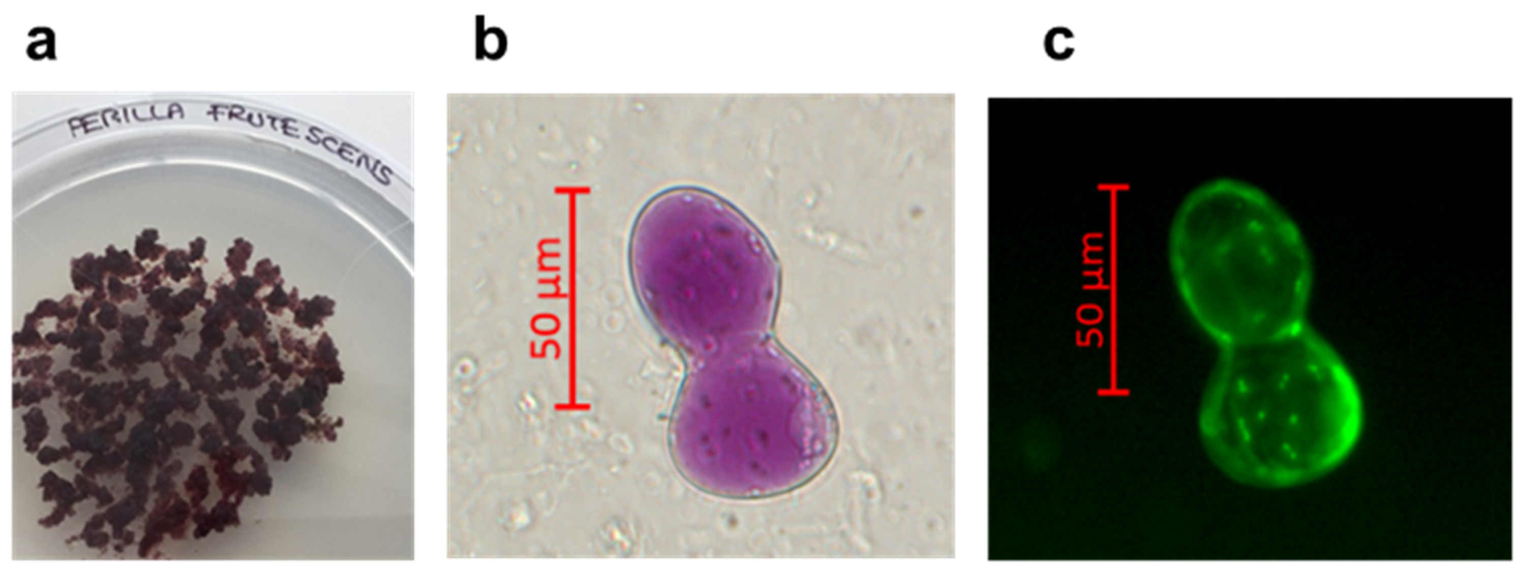
2.1. Perilla Frutescens Cell Culture
2.2. Phytocomplex Preparation from Perilla Frutescens Selected Cell Culture
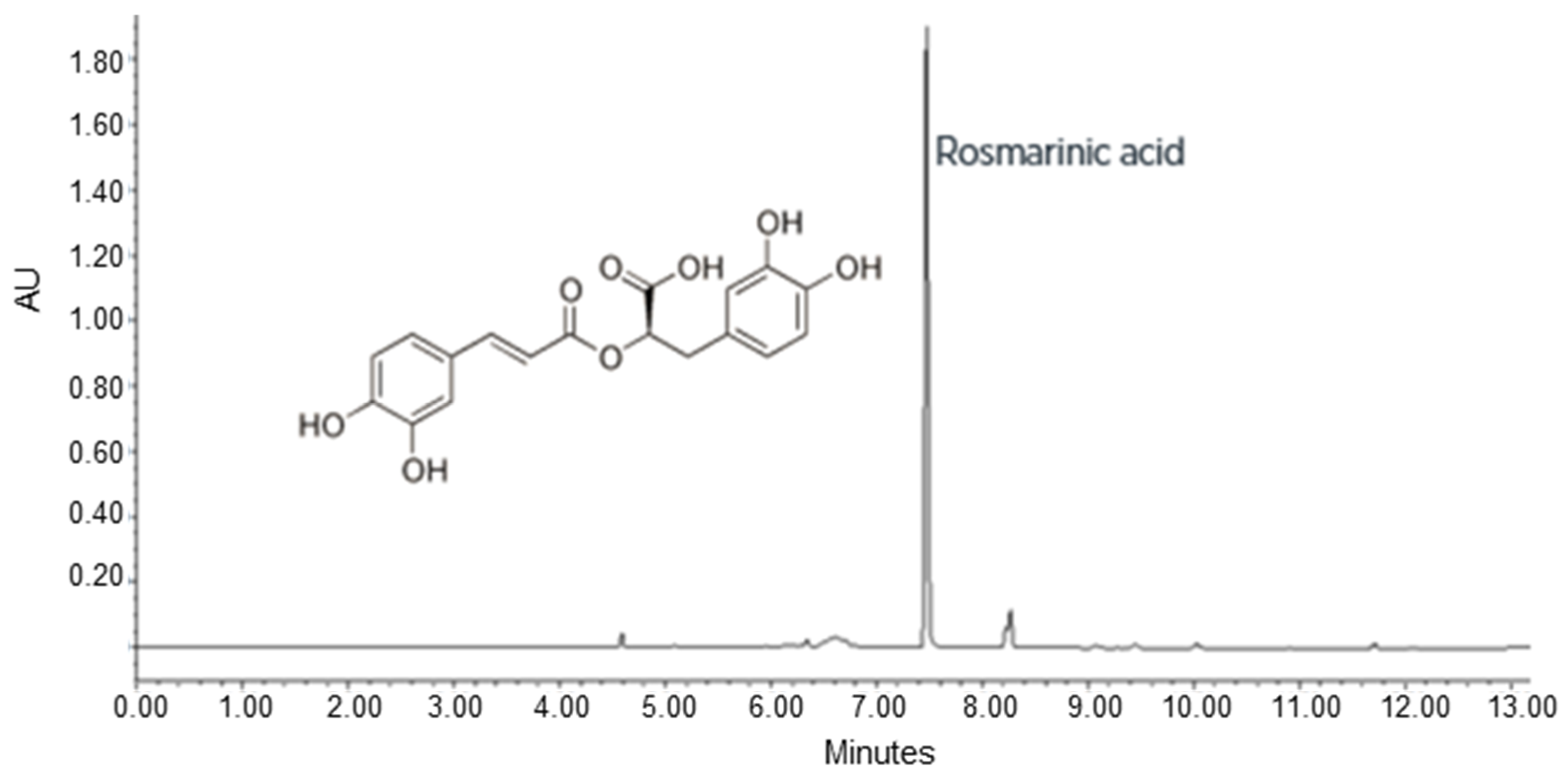
2.3. UPLC-DAD Analysis
2.4. Cell Culture and Treatments
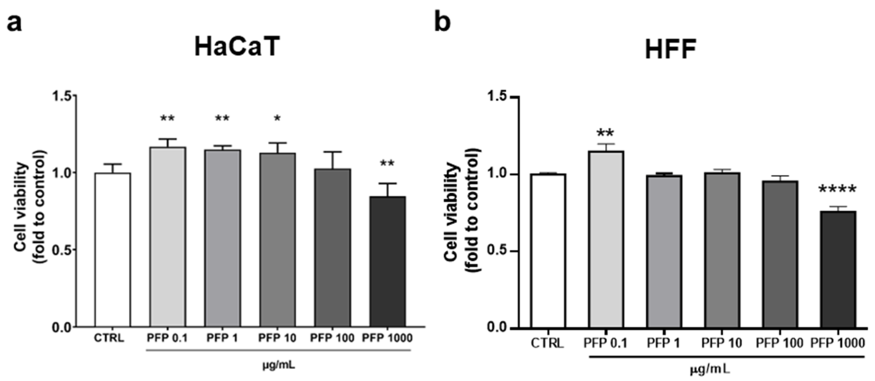
2.5. Cell Viability
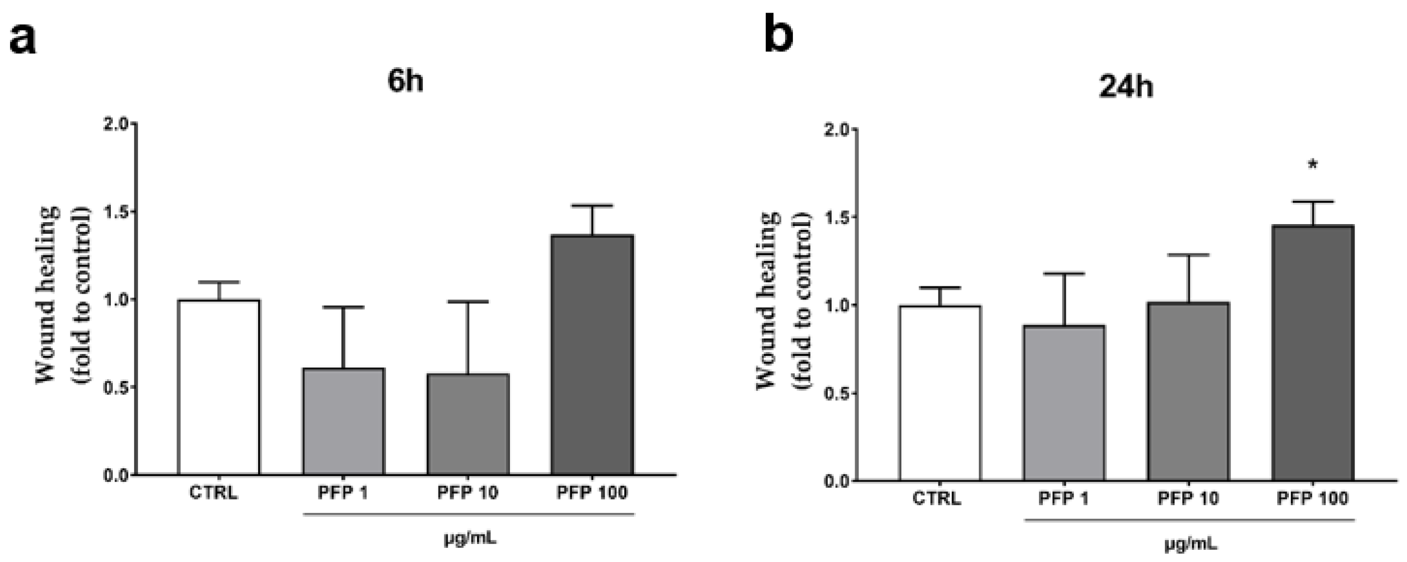
2.6. Wound Healing
2.7. Pro-Collagene I and Elastin Dosage
2.8. Cytokines Dosage
2.9. Leukocytes Infiltration Test
2.10. MAPKs Activation
2.11. Immunofluorescence
2.12. Western Blot
2.13. In Vitro Assay
2.14. In Vivo Tests
- soothing, intimate cream: once a day by applying abundantly in the anogenital and surrounding area;
- mask: 3 times a week with an application of 20 min, making sure to use a protective system to avoid getting the garment wet.
Instruments and Parameters
2.15. Statistical Analysis
- instrumental data (T0 vs. T2weeks) of both treatments were statistically compared by Student t-test for dependent and parametric data;
- clinical data (T0 vs. T2weeks) of both treatments were statistically compared by Wilcoxon test for dependent and non-parametric data;
- statistical comparisons between the active and placebo group for instrumental data were performed by Student t-test for independent and parametric data;
- statistical comparisons between active and placebo groups for clinical data were performed by U Test of Mann–Whitney for independent and non-parametric data.
3. Results
3.1. Perilla Frutescens Phytocomplex Obtaining from a Selected Cell Line and Chemical Analysis
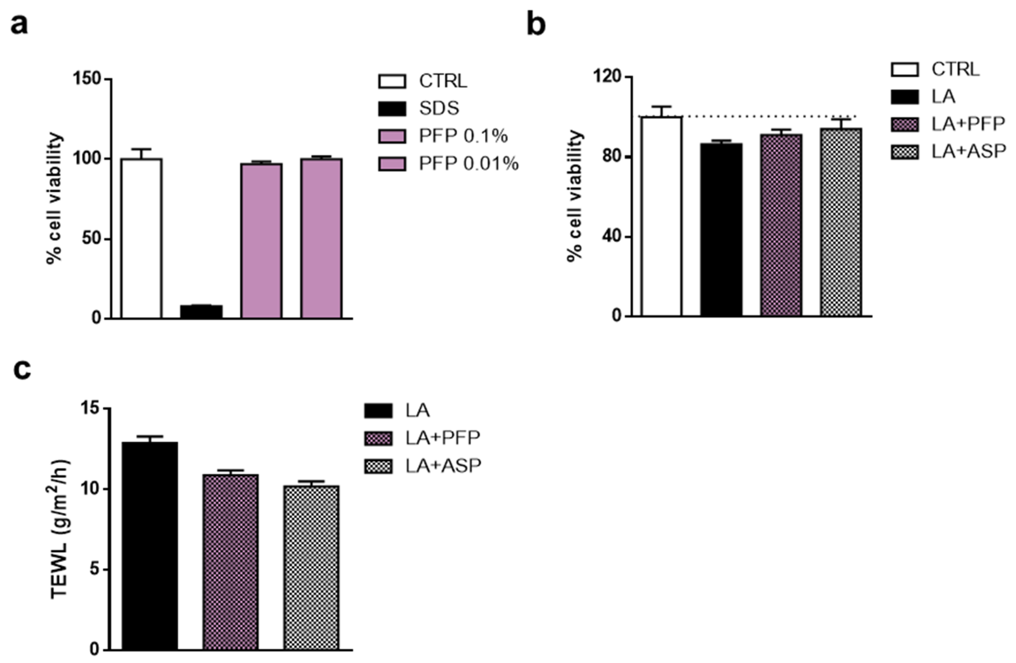
3.2. PFP Effect on the Viability of Human Keratinocytes and Fibroblasts
3.3. PFP Does Not Stimulate Pro-Collagen I and Elastin Production in HFF but Enhances the Keratinocytes Migration Ability
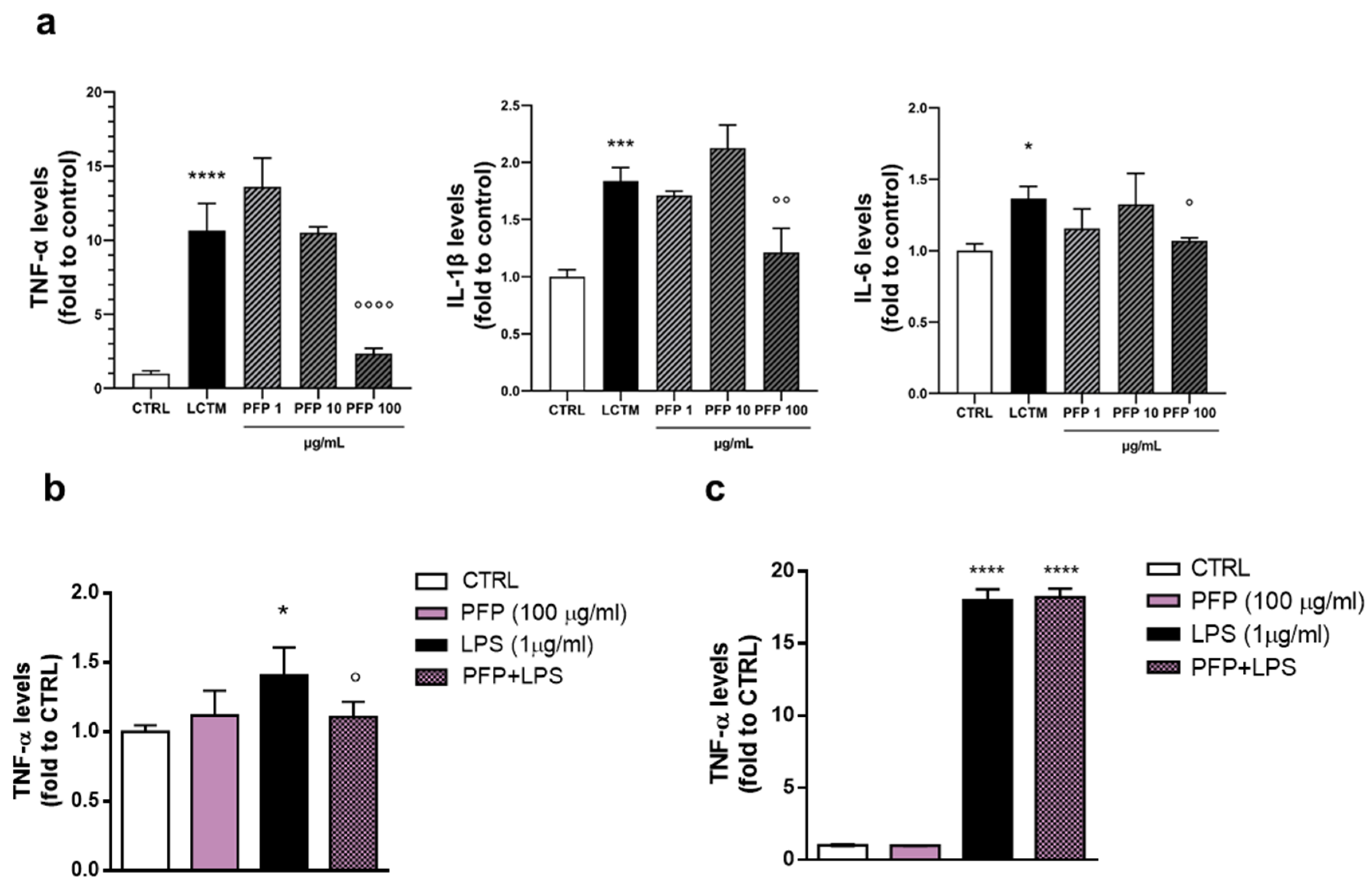
3.4. PFP Counteracts Keratinocytes Inflammatory Response by Reducing Pro-Inflammatory Cytokines Release
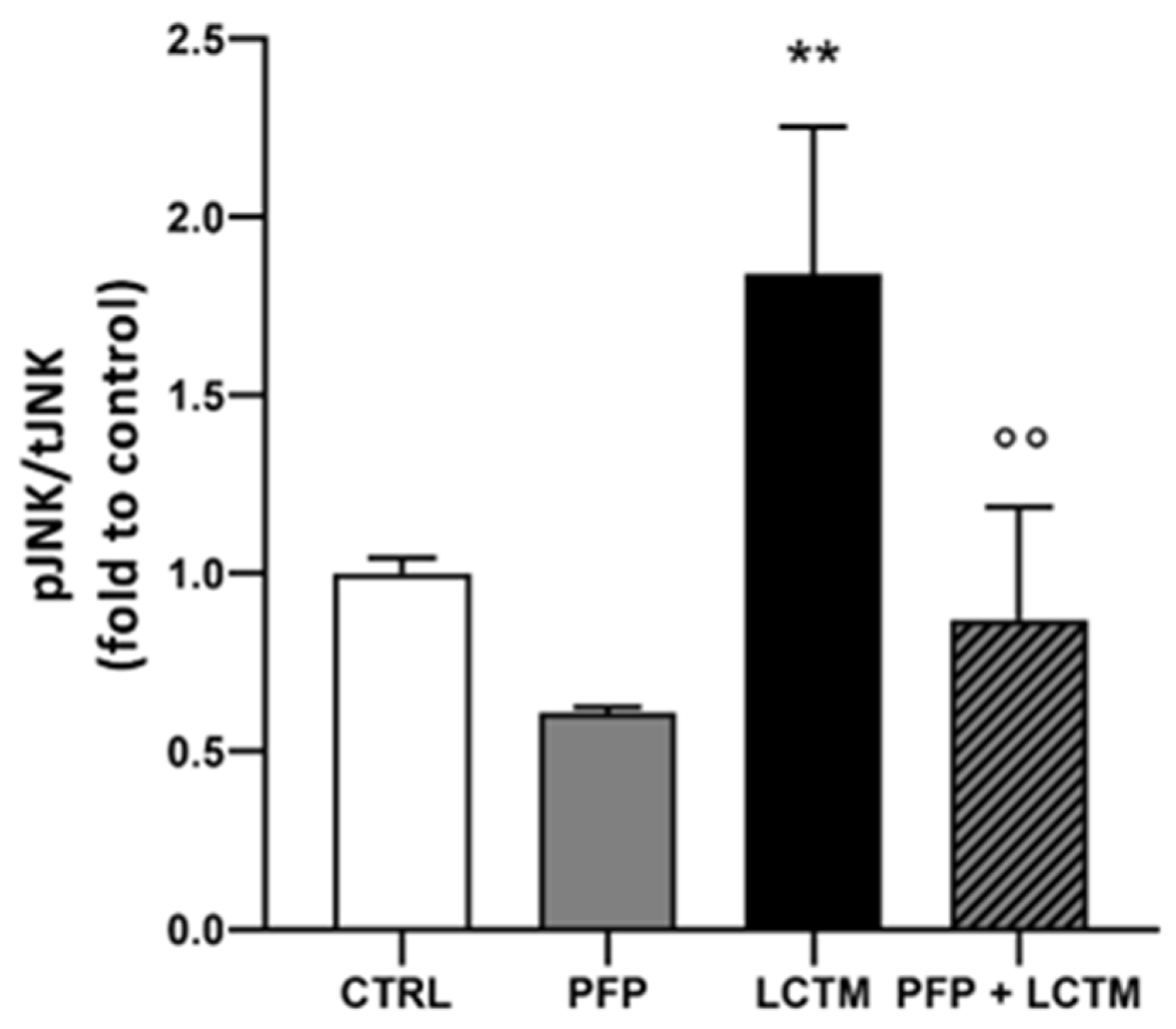
3.5. PFP Inhibits JNK Activation in HaCaT Cells Exposed to LPS-Conditioned THP-1 Medium
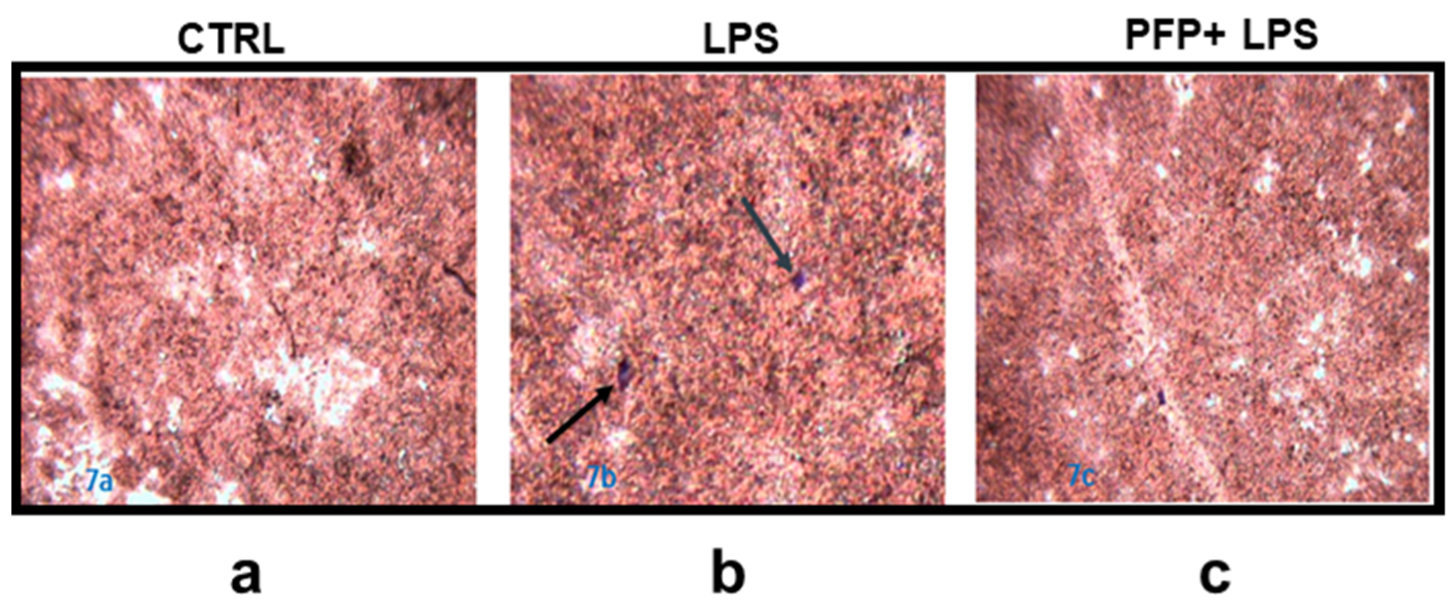
3.6. PFP Treatment Reduces the Leukocytes Infiltration Induced by Immune Stimulus
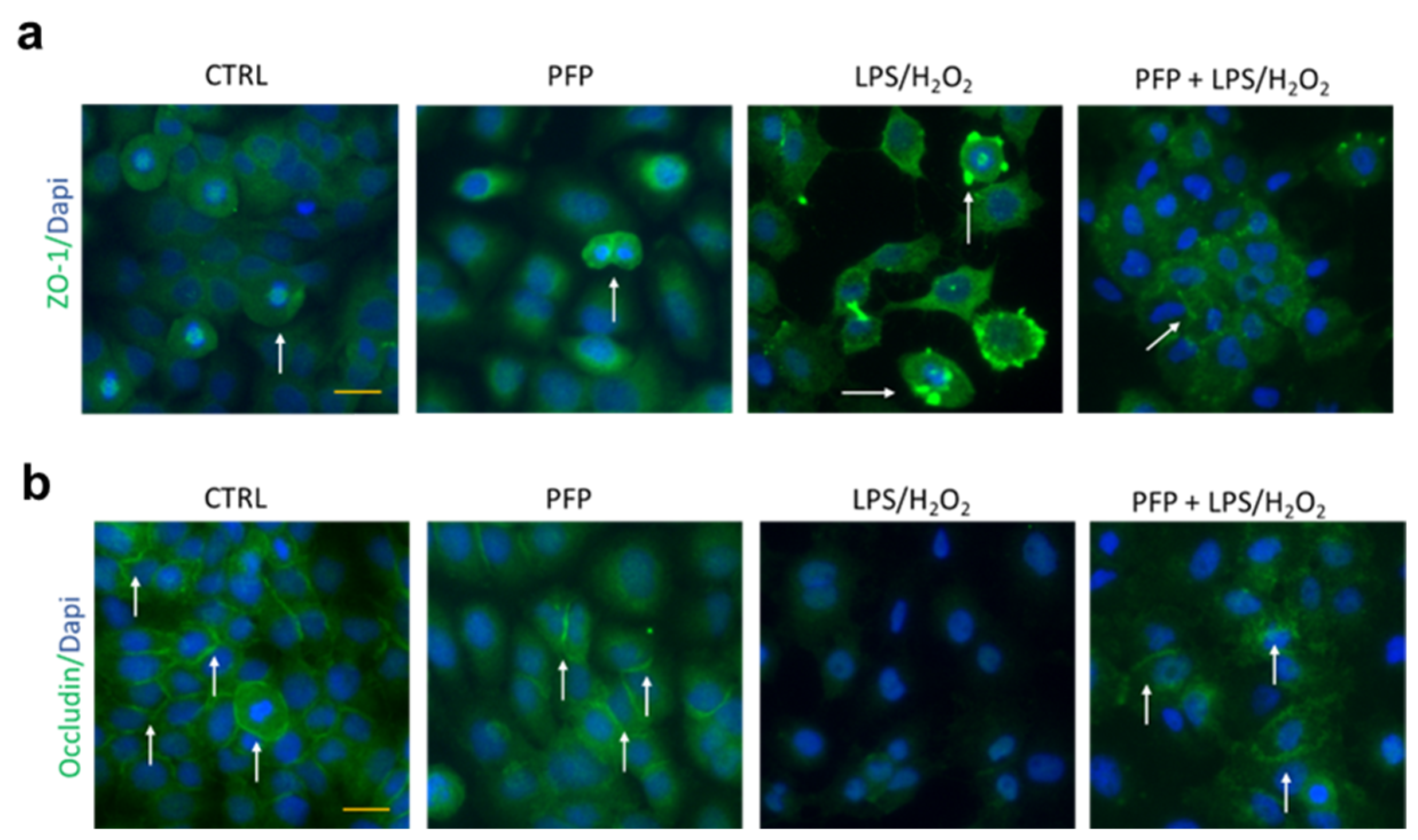
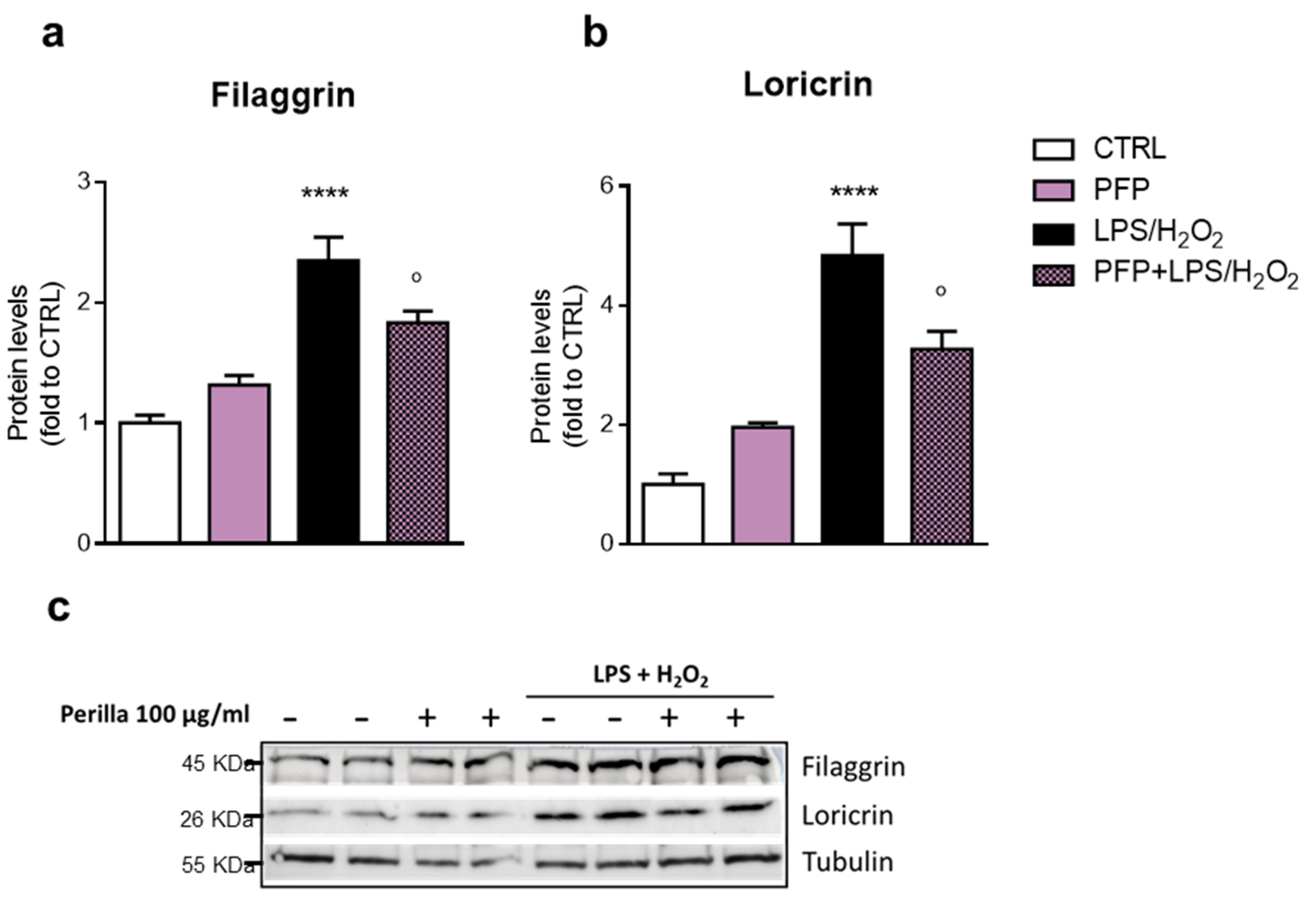
3.7. PFP Prevents Keratinocytes Tight Junctions Impairment Induced by an Inflammatory-Oxidative Damage
3.8. PFP Solution Shows Non-Irritant and Soothing Properties in an In Vitro Reconstituted Vaginal Mucosa
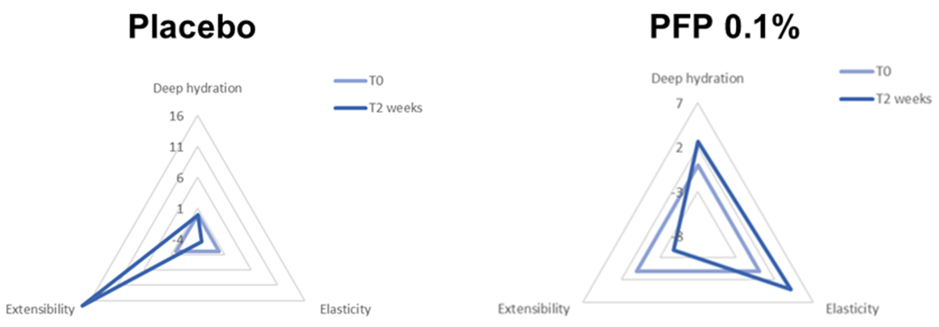
3.9. PFP Ameliorates Deep Hydration and Elasticity after In Vivo Topical Application
4. Discussion
5. Conclusions
6. Patent
Author Contributions
Funding
Institutional Review Board Statement
Informed Consent Statement
Data Availability Statement
Acknowledgments
Conflicts of Interest
References
- Takano, H.; Osakabe, N.; Sanbongi, C.; Yanagisawa, R.; Inoue, K.I.; Yasuda, A.; Natsume, M.; Baba, S.; Ichiishi, E.I.; Yoshikawa, T. Extract of Perilla frutescens enriched for rosmarinic acid, a polyphenolic phytochemical, inhibits seasonal allergic rhinoconjunctivitis in humans. Exp. Biol. Med. 2004, 229, 247–254. [Google Scholar] [CrossRef] [PubMed]
- Ken-Ichi, K.; Jun, T.; Takahiro, M.; Kentaro, K.; Takashi, S.; Yoshihiro, M.; Kaori, U.; Wakame, K. Perilla leaf extract prevents atopic dermatitis induced by an extract of Dermatophagoides farinae in NC/Nga mice. Asian Pac. J. Allergy Immunol. 2016, 34, 272–277. [Google Scholar]
- Bae, J.S.; Han, M.; Shin, H.S.; Kim, M.K.; Shin, C.Y.; Lee, D.H.; Chung, J.H. Perilla frutescens leaves extract ameliorates ultraviolet radiation-induced extracellular matrix damage in human dermal fibroblasts and hairless mice skin. J. Ethnopharmacol. 2017, 195, 334–342. [Google Scholar] [CrossRef] [PubMed]
- Osakabe, N.; Yasuda, A.; Natsume, M.; Yoshikawa, T. Rosmarinic acid inhibits epidermal inflammatory responses: Anticarcinogenic effect of Perilla frutescens extract in the murine two-stage skin model. Carcinogenesis 2004, 25, 549–557. [Google Scholar] [CrossRef]
- Kangwan, N.; Pintha, K.; Lekawanvijit, S.; Suttajit, M. Rosmarinic Acid Enriched Fraction from Perilla frutescens Leaves Strongly Protects Indomethacin-Induced Gastric Ulcer in Rats. Biomed Res. Int. 2019, 2019, 1–13. [Google Scholar] [CrossRef] [PubMed]
- Yang, L.; Zeng, Y.; Wang, J.; Zhang, Y.; Hou, Y.; Qin, Q.; Ma, W.; Wang, N. Discovery and analysis the anti-pseudo-allergic components from Perilla frutescens leaves by overexpressed MRGPRX2 cell membrane chromatography coupled with HPLC-ESI-IT-TOF system. J. Pharm. Pharmacol. 2020, 72, 852–862. [Google Scholar] [CrossRef] [PubMed]
- Yan, Y.; Yi, H.; Zhang, D.; Li, C.; Chen, L.M.; Zhao, J.Y.; Gao, H.M.; Yan, L.H.; Liu, X.Q.; Wang, Z.M. Quality standard of Perillae Folium based on multicomponent determination with HPLC method. Chin. J. Tradit. Chin. Med. 2021, 46, 4051–4060. [Google Scholar]
- Biagi, M.; Pecorari, R.; Appendino, G.; Miraldi, E.; Magnano, A.R.; Governa, P.; Cettolin, G.; Giachetti, D. Herbal products in Italy: The thin line between Phytotherapy, nutrition and Parapharmaceuticals; A normative overview of the fastest growing market in Europe. Pharmaceuticals 2016, 9, 65. [Google Scholar] [CrossRef]
- Vertuani, S.; Beghelli, E.; Scalambra, E.; Malisardi, G.; Copetti, S.; Toso, R.D.; Baldisserotto, A.; Manfredini, S. Activity and stability studies of verbascoside, a novel antioxidant, in dermo-cosmetic and pharmaceutical topical formulations. Molecules 2011, 16, 7068–7080. [Google Scholar] [CrossRef]
- Doran, P.M. Therapeutically important proteins from in vitro plant tissue culture systems. Curr. Med. Chem. 2013, 20, 1047–1055. [Google Scholar]
- Pressi, G.; Bertaiola, O.; Guarnerio, C.; Barbieri, E.; Faggian, M.; Carriero, F.; Semenzato, A.; Dall’Acqua, S. Rosa chinensis in vitro cell cultures: A phytocomplex rich of medium molecular weight polysaccharides with hydrating properties. Nat. Prod. Res. 2021, 35, 2612–2615. [Google Scholar] [CrossRef] [PubMed]
- Borgonetti, V.; Pressi, G.; Bertaiola, O.; Guarnerio, C.; Mandrone, M.; Chiocchio, I.; Galeotti, N. Attenuation of neuroinflammation in microglia cells by extracts with high content of rosmarinic acid from in vitro cultured Melissa officinalis L. cells. J. Pharm. Biomed. Anal. 2022, 220, 114969. [Google Scholar] [CrossRef] [PubMed]
- Pressi, G.; Bertaiola, O.; Guarnerio, C.; Barbieri, E.; Rigillo, G.; Governa, P.; Biagi, M.; Guzzo, F.; Semenzato, A. In Vitro Cell Culture of Rhus coriaria L.: A Standardized Phytocomplex Rich of Gallic Acid Derivatives with Antioxidant and Skin Repair Activity. Cosmet 2022, 9, 12. [Google Scholar] [CrossRef]
- Zerbinati, N.; Serati, M.; Origoni, M.; Candiani, M.; Iannitti, T.; Salvatore, S.; Marotta, F.; Calligaro, A. Microscopic and ultrastructural modifications of postmenopausal atrophic vaginal mucosa after fractional carbon dioxide laser treatment. Lasers Med. Sci. 2015, 30, 429–436. [Google Scholar] [CrossRef]
- Mac Bride, M.B.; Rhodes, D.J.; Shuster, L.T. Vulvovaginal atrophy. Mayo Clin. Proc. 2010, 85, 87–94. [Google Scholar] [CrossRef] [PubMed]
- Brincat, M.; Moniz, C.J.; Studd, J.W.W.; Darby, A.; Magos, A.; Emburey, G.; Versi, E. Long-term effects of the menopause and sex hormones on skin thickness. BJOG An Int. J. Obstet. Gynaecol. 1985, 92, 256–259. [Google Scholar] [CrossRef]
- Nybom, H.; Weising, K.; Rotter, B. DNA fingerprinting in botany: Past, present, future. Investig. Genet. 2014, 5, 1. [Google Scholar] [CrossRef]
- Gamborg, O.L.; Miller, R.A.; Ojima, K. Nutrient requirements of suspension cultures of soybean root cells. Exp. Cell Res. 1968, 50, 151–158. [Google Scholar] [CrossRef]
- Pressi, G.; BertiIola, O.; Guzzo, F.; Biagi, M. Phytocomplex and extract of a meristematic cell line selected of Perilla. frutescens. Patent ITA102020000028230/PCT/IB2021/057560, 24 November 2020. [Google Scholar]
- Borgonetti, V.; Benatti, C.; Governa, P.; Isoldi, G.; Pellati, F.; Alboni, S.; Tascedda, F.; Montopoli, M.; Galeotti, N.; Manetti, F.; et al. Non-psychotropic Cannabis sativa L. phytocomplex modulates microglial inflammatory response through CB2 receptors-, endocannabinoids-, and NF-κB-mediated signaling. Phyther. Res. 2022, 36, 2246–2263. [Google Scholar] [CrossRef]
- Governa, P.; Marchi, M.; Cocetta, V.; De Leo, B.; Saunders, P.T.K.; Catanzaro, D.; Miraldi, E.; Montopoli, M.; Biagi, M. Effects of Boswellia Serrata Roxb. and Curcuma longa L. in an In Vitro Intestinal Inflammation Model Using Immune Cells and Caco-2. Pharmaceuticals 2018, 11, 126. [Google Scholar] [CrossRef]
- Rigillo, G.; Vilella, A.; Benatti, C.; Schaeffer, L.; Brunello, N.; Blom, J.M.C.; Zoli, M.; Tascedda, F. LPS-induced histone H3 phospho(Ser10)-acetylation(Lys14) regulates neuronal and microglial neuroinflammatory response. Brain. Behav. Immun. 2018, 74, 277–290. [Google Scholar] [CrossRef] [PubMed]
- Section 4 Health Effects OECD/OCDE 431 OECD Guideline for Testing of Chemicals In Vitro Skin Corrosion: Reconstructed Human Epidermis (RhE) Test Method. OECD iLibrary, 2019. Available online: https://www.oecd-ilibrary.org/docserver/9789264264618-en.pdf?expires=1673126743&id=id&accname=guest&checksum=240742D87E4C999FF5C2C2CCA2FCF85B (accessed on 20 December 2022).
- Assessment, ECVAM. Statement on the Scientific Validity of In-Vitro Tests for Skin Irritation Testing. 2009. Available online: https://tsar.jrc.ec.europa.eu/system/files/Published/ESAC29_Skinirritation_SkinEthic%2BEpiDerm_20081201.pdf (accessed on 20 December 2022).
- Kandárová, H.; Liebsch, M.; Schmidt, E.; Genschow, E.; Traue, D.; Spielmann, H.; Meyer, K.; Steinhoff, C.; Tornier, C.; De Wever, B.; et al. Assessment of the Skin Irritation Potential of Chemicals by Using the SkinEthic Reconstructed Human Epidermal Model and the Common Skin Irritation Protocol Evaluated in the ECVAM Skin Irritation Validation Study. Sage 2006, 34, 393–406. [Google Scholar] [CrossRef] [PubMed]
- Tornier, C.; Rosdy, M.; Maibach, H.I. In vitro skin irritation testing on reconstituted human epidermis: Reproducibility for 50 chemicals tested with two protocols. Toxicol. In Vitro 2006, 20, 401–416. [Google Scholar] [CrossRef]
- Test Guideline OECD/OCDE 439: In Vitro Skin Irritation: Reconstructed Human Epidermis Test Methods. OECD iLibrary, 2021. Available online: https://www.oecd-ilibrary.org/docserver/9789264242845-en.pdf?expires=1673126594&id=id&accname=guest&checksum=F535F8A04BA926CE925B88C8D5617FCC (accessed on 20 December 2022).
- Mosmann, T. Rapid colorimetric assay for cellular growth and survival: Application to proliferation and cytotoxicity assays. J. Immunol. Methods 1983, 65, 55–63. [Google Scholar] [CrossRef] [PubMed]
- Guaschino, S.; Benvenuti, C.; Agnello, A.; Agnolotti, M.; Agostinelli, D.; Agrifoglio, V.; Albani, F.; Alesi, L.; Amadori, A.; Andresini, R.; et al. Progetto SOPHY: Studio osservazionale su pH vaginak, stile di vita e corretta igiene intima nella donna nelle diverse eta e condizioni fisiopatologiche. Parte II. Minerva Ginecol. 2008, 60, 353–362. [Google Scholar]
- Basketter, D.; Baverel, M.; Diembeck, W. Cosmetics Europe: Product test guidelines for the assessment of human skin compatibility. In COLIPA Symposium on Alternatives to Animal Testing; John Hopkins University: Baltimore, MD, USA, 1997. [Google Scholar]
- European Cosmetics Association. Cosmetics Europe: Guidelines for the Evaluation of the Efficacy of Cosmetics Products. Brussels Rev. Effic. Eval. Guidelines. 2008. Available online: https://www.cosmeticseurope.eu/files/4214/6407/6830/Guidelines_for_the_Evaluation_of_the_Efficacy_of_Cosmetic_Products_-_2008.pdf (accessed on 20 December 2022).
- Bernauer, U.; Bodin, L.; Chaudhry, Q.; Coenraads, P.J.; Dusinska, M.; Ezendam, J.; Gaffet, E.; Galli, C.L.; Granum, B.; Panteri, E.; et al. The SCCS Notes of Guidance for the Testing of Cosmetic Ingredients and their safety evaluation (SCCS/1628/21). Regul. Toxicol. Pharmacol. 2021, 127, 105052. [Google Scholar]
- Piérard, G.E.; Piérard, G.E. EEMCO Guidance to the in vivo Assessment of Tensile Functional Properties of the Skin Part 1: Relevance to the Structures and Ageing of the Skin and Subcutaneous Tissues. EEMCO Rev. Ski. Pharmacol. Appl. Ski. Physiol 1999, 12, 352–362. [Google Scholar] [CrossRef]
- Rodrigues, L. EEMCO guidance to the in vivo assessment of tensile functional properties of the skin. Part 2: Instrumentation and test modes. Skin Pharmacol. Appl. Skin Physiol. 2001, 14, 52–67. [Google Scholar] [CrossRef]
- Berardesca, E.; Loden, M.; Serup, J.; Masson, P.; Rodrigues, L.M. The revised EEMCO guidance for the in vivo measurement of water in the skin. Ski. Res. Technol. 2018, 24, 351–358. [Google Scholar] [CrossRef]
- Seidenari, S.; Andreassi, L. Diagnostica non Invasiva in Dermatologia; Edra: Palm Beach Garden, FL, USA, 1998. [Google Scholar]
- Serup, J.; Jemec, G.B.E.; Grove, G.L. Handbook of Non-Invasive Methods and the Skin; CRC Press: Boca Raton, FL, USA, 2006; p. 1029. [Google Scholar]
- Karana, E.; Rognoli, V. Materials Experience: Fundamentals of Material and Design—(Export Cit.); Butterworth-Heinemann: Oxford, UK, 2014; ISBN 9780849314377. [Google Scholar]
- Rousselle, P.; Montmasson, M.; Garnier, C. Extracellular matrix contribution to skin wound re-epithelialization. Matrix Biol. 2019, 75–76, 12–26. [Google Scholar] [CrossRef]
- Zeze, N.; Kido-Nakahara, M.; Tsuji, G.; Maehara, E.; Sato, Y.; Sakai, S.; Fujishima, K.; Hashimoto-Hachiya, A.; Furue, M.; Nakahara, T. Role of ERK Pathway in the Pathogenesis of Atopic Dermatitis and Its Potential as a Therapeutic Target. Int. J. Mol. Sci. 2022, 23, 3467. [Google Scholar] [CrossRef]
- Yang, C.C.; Hung, Y.L.; Ko, W.C.; Tsai, Y.J.; Chang, J.F.; Liang, C.W.; Chang, D.C.; Hung, C.F. Effect of Neferine on DNCB-Induced Atopic Dermatitis in HaCaT Cells and BALB/c Mice. Int. J. Mol. Sci. 2021, 22, 8237. [Google Scholar] [CrossRef] [PubMed]
- Liu, A.; Zhao, W.; Zhang, B.; Tu, Y.; Wang, Q.; Li, J. Cimifugin ameliorates imiquimod-induced psoriasis by inhibiting oxidative stress and inflammation via NF-κB/MAPK pathway. Biosci. Rep. 2020, 40, BSR20200471. [Google Scholar] [CrossRef] [PubMed]
- Liu, A.; Zhang, B.; Zhao, W.; Tu, Y.; Wang, Q.; Li, J. Catalpol ameliorates psoriasis-like phenotypes via SIRT1 mediated suppression of NF-κB and MAPKs signaling pathways. Bioengineered 2021, 12, 183–195. [Google Scholar] [CrossRef] [PubMed]
- Hammouda, M.B.; Ford, A.E.; Liu, Y.; Zhang, J.Y. The JNK Signaling Pathway in Inflammatory Skin Disorders and Cancer. Cells 2020, 9, 857. [Google Scholar] [CrossRef]
- Shyur, L.F.; Huang, C.C.; Hsu, Y.Y.; Cheng, Y.W.; Yang, S. Der A sesquiterpenol extract potently suppresses inflammation in macrophages and mice skin and prevents chronic liver damage in mice through JNK-dependent HO-1 expression. Phytochemistry 2011, 72, 391–399. [Google Scholar] [CrossRef]
- Nikoloudaki, G.; Brooks, S.; Peidl, A.P.; Tinney, D.; Hamilton, D.W. JNK Signaling as a Key Modulator of Soft Connective Tissue Physiology, Pathology, and Healing. Int. J. Mol. Sci. 2020, 21, 1015. [Google Scholar] [CrossRef]
- Langbein, L.; Grund, C.; Kuhn, C.; Praetzel, S.; Kartenbeck, J.; Brandner, J.M.; Moll, I.; Franke, W.W. Tight junctions and compositionally related junctional structures in mammalian stratified epithelia and cell cultures derived therefrom. Eur. J. Cell Biol. 2002, 81, 419–435. [Google Scholar] [CrossRef]
- Blaskewicz, C.D.; Pudney, J.; Anderson, D.J. Structure and Function of Intercellular Junctions in Human Cervical and Vaginal Mucosal Epithelia. Biol. Reprod. 2011, 85, 97. [Google Scholar] [CrossRef]
- Candi, E.; Schmidt, R.; Melino, G. The cornified envelope: A model of cell death in the skin. Nat. Rev. Mol. Cell Biol. 2005, 6, 328–340. [Google Scholar] [CrossRef]
- Kim, B.E.; Howell, M.D.; Guttman, E.; Gilleaudeau, P.M.; Cardinale, I.R.; Boguniewicz, M.; Krueger, J.G.; Leung, D.Y.M. TNF-α Downregulates Filaggrin and Loricrin through c-Jun N-terminal Kinase: Role for TNF-α Antagonists to Improve Skin Barrier. J. Invest. Dermatol. 2011, 131, 1272. [Google Scholar] [CrossRef]
- Kalinin, A.; Marekov, L.N.; Steinert, P.M. Assembly of the epidermal cornified cell envelope. J. Cell Sci. 2001, 114, 3069–3070. [Google Scholar] [CrossRef] [PubMed]
- Chiocchio, I.; Poli, F.; Governa, P.; Biagi, M.; Lianza, M. Wound healing and in vitro antiradical activity of five Sedum species grown within two sites of community importance in Emilia-Romagna (Italy). Plant Biosyst. Int. J. Deal. All Asp. Plant Biol. 2018, 153, 610–615. [Google Scholar] [CrossRef]
- Buchwald-Werner, S.; Lüscher, S.; Kudo, S.; Butterweck, V. Perilla Frutescens targets intestinal permeability in vitro study on TNF-α stress-induced barrier dysfunction in intestinal epithelial cells. Agro Food Ind. Hi Tech. 2016, 27, 13–16. [Google Scholar]
- Kim, Y.-R.; Nam, B.; Han, A.-R.; Kim, J.-B.; Jin, C.H. Isoegomaketone from Perilla frutescens (L.) Britt Stimulates MAPK/ERK Pathway in Human Keratinocyte to Promote Skin Wound Healing. Evid. Based Complement Altern. Med. 2021, 2021, 1–8. [Google Scholar] [CrossRef]
- Hou, T.; Netala, V.R.; Zhang, H.; Xing, Y.; Li, H.; Zhang, Z. Perilla frutescens: A Rich Source of Pharmacological Active Compounds. Molecules 2022, 27, 3578. [Google Scholar] [CrossRef]
- Yeo, I.J.; Park, J.H.; Jang, J.S.; Lee, D.Y.; Park, J.E.; Choi, Y.E.; Joo, J.H.; Song, J.K.; Jeon, H.O.; Hong, J.T. Inhibitory effect of Carnosol on UVB-induced inflammation via inhibition of STAT3. Arch. Pharm. Res. 2019, 42, 274–283. [Google Scholar] [CrossRef]
- Lee, H.A.; Han, J.S. Anti-inflammatory Effect of Perilla frutescens (L.) Britton var. frutescens Extract in LPS-stimulated RAW 264.7 Macrophages. Prev. Nutr. food Sci. 2012, 17, 109–115. [Google Scholar] [CrossRef] [PubMed]
- Wang, X.F.; Li, H.; Jiang, K.; Wang, Q.Q.; Zheng, Y.H.; Tang, W.; Tan, C.H. Anti-inflammatory constituents from Perilla frutescens on lipopolysaccharide-stimulated RAW264.7 cells. Fitoterapia 2018, 130, 61–65. [Google Scholar] [CrossRef]
- Jeong, Y.E.; Lee, M.Y. Anti-inflammatory activity of populus deltoides leaf extract via modulating NF-κB and p38/JNK pathways. Int. J. Mol. Sci. 2018, 19, 3706. [Google Scholar] [CrossRef]
- European Medicines Agency: Monography. Available online: https://www.ema.europa.eu/en/medicines/field_ema_web_categories%253Aname_field/Herbal/search_api_aggregation_ema_therapeutic_area_name/Skindisordersandminorwounds (accessed on 30 November 2022).
- WHO Monographs on Selected Medicinal Plants. Available online: https://apps.who.int/iris/handle/10665/42052 (accessed on 30 November 2022).
- Bertocchi, M.; Rigillo, A.; Elmi, A.; Ventrella, D.; Aniballi, C.; Scorpio, D.G.; Scozzoli, M.; Bettini, G.; Forni, M.; Bacci, M.L. Preliminary Assessment of the Mucosal Toxicity of Tea Tree (Melaleuca alternifolia) and Rosemary (Rosmarinus officinalis) Essential Oils on Novel Porcine Uterus Models. Int. J. Mol. Sci. 2020, 21, 3350. [Google Scholar] [CrossRef] [PubMed]
- Gorodeski, G.I. Estrogen modulation of epithelial permeability in cervical-vaginal cells of premenopausal and postmenopausal women. Menopause 2007, 14, 1012. [Google Scholar] [CrossRef] [PubMed]
- Alvisi, S.; Gava, G.; Orsili, I.; Giacomelli, G.; Baldassarre, M.; Seracchioli, R.; Meriggiola, M.C. Vaginal Health in Menopausal Women. Medicina 2019, 55, 615. [Google Scholar] [CrossRef] [PubMed]
- Mungmai, L.; Preedalikit, W.; Aunsri, N.; Amornlerdpison, D. Efficacy of cosmetic formulation containing perilla frutescens leaves extract for irritation and aging skin. Biomed. Pharmacol. J. 2020, 13, 779–787. [Google Scholar] [CrossRef]


| T0 | T2 Weeks | Variation % T2 Weeks-T0 | p-Value T0 vs. T2 Weeks | |
|---|---|---|---|---|
| PFP (0.1%) | 33.0 ± 4.9 | 35.6 ± 4.8 | +7.9% | p < 0.01 ** |
| PLACEBO | 35.4 ± 4.6 | 35.3 ± 5.2 | −0.1% | p > 0.05 |
| PFP vs. PLACEBO | p = 0.01 * | |||
| T0 | T2 Weeks | Variation % T2 Weeks-T0 | p-Value T0 vs. T2 Weeks | |
|---|---|---|---|---|
| PFP (0.1%) | 0.822 ± 0.084 | 0.856 ± 0.042 | +4.1% | p = 0.052 |
| PLACEBO | 0.814 ± 0.063 | 0.788 ± 0.056 | −0.1% | p > 0.05 |
| PFP vs. PLACEBO | p < 0.05 * | |||
Disclaimer/Publisher’s Note: The statements, opinions and data contained in all publications are solely those of the individual author(s) and contributor(s) and not of MDPI and/or the editor(s). MDPI and/or the editor(s) disclaim responsibility for any injury to people or property resulting from any ideas, methods, instructions or products referred to in the content. |
© 2023 by the authors. Licensee MDPI, Basel, Switzerland. This article is an open access article distributed under the terms and conditions of the Creative Commons Attribution (CC BY) license (https://creativecommons.org/licenses/by/4.0/).
Share and Cite
Pressi, G.; Rigillo, G.; Governa, P.; Borgonetti, V.; Baini, G.; Rizzi, R.; Guarnerio, C.; Bertaiola, O.; Frigo, M.; Merlin, M.; et al. A Novel Perilla frutescens (L.) Britton Cell-Derived Phytocomplex Regulates Keratinocytes Inflammatory Cascade and Barrier Function and Preserves Vaginal Mucosal Integrity In Vivo. Pharmaceutics 2023, 15, 240. https://doi.org/10.3390/pharmaceutics15010240
Pressi G, Rigillo G, Governa P, Borgonetti V, Baini G, Rizzi R, Guarnerio C, Bertaiola O, Frigo M, Merlin M, et al. A Novel Perilla frutescens (L.) Britton Cell-Derived Phytocomplex Regulates Keratinocytes Inflammatory Cascade and Barrier Function and Preserves Vaginal Mucosal Integrity In Vivo. Pharmaceutics. 2023; 15(1):240. https://doi.org/10.3390/pharmaceutics15010240
Chicago/Turabian StylePressi, Giovanna, Giovanna Rigillo, Paolo Governa, Vittoria Borgonetti, Giulia Baini, Raffaella Rizzi, Chiara Guarnerio, Oriana Bertaiola, Marco Frigo, Matilde Merlin, and et al. 2023. "A Novel Perilla frutescens (L.) Britton Cell-Derived Phytocomplex Regulates Keratinocytes Inflammatory Cascade and Barrier Function and Preserves Vaginal Mucosal Integrity In Vivo" Pharmaceutics 15, no. 1: 240. https://doi.org/10.3390/pharmaceutics15010240
APA StylePressi, G., Rigillo, G., Governa, P., Borgonetti, V., Baini, G., Rizzi, R., Guarnerio, C., Bertaiola, O., Frigo, M., Merlin, M., Paltrinieri, S., Zambonin, R., Pandolfo, S., & Biagi, M. (2023). A Novel Perilla frutescens (L.) Britton Cell-Derived Phytocomplex Regulates Keratinocytes Inflammatory Cascade and Barrier Function and Preserves Vaginal Mucosal Integrity In Vivo. Pharmaceutics, 15(1), 240. https://doi.org/10.3390/pharmaceutics15010240