


Abstract
Chronic wounds are defined as wounds that do not heal in an orderly and timely manner through the various stages of the healing process. Copper nanoparticles are essential in dressings for wound healing because they promote angiogenesis and skin regeneration, which hasten the healing process. This systematic investigation sought to explain how copper nanoparticles affect chronic wound healing in vivo and in vitro. We realized a systematic review of original articles studying the effectiveness of copper nanoparticles in the healing process of chronic wounds. The protocol was registered in the PROSPERO database. Several databases were searched between 2012 and January 2022 for English-language papers using MeSH terms and text related to chronic wounds, copper nanoparticles, and wound healing. Quality was evaluated using National Institute for Health and Care Excellence methodology and PRISMA guidelines. We looked at a total of 12 primary studies. Quantitative data were gathered and presented in all studies. Our results suggest that copper nanoparticles could have an excellent healing property, facilitating the liberation of growth factors that help the anti-inflammatory process of the wound and significantly improving antibacterial and antioxidant activities. In addition, copper presents a higher biocompatibility than other metallic ions, promoting regeneration and increasing skin quality.
1. Introduction
Chronic wounds are characterized as either failing to move through a well-ordered and suitable reparative process to produce anatomic and functional integrity within three months or continuing through the repair process without creating a sustained anatomical and functional outcome [1,2]. Chronic wounds are divided into four groups based on the etiologies that cause them: venous, arterial insufficiency, pressure, and diabetic ulcers [3]. Due to their rising prevalence and high management cost, chronic wounds are significant to the healthcare system. According to a study evaluating the cost, effect, and Medicare policy implications of chronic non-healing wounds, 8.2 million Medicare members in the United States are afflicted [4]. In recent years, impregnated dressings have enhanced wound care by enabling wound closure through in vivo and in vitro studies [5,6,7,8,9]. Moreover, there has been increased interest in using copper (Cu), gold, and silver metal nanoparticles (NPs) to limit the spread of infectious processes by impeding protein synthesis, oxidizing cell membranes, and damaging the nucleic acids of bacteria and viruses [10,11].
Gold nanoparticles are biocompatible and are now employed in medication delivery, photothermal treatment, and tissue regeneration. However, gold nanoparticles (AuNPs) containing polycaprolactone nanofibers have a limited antibacterial action against Staphylococcus aureus, Escherichia coli, Pseudomonas aeruginosa, and Candida albicans (zone of inhibition values less than 3 mm) [12]. Furthermore, while AuNPs have the ability to limit bacterial growth and promote wound healing, they should be utilized in conjunction with other antibacterial chemicals or particles [13,14].
Silver nanoparticles can be used in a variety of goods, including bandages, gauzes, sutures, plasters, and many more lotions and ointments for wound healing [15]. Along with antibacterial capabilities, silver-treated textile materials and surgical sutures display better wound healing properties in vitro, demonstrating that silver has a favorable influence on cell migration and proliferation [16,17,18]. However, silver nanoparticles are prone to aggregation, which may result in a change in size and a loss of antibacterial activity. As a result, the creation of materials with evenly dispersed silver in the polymer matrix is a hot area for future study [19].
Several metabolic activities call for modest amounts of Cu, an essential element [10,20]. In fact, under controlled circumstances, Cu performs a significant function in the healing activity by stimulating the expression of molecules found in the extracellular matrix, including integrins, the primary moderators of cell connection to the extracellular matrix, fibrinogen, and collagen formation [10,20,21,22]. However, using Cu excessively might be harmful since it produces free radicals that can produce cell death and lipid peroxidation [23,24]. For instance, just 3% of breast epithelial cells cultivated with 10 mg of Cu in nanofibers survived, indicating that the quantities of Cu released from the nanofibers are highly hazardous to cells in tissue culture [25].
Silver is a metal poorly digested by the human body; however, Cu possesses strong biocidal characteristics [26]. More significantly, Cu is essential for skin regeneration and angiogenesis [27,28]. In animal models, Cu hastens the healing process by promoting angiogenesis and vascular endothelial growth factor (VEGF) [29] through hypoxia-is-induced factor-1-alpha (HIF-1) action, where Cu increases HIF-1 expression [10] and HIF-1 binding to the essential elements in the putative enhancer and promoter regions of genes controlled by HIF-1 [30].
The two main protease groups involved in wound healing are serine proteases and matrix metalloproteinases (MMPs) [31,32]. High Cu concentrations have been observed to promote MMPs expression in fibroblasts, but low Cu levels have been reported to increase MMPs activity [33]. According to certain studies, Cu can up-regulate MMP2 and MMP3, even in a large amount of metal that suppresses MMP activity [33,34]. In essence, Cu ions might promote VEGF expression and angiogenesis, which would aid in wound healing [35]. Accordingly, copper sulfide (CuS) NPs may also be capable of photothermal therapy, which helps eradicate bacteria with a non-resistant and minimally invasive approach by near-infrared (NIR) light irradiation action [36,37]. Therefore, we aimed to assess the effectiveness of Cu NPs in the healing process of chronic wounds using in vivo and in vitro models during the last ten years.
2. Material and Methods
A systematic review of quantitative research studying the role of Cu NPs in wound healing for chronic wounds using in vivo and in vitro studies. The protocol was recorded in PROSPERO, CRD42022341892. The review is described by PRISMA [38].
2.1. Search Strategy and Selection Criteria
2.1.1. Search Strategy
Using MeSH terms (“wound healing” AND “copper nanoparticles” AND “chronic wounds”) and text words relating to the role of Cu NPs in wound healing for chronic wounds by the research question, several databases (MEDLINE, EMBASE, Scopus, and Web of Science) were examined from January 2012 to January 2022 for original articles in English. The reviews covered a variety of biological activities, including antifungal efficacy and antimicrobial, antioxidant, anti-inflammation, and wound healing investigations. Additionally, the reference lists of included studies and relevant reviews were searched.
2.1.2. Identification of Relevant Studies
Two reviewers evaluated titles, abstracts, and papers for inclusion or exclusion. Discussion with a different reviewer helped to settle any discrepancies between the results of the reviewers.
2.1.3. Types of Study and Design
The studies must have reported the role of Cu NPs in wound healing for chronic wounds or the effects of Cu NPs in biological activities and must have been English studies. The inclusion criteria were: (1). primary studies with a quantitative component; (2). studies using descriptive or inferential statistics approaches, with parametric or non-parametric methods; and (3). clinical trials, experimental studies, cross-sectional studies, or randomized controlled trials. Studies were excluded if they met the following criteria: (1). did not include or specify numerical data; (2). were not original investigations published in full; (3). were not published in a peer-reviewed journal; (4). conference abstracts; (5). systematic reviews; (6). editor letters; and (7). studies that did not focus on the role of Cu NPs in wound healing for chronic wounds, or those that did not describe antifungal efficacy and were not antimicrobial, antioxidant, anti-inflammation, and wound healing studies.
2.1.4. Population
For in vivo studies, animal models included only healthy participants, evaluating Cu NPs and their protective capacity on wound healing for chronic wounds or the effects of Cu NPs on biological activities. Pregnant animals, those with burns in other places than the skin, and/or those with comorbidity were excluded.
For studies on the human population, studies that focused just on postmenopausal women or unhealthy participants were excluded.
For in vitro studies: HDFs, HEKas, HEK, HUVECs, recombinant human bFGF, NIH-313, HFF-1, HaCaT, HCEPC, NIH-313, HEK293, bacterial cellulose (BC), and L929 cells exposed to Cu NPs were included, evaluating their protective capacity on wound healing for chronic wounds or the effects of Cu NPs in the biological activities.
2.1.5. Quality Assessment/Risk of Bias
One reviewer (GR) assessed quality using the National Institute for Health and Care Excellence (NICE) methodology [39] and another reviewer (NS) analyzed it for accuracy. Disparities between authors were solved by discussion. No studies were excluded based on assessment.
2.1.6. Data Extraction and Synthesis
Data relating to the population and study characteristics of the included studies were extracted by one reviewer and checked by another reviewer (Table 1).
Table 1.
Characteristics of included studies.
Two researchers went line by line through the results and discussion sections of each text to look for data involving the role of Cu NPs in the healing process or the effects of Cu NPs in biological activities to find information relevant to variables involved in the role of Cu NPs in the healing process using in vivo and in vitro studies. The text was reviewed in greater detail and rearranged into topics (Table 2). These were included if the study’s authors built their interpretation and concepts from the initial data.
Table 2.
Variables involved in effectiveness of copper nanoparticles in the healing process of chronic wounds in in vivo and in vitro studies.
3. Results
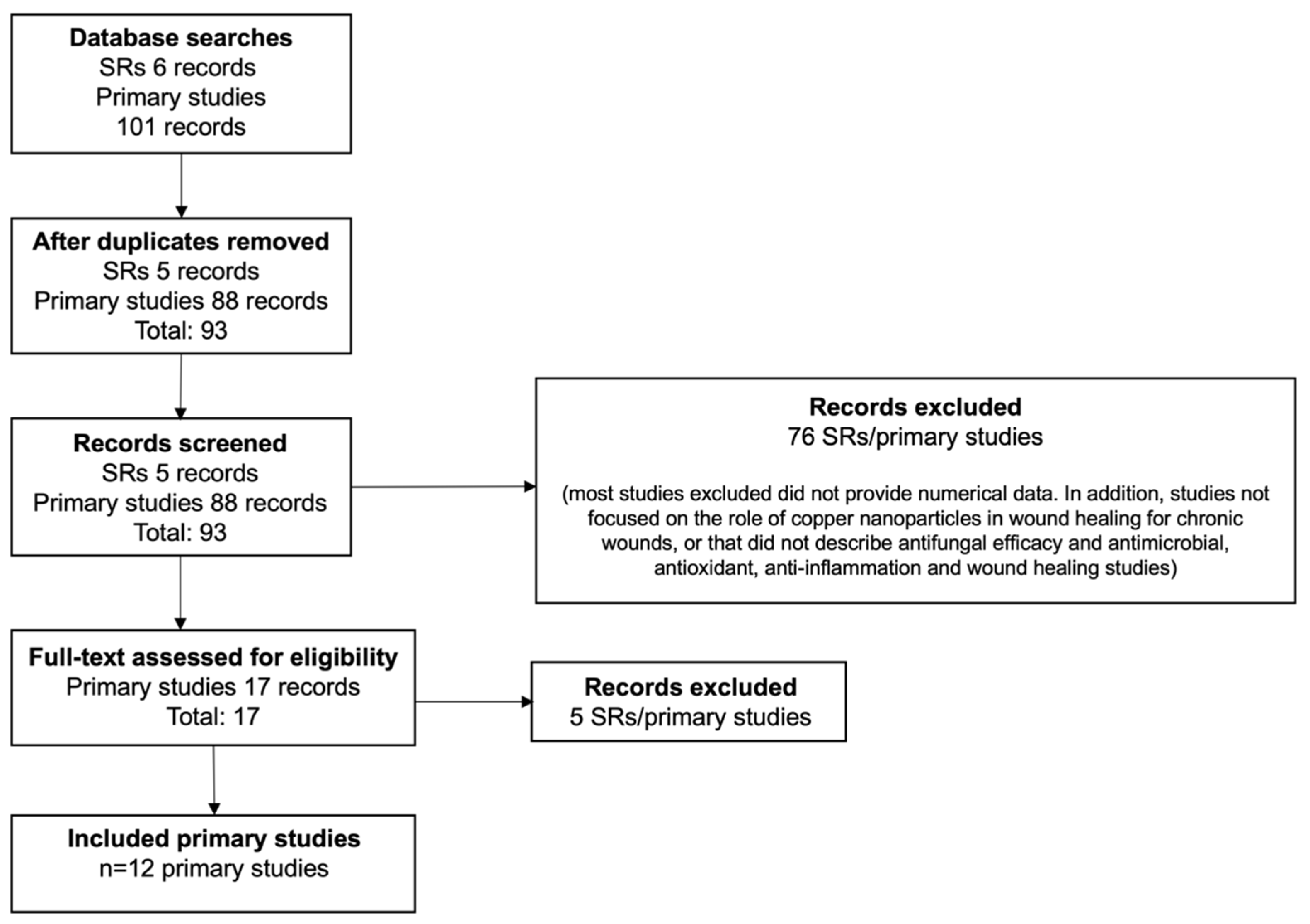
The flow chart of the selection process of the studies is shown in Figure 1 [40,41,42,43,44,45,46,48,49,50,52,53]. Table 1 lists the studies that were included, along with the demographics, environments, and contexts in which they were carried out.
Figure 1.
PRISMA flow diagram. SR: systematic review.
3.1. Description of Included Studies
Eight publications from the original studies were carried out in China, two in the US, one in Canada, and one in India.
A total of 12 original articles were analyzed. The papers collected and reported quantitative data through clinical trials or experimental studies (Table 1). Ten studies have used in vivo and in vitro models [41,43,44,45,46,48,49,50,52,53] while two have used in vitro studies [40,42]. Details of chronic wounds of each study, where available, are shown in Table 1.
The above-mentioned in vitro studies used human umbilical vein endotelial cells [43,44,49,52,53], immortalized human epithelial keratinocytes [48,50,52], human dermal fibroblasts [50,52], diabetic (db/db) mice and non-diabetic mice (C57BL/6) [50,52], human embryonic kidney [41], human keratinocytes cells [44], NIH-3T3 cell line [45,49], bacterial cellulose [40], human dermal fibroblasts extracted from split-thickness skin biopsies and obtained during breast reduction and abdominoplasty [42], human foreskin fibroblast cells [43], human corneal epithelial cells [44], fibroblast cell line [46], and recombinant human basic fibroblast growth factor [49]. For the in vivo models, female BALB/c mice [41,43,44,53], type 1 diabetic mice [48], Sprague-Dawley rats [45,49], and Wistar rats [46] were used.
3.2. Quality Assessment
Table 3 displays the quality assessment outcomes and evaluation standards for the studies. The studies’ overall quality for internal and external validity was often high or moderate. No studies were disqualified due to poor quality.
Table 3.
National Institute for Health and Care Excellence Methodology Checklist: quantitative studies.
3.3. Relation between NPs and Wound Treatment
The main advantages of Cu NPs used for wound treatment are described in Table 2. Nanoparticles with antibacterial qualities and low toxicity are excellent for use in wound dressings (Table 1). Different assays have been used to evaluate Cu tolerance, Cu toxicity, in vivo biocompatibility, and in vitro cell viability [40,41,42,43,45,46,48,49,50,52]. In addition, the antibacterial response was evaluated through different methods such as brain heart infusion [42], growth-inhibition assay [43], transmission electron microscopy [43], SYTO9/propidium iodide live/dead fluorescent staining assay [44], Luria-Bertani broth [45], or the disc diffusion method [40,46]. Finally, wound healing has been analyzed using the aortic ring assay [42], chick chorioallantoic membrane assay [42], optical coherence tomography angiography [50], photoacoustic imaging in vivo [48], immunohistochemistry [45,48,49], dermal excision wound model [52], matrigel assay [44], histochemical techniques [41,45], and ELISA kit [46].
3.4. Cytotoxicity Assays
Monitoring cell growth inhibition was assessed through cytotoxicity evaluation. However, cell cytotoxicity could be evaluated using other parameters, as shown in Table 1. In this sense, cells exposed to hollow mesoporous CuO nanospheres that resemble viruses (HvCuO@GOx) showed negligible cytotoxicity and biocompatibility when different glucose concentrations (2 mM, 4 mM, 6 mM, 8 mM, 10 mM) were used [48]. Likewise, the Cu nanodots (CuS NDs) dose did not effect the viability of the cells with or without laser irradiation. Even at a dosage of 45 g/mL, both cell lines were still alive, demonstrating the very low cytotoxicity of CuS NDs and photocytotoxicity [43].
In vitro assays have shown that the increase in human dermal fibroblasts’ (HDFs) migration due to folic acid-modified Cu-based metal–organic framework (F-HKUST-1) exposure could result from the persistent release of Cu2+ and folic acid along with modest cytotoxicity [52]. At the same time, according to CCK-8 analysis, a small quantity of ultrasmall Cu5.4O NPs (USNPs) were able to shield the cells completely from 250 μM H2O2; meanwhile, HEK293 cells had normal polygonal cytoskeleton morphology after being treated to 200 ng mL−1 Cu5.4O USNPs for 48 h, showing high biocompatibility [41]. Finally, while BC/Cu20 membranes did not exhibit cytotoxicity to normal human dermal fibroblasts (NHDFs), BC/Cu60 and BC/Cu100 membranes drastically reduced cell viability [40].
3.5. Antibacterial Response
The human body uses Cu for the innate immune response by boosting the bactericidal and phagocytic functions of neutrophils and the antimicrobial activity of macrophages [54]. In this sense, the quantity of biofilm present following treatment with Cu-containing mesoporous bioactive glasses (Cu-MBG) is reduced for both species. However, in the infected skin model, the effect of Cu-MBG on Pseudomonas aeruginosa was noticeably less pronounced than its effect on Staphylococcus aureus [42]. In the case of methicillin-resistant Staphylococcus aureus (MRSA) and extended-spectrum β-lactamase (ESBL) Escherichia coli treated with CuS NDs and NIR irradiation, where ultrasmall NDs stuck to the bacterial surface, their original form was distorted and revealed wrinkled bacterial cellular wall/membranes with visible lesions and holes, which may be due to the strong contact between CuS NDs and the bacterial cell wall [43].
Samples with higher Cu displayed bigger inhibition zones when the antibacterial activity was measured using the disk diffusion method. BC/Cu20, BC/Cu60, and BC/Cu100 had diameters of 14.7 mm, 18.0 mm, and 21.3 mm, respectively [52]. In comparison to hydrogel composite, the gelatin + CuO hydrogel (3.8 ± 0.3 in Staphylococcus aureus and 4.8 ± 0.7 in Escherichia coli) and gelatin + ZnO hydrogel (4.9 ± 0.6 in Staphylococcus aureus and 5.3 ± 0.2 in Escherichia coli) showed greater zones of inhibition [46]. Likewise, the viabilities of Staphylococcus aureus and Escherichia coli were reduced when the concentrations of Cu nanoparticles were added to hydrogel samples (93.7% to 9.2% and 92.4% to 8.8%, respectively) [45]. Finally, F-HKUST-1 slowly released Cu2+ during wound healing processes, which is recognized for having an impact on the expression and synthesis of growth factors, matrix metalloproteinases, collagen, elastin, and integrins [52].
3.6. Wound Healing
Wound evaluation has historically relied on a visual inspection by the trained clinician. However, new elements provide accurate assessment modalities. In this sense, the average small vessel length increased significantly for both Cu-MBG and mesoporous bioactive glasses (MBG) [42]. As evidence of HvCuO@GOx (HSHvCuO@GOx) superiority in new vessel development, CD34-positive cells in the HvCuO@GOx group’s adhesive hydrogel were higher than those in the pure hydrogel and HSHvCuO groups [48].
On days 7, 14, and 28, it was shown that the composite scaffold’s rate of wound contraction (13.2 ± 1.4 mm, 8.5 ± 2.9 mm, and 0.0 ± 0.01 mm, respectively) was significantly higher than cotton gauze’s (17.7 ± 3.2, 12.3 ± 2.6, and 3 ± 4.7 mm, respectively) [46]. Similarly, the wound treated with laser and AuAgCu2O NSs shrank significantly, indicating a 93.5% healing rate [44]. In fact, an almost completely healed wound in a diabetic mouse model using HSHvCuO@GOx gauze has been found. However, wounds treated with control and HvCuO-based hydrogel did not recover, showing that the HvCuO@GOx-based hydrogel can successfully promote the healing of Staphylococcus aureus-infected wounds [48]. Finally, the incorporation of folic acid into HKUST-1 also reduces Cu2+ toxicity in vivo, as has been described [52].
4. Discussion
This systematic review compiles and synthesizes the evidence of twelve quantitative studies that relate the activity of Cu NPs with their different types of antibacterial, cytotoxic, and proangiogenic properties in in vivo and in vitro studies for chronic wound healing.
4.1. Summary of Key Findings and Interpretation
The four overlapping healing processes during wound healing are hemostasis, inflammation, proliferation, and remodeling [55]. Both internal and environmental causes may hamper the healing process. Significant factors that prevent wound healing include microbial infections and reactive oxygen species (ROS). They might prolong each recovery step, resulting in less than ideal structural and functional outcomes [56].
In this regard, it is crucial to note that chronic, non-healing wounds have been associated with impaired healing when higher and continued ROS have been detected in vivo [57]. On a molecular level, high ROS and reactive nitrogen species (RNS) can affect the activity of dermal fibroblasts and keratinocytes as well as change and/or degrade extracellular matrix (ECM) proteins both directly and indirectly (through activation of proteolysis). This is in addition to ROS-mediated transcription, which can cause matrix metalloproteases to be induced and prolong pro-inflammatory cytokine secretion [58]. In fact, the adequate equilibrium between low and high levels of ROS is essential in defining functional outcomes: although low levels of ROS are required for promoting efficient healing [59], excessive ROS release damages cells and hinders wound repair [60]. Instead of directly affecting ROS, one option for wound healing might involve influencing the antioxidant system. The in vitro studies analyzed did not exhibit noticeable cytotoxicity during low levels of Cu NPs treatment because it was well-tolerated [40,41,42,43,45,46,48,49,50]; it does not deform the structure of the cell membrane, and it can be used to relieve oxidative stress at the wound site. However, this is only achieved if a Cu ion is administered in controlled release [50]. In fact, if similar amounts are administered in a short period of time and abruptly, the Cu NPs can become toxic to cells and cause apoptosis [50].
Copper-based combined pharmacological complexes are more effective as antibacterial, antifungal, and antiviral medicines [61,62]. Copper concentration has a direct relationship with the mechanism by which it has a bacteriostatic or bactericidal effect [63]. The highest recorded impact was for copper metal (99.9%), and these results were seen in alloys having at least 70% copper [61,62]. However, due to the structure of the root canal system, which contains microecological niches such as dentinal tubules where antimicrobial drugs cannot reach, nanotechnology presents as an alternative to improve treatment success and endodontic retreatment rates [64]. Although systemic antibiotic administration helps the body fight microbial infections, a locally applied antimicrobial treatment is preferred for a wound [65]. Pomades, gels, and ointments can eliminate the germs that increased in large numbers where the damage was, potentially cutting the length of the healing process [66]. These materials are most helpful to patients with compromised immune systems, such as those with diabetes, hepatitis, and acquired immune deficiency syndrome [67,68]. In fact, according to our systematic analysis, the nanocomposite hydrogels under investigation have solid antioxidant potential against MRSA and ESBL E. coli as well as broad-spectrum antibacterial activity [40,43,44,45,46,49,53], including P. aeruginosa [42]. When a wound is in the inflammatory phase, large amounts of ROS such as superoxide (O2), peroxynitrite (ONOO−), and hydroxyl radicals (OH−) are generated that damage the body’s proteins and DNAs [69]. However, free radicals can be removed from the wound site thanks to the decisive antioxidant action. In this regard, additional research has demonstrated the ability of CuO hydrogels to eradicate human-pathogenic species of Gram-positive and Gram-negative bacteria [6,70,71]. These studies show that using these hydrogels as dressings at the location of a wound speeds up the healing process. These attributes synergistically support wound healing.
In wound therapy, primarily two types of NPs are used: (1) NPs with intrinsic properties that promote wound closure; and (2) NPs used as delivery vectors for therapeutic drugs. The former can be separated into nonmetallic nanomaterials and metallic or metal oxide nanoparticles. To repair the damaged cells and restore epidermal integrity, wound healing requires the migration and proliferation of different cells, angiogenesis, and collagen deposition processes [72]. In fact, collagen formation is a very vital step [73]. Recent investigations have unmistakably demonstrated that NPs represent a crucial therapy platform for skin wounds [41,44,45,46,50,74,75]. Cu is a well-known NP with a lengthy history of direct angiogenesis involvement and antibacterial action. Additionally, Cu has been found to have a possible involvement in the healing of wounds by controlling the expression of 84 genes linked to angiogenesis and wound repair [5]. In addition, to the best knowledge, studies have reported the effects of CuNPs on keratinocyte and fibroblast cell proliferation and migration during wound healing [41,42,48,50,52].
Increasing the characteristics of polymer nanocomposites, however, is a constant challenge for their broad usage in research [76]. Wound healing, gene therapy, tissue engineering, and controlled drug administration are only a few of the uses for such biomaterials [77]. The biodegradability, nontoxicity, biocompatibility, and environmental susceptibility of polymer nanocomposites have sparked enormous interest and advancement [78].
Because of their bioavailability and repeatability, polysaccharides (e.g., sodium alginate) are often utilized in drug delivery activities. They are biocompatible and biodegradable, with low immunogenicity, and they are attractive candidates for medicine administration [79]. In this sense, sodium alginate/polyvinyl acetate nanocomposites [80], drug-loaded nanofibers [81], and polyvinyl acetate/gelatin biopolymeric films [73] could be promising therapeutic options for preventing both resistant infections and life-threatening complications in exudative wounds.
4.2. Scope and Limitations
It is essential to highlight that our objective was to evaluate the use of Cu NPs in the healing of chronic wounds. However, it is necessary to consider that multiple physiological processes are involved in the healing process. Within these mechanisms are VEGF-induction, angiogenesis, and the expression and normalization of skin proteins such as collagen and keratin. As a result, the sustained release of non-cytotoxic amounts of Cu ions promotes in vivo wound healing by inducing angiogenesis, collagen deposition, and wound re-epithelialization.
Our findings confirm that Cu NP treatment promotes the formation of new vessels and significantly increases their total length, resulting in a denser and more stable vascular network at the wound site. Furthermore, with new collagen production and epithelial cell regeneration, the time spent in the inflammatory phase was reduced, allowing for a faster transition to the late stage of angiogenesis. However, the activity of Cu NPs is restricted by their biocompatibility in certain biological activities, low toxicity, and antibacterial capabilities, which are always dependent on Cu being present in low-to-moderate concentrations so that it is not damaging to target cells.
In addition, our review had other limitations, i.e., a low quantity of articles linking Cu NPs to chronic wound and wound healing were found. Also, the intervention times were highly variable between studies, with significant differences in the number of weeks and days. Finally, some studies did not provide sufficient data to compare the results obtained before and after the intervention, and some did not even incorporate the baseline measurements for the parameters studied, which limits the extraction of information.
5. Conclusions
Cu NPs have a high antibacterial response capacity since they are prone to interacting with the bacteria membrane. In addition, they can penetrate the bacteria biofilm and release ions inside it, compromising its integrity, where the rigidity of the membrane is an important determinant of antibacterial efficiency.
Our results have shown that Cu NPs are structurally designed to have a rough surface to facilitate adhesion to the bacterial membrane, helping to reduce or prevent the formation of bacteria. This bactericidal task occurs within a few minutes of encountering the bacteria; therefore, it is fast-acting, efficient, and long-term since it lasts over time without losing its bactericidal activity. However, it has been shown that Cu NDs demonstrate a much higher antibacterial effect than Cu NPs using laser radiation and can almost completely lyse bacterial membranes.
Author Contributions
C.S. and J.F. designed this study; J.S. and V.S.-M. supervised the study; C.S., G.R., N.S. and J.S. conducted the literature searches, data extraction, and independent search and reviewing; G.R., N.S. and J.S. prepared a first draft of the manuscript; C.S., V.S.-M. and J.F. finalized the manuscript. All authors have read and agreed to the published version of the manuscript.
Funding
This research was funded by Universidad de La Frontera No. DI22-0007 and Programa de Formación de Investigadores Postdoctorales 2022, Universidad de La Frontera.
Institutional Review Board Statement
Not applicable.
Informed Consent Statement
Not applicable.
Data Availability Statement
Not applicable.
Conflicts of Interest
The authors declare no conflict of interest.
References
- Lazarus, G.S.; Cooper, D.M.; Knighton, D.R.; Margolis, D.J.; Pecoraro, R.E.; Rodeheaver, G.; Robson, M.C. Definitions and guidelines for assessment of wounds and evaluation of healing. Wound Repair Regen. 1994, 2, 165–170. [Google Scholar] [CrossRef] [Scilit]
- Werdin, F.; Tennenhaus, M.; Schaller, H.E.; Rennekampff, H.O. Evidence-based management strategies for treatment of chronic wounds. Eplasty 2009, 9, e19. [Google Scholar]
- Kirsner, R.S. The Wound Healing Society chronic wound ulcer healing guidelines update of the 2006 guidelines—Blending old with new. Wound Repair Regen. 2016, 24, 110–111. [Google Scholar] [CrossRef] [Scilit]
- Nussbaum, S.R.; Carter, M.J.; Fife, C.E.; DaVanzo, J.; Haught, R.; Nusgart, M.; Cartwright, D. An Economic Evaluation of the Impact, Cost, and Medicare Policy Implications of Chronic Nonhealing Wounds. Value Health 2018, 21, 27–32. [Google Scholar] [CrossRef] [Scilit]
- Borkow, G.; Gabbay, J.; Dardik, R.; Eidelman, A.I.; Lavie, Y.; Grunfeld, Y.; Ikher, S.; Huszar, M.; Zatcoff, R.C.; Marikovsky, M. Molecular mechanisms of enhanced wound healing by copper oxide-impregnated dressings. Wound Repair Regen. 2010, 18, 266–275. [Google Scholar] [CrossRef] [Scilit]
- Abdollahi, Z.; Zare, E.N.; Salimi, F.; Goudarzi, I.; Tay, F.R.; Makvandi, P. Bioactive Carboxymethyl Starch-Based Hydrogels Decorated with CuO Nanoparticles: Antioxidant and Antimicrobial Properties and Accelerated Wound Healing In Vivo. Int. J. Mol. Sci. 2021, 22, 2531. [Google Scholar] [CrossRef] [Scilit]
- Ghasemian Lemraski, E.; Jahangirian, H.; Dashti, M.; Khajehali, E.; Sharafinia, S.; Rafiee-Moghaddam, R.; Webster, T.M. Antimicrobial Double-Layer Wound Dressing Based on Chitosan/Polyvinyl Alcohol/Copper: In vitro and in vivo Assessment. Int. J. Nanomed. 2021, 16, 223–235. [Google Scholar] [CrossRef] [Scilit]
- Schencke, C.; Vasconcellos, A.; Sandoval, C.; Torres, P.; Acevedo, F.; del Sol, M. Morphometric evaluation of wound healing in burns treated with Ulmo (Eucryphia cordifolia) honey alone and supplemented with ascorbic acid in guinea pig (Cavia porcellus). Burns Trauma 2016, 4, 25. [Google Scholar] [CrossRef] [Scilit]
- Schencke, C.; Vásquez, B.; Sandoval, C.; del Sol, M. El Rol de la Miel en los Procesos Morfofisiológicos de Reparación de Heridas. Int. J. Morphol. 2016, 34, 385–395. [Google Scholar] [CrossRef] [Scilit]
- Faúndez, G.; Troncoso, M.; Navarrete, P.; Figueroa, G. Antimicrobial activity of copper surfaces against suspensions of Salmonella enterica and Campylobacter jejuni. BMC Microbiol. 2004, 4, 19. [Google Scholar] [CrossRef] [Scilit]
- Perelshtein, I.; Applerot, G.; Perkas, N.; Wehrschuetz-Sigl, E.; Hasmann, A.; Guebitz, G.; Gedanken, A. CuO-cotton nanocomposite: Formation, morphology, and antibacterial activity. Surf. Coat Technol. 2009, 204, 54–57. [Google Scholar] [CrossRef] [Scilit]
- El-Naggar, M.E.; Abd-Al-Aleem, A.H.; Abu-Saied, M.A.; Youssef, A.M. Synthesis of environmentally benign antimicrobial dressing nanofibers based on polycaprolactone blended with gold nanoparticles and spearmint oil nanoemulsion. J. Mater. Res. 2021, 15, 3447–3460. [Google Scholar] [CrossRef] [Scilit]
- Li, Q.; Lu, F.; Zhou, G.; Yu, K.; Lu, B.; Xiao, Y.; Dai, F.; Wu, D.; Lan, G. Silver inlaid with gold nanoparticle/chitosan wound dressing enhances antibacterial activity and porosity, and promotes wound healing. Biomacromolecules 2017, 18, 3766–3775. [Google Scholar] [CrossRef] [Scilit]
- Chen, P.; Bian, L.N.; Hu, X.Y. Synergic fabrication of gold nanoparticles embedded dextran/silk sericin nanomaterials for the treatment and care of wound healing. J. Cluster Sci. 2021, 3, 1. [Google Scholar] [CrossRef] [Scilit]
- Parveen, A.; Kulkarni, N.; Yalagatti, M.; Abbaraju, V.; Deshpande, R. In vivo efficacy of biocompatible silver nanoparticles cream for empirical wound healing. J. Tissue Viability 2018, 27, 257–261. [Google Scholar] [CrossRef] [Scilit]
- Gallo, A.L.; Paladini, F.; Romano, A.; Verri, T.; Quattrini, A.; Sannino, A.; Pollini, M. Efficacy of silver coated surgical sutures on bacterial contamination, cellular response and wound healing. Mater. Sci. Eng. C 2016, 69, 884–893. [Google Scholar] [CrossRef] [Scilit]
- Paladini, F.; De Simone, S.; Sannino, A.; Pollini, M. Antibacterial and antifungal dressings obtained by photochemical deposition of silver nanoparticles. J. Appl. Polym. Sci. 2014, 131, 4032. [Google Scholar] [CrossRef] [Scilit]
- Paladini, F.; Picca, R.A.; Sportelli, M.C.; Cioffi, N.; Sannino, A.; Pollini, M. Surface chemical and biological characterization of flax fabrics modified with silver nanoparticles for biomedical applications. Mater. Sci. Eng. C Mater. Biol. Appl. 2015, 52, 1–10. [Google Scholar] [CrossRef] [Scilit]
- Yudaev, P.A.; Maslennikova, V.V.; Konkova, A.A.; Butorova, I.A.; Chistyakov, E.M. Silver-containing hydrogel based on polyvinyl alcohol modified with nanoscale cyclotriphosphazene. Public Health Toxicol. 2021, 1, A23. [Google Scholar] [CrossRef] [Scilit]
- Uauy, R.; Olivares, M.; Gonzalez, M. Essentiality of copper in humans. Am. J. Clin. Nutr. 1998, 67, 952S–959S. [Google Scholar] [CrossRef] [Scilit]
- Tenaud, I.; Sainte-Marie, I.; Jumbou, O.; Litoux, P.; Dreno, B. In vitro modulation of keratinocyte wound healing integrins by zinc, copper and manganese. Br. J. Dermatol. 1999, 140, 26–34. [Google Scholar] [CrossRef] [Scilit] [PubMed]
- Sen, C.K.; Khanna, S.; Venojarvi, M.; Trikha, P.; Ellison, E.C.; Hunt, T.K.; Roy, S. Copper-induced vascular endothelial growth factor expression and wound healing. Am. J. Physiol. Heart Circ. Physiol. 2002, 282, H1821–H1827. [Google Scholar] [CrossRef] [Scilit] [PubMed]
- Quaranta, D.; Krans, T.; Espírito Santo, C.; Elowsky, C.G.; Domaille, D.W.; Chang, C.J.; Grass, G. Mechanisms of contact-mediated killing of yeast cells on dry metallic copper surfaces. Appl. Environ. Microbiol. 2011, 77, 416–426. [Google Scholar] [CrossRef] [Scilit] [PubMed]
- Palza, H. Antimicrobial polymers with metal nanoparticles. Int. J. Mol. Sci. 2015, 16, 2099–2116. [Google Scholar] [CrossRef] [Scilit]
- Ahire, J.J.; Hattingh, M.; Neveling, D.P.; Dicks, L.M. Copper-Containing Anti-Biofilm Nanofiber Scaffolds as a Wound Dressing Material. PLoS ONE 2016, 11, e0152755. [Google Scholar] [CrossRef] [Scilit] [PubMed]
- Borkow, G.; Gabbay, J. Copper as a biocidal tool. Curr. Med. Chem. 2005, 12, 2163–2175. [Google Scholar] [CrossRef] [Scilit]
- Alizadeh, S.; Seyedalipour, B.; Shafieyan, S.; Kheime, A.; Mohammadi, P.; Aghdami, N. Copper nanoparticles promote rapid wound healing in acute full thickness defect via acceleration of skin cell migration, proliferation, and neovascularization. Biochem. Biophys. Res. Commun. 2019, 517, 684–690. [Google Scholar] [CrossRef] [Scilit]
- Chen, M.; Li, R.; Yin, W.; Wang, T.; Kang, Y.J. Copper promotes migration of adipose-derived stem cells by enhancing vimentin- Ser39 phosphorylation. Exp. Cell Res. 2020, 388, 111859. [Google Scholar] [CrossRef] [Scilit]
- Das, A.; Sudhahar, V.; Chen, G.F.; Kim, H.W.; Youn, S.W.; Finney, L.; Vogt, S.; Yang, J.; Kweon, J.; Surenkhuu, B.; et al. Endothelial Antioxidant-1: A Key Mediator of Copper-dependent Wound Healing in vivo. Sci. Rep. 2016, 6, 33783. [Google Scholar] [CrossRef] [Scilit]
- Wu, Z.; Zhang, W.; Kang, Y.J. Copper affects the binding of HIF-1alpha to the critical motifs of its target genes. Metallomics 2019, 11, 429–438. [Google Scholar] [CrossRef] [Scilit]
- Visse, R.; Nagase, H. Matrix Metalloproteinases and Tissue Inhibitors of Metalloproteinases. Circ. Res. 2003, 92, 827–839. [Google Scholar] [CrossRef] [Scilit] [PubMed]
- Michopoulou, A.; Rousselle, P. How do epidermal matrix metalloproteinases support re-epithelialization during skin healing? Eur. J. Dermatol. 2015, 25, 33–42. [Google Scholar] [CrossRef] [Scilit]
- Philips, N.; Hwang, H.; Chauhan, S.; Leonardi, D.; Gonzalez, S. Stimulation of cell proliferation and expression of matrixmetalloproteinase-1 and interluekin-8 genes in dermal fibroblasts by copper. Connect. Tissue Res. 2010, 51, 224–229. [Google Scholar] [CrossRef] [Scilit] [PubMed]
- Adamson, I.Y.; Vincent, R.; Bakowska, J. Differential production of metalloproteinases after instilling various urban air particle samples to rat lung. Exp. Lung Res. 2003, 29, 375–388. [Google Scholar] [CrossRef] [Scilit] [PubMed]
- Salvo, J.; Sandoval, C. Role of copper nanoparticles in wound healing for chronic wounds: Literature review. Burns Trauma 2022, 10, tkab047. [Google Scholar] [CrossRef] [Scilit]
- Zhou, M.; Li, J.; Liang, S.; Sood, A.K.; Liang, D.; Li, C. CuS nanodots with ultrahigh efficient renal clearance for positron emission tomography imaging and Image-Guided photothermal therapy. ACS Nano 2015, 9, 7085–7096. [Google Scholar] [CrossRef] [Scilit]
- Feng, X.; Xu, W.; Li, Z.; Song, W.; Ding, J.; Chen, X. Immunomodulatory nanosystems. Adv. Sci. 2019, 6, 1900101. [Google Scholar] [CrossRef] [Scilit]
- Page, M.J.; McKenzie, J.E.; Bossuyt, P.M.; Boutron, I.; Hoffmann, T.C.; Mulrow, C.D.; Shamseer, L.; Tetzlaff, J.M.; Akl, E.A.; Brennan, S.E.; et al. The PRISMA 2020 statement: An updated guideline for reporting systematic reviews. PLoS Med. 2021, 18, e1003583. [Google Scholar] [CrossRef]
- National Institute for Health and Care Excellence. Appendix F Quality Appraisal Checklist—Quantitative Intervention Studies. In Methods for the Development of NICE Public Health Guidance; National Institute for Health and Care Excellence: London, UK, 2012; Available online: https://www.nice.org.uk/process/pmg4/chapter/about-this-document (accessed on 1 April 2022).
- He, W.; Huang, X.; Zheng, Y.; Sun, Y.; Xie, Y.; Wang, Y.; Yue, L. In situ synthesis of bacterial cellulose/copper nanoparticles composite membranes with long-term antibacterial property. J. Biomater. Sci. Polym. Ed. 2018, 29, 2137–2153. [Google Scholar] [CrossRef] [Scilit]
- Liu, T.; Xiao, B.; Xiang, F.; Tan, J.; Chen, Z.; Zhang, X.; Wu, C.; Mao, Z.; Luo, G.; Chen, X.; et al. Ultrasmall copper-based nanoparticles for reactive oxygen species scavenging and alleviation of inflammation related diseases. Nat. Commun. 2020, 11, 2788. [Google Scholar] [CrossRef] [Scilit]
- Paterson, T.E.; Bari, A.; Bullock, A.J.; Turner, R.; Montalbano, G.; Fiorilli, S.; Vitale-Brovarone, C.; MacNeil, S.; Shepherd, J. Multifunctional Copper-Containing Mesoporous Glass Nanoparticles as Antibacterial and Proangiogenic Agents for Chronic Wounds. Front. Bioeng. Biotechnol. 2020, 8, 246. [Google Scholar] [CrossRef] [Scilit] [PubMed]
- Qiao, Y.; Ping, Y.; Zhang, H.; Zhou, B.; Liu, F.; Yu, Y.; Xie, T.; Li, W.; Zhong, D.; Zhang, Y.; et al. Laser-Activatable CuS Nanodots to Treat Multidrug-Resistant Bacteria and Release Copper Ion to Accelerate Healing of Infected Chronic Nonhealing Wounds. ACS Appl. Mater. Interfaces 2019, 11, 3809–3822. [Google Scholar] [CrossRef] [Scilit] [PubMed]
- Qiao, Y.; He, J.; Chen, W.; Yu, Y.; Li, W.; Du, Z.; Xie, T.; Ye, Y.; Hua, S.Y.; Zhong, D.; et al. Light-Activatable Synergistic Therapy of Drug-Resistant Bacteria-Infected Cutaneous Chronic Wounds and Nonhealing Keratitis by Cupriferous Hollow Nanoshells. ACS Nano 2020, 14, 3299–3315. [Google Scholar] [CrossRef] [Scilit] [PubMed]
- Tao, B.; Lin, C.; Deng, Y.; Yuan, Z.; Shen, X.; Chen, M.; He, Y.; Peng, Z.; Hu, Y.; Cai, K. Copper-nanoparticle-embedded hydrogel for killing bacteria and promoting wound healing with photothermal therapy. J. Mater. Chem. B 2019, 7, 2534–2548. [Google Scholar] [CrossRef] [Scilit] [PubMed]
- Thanusha, A.V.; Dinda, A.K.; Koul, V. Evaluation of nano hydrogel composite based on gelatin/HA/CS suffused with Asiatic acid/ZnO and CuO nanoparticles for second degree burns. Mater. Sci. Eng. C 2018, 89, 378–386. [Google Scholar] [CrossRef] [Scilit]
- Kuijpers, A.J.; van Wachem, P.B.; van Luyn, M.J.; Brouwer, L.A.; Engbers, G.H.; Krijgsveld, J.; Zaat, S.A.; Dankert, J.; Feijen, J. In vitro and in vivo evaluation of gelatin-chondroitin sulphate hydrogels for controlled release of antibacterial proteins. Biomaterials 2000, 21, 1763–1772. [Google Scholar] [CrossRef] [Scilit]
- Wang, P.; Peng, L.; Lin, J.; Li, Y.; Luo, Q.; Jiang, S.; Tian, H.; Zhang, Y.; Liu, X.; Liu, J. Enzyme hybrid virus-like hollow mesoporous CuO adhesive hydrogel spray through glucose-activated cascade reaction to efficiently promote diabetic wound healing. Chem. Eng. J. 2021, 415, 128901. [Google Scholar] [CrossRef] [Scilit]
- Wang, T.L.; Zhou, Z.F.; Liu, J.F.; Hou, X.D.; Zhou, Z.; Dai, Y.L.; Hou, Z.Y.; Chen, F.; Zheng, L.P. Donut-like MOFs of copper/nicotinic acid and composite hydrogels with superior bioactivity for rh-bFGF delivering and skin wound healing. J. Nanobiotechnology 2021, 19, 275. [Google Scholar] [CrossRef] [Scilit]
- Xiao, J.; Chen, S.; Yi, J.; Zhang, H.; Ameer, G.A. A Cooperative Copper Metal-Organic Framework-Hydrogel System Improves Wound Healing in Diabetes. Adv. Funct. Mater. 2017, 27, 1604872. [Google Scholar] [CrossRef] [Scilit]
- Xiao, J.; Zhu, Y.; Huddleston, S.; Li, P.; Xiao, B.; Farha, O.K.; Ameer, G.A. Copper Metal-Organic Framework Nanoparticles Stabilized with Folic Acid Improve Wound Healing in Diabetes. ACS Nano 2018, 12, 1023–1032. [Google Scholar] [CrossRef] [Scilit]
- Zhou, Y.; Feng, H.; Jiang, Y.; Hua, G.; Zhang, Q.; Zeng, S.; Li, W.; Li, L.; Kang, N.; Ren, L. Nanoliquid Dressing with Enhancing Anti-Infection Performance under the Moderate Photothermal Effect for Wound Treatment. ACS Appl. Mater. Interfaces 2021, 13, 18443–18453. [Google Scholar] [CrossRef] [Scilit] [PubMed]
- Yang, J.; van Lith, R.; Baler, K.; Hoshi, R.A.; Ameer, G.A. A Thermoresponsive Biodegradable Polymer with Intrinsic Antioxidant Properties. Biomacromolecules 2014, 15, 3942–3952. [Google Scholar] [CrossRef] [Scilit]
- Djoko, K.Y.; Ong, C.Y.; Walker, M.J.; McEwan, A.G. The role of copper and zinc toxicity in innate immune defense against bacterial pathogens. J. Biol. Chem. 2015, 290, 18954–18961. [Google Scholar] [CrossRef] [Scilit]
- Shariati, A.; Moradabadi, A.; Azimi, T.; Ghaznavi-Rad, E. Wound healing properties and antimicrobial activity of platelet-derived biomaterials. Sci. Rep. 2020, 10, 1032. [Google Scholar] [CrossRef] [Scilit]
- Dev, S.K.; Choudhury, P.K.; Srivastava, R.; Sharma, M. Antimicrobial, anti-inflammatory and wound healing activity of polyherbal formulation. Biomed. Pharmacother. 2019, 111, 555–567. [Google Scholar] [CrossRef] [Scilit] [PubMed]
- Schafer, M.; Werner, S. Oxidative stress in normal and impaired wound repair. Pharmacol. Res. 2008, 58, 165–171. [Google Scholar] [CrossRef] [Scilit] [PubMed]
- Moseley, R.; Stewart, J.E.; Stephens, P.; Waddington, R.J.; Thomas, D.W. Extracellular matrix metabolites as potential biomarkers of disease activity in wound fluid: Lessons learned from other inflammatory diseases? Br. J. Dermatol. 2004, 150, 401–413. [Google Scholar] [CrossRef] [Scilit] [PubMed]
- Rodriguez, P.G.; Felix, F.N.; Woodley, D.T.; Shim, E.K. The role of oxygen in wound healing: A review of the literature. Dermatol. Surg. 2008, 34, 1159–1169. [Google Scholar] [CrossRef] [Scilit]
- Ponugoti, B.; Xu, F.; Zhang, C.; Tian, C.; Pacios, S.; Graves, D.T. FOXO1 promotes wound healing through the up-regulation of TGF-beta1 and prevention of oxidative stress. J. Cell Biol. 2013, 203, 327–343. [Google Scholar] [CrossRef] [Scilit]
- Chandraleka, S.; Ramya, K.; Chandramohan, G.; Dhanasekaran, D.; Priyadharshini, A.; Panneerselvam, A. Antimicrobial mechanism of copper (II) 1,10-phenanthroline and 2,2′-bipyridyl complex on bacterial and fungal pathogens. J. Saudi Chem. Soc. 2014, 18, 953–962. [Google Scholar] [CrossRef] [Scilit]
- O’Gorman, J.; Humphreys, H. Application of copper to prevent and control infection. Where are we now? J. Hosp. Infect. 2012, 81, 217–223. [Google Scholar] [CrossRef] [Scilit]
- Gordon, A.S.; Howell, L.D.; Harwood, V. Responses of diverse heterotrophic bacteria to elevated copper concentrations. Can. J. Microbiol. 1994, 40, 408–411. [Google Scholar] [CrossRef] [Scilit] [PubMed]
- Javidi, M.; Afkhami, F.; Zarei, M.; Ghazvini, K.; Rajabi, O. Efficacy of a combined nanoparticulate/calcium hydroxide root canal medication on elimination of Enterococcus faecalis. Aust. Endod. J. 2014, 40, 61–65. [Google Scholar] [CrossRef] [Scilit] [PubMed]
- Levy, S.B. The challenge of antibiotic resistance. Sci. Am. 1998, 278, 46–53. [Google Scholar] [CrossRef] [Scilit] [PubMed]
- Makvandi, P.; Ali, G.W.; Della Sala, F.; Abdel-Fattah, W.I.; Borzacchiello, A. Biosynthesis and characterization of antibacterial thermosensitive hydrogels based on corn silk extract, hyaluronic acid and nanosilver for potential wound healing. Carbohydr. Polym. 2019, 223, 115023. [Google Scholar] [CrossRef] [Scilit]
- Casqueiro, J.; Casqueiro, J.; Alves, C. Infections in patients with diabetes mellitus: A review of pathogenesis. Indian J. Endocrinol. Metab. 2012, 16, S27. [Google Scholar] [CrossRef] [Scilit]
- Nikolopoulo, G.K.; Paraskevis, D.; Hatzitheodorou, E.; Moschidis, Z.; Sypsa, V.; Zavitsanos, X.; Kalapothaki, V.; Hatzakis, A. Impact of hepatitis B virus infection on the progression of AIDS and mortality in HIV-infected individuals: A cohort study and meta-analysis. Clin. Infect. Dis. 2009, 48, 1763–1771. [Google Scholar] [CrossRef] [Scilit]
- Martins-Green, M.; Saeed, S. Role of Oxidants and Antioxidants in Diabetic Wound Healing. In Wound Healing, Tissue Repair, and Regeneration in Diabetes; Bagchi, D., Das, A., Roy, S., Eds.; Elsevier: Amsterdam, The Netherlands, 2020; pp. 13–38. [Google Scholar] [CrossRef] [Scilit]
- Wahid, F.; Wang, H.S.; Lu, Y.S.; Zhong, C.; Chu, L.Q. Preparation, characterization and antibacterial applications of carboxymethyl chitosan/CuO nanocomposite hydrogels. Int. J. Biol. Macromol. 2017, 101, 690–695. [Google Scholar] [CrossRef] [Scilit]
- Sattari, S.; Dadkhah Tehrani, A.; Adeli, M. pH-Responsive Hybrid Hydrogels as Antibacterial and Drug Delivery Systems. Polymers 2018, 10, 660. [Google Scholar] [CrossRef] [Scilit]
- Greaves, N.S.; Ashcroft, K.J.; Baguneid, M.; Bayat, A. Current understanding of molecular and cellular mechanisms in fibroplasia and angiogenesis during acute wound healing. J. Dermatol. Sci. 2013, 72, 206–217. [Google Scholar] [CrossRef] [Scilit]
- Khan, B.A.; Ullah, S.; Khan, M.K.; Uzair, B.; Menaa, F.; Braga, V.A. Fabrication, Physical Characterizations, and In Vitro, In Vivo Evaluation of Ginger Extract-Loaded Gelatin/Poly(Vinyl Alcohol) Hydrogel Films Against Burn Wound Healing in Animal Model. AAPS Pharm. Sci. Tech. 2020, 21, 323. [Google Scholar] [CrossRef] [Scilit] [PubMed]
- Mofazzal Jahromi, M.A.; Sahandi Zangabad, P.; Moosavi Basri, S.M.; Sahandi Zangabad, K.; Ghamarypour, A.; Aref, A.R.; Karimi, M.; Hamblin, M.R. Nanomedicine and advanced technologies for burns: Preventing infection and facilitating wound healing. Adv. Drug Deliv. Rev. 2018, 123, 33–64. [Google Scholar] [CrossRef] [Scilit] [PubMed]
- Wang, L.; Hu, C.; Shao, L. The antimicrobial activity of nanoparticles: Present situation and prospects for the future. Int. J. Nanomed. 2017, 12, 1227–1249. [Google Scholar] [CrossRef] [Scilit] [PubMed]
- Winey, K.I.; Vaia, R.A. Polymer nanocomposites. MRS Bull. 2007, 32, 314–322. [Google Scholar] [CrossRef] [Scilit]
- Rong, M.Z.; Zhang, M.Q.; Ruan, W.H. Surface modification of nanoscale fillers for improving properties of polymer nanocomposites: A review. Mater. Sci. Technol. 2006, 22, 787–796. [Google Scholar] [CrossRef] [Scilit]
- Jana, S.; Sen, K.K.; Gandhi, A. Alginate Based Nanocarriers for Drug Delivery Applications. Curr. Pharm. Des. 2016, 22, 3399–3410. [Google Scholar] [CrossRef] [Scilit] [PubMed]
- Niculescu, A.G.; Grumezescu, A.M. Applications of Chitosan-Alginate-Based Nanoparticles—An Up-to-Date Review. Nanomaterials 2022, 12, 186. [Google Scholar] [CrossRef] [Scilit]
- Bibi, S.; Mir, S.; Rehman, W.; Menaa, F.; Gul, A.; Alaryani, F.S.S.; Alqahtani, A.M.; Haq, S.; Abdellatif, M.H. Synthesis and In Vitro/Ex Vivo Characterizations of Ceftriaxone-Loaded Sodium Alginate/poly(vinyl alcohol) Clay Reinforced Nanocomposites: Possible Applications in Wound Healing. Materials 2022, 15, 3885. [Google Scholar] [CrossRef] [Scilit]
- Razzaq, A.; Khan, Z.U.; Saeed, A.; Shah, K.A.; Khan, N.U.; Menaa, B.; Iqbal, H.; Menaa, F. Development of Cephradine-Loaded Gelatin/Polyvinyl Alcohol Electrospun Nanofibers for Effective Diabetic Wound Healing: In-Vitro and In-Vivo Assessments. Pharmaceutics 2021, 13, 349. [Google Scholar] [CrossRef] [Scilit] [PubMed]

Publisher’s Note: MDPI stays neutral with regard to jurisdictional claims in published maps and institutional affiliations. |
© 2022 by the authors. Licensee MDPI, Basel, Switzerland. This article is an open access article distributed under the terms and conditions of the Creative Commons Attribution (CC BY) license (https://creativecommons.org/licenses/by/4.0/).