
Genetic Polymorphisms in VEGFR Coding Genes (FLT1/KDR) on Ranibizumab Response in High Myopia and Choroidal Neovascularization Patients

 , ,
, ,  ,
,
Abstract
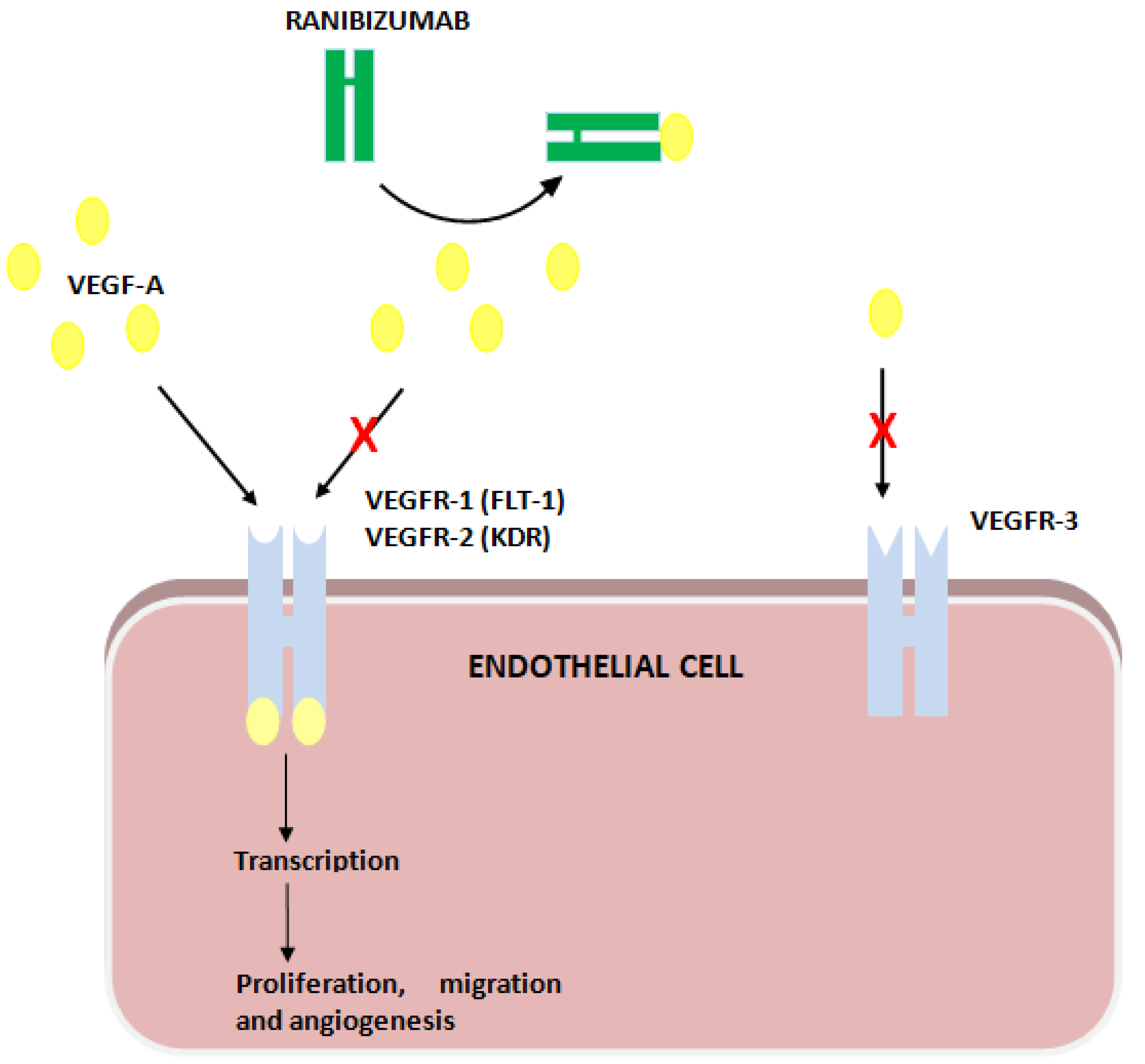
1. Introduction
2. Materials and Methods
2.1. Procedures for the Inclusion of Genetic Variants in the Study
2.2. DNA Extraction and Genotyping
2.3. Statistical Analysis
3. Results
3.1. Genotypic Distribution, H–W Equilibrium, Association of Genetic Variants with High Myopia and CNV, and Linkage Disequilibrium Analysis
3.2. Association of Genetic Polymorphisms with Response to Ranibizumab
3.2.1. Genotype Association Study with Response
3.2.2. Alleles Association Study with Response
3.3. Haplotype Association Study with Response
4. Discussion
4.1. FLT1 Genetic Polymorphisms and Ranibizumab
4.2. KDR Genetic Polymorphisms and Ranibizumab
5. Conclusions
Supplementary Materials
Author Contributions
Funding
Institutional Review Board Statement
Informed Consent Statement
Data Availability Statement
Acknowledgments
Conflicts of Interest
References
- Fredrick, D.R. Myopia. BMJ 2002, 324, 1195–1199. [Google Scholar] [CrossRef] [PubMed]
- Kempen, J.H.; Mitchell, P.; Lee, K.E.; Tielsch, J.M.; Broman, A.T.; Taylor, H.R.; Ikram, M.K.; Congdon, N.G.; O’Colmain, B.J.; Eye Diseases Prevalence Research Group. The prevalence of refractive errors among adults in the United States, Western Europe, and Australia. Arch. Ophthalmol. 2004, 122, 495–505. [Google Scholar] [PubMed]
- Wong, T.Y.; Foster, P.J.; Hee, J.; Ng, T.P.; Tielsch, J.M.; Chew, S.J.; Johnson, G.J.; Seah, S.K. Prevalence and risk factors for refractive errors in adult Chinese in Singapore. Investig. Ophthalmol. Vis. Sci. 2000, 41, 2486–2494. [Google Scholar]
- Wolf, S.; Balciuniene, V.J.; Laganovska, G.; Menchini, U.; Ohno-Matsui, K.; Sharma, T.; Wong, T.Y.; Silva, R.; Pilz, S.; Gekkieva, M.; et al. RADIANCE: A randomized controlled study of ranibizumab in patients with choroidal neovascularization secondary to pathologic myopia. Ophthalmology 2014, 121, 682–692.e2. [Google Scholar] [CrossRef]
- Miller, D.G.; Singerman, L.J. Natural history of choroidal neovascularization in high myopia. Curr. Opin. Ophthalmol. 2001, 12, 222–224. [Google Scholar] [CrossRef] [PubMed]
- Chung, Y.W.; Choi, M.Y.; Kim, J.S.; Kwon, J.W. The Association between Macular Thickness and Axial Length in Myopic Eyes. Biomed. Res. Int. 2019, 2019, 8913582. [Google Scholar] [CrossRef] [PubMed]
- Saw, S.M.; Gazzard, G.; Shih-Yen, E.C.; Chua, W.H. Myopia and associated pathological complications. Ophthalmic Physiol. Opt. 2005, 25, 381–391. [Google Scholar] [CrossRef]
- Chuck, R.S.; Jacobs, D.S.; Lee, J.K.; Afshari, N.A.; Vitale, S.; Shen, T.T.; Keenan, J.D.; American Academy of Ophthalmology Preferred Practice Pattern Refractive Management/Intervention Panel. Refractive Errors & Refractive Surgery Preferred Practice Pattern®. Ophthalmology 2018, 125, P1–P104. [Google Scholar] [PubMed]
- Wong, T.Y.; Ferreira, A.; Hughes, R.; Carter, G.; Mitchell, P. Epidemiology and disease burden of pathologic myopia and myopic choroidal neovascularization: An evidence-based systematic review. Am. J. Ophthalmol. 2014, 157, 9–25.e12. [Google Scholar] [CrossRef] [PubMed]
- Hayashi, K.; Ohno-Matsui, K.; Shimada, N.; Moriyama, M.; Kojima, A.; Hayashi, W.; Yasuzumi, K.; Nagaoka, N.; Saka, N.; Yoshida, T.; et al. Long-term pattern of progression of myopic maculopathy: A natural history study. Ophthalmology 2010, 117, 1595–1611, 1611.e1–4. [Google Scholar] [CrossRef]
- Ng, D.; Fung, N.; Yip, F.; Lai, T. Ranibizumab for myopic choroidal neovascularization. Expert Opin. Biol. Ther. 2020, 20, 1385–1393. [Google Scholar] [CrossRef]
- Chan, W.M.; Ohji, M.; Lai, T.Y.; Liu, D.T.; Tano, Y.; Lam, D.S. Choroidal neovascularisation in pathological myopia: An update in management. Br. J. Ophthalmol. 2005, 89, 1522–1528. [Google Scholar] [CrossRef]
- Soubrane, G. Choroidal neovascularization in pathologic myopia: Recent developments in diagnosis and treatment. Surv. Ophthalmol. 2008, 53, 121–138. [Google Scholar] [CrossRef] [PubMed]
- Uemura, A.; Thomas, M.A. Subretinal surgery for choroidal neovascularization in patients with high myopia. Arch. Ophthalmol. 2000, 118, 344–350. [Google Scholar] [CrossRef]
- Ruiz-Moreno, J.M.; Montero, J.A. Long-term visual acuity after argon green laser photocoagulation of juxtafoveal choroidal neovascularization in highly myopic eyes. Eur. J. Ophthalmol. 2002, 12, 117–122. [Google Scholar] [CrossRef]
- Parodi, M.B.; Iacono, P.; Papayannis, A.; Sheth, S.; Bandello, F. Laser photocoagulation, photodynamic therapy, and intravitreal bevacizumab for the treatment of juxtafoveal choroidal neovascularization secondary to pathologic myopia. Arch. Ophthalmol. 2010, 128, 437–442. [Google Scholar] [CrossRef] [PubMed]
- Verteporfin in Photodynamic Therapy Study Group. Photodynamic therapy of subfoveal choroidal neovascularization in pathologic myopia with verteporfin. 1-year results of a randomized clinical trial—VIP report no. 1. Ophthalmology 2001, 108, 841–852. [Google Scholar] [CrossRef]
- Blinder, K.J.; Blumenkranz, M.S.; Bressler, N.M.; Bressler, S.B.; Donato, G.; Lewis, H.; Lim, J.I.; Menchini, U.; Miller, J.W.; Mones, J.M.; et al. Verteporfin therapy of subfoveal choroidal neovascularization in pathologic myopia: 2-year results of a randomized clinical trial—VIP report no. 3. Ophthalmology 2003, 110, 667–673. [Google Scholar]
- Giansanti, F.; Virgili, G.; Donati, M.C.; Giuntoli, M.; Pieretti, G.; Abbruzzese, G.; Menchini, U. Long-term results of photodynamic therapy for subfoveal choroidal neovascularization with pathologic myopia. Retina 2012, 32, 1547–1552. [Google Scholar] [CrossRef] [PubMed]
- Claxton, L.; Malcolm, B.; Taylor, M.; Haig, J.; Leteneux, C. Ranibizumab, verteporfin photodynamic therapy or observation for the treatment of myopic choroidal neovascularization: Cost effectiveness in the UK. Drugs Aging 2014, 31, 837–848. [Google Scholar] [CrossRef][Green Version]
- Deeks, E.D. Ranibizumab: A review of its use in myopic choroidal neovascularization. BioDrugs 2014, 28, 403–410. [Google Scholar] [CrossRef]
- Holz, F.G.; Tufail, A.; Leveziel, N.; Lai, T.Y.; Lanzetta, P.; Wong, T.Y.; Yu, H.G.; Chen, Y.X.; Heinrichs, N.; Pilz, S.; et al. Ranibizumab in Myopic Choroidal Neovascularization: A Subgroup Analysis by Ethnicity, Age, and Ocular Characteristics in RADIANCE. Ophthalmologica 2016, 236, 19–28. [Google Scholar] [CrossRef]
- Cobos, E.; Recalde, S.; Anter, J.; Hernandez-Sanchez, M.; Barreales, C.; Olavarrieta, L.; Valverde, A.; Suarez-Figueroa, M.; Cruz, F.; Abraldes, M.; et al. Association between CFH, CFB, ARMS2, SERPINF1, VEGFR1 and VEGF polymorphisms and anatomical and functional response to ranibizumab treatment in neovascular age-related macular degeneration. Acta Ophthalmol. 2018, 96, e201–e212. [Google Scholar] [CrossRef] [PubMed]
- Beuselinck, B.; Jean-Baptiste, J.; Schöffski, P.; Couchy, G.; Meiller, C.; Rolland, F.; Allory, Y.; Joniau, S.; Verkarre, V.; Elaidi, R.; et al. Validation of VEGFR1 rs9582036 as predictive biomarker in metastatic clear-cell renal cell carcinoma patients treated with sunitinib. BJU Int. 2016, 118, 890–901. [Google Scholar] [CrossRef]
- Beuselinck, B.; Karadimou, A.; Lambrechts, D.; Claes, B.; Wolter, P.; Couchy, G.; Berkers, J.; van Poppel, H.; Paridaens, R.; Schöffski, P.; et al. VEGFR1 single nucleotide polymorphisms associated with outcome in patients with metastatic renal cell carcinoma treated with sunitinib—A multicentric retrospective analysis. Acta Oncol. 2014, 53, 103–112. [Google Scholar] [CrossRef] [PubMed]
- Dornbusch, J.; Walter, M.; Gottschalk, A.; Obaje, A.; Junker, K.; Ohlmann, C.H.; Meinhardt, M.; Zacharis, A.; Zastrow, S.; Schoffer, O.; et al. Evaluation of polymorphisms in angiogenesis-related genes as predictive and prognostic markers for sunitinib-treated metastatic renal cell carcinoma patients. J. Cancer Res. Clin. Oncol. 2016, 142, 1171–1182. [Google Scholar] [CrossRef] [PubMed]
- Lazzeri, S.; Orlandi, P.; Piaggi, P.; Sartini, M.S.; Casini, G.; Guidi, G.; Figus, M.; Fioravanti, A.; Di Desidero, T.; Ripandelli, G.; et al. IL-8 and VEGFR-2 polymorphisms modulate long-term functional response to intravitreal ranibizumab in exudative age-related macular degeneration. Pharmacogenomics 2016, 17, 35–39. [Google Scholar] [CrossRef] [PubMed]
- Hermann, M.M.; van Asten, F.; Muether, P.S.; Smailhodzic, D.; Lichtner, P.; Hoyng, C.B.; Kirchhof, B.; Grefkes, C.; den Hollander, A.I.; Fauser, S. Polymorphisms in vascular endothelial growth factor receptor 2 are associated with better response rates to ranibizumab treatment in age-related macular degeneration. Ophthalmology 2014, 121, 905–910. [Google Scholar] [CrossRef]
- Blánquez-Martínez, D.; Díaz-Villamarín, X.; Antúnez-Rodríguez, A.; Pozo-Agundo, A.; Muñoz-Ávila, J.I.; Martínez-González, L.J.; Dávila-Fajardo, C.L. Genetic Polymorphisms Affecting Ranibizumab Response in High Myopia Patients. Pharmaceutics 2021, 13, 1973. [Google Scholar] [CrossRef]
- Freeman, B.; Smith, N.; Curtis, C.; Huckett, L.; Mill, J.; Craig, I. DNA from buccal swabs recruited by mail: Evaluation of storage effects on long-term stability and suitability for multiplex polymerase chain reaction genotyping. Behav. Genet. 2003, 33, 67–72. [Google Scholar] [CrossRef]
- Gómez-Martín, A.; Hernández, A.F.; Martínez-González, L.J.; González-Alzaga, B.; Rodríguez-Barranco, M.; López-Flores, I.; Aguilar-Garduno, C.; Lacasana, M. Polymorphisms of pesticide-metabolizing genes in children living in intensive farming communities. Chemosphere 2015, 139, 534–540. [Google Scholar] [CrossRef]
- Solé, X.; Guinó, E.; Valls, J.; Iniesta, R.; Moreno, V. SNPStats: A web tool for the analysis of association studies. Bioinformatics 2006, 22, 1928–19289. [Google Scholar] [CrossRef] [PubMed]
- Rinaldi, C.; Bramanti, P.; Scimone, C.; Donato, L.; Alafaci, C.; D’Angelo, R.; Sidoti, A. Relevance of CCM gene polymorphisms for clinical management of sporadic cerebral cavernous malformations. J. Neurol. Sci. 2017, 380, 31–37. [Google Scholar] [CrossRef] [PubMed]
- Scimone, C.; Donato, L.; Katsarou, Z.; Bostantjopoulou, S.; D’Angelo, R.; Sidoti, A. Two Novel KRIT1 and CCM2 Mutations in Patients Affected by Cerebral Cavernous Malformations: New Information on CCM2 Penetrance. Front. Neurol. 2018, 9, 953. [Google Scholar] [CrossRef]
- Scimone, C.; Bramanti, P.; Ruggeri, A.; Donato, L.; Alafaci, C.; Crisafulli, C.; Mucciardi, M.; Rinaldi, C.; Sidoti, A.; D’Angelo, R. CCM3/SERPINI1 bidirectional promoter variants in patients with cerebral cavernous malformations: A molecular and functional study. BMC Med. Genet. 2016, 17, 74. [Google Scholar] [CrossRef] [PubMed]
- Scimone, C.; Bramanti, P.; Ruggeri, A.; Katsarou, Z.; Donato, L.; Sidoti, A.; D’Angelo, R. Detection of Novel Mutation in Ccm3 Causes Familial Cerebral Cavernous Malformations. J. Mol. Neurosci. 2015, 57, 400–403. [Google Scholar] [CrossRef] [PubMed]
- Smailhodzic, D.; Muether, P.S.; Chen, J.; Kwestro, A.; Zhang, A.Y.; Omar, A.; Van de Ven, J.P.; Keunen, J.E.; Kirchhof, B.; Hoyng, C.B.; et al. Cumulative effect of risk alleles in CFH, ARMS2, and VEGFA on the response to ranibizumab treatment in age-related macular degeneration. Ophthalmology 2012, 119, 2304–2311. [Google Scholar] [CrossRef]
- Tian, J.; Qin, X.; Fang, K.; Chen, Q.; Hou, J.; Li, J.; Yu, W.; Chen, D.; Hu, Y.; Li, X. Association of genetic polymorphisms with response to bevacizumab for neovascular age-related macular degeneration in the Chinese population. Pharmacogenomics 2012, 13, 779–787. [Google Scholar] [CrossRef]
- Abedi, F.; Wickremasinghe, S.; Richardson, A.J.; Makalic, E.; Schmidt, D.F.; Sandhu, S.S.; Baird, P.N.; Guymer, R.H. Variants in the VEGFA gene and treatment outcome after anti-VEGF treatment for neovascular age-related macular degeneration. Ophthalmology 2013, 120, 115–121. [Google Scholar] [CrossRef]
- Chang, W.; Noh, D.H.; Sagong, M.; Kim, I.T. Pharmacogenetic association with early response to intravitreal ranibizumab for age-related macular degeneration in a Korean population. Mol. Vis. 2013, 19, 702–709. [Google Scholar]
- Lazzeri, S.; Figus, M.; Orlandi, P.; Fioravanti, A.; Di Desidero, T.; Agosta, E.; Sartini, M.S.; Posarelli, C.; Nardi, M.; Danesi, R.; et al. VEGF-A polymorphisms predict short-term functional response to intravitreal ranibizumab in exudative age-related macular degeneration. Pharmacogenomics 2013, 14, 623–630. [Google Scholar] [CrossRef] [PubMed]
- Lorés-Motta, L.; van Asten, F.; Muether, P.S.; Smailhodzic, D.; Groenewoud, J.M.; Omar, A.; Chen, J.; Koenekoop, R.K.; Fauser, S.; Hoyng, C.B.; et al. A genetic variant in NRP1 is associated with worse response to ranibizumab treatment in neovascular age-related macular degeneration. Pharmacogenet. Genom. 2016, 26, 20–27. [Google Scholar] [CrossRef]
- Miyake, M.; Yamashiro, K.; Akagi-Kurashige, Y.; Kumagai, K.; Nakata, I.; Nakanishi, H.; Oishi, A.; Tsujikawa, A.; Yamada, R.; Matsuda, F.; et al. Vascular endothelial growth factor gene and the response to anti-vascular endothelial growth factor treatment for choroidal neovascularization in high myopia. Ophthalmology 2014, 121, 225–233. [Google Scholar] [CrossRef] [PubMed]
- Park, U.C.; Shin, J.Y.; Chung, H.; Yu, H.G. Association of ARMS2 genotype with response to anti-vascular endothelial growth factor treatment in polypoidal choroidal vasculopathy. BMC Ophthalmol. 2017, 17, 241. [Google Scholar] [CrossRef]
- Díaz-Villamarín, X.; Blánquez-Martínez, D.; Pozo-Agundo, A.; Pérez-Gutiérrez, A.M.; Muñoz-Ávila, J.I.; Antúnez-Rodríguez, A.; Fernández-Gómez, A.E.; García-Navas, P.; Martínez-González, L.J.; Dávila-Fajardo, C.L. Genetic Variants Affecting Anti-VEGF Drug Response in Polypoidal Choroidal Vasculopathy Patients: A Systematic Review and Meta-Analysis. Genes 2020, 11, 1335. [Google Scholar] [CrossRef]
- Lotery, A.J.; Gibson, J.; Cree, A.J.; Downes, S.M.; Harding, S.P.; Rogers, C.A.; Reeves, B.C.; Ennis, S.; Chakravarthy, U.; Alternative Treatments to Inhibit VEGF in Patients with Age-Related Choroidal Neovascularisation (IVAN) Study Group. Pharmacogenetic associations with vascular endothelial growth factor inhibition in participants with neovascular age-related macular degeneration in the IVAN Study. Ophthalmology 2013, 120, 2637–2643. [Google Scholar] [CrossRef]
- Apellániz-Ruiz, M.; Diekstra, M.H.; Roldán, J.M.; Boven, E.; Castellano, D.; Gelderblom, H.; Mathijssen, R.; Swen, J.J.; Böhringer, S.; García-Donás, J.; et al. Evaluation of KDR rs34231037 as a predictor of sunitinib efficacy in patients with metastatic renal cell carcinoma. Pharmacogenet. Genom. 2017, 27, 227–231. [Google Scholar] [CrossRef] [PubMed]
- Zheng, Y.B.; Zhan, M.X.; Zhao, W.; Liu, B.; Huang, J.W.; He, X.; Fu, S.R.; Zhao, Y.; Li, Y.; Hu, B.S.; et al. The relationship of kinase insert domain receptor gene polymorphisms and clinical outcome in advanced hepatocellular carcinoma patients treated with sorafenib. Med. Oncol. 2014, 31, 209. [Google Scholar] [CrossRef]
- Scartozzi, M.; Faloppi, L.; Svegliati Baroni, G.; Loretelli, C.; Piscaglia, F.; Iavarone, M.; Toniutto, P.; Fava, G.; De Minicis, S.; Mandolesi, A.; et al. VEGF and VEGFR genotyping in the prediction of clinical outcome for HCC patients receiving sorafenib: The ALICE-1 study. Int. J. Cancer 2014, 135, 1247–1256. [Google Scholar] [CrossRef]
- Escudier, B.; Rini, B.I.; Motzer, R.J.; Tarazi, J.; Kim, S.; Huang, X.; Rosbrook, B.; English, P.A.; Loomis, A.K.; Williams, J.A. Genotype Correlations With Blood Pressure and Efficacy From a Randomized Phase III Trial of Second-Line Axitinib Versus Sorafenib in Metastatic Renal Cell Carcinoma. Clin. Genitourin. Cancer 2015, 13, 328–337.e3. [Google Scholar] [CrossRef]
- Jain, L.; Sissung, T.M.; Danesi, R.; Kohn, E.C.; Dahut, W.L.; Kummar, S.; Venzon, D.; Liewehr, D.; English, B.C.; Baum, C.E.; et al. Hypertension and hand-foot skin reactions related to VEGFR2 genotype and improved clinical outcome following bevacizumab and sorafenib. J. Exp. Clin. Cancer Res. 2010, 29, 95. [Google Scholar] [CrossRef]
- Zhang, L.J.; Zhang, Y.Q.; Han, X.; Zhang, Z.T.; Zhang, Z.Q. Association of VEGFR-2 Gene Polymorphisms with Clopidogrel Resistance in Patients with Coronary Heart Disease. Am. J. Ther. 2016, 23, e1663–e1670. [Google Scholar] [CrossRef]


| Variable | Ranibizumab Mean ± SD or n (%) | |
|---|---|---|
| Study | Control | |
| Baseline characteristics | ||
| Total eyes (n) | 112 | 219 |
| Mean age (years) | 57.5 ± 13.9 | 57.5 ± 15.1 |
| Sex (Male:Female; %) | 25:75 | 32:68 |
| Mean SERE (Diopters) | 12.1 ± 5.4 | 12.3 ± 4.9 |
| Mean AL (mm) | 28.8 ± 2.1 | 28.3 ± 1.9 |
| Affected eye | ||
| RE | 61 (54.5) | |
| LE | 51 (45.5) | |
| CNV Location | ||
| Subfoveal | 30 (26.8) | |
| Juxtafoveal | 74 (66.1) | |
| Extrafoveal | 8 (7.1) | |
| Previous treatment | ||
| None | 103 (92) | |
| LP | 8 (7.1) | |
| PDT | 1 (0.9) | |
| BCVA (logMAR) at BL | 0.62 ± 0.48 | |
| 12-month follow-up characteristics | ||
| BCVA (logMAR) | 0.34 ± 0.38 | |
| BCVA change (logMAR) | −0.28 ± 0.37 | |
| BCVA improvement: | ||
| Improvement | 81 (72.3) | |
| Non improvement | 24 (21.4) | |
| Worsening | 7 (6.3) | |
| SNP | TOTAL n = 215 | Control Group n = 116 | Study Group n = 99 | Control vs. Study | ||||||||||||
|---|---|---|---|---|---|---|---|---|---|---|---|---|---|---|---|---|
| Genotypes n (%) | MAF | H-W | Genotypes n (%) | MAF | H-W | Genotypes n (%) | MAF | H-W | p-Value | |||||||
| Wt | Het | Hom | Wt | Het | Hom | Wt | Het | Hom | ||||||||
| KDR G > A | 114 | 90 | 11 | 0.261 | 0.29 | 59 | 51 | 6 | 0.272 | 0.35 | 55 | 39 | 5 | 0.247 | 0.79 | 0.783 |
| rs2239702 | (53.02) | (41.86) | (5.12) | (50.9) | (44.0) | (5.2) | (55.6) | (39.4) | (5.1) | |||||||
| KDR C > T | 166 | 48 | 1 | 0.116 | 0.32 | 92 | 24 | 0 | 0.103 | 0.61 | 74 | 24 | 1 | 0.131 | 1 | 0.445 |
| rs2305948 | (77.21) | (22.33) | (0.47) | (79.3) | (20.7) | (0.0) | (74.7) | (24.2) | (1.0) | |||||||
| KDR C > T | 52 | 115 | 48 | 0.491 | 0.34 | 26 | 66 | 24 | 0.491 | 0.19 | 26 | 49 | 24 | 0.490 | 1 | 0.555 |
| rs7667298 | (24.19) | (53.49) | (22.33) | (22.4) | (56.9) | (20.7) | (26.3) | (49.5) | (24.2) | |||||||
| KDR T > A | 125 | 79 | 11 | 0.235 | 0.85 | 63 | 45 | 8 | 0.263 | 1 | 62 | 34 | 3 | 0.202 | 0.76 | 0.289 |
| rs1870377 | (58.14) | (36.74) | (5.12) | (54.3) | (38.8) | (6.9) | (62.6) | (34.3) | (3.0) | |||||||
| KDR C > T | 51 | 116 | 48 | 0.493 | 0.28 | 25 | 68 | 23 | 0.491 | 0.10 | 26 | 48 | 25 | 0.495 | 0.84 | 0.329 |
| rs2071559 | (23.72) | (53.95) | (22.33) | (21.6) | (58.6) | (19.8) | (26.3) | (48.5) | (25.3) | |||||||
| FLT1 C > T | 183 (85.12) | 31 | 1 | 0.077 | 1 | 96 | 19 | 1 | 0.091 | 1 | 87 | 12 | 0 | 0.061 | 1 | 0.430 |
| rs664393 | (14.42) | (0.47) | (82.8) | (16.4) | (0.9) | (87.9) | (12.1) | (0.0) | ||||||||
| FLT1 A > G | 128 | 80 | 7 | 0.219 | 0.23 | 68 | 43 | 5 | 0.228 | 0.79 | 60 | 37 | 2 | 0.207 | 0.23 | 0.639 |
| rs7993418 | (59.53) | (37.21) | (3.26) | (58.6) | (37.1) | (4.3) | (60.6) | (37.4) | (2.0) | |||||||
| FLT1 C > A | 73 | 103 | 39 | 0.421 | 0.78 | 33 | 65 | 18 | 0.435 | 0.19 | 40 | 38 | 21 | 0.404 | 0.06 | 0.035 |
| rs9554320 | (33.95) | (47.91) | (18.14) | (28.4) | (56.0) | (15.5) | (40.4) | (38.4) | (21.2) | |||||||
| FLT1 A > C | 100 | 98 | 17 | 0.307 | 0.34 | 51 | 56 | 9 | 0.319 | 0.29 | 49 | 42 | 8 | 0.293 | 1 | 0.684 |
| rs9582036 | (46.51) | (45.58) | (7.91) | (44.0) | (48.3) | (7.8) | (49.5) | (42.4) | (8.1) | |||||||
| SNP | Genotype | Improvement | ||||||
| YES n (%) | NO n (%) | Genetic Model (Reference) | OR (95%CI) | p-Value | AIC | BIC | ||
| FLT1 A > G rs7993418 | A/A | 50 (61.7) | 18 (58.1) | Codominant (AA) a | 1.11 (0.47–2.62) | 0.770 | 137.6 | 145.8 |
| G/A | 30 (37) | 12 (38.7) | Codominant (AA) b | 2.78 (0.16–46.78) | ||||
| G/G | 1 (1.2) | 1 (3.2) | Dominant (AA) | 1.16 (0.50–2.70) | 0.720 | 136 | 141.4 | |
| Recessive (GG) | 0.38 (0.02–6.19) | 0.500 | 135.7 | 141.1 | ||||
| Log-additive | 0.82 (0.38–1.78) | 0.610 | 135.9 | 141.3 | ||||
| FLT1 C > A rs9554320 | C/C | 33 (40.7) | 11 (35.5) | Codominant (CC) c | 1.16 (0.45–3.01) | 0.820 | 137.7 | 145.9 |
| C/A | 31 (38.3) | 12 (38.7) | Codominant (CC) d | 1.41 (0.48–4.17) | ||||
| A/A | 17 (21) | 8 (25.8) | Dominant (CC) | 1.25 (0.53–2.95) | 0.610 | 135.9 | 141.3 | |
| Recessive (AA) | 0.76 (0.29–2.01) | 0.590 | 135.8 | 141.3 | ||||
| Log-additive | 0.84 (0.49–1.44) | 0.530 | 135.7 | 141.2 | ||||
| FLT1 A > C rs9582036 | A/A | 41 (50.6) | 15 (48.4) | Codominant (AA) e | 0.81 (0.33–1.99) | 0.091 | 133.3 | 141.5 |
| C/A | 37 (45.7) | 11 (35.5) | Codominant (AA) f | 4.56 (0.97–21.44) | ||||
| C/C | 3 (3.7) | 5 (16.1) | Dominant (AA) | 1.09 (0.48–2.50) | 0.830 | 136.1 | 141.5 | |
| Recessive (CC) | 0.20 (0.04–0.90) | 0.032 | 131.6 | 137 | ||||
| Log-additive | 0.69 (0.36–1.33) | 0.270 | 134.9 | 140.4 | ||||
| SNP | Genotype | Worsening | ||||||
| YES n (%) | NO n (%) | Genetic model | OR (95%CI) | p-value | AIC | BIC | ||
| FLT1 A > G rs7993418 | A/A | 6 (100) | 62 (58.5) | Codominant (AA) g | NA (0.00-NA) | 0.045 | 46.6 | 54.7 |
| G/A | 0 (0) | 42 (39.6) | Codominant (AA) h | NA (0.00-NA) | ||||
| G/G | 0 (0) | 2 (1.9) | Dominant (AA) | NA (0.00-NA) | 0.013 | 44.6 | 50 | |
| Recessive (GG) | 0.00 (0.00-NA) | 0.640 | 50.6 | 56 | ||||
| Log-additive | 0.00 (0.00-NA) | 0.013 | 44.6 | 50 | ||||
| FLT1 C > A rs9554320 | C/C | 3 (50) | 41 (38.7) | Codominant (CC) i | 0.98 (0.19–5.12) | 0.210 | 49.7 | 57.8 |
| C/A | 3 (50) | 40 (37.7) | Codominant (CC) j | NA (0.00-NA) | ||||
| A/A | 0 (0) | 25 (23.6) | Dominant (CC) | 1.59 (0.31–8.23) | 0.580 | 50.5 | 55.9 | |
| Recessive (AA) | 0.00 (0.00-NA) | 0.077 | 47.7 | 53.1 | ||||
| Log-additive | 0.51 (0.15–1.77) | 0.260 | 49.5 | 55 | ||||
| FLT1 A > C rs9582036 | A/A | 4 (66.7) | 52 (49.1) | Codominant (AA) k | 1.77 (0.31–10.11) | 0.510 | 51.4 | 59.6 |
| C/A | 2 (33.3) | 46 (43.4) | Codominant (AA) l | NA (0.00-NA) | ||||
| C/C | 0 (0) | 8 (7.5) | Dominant (AA) | 2.08 (0.36–11.83) | 0.400 | 50.1 | 55.5 | |
| Recessive (CC) | 0.00 (0.00-NA) | 0.340 | 49.9 | 55.3 | ||||
| Log-additive | 0.47 (0.10–2.27) | 0.310 | 49.8 | 55.2 | ||||
| SNP | Genotype | Improvement | ||||||
|---|---|---|---|---|---|---|---|---|
| YES n (%) | NO n (%) | Genetic Model (Reference) | OR (95%CI) | p-Value | AIC | BIC | ||
| KDR G > A rs2239702 | G/G | 44 (54.3) | 18 (58.1) | Codominant (GG) a | 0.89 (0.38–2.10) | 0.890 | 137.9 | 146.1 |
| G/A | 33 (40.7) | 12 (38.7) | Codominant (GG) b | 0.61 (0.06–5.85) | ||||
| A/A | 4 (4.9) | 1 (3.2) | Dominant (GG) | 0.86 (0.37–1.98) | 0.720 | 136 | 141.4 | |
| Recessive (AA) | 1.56 (0.17–14.52) | 0.690 | 136 | 141.4 | ||||
| Log-additive | 1.18 (0.57–2.43) | 0.660 | 135.9 | 141.4 | ||||
| KDR C > T rs2305948 | C/C | 57 (70.4) | 27 (87.1) | Codominant (CC) c | 0.37 (0.12–1.17) | 0.140 | 134.1 | 142.3 |
| T/C | 23 (28.4) | 4 (12.9) | Codominant (CC) d | 0.00 (0.00-NA) | ||||
| T/T | 1 (1.2) | 0 (0) | Dominant (CC) | 0.35 (0.11–1.11) | 0.055 | 132.5 | 137.9 | |
| Recessive (TT) | NA (0.00-NA) | 0.420 | 135.5 | 140.9 | ||||
| Log-additive | 2.82 (0.91–8.76) | 0.049 | 132.3 | 137.7 | ||||
| KDR C > T rs7667298 | C/C | 19 (23.5) | 10 (32.3) | Codominant (CC) e | 0.48 (0.17–1.31) | 0.200 | 134.9 | 143.1 |
| T/C | 44 (54.3) | 11 (35.5) | Codominant (CC) f | 1.06 (0.36–3.13) | ||||
| T/T | 18 (22.2) | 10 (32.3) | Dominant (CC) | 0.64 (0.26–1.60) | 0.350 | 135.3 | 140.7 | |
| Recessive (TT) | 0.60 (0.24–1.50) | 0.280 | 135 | 140.4 | ||||
| Log-additive | 0.98 (0.55–1.74) | 0.930 | 136.1 | 141.6 | ||||
| KDR T > A rs1870377 | T/T | 48 (59.3) | 23 (74.2) | Codominant (TT) g | 0.58 (0.23–1.46) | 0.130 | 134.1 | 142.2 |
| T/A | 29 (35.8) | 8 (25.8) | Codominant (TT) h | 0.00 (0.00-NA) | ||||
| A/A | 4 (4.9) | 0 (0) | Dominant (TT) | 0.51 (0.20–1.27) | 0.140 | 133.9 | 139.3 | |
| Recessive (AA) | NA (0.00-NA) | 0.100 | 133.5 | 138.9 | ||||
| Log-additive | 2.05 (0.88–4.79) | 0.080 | 133.1 | 138.5 | ||||
| KDR C > T rs2071559 | C/C | 19 (23.5) | 12 (38.7) | Codominant (CC) i | 0.32 (0.12–0.90) | 0.051 | 132.2 | 140.3 |
| T/C | 44 (54.3) | 9 (29) | Codominant (CC) j | 0.88 (0.31–2.53) | ||||
| T/T | 18 (22.2) | 10 (32.3) | Dominant (CC) | 0.49 (0.20–1.18) | 0.110 | 133.6 | 139.1 | |
| Recessive (TT) | 0.60 (0.24–1.50) | 0.280 | 135 | 140.4 | ||||
| Log-additive | 1.10 (0.62–1.96) | 0.730 | 136 | 141.5 | ||||
| Worsening | ||||||||
| KDR G > A rs2239702 | G/G | 1 (16.7) | 61 (57.5) | Codominant (GG) k | 0.17 (0.02–1.56) | 0.100 | 48.2 | 56.4 |
| G/A | 4 (66.7) | 41 (38.7) | Codominant (GG) l | 0.07 (0.00–1.25) | ||||
| A/A | 1 (16.7) | 4 (3.8) | Dominant (GG) | 0.15 (0.02–1.31) | 0.044 | 46.7 | 52.2 | |
| Recessive (AA) | 5.10 (0.48–54.45) | 0.240 | 49.4 | 54.8 | ||||
| Log-additive | 4.03 (1.06–15.28) | 0.037 | 46.5 | 51.9 | ||||
| KDR C > T rs2305948 | C/C | 5 (83.3) | 79 (74.5) | Codominant (CC) m | 1.65 (0.18–14.74) | 0.850 | 52.5 | 60.6 |
| T/C | 1 (16.7) | 26 (24.5) | Codominant (CC) n | NA (0.00-NA) | ||||
| T/T | 0 (0) | 1 (0.9) | Dominant (CC) | 1.71 (0.19–15.29) | 0.610 | 50.5 | 56 | |
| Recessive (TT) | 0.00 (0.00-NA) | 0.740 | 50.7 | 56.1 | ||||
| Log-additive | 0.58 (0.07–4.90) | 0.590 | 50.5 | 55.9 | ||||
| KDR C > T rs7667298 | C/C | 0 (0) | 29 (27.4) | Codominant (CC) o | 0.00 (0.00-NA) | 0.160 | 49.1 | 57.2 |
| T/C | 4 (66.7) | 51 (48.1) | Codominant (CC) p | 0.00 (0.00-NA) | ||||
| T/T | 2 (33.3) | 26 (24.5) | Dominant (CC) | 0.00 (0.00-NA) | 0.054 | 47.1 | 52.5 | |
| Recessive (TT) | 1.54 (0.27–8.89) | 0.640 | 50.6 | 56 | ||||
| Log-additive | 2.10 (0.61–7.19) | 0.220 | 49.3 | 54.7 | ||||
| KDR T > A rs1870377 | T/T | 4 (66.7) | 67 (63.2) | Codominant (TT) q | 1.04 (0.18–5.99) | 0.800 | 52.3 | 60.5 |
| T/A | 2 (33.3) | 35 (33) | Codominant (TT) r | NA (0.00-NA) | ||||
| A/A | 0 (0) | 4 (3.8) | Dominant (TT) | 1.16 (0.20–6.65) | 0.860 | 50.8 | 56.2 | |
| Recessive (AA) | 0.00 (0.00-NA) | 0.500 | 50.3 | 55.8 | ||||
| Log-additive | 0.78 (0.16–3.77) | 0.750 | 50.7 | 56.1 | ||||
| KDR C > T rs2071559 | C/C | 2 (33.3) | 29 (27.4) | Codominant (CC) s | 0.84 (0.15–4.90) | 0.170 | 49.2 | 57.3 |
| T/C | 4 (66.7) | 49 (46.2) | Codominant (CC) t | NA (0.00-NA) | ||||
| T/T | 0 (0) | 28 (26.4) | Dominant (CC) | 1.33 (0.23–7.64) | 0.750 | 50.7 | 56.1 | |
| Recessive (TT) | 0.00 (0.00-NA) | 0.059 | 47.2 | 52.7 | ||||
| Log-additive | 0.52 (0.16–1.76) | 0.280 | 49.6 | 55.1 | ||||
| SNP Major > Minor | Allele | Improvement | Worsening | ||||||
|---|---|---|---|---|---|---|---|---|---|
| YES n (%) | NO n (%) | OR (95%CI) | p-Value | YES n (%) | NO n (%) | OR (95%CI) | p-Value | ||
| FLT1 rs664393 C > T | T | 10 (6.2) | 4 (6.5) | 0.95 (0.29–3.16) | 1 Fischer | 1 (8.3) | 13 (6.1) | 1.39 (0.17–11.62) | 0.548 Fischer |
| C | 152 (93.8) | 58 (93.5) | 11 (91.7) | 199 (93.9) | |||||
| FLT1 rs7993418 A > G | G | 32 (19.8) | 14 (22.6) | 0.84 (0.41–1.72) | 0.639 | 0 (0) | 46 (21.7) | 0 (0.0-NA) | 0.133 Fischer |
| A | 130 (80.2) | 48 (77.4) | 12 (100) | 166 (78.3) | |||||
| FLT1 rs9554320 C > A | A | 65 (40.1) | 28 (45.2) | 0.81 (0.45–1.47) | 0.494 | 3 (25.0) | 90 (42.5) | 0.45 (0.12–1.72) | 0.367 Fischer |
| C | 97 (59.9) | 34 (54.8) | 9 (75.0) | 122 (57.5) | |||||
| FLT1 rs9582036 A > C | C | 43 (26.5) | 21 (33.9) | 0.71 (0.38–1.33) | 0.277 | 2 (16.7) | 62 (29.2) | 0.48 (0.1–2.27) | 0.516 Fischer |
| A | 119 (73.5) | 41 (66.1) | 10 (83.3) | 150 (70.8) | |||||
| KDR rs2239702 G > A | A | 41 (25.3) | 14 (22.6) | 1.16 (0.58–2.32) | 0.671 | 6 (50.0) | 49 (23.1) | 3.33 (1.03–10.78) | 0.035 |
| G | 121 (74.7) | 48 (77.4) | 6 (50.0) | 163 (76.9) | |||||
| KDR rs2305948 C > T | T | 25 (15.4) | 4 (6.5) | 2.65 (0.88–7.94) | 0.073 | 1 (8.3) | 28 (13.2) | 0.6 (0.07–4.81) | 1 Fischer |
| C | 137 (84.6) | 58 (93.5) | 11 (91.7) | 184 (86.8) | |||||
| KDR rs7667298 T > C | C | 82 (50.6) | 31 (50.0) | 1.03 (0.57–1.84) | 0.934 | 4 (33.3) | 109 (51.4) | 0.47 (0.14–1.62) | 0.251 Fischer |
| T | 80 (49.4) | 31 (50.0) | 8 (66.7) | 103 (48.6) | |||||
| KDR rs1870377 T > A | A | 37 (22.8) | 8 (12.9) | 2 (0.87–4.57) | 0.097 | 2 (16.7) | 43 (20.3) | 0.79 (0.17–3.72) | 1 Fischer |
| T | 125 (77.2) | 54 (87.1) | 10 (83.3) | 169 (79.7) | |||||
| KDR rs2071559 C > T | C | 82 (50.6) | 33 (53.2) | 0.9 (0.5–1.62) | 0.727 | 8 (66.7) | 107 (50.5) | 1.96 (0.57–6.71) | 0.376 Fischer |
| T | 80 (49.4) | 29 (46.8) | 4 (33.3) | 105 (49.5) | |||||
Publisher’s Note: MDPI stays neutral with regard to jurisdictional claims in published maps and institutional affiliations. |
© 2022 by the authors. Licensee MDPI, Basel, Switzerland. This article is an open access article distributed under the terms and conditions of the Creative Commons Attribution (CC BY) license (https://creativecommons.org/licenses/by/4.0/).
Share and Cite
Blánquez-Martínez, D.; Díaz-Villamarín, X.; García-Rodríguez, S.; Antúnez-Rodríguez, A.; Pozo-Agundo, A.; Martínez-González, L.J.; Muñoz-Ávila, J.I.; Dávila-Fajardo, C.L. Genetic Polymorphisms in VEGFR Coding Genes (FLT1/KDR) on Ranibizumab Response in High Myopia and Choroidal Neovascularization Patients. Pharmaceutics 2022, 14, 1555. https://doi.org/10.3390/pharmaceutics14081555
Blánquez-Martínez D, Díaz-Villamarín X, García-Rodríguez S, Antúnez-Rodríguez A, Pozo-Agundo A, Martínez-González LJ, Muñoz-Ávila JI, Dávila-Fajardo CL. Genetic Polymorphisms in VEGFR Coding Genes (FLT1/KDR) on Ranibizumab Response in High Myopia and Choroidal Neovascularization Patients. Pharmaceutics. 2022; 14(8):1555. https://doi.org/10.3390/pharmaceutics14081555
Chicago/Turabian StyleBlánquez-Martínez, David, Xando Díaz-Villamarín, Sonia García-Rodríguez, Alba Antúnez-Rodríguez, Ana Pozo-Agundo, Luis Javier Martínez-González, José Ignacio Muñoz-Ávila, and Cristina Lucía Dávila-Fajardo. 2022. "Genetic Polymorphisms in VEGFR Coding Genes (FLT1/KDR) on Ranibizumab Response in High Myopia and Choroidal Neovascularization Patients" Pharmaceutics 14, no. 8: 1555. https://doi.org/10.3390/pharmaceutics14081555
APA StyleBlánquez-Martínez, D., Díaz-Villamarín, X., García-Rodríguez, S., Antúnez-Rodríguez, A., Pozo-Agundo, A., Martínez-González, L. J., Muñoz-Ávila, J. I., & Dávila-Fajardo, C. L. (2022). Genetic Polymorphisms in VEGFR Coding Genes (FLT1/KDR) on Ranibizumab Response in High Myopia and Choroidal Neovascularization Patients. Pharmaceutics, 14(8), 1555. https://doi.org/10.3390/pharmaceutics14081555