Article

Effect of the Silica–Magnetite Nanocomposite Coating Functionalization on the Doxorubicin Sorption/Desorption

by
Alexander M. Demin
1,*,
Alexander V. Vakhrushev
1,
Marina S. Valova
1,
Marina A. Korolyova
1,
Mikhail A. Uimin
2,
Artem S. Minin
2,
Varvara A. Pozdina
3,4,
Iliya V. Byzov
2,
Andrey A. Tumashov
1,
Konstantin A. Chistyakov
1,
Galina L. Levit
1,
Victor P. Krasnov
1 and
Valery N. Charushin
1,5
1
Postovsky Institute of Organic Synthesis, Russian Academy of Sciences (Ural Branch), Ekaterinburg 620108, Russia
2
Mikheev Institute of Metal Physics, Russian Academy of Sciences (Ural Branch), Ekaterinburg 620990, Russia
3
Institute of Immunology and Physiology, Russian Academy of Sciences (Ural Branch), Ekaterinburg 620049, Russia
4
Institute of Natural Sciences and Mathematics, Ural Federal University, Ekaterinburg 620002, Russia
5
Institute of Chemical Engineering, Ural Federal University, Ekaterinburg 620002, Russia
*
Author to whom correspondence should be addressed.
Pharmaceutics 2022, 14(11), 2271; https://doi.org/10.3390/pharmaceutics14112271
Submission received: 20 September 2022 / Revised: 18 October 2022 / Accepted: 20 October 2022 / Published: 24 October 2022
(This article belongs to the Special Issue Magnetic Nanomaterials – a Promising Approach in Cancer Therapy)

Abstract

:
A series of new composite materials based on Fe3O4 magnetic nanoparticles coated with SiO2 (or aminated SiO2) were synthesized. It has been shown that the use of N-(phosphonomethyl)iminodiacetic acid (PMIDA) to stabilize nanoparticles before silanization ensures the increased content of a SiO2 phase in the Fe3O4@SiO2 nanocomposites (NCs) in comparison with materials obtained under similar conditions, but without PMIDA. It has been demonstrated for the first time that the presence of PMIDA on the surface of NCs increases the level of Dox loading due to specific binding, while surface modification with 3-aminopropylsilane, on the contrary, significantly reduces the sorption capacity of materials. These regularities were in accordance with the results of quantum chemical calculations. It has been shown that the energies of Dox binding to the functional groups of NCs are in good agreement with the experimental data on the Dox sorption on these NCs. The mechanisms of Dox binding to the surface of NCs were proposed: simultaneous coordination of Dox on the PMIDA molecule and silanol groups at the NC surface leads to a synergistic effect in Dox binding. The synthesized NCs exhibited pH-dependent Dox release, as well as dose-dependent cytotoxicity in in vitro experiments. The cytotoxic effects of the studied materials correspond to their calculated IC50 values. NCs with a SiO2 shell obtained using PMIDA exhibited the highest effect. At the same time, the presence of PMIDA in NCs makes it possible to increase the Dox loading, as well as to reduce its desorption rate, which may be useful in the design of drug delivery vehicles with a prolonged action. We believe that the data obtained can be further used to develop stimuli-responsive materials for targeted cancer chemotherapy.

1. Introduction

Cancer is regarded as the leading cause of death in every country in the world. For example, according to the World Health Organization, in 2019, cancer was the first or second leading cause of death in patients under the age of 70 in 112 out of 183 countries [1]. Therefore, at present, great attention is paid to increasing the efficiency and reducing the toxicity of known antitumor agents (doxorubicin (Dox), paclitaxel (Ptx), methotrexate (Mtx), camptothecin (Cpt), etc.), and, in particular, to the design of new systems of their delivery to tumor tissue. The development of micro- and nanocomposite (NC) materials based on mesoporous SiO2, which is characterized by a high sorption capacity for antitumor agents and biocompatibility, is one of the main approaches to the design of drug delivery vehicles [2,3]. The efficiency of such materials can be increased by introducing additional components into them: polyethylene glycol or other polymers that impart a “stealth” effect to the particles, which makes it possible to reduce the probability of their absorption by the reticuloendothelial system [4]; molecules providing active drug targeting to the tumor [5,6]; photosensitizers capable of generating singlet oxygen to enable photodynamic therapy [6,7]; photothermal agents for photothermal therapy [7]; low-molecular-weight compounds or polymers (biopolymers) capable of stimuli-responsive release of material in a tumor, and luminophores, which make it possible to visualize the accumulation of particles in the body [7,8,9,10]. One of the most promising directions is the preparation of multimodal composite materials based on SiO2 and magnetic nanoparticles (MNPs). The presence of a magnetic core in NCs enables simultaneous visualization of their distribution in the body using magnetic resonance imaging (MRI) [11,12,13,14,15] or magnetic particle imaging (MPI) [16] and tumor therapy by heating tissues above 42 °C with a high-frequency magnetic field (magnetic hyperthermia effect) [17,18], NIR irradiation (photothermal effect) [19,20,21], or high-intensity focused ultrasound (HIFU) [22]. Moreover, the release of cytostatic agents from the delivery vehicle can be magnetically controlled [15,23,24,25,26,27] and additionally pH- [28,29,30,31,32] and glutathione-mediated [33]. As a result, a synergistic effect of hyperthermia and chemotherapy for cancer treatment can be achieved.
Dox is a broad-spectrum anthracycline antibiotic that is one of the most commonly prescribed drugs for cancer treatment. Since it has a rather pronounced cardiotoxicity, obtaining its new dosage forms that can reduce the negative effects of therapy remains an urgent task to this day. In materials based on Fe3O4 MNPs and SiO2, Dox can be fixed covalently (most often when using alkoxysilane derivatives) [10,31,32] and also due to noncovalent interactions with the surface functional groups [4,5,6,7,8,9]. Immobilization of Dox on the surface due to sorption seems to be more preferable, since it is not associated with a change in the structure of the Dox molecule (and, accordingly, a change in its biological activity), which can occur during chemical coupling with activated functional groups on the material surface. In most cases, for materials with a SiO2 shell (primarily mesoporous), due to the presence of a developed porous hydrophilic surface rich in active sites, it is possible to achieve a higher level of drug loading [7,8,9,19,34,35,36,37] in comparison with nanomaterials with coatings based on polyethylene glycol (PEG) [8,15,38], polyethyleneimine (PEI) [5,29,39], albumin [40], chitosan [41], polymethyl methacrylate (PMMA) [42], polypyrrole [43], Pluronic F127 [44,45], PEG–PEI copolymers [4], polyamidoamine (PAA) and PEG copolymers [46], other copolymers [29,47,48], and low-molecular-weight compounds [49,50,51,52].
It should be noted that, despite the abundance of works on the synthesis of SiO2-based NC materials as drug delivery systems, very little attention has been paid to a comprehensive study of the features of Dox sorption on materials depending on their size, morphology, or the presence of various functional groups on their surface. So, the purpose of this work was to synthesize a number of NC materials based on Fe3O4 MNPs with SiO2 or aminated SiO2 coatings of various structures, including those using N-phosphonomethyliminodiacetic acid (PMIDA) in the synthesis, to evaluate the efficiency of Dox sorption/desorption depending on the presence of various functional groups on their surface, including the use of quantum chemical calculations in the framework of the electron density functional theory (DFT), and to study NC cytotoxicity in in vitro experiments.

2. Materials and Methods

2.1. Materials

FeCl3 × 6H2O and FeSO4 × 7H2O (Sigma-Aldrich, St. Louis, MO, USA), N-(phosphonomethyl)iminodiacetic acid (PMIDA, Sigma-Aldrich, St. Louis, MO, USA), tetraethoxysilane (TEOS, Alfa Aesar, Heysham, Lancashire, UK), (3-aminopropyl)trimethoxysilane (APTMS, Alfa Aesar, Heysham, Lancashire, UK), and doxorubicin hydrochloride (Dox, Sigma-Aldrich, St. Louis, MO, USA) were used.

2.2. Synthesis of Starting MNPs 1 and Their Stabilization with PMIDA (MNPs-PMIDA 2)

The synthesis was carried out by analogy with [53,54], but with minor changes. A solution of FeCl3 × 6H2O (2.335 g, 8.64 mmol) and FeSO4 × 7H2O (1.200 g, 4.32 mmol) in water (100 mL) was heated to 40 °C. A saturated NH4OH solution (10 mL) was rapidly added under sonification (10 min) and stirring with an overhead stirrer. After 10 min, MNPs were precipitated with a magnet, washed with water (3 × 50 mL) until neutral pH, and dispersed in water (100 mL) to afford a colloidal solution of MNPs 1 at a concentration of 10.8 mg/mL. The subsequent stabilization of MNPs was carried out by analogy with [55,56], but with minor changes. To do this, the resulting colloidal solution of MNPs 1 (1.08 g, 4.66 mmol) in water (100 mL) was heated under sonification to 40 °C. After 10 min, a solution of PMIDA (0.265 g, 1.17 mmol) in water (30 mL) was added. The reaction mixture was stirred with an overhead stirrer at 40 °C for 4 h, and at 20 °C for 16 h. The resulting solution of MNPs-PMIDA 2 was further used for silanization.

2.3. Synthesis of Fe3O4 MNP-Based Composite Materials Coated with SiO2 (MNP@SiO2 38) or Aminated SiO2 (MNP@SiO2-APS 914)

EtOH (80 mL) was added to a solution of MNPs 1 or 2 (0.435 g, 1.88 mmol) in water (40 mL) and heated under sonification to 40 °C, sonicated for 10 min, then a solution of TEOS (0.630, 1.26, and 2.10 mL; 1.5, 3.0, and 5.0 molar excess per MNP, respectively) in EtOH (20 mL) was added, and a saturated NH4OH solution (6, 12, and 20 mL, respectively) was added dropwise under sonification (10 min) and continuous stirring. The reaction mixture was stirred at 40 °C for 4 h and at 20 °C for 16 h to afford colloidal solutions of MNP@SiO2 38 that were further used for amination with APTMS. For this, a solution of APTMS (3 mmol per 1 g MNPs) in EtOH (20 mL) was added to a solution of MNP@SiO2 (0.473 mg of MNPs 3, 6; 0.544 mg of MNPs 4, 7; and 0.651 mg of MNPs 5, 8) in water (80 mL) under stirring and sonification at 40 °C for 10 min. The reaction mixture was stirred at 40 °C for 4 h, then at 20 °C for 16 h. MNPs were precipitated with a magnet, washed with EtOH (50 mL) and water (2 × 50 mL), and dispersed in water (40 mL) to afford colloidal solutions of MNPs 914.

2.4. Synthesis of Dox-Containing Composite Materials (1526) and Study of Dox Sorption/Desorption

A solution of Dox (10 mg) in water (1 mL) was added to a solution of NCs 38 and 914 (10 mg) in water (9 mL) and mixed using a vortex at 25 °C for 24 h. NCs were centrifuged at 16,000× g rpm for 15 min, and the precipitate was washed with water (2 × 5 mL) and dispersed in water (2 mL) to afford NCs 1526. After Dox sorption, supernatant was analyzed by UV spectrometry (at λmax = 480 nm) and fluorescence spectrometry (at λex = 480 nm and λem = 594 nm). The Dox loading efficiency (LE, wt.%) and loading capacity (LC, wt.%) were calculated by Formulas (1) and (2), respectively, as described in [27]:
LE = (mDox loadmDox) × 100%/mDox load,
LC = (mDox loadmDox) × 100%/mnanocomposite,
where mDox load is the mass (mg) of Dox loaded in the reaction, mDox is the amount (mg) of Dox in supernatant, mnanocomposite is the mass (mg) of Dox-containing nanocomposite.
To prepare a phosphate buffer solution (pH = 5.8), Na2HPO4 × 2H2O (1.19 g) and KH2PO4 (8.25 g) were dissolved in deionized water (1 L). To prepare a phosphate buffer solution (pH = 7.4), Na2HPO4 × 2H2O (2.38 g) and KH2PO4 (0.19 g) were dissolved in deionized water (1 L). An aliquot (500 µL) was taken from an aqueous colloidal solution of NCs 1114; NCs were precipitated by centrifugation (10,000× g rpm, 10 min), then 3 mL of a buffer solution (pH = 5.8 or 7.4) was added to the precipitate and sonificated. The resulting solutions were stirred at 37 °C for 24 h. To determine the amount of desorbed Dox, the samples were centrifuged (10,000× g rpm, 10 min) and analyzed by UV spectrometry and fluorescence spectrometry.

2.5. Characterization of Nanocomposites

Transmission electron microscopy (TEM) images were obtained on a Tecnai G2 30 Twin transmission electron microscope (Thermo Fisher Scientific, Waltham, MA, USA). The EDX spectra and Fe, Si and P fractions were determined on an EDX-7000 X-ray fluorescence spectrometer (Shimadzu, Kyoto, Japan) with a range of detected elements: 11Na to 92U. Microanalyses were performed using a CHN EuroEA 3000 automatic analyzer (EuroVector Instruments & Software, Milan, Italy). The IR spectra were recorded on a Perkin Elmer Spectrum Two FT-IR spectrometer (Perkin Elmer, Waltham, MA, USA) equipped with the ultra-attenuated total reflection (UATR) accessory on the diamond crystal. Quantification of Dox in water solutions was carried out using a UV 2600 spectrometer (Shimadzu, Kyoto, Japan) and a Cary Eclipse fluorescence spectrometer (Agilent, Santa Clara, CA, USA). DLS characterization and zeta potential measurement were carried out on a Malvern Zetasizer Nano ZS instrument (Malvern Instruments, Malvern, UK). The magnetic properties were studied on a vibrating-sample magnetometer in magnetic fields up to 25 kOe at room temperature. The relaxivity of aqueous colloidal solutions of MNPs at a [Fe] concentration ranging from 0.2 mM to 0.2 µM was determined using a relaxometer (ECOTEK Corporation, Houston, TX, USA). The T2 measurements were carried out using the Carr–Purcell–Meiboom–Gill pulse sequence (90°–echo time [TE]/2–180°–echo) (1 kOe, 4.31 MHz, echo 1 ms).

2.6. DFT Calculations of the Total Electronic Energies of the Complexes of Doxorubicin Base with a Small SiO2 Cluster and Its Conjugates with APS and PMIDA

All DFT calculations were performed using the ORCA 4.0.1 software (Max Planck Institute for Chemical Energy Conversion, Mülheim a. d. Ruhr, Germany) [57]. The geometric optimization of the initial clusters, Dox, and its complexes was performed using the Becke’s hybrid three-parameter nonlocal exchange functional combined with the Lee–Yang–Parr correlation functional B3LYP [58] and the def2-SVP Alrichs basis set [59] for all atoms. Corrections for dispersion effects were introduced according to the semiempirical scheme of pairwise Grimme correction (D3) [60]. At every optimization step, total electronic energies E were calculated at the B3LYP-D3-gCP/def2-SVP level of theory using D3 and geometrical counterpoise (gCP) correction of basis set superposition error [61]. The solvent (water) effects were simulated using a conductor type polarizable continuum model (CPCM) [62].
Before the DFT calculation, we simulated Dox complexes using the VEGA ZZ program [63] by orienting the Dox molecule (in the form of a base) (xyz structure is publicly available in the PubChem databank [https://pubchem.ncbi.nlm.nih.gov/compound/Doxorubicin] (accessed on 19 October 2022) in different positions relative to the SiO2 cluster, APS residue, and PMIDA conjugates at a distance of 1.8–2 Å. At first, we optimized the structure of the Dox complexes in the gas phase; then, the found minimum was optimized taking into account water according to CPCM; the general level of the theory is CPCM-H2O-B3LYP-D3-gCP/def2-SVP//B3LYP-D3-gCP/def2-SVP. The energy of Dox binding (Eb) with clusters in water was determined by Formula (3) [64]:
Eb = Ec − (EDox + Ecl),
where Ec, EDox and Ecl are the total electronic energies of complexes, Dox and clusters, respectively.
For optimized geometries of Dox complexes with the most favorable binding energy Eb, we calculated nonvalent interactions at the pro-molecular level using the method for calculating and imaging noncovalent interactions (NCIPlot) [65].

2.7. Assessment of Cytotoxicity of the Obtained NCs

HEK-293 cell culture was obtained from the American Type Culture Collection (ATCC, Manassas, VA, USA). Cells were cultured with DMEM (Gibco, Grand Island, NY, USA) supplemented with 10% fetal bovine serum (BioloT, St.-Petersburg, Russia) and 1% penicillin/streptomycin (BioloT, St.-Petersburg, Russia) in tissue culture dishes in a humidified incubator at 37 °C in an atmosphere of 95% air and 5% CO2.
For the MTT assay, 4 × 10⁴ HEK-293 cells in 200 µL of complete medium were plated into the wells of a 96-well plate. After 24 h, the medium was removed from the wells, and 200 µL of a complete medium and 50 µL MTT reagent (DIA-M, Moscow, Russia) at a concentration of 5 mg/mL were added to each well and incubated for 4 h at 37 °C. Formazan crystals were dissolved in DMSO (150 µL), and the absorbance was measured in the wells at 570 nm (Vector-Best, Novosibirsk, Russia).

3. Results and Discussion

3.1. Synthesis and Characterization of Fe3O4@SiO2 Nanocomposite Materials

A series of NCs based on Fe3O4 MNPs obtained by co-precipitation and coated with SiO2 were synthesized according to Scheme 1.
It is known that various surfactants used to stabilize MNPs significantly affect the morphology and properties of the coating formed on particles during silanization [66].
Before applying the SiO2 coating, MNPs are commonly stabilized with organic compounds of various nature such as citric acid [19,67,68], oleic acid [69], poly(vinylpyrrolidone) [19,70], poly(oxyethylene)nonylphenyl ether [71,72], lactose-f-chitosan [73], cetyltrimethylammonium bromide [74,75], and others. Phosphonic acid derivatives can also be used to obtain stable colloidal solutions of MNPs [76,77]. Previously, we have demonstrated that the use of PMIDA allows obtaining materials with a large specific area, which makes it possible to increase the level of covalent or noncovalent immobilization of biomolecules [12,35,53]. Nevertheless, in this work, we have for the first time carried out a comprehensive study of the effect of PMIDA and the amount of TEOS added to the reaction on the formation of the SiO2 shell and, accordingly, on the size and morphology of the obtained MNP-based NCs. So, PMIDA was used to stabilize some of the obtained MNPs 1 by analogy with [55,56]. To form a SiO2 shell on the initial MNPs 1 and MNPs-PMIDA 2, various amounts of tetraethyl orthosilicate (TEOS) (1.5, 3.0, 5.0 mol per 1 mol of Fe3O4) were used to obtain MNP@SiO2 35 and MNP(PMIDA)@SiO2 68. The use of a larger amount of TEOS in the corresponding series led to an increase in the content of the SiO2 phase in NCs, which was estimated from the energy dispersive X-ray (EDX) spectral data (Table 1). Some synthesized NC materials were aminated with (3-aminopropyl)trimethoxysilane (APTMS) [54,78] to obtain MNP@SiO2-APS 911 and MNP(PMIDA)@SiO2-APS 1214, respectively.
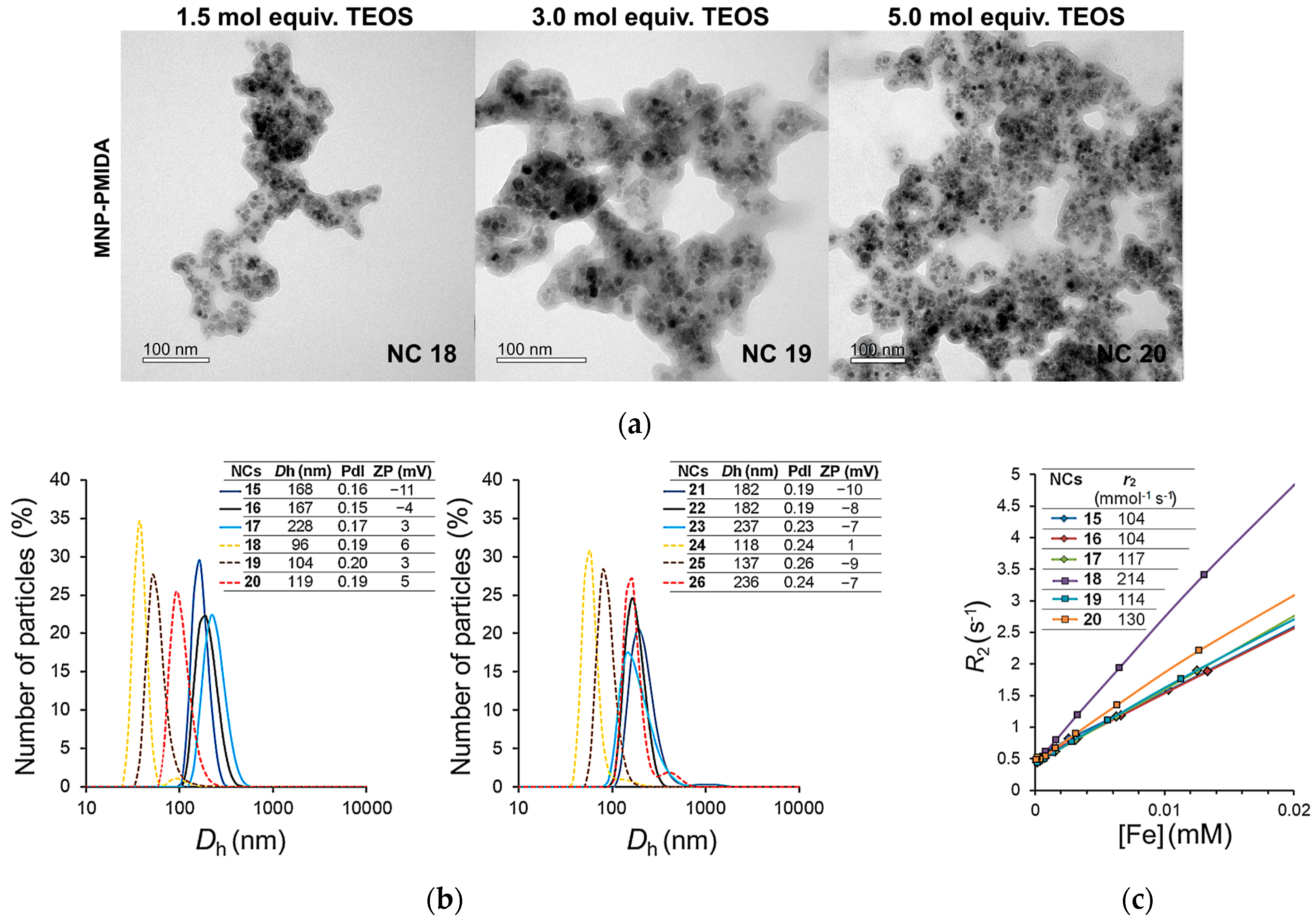
Based on the data of EDX and especially FTIR spectroscopy, it was shown that during silanization of MNPs with TEOS, the presence of PMIDA promotes an increase in the relative content of SiO2 phase in composites 911 compared with materials 35 obtained under similar conditions, but without PMIDA (Table 1), and, accordingly, determines their morphology. MNPs obtained without PMIDA did not have a common pronounced shell. Thus, according to TEM data, NCs 3 and 4 were MNPs (10 nm) coated with a thin SiO2 shell 2–5 nm thick (Figure 1a) and combined into agglomerates with an average size of ~240 nm according to DLS data (Figure 1b). NCs 5 were chemically cross-linked agglomerates up to 200 nm in size (Figure 1a) (with an average hydrodynamic size of ~270 nm (Figure 1b)).
When starting from MNPs-PMIDA 2, NCs 68 were formed with a thick SiO2 shell (increasing with an increase in the amount of TEOS). NCs 68 contained several MNP cores (10 nm) united by a pronounced SiO2 shell. The average sizes of NCs 68 were 26–50 nm (117, 180, and 234 nm, respectively, according to DLS data (Figure 1b)) with a common shell of a thickness of 3.5–5.5 nm (Figure 1a). The synthesized NCs differed in their structure from the core-shell MNPs previously synthesized by our group using PMIDA [12,53]. This can be explained by a new protocol for applying the SiO2 coating. Thus, the obtained MNPs 2 were not washed out from excess PMIDA after the stabilization procedure, as in [12,53], and were used in further modifications without additional treatment. The content of PMIDA in MNPs 2 was 0.97 mmol/g, which was ~1.5 times higher than in the samples with a PMIDA monolayer (0.61–0.67 mmol/g) [53,55,56]. In addition, unbound acid was present in the reaction mixture during silanization and probably catalyzed the hydrolysis of TEOS alkoxysilane groups both on the particle surface and throughout the entire volume. As a result, clusters of individual MNPs with an already formed thin SiO2 coating were subjected to additional silanization by partially hydrolyzed TEOS from the bulk of the reaction mixture to form a common coating, the thickness of which increased with increasing amount of silane (Figure 1a). It is interesting to note that with an increase in the SiO2 proportion in NCs, the degree of their subsequent APS functionalization decreased (Table 1).
It has been previously shown that during silanization, PMIDA molecules are displaced by silane molecules [12,53]. The degree of displacement was determined by the proportion of SiO2 in NCs. As a result, after deposition of the SiO2 shell, from 65 to 32% of the amount of PMIDA in the initial MNPs remained in the NC materials, and after amination, from 27 to 17% remained (Table 1). The ability of PMIDA to bind to the SiO2 surface was demonstrated in [79]. In general, in aqueous solutions, the synthesized NCs obtained using PMIDA had smaller hydrodynamic sizes (Figure 1b), which is more preferable for their medical applications.
The efficiency of nanoparticle silanization was evaluated by FTIR spectroscopy [80], which is often used to study both the features of the molecular structure and to qualitatively and quantitatively assess the composition of nanocomposite materials [81,82,83]. Thus, according to UATR-FTIR spectroscopy, it was possible to semi-quantitatively estimate an increase in the proportion of SiO2 in the NC composition depending on the increase in the amount of TEOS or PMIDA [12]. The most characteristic bands for SiO2 materials are bands in the region of 1300–750 cm−1 (caused mainly by νas Si–O–Si (~1070 cm−1)), ν Si–OH (~950 cm−1) and νs Si–O–Si (~789 cm−1)); and for iron oxides, a band in the region of 750–500 cm−1 (due to ν Fe–O at ~ 550 cm−1). From the ratio of areas under the lines of the absorption bands of Si–O and Fe–O bonds in the IR spectra (SSi–O/SFe–O), one can estimate the relative content of SiO2 in NCs by analogy with [82] (Supplementary Materials, Figure S1). Thus, with an increase in the amount of TEOS used for preparation of NCs 38, the intensity of the characteristic Si–O bands increased significantly; the SSi–O/SFe–O values also increased (Table 1, Figure 2).
The UATR-FTIR spectral data correlated with EDX data. It should be noted that with an increase in the proportion of SiO2, the number of Si–OH groups increased, as evidenced by an increase in the intensity of the absorption band in the region of 977–949 cm−1. A decrease in the amount of PMIDA in NCs 68 and 1214 could also be observed by a decrease in the intensity of the absorption bands at 1436 and 1402 cm−1, which belonged to the symmetric stretching vibrations of the PMIDA COO group. It is worth noting their shift relative to the bands at 1417 and 1375 cm−1 in the spectrum of the initial MNPs 2, which can be explained by the fact that, in one case, the PMIDA COO group bound to the surface Fe–OH and in the other case, to Si–OH. The bands related to the asymmetric vibrations of the COO and PO32− groups of PMIDA [55,56] were superimposed on the bands associated with the vibrations of crystallization water (1615 cm−1) and Si–O–Si (1070 cm−1) and were difficult to identify.
The obtained materials had high saturation magnetization (MS), low remanence magnetization (MR) (up to 0.5 emu/g) and coercive force (HC) (up to 3 Oe) (Supplementary Materials, Figure S2). The magnetization of NCs decreased in accordance with the content of the nonmagnetic SiO2 phase in them.

3.2. Synthesis of Dox-Containing Nanocomposite Materials

3.2.1. Study of Dox Sorption/Desorption

Noncovalent immobilization of antitumor agents on nanoparticles can be carried out due to electrostatic and van der Waals interactions, hydrogen bonding, and π-π stacking of the Dox anthracycline fragment with aromatic fragments of the coating material. Therefore, the immobilization efficiency and level are determined by a number of factors, such as particle morphology, surface porosity, charge state or the presence of functional groups on the surface, pH of a solution, and nanoparticle-to-agent ratio.
The Dox molecule (pKa = 8.2) is positively charged in aqueous solutions with neutral pH; therefore, it can be adsorbed on the surface due to electrostatic interactions with negatively charged functional groups (for example, silanol ones in the case of materials with a SiO2-based surface). Thus, it has been demonstrated that Dox sorption proceeds better at pH = 7.4 compared to lower pH values [19]. The presence of negatively charged groups on the surface of the material promotes the Dox sorption, which has been experimentally demonstrated in a number of works. Nevertheless, an increase in the efficiency and level of Dox loading on hollow aminated (with different degrees of amination) mesoporous SiO2 nanoparticles was shown to be increased in comparison with non-aminated ones [34]. Therefore, the question of the effect of the amino groups on the nanomaterial surface on the efficiency of Dox sorption remains open.
It should be noted that in this work, we used NCs 314 with a negatively charged surface. Due to the presence of an excess amount of silanol groups on the surface, the aminated NCs 914 also had a negative charge (Figure 1b).
The increased Dox-to-nanoparticle ratio leads to an increase in the level of Dox loading, but to a decrease in its efficiency [9,15,19,47,50]. The option when the maximum loading capacity (LC) is reached at the maximum possible loading efficiency (LE) can be considered as the optimal one [15,47]. In this work, we studied the sorption of Dox on all types of synthesized NCs 314 (Scheme 1). Immobilization was carried out in an aqueous solution with a NCs–Dox mass ratio of 1:1 at room temperature. After Dox sorption on NCs 314, the reaction mixtures were centrifuged, and the resulting precipitates were washed with water, dried, and analyzed. The supernatants were analyzed by UV and fluorescence spectrometry.
The Dox content in the powders of NCs 1526 was qualitatively assessed by UATR-FTIR spectroscopy (Figure 3). In the spectra of NCs 1820, which were obtained based on PMIDA-modified MNPs with a SiO2 shell, there were well-defined absorption bands characteristic of Dox: 1720 cm−1 (ν C = O of the side chain), 1617 cm−1 (ν C = O of anthracene fragment), 1580 cm−1 (ν NH), and a group of bands at 1470, 1446, and 1413 cm−1 (ν C–H). In the spectra of NCs 1517, these bands were much less pronounced, which indicated a lower content of Dox in them. Based on the even weaker prominence of these bands in the spectra of NCs 2126 containing APS on the surface, it can be concluded that the Dox loading capacity was much lower than in the corresponding NCs 1517 and 1820.
The presence of Dox in the composition of NCs was also confirmed by the appearance of an absorption maximum at 490 nm in the UV-Vis spectra and an emission maximum at 590 nm in the fluorescence spectra (see the Supplementary Materials; Figure S1a shows the UV-Vis and fluorescence spectra of NCs 15 and 18 as examples). A significant decrease in fluorescence (fluorescence quenching) is probably associated with the interaction of the Dox molecule with the NC surface.
After sorption, the sizes of NCs were not changed according to TEM data. Figure 4a shows TEM images of NCs 1820 with the highest Dox content as examples.
The main phase of the cores of the described NCs corresponds to magnetite, and it is not changed during modification, which was confirmed by the electron diffraction pattern of NC 18 (JCPDS Card No. (79-0417)) as an example (Supplementary Materials, Figure S3a). The mean core size in the synthesized NCs, which was determined from TEM data, was 8.8 nm (Supplementary Materials, Figure S3b). The core size histogram was constructed using the Sturges’ method by analogy with [84].
The hydrodynamic diameter of particles with the SiO2 shell (NCs 1520) somewhat decreased, while that of NCs with the aminated SiO2 shell (NCs 2126) actually remained the same (Figure 4b). Aqueous colloidal solutions of NCs obtained using PMIDA had the highest stability. Due to the positive charge of Dox molecules, the negative surface charge of NCs is partially or completely compensated during Dox sorption on them. As a result, the ζ-potential of NCs 1520 becomes weakly negative or even positive (Figure 4b).
Relaxivity (r2) (Figure 4c) is a key parameter that makes it possible to judge the possibility of using materials as T2 contrast agents in MRI investigations. We measured the relaxivity r2 of colloidal solutions of NCs 1520 with a SiO2 shell (Figure 5c), which are more stable than their aminated analogues and have a high Dox loading. The studied NCs had the r2 values comparable to those of the FDA-approved commercial agents: Sinerem® (Dh ~ 50 nm, r2 = 60 mmol−1 s−1), Feridex® (Dh ~ 200 nm, r2 = 107 mmol−1 s−1), and Resovist® (Dh ~ 60 nm, r2 = 190 mmol−1 s−1) [85]. Among the materials studied by us, NC 18 with the smallest diameter had twice the r2 value, which is comparable with the parameters of previously synthesized MNPs used by us as contrast-enhancing MRI agents [12,13,55]. This indicates the possibility of MRI evaluation of the biodistribution and accumulation of the obtained NCs in in vivo experiments.

3.2.2. Quantitative Estimation of Dox Content

To quantify the content of antitumor agents (in particular, Dox) in the active pharmaceutical substances, liquid chromatography with UV-Vis, fluorescent, mass-spectrometric and electrochemical detection techniques, as well as UV-Vis and fluorescence spectrometry are most often used [86,87]. In the development of drug delivery systems, the Dox content is determined indirectly by UV-Vis spectrometry [28,30,36,47,48,49], fluorescence spectrometry [45,51,52], and high-performance liquid chromatography (HPLC) [15]. The use of the direct method for the quantitative determination of Dox in NCs by the above methods does not seem to be correct, since there is a significant change in the optical properties of Dox when it is in contact with any material. Thus, Dox molecules can interact with surface groups (for example, Si–OH in the SiO2-based materials) due to electrostatic interactions with charge transfer, and also form molecular aggregates. As a result, the optical properties (molecular extinction coefficient and fluorescence quantum yield) of adsorbed Dox are changed [88,89,90,91], which will lead to changes in the absorption and fluorescence spectra and distortion of the results of calculations of the amount of Dox on the surface of materials. In this regard, there is a need for the careful development of methods for determination of Dox in filtrates after sorption on any nanomaterials, which, in our opinion, is not given enough attention in the literature. Therefore, a separate task of the work was to study the features of using UV and fluorescence spectrometry for the analysis of Dox in synthesized materials.
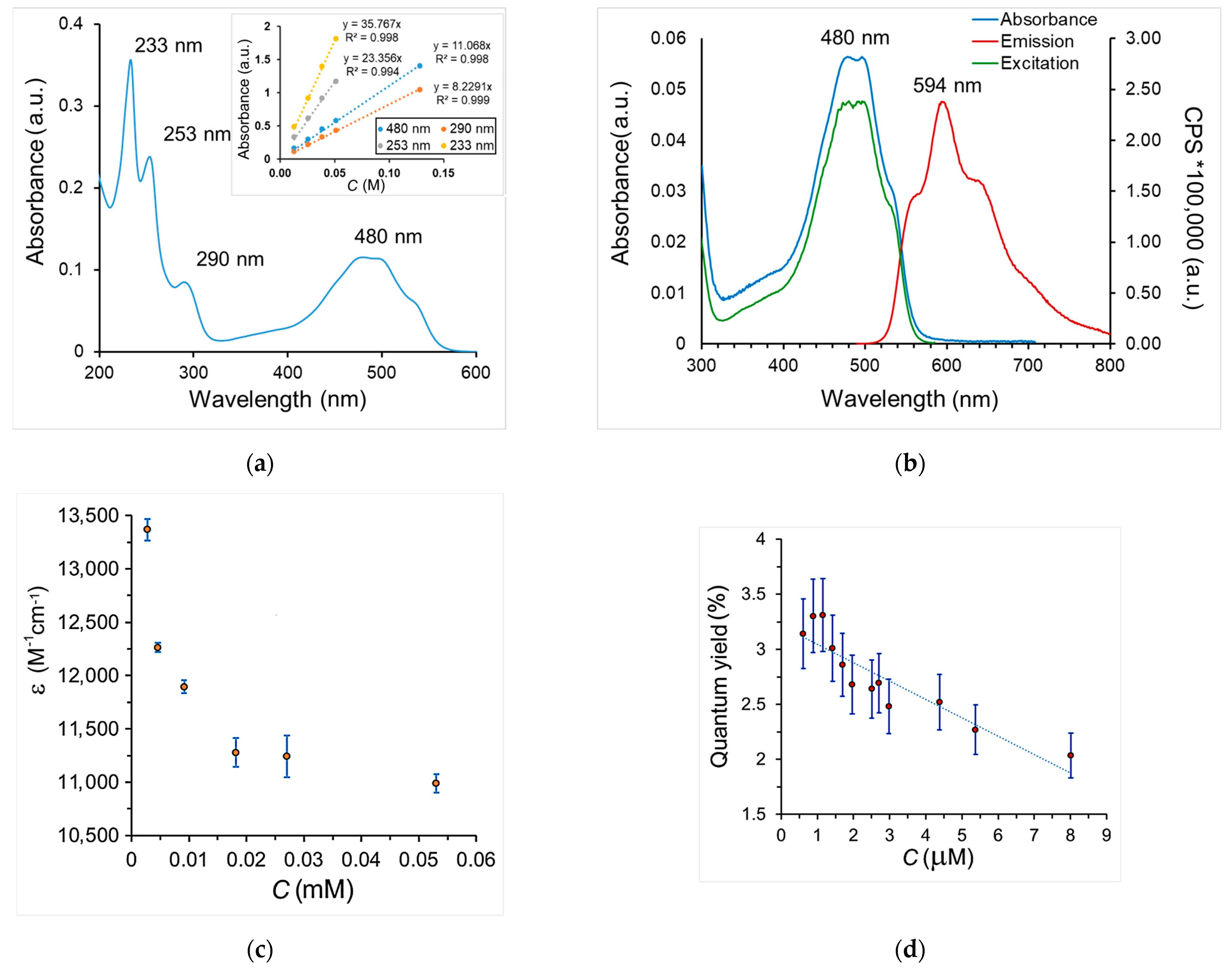
To quantify the Dox content in the samples, we used UV and fluorescence spectrometry. Several maxima can be distinguished in the UV spectra of a Dox solution (Figure 5a). For analysis by UV spectrometry, a maximum at 480 nm corresponding to the π→π* transition in the anthracycline Dox fragment was chosen, since there is no absorption of other components of the nanocomposite in this region of the spectrum. In the fluorescence spectra of Dox, there is only one pronounced maximum at 594 nm (Figure 5b) (the emission and absorption spectra with the main maximums at 480 nm coincided in shape). It should be noted that a significant contribution to the analysis error is known to be made by the Dox sorption on the surface of PTFE, glass (or quartz) cuvettes [92], and laboratory glassware, as well as on the material of microfiltration filters and dialysis membranes used to isolate samples (filtrates) from the reaction mixtures. Thus, we failed to clean quartz cuvettes from Dox after analysis, despite repeated washings with water. Therefore, sorption was performed in polypropylene test tubes, and the analysis of aqueous Dox solutions was carried out in acrylic cuvettes (fluorescence spectrometry showed no traces of Dox on the walls of the cuvettes after removal of the analyzed solutions and a single washing with water).
When quantifying the Dox content by UV spectrometry, at first we experimentally determined the molecular extinction coefficient (ε) of Dox. To do this, the calibration curves of the optical density vs. the Dox concentration were plotted in the range of absorbance (A) of 0.2–1.0 a.u. (see Supplementary Materials, Figures S4b,c). At a concentration below 0.011 mM, the shoulder at 480 nm increases and the contribution of the band at 493 nm decreases (Figure S4d). In most cases, researchers rely on the values ε = 10,067–11,500 M−1 cm−1 [93,94,95].
However, it is known that Dox in aqueous solutions exists in the monomolecular state and as molecular dimers, which are characterized by different values of ε and luminescence/fluorescence lifetime (1 ns, about 0.2 ns, and about 0.4 fs, respectively) [91]. It has been shown that if in highly diluted solutions Dox is present predominantly in the monomeric form, then already at a concentration of 38 µM, its content is 35%. Therefore, the calculation of the Dox concentration in the studied solutions from the average value ε = 10,850 M−1 cm−1 can lead to a significant error, especially in the case of a low Dox concentration. Thus, a nonlinear nature of the change in ε was observed (Figure 5c). Depending on the Dox concentration, we observed a change in the shape of the UV-Vis absorption spectrum (Supplementary Materials, Figure S4d), which can also be explained by the redistribution of the contributions of the monomolecular form or dimers. In this work, to calculate the Dox concentration, we used the arithmetic mean values of ε for three concentration ranges: ε = 12,900, ε = 12,000, and ε = 11,500 M−1 cm−1 for solutions with A < 0.1, 0.5 > A > 0.1, and A > 0.5 a.u., respectively.
When analyzing samples with a Dox concentration of less than 0.01 mM (A < 0.06 a.u.), the error increases significantly, so the determination of the Dox concentration in solutions by UV spectrometry may be incorrect. For example, when studying the process of Dox desorption from synthesized NCs, it becomes relevant to use fluorescence spectrometry to determine the Dox concentration, since that makes it possible to work with solutions containing 0.001–0.01 mM Dox.
To determine the Dox concentration in solutions by fluorescence spectrometry, a calibration plot of fluorescence intensity at 594 nm vs. Dox concentration (for studied solutions, A < 0.1 a.u.) was built (Supplementary Materials, Figure S5). As we have shown, the dependence of the luminescence intensity (as well as that of UV absorbance A) on the Dox concentration is not linear. The change in the optical properties is associated with different ratios of the Dox monomolecular form and molecular dimers of two types in solutions. The formation of dimers occurs due to the stacking of Dox aglycone fragments in parallel or antiparallel orientation, which are characterized by different fluorescence lifetimes: 1 ns, about 0.2 ns, and about 0.4 fs, respectively. The ratio of monomolecular and dimer forms is determined by the Dox concentration [87,91]. It should be noted that registration of fluorescence with a lifetime of less than 0.1 ns requires special instrumental capabilities. Thus, the flash xenon lamp, which most devices are equipped with, does not allow recording such signals; therefore, it can be expected that the total fluorescence intensity of the analyzed Dox solutions will be underestimated, which, in turn, will introduce an error in the concentration estimate.

3.2.3. Quantitative Assessment of the Dox Content in NCs after Sorption/Desorption

Sorption. Dox loading efficiency (LE, wt.%) and loading capacity (LC, wt.%) (Figure 6a) were calculated based on the UV spectrometry data using Formulas (1) and (2), respectively. It was demonstrated that the initial MNPs 1 and 2 adsorbed Dox within 24 h with LE 7% and 23%, respectively (Supplementary Materials, Figure S6). The SiO2-coated NCs adsorbed Dox much more efficiently than their APS-modified analogues, with LC differing by a factor of 1.5–3.0. It should be noted that the Dox loading efficiency for materials 1820 and 2426 obtained using PMIDA was higher by a factor of 1.4–6.0 compared to NCs 1517 and 2123. Increasing the thickness of the SiO2 shell leads to an increase in Dox LC (Figure 6a). The obtained data are consistent with the IR spectroscopy data (Figure 3). The LC value (28%) for NCs 19, 20 exceeded the loading capacity of some materials described in the literature, including those based on silica-coated MNPs [26,28,35,36] or mesoporous SiO2 particles [8,9,34] (LC up to 26%).
Desorption. We studied the Dox desorption from NCs 1526. For this, colloidal solutions of NCs 1526 in phosphate buffer solutions (pH = 5.8 and 7.4) were kept at 37 °C for 24 h. After that, the reaction mixtures were centrifuged, and supernatants were analyzed by fluorescence spectrometry. The pH-dependent desorption of Dox from the synthesized materials was demonstrated (Figure 6b), which can be explained by the fact that the protonation of silanol groups and the hydrophobization of the SiO2 surface occur at low pH. In this case, the Dox amino groups are protonated (pKa = 8.2), and noncovalent bonds with the surface atoms of NCs are broken. It is interesting to note that for NCs 1517 and 2123 synthesized without PMIDA, the amount of desorbed Dox (as a percentage of the Dox amount loaded onto NCs) was greater than for PMIDA-modified analogues 1820 and 2426. This can be explained by the fact that in NCs 1820 and 2426, Dox is retained due to stronger bonding with PMIDA molecules on the particle surface. On the contrary, the presence of APS molecules in NCs promotes the desorption process (for example, for NCs 22 and 23). It is interesting that in the case of NCs 2426 containing both PMIDA and APS molecules, desorption is lowest, and the difference in the Dox desorption at pH 5.8 and 7.4 levels out. It can be assumed that the binding of Dox in such NCs is largely due to the strong interaction with the phosphonic and/or carboxyl groups of PMIDA. This is consistent with DFT calculations of the binding of the Dox molecule into complexes with the surface functional groups of the synthesized NCs (see Section 3.2.4).
Thus, it has been shown that the presence of PMIDA in NCs makes it possible to increase the Dox loading, as well as to reduce its desorption rate, which may be useful in the design of drug delivery vehicles with a prolonged action. So, slow release can determine the reduced toxicity of the agent when administered intravenously. The drug can begin to act after accumulation in target tissues due to destruction in tumor cell lysosomes, or drug release can be activated by exposure to an alternating magnetic field (by analogy with [27]).

3.2.4. DFT Calculations of the Total Electronic Energies of Dox Complexes with a Small SiO2 Cluster and Its Conjugates with APS and PMIDA

To assess the possibility of specific binding of the Dox-base molecule to the surface groups of synthesized NCs, we performed the density functional theory (DFT) simulation of its complexes with small SiO2 clusters (which were described in [96] as imitating SiO2 nanoparticles), including their conjugates with one (SiO2-APS) or two APS molecules (SiO2-2APS), as well as with the adsorbed PMIDA molecule (SiO2-PMIDA) (Figure 7; see Supplementary Materials). In our study, a small SiO2 cluster imitated the outer layer of synthesized nanocomposites. The binding energies (Eb) of Dox with these clusters were calculated at the B3LYP-D3-gCP/def2-SVP level of theory in gas phase and in water using ORCA 4.0.1 software [57] (Supplementary Materials, Tables S1 and S2).
Calculations have shown that the Eb value (–224.88 kJ/mol) for the Dox molecule in the SiO2-PMIDA…Dox salt complex is less than that of the most energy favorable SiO2…Dox complex (Figure 7d).
This may indicate the strongest binding of Dox to the surface of NCs containing SiO2 and PMIDA fragments. Dox coordination in the SiO2-PMIDA…Dox complex (Figure 7d) occurs due to the formation of various bonds: an ionic bond between the Dox amino group and the PMIDA phosphonic group; two hydrogen bonds between C(8)–O(H) and C(8)–COCH2–O(H) of the Dox molecule and the PMIDA COOH groups; and a hydrogen bond between OH in the Dox pyranosyl group and Si-OH (H-bonded oxygen and hydrogen atoms are highlighted in white). At the same time, the energy of Dox binding into the complex in which PMIDA (see Supplementary Materials, PMIDA-Dox salt A) is higher than Eb calculated for the SiO2-PMIDA…Dox salt (see Supplementary Materials, SiO2-PMIDA-Dox salt 2). Thus, it can be said that the simultaneous coordination of Dox with the PMIDA molecule and the silanol groups results in a synergistic effect of decreasing the binding energy of Dox with the SiO2-PMIDA cluster in comparison with the binding energies of Dox with the SiO2 cluster. Such synergy arises from the fact that Dox is attached to the conjugates not only through ionic and H-bonds, but also through the dispersion and dipole–dipole interactions with PMIDA functional groups, which is clearly seen in the NCI D3 plots (Figure 8, right).
Thus, the Eb value for SiO2-APS…Dox complexes (regardless of the number of APS molecules in them) is higher than for SiO2…Dox complexes, and even more so for SiO2-PMIDA…Dox. This can explain the fact that the Dox molecule is adsorbed to a lesser extent on NCs containing APS fragments than on NCs with a SiO2 or SiO2-PMIDA shell.
The calculated data on the energy of Dox binding (Figure 7 and Figure 8) to the functional groups of clusters that mimic the surface of the synthesized NCs are in good agreement with the experimental data on the Dox sorption on these NCs (Figure 6a).
Thus, the lowest sorption was observed in the case of APS-modified NCs 2123 obtained without PMIDA (LC 5–10%), which corresponds to the highest Eb value of Dox complexes with SiO2–APS cluster, Eb = –73.09 kJ/mol for SiO2-APS…Dox complex. Mean values of sorption (LC 20.5%) were achieved for NCs 1517, Eb = –186.41 kJ/mol for the SiO2…Dox complex, while the highest sorption (LC up to 28%) was achieved for NCs 1820 with the lowest value of Eb = –224.88 kJ/mol for the SiO2–PMIDA…Dox complex, which is due to the combination of the most energy favorable binding of Dox to the phosphonic or carboxyl groups of PMIDA.

3.3. Cytotoxicity Assessment

We carried out a comparative study of the cytotoxicity of synthesized NCs 1526 and initial NCs 314 towards HEK-293 cells using the MTT assay. Figure S7a shows cell survival data as a function of Dox concentration (0.1–0.005 mg/mL) calculated from its loading capacity (LC) in the studied NCs. It has been shown that, although according to Figure 7b, PMIDA-containing NCs 1820 exhibited the least desorption of Dox within 24 h, their cytotoxic effect was comparable to the initial Dox at a concentration of 0.1 mg/mL and turned out to be the highest among synthesized NCs. Their PMIDA-free analogues (NCs 1517) or APS-modified NCs 2426 exhibited markedly lower cytotoxicity at Dox concentrations < 0.1 mg/mL. The aminated NCs 2123, which did not contain PMIDA, as well as the starting NCs 314 (see Supplementary Materials, Figure S7b), did not exhibit significant toxicity. NCs actively interacted with cells (penetrated into them or were adsorbed on the surface), which is demonstrated in Figures S8 and S9.
As a result, it has been shown that the cytotoxic effect of NCs corresponds to their calculated IC50 values in the framework of the Hill model by analogy with [97]. The IC50 values of NCs 1820 and free Dox are comparable (Table 2, Figure S10). Based on the data obtained, it can be concluded that the cytotoxicity of the studied NCs is determined mainly by the total Dox content in them (Figure 6a), and not by the magnitude of desorption in a model experiment conducted for 24 h at different pH (Figure 6b).
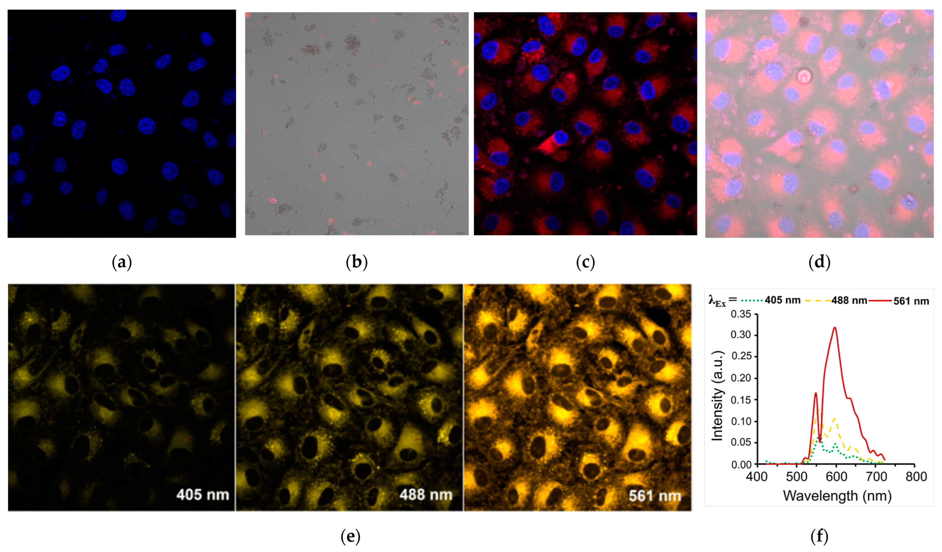
Using confocal microscopy, we demonstrated the possibility of Dox desorption and its accumulation inside cells by the example of cell incubation with NC 18 (Figure 9).
For better cell visualization, cell nuclei were stained with Hoechst 33,342 (blue) (Figure 9a). NC 18 under confocal microscopy had a weak fluorescence (Figure 9b), which is consistent with the data of fluorescence spectrometry (Figure S5b). However, after the cells were incubated with NC 18 for 24 h, we observed additional staining of the cells due to the accumulation of Dox (red) desorbed from the nanoparticles. To obtain an image with maximum detail, the channel mode of a confocal microscope was used, with excitation at 405 (for nuclei stained by Hoechst dye) and 561 nm (for Dox) (Figure 9c,d).
To obtain an informative fluorescent image in ZEN software (2009 (Version 6.0), Carl Zeiss, Oberkochen, Germany), a special lambda mode was used, which allowed determining the emission range with the maximum contrast for this material (Figure 9e). The fluorescence spectra of the investigated nanomaterials in the cells were extracted from the images obtained in the lambda mode (Figure 9f). They corresponded to spectra of Dox at λex = 561 nm (Figure 5b).

4. Conclusions

In summary, a series of new composite materials based on Fe3O4 magnetic nanoparticles (MNPs) coated with SiO2 or aminated SiO2 were synthesized, including the materials obtained using N-(phosphonomethyl)iminodiacetic acid (PMIDA). For the first time, the role of PMIDA in the formation of the SiO2 coating and in the process of the doxorubicin sorption/desorption was revealed. It has been shown that the use of PMIDA to stabilize nanoparticles before silanization promotes an increase in the relative content of the SiO2 phase in Fe3O4@SiO2 nanocomposites compared to materials obtained under similar conditions, but without PMIDA. The conditions of doxorubicin (Dox) immobilization on the synthesized materials were studied by UV and fluorescence spectrometry. It has been shown that the first of these methods is most suitable for studying the process of Dox sorption; the second, for the study of Dox desorption. It has been demonstrated for the first time that the presence of PMIDA on the surface of nanocomposites increases the level of Dox loading (up to 28 mg per 1 mg of MNPs) due to specific binding, while modification of their surface with 3-aminopropylsilane (APS), on the contrary, significantly reduces the sorption capacity of materials (up to 5 mg per 1 mg of MNPs). These regularities were in accordance with the results of quantum chemical calculations. It has been shown that the energies of Dox binding to the functional groups of NCs are in good agreement with the experimental data on the sorption of Dox on these NCs. The mechanisms of Dox binding to the surface of NCs were proposed: simultaneous coordination of Dox on the PMIDA molecule and silanol groups at the NC surface leads to a synergistic effect in Dox binding. Simultaneous coordination of Dox with the PMIDA molecule and silanol groups of the nanocomposite surface leads to a synergistic effect, and the 3-aminopropyl fragment of the APS-modified particles is almost not involved in Dox binding. Synthesized nanocomposites allow achieving a high level of Dox loading and a long pH-determined release. At the same time, the presence of PMIDA in NCs makes it possible to reduce the Dox desorption rate, which may be useful in the design of drug delivery vehicles with a prolonged action. The materials showed dose-dependent cytotoxicity in in vitro experiments on HEK-293 cells. The cytotoxic effects of NCs correspond to their calculated IC50 values in the framework of the Hill model. Nanocomposites with a SiO2 shell obtained using PMIDA had the greatest effect (their IC50 values corresponded to IC50 of the starting Dox). We believe that the data obtained can be further used to develop stimuli-responsive materials for targeted cancer chemotherapy.

Supplementary Materials

The following supporting information can be downloaded at: https://www.mdpi.com/article/10.3390/pharmaceutics14112271/s1, Figure S1: Calculation of areas of absorption bands related to vibrations of Si–O and Fe–O bonds in UATR-FTIR spectra of NC 6: (a) reflectance and (b) absorbance spectra; calculation of areas of absorption bands related to vibrations of (c) Si–O and (d) Fe–O bonds; Figure S2: Magnetization reversal curves for NCs 1, 35, 911 (left) and 2, 68, 1214 (right); Figure S3: (a) Electron diffraction pattern of NC 18 (corresponds to JCPDS Card No. (79-0417) Magnetite—synthetic). (b) The size distribution of Fe3O4 MNPs (core) in composition of NC 18 from TEM data; Figure S4: (a) Absorption and fluorescence spectra of colloidal solutions of NCs 15 and 18. (b) Determination of the molecular extinction coefficient of Dox at 480 nm, concentration from 2 × 10⁻⁶ to 7 × 10⁻⁵ M. (c) Fragment of the absorption spectra of analyzed Dox solutions in the range of 300–600 nm. (d) Normalized absorption spectra of Dox at various concentrations; Figure S5: (a) Calibration plot of fluorescence intensity at 594 nm vs. Dox concentration. (b) Fragment of the emission spectra of analyzed Dox solutions in the range of 500–800 nm; Figure S6: The efficiency of Dox sorption onto MNPs 1 and 2 for (a) 1 min and (b) 24 h; MNPs–Dox ratio 50:50 or 95:5. Dox loading efficiency (LE, %) was calculated from the UV-Vis and fluorescence spectrometry data; Table S1: Total energies E of the optimized complexes of Dox and corresponding sums of Dox and SiO2 conjugate energies; Table S2: Binding energies Eb for the optimized complexes of Dox; Figure S7. Viability of HEK-293 cells after 48 h incubation with (a) Dox-loaded NCs 1526 and (b) NCs 314 (MTT assay); Figure S8: Optical microscope images (×200) of HEK-293 cells incubated with NCs 1526 at concentrations of (a) 0.1, (b) 0.05, (c) 0.01, and (d) 0.005 mg/mL; Figure S9: Optical microscope images (×400) of HEK-293 cells incubated with NC 18 at concentrations of (a) 0.1, (b) 0.05, (c) 0.01, and (d) 0.005 mg/mL; Figure S10: IC50 curves.

Author Contributions

Conceptualization, A.M.D.; methodology, A.M.D.; investigation, A.M.D., A.V.V., M.S.V., M.A.K., M.A.U., A.S.M., V.A.P., I.V.B., A.A.T. and K.A.C.; writing—original draft preparation, A.M.D.; writing—review and editing, G.L.L. and V.P.K.; visualization, A.M.D.; supervision, V.P.K.; funding acquisition, V.N.C.; project administration, V.N.C. All authors have read and agreed to the published version of the manuscript.

Funding

This research was funded by the Ministry of Science and Higher Education of the Russian Federation, grant number 075-15-2020-777.

Institutional Review Board Statement

Not applicable.

Informed Consent Statement

Not applicable.

Data Availability Statement

Not applicable.

Acknowledgments

The equipment of the Centre for Joint Use “Spectroscopy and Analysis of Organic Compounds” at the Postovsky Institute of Organic Synthesis was used. Quantum chemical calculations were performed using the computing resources of the Centre for Joint Use “Supercomputer Centre” at the Krasovskii Institute of Mathematics and Mechanics, Russian Academy of Sciences (Ural Branch), Ekaterinburg, Russia. Biological experiments were performed using the equipment of the Shared Research Center of Scientific Equipment at the Institute of Immunology and Physiology, Russian Academy of Sciences (Ural Branch), Ekaterinburg, Russia.

Conflicts of Interest

The authors declare no conflict of interest.

Sample Availability

Not applicable.

References

  1. Sung, H.; Ferlay, J.; Siegel, R.L.; Laversanne, M.; Soerjomataram, I.; Jemal, A.; Bray, F. Global cancer statistics 2020: GLOBOCAN estimates of incidence and mortality worldwide for 36 cancers in 185 countries. CA Cancer J. Clin. 2021, 71, 209–249. [Google Scholar] [CrossRef] [PubMed]
  2. Taleghani, A.S.; Nakhjiri, A.T.; Khakzad, M.J.; Rezayat, S.M.; Ebrahimnejad, P.; Heydarinasab, A.; Akbarzadeh, A.; Marjani, A. Mesoporous silica nanoparticles as a versatile nanocarrier for cancer treatment: A review. J. Mol. Liq. 2021, 328, 115417. [Google Scholar] [CrossRef]
  3. Paris, J.L.; Vallet-Regí, M. Mesoporous silica nanoparticles for co-delivery of drugs and nucleic acids in oncology: A review. Pharmaceutics 2020, 12, 526. [Google Scholar] [CrossRef] [PubMed]
  4. Meng, H.; Xue, M.; Xia, T.; Ji, Z.; Tarn, D.Y.; Zink, J.I.; Nel, A.E. Use of Size and a Copolymer Design Feature to Improve the Biodistribution and the Enhanced Permeability and Retention Effect of Doxorubicin-Loaded Mesoporous Silica Nanoparticles in a Murine Xenograft Tumor Model. ACS Nano 2011, 5, 4131–4144. [Google Scholar] [CrossRef] [PubMed] [Green Version]
  5. Thorat, N.D.; Bauer, J.; Tofail, S.A.M.; Pérez, V.G.; Bohara, R.A.; Yadav, H.M. Silica nano supra-assembly for the targeted delivery of therapeutic cargo to overcome chemoresistance in cancer. Colloids Surf. B Biointerfaces 2020, 185, 110571. [Google Scholar] [CrossRef] [PubMed]
  6. Li, S.; Zhang, Y.; He, X.-W.; Li, W.-Y.; Zhang, Y.-K. Multifunctional mesoporous silica nanoplatform based on silicon nanoparticles for targeted two-photon-excited fluorescence imaging-guided chemo/photodynamic synergetic therapy in vitro. Talanta 2020, 209, 120552. [Google Scholar] [CrossRef] [PubMed]
  7. Wang, Y.; Yang, G.; Wang, Y.; Zhao, Y.; Jiang, H.; Han, Y.; Yang, P. Multiple imaging and excellent anticancer efficiency of an upconverting nanocarrier mediated by single near infrared light. Nanoscale 2017, 9, 4759–4769. [Google Scholar] [CrossRef]
  8. Zhang, Y.; Dang, M.; Tian, Y.; Zhu, Y.; Liu, W.; Tian, W.; Su, Y.; Ni, Q.; Xu, C.; Lu, N.; et al. Tumor acidic microenvironment targeted drug delivery based on pHLIP modified mesoporous organosilica nanoparticles. ACS Appl. Mater. Interfaces 2017, 9, 30543–30552. [Google Scholar] [CrossRef]
  9. Hu, L.-L.; Meng, J.; Zhang, D.-D.; Chen, M.-L.; Shu, Y.; Wang, J.-H. Functionalization of mesoporous organosilica nanocarrier for pH/glutathione dual-responsive drug delivery and imaging of cancer therapy process. Talanta 2018, 177, 203–211. [Google Scholar] [CrossRef]
  10. Zhang, P.; Kong, J. Doxorubicin-tethered fluorescent silica nanoparticles for pH-responsive anticancer drug delivery. Talanta 2015, 134, 501–507. [Google Scholar] [CrossRef]
  11. Zhang, W.; Liu, L.; Chen, H.; Hu, K.; Delahunty, I.; Gao, S.; Xie, J. Surface impact on nanoparticle-based magnetic resonance imaging contrast agents. Theranostics 2018, 8, 2521–2548. [Google Scholar] [CrossRef] [PubMed]
  12. Demin, A.M.; Pershina, A.G.; Minin, A.S.; Brikunova, O.Y.; Murzakaev, A.M.; Perekucha, N.A.; Romashchenko, A.V.; Shevelev, O.B.; Uimin, M.A.; Byzov, I.V.; et al. Smart design of a pH-responsive system based on pHLIP-modified magnetite nanoparticles for tumor MRI. ACS Appl. Mater. Interfaces 2021, 13, 36800–36815. [Google Scholar] [CrossRef] [PubMed]
  13. Pershina, A.G.; Brikunova, O.Y.; Demin, A.M.; Abakumov, M.A.; Vaneev, A.N.; Naumenko, V.A.; Erofeev, A.S.; Gorelkin, P.V.; Nizamov, T.R.; Muslimov, A.R.; et al. Variation in tumor pH affects pH-triggered delivery of peptide-modified magnetic nanoparticles. Nanomed. Nanotechnol. Biol. Med. 2021, 32, 102317. [Google Scholar] [CrossRef] [PubMed]
  14. Liu, D.; Li, J.; Wang, C.; An, L.; Lin, J.; Tian, Q.; Yang, S. Ultrasmall Fe@Fe3O4 nanoparticles as T1T2 dual-mode MRI contrast agents for targeted tumor imaging. Nanomed. Nanotechnol. Biol. Med. 2021, 32, 102335. [Google Scholar] [CrossRef] [PubMed]
  15. Liang, P.-C.; Chen, Y.-C.; Chiang, C.-F.; Mo, L.-R.; Wei, S.-Y.; Hsieh, W.-Y.; Lin, W.-L. Doxorubicin-modified magnetic nanoparticles as a drug delivery system for magnetic resonance imaging-monitoring magnet-enhancing tumor chemotherapy. Int. J. Nanomed. 2016, 11, 2021–2037. [Google Scholar] [CrossRef] [Green Version]
  16. Bulte, J.W.M. Superparamagnetic iron oxides as MPI tracers: A primer and review of early applications. Adv. Drug Deliv. Rev. 2019, 138, 293–301. [Google Scholar] [CrossRef]
  17. Liu, X.; Zhang, Y.; Wang, Y.; Zhu, W.; Li, G.; Ma, X.; Zhang, Y.; Chen, S.; Tiwari, S.; Shi, K.; et al. Comprehensive understanding of magnetic hyperthermia for improving antitumor therapeutic efficacy. Theranostics 2020, 10, 3793–3815. [Google Scholar] [CrossRef]
  18. Vilas-Boas, V.; Carvalho, F.; Espiña, B. Magnetic hyperthermia for cancer treatment: Main parameters affecting the outcome of in vitro and in vivo studies. Molecules 2020, 25, 2874. [Google Scholar] [CrossRef]
  19. Dai, Z.; Wen, W.; Guo, Z.; Song, X.-Z.; Zheng, K.; Xu, X.; Qi, X.; Tan, Z. SiO2-coated magnetic nano-Fe3O4 photosensitizer for synergistic tumour targeted chemo-photothermal therapy. Colloids Surf. B Biointerfaces 2020, 195, 111274. [Google Scholar] [CrossRef]
  20. Kolosnjaj-Tabi, J.; Kralj, S.; Griseti, E.; Nemec, S.; Wilhelm, C.; Sangnier, A.P.; Bellard, E.; Fourquaux, I.; Golzio, M.; Rols, M.-P. Magnetic silica-coated iron oxide nanochains as photothermal agents, disrupting the extracellular matrix, and eradicating cancer cells. Cancers 2019, 11, 2040. [Google Scholar] [CrossRef]
  21. Jabalera, Y.; Sola-Leyva, A.; Carrasco-Jiménez, M.P.; Iglesias, G.R.; Jimenez-Lopez, C. Synergistic photothermal-chemotherapy based on the use of biomimetic magnetic nanoparticles. Pharmaceutics 2021, 13, 625. [Google Scholar] [CrossRef] [PubMed]
  22. Novoselova, M.V.; German, S.V.; Abakumova, T.O.; Perevoschikov, S.V.; Sergeeva, O.V.; Nesterchuk, M.V.; Efimova, O.I.; Petrov, K.S.; Chernyshev, V.S.; Zatsepin, T.S.; et al. Multifunctional nanostructured drug delivery carriers for cancer therapy: Multimodal imaging and ultrasound-induced drug release. Colloids Surf. B Biointerfaces 2021, 200, 111576. [Google Scholar] [CrossRef] [PubMed]
  23. Guisasola, E.; Asín, L.; Beola, L.; de la Fuente, J.M.; Baeza, A.; Vallet-Regí, M. Beyond traditional hyperthermia: In vivo cancer treatment with magnetic-responsive mesoporous silica nanocarriers. ACS Appl. Mater. Interfaces 2018, 10, 12518–12525. [Google Scholar] [CrossRef] [PubMed] [Green Version]
  24. Moros, M.; Idiago-López, J.; Asín, L.; Moreno-Antolín, E.; Beola, L.; Grazú, V.; Fratila, R.M.; Gutiérrez, L.; de la Fuente, J.M. Triggering antitumoural drug release and gene expression by magnetic hyperthermia. Adv. Drug Deliv. Rev. 2019, 138, 326–343. [Google Scholar] [CrossRef] [PubMed]
  25. Kim, D.H.; Kim, D.W.; Jang, J.Y.; Lee, N.; Ko, Y.-J.; Lee, S.M.; Kim, H.J.; Na, K.; Son, S.U. Fe3O4@void@microporous organic polymer-based multifunctional drug delivery systems: Targeting, imaging, and magneto-thermal behaviors. ACS Appl. Mater. Interfaces 2020, 12, 37628–37636. [Google Scholar] [CrossRef]
  26. Zhu, Y.; Tao, C. DNA-capped Fe3O4/SiO2 magnetic mesoporous silica nanoparticles for potential controlled drug release and hyperthermia. RSC Adv. 2015, 5, 22365–22372. [Google Scholar] [CrossRef]
  27. Demin, A.M.; Vakhrushev, A.V.; Pershina, A.G.; Valova, M.S.; Efimova, L.V.; Syomchina, A.A.; Uimin, M.A.; Minin, A.S.; Levit, G.L.; Krasnov, V.P.; et al. Magnetic-responsive doxorubicin-containing materials based on Fe3O4 nanoparticles with a SiO2/PEG shell and study of their effects on cancer cell lines. Int. J. Mol. Sci. 2022, 23, 9093. [Google Scholar] [CrossRef]
  28. Javanbakht, S.; Shadi, M.; Mohammadian, R.; Shaabani, A.; Ghorbani, M.; Rabiee, G.; Amini, M.M. Preparation of Fe3O4@SiO2@Tannic acid double core-shell magnetic nanoparticles via the Ugi multicomponent reaction strategy as a pH-responsive co-delivery of doxorubicin and methotrexate. Mater. Chem. Phys. 2020, 247, 122857. [Google Scholar] [CrossRef]
  29. Zaaeri, F.; Khoobi, M.; Rouini, M.; Javar, H.A. pH-responsive polymer in a core–shell magnetic structure as an efficient carrier for delivery of doxorubicin to tumor cells. Int. J. Polym. Mater. Polym. Biomater. 2018, 67, 967–977. [Google Scholar] [CrossRef]
  30. Hervault, A.; Dunn, A.E.; Lim, M.; Boyer, C.; Mott, D.; Maenosono, S.; Thanh, N.T.K. Doxorubicin loaded dual pH- and thermoresponsive magnetic nanocarrier for combined magnetic hyperthermia and targeted controlled drug delivery applications. Nanoscale 2016, 8, 12152–12161. [Google Scholar] [CrossRef]
  31. Chen, F.H.; Gao, Q.; Ni, J.Z. The grafting and release behavior of doxorubincin from Fe3O4@SiO2 core–shell structure nanoparticles via an acid cleaving amide bond: The potential for magnetic targeting drug delivery. Nanotechnology 2008, 19, 165103. [Google Scholar] [CrossRef] [PubMed]
  32. Chen, F.-H.; Zhang, L.-M.; Chen, Q.-T.; Zhang, Y.; Zhang, Z.-J. Synthesis of a novel magnetic drug delivery system composed of doxorubicin-conjugated Fe3O4 nanoparticle cores and a PEG-functionalized porous silica shell. Chem. Commun. 2010, 46, 8633–8635. [Google Scholar] [CrossRef] [PubMed] [Green Version]
  33. Yoon, H.-M.; Kang, M.-S.; Choi, G.-E.; Kim, Y.-J.; Bae, C.-H.; Yu, Y.-B.; Jeong, Y.-I. Stimuli-responsive drug delivery of doxorubicin using magnetic nanoparticle conjugated poly(ethylene glycol)-g-chitosan copolymer. Int. J. Mol. Sci. 2021, 22, 13169. [Google Scholar] [CrossRef] [PubMed]
  34. Nguyen, T.N.T.; Le, N.T.T.; Nguyen, N.H.; Ly, B.T.K.; Nguyen, T.D.; Nguyen, D.H. Aminated hollow mesoporous silica nanoparticles as an enhanced loading and sustained releasing carrier for doxorubicin delivery. Microporous Mesoporous Mater. 2020, 309, 110543. [Google Scholar] [CrossRef]
  35. Demin, A.M.; Vakhrushev, A.V.; Valova, M.S.; Minin, A.S.; Kuznetsov, D.K.; Uimin, M.A.; Shur, V.Y.; Krasnov, V.P.; Charushin, V.N. Design of SiO2/aminopropylsilane-modified magnetic Fe3O4 nanoparticles for doxorubicin immobilization. Russ. Chem. Bull. 2021, 70, 987–994. [Google Scholar] [CrossRef]
  36. Sadighian, S.; Rostamizadeh, K.; Hosseini-Monfared, H.; Hamidi, M. Doxorubicin-conjugated core–shell magnetite nanoparticles as dual-targeting carriers for anticancer drug delivery. Colloids Surf. B Biointerfaces 2014, 117, 406–413. [Google Scholar] [CrossRef]
  37. Su, X.; Chan, C.; Shi, J.; Tsang, M.-K.; Pan, Y.; Cheng, C.; Gerile, O.; Yang, M. A graphene quantum dot@Fe3O4@SiO2 based nanoprobe for drug delivery sensing and dual-modal fluorescence and MRI imaging in cancer cells. Biosens. Bioelectron. 2017, 92, 489–495. [Google Scholar] [CrossRef]
  38. Popescu, R.C.; Savu, D.; Dorobantu, I.; Vasile, B.S.; Hosser, H.; Boldeiu, A.; Temelie, M.; Straticiuc, M.; Iancu, D.A.; Andronescu, E.; et al. Efficient uptake and retention of iron oxide-based nanoparticles in HeLa cells leads to an effective intracellular delivery of doxorubicin. Sci. Rep. 2020, 10, 10530. [Google Scholar] [CrossRef]
  39. Meng, H.; Liong, M.; Xia, T.; Li, Z.; Ji, Z.; Zink, J.I.; Nel, A.E. Engineered design of mesoporous silica nanoparticles to deliver doxorubicin and P-glycoprotein siRNA to overcome drug resistance in a cancer cell line. ACS Nano 2010, 4, 4539–4550. [Google Scholar] [CrossRef]
  40. Semkina, A.; Abakumov, M.; Grinenko, N.; Abakumov, A.; Skorikov, A.; Mironova, E.; Davydova, G.; Majouga, A.G.; Nukolova, N.; Kabanov, A.; et al. Core–shell–corona doxorubicin-loaded superparamagnetic Fe3O4 nanoparticles for cancer theranostics. Colloids Surf. B Biointerfaces 2015, 136, 1073–1080. [Google Scholar] [CrossRef]
  41. Zarrin, A.; Sadighian, S.; Rostamizadeh, K.; Firuzi, O.; Hamidi, M.; Mohammadi-Samani, S.; Miri, R. Design, preparation, and in vitro characterization of a trimodally-targeted nanomagnetic onco-theranostic system for cancer diagnosis and therapy. Int. J. Pharm. 2016, 500, 62–76. [Google Scholar] [CrossRef] [PubMed]
  42. Liang, B.; Zuo, D.; Yu, K.; Cai, X.; Qiao, B.; Deng, R.; Yang, J.; Chu, L.; Deng, Z.; Zheng, Y.; et al. Multifunctional bone cement for synergistic magnetic hyperthermia ablation and chemotherapy of osteosarcoma. Mater. Sci. Eng. C 2020, 108, 110460. [Google Scholar] [CrossRef] [PubMed]
  43. Zhou, J.; Li, J.; Ding, X.; Liu, J.; Luo, Z.; Liu, Y.; Ran, Q.; Cai, K. Multifunctional Fe2O3@PPy-PEG nanocomposite for combination cancer therapy with MR imaging. Nanotechnology 2015, 26, 425101. [Google Scholar] [CrossRef] [PubMed]
  44. Jain, T.K.; Richey, J.; Strand, M.; Leslie-Pelecky, D.L.; Flask, C.A.; Labhasetwar, V. Magnetic nanoparticles with dual functional properties: Drug delivery and magnetic resonance imaging. Biomaterials 2008, 29, 4012–4021. [Google Scholar] [CrossRef] [PubMed] [Green Version]
  45. Mdlovu, N.V.; Mavuso, F.A.; Lin, K.-S.; Chang, T.-W.; Chen, Y.; Wang, S.S.-S.; Wu, C.-M.; Mdlovu, N.B.; Lin, Y.-S. Iron oxide-pluronic F127 polymer nanocomposites as carriers for a doxorubicin drug delivery system. Colloids Surf. A Physicochem. Eng. Asp. 2019, 562, 361–369. [Google Scholar] [CrossRef]
  46. Chen, J.; Shi, M.; Liu, P.; Ko, A.; Zhong, W.; Liao, W.; Xing, M.M.Q. Reducible polyamidoamine-magnetic iron oxide self-assembled nanoparticles for doxorubicin delivery. Biomaterials 2014, 35, 1240–1248. [Google Scholar] [CrossRef]
  47. Zohreh, N.; Rastegaran, Z.; Hosseini, S.H.; Akhlaghi, M.; Istrate, C.; Busuioc, C. pH-triggered intracellular release of doxorubicin by a poly(glycidyl methacrylate)-based double-shell magnetic nanocarrier. Mater. Sci. Eng. C 2021, 118, 111498. [Google Scholar] [CrossRef]
  48. Li, M.; Bu, W.; Ren, J.; Li, J.; Deng, L.; Gao, M.; Gao, X.; Wang, P. Enhanced synergism of thermo-chemotherapy for liver cancer with magnetothermally responsive nanocarriers. Theranostics 2018, 8, 693–709. [Google Scholar] [CrossRef] [Green Version]
  49. Dutta, B.; Nema, A.; Shetake, N.G.; Gupta, J.; Barick, K.C.; Lawande, M.A.; Pandey, B.N.; Priyadarsini, I.K.; Hassan, P.A. Glutamic acid-coated Fe3O4 nanoparticles for tumor-targeted imaging and therapeutics. Mater. Sci. Eng. C 2020, 112, 110915. [Google Scholar] [CrossRef]
  50. Nigam, S.; Barick, K.C.; Bahadur, D. Development of citrate-stabilized Fe3O4 nanoparticles: Conjugation and release of doxorubicin for therapeutic applications. J. Magn. Magn. Mater. 2011, 323, 237–243. [Google Scholar] [CrossRef]
  51. Rana, S.; Shetake, N.G.; Barick, K.C.; Pandey, B.N.; Salunke, H.G.; Hassan, P.A. Folic acid conjugated Fe3O4 magnetic nanoparticles for targeted delivery of doxorubicin. Dalton Trans. 2016, 45, 17401–17408. [Google Scholar] [CrossRef] [PubMed]
  52. Nieciecka, D.; Celej, J.; Żuk, M.; Majkowska-Pilip, A.; Żelechowska-Matysiak, K.; Lis, A.; Osial, M. Hybrid system for local drug delivery and magnetic hyperthermia based on SPIONs loaded with doxorubicin and epirubicin. Pharmaceutics 2021, 13, 480. [Google Scholar] [CrossRef] [PubMed]
  53. Demin, A.M.; Maksimovskikh, A.I.; Mekhaev, A.V.; Kuznetsov, D.K.; Minin, A.S.; Pershina, A.G.; Uimin, M.A.; Shur, V.Y.; Krasnov, V.P. Silica coating of Fe3O4 magnetic nanoparticles with PMIDA assistance to increase the surface area and enhance peptide immobilization efficiency. Ceram. Int. 2021, 47, 23078–23087. [Google Scholar] [CrossRef]
  54. Demin, A.M.; Mekhaev, A.V.; Kandarakov, O.F.; Popenko, V.I.; Leonova, O.G.; Murzakaev, A.M.; Kuznetsov, D.K.; Uimin, M.A.; Minin, A.S.; Shur, V.Y.; et al. L-Lysine-modified Fe3O4 nanoparticles for magnetic cell labelling. Colloids Surf. B Biointerfaces 2020, 190, 110879. [Google Scholar] [CrossRef] [PubMed]
  55. Demin, A.M.; Pershina, A.G.; Minin, A.S.; Mekhaev, A.V.; Ivanov, V.V.; Lezhava, S.P.; Zakharova, A.A.; Byzov, I.V.; Uimin, M.A.; Krasnov, V.P.; et al. PMIDA-modified Fe3O4 magnetic nanoparticles: Synthesis and application for liver MRI. Langmuir 2018, 34, 3449–3458. [Google Scholar] [CrossRef]
  56. Demin, A.M.; Mekhaev, A.V.; Esin, A.A.; Kuznetsov, D.K.; Zelenovskiy, P.S.; Shur, V.Y.; Krasnov, V.P. Immobilization of PMIDA on Fe3O4 magnetic nanoparticles surface: Mechanism of bonding. Appl. Surf. Sci. 2018, 440, 1196–1203. [Google Scholar] [CrossRef]
  57. Neese, F. The ORCA program system. Wiley Interdiscip. Rev. Comput. Mol. Sci. 2012, 2, 73–78. [Google Scholar] [CrossRef]
  58. Becke, A.D. A new mixing of Hartree-Fock and local density functional theories. J. Chem. Phys. 1993, 98, 1372–1377. [Google Scholar] [CrossRef]
  59. Weigend, F.; Ahlrichs, R. Balanced basis sets of split valence, triple zeta valence and quadruple zeta valence quality for H to Rn: Design and assessment of accuracy. J. Phys. Chem. Chem. Phys. 2005, 7, 3297–3305. [Google Scholar] [CrossRef]
  60. Grimme, S.; Hansen, A.; Brandenburg, J.G.; Bannwarth, C. Dispersion-corrected mean-field electronic structure methods. Chem. Rev. 2016, 116, 5105–5154. [Google Scholar] [CrossRef]
  61. Kruse, H.; Grimme, S. A geometrical correction for the inter- and intra-molecular basis set superposition error in Hartree-Fock and density functional theory calculations for large systems. J. Chem. Phys. 2012, 136, 154101. [Google Scholar] [CrossRef] [PubMed] [Green Version]
  62. Cossi, M.; Rega, N.; Scalmani, G.; Barone, V. Energies, structures, and electronic properties of molecules in solution with the C-PCM solvation model. J. Comput. Chem. 2003, 24, 669–681. [Google Scholar] [CrossRef] [PubMed]
  63. Pedretti, A.; Mazzolari, A.; Gervasoni, S.; Fumagalli, L.; Vistoli, G. The VEGA suite of programs: An versatile platform for cheminformatics and drug design projects. Bioinformatics 2021, 37, 1174–1175. [Google Scholar] [CrossRef] [PubMed]
  64. Kaviani, M.; Di Valentin, C. Rational design of nanosystems for simultaneous drug delivery and photodynamic therapy by quantum mechanical modeling. Nanoscale 2019, 11, 15576–15588. [Google Scholar] [CrossRef] [PubMed] [Green Version]
  65. Contreras-Garcia, J.; Johnson, E.R.; Keinan, S.; Chaudret, R.; Piquemal, J.-P.; Beratan, D.N.; Yang, W. NCIPLOT: A program for plotting noncovalent interaction regions. J. Chem. Theory Comput. 2011, 7, 625–632. [Google Scholar] [CrossRef] [PubMed]
  66. Zhang, R.; Hua, M.; Liu, H.; Li, J. How to design nanoporous silica nanoparticles in regulating drug delivery: Surface modification and porous control. Mater. Sci. Eng. B 2021, 263, 114835. [Google Scholar] [CrossRef]
  67. Yang, D.; Hu, J.; Fu, S. Controlled synthesis of magnetite-silica nanocomposites via a seeded sol-gel approach. J. Phys. Chem. C 2009, 113, 7646–7651. [Google Scholar] [CrossRef]
  68. Mojić, B.; Giannakopoulos, K.P.; Cvejić, Ž.; Srdić, V.V. Silica coated ferrite nanoparticles: Influence of citrate functionalization procedure on final particle morphology. Ceram. Int. 2012, 38, 6635–6641. [Google Scholar] [CrossRef]
  69. Branca, M.; Marciello, M.; Ciuculescu-Pradines, D.; Respaud, M.; Morales, M.D.P.; Serra, R.; Casanove, M.-J.; Amiens, C. Towards MRI T2 contrast agents of increased efficiency. J. Magn. Magn. Mater. 2015, 377, 348–353. [Google Scholar] [CrossRef]
  70. Abbas, M.; Abdel-Hamed, M.O.; Chen, J. Efficient one-pot sonochemical synthesis of thickness-controlled silica-coated superparamagnetic iron oxide (Fe3O4/SiO2) nanospheres. Appl. Phys. A 2017, 123, 775. [Google Scholar] [CrossRef]
  71. Kang, K.; Choi, J.; Nam, J.H.; Lee, S.C.; Kim, K.J.; Lee, S.-W.; Chang, J.H. Preparation and characterization of chemically functionalized silica-coated magnetic nanoparticles as a DNA separator. J. Phys. Chem. B 2009, 113, 536–543. [Google Scholar] [CrossRef] [PubMed]
  72. Souza, C.G.S.; Beck, W., Jr.; Varanda, L.C. Multifunctional luminomagnetic FePt@Fe3O4/SiO2/Rhodamine B/SiO2 nanoparticles with high magnetic emanation for biomedical applications. J. Nanopart. Res. 2013, 15, 1545. [Google Scholar] [CrossRef]
  73. Pan, X.; Ren, W.; Wang, G.; Cheng, W.; Liu, Y. Preparation of chitosan wrapped Fe3O4@SiO2 nanoparticles. J. Nanosci. Nanotechnol. 2016, 16, 12662–12665. [Google Scholar] [CrossRef]
  74. Schneid, A.D.C.; Silveira, C.P.; Galdino, F.E.; Ferreira, L.F.; Bouchmella, K.; Cardoso, M.B. Colloidal stability and re-dispersibility of mesoporous silica nanoparticles in biological media. Langmuir 2020, 36, 11442–11449. [Google Scholar] [CrossRef] [PubMed]
  75. Demin, A.M.; Nizamov, T.R.; Pershina, A.G.; Mekhaev, A.V.; Uimin, M.A.; Minin, A.S.; Zakharova, A.A.; Krasnov, V.P.; Abakumov, M.A.; Zhukov, D.G.; et al. Immobilization of a pH-low insertion peptide onto SiO2/aminosilane-coated magnetite nanoparticles. Mendeleev Commun. 2019, 29, 631–634. [Google Scholar] [CrossRef]
  76. Neto, D.M.A.; da Costa, L.S.; de Menezes, F.L.; Fechine, L.M.U.D.; Freire, R.M.; Denardin, J.C.; Bañobre-López, M.; Vasconcelos, I.F.; Ribeiro, T.S.; Leal, L.K.A.M.; et al. A novel amino phosphonate-coated magnetic nanoparticle as MRI contrast agent. Appl. Surf. Sci. 2021, 543, 148824. [Google Scholar] [CrossRef]
  77. Akhlaghi, N.; Najafpour-Darzi, G. Multifunctional metal-chelated phosphonate/Fe3O4 magnetic nanocomposite particles for defeating antibiotic-resistant bacteria. Powder Technol. 2021, 384, 1–8. [Google Scholar] [CrossRef]
  78. Demin, A.M.; Krasnov, V.P.; Charushin, V.N. Covalent Surface Modification of Fe3O4 Magnetic Nanoparticles with Alkoxy Silanes and Amino Acids. Mendeleev Commun. 2013, 23, 14–16. [Google Scholar] [CrossRef]
  79. Majewski, P.; Albrecht, T.; Weber, S. COOH-functionalisation of silica particles. Appl. Surf. Sci. 2011, 257, 9282–9286. [Google Scholar] [CrossRef]
  80. López-Lorente, A.I.; Mizaikoff, B. Recent advances on the characterization of nanoparticles using infrared spectroscopy. TrAC Trends Anal. Chem. 2016, 84, 97–106. [Google Scholar] [CrossRef]
  81. Vargas, A.; Shnitko, I.; Teleki, A.; Weyeneth, S.; Pratsinis, S.E.; Baiker, A. Structural dependence of the efficiency of functionalization of silica-coated FeOx magnetic nanoparticles studied by ATR-IR. Appl. Surf. Sci. 2011, 257, 2861–2869. [Google Scholar] [CrossRef]
  82. Demin, A.M.; Koryakova, O.V.; Krasnov, V.P. Quantitative determination of 3-aminopropylsilane on the surface of Fe3O4 nanoparticles by attenuated total reflection infrared spectroscopy. J. Appl. Spectrosc. 2014, 81, 565–569. [Google Scholar] [CrossRef]
  83. Fidalgo, A.; Ilharco, L.M. Correlation between physical properties and structure of silica xerogels. J. Non-Cryst. Solids 2004, 347, 128–137. [Google Scholar] [CrossRef]
  84. Aragón, F.H.; Coaquira, J.A.H.; Villegas-Lelovsky, L.; da Silva, S.W.; Cesar, D.F.; Nagamine, L.C.C.M.; Cohen, R.; Menéndez-Proupin, E.; Morais, P.C. Evolution of the doping regimes in the Al-doped SnO2 nanoparticles prepared by a polymer precursor method. J. Phys. Condens. Matter 2015, 27, 095301. [Google Scholar] [CrossRef] [PubMed]
  85. Geraldes, C.F.G.C.; Laurent, S. Classification and basic properties of contrast agents for magnetic resonance imaging. Contrast Media Mol. Imaging 2009, 4, 1–23. [Google Scholar] [CrossRef] [PubMed]
  86. Nussbaumer, S.; Bonnabry, P.; Veuthey, J.-L.; Fleury-Souverain, S. Analysis of anticancer drugs: A review. Talanta 2011, 85, 2265–2289. [Google Scholar] [CrossRef] [PubMed]
  87. Safaei, M.; Shishehbore, M.R. A review on analytical methods with special reference to electroanalytical methods for the determination of some anticancer drugs in pharmaceutical and biological samples. Talanta 2021, 229, 122247. [Google Scholar] [CrossRef]
  88. Prokopowicz, M. Characterization of low-dose doxorubicin-loaded silica-based nanocomposites. Appl. Surf. Sci. 2018, 427, 55–63. [Google Scholar] [CrossRef]
  89. Yang, X.; Zhang, X.; Liu, Z.; Ma, Y.; Huang, Y.; Chen, Y. High-efficiency loading and controlled release of doxorubicin hydrochloride on graphene oxide. J. Phys. Chem. C 2008, 112, 17554–17558. [Google Scholar] [CrossRef]
  90. Su, T.; Peng, X.; Cao, J.; Chang, J.; Liu, R.; Gu, Z.; He, B. Functionalization of biodegradable hyperbranched poly(α,β-malic acid) as a nanocarrier platform for anticancer drug delivery. RSC Adv. 2015, 5, 13157–13165. [Google Scholar] [CrossRef]
  91. Changenet-Barret, P.; Gustavsson, T.; Markovitsi, D.; Manet, I.; Monti, S. Unravelling molecular mechanisms in the fluorescence spectra of doxorubicin in aqueous solution by femtosecond fluorescence spectroscopy. Phys. Chem. Chem. Phys. 2013, 15, 2937–2944. [Google Scholar] [CrossRef] [PubMed] [Green Version]
  92. Prokopowicz, M.; Łukasiak, J.; Przyjazny, A. Synthesis and application of doxorubicin-loaded silica gels as solid materials for spectral analysis. Talanta 2005, 65, 663–671. [Google Scholar] [CrossRef] [PubMed]
  93. Quintos-Meneses, H.A.; Aranda-Lara, L.; Morales-Ávila, E.; Torres-García, E.; Camacho-López, M.A.; Sánchez-Holguín, M.; Luna-Gutiérrez, M.A.; Ramírez-Durán, N.; Isaac-Olivé, K. In vitro irradiation of doxorubicin with 18F-FDG Cerenkov radiation and its potential application as a theragnostic system. J. Photochem. Photobiol. B Biol. 2020, 210, 111961. [Google Scholar] [CrossRef] [PubMed]
  94. Stepankova, J.; Studenovsky, M.; Malina, J.; Kasparkova, J.; Liskova, B.; Novakova, O.; Ulbrich, K.; Brabec, V. DNA interactions of 2-pyrrolinodoxorubicin, a distinctively more potent daunosamine-modified analogue of doxorubicin. Biochem. Pharmacol. 2011, 82, 227–235. [Google Scholar] [CrossRef] [Green Version]
  95. Tian, Y.; Bromberg, L.; Lin, S.N.; Hatton, T.A.; Tam, K.C. Complexation and release of doxorubicin from its complexes with pluronic P85-b-poly(acrylic acid) block copolymers. J. Control. Release 2007, 121, 137–145. [Google Scholar] [CrossRef]
  96. Najafi, M.; Morsali, A.; Bozorgmehr, M.R. DFT study of SiO2 nanoparticles as a drug delivery system: Structural and mechanistic aspects. Struct. Chem. 2019, 30, 715–726. [Google Scholar] [CrossRef]
  97. Li, Z.; Wang, D.; Xu, M.; Wang, J.; Hu, X.; Anwar, S.; Tedesco, A.C.; Morais, P.C.; Bi, H. Fluorine-containing graphene quantum dots with a high singlet oxygen generation applied for photodynamic therapy. J. Mater. Chem. B 2020, 8, 2598–2606. [Google Scholar] [CrossRef]
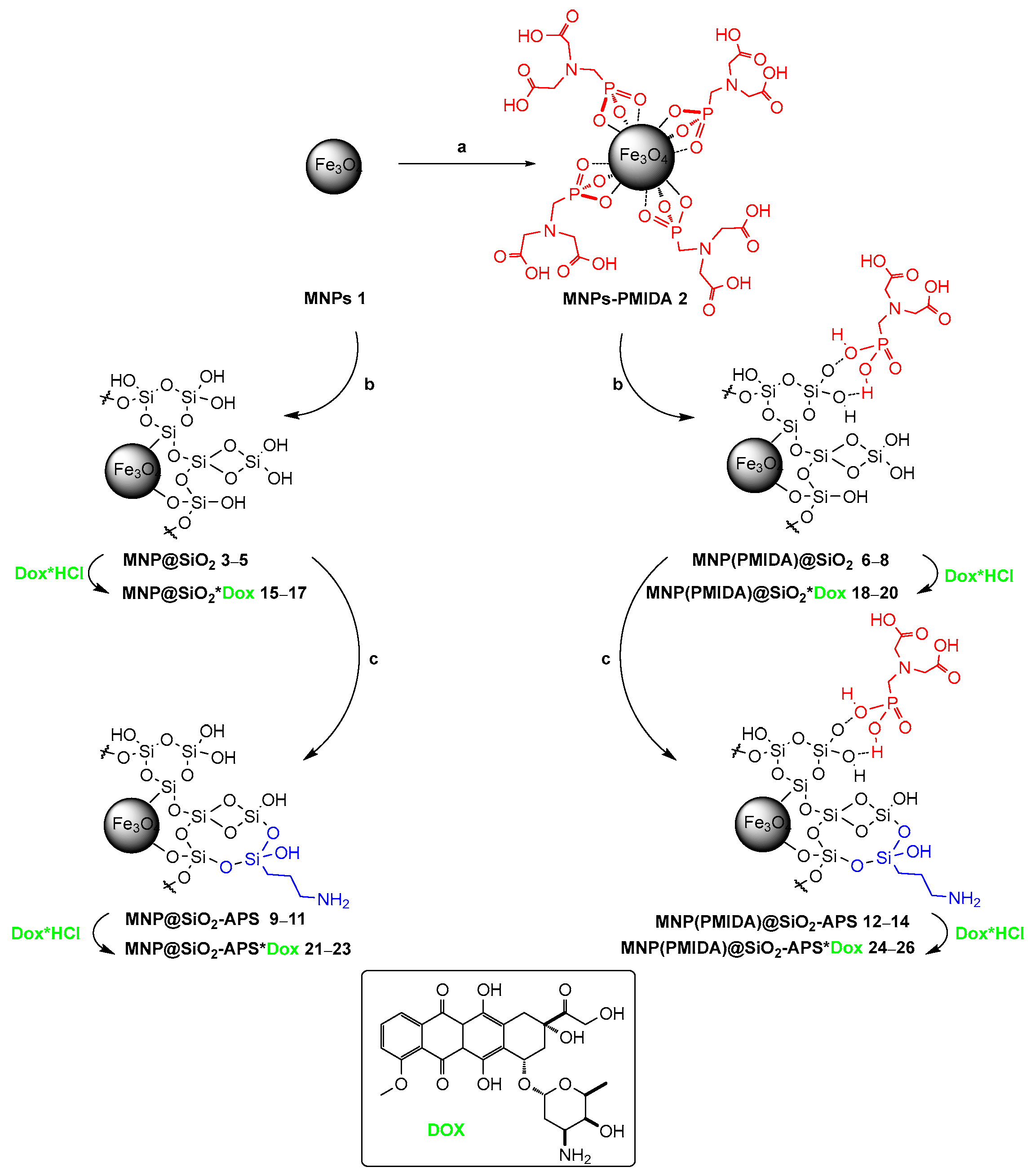
Scheme 1. Synthesis of NCs and Dox loading on them. (a) PMIDA, 40 °C, 4 h, 20 °C, 16 h; (b) TEOS (1.5, 3.0, and 5.0 equiv.), NH4OH, EtOH/H2O, 40 °C, 4 h, 20 °C, 16 h; (c) APTMS, EtOH/H2O, 40 °C, 4 h, 20 °C, 16 h.
Pharmaceutics 14 02271 sch001
Figure 1. (a) TEM images of Fe3O4@SiO2 MNP-based composite materials 38 obtained using 1.5, 3.0, and 5.0 mol equiv. TEOS, respectively. (b) DLS data for colloidal solutions of NCs 314.
Pharmaceutics 14 02271 g001aPharmaceutics 14 02271 g001b
Figure 2. UATR-FTIR spectra of initial MNPs 1 and 2, as well as SiO2-coated (38) and SiO2/APS-coated NCs (914); the inset shows an enlarged fragment of the spectrum in the region 1700–1270 cm−1. The spectra are normalized by the height of the bands corresponding to the Fe–O vibrations.
Pharmaceutics 14 02271 g002
Figure 3. UATR-FTIR spectra of Dox-containing NCs 1520 (left) and 2126 (right); the inset shows an enlarged fragment of the spectrum in the region 1800–1350 cm−1.
Pharmaceutics 14 02271 g003
Figure 4. (a) TEM images of NCs 1820. (b) DLS data for colloidal solutions of Dox-loaded NCs 1526. (c) Relaxation rate (R2) of colloidal solutions of NCs 1520.
Pharmaceutics 14 02271 g004
Figure 5. (a) Dox absorption spectrum; the inset shows a calibration plot for determination of the extinction coefficient (εDox) at wavelengths of 480, 290, 253, and 233 nm vs. concentration. (b) Absorption, excitation and emission spectra. (c) Dependence of εDox (at 480 nm) on Dox concentration. Data are presented as M ± SD. (d) Concentration dependence of the relative quantum yield of Dox fluorescence (quantum yield (±10%) was determined relative to fluorescein in 0.1 M KOH as a standard (φ = 0.85, λex = 470 nm)). Data are presented as M ± SD.
Pharmaceutics 14 02271 g005
Figure 6. (a) Sorption and (b) desorption of Dox on NCs 1526.
Pharmaceutics 14 02271 g006
Figure 7. Images of Dox complexes with (a) SiO2 cluster, (b) SiO2-APS, (c) SiO2-2APS and (d) SiO2-PMIDA. The binding energies (Eb) were calculated at the B3LYP-D3-gCP/def2-SVP level of theory in water using ORCA 4.0.1 software [57]. The atoms involved in the binding of molecules on the cluster surface are highlighted in red.
Pharmaceutics 14 02271 g007
Figure 8. NCIplot images of noncovalent interactions in Dox complexes with a SiO2-2APS and SiO2-PMIDA-Dox salt 2 in water. Electron density iso-surfaces in the range of reduced electron density gradient (s) –0.07 < s < 0.07 at. units. Coloring in the BGR palette from blue (attraction) to red (repulsion).
Pharmaceutics 14 02271 g008
Figure 9. Images of (a) cells stained with nuclear dye and captured in channel mode with 405 nm excitation; (b) NC 18 taken in channel mode with 561 nm excitation, overlapped on image captured in transmission light mode; (c) cells stained with nuclear dye (blue) and Dox (red) desorbed from NC 18 (captured in channel mode with λEx = 405 and 561 nm), and (d) overlapped on image captured in transmission light mode. (e) Cell images taken in the lambda mode when excited by lasers of different wavelengths (405, 488 and 561 nm). (f) Fluorescence spectra extracted from the lambda-images with different excitation.
Pharmaceutics 14 02271 g009
Table 1. Characterization of MNPs 114.
MNPsComponentsRelative Content of Inorganic Elements (%) 1EA Data (%)Inorganic Component RatioConcentration of Organic Components (mmol/g)MS (emu/g)
PMIDATEOS (Equiv.)APTMS (Equiv.)FeSiPCSiO2:Fe3O4 2SSi–O/SFe–O 3CAPS 4CPMIDA 5
11000000074
2+94.3405.665.8500.9761
31.577.1222.880031:692.330051
43.064.0835.920046:543.860039
55.050.7849.220060:406.610026
6+1.571.3224.873.801.7235:653.5600.6340
7+3.057.1339.932.941.1852:486.1300.4928
8+5.046.3851.741.871.0063:3713.0500.3121
91.53.074.6525.3503.4934:664.180.48049
103.03.063.4636.5402.7147:537.730.32038
115.03.050.3449.6601.8060:4013.340.28027
12+1.53.068.8229.691.494.3140:603.970.660.2637
13+3.03.057.2741.501.232.6347:537.580.330.2128
14+5.03.045.7053.291.012.0364:3613.040.340.1721
1 EDX data. 2 Calculated from the EDX data by analogy with [27]. 3 Calculated from the FTIR spectra. 4 Calculated based on the CHN-elemental analysis data by analogy with [78]. 5 Calculated from the EDX data.
Table 2. IC50 values of Dox and NCs 1526 for HEK-293 cells after incubation for 48 h (MTT assay). Data are presented as M ± SD.
IC50, μM
DoxNCs
151617181920212223242526
0.289 ± 0.0472.235 ± 0.0951.137 ± 0.0591.125 ± 0.0780.574 ± 0.0450.341 ± 0.0790.989 ± 0.0951.099 ± 0.0811.111 ± 0.0731.012 ± 0.0871.129 ± 0.0621.114 ± 0.0561.111 ± 0.059
Publisher’s Note: MDPI stays neutral with regard to jurisdictional claims in published maps and institutional affiliations.