
Application of Electrospun Nanofiber Membrane in the Treatment of Diabetic Wounds

Abstract
:1. Introduction
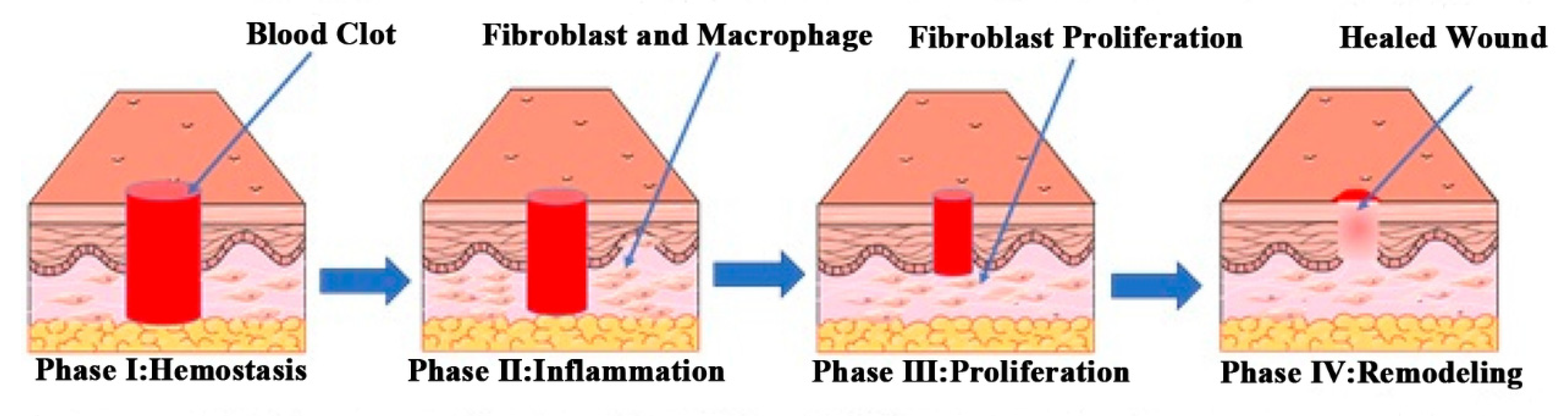
1.1. Normal Wound Healing Process
1.1.1. Hemostasis Stage
1.1.2. Inflammation Stage
1.1.3. Proliferation Stage
1.1.4. Remodeling Stage
1.2. Diabetic Wounds
2. Electrospinning
2.1. Critical Parameters of Electrospinning Process
2.1.1. Properties of Polymer Solution
- (1)
- Relative Molecular Mass of Polymer
- (2)
- Solution Concentration and Viscosity
- (3)
- Solvent Properties
2.1.2. Process Parameters
- (1)
- Applied Voltage
- (2)
- Solution Injection Speed
- (3)
- The Receiving Distance of the Fiber
2.1.3. Environmental Conditions
2.2. Advantages of Electrospun Nanofiber Membranes in the Treatment of Diabetic Wounds
2.3. Preparation Methods of Electrospun Nanofiber Membranes
2.3.1. Uniaxial Electrospinning
2.3.2. Emulsion Electrospinning
2.3.3. Coaxial Electrospinning
3. Application of Electrospun Nanofiber Membranes in the Treatment of Diabetic Wounds
3.1. Treatment of Diabetic Wounds with Polymer Electrospun Fibers
3.1.1. Treatment of Diabetic Wounds with Electrospun Synthetic Polymer Fibers
3.1.2. Natural/Synthetic Polymer Blended Electrospun Fibers in the Treatment of Diabetic Wounds
3.2. Nanoparticle-Loaded Electrospun Fibers in the Treatment of Diabetic Wounds
3.2.1. Nanoparticles/Synthetic Polymer Electrospun Fibers
3.2.2. Nanoparticles/Synthetic Polymers/Natural Polymer Electrospun Fibers
3.3. Drug-Loaded Electrospun Fibers for Collaborative Therapy of Diabetic Wounds
3.3.1. Drugs/Natural Polymer Electrospun Fibers
3.3.2. Drugs/Synthetic Polymer Electrospun Fibers
3.3.3. Drugs/Synthetic Polymer/Natural Polymer Electrospun Fibers
3.4. Drug/Nanoparticle/Polymer Electrospun Fibers
3.5. Cell Loaded Electrospun Fiber Membranes for Diabetic Wound Treatment
4. Outlook
Author Contributions
Funding
Institutional Review Board Statement
Informed Consent Statement
Data Availability Statement
Acknowledgments
Conflicts of Interest
References
- Childs, D.R.; Murthy, A.S. Overview of Wound Healing and Management. Surg. Clin. North Am. 2017, 97, 189–207. [Google Scholar] [CrossRef]
- Baltzis, D.; Eleftheriadou, I.; Veves, A. Pathogenesis and treatment of impaired wound healing in diabetes mellitus: New insights. Adv. Ther. 2014, 31, 817–836. [Google Scholar] [CrossRef] [PubMed]
- Huayllani, M.T.; Sarabia-Estrada, R.; Restrepo, D.J.; Boczar, D.; Sisti, A.; Nguyen, J.H.; Rinker, B.D.; Moran, S.L.; Quinones-Hinojosa, A.; Forte, A.J. Adipose-derived stem cells in wound healing of full-thickness skin defects: A review of the literature. J. Plast. Surg. Hand Surg. 2020, 54, 263–279. [Google Scholar] [CrossRef] [PubMed]
- Gao, J.; Wang, L.; Xia, C.; Yang, X.; Cao, Z.; Zheng, L.; Ko, R.; Shen, C.; Yang, C.; Cheng, C. Cold atmospheric plasma promotes different types of superficial skin erosion wounds healing. Int. Wound J. 2019, 16, 1103–1111. [Google Scholar] [CrossRef] [PubMed]
- Markova, A.; Mostow, E.N. US skin disease assessment: Ulcer and wound care. Dermatol. Clin. 2012, 30, 107–111. [Google Scholar] [CrossRef]
- Yang, C.; Goss, S.G.; Alcantara, S.; Schultz, G.; Lantis, I.J.C. Effect of Negative Pressure Wound Therapy With Instillation on Bioburden in Chronically Infected Wounds. Wounds Compend. Clin. Res. Pract. 2017, 29, 240–246. [Google Scholar]
- Xuan, X.; Zhou, Y.; Chen, A.; Zheng, S.; An, Y.; He, H.; Huang, W.; Chen, Y.; Yang, Y.; Li, S.; et al. Silver crosslinked injectable bFGF-eluting supramolecular hydrogels speed up infected wound healing. J. Mater. Chem. B 2020, 8, 1359–1370. [Google Scholar] [CrossRef]
- Reinke, J.M.; Sorg, H. Wound repair and regeneration. Eur. Surg. Res. 2012, 49, 35–43. [Google Scholar] [CrossRef]
- Bhagavathula, N.; Warner, R.L.; DaSilva, M.; McClintock, S.D.; Barron, A.; Aslam, M.N.; Johnson, K.J.; Varani, J. A combination of curcumin and ginger extract improves abrasion wound healing in corticosteroid-impaired hairless rat skin. Wound Repair Regen. 2009, 17, 360–366. [Google Scholar] [CrossRef] [Green Version]
- Nolff, M.C.; Albert, R.; Reese, S.; Meyer-Lindenberg, A. Comparison of Negative Pressure Wound Therapy and Silver-Coated Foam Dressings in Open Wound Treatment in Dogs: A Prospective Controlled Clinical Trial. Vet. Comp. Orthop. Traumatol. 2018, 31, 229–238. [Google Scholar] [CrossRef]
- Javierre, E. Impact of anomalous transport kinetics on the progress of wound healing. Med. Eng. Phys. 2016, 38, 885–894. [Google Scholar] [CrossRef]
- Sivakumar, B.S.; Athreya, P.J.; Chow, J.; Suthersan, M.; Symes, M.; O’Leary, E.; Martin, B. Internal degloving injury of the foot. ANZ J. Surg. 2020, 90, 926–927. [Google Scholar] [CrossRef]
- Sorg, H.; Tilkorn, D.J.; Hager, S.; Hauser, J.; Mirastschijski, U. Skin Wound Healing: An Update on the Current Knowledge and Concepts. Eur. Surg. Res. 2017, 58, 81–94. [Google Scholar] [CrossRef]
- Bielefeld, K.A.; Amini-Nik, S.; Alman, B.A. Cutaneous wound healing: Recruiting developmental pathways for regeneration. Cell Mol. Life Sci. 2013, 70, 2059–2081. [Google Scholar] [CrossRef] [PubMed] [Green Version]
- Brugues, A.; Anon, E.; Conte, V.; Veldhuis, J.H.; Gupta, M.; Colombelli, J.; Munoz, J.J.; Brodland, G.W.; Ladoux, B.; Trepat, X. Forces driving epithelial wound healing. Nat. Phys. 2014, 10, 683–690. [Google Scholar] [CrossRef] [Green Version]
- Wang, P.H.; Huang, B.S.; Horng, H.C.; Yeh, C.C.; Chen, Y.J. Wound healing. J. Chin. Med. Assoc. 2018, 81, 94–101. [Google Scholar] [CrossRef] [PubMed]
- Memic, A.; Abudula, T.; Mohammed, H.S.; Navare, J.K.; Colombani, T.; Bencherif, S.A. Latest Progress in Electrospun Nanofibers for Wound Healing Applications. ACS Appl. Bio. Mater. 2019, 2, 952–969. [Google Scholar] [CrossRef]
- Sinegre, T.; Teissandier, D.; Milenkovic, D.; Morand, C.; Lebreton, A. Epicatechin influences primary hemostasis, coagulation and fibrinolysis. Food Funct. 2019, 10, 7291–7298. [Google Scholar] [CrossRef] [PubMed]
- Rojano, M.R.; Mendez, S.; Lucor, D.; Ranc, A.; Giansily-Blaizot, M.; Schved, J.F.; Nicoud, F. Kinetics of the coagulation cascade including the contact activation system: Sensitivity analysis and model reduction. Biomech. Model Mechanobiol. 2019, 18, 1139–1153. [Google Scholar] [CrossRef]
- Wan, R.; Weissman, J.P.; Grundman, K.; Lang, L.; Grybowski, D.J.; Galiano, R.D. Diabetic wound healing: The impact of diabetes on myofibroblast activity and its potential therapeutic treatments. Wound Repair Regen. 2021, 29, 573–581. [Google Scholar] [CrossRef]
- Golebiewska, E.M.; Poole, A.W. Platelet secretion: From haemostasis to wound healing and beyond. Blood Rev. 2015, 29, 153–162. [Google Scholar] [CrossRef] [Green Version]
- Rubenstein, D.A.; Yin, W. Platelet-Activation Mechanisms and Vascular Remodeling. Compr. Physiol. 2018, 8, 1117–1156. [Google Scholar] [CrossRef]
- Rodrigues, M.; Kosaric, N.; Bonham, C.A.; Gurtner, G.C. Wound Healing: A Cellular Perspective. Physiol. Rev. 2019, 99, 665–706. [Google Scholar] [CrossRef]
- Yang, W.; Tao, Y.; Wu, Y.; Zhao, X.; Ye, W.; Zhao, D.; Fu, L.; Tian, C.; Yang, J.; He, F.; et al. Neutrophils promote the development of reparative macrophages mediated by ROS to orchestrate liver repair. Nat. Commun. 2019, 10, 1076. [Google Scholar] [CrossRef] [PubMed] [Green Version]
- Medina, C.B.; Mehrotra, P.; Arandjelovic, S.; Perry, J.S.A.; Guo, Y.; Morioka, S.; Barron, B.; Walk, S.F.; Ghesquiere, B.; Krupnick, A.S.; et al. Metabolites released from apoptotic cells act as tissue messengers. Nature 2020, 580, 130–135. [Google Scholar] [CrossRef] [PubMed]
- Behm, B.; Babilas, P.; Landthaler, M.; Schreml, S. Cytokines, chemokines and growth factors in wound healing. J. Eur. Acad. Dermatol. Venereol. 2012, 26, 812–820. [Google Scholar] [CrossRef] [PubMed]
- Vestweber, D. How leukocytes cross the vascular endothelium. Nat. Rev. Immunol. 2015, 15, 692–704. [Google Scholar] [CrossRef]
- Prasad, A.; Lin, F.; Clark, R.A.F. Fibronectin-derived Epiviosamines enhance PDGF-BB-stimulated human dermal fibroblast migration in vitro and granulation tissue formation in vivo. Wound Repair Regen. 2019, 27, 634–649. [Google Scholar] [CrossRef]
- Xue, M.; Jackson, C.J. Extracellular Matrix Reorganization During Wound Healing and Its Impact on Abnormal Scarring. Adv. Wound Care 2015, 4, 119–136. [Google Scholar] [CrossRef] [Green Version]
- Decker, C.G.; Wang, Y.; Paluck, S.J.; Shen, L.; Loo, J.A.; Levine, A.J.; Miller, L.S.; Maynard, H.D. Fibroblast growth factor 2 dimer with superagonist in vitro activity improves granulation tissue formation during wound healing. Biomaterials 2016, 81, 157–168. [Google Scholar] [CrossRef] [Green Version]
- Lau, K.; Paus, R.; Tiede, S.; Day, P.; Bayat, A. Exploring the role of stem cells in cutaneous wound healing. Exp. Dermatol. 2009, 18, 921–933. [Google Scholar] [CrossRef]
- Pan, H.; Shi, C.; Yang, R.; Xi, G.; Lu, C.; Yang, X.; Chen, J.; Wang, X.; Chen, L.; Pan, J. Controlled release of KGF-2 for regulation of wound healing by KGF-2 complexed with “lotus seedpod surface-like” porous microspheres. J. Mater. Chem. B 2021, 9, 4039–4049. [Google Scholar] [CrossRef] [PubMed]
- Brandi, J.; Cheri, S.; Manfredi, M.; Di Carlo, C.; Vanella, V.V.; Federici, F.; Bombiero, E.; Bazaj, A.; Rizzi, E.; Manna, L.; et al. Exploring the wound healing, anti-inflammatory, anti-pathogenic and proteomic effects of lactic acid bacteria on keratinocytes. Sci. Rep. 2020, 10, 11572. [Google Scholar] [CrossRef] [PubMed]
- Hinz, B. The role of myofibroblasts in wound healing. Curr. Res. Transl. Med. 2016, 64, 171–177. [Google Scholar] [CrossRef]
- Holl, J.; Kowalewski, C.; Zimek, Z.; Fiedor, P.; Kaminski, A.; Oldak, T.; Moniuszko, M.; Eljaszewicz, A. Chronic Diabetic Wounds and Their Treatment with Skin Substitutes. Cells 2021, 10, 655. [Google Scholar] [CrossRef] [PubMed]
- Tiwari, R.; Tiwari, G.; Lahiri, A.; R, V.; Rai, A.K. Localized Delivery of Drugs through Medical Textiles for Treatment of Burns: A Perspective Approach. Adv. Pharm. Bull. 2021, 11, 248–260. [Google Scholar] [CrossRef]
- Kaplani, K.; Koutsi, S.; Armenis, V.; Skondra, F.G.; Karantzelis, N.; Tsaniras, C.S.; Taraviras, S. Wound healing related agents: Ongoing research and perspectives. Adv. Drug. Deliv. Rev. 2018, 129, 242–253. [Google Scholar] [CrossRef]
- Davis, S.C.; Li, J.; Gil, J.; Valdes, J.; Solis, M.; Higa, A.; Bowler, P. The wound-healing effects of a next-generation anti-biofilm silver Hydrofiber wound dressing on deep partial-thickness wounds using a porcine model. Int. Wound J. 2018, 15, 834–839. [Google Scholar] [CrossRef]
- Hotkar, M.S.; Avachat, A.M.; Bhosale, S.S.; Oswal, Y.M. Preliminary investigation of topical nitroglycerin formulations containing natural wound healing agent in diabetes-induced foot ulcer. Int. Wound J. 2015, 12, 210–217. [Google Scholar] [CrossRef]
- Lim, J.Z.; Ng, N.S.; Thomas, C. Prevention and treatment of diabetic foot ulcers. J. R. Soc. Med. 2017, 110, 104–109. [Google Scholar] [CrossRef] [Green Version]
- da Silva, L.P.; Reis, R.L.; Correlo, V.M.; Marques, A.P. Hydrogel-Based Strategies to Advance Therapies for Chronic Skin Wounds. Annu. Rev. Biomed. Eng. 2019, 21, 145–169. [Google Scholar] [CrossRef] [Green Version]
- Peng, Y.; He, D.; Ge, X.; Lu, Y.; Chai, Y.; Zhang, Y.; Mao, Z.; Luo, G.; Deng, J.; Zhang, Y. Construction of heparin-based hydrogel incorporated with Cu5.4O ultrasmall nanozymes for wound healing and inflammation inhibition. Bioact. Mater. 2021, 6, 3109–3124. [Google Scholar] [CrossRef] [PubMed]
- Salazar, J.J.; Ennis, W.J.; Koh, T.J. Diabetes medications: Impact on inflammation and wound healing. J. Diabetes Complicat. 2016, 30, 746–752. [Google Scholar] [CrossRef] [PubMed] [Green Version]
- Lohmann, N.; Schirmer, L.; Atallah, P.; Wandel, E.; Ferrer, R.A.; Werner, C.; Simon, J.C.; Franz, S.; Freudenberg, U. Glycosaminoglycan-based hydrogels capture inflammatory chemokines and rescue defective wound healing in mice. Sci. Transl. Med. 2017, 9, 386. [Google Scholar] [CrossRef]
- Boniakowski, A.E.; Kimball, A.S.; Jacobs, B.N.; Kunkel, S.L.; Gallagher, K.A. Macrophage-Mediated Inflammation in Normal and Diabetic Wound Healing. J. Immunol. 2017, 199, 17–24. [Google Scholar] [CrossRef] [Green Version]
- Roep, B.O.; Wheeler, D.C.S.; Peakman, M. Antigen-based immune modulation therapy for type 1 diabetes: The era of precision medicine. Lancet Diabetes Endocrinol. 2019, 7, 65–74. [Google Scholar] [CrossRef]
- Han, G.; Ceilley, R. Chronic Wound Healing: A Review of Current Management and Treatments. Adv. Ther. 2017, 34, 599–610. [Google Scholar] [CrossRef] [PubMed] [Green Version]
- Shin, L.; Peterson, D.A. Human mesenchymal stem cell grafts enhance normal and impaired wound healing by recruiting existing endogenous tissue stem/progenitor cells. Stem Cells Transl. Med. 2013, 2, 33–42. [Google Scholar] [CrossRef]
- Bjarnsholt, T.; Kirketerp-Moller, K.; Jensen, P.O.; Madsen, K.G.; Phipps, R.; Krogfelt, K.; Hoiby, N.; Givskov, M. Why chronic wounds will not heal: A novel hypothesis. Wound Repair Regen. 2008, 16, 2–10. [Google Scholar] [CrossRef]
- Gueraud, F.; Atalay, M.; Bresgen, N.; Cipak, A.; Eckl, P.M.; Huc, L.; Jouanin, I.; Siems, W.; Uchida, K. Chemistry and biochemistry of lipid peroxidation products. Free Radic. Res. 2010, 44, 1098–1124. [Google Scholar] [CrossRef] [PubMed]
- Schreml, S.; Szeimies, R.M.; Prantl, L.; Karrer, S.; Landthaler, M.; Babilas, P. Oxygen in acute and chronic wound healing. Br. J. Dermatol. 2010, 163, 257–268. [Google Scholar] [CrossRef] [PubMed]
- Dai, J.; Zhang, X.; Wang, Y.; Chen, H.; Chai, Y. ROS-activated NLRP3 inflammasome initiates inflammation in delayed wound healing in diabetic rats. Int. J. Clin. Exp. Pathol. 2017, 10, 9902–9909. [Google Scholar] [PubMed]
- Choi, S.; Lee, H.M.; Kim, H.S. Effect of molecular weight on humidity-sensitive characteristics of electrospun polyethylene oxide. Sens. Actuators A Phys. 2019, 294, 194–202. [Google Scholar] [CrossRef]
- Nezarati, R.M.; Eifert, M.B.; Cosgriff-Hernandez, E. Effects of humidity and solution viscosity on electrospun fiber morphology. Tissue Eng. Part C Methods 2013, 19, 810–819. [Google Scholar] [CrossRef] [PubMed] [Green Version]
- Lauricella, M.; Cipolletta, F.; Pontrelli, G.; Pisignano, D.; Succi, S. Effects of orthogonal rotating electric fields on electrospinning process. Phys. Fluids 2017, 29, 082003. [Google Scholar] [CrossRef] [Green Version]
- Yu, H.; Chen, X.; Cai, J.; Ye, D.; Wu, Y.; Liu, P. Dual controlled release nanomicelle-in-nanofiber system for long-term antibacterial medical dressings. J. Biomater. Sci. Polym. Ed. 2019, 30, 64–76. [Google Scholar] [CrossRef]
- Xie, Z.; Paras, C.B.; Weng, H.; Punnakitikashem, P.; Su, L.C.; Vu, K.; Tang, L.; Yang, J.; Nguyen, K.T. Dual growth factor releasing multi-functional nanofibers for wound healing. Acta Biomater. 2013, 9, 9351–9359. [Google Scholar] [CrossRef] [Green Version]
- Pal, P.; Dadhich, P.; Srivas, P.K.; Das, B.; Maulik, D.; Dhara, S. Bilayered nanofibrous 3D hierarchy as skin rudiment by emulsion electrospinning for burn wound management. Biomater. Sci. 2017, 5, 1786–1799. [Google Scholar] [CrossRef]
- Peh, P.; Lim, N.S.; Blocki, A.; Chee, S.M.; Park, H.C.; Liao, S.; Chan, C.; Raghunath, M. Simultaneous Delivery of Highly Diverse Bioactive Compounds from Blend Electrospun Fibers for Skin Wound Healing. Bioconjug. Chem. 2015, 26, 1348–1358. [Google Scholar] [CrossRef]
- Han, D.; Steckl, A.J. Coaxial Electrospinning Formation of Complex Polymer Fibers and their Applications. Chempluschem 2019, 84, 1453–1497. [Google Scholar] [CrossRef]
- Lee, C.H.; Hung, K.C.; Hsieh, M.J.; Chang, S.H.; Juang, J.H.; Hsieh, I.C.; Wen, M.S.; Liu, S.J. Core-shell insulin-loaded nanofibrous scaffolds for repairing diabetic wounds. Nanomedicine 2020, 24, 102123. [Google Scholar] [CrossRef]
- Li, R.; Cheng, Z.; Wen, R.; Zhao, X.; Yu, X.; Sun, L.; Zhang, Y.; Han, Z.; Yuan, Y.; Kang, L. Novel SA@Ca2+/RCSPs core–shell structure nanofibers by electrospinning for wound dressings. RSC Adv. 2018, 8, 15558–15566. [Google Scholar] [CrossRef] [Green Version]
- Li, J.; Xu, W.; Li, D.; Liu, T.; Zhang, Y.S.; Ding, J.; Chen, X. Locally Deployable Nanofiber Patch for Sequential Drug Delivery in Treatment of Primary and Advanced Orthotopic Hepatomas. ACS Nano 2018, 12, 6685–6699. [Google Scholar] [CrossRef] [PubMed]
- de Souza, S.O.L.; Guerra, M.C.A.; Heneine, L.G.D.; de Oliveira, C.R.; Junior, C.A.D.S.; Fialho, S.L.; Orefice, R.L. Biodegradable core-shell electrospun nanofibers containing bevacizumab to treat age-related macular degeneration. J. Mater. Sci. Mater. Med. 2018, 29, 173. [Google Scholar] [CrossRef] [PubMed]
- Maggay, I.V.; Venault, A.; Fang, C.Y.; Yang, C.C.; Hsu, C.H.; Chou, C.Y.; Ishihara, K.; Chang, Y. Zwitterionized Nanofibrous Poly(vinylidene fluoride) Membranes for Improving the Healing of Diabetic Wounds. ACS Biomater. Sci. Eng. 2021, 7, 562–576. [Google Scholar] [CrossRef]
- Gholipour-Kanani, A.; Bahrami, S.H.; Rabbani, S. Effect of novel blend nanofibrous scaffolds on diabetic wounds healing. IET Nanobiotechnol. 2016, 10, 1–7. [Google Scholar] [CrossRef]
- Sun, L.; Gao, W.; Fu, X.; Shi, M.; Xie, W.; Zhang, W.; Zhao, F.; Chen, X. Enhanced wound healing in diabetic rats by nanofibrous scaffolds mimicking the basketweave pattern of collagen fibrils in native skin. Biomater. Sci. 2018, 6, 340–349. [Google Scholar] [CrossRef]
- Ajmal, G.; Bonde, G.V.; Mittal, P.; Khan, G.; Pandey, V.K.; Bakade, B.V.; Mishra, B. Biomimetic PCL-gelatin based nanofibers loaded with ciprofloxacin hydrochloride and quercetin: A potential antibacterial and anti-oxidant dressing material for accelerated healing of a full thickness wound. Int. J. Pharm. 2019, 567, 118480. [Google Scholar] [CrossRef] [PubMed]
- Sanhueza, C.; Hermosilla, J.; Bugallo-Casal, A.; Da Silva-Candal, A.; Taboada, C.; Millan, R.; Concheiro, A.; Alvarez-Lorenzo, C.; Acevedo, F. One-step electrospun scaffold of dual-sized gelatin/poly-3-hydroxybutyrate nano/microfibers for skin regeneration in diabetic wound. Mater. Sci. Eng. C Mater. Biol. Appl. 2021, 119, 111602. [Google Scholar] [CrossRef] [PubMed]
- Gilotra, S.; Chouhan, D.; Bhardwaj, N.; Nandi, S.K.; Mandal, B.B. Potential of silk sericin based nanofibrous mats for wound dressing applications. Mater. Sci. Eng. C Mater. Biol. Appl. 2018, 90, 420–432. [Google Scholar] [CrossRef]
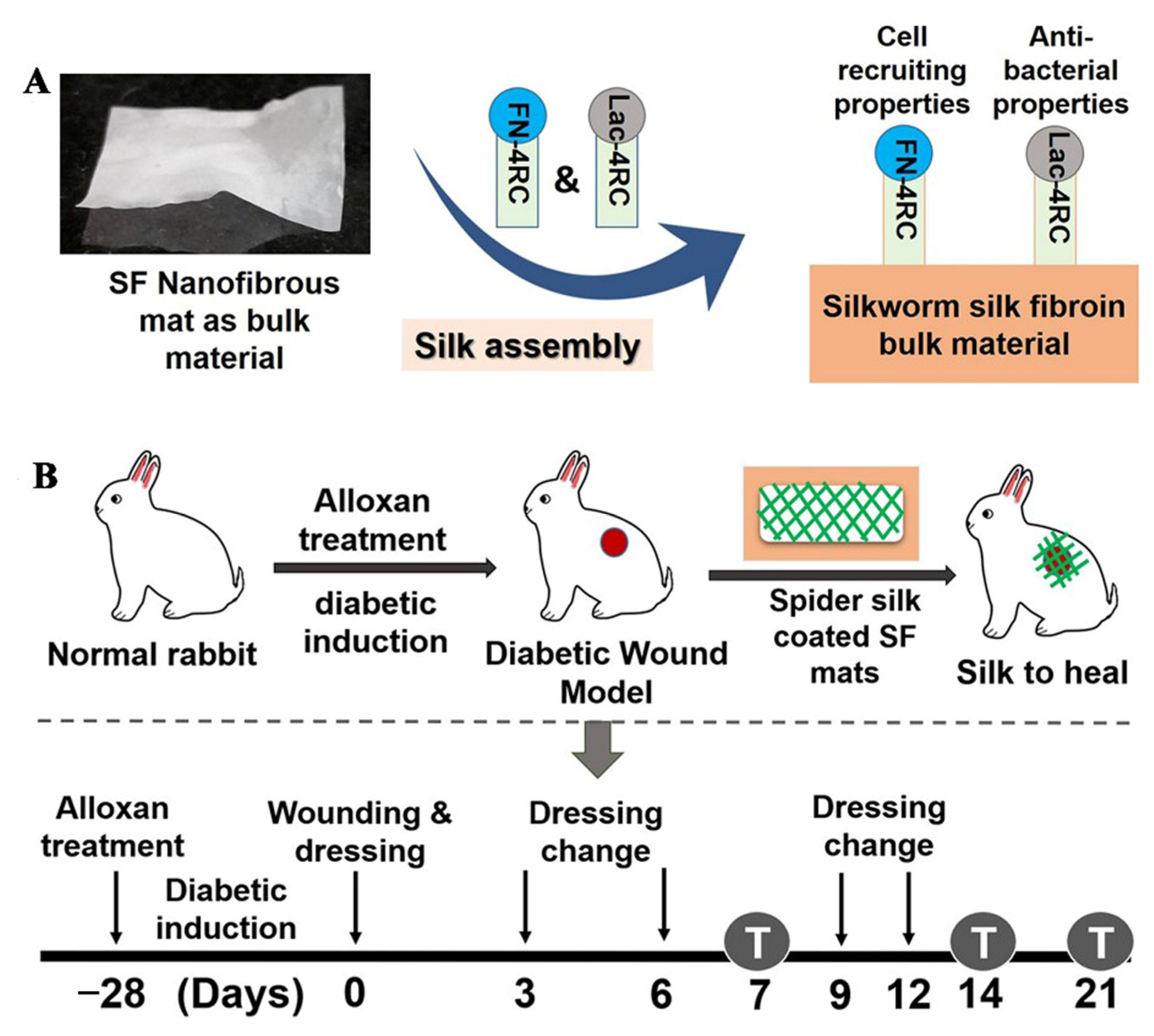
- Chouhan, D.; Das, P.; Thatikonda, N.; Nandi, S.K.; Hedhammar, M.; Mandal, B.B. Silkworm Silk Matrices Coated with Functionalized Spider Silk Accelerate Healing of Diabetic Wounds. ACS Biomater. Sci. Eng. 2019, 5, 3537–3548. [Google Scholar] [CrossRef]
- Grip, J.; Engstad, R.E.; Skjaeveland, I.; Skalko-Basnet, N.; Isaksson, J.; Basnet, P.; Holsaeter, A.M. Beta-glucan-loaded nanofiber dressing improves wound healing in diabetic mice. Eur. J. Pharm. Sci. 2018, 121, 269–280. [Google Scholar] [CrossRef] [Green Version]
- Zhang, P.; Li, Y.; Tang, Y.; Shen, H.; Li, J.; Yi, Z.; Ke, Q.; Xu, H. Copper-Based Metal-Organic Framework as a Controllable Nitric Oxide-Releasing Vehicle for Enhanced Diabetic Wound Healing. ACS Appl. Mater. Interfaces 2020, 12, 18319–18331. [Google Scholar] [CrossRef] [PubMed]
- Elshazly, N.; Khalil, A.; Saad, M.; Patruno, M.; Chakraborty, J.; Marei, M. Efficacy of Bioactive Glass Nanofibers Tested for Oral Mucosal Regeneration in Rabbits with Induced Diabetes. Materials 2020, 13, 2603. [Google Scholar] [CrossRef] [PubMed]
- Jiang, Y.; Li, Y.; Li, J.; Han, Y.; Zhang, P.; Yi, Z.; Ke, Q.; Xu, H. A Mussel-Inspired Extracellular Matrix-Mimicking Composite Scaffold for Diabetic Wound Healing. ACS Appl. Bio Mater. 2020, 3, 4052–4061. [Google Scholar] [CrossRef]
- Zehra, M.; Zubairi, W.; Hasan, A.; Butt, H.; Ramzan, A.; Azam, M.; Mehmood, A.; Falahati, M.; Chaudhry, A.A.; Rehman, I.U.; et al. Oxygen Generating Polymeric Nano Fibers That Stimulate Angiogenesis and Show Efficient Wound Healing in a Diabetic Wound Model. Int. J. Nanomed. 2020, 15, 3511–3522. [Google Scholar] [CrossRef]
- Augustine, R.; Hasan, A.; Patan, N.K.; Dalvi, Y.B.; Varghese, R.; Antony, A.; Unni, R.N.; Sandhyarani, N.; Moustafa, A.A. Cerium Oxide Nanoparticle Incorporated Electrospun Poly(3-hydroxybutyrate-co-3-hydroxyvalerate) Membranes for Diabetic Wound Healing Applications. ACS Biomater. Sci. Eng. 2020, 6, 58–70. [Google Scholar] [CrossRef]
- Zhang, P.; Jiang, Y.; Liu, D.; Liu, Y.; Ke, Q.; Xu, H. A bioglass sustained-release scaffold with ECM-like structure for enhanced diabetic wound healing. Nanomedicine 2020, 15, 2241–2253. [Google Scholar] [CrossRef]
- Chen, Q.; Wu, J.; Liu, Y.; Li, Y.; Zhang, C.; Qi, W.; Yeung, K.W.K.; Wong, T.M.; Zhao, X.; Pan, H. Electrospun chitosan/PVA/bioglass Nanofibrous membrane with spatially designed structure for accelerating chronic wound healing. Mater. Sci. Eng. C Mater. Biol. Appl. 2019, 105, 110083. [Google Scholar] [CrossRef]
- Lv, F.; Wang, J.; Xu, P.; Han, Y.; Ma, H.; Xu, H.; Chen, S.; Chang, J.; Ke, Q.; Liu, M.; et al. A conducive bioceramic/polymer composite biomaterial for diabetic wound healing. Acta Biomater. 2017, 60, 128–143. [Google Scholar] [CrossRef] [PubMed]
- Ahmed, R.; Tariq, M.; Ali, I.; Asghar, R.; Khanam, N.P.; Augustine, R.; Hasan, A. Novel electrospun chitosan/polyvinyl alcohol/zinc oxide nanofibrous mats with antibacterial and antioxidant properties for diabetic wound healing. Int. J. Biol. Macromol. 2018, 120, 385–393. [Google Scholar] [CrossRef]
- Simoes, D.; Miguel, S.P.; Ribeiro, M.P.; Coutinho, P.; Mendonca, A.G.; Correia, I.J. Recent advances on antimicrobial wound dressing: A review. Eur. J. Pharm. Biopharm. 2018, 127, 130–141. [Google Scholar] [CrossRef]
- Liu, F.; Li, X.; Wang, L.; Yan, X.; Ma, D.; Liu, Z.; Liu, X. Sesamol incorporated cellulose acetate-zein composite nanofiber membrane: An efficient strategy to accelerate diabetic wound healing. Int. J. Biol. Macromol. 2020, 149, 627–638. [Google Scholar] [CrossRef]
- Losi, P.; Al Kayal, T.; Buscemi, M.; Foffa, I.; Cavallo, A.; Soldani, G. Bilayered Fibrin-Based Electrospun-Sprayed Scaffold Loaded with Platelet Lysate Enhances Wound Healing in a Diabetic Mouse Model. Nanomaterials 2020, 10, 2128. [Google Scholar] [CrossRef] [PubMed]
- Augustine, R.; Zahid, A.A.; Hasan, A.; Wang, M.; Webster, T.J. CTGF Loaded Electrospun Dual Porous Core-Shell Membrane For Diabetic Wound Healing. Int. J. Nanomed. 2019, 14, 8573–8588. [Google Scholar] [CrossRef] [PubMed] [Green Version]
- Su, Y.; Wang, H.; Mishra, B.; Narayana, L.J.; Jiang, J.; Reilly, D.A.; Hollins, R.R.; Carlson, M.A.; Wang, G.; Xie, J. Nanofiber Dressings Topically Delivering Molecularly Engineered Human Cathelicidin Peptides for the Treatment of Biofilms in Chronic Wounds. Mol. Pharm. 2019, 16, 2011–2020. [Google Scholar] [CrossRef] [PubMed]
- Mabrouk, M.; Kumar, P.; Choonara, Y.E.; du Toit, L.C.; Pillay, V. Artificial, Triple-Layered, Nanomembranous Wound Patch for Potential Diabetic Foot Ulcer Intervention. Materials 2018, 11, 2128. [Google Scholar] [CrossRef] [Green Version]
- Cui, S.; Sun, X.; Li, K.; Gou, D.; Zhou, Y.; Hu, J.; Liu, Y. Polylactide nanofibers delivering doxycycline for chronic wound treatment. Mater. Sci. Eng. C Mater. Biol. Appl. 2019, 104, 109745. [Google Scholar] [CrossRef] [PubMed]
- Zhang, Q.; Oh, J.H.; Park, C.H.; Baek, J.H.; Ryoo, H.M.; Woo, K.M. Effects of Dimethyloxalylglycine-Embedded Poly(epsilon-caprolactone) Fiber Meshes on Wound Healing in Diabetic Rats. ACS Appl. Mater. Interfaces 2017, 9, 7950–7963. [Google Scholar] [CrossRef]
- Thakkar, S.; More, N.; Sharma, D.; Kapusetti, G.; Kalia, K.; Misra, M. Fast dissolving electrospun polymeric films of anti-diabetic drug repaglinide: Formulation and evaluation. Drug. Dev. Ind. Pharm. 2019, 45, 1921–1930. [Google Scholar] [CrossRef]
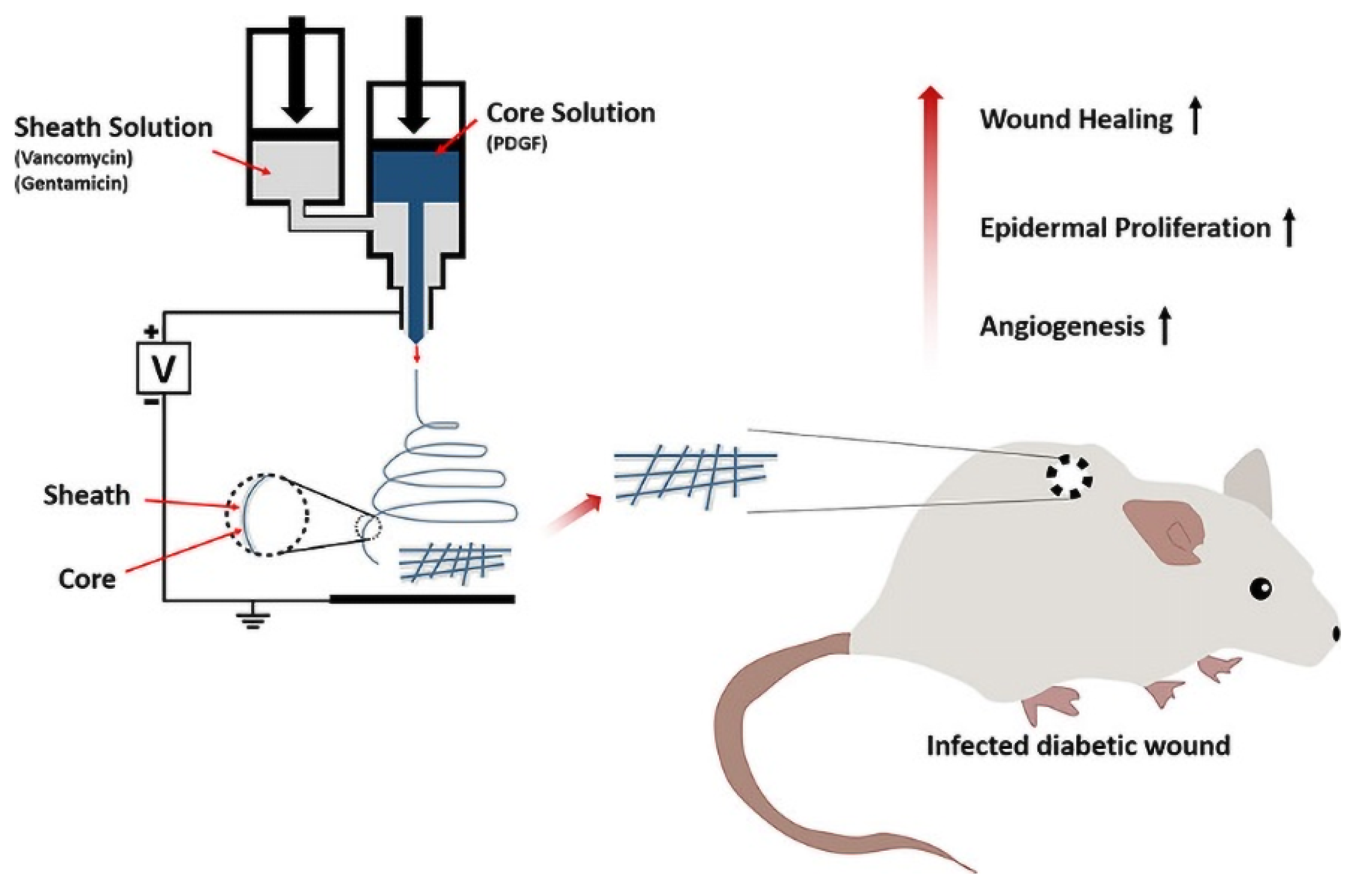
- Lee, C.H.; Liu, K.S.; Cheng, C.W.; Chan, E.C.; Hung, K.C.; Hsieh, M.J.; Chang, S.H.; Fu, X.; Juang, J.H.; Hsieh, I.C.; et al. Codelivery of Sustainable Antimicrobial Agents and Platelet-Derived Growth Factor via Biodegradable Nanofibers for Repair of Diabetic Infectious Wounds. ACS Infect. Dis. 2020, 6, 2688–2697. [Google Scholar] [CrossRef]
- Dwivedi, C.; Pandey, I.; Pandey, H.; Patil, S.; Mishra, S.B.; Pandey, A.C.; Zamboni, P.; Ramteke, P.W.; Singh, A.V. In vivo diabetic wound healing with nanofibrous scaffolds modified with gentamicin and recombinant human epidermal growth factor. J. Biomed. Mater. Res. A 2018, 106, 641–651. [Google Scholar] [CrossRef]
- Yin, H.; Ding, G.; Shi, X.; Guo, W.; Ni, Z.; Fu, H.; Fu, Z. A bioengineered drug-Eluting scaffold accelerated cutaneous wound healing In diabetic mice. Colloids Surf. B Biointerfaces 2016, 145, 226–231. [Google Scholar] [CrossRef] [PubMed]
- Cheng, P.K.; Chen, X.L.; Su, X.X.; Su, X.J.; Hou, C.L. A novel dressing seeded with embryonic artery CD133(+) cells and loaded with the Sirt1 agonist SRT1720 accelerates the healing of diabetic ischemic ulcers. Exp. Ther. Med. 2018, 15, 5243–5250. [Google Scholar] [CrossRef] [PubMed]
- Yu, B.; He, C.; Wang, W.; Ren, Y.; Yang, J.; Guo, S.; Zheng, Y.; Shi, X. Asymmetric Wettable Composite Wound Dressing Prepared by Electrospinning with Bioinspired Micropatterning Enhances Diabetic Wound Healing. ACS Appl. Bio Mater. 2020, 3, 5383–5394. [Google Scholar] [CrossRef]
- Shin, Y.C.; Shin, D.M.; Lee, E.J.; Lee, J.H.; Kim, J.E.; Song, S.H.; Hwang, D.Y.; Lee, J.J.; Kim, B.; Lim, D.; et al. Hyaluronic Acid/PLGA Core/Shell Fiber Matrices Loaded with EGCG Beneficial to Diabetic Wound Healing. Adv. Healthc. Mater. 2016, 5, 3035–3045. [Google Scholar] [CrossRef] [PubMed]
- Khazaeli, P.; Alaei, M.; Khaksarihadad, M.; Ranjbar, M. Preparation of PLA/chitosan nanoscaffolds containing cod liver oil and experimental diabetic wound healing in male rats study. J Nanobiotechnology 2020, 18, 176. [Google Scholar] [CrossRef]
- Chouhan, D.; Janani, G.; Chakraborty, B.; Nandi, S.K.; Mandal, B.B. Functionalized PVA-silk blended nanofibrous mats promote diabetic wound healing via regulation of extracellular matrix and tissue remodelling. J. Tissue Eng. Regen. Med. 2018, 12, e1559–e1570. [Google Scholar] [CrossRef]
- Chen, Y.P.; Lo, T.S.; Lin, Y.T.; Chien, Y.H.; Lu, C.J.; Liu, S.J. Fabrication of Drug-Eluting Polycaprolactone/poly(lactic-co-glycolic Acid) Prolapse Mats Using Solution-Extrusion 3D Printing and Coaxial Electrospinning Techniques. Polymers 2021, 13, 2295. [Google Scholar] [CrossRef]
- Alhusein, N.; De Bank, P.A.; Blagbrough, I.S.; Bolhuis, A. Killing bacteria within biofilms by sustained release of tetracycline from triple-layered electrospun micro/nanofibre matrices of polycaprolactone and poly(ethylene-co-vinyl acetate). Drug Deliv. Transl. Res. 2013, 3, 531–541. [Google Scholar] [CrossRef] [Green Version]
- Zhu, T.; Park, H.C.; Son, K.M.; Yang, H.C. Effects of dimethyloxalylglycine on wound healing of palatal mucosa in a rat model. BMC Oral Health 2015, 15, 60. [Google Scholar] [CrossRef] [Green Version]
- Ren, X.; Han, Y.; Wang, J.; Jiang, Y.; Yi, Z.; Xu, H.; Ke, Q. An aligned porous electrospun fibrous membrane with controlled drug delivery—An efficient strategy to accelerate diabetic wound healing with improved angiogenesis. Acta Biomater. 2018, 70, 140–153. [Google Scholar] [CrossRef]
- Lawall, H.; Diehm, C. Diabetic foot syndrome from the perspective of angiology and diabetology. Orthopade 2009, 38, 1149–1159. [Google Scholar] [CrossRef]
- Zhao, W.N.; Xu, S.Q.; Liang, J.F.; Peng, L.; Liu, H.L.; Wang, Z.; Fang, Q.; Wang, M.; Yin, W.Q.; Zhang, W.J.; et al. Endothelial progenitor cells from human fetal aorta cure diabetic foot in a rat model. Metabolism 2016, 65, 1755–1767. [Google Scholar] [CrossRef]
- Franz, S.; Allenstein, F.; Kajahn, J.; Forstreuter, I.; Hintze, V.; Moller, S.; Simon, J.C. Artificial extracellular matrices composed of collagen I and high-sulfated hyaluronan promote phenotypic and functional modulation of human pro-inflammatory M1 macrophages. Acta Biomater. 2013, 9, 5621–5629. [Google Scholar] [CrossRef] [PubMed]
- Khanna, P.K.; Nair, C.K.K. Synthesis of Silver Nanoparticles Using Cod Liver Oil (Fish Oil): Green Approach to Nanotechnology. Int. J. Green Nanotechnol. Phys. Chem. 2009, 1, P3–P9. [Google Scholar] [CrossRef]
- Jafari, A.; Amirsadeghi, A.; Hassanajili, S.; Azarpira, N. Bioactive antibacterial bilayer PCL/gelatin nanofibrous scaffold promotes full-thickness wound healing. Int. J. Pharm. 2020, 583, 119413. [Google Scholar] [CrossRef] [PubMed]
- Gunal, O.; Tuncel, U.; Turan, A.; Barut, S.; Kostakoglu, N. The Use of Vacuum-Assisted Closure and GranuFoam Silver(R) Dressing in the Management of Diabetic Foot Ulcer. Surg. Infect. 2015, 16, 558–565. [Google Scholar] [CrossRef]
- Liu, Z.; Peng, Y.; Yang, L.; Zhang, G. Poly(lactic-co-glycolic acid)-Chitosan-Gelatin Composite Nanomaterials for the Treatment of Diabetic Foot Ulcer Wound Infection. J. Nanosci. Nanotechnol. 2021, 21, 1070–1078. [Google Scholar] [CrossRef] [PubMed]
- Kim, S.E.; Tiwari, A.P. Three dimensional polycaprolactone/cellulose scaffold containing calcium-based particles: A new platform for bone regeneration. Carbohydr. Polym. 2020, 250, 116880. [Google Scholar] [CrossRef]
- Moon, J.Y.; Lee, J.; Hwang, T.I.; Park, C.H.; Kim, C.S. A multifunctional, one-step gas foaming strategy for antimicrobial silver nanoparticle-decorated 3D cellulose nanofiber scaffolds. Carbohydr. Polym. 2021, 273, 118603. [Google Scholar] [CrossRef]
- Kim, J.H.; Unnithan, A.R.; Kim, H.J.; Tiwari, A.P.; Park, C.H.; Kim, C.S. Electrospun badger (Meles meles) oil/Ag nanoparticle based anti-bacterial mats for biomedical applications. J. Ind. Eng. Chem. 2015, 30, 254–260. [Google Scholar] [CrossRef]
- Aljohani, M.; Alkabli, J.; Abualnaja, M.M.; Alrefaei, A.F.; Almehmadi, S.J.; Mahmoud, M.H.H.; El-Metwaly, N.M. Electrospun AgNPs-polylactate nanofibers and their antimicrobial applications. React. Funct. Polym. 2021, 167, 104999. [Google Scholar] [CrossRef]
- Zienkiewicz-Strzalka, M.; Derylo-Marczewska, A.; Skorik, Y.A.; Petrova, V.A.; Choma, A.; Komaniecka, I. Silver Nanoparticles on Chitosan/Silica Nanofibers: Characterization and Antibacterial Activity. Int. J. Mol. Sci. 2019, 21, 166. [Google Scholar] [CrossRef] [PubMed] [Green Version]


| Name of Electrospun Fiber Membrane | Spinning Polymers | Active Ingredient | Mechanism of Action | Reference |
|---|---|---|---|---|
| βG-loaded hydroxypropyl methylcellulose (HPMC)/PEO nanofiber | HPMC, PEO | βG | βG activates the innate immune system by binding to dectin-1 receptors on macrophages, dendritic cells, and neutrophils to transform macrophages from M1 to M2. | [72] |
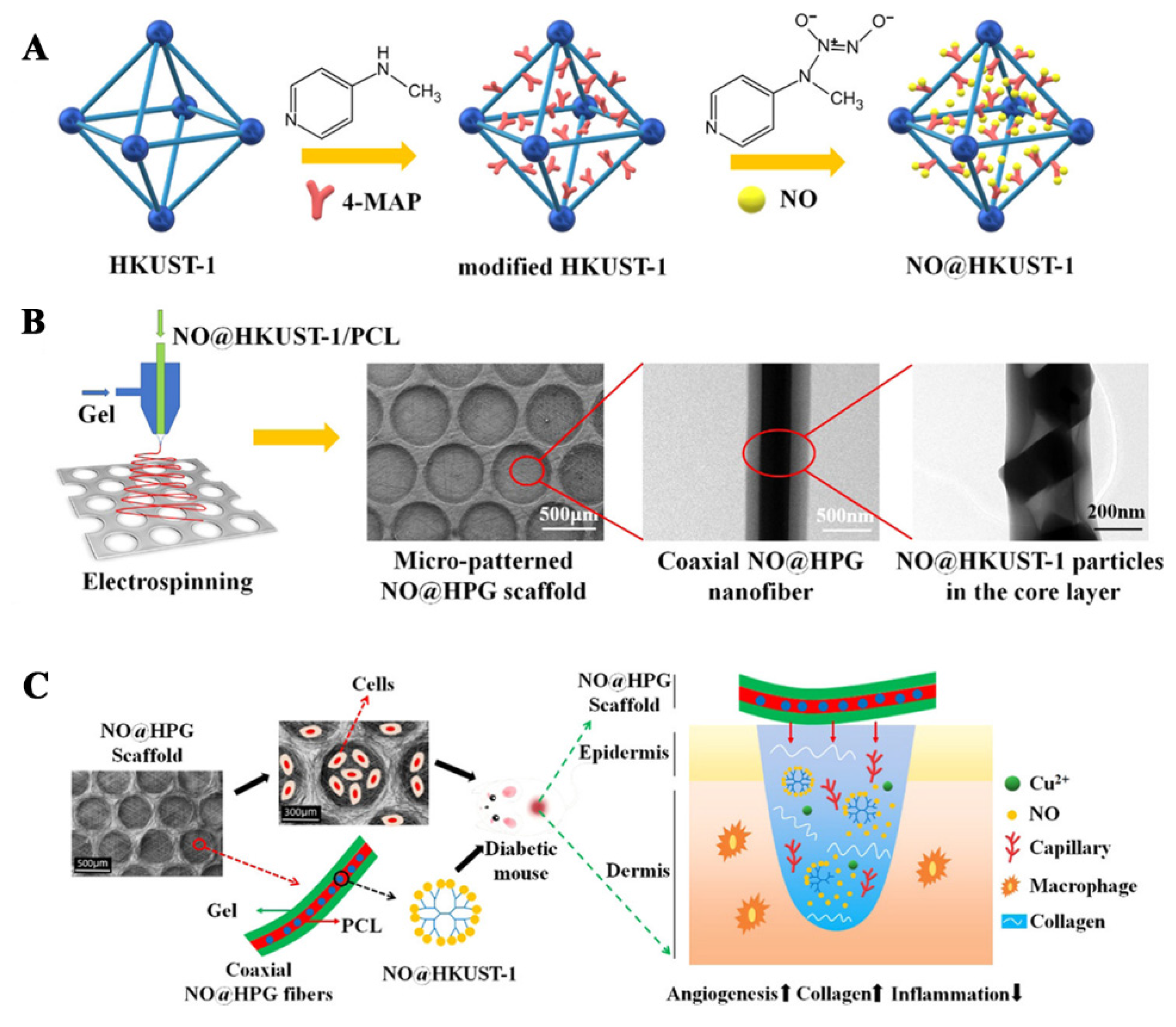
| NO@HKUST-1 (MOFs)/PCL/Gel nanofibrous membranes | PCL, Gel | NO-loaded HKUST-1 particles | NO@HKUST-1/PCL/Gel nanofiber membrane can promote angiogenesis and inhibit inflammation. Cu2+ released by HKUST-1 and NO can cooperatively promote endothelial cell growth. | [73] |
| BGs nanofibers (BGnf) | Polyvinyl butyral (PVB) | BGs | BGs can change the cell microenvironment by releasing Si4+, which stimulates expression of hypoxia inducible factor-α (HIF-α) and thus promotes the angiogenesis of endothelial cells | [74] |
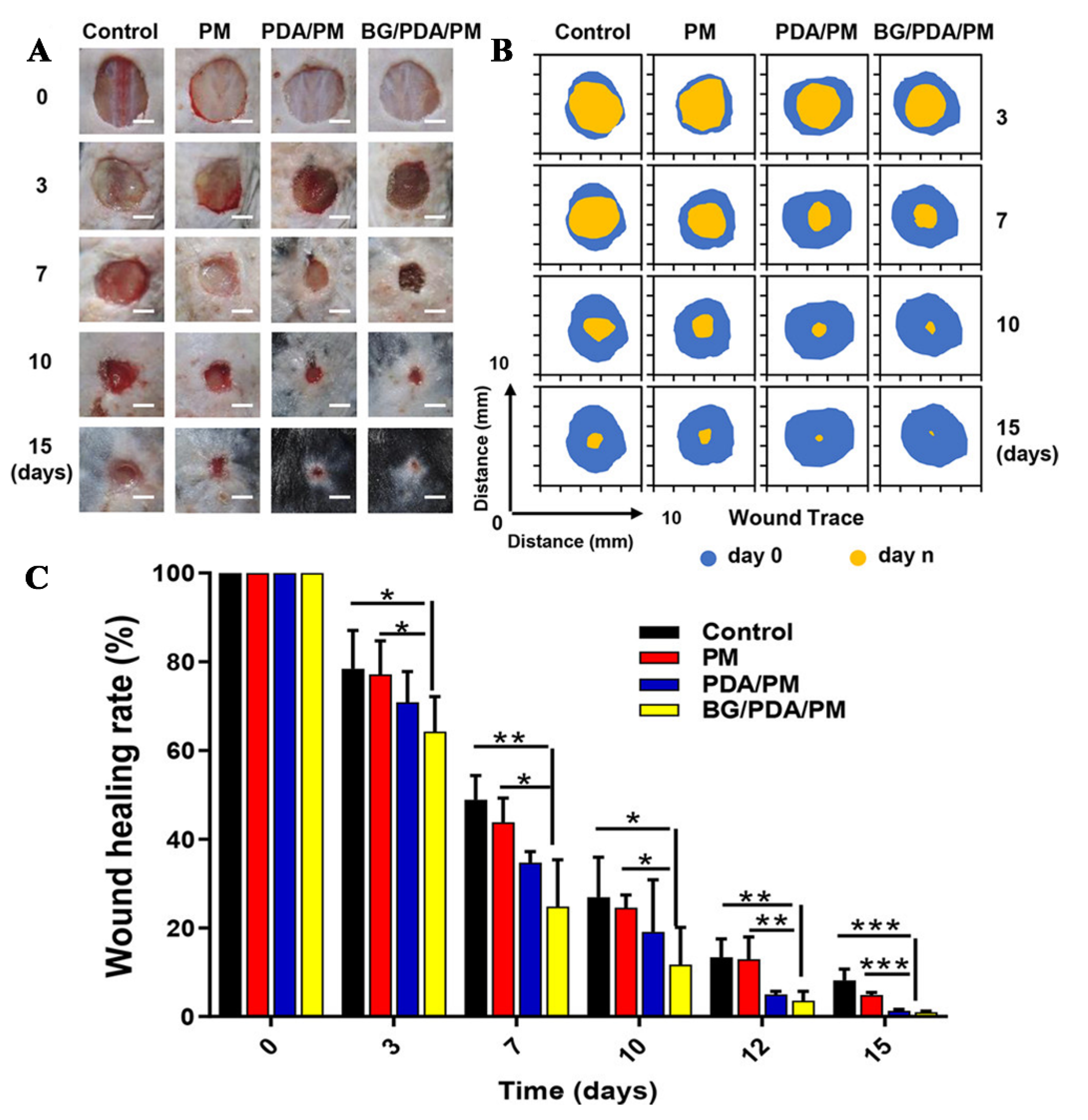
| BGs loaded polydopamine (PDA)-modified polylactic acid (PLA)/PCL nanofibrous membranes (BGs/PDA/PM) | PLA, PCL | BGs | Si4+ released from BGs/PDA/PM nanofiber membranes can stimulate expression of HIF-α and promote angiogenesis | [75] |
| SPC-loaded PCL nanofibrous membranes | PCL | SPC | Hypoxia inducible factor-1α (HIF-1α) promotes diabetic wound healing by promoting angiogenesis. Long-term hypoxia will cause HIF-1α deficiency. The oxygen supply of SPC plays an important role in diabetic wound healing | [76] |
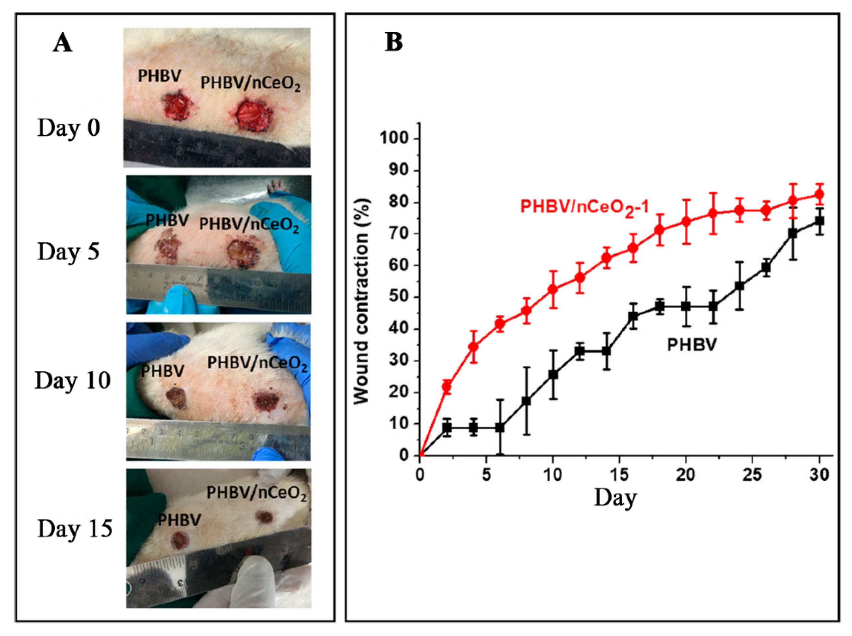
| nCeO2-incorporated poly (3-hydroxybutyrae-co-3- hydroxyvalerate) (PHBV) membranes | PHBV | nCeO2 | During the inflammation phase, ROS produced by nCeO2 can inhibit bacterial growth and promote diabetic wound healing | [77] |
| BGs@PLA/Gel nanofibrous membranes | PLA, Gel | BGs | Si4+ released from BGs@PLA /Gel nanofiber membrane can up-regulate expression of hypoxia inducible factor-1 (HIF-1), and thus up-regulate expression of pro-angiogenic factors such as bFGF and VEGF | [78] |
| BGs-incorporated CS-PVA trilayer nanofibrous membrane (BGs-TFM) | PVA, CS | BGs | BGs-TFM up-regulates growth factors VEGF and TGF-β, down-regulates inflammatory factors TNF-α and IL-1β, and promoted epithelial regeneration and collagen deposition | [79] |
| PCL/gel nanofibrous composite scaffold containing silicate-based bioceramic particles (NAGEL) | PCL, Gel | NAGEL | PCL/Gel nanofibrous composite scaffold can promote diabetic wound healing by promoting angiogenesis, collagen deposition, re-epithelialization, and inhibiting inflammation | [80] |
| CS/PVA/ZnO nanofibrous membranes | PVA, CS | ZnO nanoparticles | ZnO nanoparticles have bactericidal properties, and the porous structure of the fiber membrane can promote the proliferation of fibroblasts and the recruitment of macrophages, and thus accelerate wound contraction | [81] |
| Name of Electrospun Fiber Membrane | Spinning Polymers | Active Ingredient | Mechanism of Action | Reference |
|---|---|---|---|---|
| Sesamol-loaded cellulose acetate (CA) /zein nanofiber membranes | CA, zein | Sesamol | Sesamol can down-regulate expression of inflammatory cytokines, such as IL-1β and TNF-α, and up-regulate expression of interleukin-6 (IL-6) (anti-inflammatory cytokines) | [83] |
| Bi-layered fibrin/ poly(ether)urethane scaffold loaded with PL | poly(ether)urethane | PL | PDGF and VEGF released by PL can promote collagen deposition and re-epithelialization, thus promote diabetic wound healing | [84] |
| PVA-connective tissue growth factor (CTGF) /PLA core/shell nanofibrous membranes | PVA, PLA | CTGF | PVA-CTGF/PLA core/shell nanofibrous membranes are conducive to the proliferation and migration of fibroblasts, keratinocytes and other cells, which are beneficial to diabetic wound healing | [85] |
| 17BIPHE2-PCL /pluronic F127 core/shell nanofibers | PCL, pluronic F127 | Antimicrobial peptide 17BIPHE2 | 17BIPHE2-PCL/pluronic F127 core/shell nanofibers promote wound healing by removing bacterial biofilms from diabetic wounds | [86] |
| Poly (acrylic acid) (PAA)/polyvinyl pyrrolidone (PVP)-CFX /PCL triple-layered nanofibrous membranes | PAA, PVP, PCL | CFX | CFX has antibacterial effects on gram-negative and gram-positive bacteria. The antibacterial activity of PAA/PVP-CFX/PCL nanofibrous membranes gives it the potential to promote diabetic wound healing | [87] |
| DCH-loaded PLA nanofibrous membranes | PLA | DCH | High levels of MMPs and TNF-α converting enzyme (TACE) can prevent wound healing, and DCH can inhibit the activity of MMPs and TACE, thus promoting diabetic wound healing | [88] |
| Dimethyloxalylglycine (DMOG)-embedded PCL fiber membranes | PCL | DMOG | DMOG is a small molecule inhibitor of non-specific prolyl hydroxylases, which can inhibit the decomposition of HIF-α, create a cellular microenvironment similar to hypoxia, thus accelerating wound healing by activating angiogenesis and fiber regeneration | [89] |
| Repaglinide-loaded PVA/PVP nanofibers | PVA, PVP | Repaglinide | Repaglinide-loaded PVA/PVP nanofibers can solve the problems of poor water solubility and unstable drug absorption of hypoglycemic drug repaglinide, significantly reduce blood glucose level, and promote diabetic wound healing | [90] |
| Bioactive antibiotics and PDGF loaded PDGF/PLGA-antibiotic core/sheath nanofibrous | PLGA | PDGF, gentamicin, vancomycin | PDGF/PLGA-antibiotic core/sheath nanofibrous promote angiogenesis and epidermal hyperplasia through the synergistic effect of PDGF and antibiotics | [91] |
| Gentamicin sulfate (GS) and recombinant human epidermal growth factor (rhEGF) co-loaded Eudragit RL/RS nanofibers | Eudragit RL-100 and Eudragit RS-100 | GS, rhEGF | Bacterial inhibitor GS can reduce inflammation of diabetic wounds, and rhEGF can promote granulation tissue formation and angiogenesis at the wound | [92] |
| Monocyte chemoattractant protein-1(MCP-1) loaded polyglycolic acid (PGA)-Gel electrospun scaffold | PGA, Gel | MCP-1 | MCP-1 promotes macrophages to participate in the wound healing process, thus the growth factors VEGF and PDGF secreted by macrophages can promote wound healing | [93] |
| Sirt1 agonist (SRT1720) loaded PLGA/collagen protein /silk membranes inoculated with embryonic artery cluster of differentiation 133+ cells (EACCs) (PCSS-EACCs) | PLGA, collagen protein, silk | SRT1720, EACCs | PCSS-EACCs can steadily release SRT1720 for 15 days, thus improving the survival rate of EACCs in a high glucose environment. The release of vascular endothelial growth factor A (VEGFA) and interleukin-8 (IL-8) from EACCs ultimately promote endothelial cell proliferation, migration, and angiogenesis | [94] |
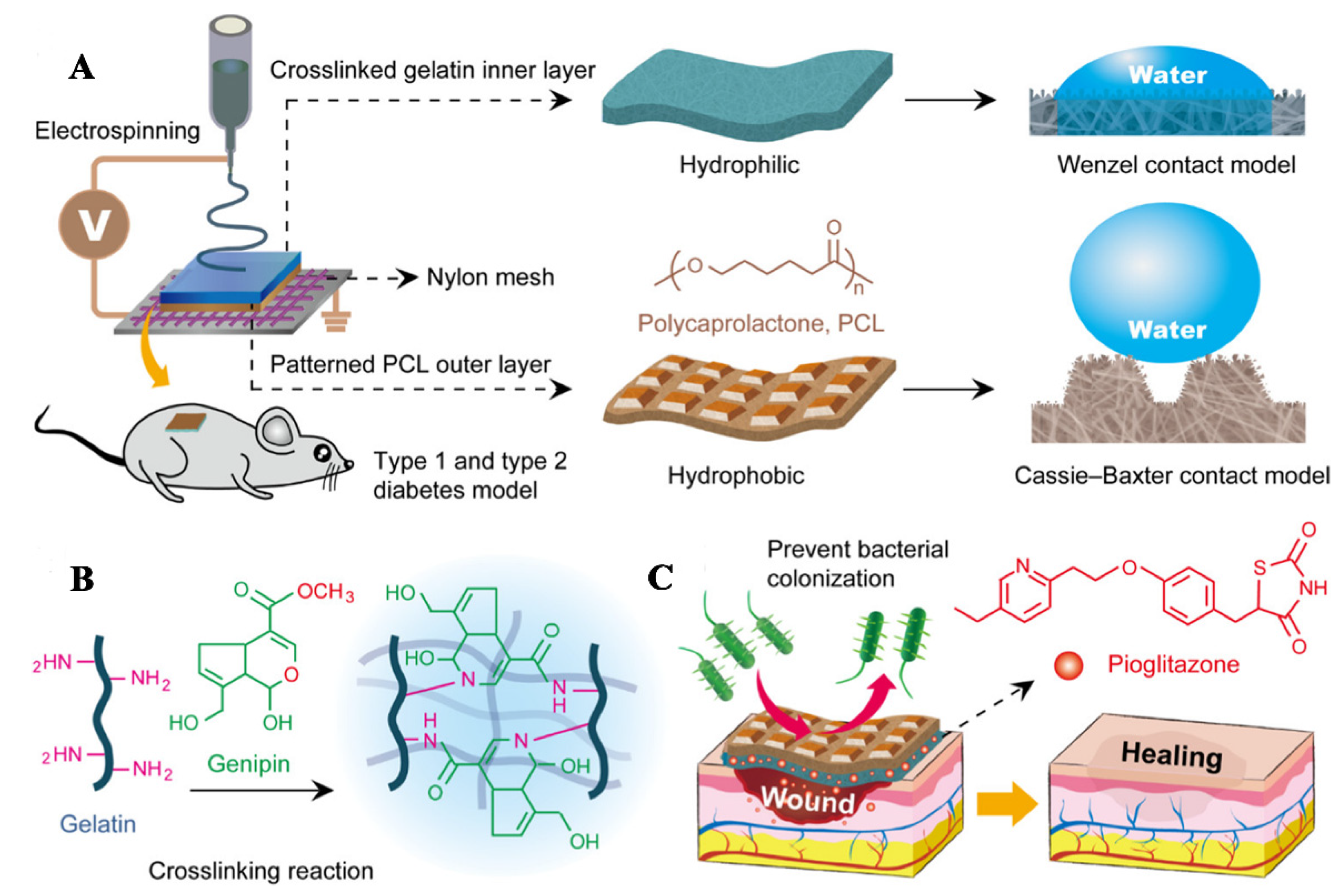
| PCL/Gel-pioglitazone nanofibrous membranes | PCL, Gel | Pioglitazone | PCL/Gel-pioglitazone nanofibrous membranes reduce expression of MMP-9, IL-1β, and IL-6 to reduce wound inflammation, and upregulate expression of macrophage inflammatory protein-2 (MIP-2), TNF-α, and VEGF to promote wound healing | [95] |
| Hyaluronic acid (HA) /PLGA core/shell fiber loaded with EGCG | PLGA, HA | EGCG | EGCG can promote diabetic wound healing by promoting capillary formation and epithelial cell proliferation | [96] |
| PLA/CS nanoscaffolds containing cod liver oil | PLA, CS | Cod liver oil | Cod liver oil enhances the activity of the growth factor, promotes cell differentiation, reduces inflammation, and increase the production of IL-1, which promotes diabetic wound healing | [97] |
| EGF, bFGF, antimicrobial peptide LL-37 co-loaded PVA- Silk fibroin (SF) nanofiber membrane | PVA, SF | EGF, bFGF, antimicrobial peptide LL-37 | EGF and bFGF can promote the proliferation of fibroblasts, keratinocytes, and endothelial cells, antimicrobial peptide LL-37 can reduce the inflammation of the wound, EGF, bFGF, and antimicrobial peptide LL-37 can cooperate to promote diabetic wound healing | [98] |
Publisher’s Note: MDPI stays neutral with regard to jurisdictional claims in published maps and institutional affiliations. |
© 2021 by the authors. Licensee MDPI, Basel, Switzerland. This article is an open access article distributed under the terms and conditions of the Creative Commons Attribution (CC BY) license (https://creativecommons.org/licenses/by/4.0/).
Share and Cite
Gao, Z.; Wang, Q.; Yao, Q.; Zhang, P. Application of Electrospun Nanofiber Membrane in the Treatment of Diabetic Wounds. Pharmaceutics 2022, 14, 6. https://doi.org/10.3390/pharmaceutics14010006
Gao Z, Wang Q, Yao Q, Zhang P. Application of Electrospun Nanofiber Membrane in the Treatment of Diabetic Wounds. Pharmaceutics. 2022; 14(1):6. https://doi.org/10.3390/pharmaceutics14010006
Chicago/Turabian StyleGao, Zhaoju, Qiuxiang Wang, Qingqiang Yao, and Pingping Zhang. 2022. "Application of Electrospun Nanofiber Membrane in the Treatment of Diabetic Wounds" Pharmaceutics 14, no. 1: 6. https://doi.org/10.3390/pharmaceutics14010006
APA StyleGao, Z., Wang, Q., Yao, Q., & Zhang, P. (2022). Application of Electrospun Nanofiber Membrane in the Treatment of Diabetic Wounds. Pharmaceutics, 14(1), 6. https://doi.org/10.3390/pharmaceutics14010006