
Polymeric Lipid Hybrid Nanoparticles (PLNs) as Emerging Drug Delivery Platform—A Comprehensive Review of Their Properties, Preparation Methods, and Therapeutic Applications

 , ,
, ,
Abstract
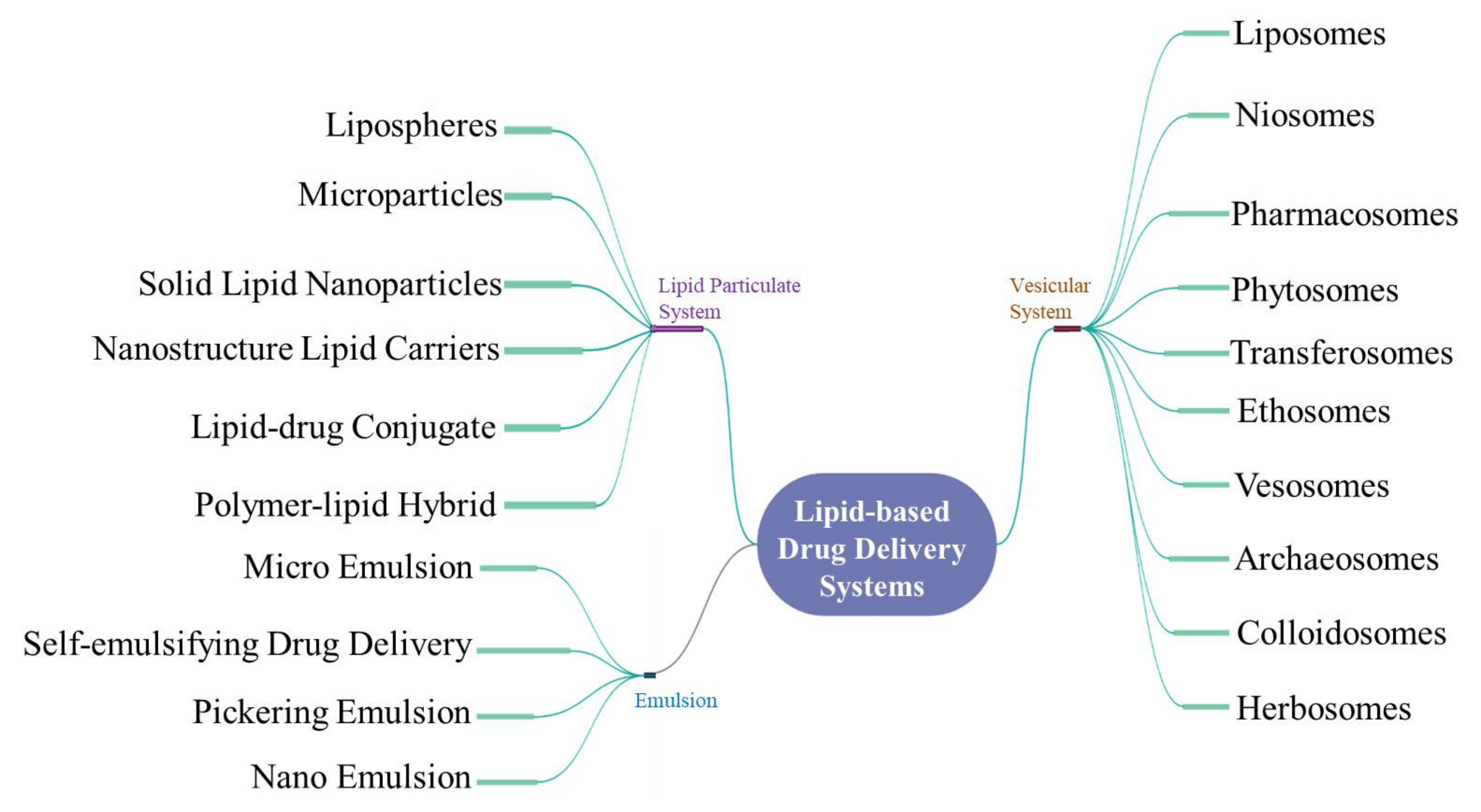
:1. Introduction of Lipid-Based Drug Delivery Systems
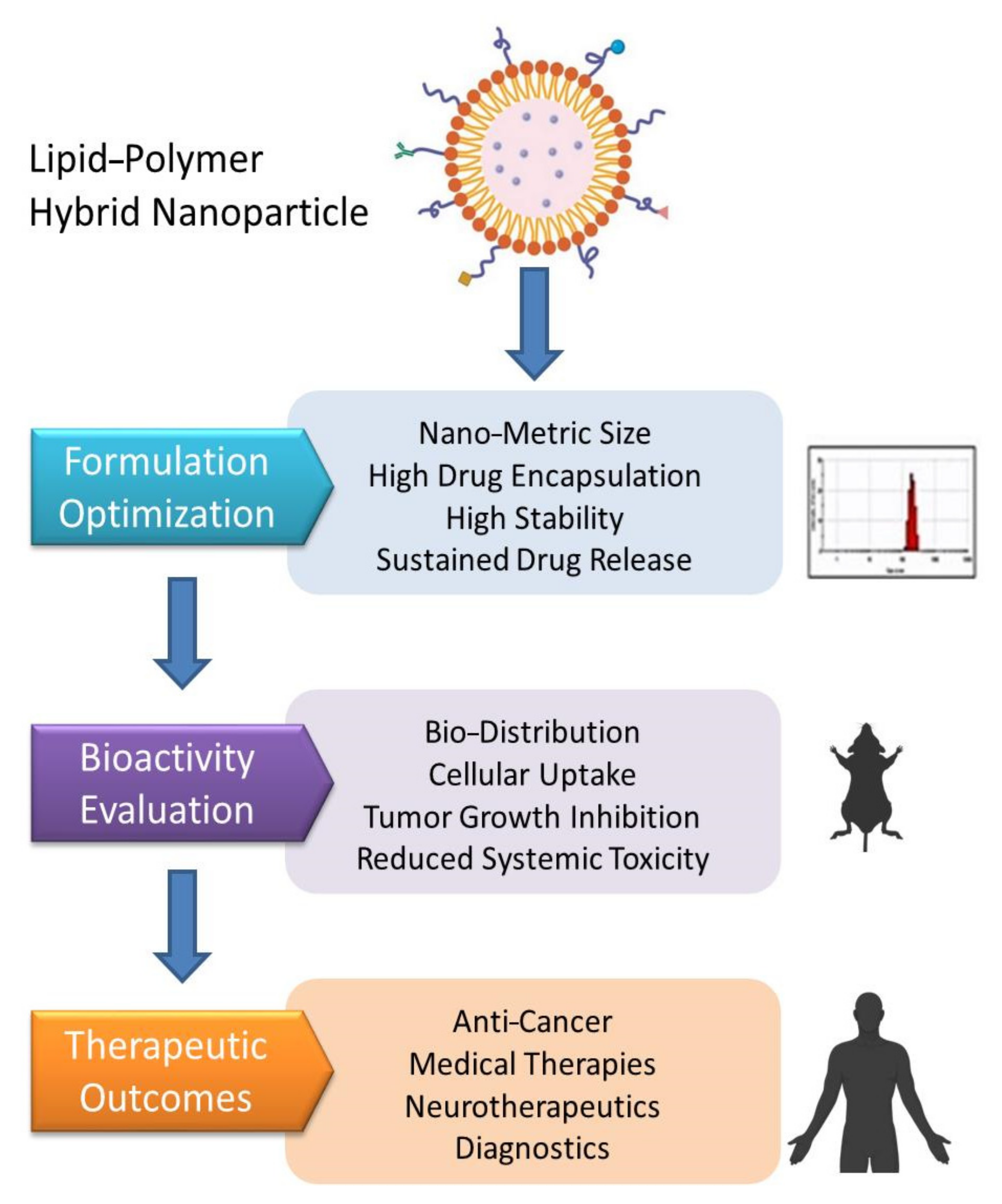
2. The Polymeric Lipid Hybrid Nanoparticles (PLNs)
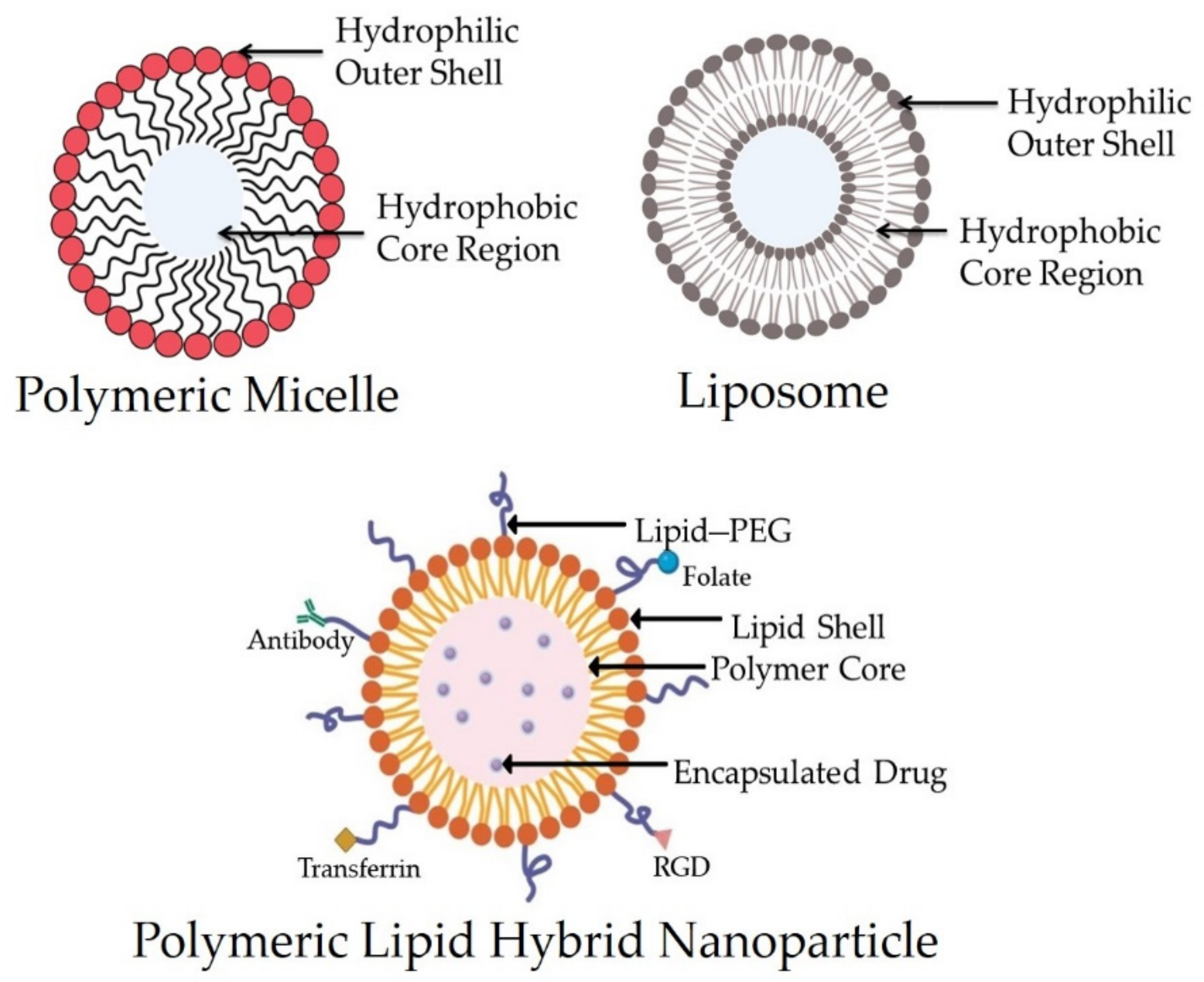
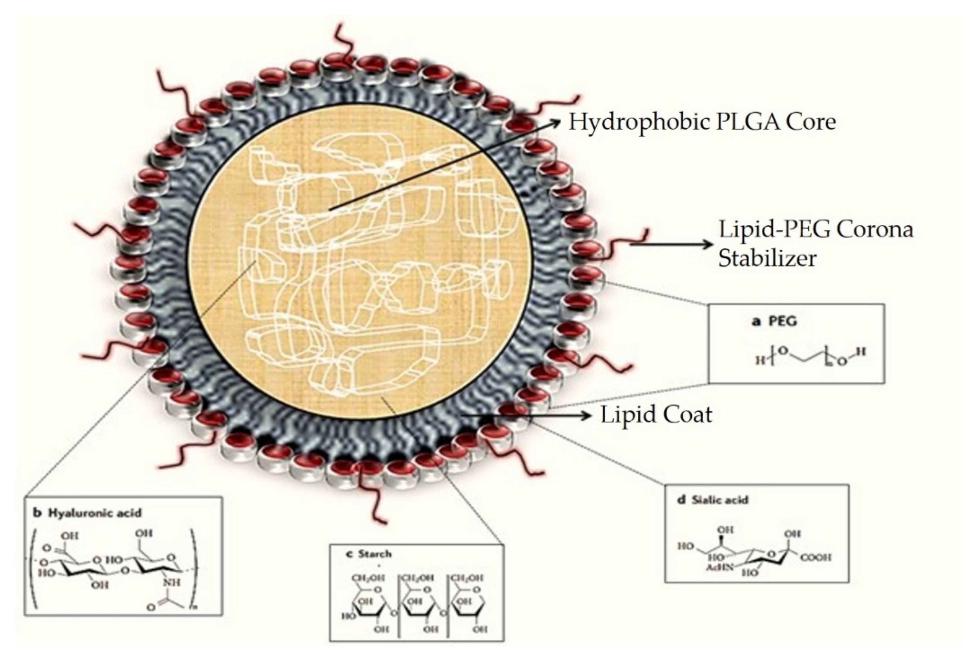
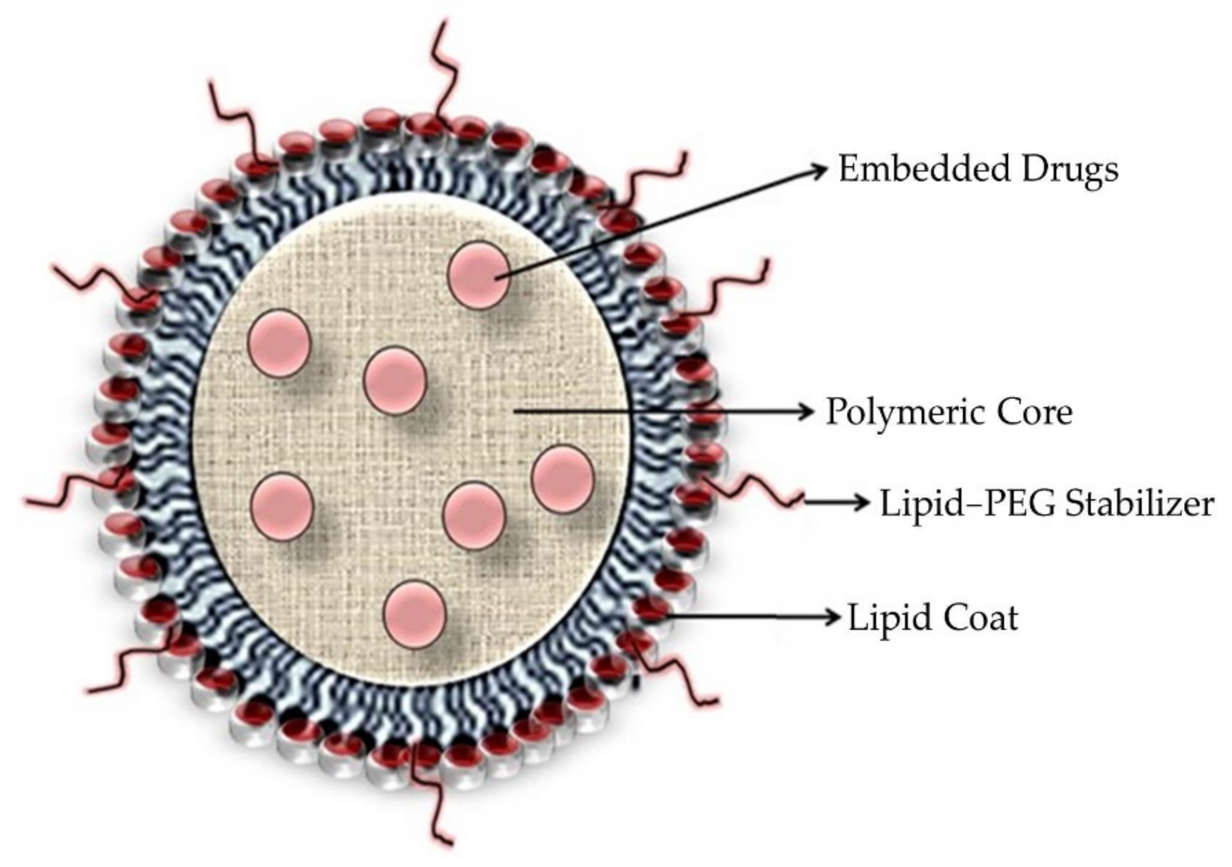
2.1. Structure and Components of PLNs
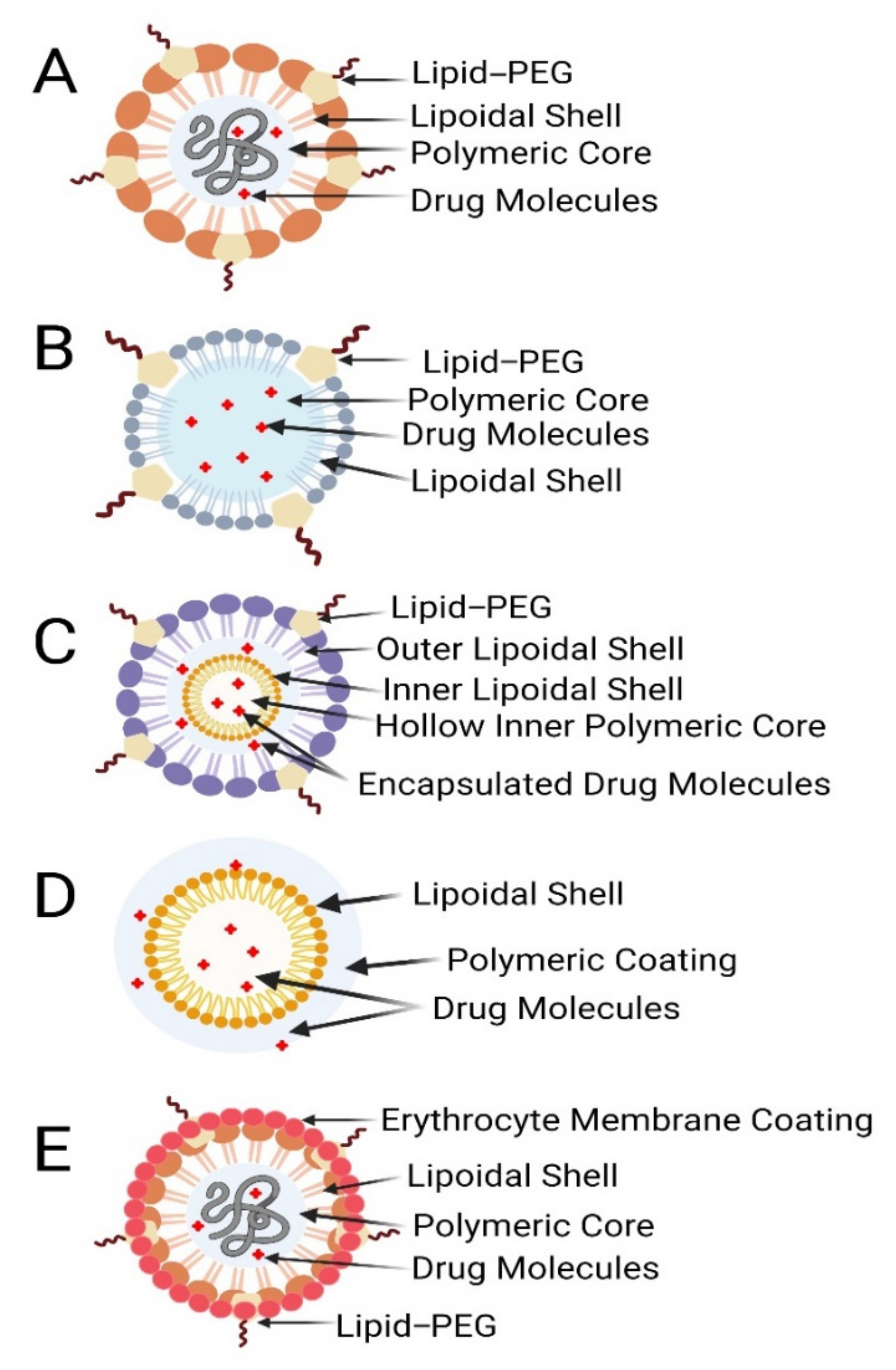
2.2. Advantages and Classification of PLNs
- Enhanced stability and better biocompatibility owing to the lipid–PEG shell;
- Controlled or regulated delivery because of the polymeric core.
- Polymer core–lipid shell hybrid NPs;
- Lipid-bilayer-coated NPs;
- Polymer-caged nanobins;
- Monolithic PLN/mixed polymer–lipid hybrid NPs;
- Hollow-core NP/lipid–polymer–lipid NPs.

| Type of PLNs | Advantages | Disadvantages |
|---|---|---|
| Polymer core–lipid shell type | Improved encapsulation efficiency over liposomes. Optimization of core–shell layer will promote sustained release profile of drugs. Lipophilic and hydrophilic drugs can be easily entrapped | Poor drug-loading and entrapment efficiency |
| Core–shell-type hollow lipid–polymer nanoparticles | Delivery of si-RNA and mRNA by reducing its susceptibility to serum nucleases and phagocytic uptake | Toxicity of cationic lipids arises due to inflammation and tissue damage, rapid inactivation of cationic lipids in the presence of serum |
| Polymer-caged nanobins | Deliver cytotoxic chemotherapeutic agents to the targeted site, thereby reducing the systemic toxicity | Scale-up of polymer-caged nanobins is in its primitive stage |
| Glued cell membrane camouflaged polymeric nanoparticles | Promote extensive systemic circulation, active targeting of drugs to the site of action | Large-scale production with batch- to-batch variation is a concern; quality control is another major challenge |
2.3. Methods of Preparation of PLNs
2.4. Characterization of PLNs
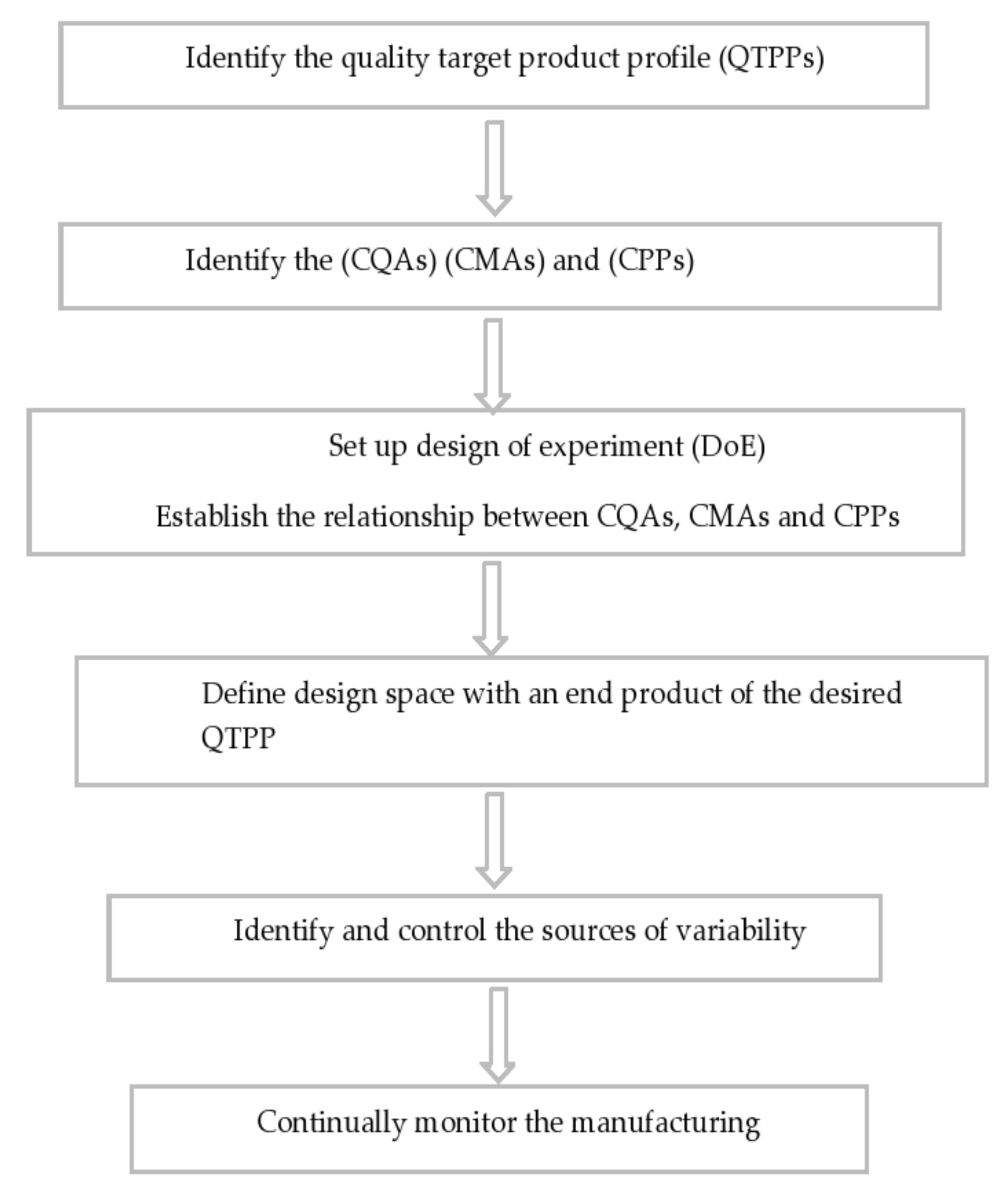
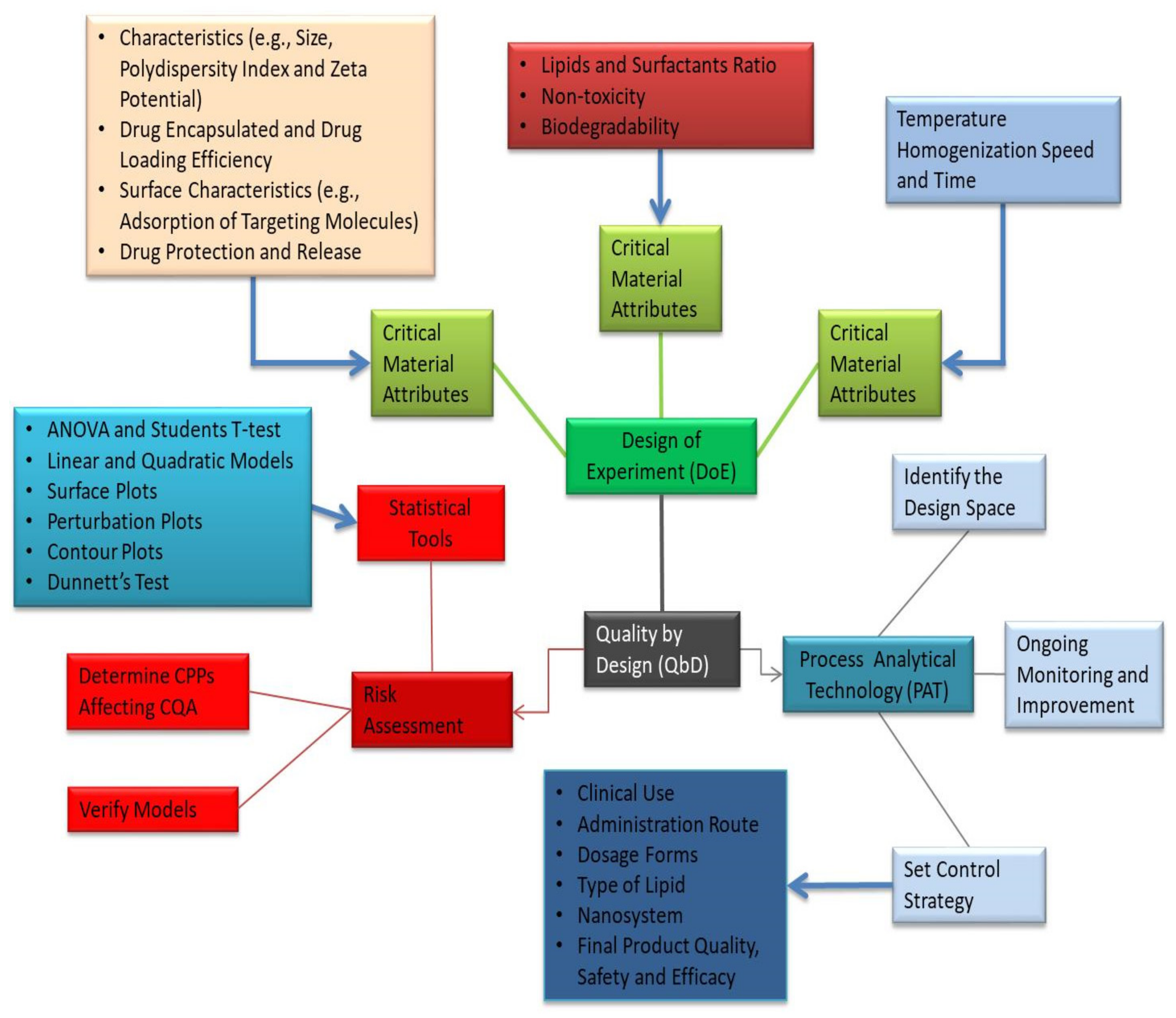
3. Optimization of Formulation Parameters by Quality by Design Approach (QbD) in the Development of PLNs
4. Drug Delivery Mechanisms of PLNs
5. Therapeutic Applications of PLNs
6. Recent Clinical Trials in PLN and Liposomal Nanocarrier-Based Therapy
7. Biopharmaceutical Aspects of PLNs
8. Pharmacokinetic Properties of PLNs
9. Limitation of PLNs
10. Conclusions and Future Perspectives
Author Contributions
Funding
Institutional Review Board Statement
Informed Consent Statement
Data Availability Statement
Conflicts of Interest
References
- Farokhzad, O.C.; Langer, R. Nanomedicine: Developing smarter therapeutic and diagnostic modalities. Adv. Drug Deliv. Rev. 2006, 58, 1456–1459. [Google Scholar] [CrossRef]
- Kamaly, N.; Xiao, Z.; Valencia, P.M.; Radovic-Moreno, A.F.; Farokhzad, O.C. Targeted polymeric therapeutic nanoparticles: Design, development and clinical translation. Chem. Soc. Rev. 2012, 41, 2971–3010. [Google Scholar] [CrossRef] [PubMed]
- Kamaly, N.; Yameen, B.; Wu, J.; Farokhzad, O.C. Degradable controlled-release polymers and polymeric nanoparticles: Mechanisms of controlling drug release. Chem. Rev. 2016, 116, 2602–2663. [Google Scholar] [CrossRef] [PubMed] [Green Version]
- Albanese, A.; Tang, P.S.; Chan, W.C. The effect of nanoparticle size, shape, and surface chemistry on biological systems. Annu. Rev. Biomed. Eng. 2012, 14, 1–16. [Google Scholar] [CrossRef] [PubMed] [Green Version]
- Alexis, F.; Pridgen, E.; Molnar, L.K.; Farokhzad, O.C. Factors affecting the clearance and biodistribution of polymeric nanoparticles. Mol. Pharm. 2008, 5, 505–515. [Google Scholar] [CrossRef] [PubMed] [Green Version]
- Burgess, P.; Hutt, P.B.; Farokhzad, O.C.; Langer, R.; Minick, S.; Zale, S. On firm ground: IP protection of therapeutic nanoparticles. Nat. Biotechnol. 2010, 28, 1267–1270. [Google Scholar] [CrossRef]
- Farokhzad, O.C.; Langer, R. Impact of nanotechnology on drug delivery. ACS Nano 2009, 3, 16–20. [Google Scholar] [CrossRef]
- Jyoti, K.; Naik, S.K.; Mandal, M.; Das, D.K. Performance of different sources of sulphur on the yield and quality of rapeseed (Brassica campestris L.). J. Indian Soc. Soil Sci. 2012, 60, 218–224. [Google Scholar]
- Cole, A.J.; Yang, V.C.; David, A.E. Cancer theranostics: The rise of targeted magnetic nanoparticles. Trends Biotechnol. 2011, 29, 323–332. [Google Scholar] [CrossRef] [Green Version]
- Attama, A.A.; Nkemnele, M.O. In vitro evaluation of drug release from self micro-emulsifying drug delivery systems using a biodegradable homolipid from Capra hircus. Int. J. Pharm. 2005, 304, 4–10. [Google Scholar] [CrossRef]
- Fouad, D.M.; Mohamed, M.B. Studies on the photo-catalytic activity of semiconductor nanostructures and their gold core–shell on the photodegradation of malathion. Nanotechnology 2011, 22, 455705. [Google Scholar] [CrossRef] [Green Version]
- Chime, S.A.; Onyishi, I.V. Lipid-based drug delivery systems (LDDS): Recent advances and applications of lipids in drug delivery. Afr. J. Pharm. Pharmacol. 2013, 7, 3034–3059. [Google Scholar] [CrossRef] [Green Version]
- Umeyor, C.E.; Kenechukwu, F.C.; Uronnachi, E.M.; Osonwa, U.E.; Nwakile, C.D. Solid lipid microparticles (SLMs): An effective lipid based technology for controlled drug delivery. Am. J. PharmTech Res. 2012, 2, 1–18. [Google Scholar]
- Arnold, Y.; Gonzalez, R.B.; Versace, H.; Kuentz, M. Comparison of different in vitro tests to assess oral lipid-based formulations using a poorly soluble acidic drug. J. Drug Deliv. Sci. Technol. 2010, 20, 143–148. [Google Scholar] [CrossRef]
- Aungst, B.J.; Saitoh, H.; Burcham, D.L.; Huang, S.M.; Mousa, S.A.; Hussain, M.A. Enhancement of the intestinal absorption of peptides and nonpeptides. J. Control. Release 1996, 41, 19–31. [Google Scholar] [CrossRef]
- Kumar, P.; Kumar, P.; Kumar, R.; Kumar, N.; Kumar, R. An overview on lipid based formulation for oral drug delivery. Drug Invent. Today 2010, 2, 390–395. [Google Scholar]
- Pang, X.; Zhitomirsky, I. Electrophoretic deposition of composite hydroxyapatite-chitosan coatings. Mater. Charact. 2007, 58, 339–348. [Google Scholar] [CrossRef]
- Moghimi, S.M.; Szebeni, J. Stealth liposomes and long circulating nanoparticles: Critical issues in pharmacokinetics, opsonization and protein-binding properties. Prog. Lipid Res. 2003, 42, 463–478. [Google Scholar] [CrossRef]
- Barenholz, Y.C. Doxil®—The first FDA-approved nano-drug: Lessons learned. J. Control. Release 2012, 160, 117–134. [Google Scholar] [CrossRef] [PubMed]
- Bulbake, U.; Doppalapudi, S.; Kommineni, N.; Khan, W. Liposomal formulations in clinical use: An updated review. Pharmaceutics 2017, 9, 12. [Google Scholar] [CrossRef] [PubMed]
- Petersen, G.H.; Alzghari, S.K.; Chee, W.; Sankari, S.S.; La-Beck, N.M. Meta-analysis of clinical and preclinical studies comparing the anticancer efficacy of liposomal versus conventional non-liposomal doxorubicin. J. Control. Release 2016, 232, 255–264. [Google Scholar] [CrossRef] [PubMed]
- Kim, T.; Murdande, S.; Gruber, A.; Kim, S. Sustained-release morphine for epidural analgesia in rats. J. ASA 1996, 85, 331–338. [Google Scholar] [CrossRef] [PubMed]
- Zhang, L.; Chan, J.M.; Gu, F.X.; Rhee, J.W.; Wang, A.Z.; Radovic-Moreno, A.F.; Alexis, F.; Langer, R.; Farokhzad, O.C. Self-assembled lipid−polymer hybrid nanoparticles: A robust drug delivery platform. ACS Nano 2008, 2, 1696–1702. [Google Scholar] [CrossRef] [PubMed] [Green Version]
- Kumari, M.; Mukherjee, A.; Chandrasekaran, N. Genotoxicity of silver nanoparticles in Allium cepa. Sci. Total Environ. 2009, 407, 5243–5246. [Google Scholar] [CrossRef]
- Zielińska, A.; Ferreira, N.R.; Feliczak-Guzik, A.; Nowak, I.; Souto, E.B. Loading, release profile and accelerated stability assessment of monoterpenes-loaded Solid Lipid Nanoparticles (SLN). Pharm. Dev. Technol. 2020, 25, 832–844. [Google Scholar] [CrossRef]
- Lockman, P.R.; Mumper, R.J.; Khan, M.A.; Allen, D.D. Nanoparticle technology for drug delivery across the blood-brain barrier. Drug. Dev. Ind. Pharm. 2002, 28, 1–13. [Google Scholar] [CrossRef]
- Hallan, S.S.; Kaur, P.; Kaur, V.; Mishra, N.; Vaidya, B. Lipid polymer hybrid as emerging tool in nanocarriers for oral drug delivery. Artif. Cells Nanomed. Biotechnol. 2016, 44, 334–349. [Google Scholar] [CrossRef]
- Wu, X.Y. Strategies for optimizing polymer-lipid hybrid nanoparticle-mediated drug delivery. Expert Opin. Drug Deliv. 2016, 13, 609–612. [Google Scholar] [CrossRef]
- Li, Q.; Cai, T.; Huang, Y.; Xia, X.; Cole, S.P.; Cai, Y. A review of the structure, preparation, and application of NLCs, PNPs, and PLNs. Nanomaterials 2017, 7, 122. [Google Scholar] [CrossRef]
- Chan, J.M.; Zhang, L.; Yuet, K.P.; Liao, G.; Rhee, J.W.; Langer, R.; Farokhzad, O.C. PLGA–lecithin–PEG core–shell nanoparticles for controlled drug delivery. Biomaterials 2009, 30, 1627–1634. [Google Scholar] [CrossRef]
- Salvador-Morales, C.; Zhang, L.; Langer, R.; Farokhzad, O.C. Immunocompatibility properties of lipid–polymer hybrid nanoparticles with heterogeneous surface functional groups. Biomaterials 2009, 30, 2231–2240. [Google Scholar] [CrossRef] [PubMed] [Green Version]
- Mandal, B.; Bhattacharjee, H.; Mittal, N.; Sah, H.; Balabathula, P.; Thoma, L.A.; Wood, G.C. Core–shell-type lipid–polymer hybrid nanoparticles as a drug delivery platform. Nanomed. Nanomed.-Nanotechnol. 2013, 9, 474–491. [Google Scholar] [CrossRef] [PubMed]
- Thevenot, J.; Troutier, A.L.; David, L.; Delair, T.; Ladavière, C. Steric stabilization of lipid/polymer particle assemblies by poly (ethylene glycol)-lipids. Biomacromolecules 2007, 8, 3651–3660. [Google Scholar] [CrossRef]
- Dong, W.; Wang, X.; Liu, C.; Zhang, X.; Zhang, X.; Chen, X.; Kou, Y.; Mao, S. Chitosan based polymer-lipid hybrid nanoparticles for oral delivery of enoxaparin. Int. J. Pharm. 2018, 547, 499–505. [Google Scholar] [CrossRef]
- Fang, D.L.; Chen, Y.; Xu, B.; Ren, K.; He, Z.Y.; He, L.L.; Lei, Y.; Fan, C.M.; Song, X.R. Development of lipid-shell and polymer core nanoparticles with water-soluble salidroside for anti-cancer therapy. Int. J. Mol. Sci. 2014, 15, 3373–3388. [Google Scholar] [CrossRef] [Green Version]
- Bagre, A.P.; Jain, K.; Jain, N.K. Alginate coated chitosan core shell nanoparticles for oral delivery of enoxaparin: In vitro and in vivo assessment. Int. J. Pharm. 2013, 456, 31–40. [Google Scholar] [CrossRef]
- Hu, J.; Zhang, G.; Liu, S. Enzyme-responsive polymeric assemblies, nanoparticles and hydrogels. Chem. Soc. Rev. 2012, 41, 5933–5949. [Google Scholar] [CrossRef]
- Hu, C.M.; Zhang, L.; Aryal, S.; Cheung, C.; Fang, R.H.; Zhang, L. Erythrocyte membrane-camouflaged polymeric nanoparticles as a biomimetic delivery platform. Proc. Natl. Acad. Sci. USA 2011, 108, 10980–10985. [Google Scholar] [CrossRef] [PubMed] [Green Version]
- Lallana, E.; Sousa-Herves, A.; Fernandez-Trillo, F.; Riguera, R.; Fernandez-Megia, E. Click chemistry for drug delivery nanosystems. Pharm. Res. 2012, 29, 1–34. [Google Scholar] [CrossRef] [PubMed]
- Lee, S.Y.; Kim, H.J.; Patel, R.; Im, S.J.; Kim, J.H.; Min, B.R. Silver nanoparticles immobilized on thin film composite polyamide membrane: Characterization, nanofiltration, antifouling properties. Polym. Advan. Technol. 2007, 18, 562–568. [Google Scholar] [CrossRef]
- Lee, H.J.; Cho, H.Y.; Oh, J.H.; Namkoong, K.; Lee, J.G.; Park, J.M.; Lee, S.S.; Huh, N.; Choi, J.W. Simultaneous capture and in situ analysis of circulating tumor cells using multiple hybrid nanoparticles. Biosens. Bioelectron. 2013, 47, 508–514. [Google Scholar] [CrossRef]
- Gao, J.; Lü, C.; Lü, X.; Du, Y. APhen-functionalized nanoparticles–polymer fluorescent nanocomposites via ligand exchange and in situ bulk polymerization. J. Mater. Chem. 2007, 17, 4591–4597. [Google Scholar] [CrossRef]
- Lin, C.I.; Joseph, A.K.; Chang, C.K.; Der Lee, Y. Synthesis and photoluminescence study of molecularly imprinted polymers appended onto CdSe/ZnS core-shells. Biosens. Bioelectron. 2004, 20, 127–131. [Google Scholar] [CrossRef] [PubMed]
- Shi, Q.; Vitchuli, N.; Nowak, J.; Caldwell, J.M.; Breidt, F.; Bourham, M.; Zhang, X.; McCord, M. Durable antibacterial Ag/polyacrylonitrile (Ag/PAN) hybrid nanofibers prepared by atmospheric plasma treatment and electrospinning. Eur. Poly. J. 2011, 47, 1402–1409. [Google Scholar] [CrossRef]
- Hu, Q.; Katti, P.S.; Gu, Z. Enzyme-responsive nanomaterials for controlled drug delivery. Nanoscale 2014, 6, 12273–12286. [Google Scholar] [CrossRef]
- Jose, C.; Amra, K.; Bhavsar, C.; Momin, M.; Omri, A. Polymeric Lipid Hybrid Nanoparticles: Properties and Therapeutic Applications. Crit. Rev. Ther. Drug Carrier Syst. 2018, 35, 555–588. [Google Scholar] [CrossRef]
- Zhang, L.I.; Zhang, L. Lipid–polymer hybrid nanoparticles: Synthesis, characterization and applications. Nano Life 2010, 1, 163–173. [Google Scholar] [CrossRef]
- Hadinoto, K.; Sundaresan, A.; Cheow, W.S. Lipid–polymer hybrid nanoparticles as a new generation therapeutic delivery platform: A review. Eur. J. Pharm. Biopharm. 2013, 85, 427–443. [Google Scholar] [CrossRef]
- Cohen-Sela, E.; Chorny, M.; Koroukhov, N.; Danenberg, H.D.; Golomb, G. A new double emulsion solvent diffusion technique for encapsulating hydrophilic molecules in PLGA nanoparticles. J. Control. Release 2009, 133, 90–95. [Google Scholar] [CrossRef] [PubMed]
- Kang, F.; Singh, J. Effect of additives on the release of a model protein from PLGA microspheres. AAPS PharmSciTech 2001, 2, 86–92. [Google Scholar] [CrossRef] [PubMed]
- Betancourt, T.; Brown, B.; Brannon-Peppas, L. Doxorubicin-loaded PLGA nanoparticles by nanoprecipitation: Preparation, characterization and in vitro evaluation. Nanomedicine 2007, 2, 219–232. [Google Scholar] [CrossRef]
- Govender, T.; Stolnik, S.; Garnett, M.C.; Illum, L.; Davis, S.S. PLGA nanoparticles prepared by nanoprecipitation: Drug loading and release studies of a water soluble drug. J. Control. Release 1999, 57, 171–185. [Google Scholar] [CrossRef]
- Perrault, S.D.; Chan, W.C. Synthesis and surface modification of highly monodispersed, spherical gold nanoparticles of 50–200 nm. J. Am. Chem. Soc. 2009, 131, 17042–17043. [Google Scholar] [CrossRef] [PubMed]
- Valencia, P.M.; Basto, P.A.; Zhang, L.; Rhee, M.; Langer, R.; Farokhzad, O.C.; Karnik, R. Single-step assembly of homogenous lipid–polymeric and lipid–quantum dot nanoparticles enabled by microfluidic rapid mixing. ACS Nano 2010, 4, 1671–1679. [Google Scholar] [CrossRef] [PubMed] [Green Version]
- Liu, Y.; Li, K.; Pan, J.; Liu, B.; Feng, S.S. Folic acid conjugated nanoparticles of mixed lipid monolayer shell and biodegradable polymer core for targeted delivery of Docetaxel. Biomaterials 2010, 31, 330–338. [Google Scholar] [CrossRef] [PubMed]
- Wissing, S.A.; Kayser, O.; Müller, R.H. Solid lipid nanoparticles for parenteral drug delivery. Adv. Drug Deliv. Rev. 2004, 56, 1257–1272. [Google Scholar] [CrossRef] [PubMed]
- Crucho, C.I.; Barros, M.T. Polymeric nanoparticles: A study on the preparation variables and characterization methods. Mater. Sci. Eng. C 2017, 80, 771–784. [Google Scholar] [CrossRef] [PubMed]
- Muller, R.H.; Keck, C.M. Challenges and solutions for the delivery of biotech drugs–a review of drug nanocrystal technology and lipid nanoparticles. J. Biotechnol. 2004, 113, 151–170. [Google Scholar] [CrossRef]
- Gu, F.; Zhang, L.; Teply, B.A.; Mann, N.; Wang, A.; Radovic-Moreno, A.F.; Langer, R.; Farokhzad, O.C. Precise engineering of targeted nanoparticles by using self-assembled biointegrated block copolymers. Proc. Natl. Acad. Sci. USA 2008, 105, 2586–2591. [Google Scholar] [CrossRef] [PubMed] [Green Version]
- Zhang, X.F.; Liu, Z.G.; Shen, W.; Gurunathan, S. Silver nanoparticles: Synthesis, characterization, properties, applications, and therapeutic approaches. Int. J. Mol. Sci. 2016, 17, 1534. [Google Scholar] [CrossRef]
- Lionberger, R.A.; Lee, S.L.; Lee, L.; Raw, A.; Lawrence, X.Y. Quality by design: Concepts for ANDAs. AAPS 2008, 10, 268–276. [Google Scholar] [CrossRef] [Green Version]
- ICH. International Conference on Harmonisation of Technical Requirements for Registration of Pharmaceuticals for Human Use. 2009. Available online: https://database.ich.org/sites/default/files/Q8_R2_Guideline.pdf (accessed on 3 July 2021).
- Yu, L.X.; Raw, A.; Lionberger, R.; Rajagopalan, R.; Lee, L.M.; Holcombe, F.; Patel, R.; Fang, F.; Sayeed, V.; Schwartz, P.; et al. US FDA question-based review for generic drugs: A new pharmaceutical quality assessment system. J. Gener. Med. 2007, 4, 239–246. [Google Scholar] [CrossRef]
- Anderson, M.J.W.P. Design of Experiments; Wiley Online Library: Hoboken, NJ, USA, 1974. [Google Scholar]
- Dahmash, E.Z.; Al-Khattawi, A.; Iyire, A.; Al-Yami, H.; Dennison, T.J.; Mohammed, A.R. Quality by Design (QbD) based process optimisation to develop functionalised particles with modified release properties using novel dry particle coating technique. PLoS ONE 2018, 13, e0206651. [Google Scholar] [CrossRef] [Green Version]
- Bastogne, T. Quality-by-design of nanopharmaceuticals—A state of the art. Nanomed.-Nanotechnol. 2017, 13, 2151–2157. [Google Scholar] [CrossRef] [PubMed] [Green Version]
- Beg, S.; Rahman, M.; Kohli, K. Quality-by-design approach as a systematic tool for the development of nanopharmaceutical products. Drug Discov. 2019, 24, 717–725. [Google Scholar] [CrossRef] [PubMed]
- Sainz, V.; Conniot, J.; Matos, A.I.; Peres, C.; Zupanǒiǒ, E.; Moura, L.; Silva, L.C.; Florindo, H.F.; Gaspar, R.S. Regulatory aspects on nanomedicines. Biochem. Biophys. Res. Commun. 2015, 468, 504–510. [Google Scholar] [CrossRef] [PubMed]
- Singh, B.; Kumar, R.; Ahuja, N. Optimizing drug delivery systems using systematic design of experiments. Part I: Fundamental aspects. Crit. Rev. Ther. Drug. 2005, 22, 27–105. [Google Scholar] [CrossRef] [PubMed]
- Singh, B.; Kapil, R.; Nandi, M.; Ahuja, N. Developing oral drug delivery systems using formulation by design: Vital precepts, retrospect and prospects. Expert Opin. Drug Deliv. 2011, 8, 1341–1360. [Google Scholar] [CrossRef] [PubMed]
- Patil, G.B.; Patil, N.D.; Deshmukh, P.K.; Patil, P.O.; Bari, S.B. Nanostructured lipid carriers as a potential vehicle for Carvedilol delivery: Application of factorial design approach. Artif. Cells Nanomed. Biotechnol. 2016, 44, 12–19. [Google Scholar] [CrossRef]
- Costa, C.; Moreira, J.N.; Amaral, M.H.; Lobo, J.S.; Silva, A.C. Nose-to-brain delivery of lipid-based nanosystems for epileptic seizures and anxiety crisis. J. Control. Release 2019, 295, 187–200. [Google Scholar] [CrossRef]
- Garcês, A.; Amaral, M.H.; Lobo, J.S.; Silva, A.C. Formulations based on solid lipid nanoparticles (SLN) and nanostructured lipid carriers (NLC) for cutaneous use: A review. Eur. J. Pharm. Sci. 2018, 112, 159–167. [Google Scholar] [CrossRef] [PubMed]
- Silva, M.M.; Calado, R.; Marto, J.; Bettencourt, A.; Almeida, A.J.; Gonçalves, L. Chitosan nanoparticles as a mucoadhesive drug delivery system for ocular administration. Mar. Drugs 2017, 15, 370. [Google Scholar] [CrossRef] [PubMed] [Green Version]
- Cunha, S.; Costa, C.P.; Moreira, J.N.; Lobo, J.M.; Silva, A.C. Using the quality by design (QbD) approach to optimize formulations of lipid nanoparticles and nanoemulsions: A review. Nanomed.-Nanotechnol. 2020, 28, 102–106. [Google Scholar] [CrossRef]
- Soni, G.; Kale, K.; Shetty, S.; Gupta, M.K.; Yadav, K.S. Quality by design (QbD) approach in processing poly meric nanoparticles loading anticancer drugs by high pressure homogenizer. Heliyon 2020, 6, e03846. [Google Scholar] [CrossRef]
- Hu, C.M.; Aryal, S.; Zhang, L. Nanoparticle-assisted combination therapies for effective cancer treatment. Ther. Deliv. 2010, 1, 323–334. [Google Scholar] [CrossRef]
- Zhang, L.; Jiang, Y.; Ding, Y.; Povey, M.; York, D. Investigation into the antibacterial behaviour of suspensions of ZnO nanoparticles (ZnO nanofluids). J. Nanopart. Res. 2007, 9, 479–489. [Google Scholar] [CrossRef]
- Kundu, J.; Chung, Y.I.; Kim, Y.H.; Tae, G.; Kundu, S.C. Silk fibroin nanoparticles for cellular uptake and control release. Int. J. Pharm. 2010, 388, 242–250. [Google Scholar] [CrossRef] [PubMed]
- Yoo, H.S.; Park, T.G. Biodegradable polymeric micelles composed of doxorubicin conjugated PLGA–PEG block copolymer. J. Control. Release 2001, 70, 63–70. [Google Scholar] [CrossRef]
- Mukherjee, A.; Waters, A.K.; Kalyan, P.; Achrol, A.S.; Kesari, S.; Yenugonda, V.M. Lipid–polymer hybrid nanoparticles as a nextgeneration drug delivery platform: State of the art, emerging technologies, and perspectives. Int. J. Nanomed. 2019, 14, 1937–1952. [Google Scholar] [CrossRef] [Green Version]
- Chan, J.M.; Zhang, L.; Tong, R.; Ghosh, D.; Gao, W.; Liao, G.; Yuet, K.P.; Gray, D.; Rhee, J.W.; Cheng, J.; et al. Spatiotemporal controlled delivery of nanoparticles to injured vasculature. Proc. Natl. Acad. Sci. USA 2010, 107, 2213–2218. [Google Scholar] [CrossRef] [Green Version]
- Lian, T.; Ho, R.J. Trends and developments in liposome drug delivery systems. J. Pharm. Sci. 2001, 90, 667–680. [Google Scholar] [CrossRef] [PubMed]
- Lasic, D.D.; Čeh, B.; Stuart, M.C.; Guo, L.; Frederik, P.M.; Barenholz, Y. Transmembrane gradient driven phase transitions within vesicles: Lessons for drug delivery. Biochim. Biophys. Acta Biomembr. 1995, 1239, 145–156. [Google Scholar] [CrossRef] [Green Version]
- Gabizon, A.A. Liposome circulation time and tumor targeting: Implications for cancer chemotherapy. Adv. Drug Deliv. Rev. 1995, 16, 285–294. [Google Scholar] [CrossRef]
- Richter, A.M.; Waterfield, E.; Jain, A.K.; Canaan, A.J.; Allison, B.A.; Levy, J.G. Liposomal delivery of a photosensitizer, benzoporphyrin derivative monoacid ring A (BPD), to tumor tissue in a mouse tumor model. Photochem. Photobiol. 1993, 57, 1000–1006. [Google Scholar] [CrossRef]
- Bressler, N.M. Treatment of Age-Related Macular Degeneration with Photodynamic Therapy (TAP) Study Group. Photodynamic therapy of subfoveal choroidal neovascularization in age-related macular degeneration with verteporfin: One-year results of 2 randomized clinical trials-TAP report 1. Arch. Ophthalmol. 1999, 117, 1329–1345. [Google Scholar]
- Alam Masroor Craig, T.H. Extendedrroorrr5. TAP report 1.ze2 rDepoDuron with Photodynamih a new profile. Pain Pract. 2005, 5, 349–353. [Google Scholar]
- Kim, D.D.; Chien, Y.W. Transdermal delivery of dideoxynucleoside-type anti-HIV drugs. 2. The effect of vehicle and enhancer on skin permeation. J. Pharm. Sci. 1996, 85, 214–219. [Google Scholar] [CrossRef]
- Sehouli, J.; Stengel, D.; Oskay, G.; Camara, O.; Hindenburg, H.J.; Klare, P.; Blohmer, J.; Heinrich, G.; Elling, D.; Ledwon, P.; et al. A phase II study of topotecan plus gemcitabine in the treatment of patients with relapsed ovarian cancer after failure of first-line chemotherapy. Ann. Oncol. 2002, 13, 1749–1755. [Google Scholar] [CrossRef] [PubMed]
- Mitragotri, S.; Burke, P.A.; Langer, R. Overcoming the challenges in administering biopharmaceuticals: Formulation and delivery strategies. Nat. Rev. Drug Discov. 2014, 13, 655–672. [Google Scholar] [CrossRef] [Green Version]
- Chen, Y.; Dalwadi, G.; Benson, H.A. Drug delivery across the blood-brain barrier. Curr. Drug Deliv. 2004, 1, 361–376. [Google Scholar] [CrossRef]
- Hu, C.; Zhang, Z.; Liu, H.; Gao, P.; Wang, Z.L. Direct synthesis and structure characterization of ultrafine CeO2 nanoparticles. Nanotechnol 2006, 17, 5983. [Google Scholar] [CrossRef]
- Manjunath, K.; Venkateswarlu, V. Pharmacokinetics, tissue distribution and bioavailability of nitrendipine solid lipid nanoparticles after intravenous and intraduodenal administration. J. Drug Target. 2006, 14, 632–645. [Google Scholar] [CrossRef]
- Vandorpe, J.; Schacht, E.; Dunn, S.; Hawley, A.; Stolnik, S.; Davis, S.S.; Garnett, M.C.; Davies, M.C.; Illum, L. Long circulating biodegradable poly (phosphazene) nanoparticles surface modified with poly (phosphazene)-poly (ethylene oxide) copolymer. Biomaterials 1997, 18, 1147–1152. [Google Scholar] [CrossRef]
- Dong, Y.; Feng, S.S. Methoxy poly (ethylene glycol)-poly (lactide) (MPEG-PLA) nanoparticles for controlled delivery of anticancer drugs. Biomaterials 2004, 25, 2843–2849. [Google Scholar] [CrossRef]
- Vonarbourg, A.; Passirani, C.; Saulnier, P.; Simard, P.; Leroux, J.C.; Benoit, J.P. Evaluation of pegylated lipid nanocapsules versus complement system activation and macrophage uptake. J. Biomed. Mater. Res. Part A 2006, 78, 620–628. [Google Scholar] [CrossRef]
- Sengupta, S.; Eavarone, D.; Capila, I.; Zhao, G.; Watson, N.; Kiziltepe, T.; Sasisekharan, R. Temporal targeting of tumour cells and neovasculature with a nanoscale delivery system. Nature 2005, 436, 568–572. [Google Scholar] [CrossRef] [PubMed]
- Chen, S.; Sommers, J.M. Alkanethiolate-protected copper nanoparticles: Spectroscopy, electrochemistry, and solid-state morphological evolution. J. Phys. Chem. B 2001, 105, 8816–8820. [Google Scholar] [CrossRef] [Green Version]
- Freitas, C.; Müller, R.H. Effect of light and temperature on zeta potential and physical stability in solid lipid nanoparticle (SLN™) dispersions. Int. J. Pharm. 1998, 168, 221–229. [Google Scholar] [CrossRef]
- Manjunath, K.; Venkateswarlu, V. Pharmacokinetics, tissue distribution and bioavailability of clozapine solid lipid nanoparticles after intravenous and intraduodenal administration. J. Control. Release 2005, 107, 215–228. [Google Scholar] [CrossRef] [PubMed]
- Kaur, A.; Jain, S.; Tiwary, A.K. Mannan-coated gelatin nanoparticles for sustained and targeted delivery of didanosine: In vitro and in vivo evaluation. Acta Pharm. 2008, 58, 61–74. [Google Scholar] [CrossRef] [PubMed] [Green Version]
- Wong, H.L.; Bendayan, R.; Rauth, A.M.; Li, Y.; Wu, X.Y. Chemotherapy with anticancer drugs encapsulated in solid lipid nanoparticles. Adv. Drug Deliv. Rev. 2007, 59, 491–504. [Google Scholar] [CrossRef] [PubMed]
- Xie, L.; Beyer, S.; Vogel, V.; Wacker, M.G.; Mäntele, W. Assessing the drug release from nanoparticles: Overcoming the shortcomings of dialysis by using novel optical techniques and a mathematical model. Int. J. Pharm. 2015, 488, 108–119. [Google Scholar] [CrossRef] [PubMed]
- Kora, A.J.; Sashidhar, R.B.; Arunachalam, J. Gum kondagogu (Cochlospermum gossypium): A template for the green synthesis and stabilization of silver nanoparticles with antibacterial application. Carbohydr. Polym. 2010, 82, 670–679. [Google Scholar] [CrossRef]
- Wong, H.L.; Rauth, A.M.; Bendayan, R.; Manias, J.L.; Ramaswamy, M.; Liu, Z.; Erhan, S.Z.; Wu, X.Y. A new polymer–lipid hybrid nanoparticle system increases cytotoxicity of doxorubicin against multidrug-resistant human breast cancer cells. Pharm. Res. 2006, 23, 1574–1585. [Google Scholar] [CrossRef]
- Li, X.Q.; Elliott, D.W.; Zhang, W.X. Zero-valent iron nanoparticles for abatement of environmental pollutants: Materials and engineering aspects. Crit. Rev. Solid State Mater. Sci. 2006, 31, 111–122. [Google Scholar] [CrossRef]


| Techniques Used | Advantages | Drawbacks |
|---|---|---|
| Single-step method | Less time consuming, a preferred technique for designing core–shell-type hollow lipid–polymer nanoparticles | The use of organic solvents limits this method to prepare nanoparticles |
| Two-step method | Sonication and extrusion enable small uniform particle size with a low polydispersity index (PDI). Controlled drug release due to the polymeric core and lipid shell The theoretical amount of lipid required to uniformly coat the polymeric core can be calculated from the core and phospholipid properties | Reduction in the encapsulation efficiency of water-soluble drug during the incubation process. Less efficient technique |
| Modified nanoprecipitation method | By optimizing the polymer to lipid ratio, particle size and polydispersibility can be controlled | The use of organic solvents is a major drawback, and it is time consuming |
| Mathematical Model (Design of Experiment (DoE)) | Statistical Analysis | Independent Variables (Critical Material Attributes (CMAs) and Critical Process Parameters (CPPs)) | Dependent Variables (Critical Quality Attributes (CQAs)) | |
|---|---|---|---|---|
| Box–Behnken Method [78] | 33 factorial Box–Behnken design | ANOVA and student’s t-test Bonferroni’s post hoc test | Concentration of drugs | Particle size |
| 32 Box–Behnken design | ANOVA and Dunnett’s test | Concentration of lipids | Particle size | |
| Plackett–Burman design | Pareto chart | Concentration of emulsifier | Polydispersity index | |
| Central composite design | Linear model | Lipid–drug ratio Concentration of emulsifier | Zeta potential Ex vivo drug permeation | |
| Factorial design [66] | 32 factorial design | Linear and Quadratic models | Concentration of lipid Concentration of emulsifier | Polydispersity index |
| 23 full factorial design | Surface plot (fitted response) | Lipid–drug ratio Concentration of emulsifier | In vitro drug release | |
| 24 full factorial design | Surface and contour plot (fitted response) | Concentration of lipid Concentration of emulsifier | Percentage yield | |
| Orthogonal design | Contour and perturbation plots | Homogenization speed | Encapsulation efficiency | |
| Taguchi | Two-factor interaction | Homogenization speed, pressure, and time | Zeta potential | |
| Resolution IV experimental design [78] | Counter and surface plot | Homogenization speed, pressure, and time Sonication amplitude/ time | Zeta potential Encapsulation efficiency | |
| Polymers Used | Lipids Used | Targeting Ligand/Combinatorial Drug Delivery | Drugs | Applications |
|---|---|---|---|---|
| PLGA | Lecithin/PEG2000/DSPE–PEG2000-FA | Folate | Doxorubicin Hydrochloride | Enhanced and selective targeting to folate receptor-positive cancer cells in vitro |
| PLGA | DLPC/DSPE–PEG2000/DSPE–PEG2000-FA | Folate | Docetaxel | Maximal accumulation and penetration into the tumour cells overexpressing folate receptors |
| PCL–PEG–PCL | PEG/DSPE–PEG2000/DSPE–PEG2000-FA | Folate | Paclitaxel | Intratumoral delivery of paclitaxel-loaded folate-targeted hybrid nanoparticles showed lower toxicity and greater therapeutic efficacy |
| PLGA | DSPE–PEG2000/lecithin/DSPE–PEG2000-FA | Folate | Cisplatin/Indocyanine green | Combined chemo-photothermal therapy. Induced apoptotic cell death and inhibited the tumor recurrence |
| PLGA | Lecithin/DSPE–PEG-COOH/ DSPE–PEG2000-FA | Folate | Doxorubicin | Enhanced cellular uptake and cytotoxicity in folate overexpressing human oral cavity squamous carcinoma cells and showed greater tumour accumulation and appreciable antitumor efficacy |
| PLA | SPC/DPPE/DSPE–PEG-COOH/ DSPE–PEG2000-FA | Folate | Mitomycin C | Improved pharmacokinetic profile (compared with free drug) by extending circulation time and showed better in vitro and in vivo therapeutic efficiency |
| PLGA | EPC/DSPE–PEG/DSPE/H2N-PEG2K-OH | RGD | 10-Hydroxy camptothecin | Enhanced cytotoxicity profile of hydroxy camptothecin |
| mPEG–PLGA | Lecithin/cholesterol/Chol-PEG-RGD | RGD | Curcumin | Enhanced cytotoxicity of curcumin in vitro and prolonged survival in a subcutaneous B16 murine tumour model |
| PLGA–COOH | Lecithin/DSPE–PEG2000-Maleimide | RGD | Isoliquiritigenin | Demonstrated better cytotoxicity and apoptotic cell death of different types of breast cancer cells, prolonged in vivo circulation and exhibited higher tumor growth inhibition efficacy in 4T1-bearing breast tumor murine models |
| PLGA | Lecithin/DSPE–PEG2000-OMe/ DSPE–PEG2000-RGD | RGD | Docetaxel | The median survival times for the rats treated with RGD-functionalized docetaxel-loaded NPs were prolonged by 57 days after a series of experiments |
| PLGA | Lecithin/DSPE-PEG | Doxorubicin | Indocyanine green | Apoptotic cell death and inhibited tumor recurrence |
| PLGA | PEG-DSPE/phosphatidyl choline/cholesterol | Doxorubicin | Combretastatin | The therapeutic efficacies are validated in the murine model of melanoma and Lewis lung carcinoma |
| PLGA | Lecithin/DSPE-PEG-COOH | Paclitaxel | Gemcitabine hydrochloride | Showed enhanced cytotoxicity over their single counterparts |
| Poly-L-arginine/PLA/PEI | DSPC/cholesterol/POPG | siRNA | Doxorubicin | siRNA that downregulates a drug-resistant pathway and doxorubicin to treat triple-negative breast cancer in an MDA-MB-468 xenograft model |
| Name | Drug | Architecture of PLNs | Investigated Applications | Company | Status | Year |
|---|---|---|---|---|---|---|
| Lupron Depot® | Leuprolide | Sterile lyophilized microspheres in which the leuprolide is incorporated in a biodegradable copolymer of lactic and glycolic acids | Prostate cancer, endometriosis, central precocious puberty | Abbot Laboratories, Takeda | Approved | 1989 |
| Sandostatin Lar® | Octreotide acetate | Long-acting repeatable depot formulation consisting of biodegradable glucose star polymer, D,L-lactic acid, and glycolic acid copolymer | Acromegaly | Novartis | Approved | 1998 |
| Trelstar® | Triptorelin pamoate | Sterile lyophilized, biodegradable microgranule formulation containing triptorelin pamoate, PLGA, mannitol, carboxy methyl cellulose, and polysorbate 80 | Prostate cancer | Allergen | Approved | 1998 |
| Arestin® | Minocycline HCl | Subgingival sustained release of microspheres containing bioresorbable polymer poly (glycolide-co-dl-lactide) | As an adjunct in adult periodontitis | Bausch Health U.S. | Approved | 2001 |
| Eligard® | Leuprolide acetate | Sterile polymeric matrix formulation consisting of a biodegradable poly(D,L-lactide-co-glycolide) (PLGH or PLG) polymer forms a solid drug delivery depot | Prostate cancer | Tolmar | Approved | 2002 |
| Risperdal Consta® | Risperidone | Extended release microspheres formulation of risperidone encapsulated in polyglactin | Schizophrenia and bipolar I disorder | Janssen Pharmaceuticals Inc. | Approved | 2003 |
| Vivitrol® | Naltrexone | Extended release injectable suspension containing poly(lactide-co-glycolide) | Opioid antagonist | Alkermes Inc. | Approved | 2006 |
| Ozurdex® | Dexamethasone | Intravitreal implant contains micronized dexamethasone in a biodegradable polymer matrix | Corticosteroid | Allergan Inc. | Approved | 2009 |
| Bydureon® | Exenatide synthetic | Extended release injectable containing poly(D,L-lactide-co-glycolide) polymer along with sucrose | Type II diabetes | AstraZeneca AB | Approved | 2012 |
| Signifor Lar® | Pasireotide pamoate | It consists of pasireotide pamoate uniformly distributed within microspheres containing biodegradable copolymers of poly (D,L-lactide-co-glycolide) acids | Acromegaly | Novartis | Approved | 2014 |
| Zilretta® | Triamcinolone | Suspension of microspheres consisting of small crystals of triamcinolone acetonide, embedded in a poly-lactic-co-glycolic acid co-polymer matrix | Osteoarthritis and other corticosteroid therapy | Flexion therapeutics Inc | Approved | 2017 |
| Bydureon Bcise® | Exenatide | Extended release sterile microsphere suspension in an oil-based vehicle of medium chain triglycerides (MCT) | Type II diabetes | AstraZeneca AB | Approved | 2017 |
| Triptodur Kit® | Triptorelin pamoate | Sterile, lyophilized, biodegradable microgranule formulation, comprised of triptorelin pamoate, poly-D,L-lactide-co-glycolide, mannitol, carboxymethylcellulose sodium, and polysorbate 80 | Central precocious puberty | Arbor | Approved | 2017 |
| Sublocade® | Buprenorphine | Extended release injection in which buprenorphine is dissolved in the ATRIGEL® delivery system The ATRIGEL® delivery system consists of biodegradable poly(D,L-lactide-co-glycolide) polymer and a biocompatible solvent, N-methyl-2-pyrrolidone | Moderate to severe addiction to opioid drugs | Indivior | Approved | 2017 |
Publisher’s Note: MDPI stays neutral with regard to jurisdictional claims in published maps and institutional affiliations. |
© 2021 by the authors. Licensee MDPI, Basel, Switzerland. This article is an open access article distributed under the terms and conditions of the Creative Commons Attribution (CC BY) license (https://creativecommons.org/licenses/by/4.0/).
Share and Cite
Sivadasan, D.; Sultan, M.H.; Madkhali, O.; Almoshari, Y.; Thangavel, N. Polymeric Lipid Hybrid Nanoparticles (PLNs) as Emerging Drug Delivery Platform—A Comprehensive Review of Their Properties, Preparation Methods, and Therapeutic Applications. Pharmaceutics 2021, 13, 1291. https://doi.org/10.3390/pharmaceutics13081291
Sivadasan D, Sultan MH, Madkhali O, Almoshari Y, Thangavel N. Polymeric Lipid Hybrid Nanoparticles (PLNs) as Emerging Drug Delivery Platform—A Comprehensive Review of Their Properties, Preparation Methods, and Therapeutic Applications. Pharmaceutics. 2021; 13(8):1291. https://doi.org/10.3390/pharmaceutics13081291
Chicago/Turabian StyleSivadasan, Durgaramani, Muhammad Hadi Sultan, Osama Madkhali, Yosif Almoshari, and Neelaveni Thangavel. 2021. "Polymeric Lipid Hybrid Nanoparticles (PLNs) as Emerging Drug Delivery Platform—A Comprehensive Review of Their Properties, Preparation Methods, and Therapeutic Applications" Pharmaceutics 13, no. 8: 1291. https://doi.org/10.3390/pharmaceutics13081291
APA StyleSivadasan, D., Sultan, M. H., Madkhali, O., Almoshari, Y., & Thangavel, N. (2021). Polymeric Lipid Hybrid Nanoparticles (PLNs) as Emerging Drug Delivery Platform—A Comprehensive Review of Their Properties, Preparation Methods, and Therapeutic Applications. Pharmaceutics, 13(8), 1291. https://doi.org/10.3390/pharmaceutics13081291