
B3Pred: A Random-Forest-Based Method for Predicting and Designing Blood–Brain Barrier Penetrating Peptides


 ,
, 
Abstract
:
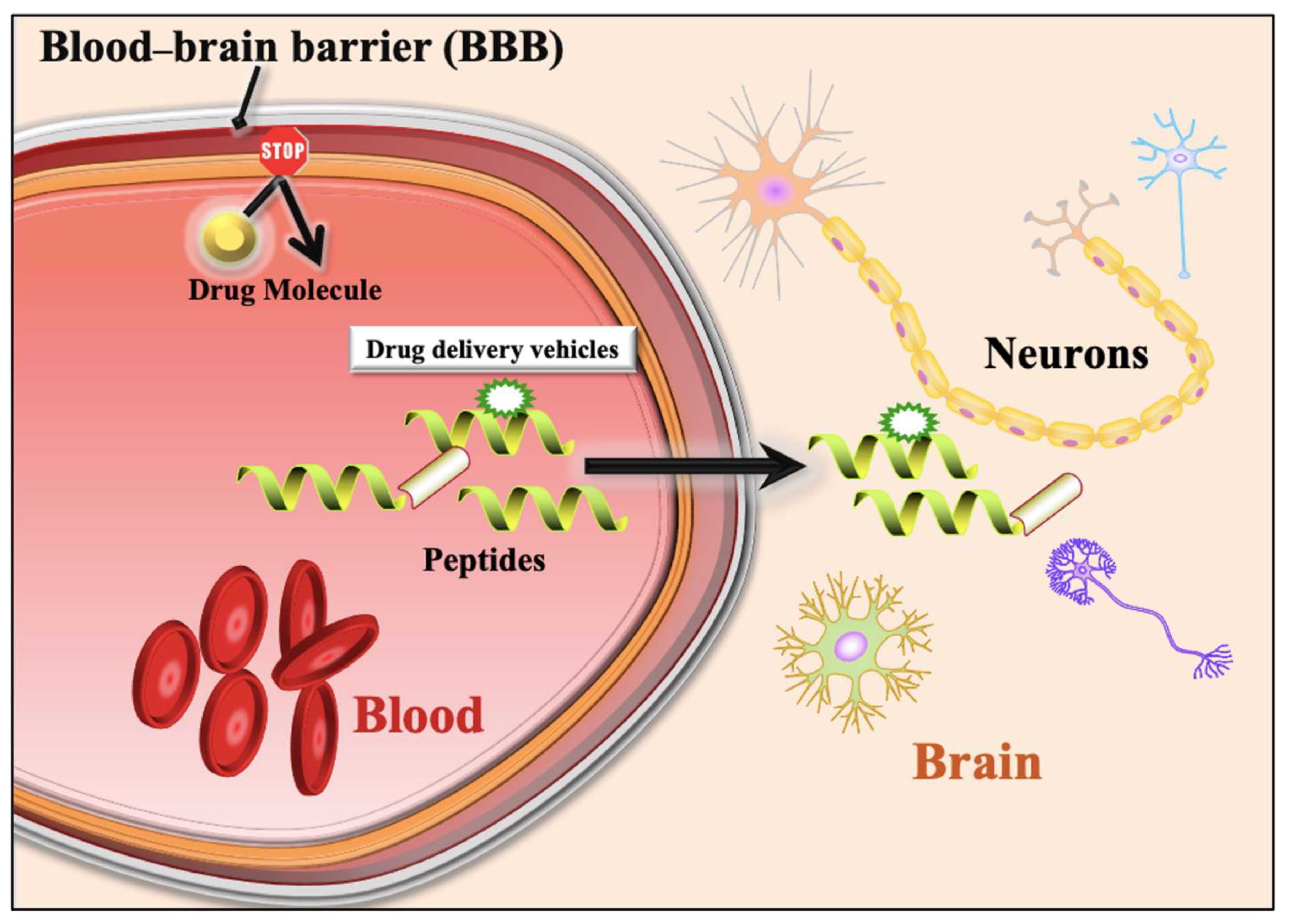
1. Introduction
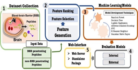
2. Materials and Methods
2.1. Dataset Collection
2.2. Amino Acid Composition
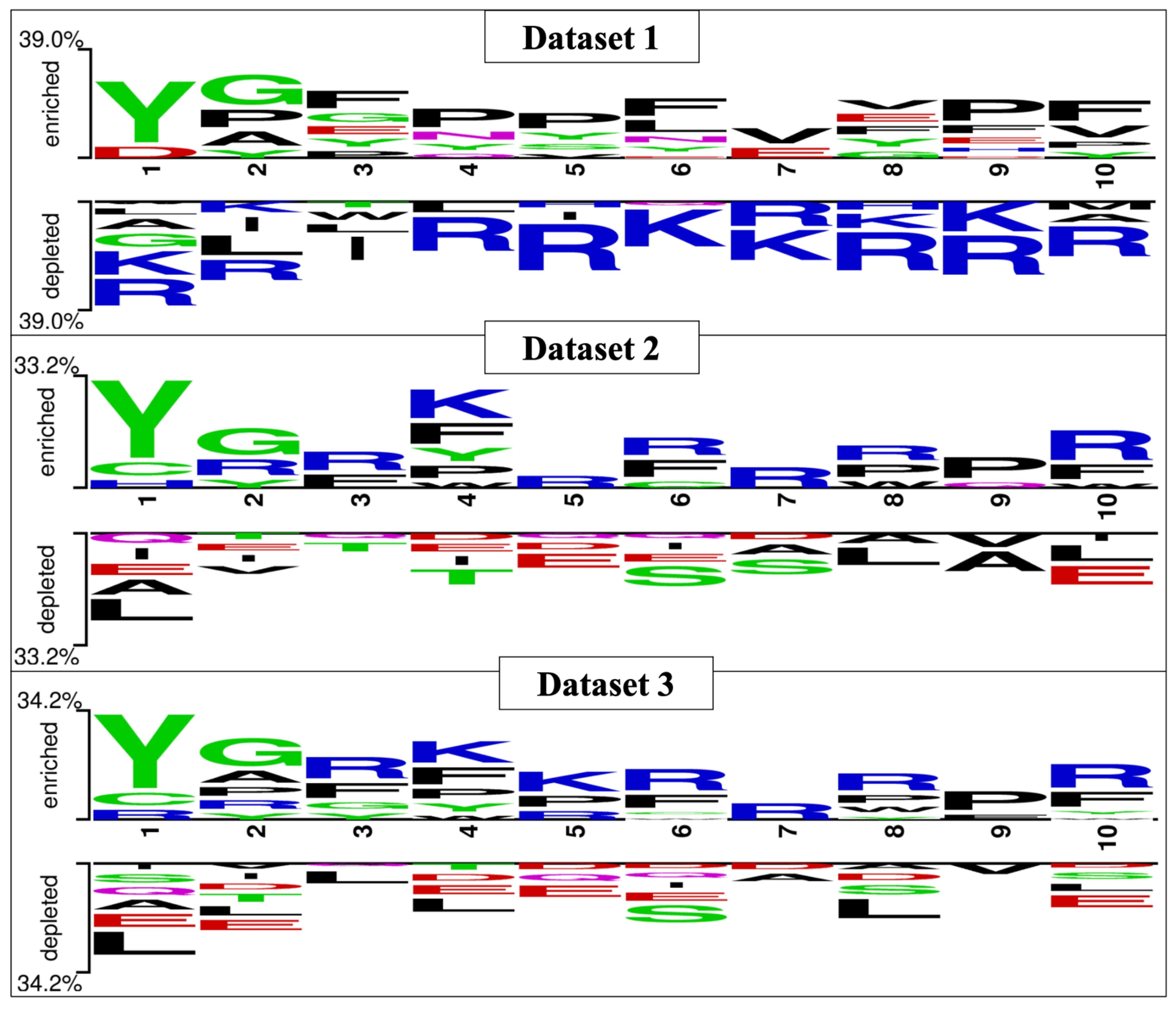
2.3. Two Sample Logo
2.4. Generation of Peptide Features
2.5. Feature Selection
2.6. Feature Ranking
2.7. Machine Learning Techniques
2.8. Cross-Validation Techniques
2.9. Performance Evaluation Parameters
2.10. Webserver Implementation
3. Results
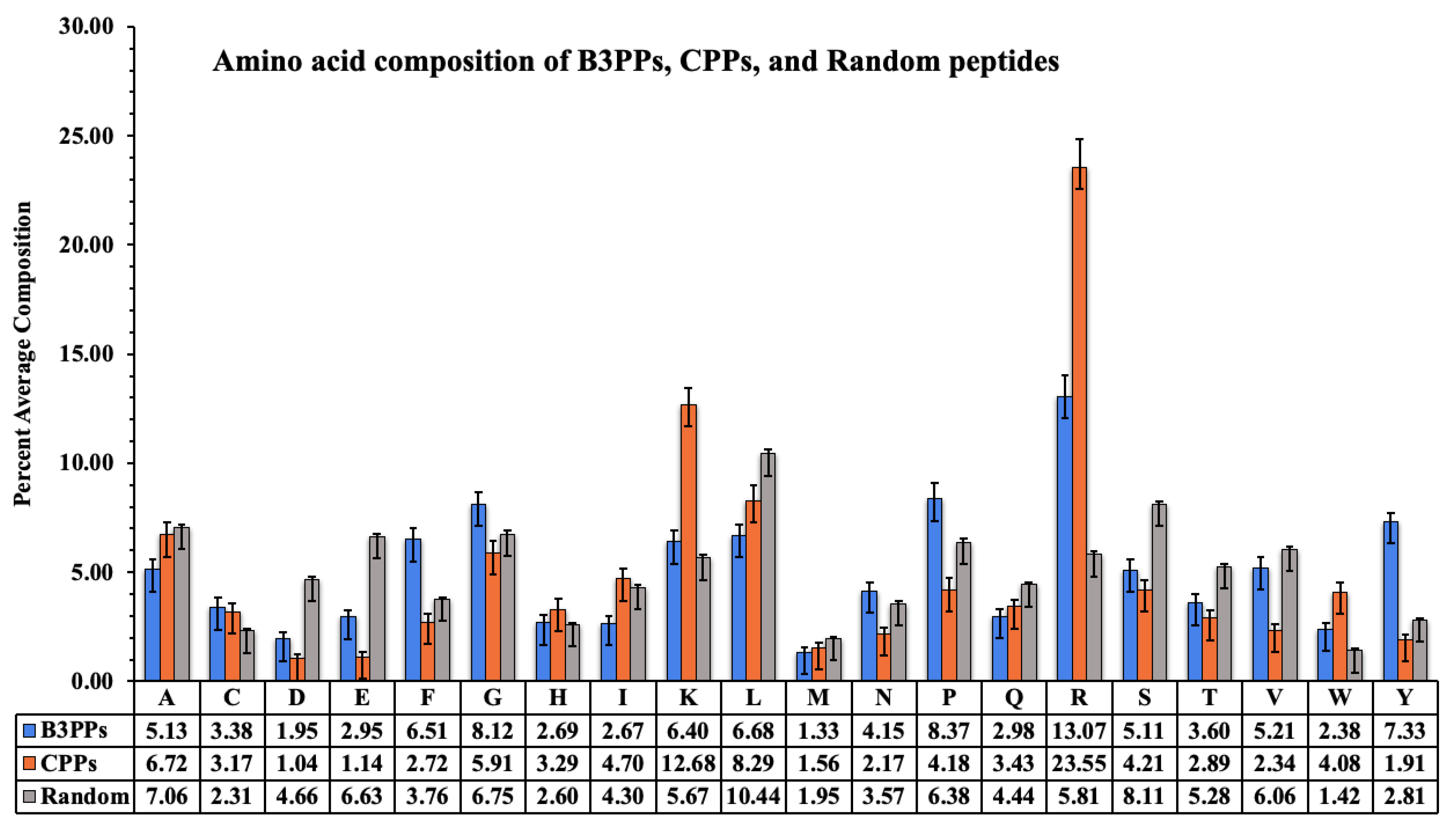
3.1. Amino Acid Composition Analysis
3.2. Amino Acid Position Analysis
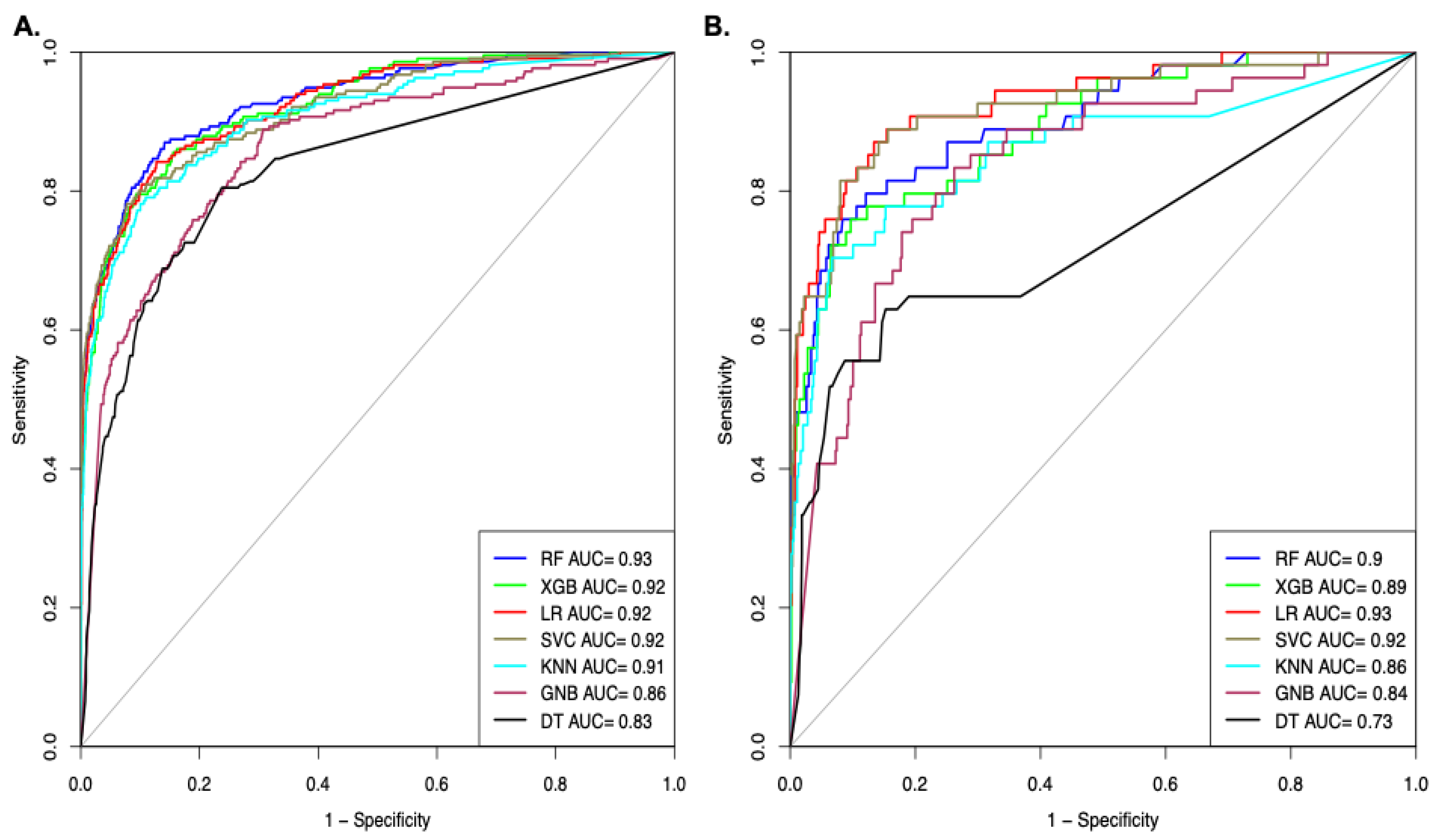
3.3. B3PPs Prediction Methods on Different Datasets
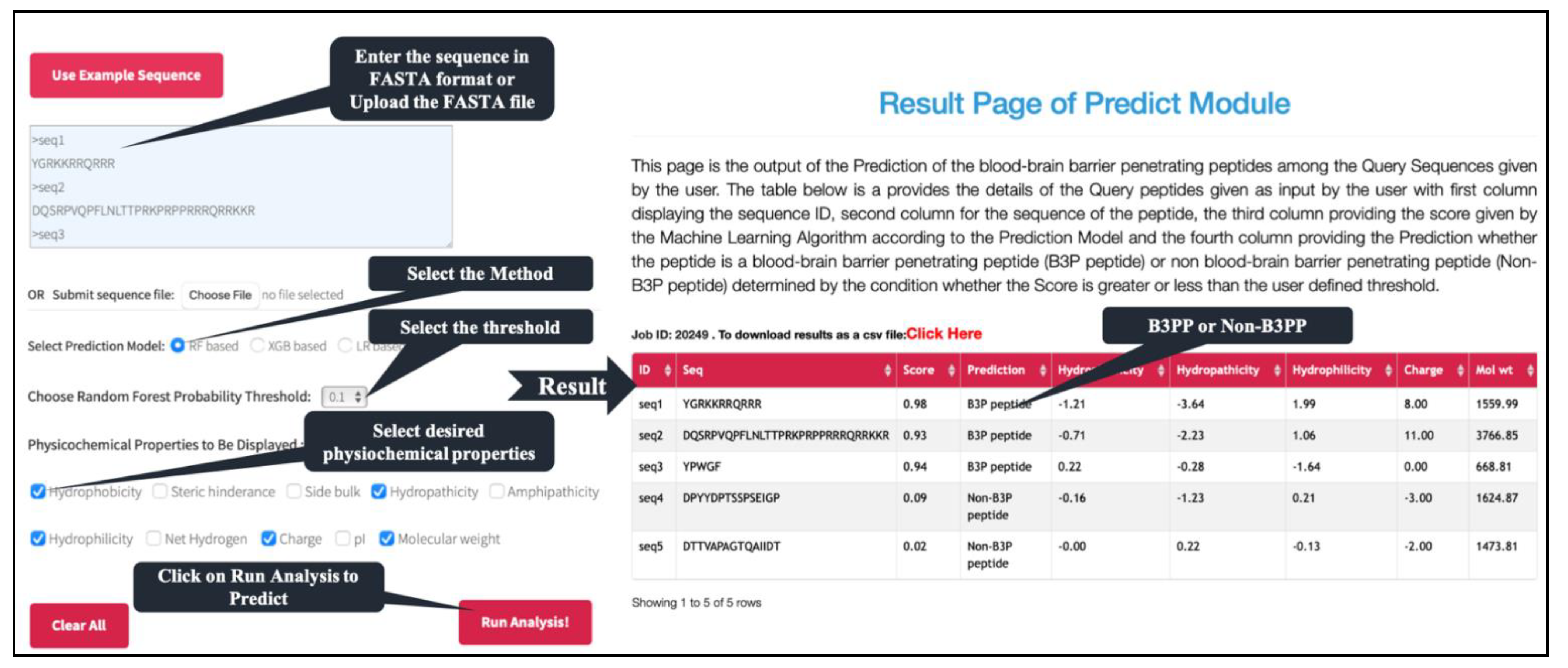
3.4. Webserver and Standalone Software
3.5. Comparison with the Existing Method
4. Discussion and Conclusions
Supplementary Materials
Author Contributions
Funding
Institutional Review Board Statement
Informed Consent Statement
Data Availability Statement
Acknowledgments
Conflicts of Interest
References
- Abbott, N.J.; Rönnbäck, L.; Hansson, E. Astrocyte-endothelial interactions at the blood-brain barrier. Nat. Rev. Neurosci. 2006, 7, 41–53. [Google Scholar] [CrossRef]
- Kniesel, U.; Wolburg, H. Tight junctions of the blood-brain barrier. Cell. Mol. Neurobiol. 2000, 20, 57–76. [Google Scholar] [CrossRef]
- Fenstermacher, J.; Gross, P.; Sposito, N.; Acuff, V.; Pettersen, S.; Gruber, K. Structural and Functional Variations in Capillary Systems within the brain. Ann. N. Y. Acad. Sci. 1988, 529, 21–30. [Google Scholar] [CrossRef] [PubMed]
- Rhea, E.M.; Banks, W.A. Role of the Blood-Brain Barrier in Central Nervous System Insulin Resistance. Front. Neurosci. 2019, 13. [Google Scholar] [CrossRef] [PubMed] [Green Version]
- Muoio, V.; Persson, P.B.; Sendeski, M.M. The neurovascular unit—Concept review. Acta Physiol. 2014, 210, 790–798. [Google Scholar] [CrossRef] [PubMed]
- Oldendorf, W.H. Brain uptake of radiolabeled amino acids, amines, and hexoses after arterial injection. Am. J. Physiol. 1971, 221, 1629–1639. [Google Scholar] [CrossRef] [Green Version]
- Tietz, S.; Engelhardt, B. Brain barriers: Crosstalk between complex tight junctions and adherens junctions. J. Cell Biol. 2015, 209, 493–506. [Google Scholar] [CrossRef] [PubMed] [Green Version]
- Pardridge, W.M. Blood-brain barrier delivery. Drug Discov. Today 2007, 12, 54–61. [Google Scholar] [CrossRef]
- Islam, Y.; Leach, A.G.; Smith, J.; Pluchino, S.; Coxon, C.; Sivakumaran, M.; Downing, J.E.; Fatokun, A.A.; Teixidò, M.; Ehtezazi, T. Peptide based drug delivery systems to the brain. Nano Express 2020, 1, 012002. [Google Scholar] [CrossRef]
- Banks, W.A. Peptides and the blood-brain barrier. Peptides 2015, 72, 16–19. [Google Scholar] [CrossRef] [Green Version]
- Funke, S.A.; Willbold, D. Peptides for Therapy and Diagnosis of Alzheimer’s Disease. Curr. Pharm. Des. 2012, 18, 755–767. [Google Scholar] [CrossRef] [Green Version]
- Baig, M.H.; Ahmad, K.; Saeed, M.; Alharbi, A.M.; Barreto, G.E.; Ashraf, G.M.; Choi, I. Peptide based therapeutics and their use for the treatment of neurodegenerative and other diseases. Biomed. Pharmacother. 2018, 103, 574–581. [Google Scholar] [CrossRef] [PubMed]
- Raucher, D. Tumor targeting peptides: Novel therapeutic strategies in glioblastoma. Curr. Opin. Pharmacol. 2019, 47, 14–19. [Google Scholar] [CrossRef] [PubMed]
- Oller-Salvia, B.; Sánchez-Navarro, M.; Giralt, E.; Teixidó, M. Blood-brain barrier shuttle peptides: An emerging paradigm for brain delivery. Chem. Soc. Rev. 2016, 45, 4690. [Google Scholar] [CrossRef] [PubMed] [Green Version]
- Wu, L.-P.; Ahmadvand, D.; Su, J.; Hall, A.; Tan, X.; Farhangrazi, Z.S.; Moghimi, S.M. Crossing the blood-brain-barrier with nanoligand drug carriers self-assembled from a phage display peptide. Nat. Commun. 2019, 10, 1–16. [Google Scholar] [CrossRef] [PubMed] [Green Version]
- Nosrati, H.; Tarantash, M.; Bochani, S.; Charmi, J.; Bagheri, Z.; Fridoni, M.; Abdollahifar, M.-A.; Davaran, S.; Danafar, H.; Manjili, H.K. Glutathione (GSH) Peptide Conjugated Magnetic Nanoparticles As Blood-Brain Barrier Shuttle for MRI-Monitored Brain Delivery of Paclitaxel. ACS Biomater. Sci. Eng. 2019, 5, 1677–1685. [Google Scholar] [CrossRef]
- Solbrig, M.V.; Koob, G.F. Epilepsy, CNS viral injury and dynorphin. Trends Pharmacol. Sci. 2004, 25, 98–104. [Google Scholar] [CrossRef] [PubMed]
- Balasubramaniam, A. Clinical potentials of neuropeptide Y family of hormones. Am. J. Surg. 2002, 183, 430–434. [Google Scholar] [CrossRef]
- Claes, S.J. Corticotropin-releasing hormone (CRH) in psychiatry: From stress to psychopathology. Ann. Med. 2004, 36, 50–61. [Google Scholar] [CrossRef]
- Ströhle, A.; Holsboer, F. Stress Responsive Neurohormones in Depression and Anxiety. Pharmacopsychiatry 2003, 36, 207–214. [Google Scholar]
- Li, C.; Wu, X.; Liu, S.; Zhao, Y.; Zhu, J.; Liu, K. Roles of Neuropeptide Y in Neurodegenerative and Neuroimmune Diseases. Front. Neurosci. 2019, 13, 869. [Google Scholar] [CrossRef]
- Dwibhashyam, V.S.N.M.; Nagappa, A. Strategies for enhanced drug delivery to the central nervous system. Indian J. Pharm. Sci. 2008, 70, 145–153. [Google Scholar]
- Reese, T.S.; Karnovsky, M.J. Fine structural localization of a blood-brain barrier to exogenous peroxidase. J. Cell Biol. 1967, 34, 207–217. [Google Scholar] [CrossRef] [PubMed] [Green Version]
- Kapoor, P.; Singh, H.; Gautam, A.; Chaudhary, K.; Kumar, R.; Raghava, G.P.S. TumorHoPe: A database of tumor homing peptides. PLoS ONE 2012, 7, e35187. [Google Scholar] [CrossRef] [Green Version]
- Gautam, A.; Singh, H.; Tyagi, A.; Chaudhary, K.; Kumar, R.; Kapoor, P.; Raghava, G.P.S. CPPsite: A curated database of cell penetrating peptides. Database 2012, 2012, bas015. [Google Scholar] [CrossRef] [Green Version]
- Sharma, A.; Kapoor, P.; Gautam, A.; Chaudhary, K.; Kumar, R.; Chauhan, J.S.; Tyagi, A.; Raghava, G.P.S. Computational approach for designing tumor homing peptides. Sci. Rep. 2013, 3, 1607. [Google Scholar] [CrossRef] [PubMed]
- Gautam, A.; Sharma, M.; Vir, P.; Chaudhary, K.; Kapoor, P.; Kumar, R.; Nath, S.K.; Raghava, G.P.S. Identification and characterization of novel protein-derived arginine-rich cell-penetrating peptides. Eur. J. Pharm. Biopharm. 2015, 89, 93–106. [Google Scholar] [CrossRef] [PubMed]
- Shergalis, A.; Bankhead, A.; Luesakul, U.; Muangsin, N.; Neamati, N. Current challenges and opportunities in treating glioblastomas. Pharmacol. Rev. 2018, 70, 412–445. [Google Scholar] [CrossRef] [Green Version]
- Stalmans, S.; Wynendaele, E.; Bracke, N.; Gevaert, B.; D’Hondt, M.; Peremans, K.; Burvenich, C.; De Spiegeleer, B. Chemical-Functional Diversity in Cell-Penetrating Peptides. PLoS ONE 2013, 8, e71752. [Google Scholar] [CrossRef] [PubMed] [Green Version]
- Stalmans, S.; Bracke, N.; Wynendaele, E.; Gevaert, B.; Peremans, K.; Burvenich, C.; Polis, I.; De Spiegeleer, B. Cell-penetrating peptides selectively cross the blood-brain barrier in vivo. PLoS ONE 2015, 10, e0139652. [Google Scholar] [CrossRef] [PubMed] [Green Version]
- Yamano, S.; Dai, J.; Hanatani, S.; Haku, K.; Yamanaka, T.; Ishioka, M.; Takayama, T.; Yuvienco, C.; Khapli, S.; Moursi, A.M.; et al. Long-term efficient gene delivery using polyethylenimine with modified Tat peptide. Biomaterials 2014, 35, 1705–1715. [Google Scholar] [CrossRef]
- Huwyler, J.; Wu, D.; Pardridge, W.M. Brain drug delivery of small molecules using immunoliposomes. Proc. Natl. Acad. Sci. USA 1996, 93, 14164–14169. [Google Scholar] [CrossRef] [Green Version]
- Knight, A.; Carvajal, J.; Schneider, H.; Coutelle, C.; Chamberlain, S.; Fairweather, N. Non-viral neuronal gene delivery mediated by the H(C) fragment of tetanus toxin. Eur. J. Biochem. 1999, 259, 762–769. [Google Scholar] [CrossRef] [PubMed] [Green Version]
- El-Andaloussi, S.; Holm, T.; Langel, U. Cell-Penetrating Peptides: Mechanisms and Applications. Curr. Pharm. Des. 2005, 11, 3597–3611. [Google Scholar] [CrossRef] [PubMed]
- Milletti, F. Cell-penetrating peptides: Classes, origin, and current landscape. Drug Discov. Today 2012, 17, 850–860. [Google Scholar] [CrossRef] [PubMed]
- Stewart, K.M.; Horton, K.L.; Kelley, S.O. Cell-penetrating peptides as delivery vehicles for biology and medicine. Org. Biomol. Chem. 2008, 6, 2242–2255. [Google Scholar] [CrossRef]
- Mueller, J.; Kretzschmar, I.; Volkmer, R.; Boisguerin, P. Comparison of cellular uptake using 22 CPPs in 4 different cell lines. Bioconjug. Chem. 2008, 19, 2363–2374. [Google Scholar] [CrossRef] [PubMed]
- Meade, A.J.; Meloni, B.P.; Mastaglia, F.L.; Knuckey, N. The application of cell penetrating peptides for the delivery of neuroprotective peptides/proteins in experimental cerebral ischaemia studies. J. Exp. Stroke Transl. Med. 2009, 2, 22–40. [Google Scholar] [CrossRef]
- Mathur, D.; Prakash, S.; Anand, P.; Kaur, H.; Agrawal, P.; Mehta, A.; Kumar, R.; Singh, S.; Raghava, G.P.S. PEPlife: A Repository of the Half-life of Peptides. Sci. Rep. 2016, 6, 36617. [Google Scholar] [CrossRef] [Green Version]
- Gautam, A.; Chaudhary, K.; Kumar, R.; Sharma, A.; Kapoor, P.; Tyagi, A.; Raghava, G.P.S. In silico approaches for designing highly effective cell penetrating peptides. J. Transl. Med. 2013, 11, 74. [Google Scholar] [CrossRef] [Green Version]
- Wei, L.; Tang, J.; Zou, Q. SkipCPP-Pred: An improved and promising sequence-based predictor for predicting cell-penetrating peptides. BMC Genom. 2017, 18, 742. [Google Scholar] [CrossRef] [Green Version]
- Wei, L.; Xing, P.; Su, R.; Shi, G.; Ma, Z.S.; Zou, Q. CPPred-RF: A Sequence-based Predictor for Identifying Cell-Penetrating Peptides and Their Uptake Efficiency. J. Proteome Res. 2017, 16, 2044–2053. [Google Scholar] [CrossRef]
- Pandey, P.; Patel, V.; George, N.V.; Mallajosyula, S.S. KELM-CPPpred: Kernel Extreme Learning Machine Based Prediction Model for Cell-Penetrating Peptides. J. Proteome Res. 2018, 17, 3214–3222. [Google Scholar] [CrossRef]
- Kumar, V.; Agrawal, P.; Kumar, R.; Bhalla, S.; Usmani, S.S.; Varshney, G.C.; Raghava, G.P.S. Prediction of cell-penetrating potential of modified peptides containing natural and chemically modified residues. Front. Microbiol. 2018, 9, 725. [Google Scholar] [CrossRef]
- Qiang, X.; Zhou, C.; Ye, X.; Du, P.-F.; Su, R.; Wei, L. CPPred-FL: A sequence-based predictor for large-scale identification of cell-penetrating peptides by feature representation learning. Brief. Bioinform. 2018, 21, 11–23. [Google Scholar] [CrossRef] [PubMed]
- Shaker, B.; Yu, M.-S.; Song, J.S.; Ahn, S.; Ryu, J.Y.; Oh, K.-S.; Na, D. LightBBB: Computational prediction model of blood–brain-barrier penetration based on LightGBM. Bioinformatics 2021, 37, 1135–1139. [Google Scholar] [CrossRef] [PubMed]
- Carpenter, T.; Kirshner, D.A.; Lau, E.Y.; Wong, S.E.; Nilmeier, J.P.; Lightstone, F.C. A Method to Predict Blood-Brain Barrier Permeability of Drug-Like Compounds Using Molecular Dynamics Simulations. Biophys. J. 2014, 107, 630–641. [Google Scholar] [CrossRef] [PubMed] [Green Version]
- Mensch, J.; Oyarzabal, J.; Mackie, C.; Augustijns, P. In vivo, in vitro and in silico methods for small molecule transfer across the BBB. J. Pharm. Sci. 2009, 98, 4429–4468. [Google Scholar] [CrossRef] [PubMed]
- Dai, R.; Zhang, W.; Tang, W.; Wynendaele, E.; Zhu, Q.; Bin, Y.; De Spiegeleer, B.; Xia, J. BBPpred: Sequence-Based Prediction of Blood-Brain Barrier Peptides with Feature Representation Learning and Logistic Regression. J. Chem. Inf. Model. 2021, 61, 525–534. [Google Scholar] [CrossRef]
- Kumar, V.; Patiyal, S.; Kumar, R.; Sahai, S.; Kaur, D.; Lathwal, A.; Raghava, G.P.S. B3Pdb: An archive of blood–brain barrier-penetrating peptides. Brain Struct. Funct. 2021. [Google Scholar] [CrossRef]
- Agrawal, P.; Bhalla, S.; Usmani, S.S.; Singh, S.; Chaudhary, K.; Raghava, G.P.S.; Gautam, A. CPPsite 2.0: A repository of experimentally validated cell-penetrating peptides. Nucleic Acids Res. 2016, 44, D1098–D1103. [Google Scholar] [CrossRef] [PubMed]
- Boutet, E.; Lieberherr, D.; Tognolli, M.; Schneider, M.; Bansal, P.; Bridge, A.; Poux, S.; Bougueleret, L.; Xenarios, I.; Boutet, E. Uniprotkb/swiss-prot, the manually annotated section of the uniprot knowledgebase: How to use the entry view. Methods Mol. Biol. 2016, 1374, 23–54. [Google Scholar] [PubMed]
- Agrawal, P.; Raghava, G.P.S. Prediction of Antimicrobial Potential of a Chemically Modified Peptide From Its Tertiary Structure. Front. Microbiol. 2018, 9, 2551. [Google Scholar] [CrossRef] [PubMed] [Green Version]
- Vacic, V.; Iakoucheva, L.M.; Radivojac, P. Two Sample Logo: A graphical representation of the differences between two sets of sequence alignments. Bioinformatics 2006, 22, 1536–1537. [Google Scholar] [CrossRef] [Green Version]
- Pande, A.; Patiyal, S.; Lathwal, A.; Arora, C.; Kaur, D.; Dhall, A.; Mishra, G.; Kaur, H.; Sharma, N.; Jain, S.; et al. Computing wide range of protein/peptide features from their sequence and structure. bioRxiv 2019. [Google Scholar] [CrossRef]
- Tang, J.; Alelyani, S.; Liu, H. Feature selection for classification: A review. Data Classif Algorithms Appl. 2014, 37–64. Available online: https://www.cvs.edu.in/upload/feature_selection_for_classification.pdf (accessed on 22 February 2021).
- Chang, K.-W.; Hsieh, C.-J.; Lin, C.-J. LIBLINEAR: A Library for Large Linear Classification Rong-En Fan Xiang-Rui Wang. J. Mach. Learn. Res. 2008, 9, 1871–1874. [Google Scholar]
- Ke, G.; Meng, Q.; Finley, T.; Wang, T.; Chen, W.; Ma, W.; Ye, Q.; Liu, T.-Y. LightGBM: A Highly Efficient Gradient Boosting Decision Tree. Adv. Neural Inf. Process. Syst. 2017, 30, 3146–3154. [Google Scholar]
- Pedregosa, F.; Varoquaux, G.; Gramfort, A.; Michel, V.; Thirion, B.; Grisel, O.; Blondel, M.; Prettenhofer, P.; Weiss, R.; Dubourg, V.; et al. Scikit-learn: Machine Learning in Python. J. Mach. Learn. Res. 2011, 12, 2825–2830. [Google Scholar]
- Webb, G.I. Decision Tree Grafting From the All-Tests-But-One Partition. In Proceedings of the Sixteenth International Joint Conference on Artificial Intelligence, IJCAI 99, Stockholm, Sweden, 31 July–6 August 1999. [Google Scholar]
- Zhang, H. Exploring conditions for the optimality of naïve bayes. Int. J. Pattern Recognit. Artif. Intell. 2005, 19, 183–198. [Google Scholar] [CrossRef]
- Geurts, P.; Ernst, D.; Wehenkel, L. Extremely randomized trees. Mach. Learn. 2006, 63, 3–42. [Google Scholar] [CrossRef] [Green Version]
- Tolles, J.; Meurer, W.J. Logistic regression: Relating patient characteristics to outcomes. J. Am. Med. Assoc. 2016, 316, 533–534. [Google Scholar] [CrossRef] [PubMed]
- Mucherino, A.; Papajorgji, P.J.; Pardalos, P.M. Data Mining in Agriculture. Dyn. Disasters 2009, 34. [Google Scholar] [CrossRef]
- Chen, T.; Guestrin, C. XGBoost: A Scalable Tree Boosting System. In Proceedings of the 22nd ACM SIGKDD International Conference on Knowledge Discovery and Data Mining, San Francisco, CA, USA, 13–17 August 2016. [Google Scholar]
- Chang, C.-C.; Lin, C.-J. LIBSVM: A Library for Support Vector Machines. ACM Trans. Intell. Syst. Technol. 2011, 2, 1–27. [Google Scholar] [CrossRef]
- Kumar, V.; Kumar, R.; Agrawal, P.; Patiyal, S.; Raghava, G.P.S. A Method for Predicting Hemolytic Potency of Chemically Modified Peptides From Its Structure. Front. Pharmacol. 2020, 11, 54. [Google Scholar] [CrossRef] [PubMed] [Green Version]
- Dhall, A.; Patiyal, S.; Sharma, N.; Usmani, S.S.; Raghava, G.P.S. Computer-aided prediction and design of IL-6 inducing peptides: IL-6 plays a crucial role in COVID-19. Brief. Bioinform. 2021, 22, 936–945. [Google Scholar] [CrossRef] [PubMed]
- Thangudu, S.; Cheng, F.-Y.; Su, C.-H. Advancements in the Blood-Brain Barrier Penetrating Nanoplatforms for Brain Related Disease Diagnostics and Therapeutic Applications. Polymers 2020, 12, 55. [Google Scholar] [CrossRef] [PubMed]
- He, Q.; Liu, J.; Liang, J.; Liu, X.; Li, W.; Liu, Z.; Ding, Z.; Tuo, D. Towards Improvements for Penetrating the Blood-Brain Barrier-Recent Progress from a Material and Pharmaceutical Perspective. Cells 2018, 7, 24. [Google Scholar] [CrossRef] [Green Version]
- Banks, W.A. From blood-brain barrier to blood-brain interface: New opportunities for CNS drug delivery. Nat. Rev. Drug Discov. 2016, 15, 275–292. [Google Scholar] [CrossRef]
- Polianova, M.T.; Ruscetti, F.W.; Pert, C.B.; Tractenberg, R.E.; Leoung, G.; Strang, S.; Ruff, M.R. Antiviral and immunological benefits in HIV patients receiving intranasal peptide T (DAPTA). Peptides 2003, 24, 1093–1098. [Google Scholar] [CrossRef]
- Barrera, C.M.; Kastin, A.J.; Banks, W.A. D-[Ala1]-peptide T-Amide is transported from blood to brain by a saturable system. Brain Res. Bull. 1987, 19, 629–633. [Google Scholar] [CrossRef]
- Jackman, J.A.; Costa, V.V.; Park, S.; Real, A.L.C.V.; Park, J.H.; Cardozo, P.L.; Ferhan, A.R.; Olmo, I.G.; Moreira, T.P.; Bambirra, J.L.; et al. Therapeutic treatment of Zika virus infection using a brain-penetrating antiviral peptide. Nat. Mater. 2018, 17, 971–977. [Google Scholar] [CrossRef] [PubMed]
- Cho, N.-J.; Dvory-Sobol, H.; Xiong, A.; Cho, S.-J.; Frank, C.W.; Glenn, J.S. Mechanism of an amphipathic α-helical peptide’s antiviral activity involves size-dependent virus particle lysis. ACS Chem. Biol. 2009, 4, 1061–1067. [Google Scholar] [CrossRef] [PubMed]
- Zou, J.; Shi, P.Y. Targeting vesicle size. Nat. Mater. 2018, 17, 955–956. [Google Scholar] [CrossRef]
- Boldescu, V.; Behnam, M.; Vasilakis, N.; Klein, C.D. Broad-spectrum agents for flaviviral infections: Dengue, Zika and beyond. Nat. Rev. Drug Discov. 2017, 16, 565–586. [Google Scholar] [CrossRef] [Green Version]
- Mao, X.Y.; Jin, W.L. The COVID-19 Pandemic: Consideration for Brain Infection. Neuroscience 2020, 437, 130–131. [Google Scholar] [CrossRef]


| Methods | Training Dataset_1 | Validation Dataset_1 | ||||||||
|---|---|---|---|---|---|---|---|---|---|---|
| Sens | Spec | Acc | AUROC | MCC | Sens | Spec | Acc | AUROC | MCC | |
| RF | 86.04 | 84.18 | 82.09 | 0.90 | 0.64 | 75.92 | 87.03 | 81.48 | 0.88 | 0.63 |
| XGB | 81.39 | 82.32 | 80.93 | 0.88 | 0.62 | 79.63 | 88.89 | 84.25 | 0.88 | 0.68 |
| LR | 82.79 | 83.25 | 83.48 | 0.90 | 0.67 | 81.48 | 87.03 | 84.26 | 0.91 | 0.69 |
| SVC | 83.25 | 82.79 | 81.86 | 0.88 | 0.64 | 74.07 | 92.59 | 83.33 | 0.91 | 0.67 |
| KNN | 66.51 | 64.65 | 65.58 | 0.74 | 0.32 | 48.18 | 77.77 | 62.93 | 0.72 | 0.27 |
| GNB | 84.18 | 82.32 | 80 | 0.86 | 0.61 | 53.70 | 94.44 | 74.07 | 0.86 | 0.52 |
| DT | 78.14 | 75.34 | 73.49 | 0.79 | 0.47 | 74.07 | 70.37 | 72.22 | 0.76 | 0.44 |
| Methods | Training Dataset_2 | Validation Dataset_2 | ||||||||
|---|---|---|---|---|---|---|---|---|---|---|
| Sens | Spec | Acc | AUROC | MCC | Sens | Spec | Acc | AUROC | MCC | |
| RF | 80.57 | 84.18 | 82.09 | 0.90 | 0.64 | 75.92 | 87.03 | 81.48 | 0.88 | 0.63 |
| XGB | 80.46 | 81.39 | 80.93 | 0.88 | 0.62 | 79.63 | 88.89 | 84.25 | 0.88 | 0.68 |
| LR | 80.46 | 86.52 | 83.48 | 0.90 | 0.67 | 81.48 | 87.03 | 84.26 | 0.91 | 0.69 |
| SVC | 79.07 | 84.65 | 81.86 | 0.88 | 0.64 | 74.07 | 92.59 | 83.33 | 0.91 | 0.67 |
| KNN | 50.23 | 80.93 | 65.58 | 0.74 | 0.32 | 48.18 | 77.77 | 62.93 | 0.72 | 0.27 |
| GNB | 72.55 | 87.44 | 80 | 0.86 | 0.61 | 53.70 | 94.44 | 74.07 | 0.86 | 0.52 |
| DT | 73.02 | 73.95 | 73.49 | 0.79 | 0.47 | 74.07 | 70.37 | 72.22 | 0.76 | 0.44 |
| Methods | Training Dataset_3 | Validation Dataset_3 | ||||||||
|---|---|---|---|---|---|---|---|---|---|---|
| Sens | Spec | Acc | AUROC | MCC | Sens | Spec | Acc | AUROC | MCC | |
| RF | 86.97 | 85.08 | 85.25 | 0.93 | 0.51 | 81.48 | 83.08 | 82.93 | 0.90 | 0.44 |
| XGB | 72.55 | 93.82 | 91.88 | 0.92 | 0.58 | 72.22 | 92.00 | 90.20 | 0.892 | 0.52 |
| LR | 80.93 | 89.73 | 88.93 | 0.92 | 0.54 | 83.33 | 89.40 | 88.85 | 0.93 | 0.55 |
| SVC | 80.00 | 84.75 | 84.32 | 0.90 | 0.45 | 85.18 | 82.15 | 82.43 | 0.90 | 0.45 |
| KNN | 83.72 | 80.76 | 81.03 | 0.88 | 0.43 | 79.63 | 78.44 | 78.54 | 0.84 | 0.37 |
| GNB | 80.46 | 75.74 | 76.20 | 0.84 | 0.35 | 83.33 | 72.67 | 73.65 | 0.86 | 0.34 |
| DT | 85.11 | 65.00 | 66.83 | 0.82 | 0.30 | 64.82 | 63.20 | 63.40 | 0.72 | 0.20 |
Publisher’s Note: MDPI stays neutral with regard to jurisdictional claims in published maps and institutional affiliations. |
© 2021 by the authors. Licensee MDPI, Basel, Switzerland. This article is an open access article distributed under the terms and conditions of the Creative Commons Attribution (CC BY) license (https://creativecommons.org/licenses/by/4.0/).
Share and Cite
Kumar, V.; Patiyal, S.; Dhall, A.; Sharma, N.; Raghava, G.P.S. B3Pred: A Random-Forest-Based Method for Predicting and Designing Blood–Brain Barrier Penetrating Peptides. Pharmaceutics 2021, 13, 1237. https://doi.org/10.3390/pharmaceutics13081237
Kumar V, Patiyal S, Dhall A, Sharma N, Raghava GPS. B3Pred: A Random-Forest-Based Method for Predicting and Designing Blood–Brain Barrier Penetrating Peptides. Pharmaceutics. 2021; 13(8):1237. https://doi.org/10.3390/pharmaceutics13081237
Chicago/Turabian StyleKumar, Vinod, Sumeet Patiyal, Anjali Dhall, Neelam Sharma, and Gajendra Pal Singh Raghava. 2021. "B3Pred: A Random-Forest-Based Method for Predicting and Designing Blood–Brain Barrier Penetrating Peptides" Pharmaceutics 13, no. 8: 1237. https://doi.org/10.3390/pharmaceutics13081237
APA StyleKumar, V., Patiyal, S., Dhall, A., Sharma, N., & Raghava, G. P. S. (2021). B3Pred: A Random-Forest-Based Method for Predicting and Designing Blood–Brain Barrier Penetrating Peptides. Pharmaceutics, 13(8), 1237. https://doi.org/10.3390/pharmaceutics13081237