
Inorganic Element Determination of Romanian Populus nigra L. Buds Extract and In Vitro Antiproliferative and Pro-Apoptotic Evaluation on A549 Human Lung Cancer Cell Line

 ,
,  ,
,  ,
,  ,
, 

Abstract
1. Introduction
2. Materials and Methods
2.1. Plant Materials and Reagents
2.2. Phytochemical Composition—HPLC-DAD
2.3. Inorganic Element Determination of Pg Extracts by GF-AAS—Sampling and Sample Preparation
2.4. Cell Culture
2.5. MTT Assay
2.6. Scratch Assay
2.7. Cell Cycle Analysis
2.8. Detection of Apoptosis via 4′,6-Diamidino-2-Phenylindole (DAPI) Staining
2.9. Annexin V-FITC Apoptosis Assay
2.10. LDH Assay
2.11. Statistical Analysis
3. Results
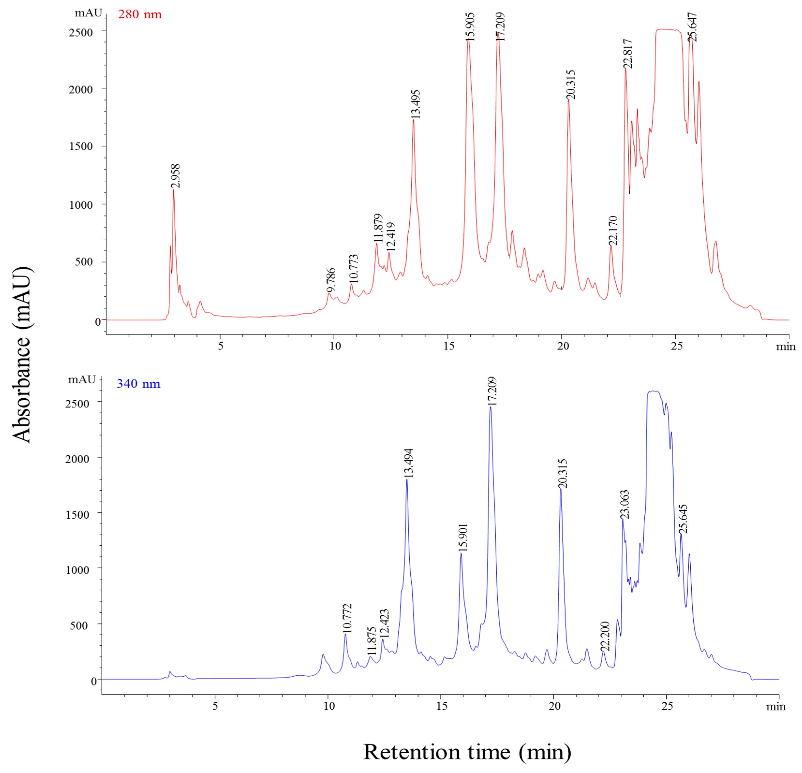
3.1. Phytochemical Composition
3.2. Inorganic Elements
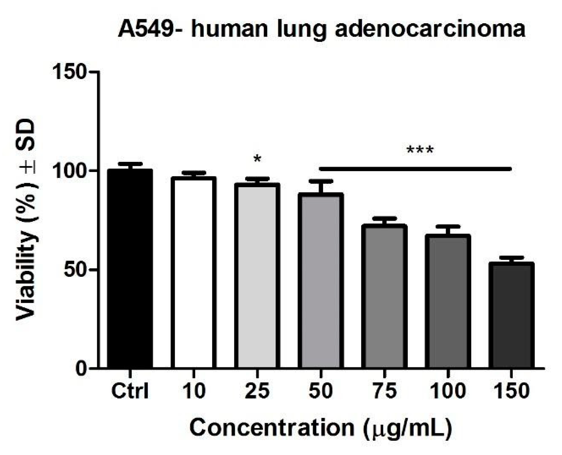
3.3. MTT Assay
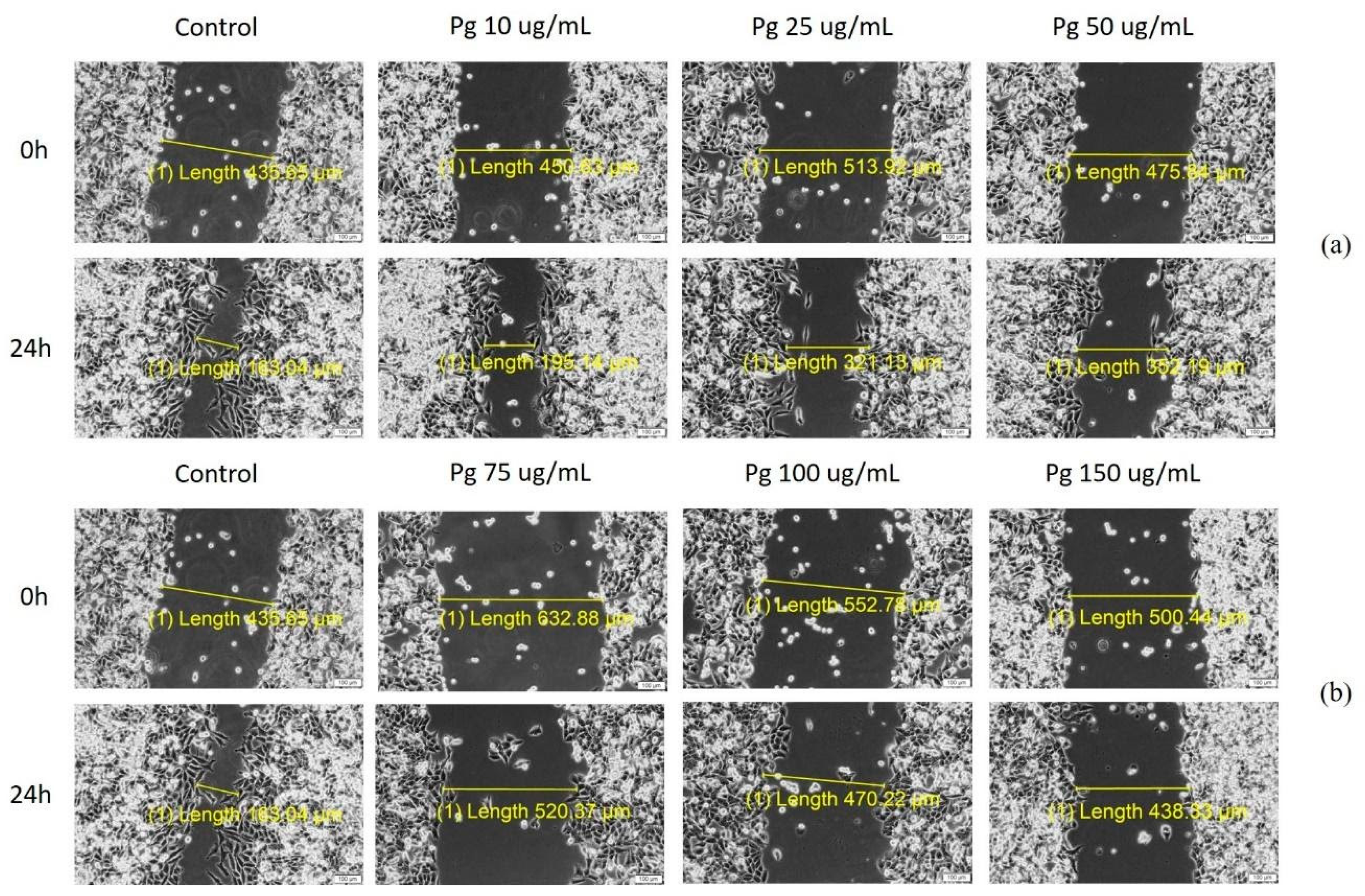
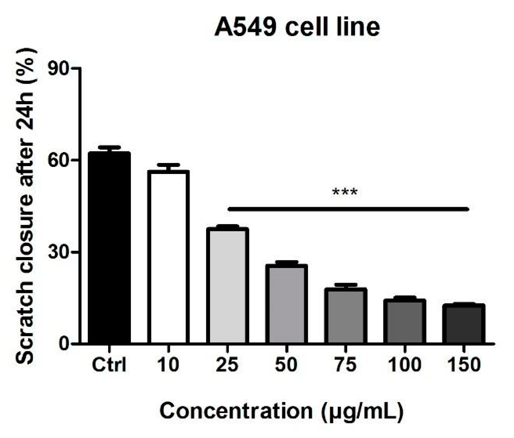
3.4. Scratch Assay
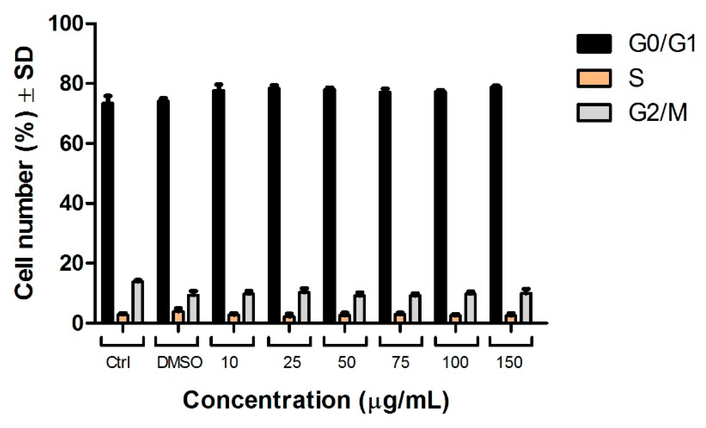
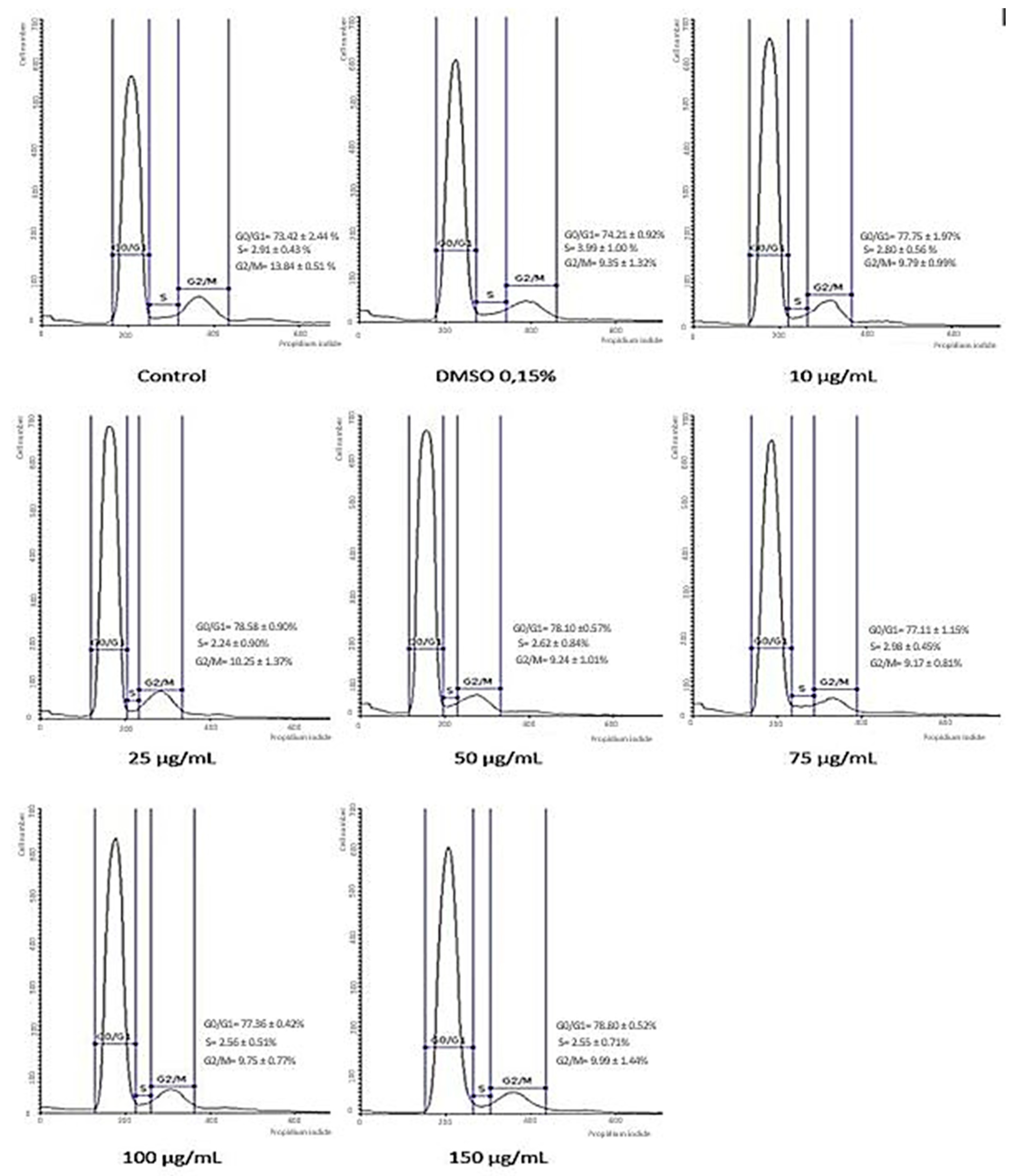
3.5. Cell Cycle Analysis
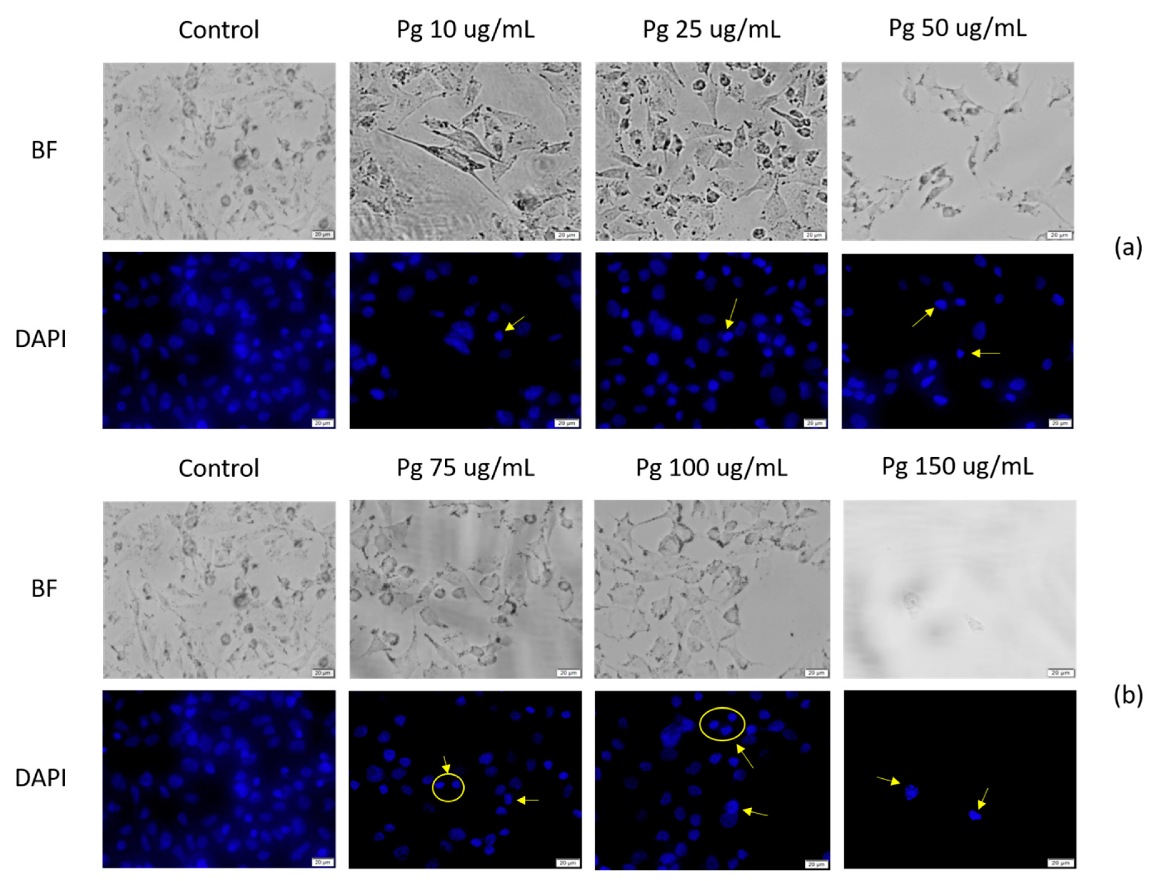
3.6. DAPI Staining
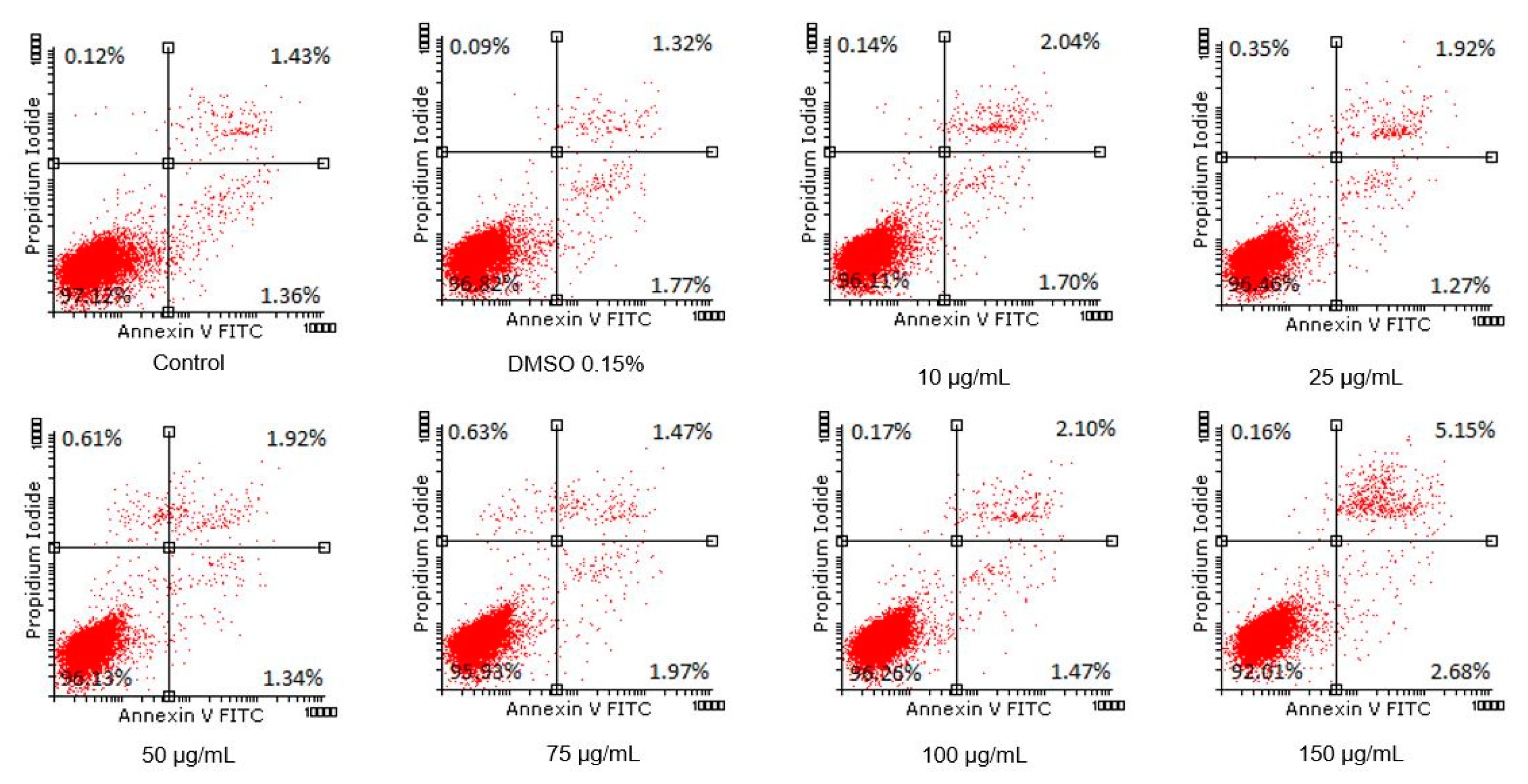
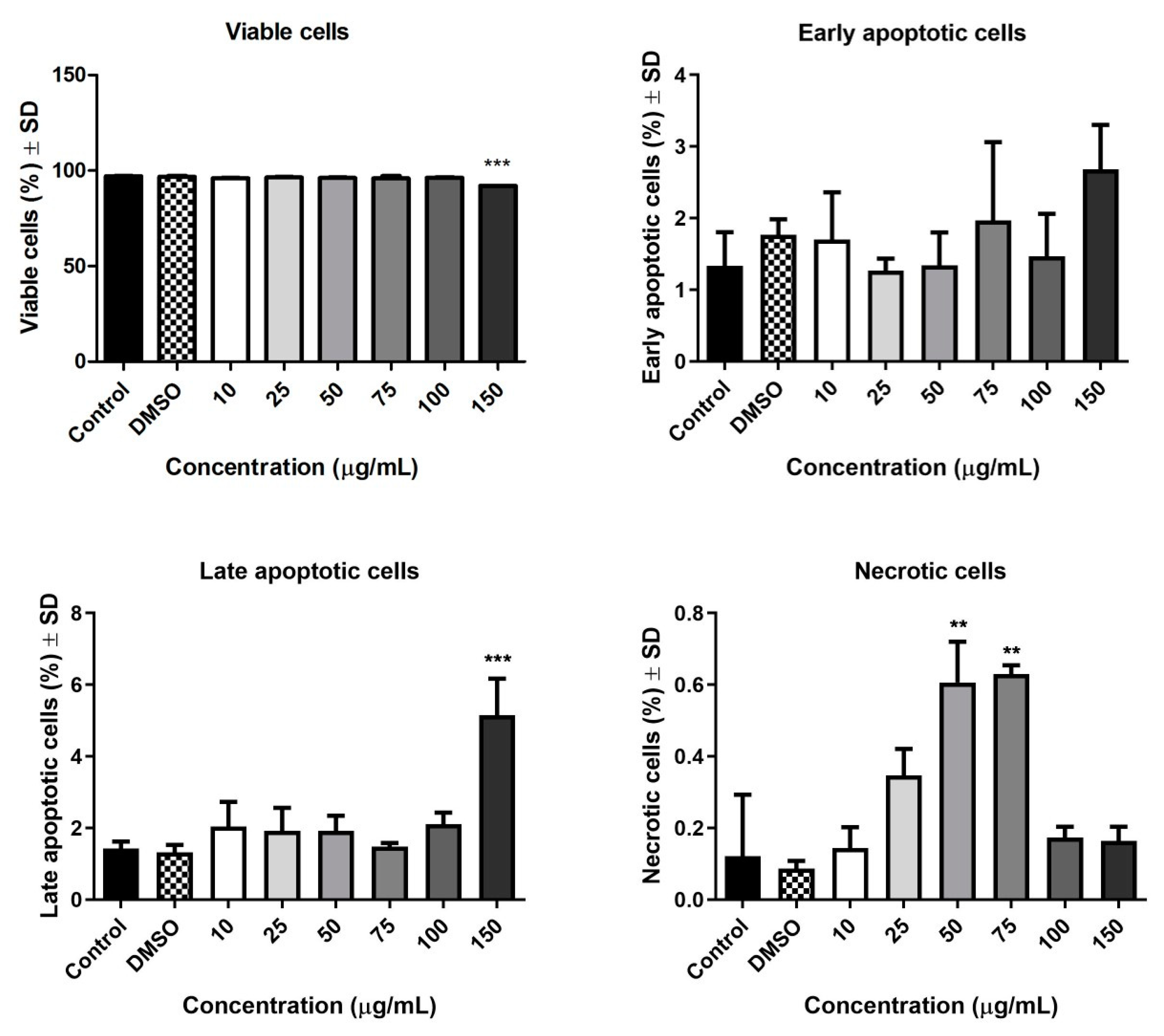
3.7. Annexin PI
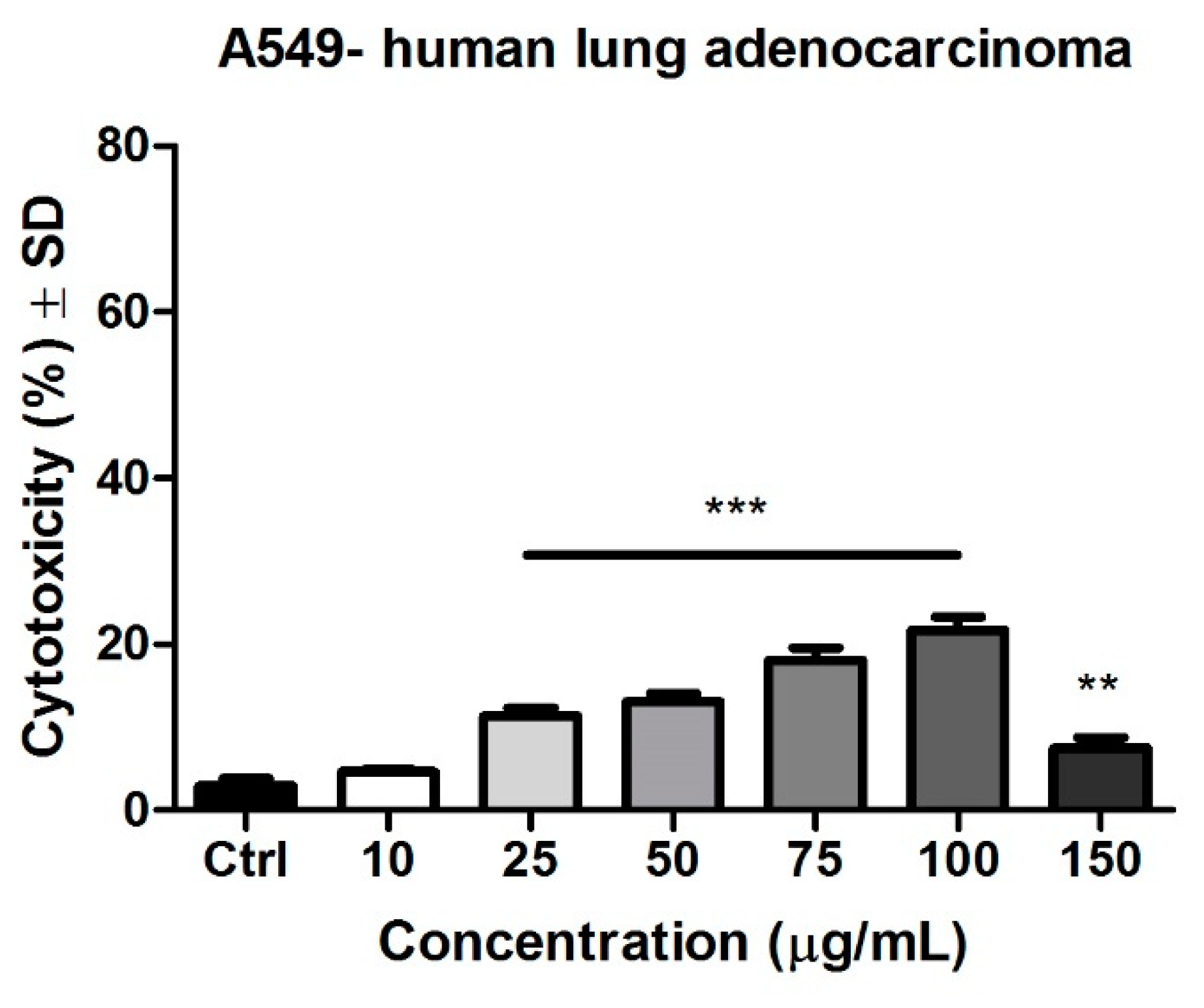
3.8. Determination of the Cytotoxic Potential by LDH Release
4. Discussion
5. Conclusions
Author Contributions
Funding
Institutional Review Board Statement
Informed Consent Statement
Data Availability Statement
Conflicts of Interest
References
- Petrovska, B.B. Historical review of medicinal plants usage. Pharmacogn. Rev. 2012, 6, 1–5. [Google Scholar] [CrossRef] [PubMed]
- Kennedy, O.W.; Wightman, L.E. Herbal Extracts and Phytochemicals: Plant Secondary Metabolites and the Enhancement of Human Brain function. Adv. Nutr. 2011, 2, 32–50. [Google Scholar] [CrossRef]
- Michel, J.; Nur Zahirah, A.R.; Khairana, H. A Review on the Potential Use of Medicinal Plants From Asteraceae and Lamiaceae Plant Family in Cardiovascular Diseases. Front. Pharmacol. 2020, 11, 852. [Google Scholar] [CrossRef] [PubMed]
- Aye, M.M.; Aung, H.T.; Sein, M.M.; Armijos, C. A Review on the Phytochemistry, Medicinal Properties and Pharmacological Activities of 15 Selected Myanmar Medicinal Plants. Molecules 2019, 24, 293. [Google Scholar] [CrossRef] [PubMed]
- Azab, A.; Nassar, A.; Azab, A.N. Anti-Inflammatory Activity of Natural Products. Molecules 2016, 21, 1321. [Google Scholar] [CrossRef]
- Stănescu, U.; Hăncianu, M.; Cioancă, O.; Aprotosoaie, A.C.; Miron, A. Plante medicinale de la A la Z, a III-a, ed.; Revizuită Si adăugită; Editura Polirom: Iaşi, Romania, 2018; ISBN 978-973-46-7240-0. [Google Scholar]
- Zong, D.; Gan, P.; Zhou, A.; Zhang, Y.; Zou, X.; Duan, A.; Song, Y.; He, C. Plastome Sequences Help to Resolve Deep-Level Relationships of Populus in the Family Salicaceae. Front. Plant Sci. 2019, 10, 5. [Google Scholar] [CrossRef] [PubMed]
- Alcalde-Eon, C.; García-Estévez, I.; Rivas-Gonzalo, J.; Rodríguez de la Cruz, D.; Escribano, T. Anthocyanins of the anthers as chemotaxonomic markers in the genus Populus L. Differentiation between Populus nigra, Populus alba and Populus tremula. Phytochemistry 2016, 128. [Google Scholar] [CrossRef]
- Hage, S.; Morlock, G.E. Bioprofiling of Salicaceae bud extracts through high-performance thin-layer chromatography hyphenated to biochemical, microbiological and chemical detections. J. Chromatogr. A 2017, 1490, 201–211. [Google Scholar] [CrossRef]
- Yang, S.; Zhou, Y.; Ye, J.; Fan, G.; Peng, L.; Pan, S. Effects of poplar buds as an alternative to propolis on postharvest diseases control of strawberry fruits. J. Sci. Food Agric. 2016, 96, 2136–2141. [Google Scholar] [CrossRef] [PubMed]
- Pavlovic, R.; Borgonovo, G.; Leoni, V.; Giupponi, L.; Ceciliani, G.; Sala, S.; Bassoli, A.; Giorgi, A. Effectiveness of Different Analytical Methods for the Characterization of Propolis: A Case of Study in Northern Italy. Molecules 2020, 25, 504. [Google Scholar] [CrossRef]
- Lee, Y.S.; Cui, C.B.; Kim, J.K.; Bae, Y.S.; Lee, J.Y.; Kang, I.J.; Lim, S.S. Inhibitory Effect of Populoside from the Bark of Populus nigra on Aldose Reductase. J. Korean Soc. Appl. Biol. Chem. 2010, 53, 729–733. [Google Scholar] [CrossRef]
- Stănescu, U.; Hăncianu, M.; Gîrd, C.E. Farmacognozie. Produse Vegetale cu Substanțe Bioactive; Editura Polirom: Iasi, Romania, 2020; ISBN 978-973-46-7996-6. [Google Scholar]
- Jerkovic, I.; Mastelic, J. Volatile compounds from leaf-buds of Populus nigra L. (Salicaceae). Phytochemistry 2003, 63, 109–113. [Google Scholar] [CrossRef]
- Isidorov, V.A.; Vinogorova, V.T. GC-MS analysis of compounds extracted from buds of Populus balsamifera and Populus nigra. Z. Nat. C J. Biosci. 2003, 58, 355–360. [Google Scholar] [CrossRef]
- Cooke, J.E.; Martin, T.A.; Davis, J.M. Short-term physiological and developmental responses to nitrogen availability in hybrid poplar. New Phytol. 2005, 167, 41–52. [Google Scholar] [CrossRef]
- Bélanger, A.; Grenier, A.; Simard, F.; Gendreau, I.; Pichette, A.; Legault, J.; Pouliot, R. Dihydrochalcone Derivatives from Populus balsamifera L. Buds for the Treatment of Psoriasis. Int. J. Mol. Sci. 2019, 21, 256. [Google Scholar] [CrossRef] [PubMed]
- Merghache, D.; Boucherit-Otmani, Z.; El Haci, I.; Merghachec, S.; Chikhid, I.; Boucherita, K. Antioxidant and antimicrobial activities of algerian Populus nigra L. buds extracts. Braz. J. Pharm. Sci. 2016, 3, 1–8. [Google Scholar] [CrossRef]
- Debbache, N.; Atmani, D.; Atmani, D. Chemical analysis and biological activities of Populus nigra, flower buds extracts as source of propolis in Algeria. Ind. Crops Prod. 2014, 53, 85–92. [Google Scholar] [CrossRef]
- Mainar, A.M.; Langa, E.; Berrueco, B.; Maestro, C.; Urieta, J.S. Antioxidant Activity of Supercritical Extracts of Populus Buds. In Proceedings of the 11th European Meeting on Supercritical Fluids, Barcelona, Spain, 4–7 May 2008; pp. 2–7. [Google Scholar]
- Pandey, K.B.; Rizvi, S.I. Plant polyphenols as dietary antioxidants in human health and disease. Oxid. Med. Cell. Longev. 2009, 2, 270–278. [Google Scholar] [CrossRef] [PubMed]
- Uttara, B.; Singh, A.V.; Zamboni, P.; Mahajan, R.T. Oxidative stress and neurodegenerative diseases: A review of upstream and downstream antioxidant therapeutic options. Curr. Neuropharmacol. 2009, 7, 65–74. [Google Scholar] [CrossRef]
- Vlietinck, A.; Pieters, L.; Apers, S. Legal requirements for the quality of herbal substances and herbal preparations for the manufacturing of herbal medicinal products in the European union. Planta Med. 2009, 75, 683–688. [Google Scholar] [CrossRef] [PubMed]
- Gasser, U.; Klier, B.; Kühn, A.V.; Steinhoff, B. Current findings on the heavy metal content in herbal drugs. Pharmeuropa 2009, 2009, 37–50. [Google Scholar]
- Bouin, A.S.; Wierer, M. Quality standards of the European Pharmacopoeia. J. Ethnopharmacol. 2014, 158, 454–457. [Google Scholar] [CrossRef] [PubMed]
- Okinczyc, P.; Szumny, A.; Szperlik, J.; Kulma, A.; Franiczek, R.; Zbikowska, B.; Krzyzanowska, B.; Sroka, Z. Profile of Polyphenolic and Essential Oil Composition of Polish Propolis, Black Poplar and Aspens Buds. Molecules 2018, 23, 1262. [Google Scholar] [CrossRef] [PubMed]
- Dudonné, S.; Poupard, P.; Coutière, P.; Woillez, M.; Richard, T.; Mérillon, J.M.; Vitrac, X. Phenolic Composition and Antioxidant Properties of Poplar Bud (Populus nigra) Extract: Individual Antioxidant Contribution of Phenolics and Transcriptional Effect on Skin Aging. J. Agric. Food Chem. 2011, 59, 4527–4536. [Google Scholar] [CrossRef]
- Vardar-Ünlü, G.; Sibel, S.; Unlu, M. Composition and in vitro antimicrobial activity of Populus buds and poplar-type propolis. World J. Microbiol. Biotechnol. 2007, 24, 1011–1017. [Google Scholar] [CrossRef]
- Kostic, D.; Mitic, S.; Zarubica, A.; Mitić, M.; Veličković, J.; Randjelović, S. Content of trace metals in medicinal plants and their extracts. Hemijska Industrija. 2011, 65, 165–170. [Google Scholar] [CrossRef]
- El-Mahrouk, E.M.; Eisa, E.A.E.; Ali, H.M.; Hegazy, M.A.E.; Abd El-Gayed, M.E.S. Populus nigra as a Phytoremediator for Cd, Cu, and Pb in Contaminated Soil. BioResources 2020, 15, 869–893. [Google Scholar] [CrossRef]
- Hermle, S.; Vollenweider, P.; Günthardt-Goerg, M.; Mcquattie, C.; Matyssek, R. Leaf responsiveness of Populus tremula and Salix viminalis to soil contaminated with heavy metals and acidic rainwater. Tree Physiol. 2007, 7, 1517–1531. [Google Scholar] [CrossRef]
- Vollenweider, P.; Menard, T.; Günthardt-Goerg, M.S. Compartmentation of metals in foliage of Populus tremula grown on soils with mixed contamination. I. From the tree crown to leaf cell level. Environ. Pollut. 2011, 159, 324–336. [Google Scholar] [CrossRef]
- Danciu, C.; Muntean, D.; Alexa, E.; Farcas, C.; Oprean, C.; Zupko, I.; Bor, A.; Minda, D.; Proks, M.; Buda, V. Phytochemical characterization and evaluation of the antimicrobial, antiproliferative and pro-apoptotic potential of Ephedra alata Decne. hydroalcoholic extract against the MCF-7 breast cancer cell line. Molecules 2019, 24, 13. [Google Scholar] [CrossRef] [PubMed]
- Ghițu, A.; Schwiebs, A.; Radeke, H.H.; Avram, S.; Zupko, I.; Bor, A.; Pavel, I.Z.; Dehelean, C.A.; Oprean, C.; Bojin, F.; et al. A Comprehensive Assessment of Apigenin as an Antiproliferative, Proapoptotic, Antiangiogenic and Immunomodulatory Phytocompound. Nutrients 2019, 11, 858. [Google Scholar] [CrossRef] [PubMed]
- Danciu, C.; Pavel, I.Z.; Babuta, R.; Ersilia, A.; Oana, S.; Pop, G.; Soica, C.; Dehelean, C.; Radulov, I. Total phenolic content, FTIR analysis, and antiproliferative evaluation of lupin seeds harvest from western Romania. Ann. Agric. Environ. Med. 2017, 24, 726–731. [Google Scholar] [CrossRef]
- Danciu, C.; Zupko, I.; Bor, A.; Schwiebs, A.; Radeke, H.; Hancianu, M.; Cioanca, O.; Alexa, E.; Oprean, C.; Bojin, F.; et al. Botanical Therapeutics: Phytochemical Screening and Biological Assessment of Chamomile, Parsley and Celery Extracts against A375 Human Melanoma and Dendritic Cells. Int. J. Mol. Sci. 2018, 19, 3624. [Google Scholar] [CrossRef]
- Fecker, R.; Buda, V.; Alexa, E.; Avram, S.; Pavel, I.Z.; Muntean, D.; Cocan, I.; Watz, C.; Minda, D.; Dehelean, C.A.; et al. Phytochemical and Biological Screening of Oenothera Biennis L. Hydroalcoholic Extract. Biomolecules 2020, 10, 818. [Google Scholar] [CrossRef] [PubMed]
- Danciu, C.; Cioanca, O.; Watz, F.C.; Hancianu, M.; Racoviceanu, R.; Muntean, D.; Zupko, I.; Oprean, C.; Tatu, C.; Paunescu, V.; et al. Botanical Therapeutics (Part II): Antimicrobial and In Vitro Anticancer Activity against MCF7 Human Breast Cancer Cells of Chamomile, Parsley and Celery Alcoholic Extracts. Anticancer. Agents. Med. Chem. 2021, 21, 187–200. [Google Scholar] [CrossRef]
- Diaconeasa, Z.; Iuhas, C.I.; Ayvaz, H.; Rugină, D.; Stanilă, A.; Dulf, F.; Bunea, A.; Socaci, S.A.; Socaciu, C.; Pintea, A. Phytochemical Characterization of Commercial Processed Blueberry, Blackberry, Blackcurrant, Cranberry, and Raspberry and Their Antioxidant Activity. Antioxidants 2019, 8, 540. [Google Scholar] [CrossRef]
- Rubiolo, P.; Casetta, C.; Cagliero, C.; Brevard, H.; Sgorbini, B.; Bicchi, C. Populus nigra L. bud absolute: A case study for a strategy of analysis of natural complex substances. Anal. Bioanal. Chem. 2013, 405, 1223–1235. [Google Scholar] [CrossRef] [PubMed]
- Tawfeek, N.; Mahmoud, M.F.; Hamdan, D.I.; Sobeh, M.; Farrag, N.; Wink, M.; El-Shazly Assem, M. Phytochemistry, Pharmacology and Medicinal Uses of Plants of the Genus Salix: An Updated Review. Front. Pharmacol. 2021, 12, 50. [Google Scholar] [CrossRef] [PubMed]
- Santos, A.L.; Soares, M.G.; de Medeiros, L.S.; Ferreira, M.J.P.; Sartorelli, P. Identification of flavonoid-3-O-glycosides from leaves of Casearia arborea (Salicaceae) by UHPLC-DAD-ESI-HRMS/MS combined with molecular networking and NMR. Phytochem. Anal. 2021. [Google Scholar] [CrossRef] [PubMed]
- Allaway, Z.; Sosa, A. Chemical study in leaf and fruit of some species for Populus and Salix in Diwaniyah governorate using gas chromatography- mass spectrometry (GS-MS). Plant Archives. 2019, 19, 102–111. [Google Scholar]
- Djouossi, M.G.; Tamokou, J.D.; Ngnokam, D.; Kuiate, J.R.; Tapondjou, L.A.; Harakat, D.; Voutquenne-Nazabadioko, L. Antimicrobial and antioxidant flavonoids from the leaves of Oncoba spinosa Forssk. (Salicaceae). BMC Compl. Alt. Med. 2015, 15, 134. [Google Scholar] [CrossRef] [PubMed]
- Palm, E.; Guidi, N.W.; Mancuso, S.; Azzarello, E. Split-root investigation of the physiological response to heterogeneous elevated Zn exposure in poplar and willow. Env. Exper. Bot. 2020, 183, 104347. [Google Scholar] [CrossRef]
- Labancová, E.; Vivodová, Z.; Kučerová, D.; Lišková, D.; Kollárová, K. The cadmium tolerance development of poplar callus is influenced by silicon. Ecotoxicology 2020, 29. [Google Scholar] [CrossRef]
- Bardule, A.; Bērtiņš, M.; Buša, L.; Lazdina, D.; Viksna, A.; Tvrdonova, M.; Kanický, V.; Vaculovic, T. Variation of major elements and heavy metals occurrence in hybrid aspen (Populus tremuloides Michx. × P. tremula L.) tree rings in marginal land. iForest Biogeosciences For. 2020, 13, 24–32. [Google Scholar] [CrossRef]
- World Health Organization. WHO Guidelines for Assessing Quality of Herbal Medicines with Reference to Contaminants and Residues; World Health Organization: Geneva, Switzerland, 2007; Available online: https://apps.who.int/iris/handle/10665/43510 (accessed on 20 May 2021).
- Hoover, J.; Gonzales, M.; Shuey, C.; Barney, Y.; Lewis, J. Elevated Arsenic and Uranium Concentrations in Unregulated Water Sources on the Navajo Nation, USA. Expo. Health 2017, 9, 113–124. [Google Scholar] [CrossRef]
- Kumar, A.; Kumar, A.; Cabral-Pinto, M.M.S.; Chaturvedi, A.K.; Shabnam, A.A.; Subrahmanyam, G.; Mondal, R.; Gupta, D.K.; Malyan, S.K.; Dipar, K.S.; et al. Lead Toxicity: Health Hazards, Influence on Food Chain, and Sustainable Remediation Approaches. Int. J. Environ. Res. Public Health 2020, 17, 2179. [Google Scholar] [CrossRef] [PubMed]
- Chunhabundit, R. Cadmium Exposure and Potential Health Risk from Foods in Contaminated Area, Thailand. Toxicol. Res. 2016, 32, 65–72. [Google Scholar] [CrossRef] [PubMed]
- Satarug, S.; Vesey, D.A.; Gobe, G.C. Health Risk Assessment of Dietary Cadmium Intake: Do Current Guidelines Indicate How Much is Safe? Environ. Health Perspect. 2017, 125, 284–288. [Google Scholar] [CrossRef] [PubMed]
- Leyssens, L.; Vinck, B.; Straeten, C.; Wuyts, F.; Maes, L. Cobalt toxicity in humans. A review of the potential sources and systemic health effects. Toxicology 2017, 387. [Google Scholar] [CrossRef]
- Stubblefield, W.A.; Van Genderen, E.; Cardwell, A.S.; Heijerick, D.G.; Janssen, C.R.; De Schamphelaere, K.A.C. Acute and Chronic Toxicity of Cobalt to Freshwater Organisms: Using a Species Sensitivity Distribution Approach to Establish International Water Quality Standards. Environ. Toxicol. Chem. 2020, 39, 799–811. [Google Scholar] [CrossRef]
- Genchi, G.; Carocci, A.; Lauria, G.; Sinicropi, M.S.; Catalano, A. Nickel: Human Health and Environmental Toxicology. Int. J. Environ. Res. Public Health 2020, 17, 679. [Google Scholar] [CrossRef]
- Pohl, P.; Dzimitrowicz, A.; Jedryczko, D.; Szymczycha-Madeja, A.; Welna, M.; Jamroz, P. The determination of elements in herbal teas and medicinal plant formulations and their tisanes. J. Pharm. Biomed. Anal. 2016, 130, 326–335. [Google Scholar] [CrossRef]
- Available online: https://eur-lex.europa.eu/eli/dir/2020/2184/oj (accessed on 17 April 2021).
- Tietz, T.; Lenzner, A.; Kolbaum, A.E.; Zellmer, S.; Riebeling, C.; Gürtler, R.; Jung, C.; Kappenstein, O.; Tentschert, J.; Giulbudagian, M.; et al. Aggregated aluminium exposure: Risk assessment for the general population. Arch. Toxicol. 2019, 93, 3503–3521. [Google Scholar] [CrossRef]
- Cuciureanu, R.; Urzică, A.; Voitcu, M.; Antoniu, A. Estimarea aportului zilnic de aluminiu prin consum de alimente [Assessment of daily aluminum intake by food consumption]. Rev. Med. Chir. Soc. Med. Nat. Iasi. 2000, 104, 107–112. [Google Scholar]
- Haidu, D.; Párkányi, D.; Moldovan, R.I.; Savii, C.; Pinzaru, I.; Dehelean, C.; Kurunczi, L. Elemental Characterization of Romanian Crop Medicinal Plants by Neutron Activation Analysis. J. Anal. Methods Chem. 2017, 2017, 9748413. [Google Scholar] [CrossRef] [PubMed]
- Nirola, R.; Biswas, B.; Megharaj, M.; Subramanian, A.; Thavamani, P.; Aryal, R.; Saint, C. Assessment of chromium hyper-accumulative behaviour using biochemical analytical techniques of greenhouse cultivated Sonchus asper on tannery waste dump site soils. Environ. Sci. Pollut. Res. Int. 2018, 25, 26992–26999. [Google Scholar] [CrossRef]
- Artwell, K.; France, N.; Florence, K. Investigation of Some Metals in Leaves and Leaf Extracts of Lippia javanica: Its Daily Intake. J. Environ. Public Health 2017, 2017, 1476328. [Google Scholar] [CrossRef] [PubMed]
- Ancuceanu, R.; Dinu, M.; Hovaneţ, M.V.; Anghel, A.I.; Popescu, C.V.; Negreş, S. A Survey of Plant Iron Content-A Semi-Systematic Review. Nutrients 2015, 7, 10320–10351. [Google Scholar] [CrossRef] [PubMed]
- Guerra, F.; Gainza, F.; Perez, M.; Zamudio, F. Phytoremediation of heavy metals using poplars (Populus spp.): A glimpse of the plant responses to copper, cadmium and zinc stress. In Handbook of Phytoremediation; Golubev, I.A., Ed.; Nova Science: New York, NY, USA, 2011; pp. 387–413. [Google Scholar]
- Rohanizadegan, M. Analysis of circulating tumor DNA in breast cancer as a diagnostic and prognostic biomarker. Cancer Genet. 2018, 228–229, 159–168. [Google Scholar] [CrossRef]
- Wadowska, K.; Bil-Lula, I.; Trembecki, Ł.; Śliwińska-Mossoń, M. Genetic Markers in Lung Cancer Diagnosis: A Review. Int. J. Mol. Sci. 2020, 21, 4569. [Google Scholar] [CrossRef]
- Jahanban-Esfahlan, A.; Modaeinama, S.; Abasi, M.; Abbasi, M.M.; Jahanban-Esfahlan, R. Anti Proliferative Properties of Melissa officinalis in Different Human Cancer Cells. Asian Pac. J. Cancer Prev. 2015, 16, 5703–5707. [Google Scholar] [CrossRef] [PubMed][Green Version]
- Miladi, H.; Slama, R.; Mili, D.; Zouari, S.; Bakhrouf, A.; Ammar, E. Essential oil of Thymus vulgaris L. and Rosmarinus officinalis L.: Gas chromatography-mass spectrometry analysis, cytotoxicity and antioxidant properties and antibacterial activities against foodborne pathogens. Nat. Sci. 2013, 5, 729–739. [Google Scholar] [CrossRef]
- Mohammad-Hossein, D.; Kazem, A.; Mahdi, F.R. The effect of ethanolic extract of Thymus kotschyanus on cancer cell growth in vitro and depression-like behavior in the mouse. J. Tradit. Complem. Med. 2018, 8, 89–94. [Google Scholar] [CrossRef]
- Md, S.; Alhakamy, N.A.; Aldawsari, H.M.; Husain, M.; Kotta, S.; Abdullah, S.T.; A Fahmy, U.; Alfaleh, M.A.; Asfour, H.Z. Formulation Design, Statistical Optimization, and In Vitro Evaluation of a Naringenin Nanoemulsion to Enhance Apoptotic Activity in A549 Lung Cancer Cells. Pharmaceuticals 2020, 13, 152. [Google Scholar] [CrossRef]
- Poofery, J.; Khaw-on, P.; Subhawa, S.; Sripanidkulchai, B.; Tantraworasin, A.; Saeteng, S.; Siwachat, S.; Lertprasertsuke, N.; Banjerdpongchai, R. Potential of Thai Herbal Extracts on Lung Cancer Treatment by Inducing Apoptosis and Synergizing Chemotherapy. Molecules 2020, 25, 231. [Google Scholar] [CrossRef] [PubMed]
- Wihadmadyatami, H.; Karnati, S.; Hening, P.; Tjahjono, Y.; Maharjanti, F.; Kusindarta, D.L.; Triyono, T. Ethanolic extract Ocimum sanctum Linn. induces an apoptosis in human lung adenocarcinoma (A549) cells. Heliyon 2019, 5, e02772. [Google Scholar] [CrossRef] [PubMed]
- Tsai, Y.; Xia, C.; Sun, Z. The Inhibitory Effect of 6-Gingerol on Ubiquitin-Specific Peptidase 14 Enhances Autophagy-Dependent Ferroptosis and Anti-Tumor in vivo and in vitro. Front Pharmacol. 2020, 11, 598555. [Google Scholar] [CrossRef]
- Khan, I.; Bahuguna, A.; Bhardwaj, M.; Pal Khaket, T.; Kang, S.C. Carvacrol nanoemulsion evokes cell cycle arrest, apoptosis induction and autophagy inhibition in doxorubicin resistant-A549 cell line. Artif. Cells Nanomed. Biotechnol. 2018, 46, 664–675. [Google Scholar] [CrossRef]
- Yang, J.; Chen, H.; Wang, Q.; Deng, S.; Huang, M.; Ma, X.; Song, P.; Du, J.; Huang, Y.; Wen, Y. Inhibitory Effect of Kurarinone on Growth of Human Non-small Cell Lung Cancer: An Experimental Study Both in Vitro and in Vivo Studies. Front. Pharmacol. 2018, 9, 252. [Google Scholar] [CrossRef] [PubMed]
- Gezici, S.; Sekeroglu, N.; Kijjoa, A. In vitro Anticancer Activity and Antioxidant Properties of Essential Oils from Populus alba L. and Rosmarinus officinalis L. from South Eastern Anatolia of Turkey. Indian J. Pharm. Educ. Res. 2017, 51, s498–s503. [Google Scholar] [CrossRef]
- Pereira, F.G.; Marquete, R.; Leite, K.O.; Cabral, O.V.; May, B.; Mansur, E.; Moreira, D.L. Anatomical aspects, chemical analysis and cytotoxic effect of the essential oil from leaves of Casearia arborea (Salicaceae). Lat. Am. Caribb. Bull. Med. Aromat. Plants 2017, 16, 99–109. [Google Scholar]
- Li, J.; Liu, H.; Liu, X.; Hao, S.; Zhang, Z.; Xuan, H. Chinese Poplar Propolis Inhibits MDA-MB-231 Cell Proliferation in an Inflammatory Microenvironment by Targeting Enzymes of the Glycolytic Pathway. J. Immunol. Res. 2021, 2021, 6641341. [Google Scholar] [CrossRef] [PubMed]
- Smith, M.S.; Wunder, B.M.; Norris, D.A.; Shellman, Y.G. A Simple Protocol for Using a LDH-Based Cytotoxicity Assay to Assess the Effects of Death and Growth Inhibition at the Same Time. PLoS ONE 2011, 6, e26908. [Google Scholar] [CrossRef] [PubMed]


| Program Parameters | T1 | t1 | P1 | T2 | t2 | P2 | T3 | t3 | P3 |
| 160 °C | 15 min | 80% | 210 °C | 15 min | 90% | gradual decrease in temperature | 15 min | 0 |
| No. | Metal | Wavelength, λ (nm) | Lower Limit, (µg/L) | Upper Limit, (µg/L) | Calibration Curve | R2 |
|---|---|---|---|---|---|---|
| 1 | Cu | 324.8 | 3.6 | 18 | y = 0.020731 + 0.016628x | 0.9961 |
| 2 | Pb | 283.3 | 7.4 | 37 | y = 0.001778 + 0.003524x | 0.9994 |
| 3 | As | 193.7 | 13.2 | 58.1 | y = 0.00185 + 0.001544x | 0.9927 |
| 4 | Zn | 213.9 | 1 | 8 | y = 0.071658 + 0.092202x | 0.9827 |
| 5 | Mn | 297.5 | 0.84 | 4.2 | y = 0.007792 + 0.112496x | 0.9925 |
| 6 | Ni | 232.0 | 4.2 | 34.6 | y = 0.033774 + 0.011603x | 0.9967 |
| 7 | Cd | 228.8 | 0.1 | 2.2 | y = 0.004734 + 0.071971x | 0.9923 |
| 8 | Cr | 357.9 | 5 | 22.0 | y = 0.018371 + 0.018435x | 0.9961 |
| 9 | Co | 240.7 | 5.4 | 29.4 | y = 0.008353 + 0.010864x | 0.9929 |
| 10 | Al | 309.3 | 13.2 | 58.2 | y = 0.006978 + 0.00175x | 0.9971 |
| 11 | Fe | 248.3 | 3.6 | 14.4 | y = 0.02274 + 0.013974x | 0.9939 |
| Peak No. | Retention Time Rt (min) | [M+H]+ (m/z) | UV λmax (nm) | Compound | Subclass |
|---|---|---|---|---|---|
| 1 | 2.96 | 155 | 278 | Dihydroxybenzoic acid | Hydroxybenzoic acid |
| 2 | 9.78 | 155 | 280 | Protocatechuic acid | Hydroxybenzoic acid |
| 3 | 10.28 | 355, 163 | 322 | 3-Caffeoylquinic acid (Neochlorogenic acid) | Hydroxycinnamic acid |
| 4 | 11.89 | 355, 163 | 322 | 5-Caffeoylquinic acid (Chlorogenic acid) | Hydroxycinnamic acid |
| 5 | 12.52 | 181, 163 | 320 | Caffeic acid | Hydroxycinnamic acid |
| 6 | 13.52 | 475, 181 | 320 | Chicoric acid | Hydroxycinnamic acid |
| 7 | 15.90 | 447, 271 | 312, 240 | Apigenin-glucuronide | Flavone |
| 8 | 17.21 | 477 | 310, 240 | Chrysoeriol -glucuronide | Flavone |
| 9 | 20.31 | 389 | 320, 230 | Tremuloidin | Salicin benzoate ester |
| 10 | 22.17 | 287 | 278 | Salicin | Hydroxybenzoic acid |
| 11 | 22.81 | 271 | 312, 290 | Pinostrobin | Flavanone |
| 12 | 25.64 | 529 | 290, 230 | Tremulacin | Salicin ester |
| Peak No. | Rt (min) | Compound | Amount mg/g CEE |
|---|---|---|---|
| 1 | 2.96 | Dihydroxybenzoic acid | 13.022 |
| 2 | 9.78 | Protocatechuic acid | 2.674 |
| 3 | 10.28 | 3-Caffeoylquinic acid (Neochlorogenic acid) | 3.382 |
| 4 | 11.89 | 5-Caffeoylquinic acid (Chlorogenic acid) | 8.216 |
| 5 | 12.52 | Caffeic acid | 4.983 |
| 6 | 13.52 | Chicoric acid | 30.021 |
| 7 | 15.90 | Apigenin-glucuronide | 55.828 |
| 8 | 17.21 | Chry-glucuronide | 48.765 |
| 9 | 20.31 | Tremuloidin | 30.459 |
| 10 | 22.17 | Salicin | 8.874 |
| 11 | 22.81 | Pinostrobin | 18.307 |
| 12 | 25.64 | Tremulacin | 14.642 |
| Total phenolic content | 239.174 | ||
| Metal | Co | Cu | Cr | Ni | Fe | Zn | Pb | Mn | Al | As | Cd |
|---|---|---|---|---|---|---|---|---|---|---|---|
| Media | *udl | 6.66 | 0.79 | 3.28 | 39.00 | 14.84 | *udl | 0.59 | 2109.87 | *udl | 0.019 |
| SD | - | 0.10 | 0.01 | 0.01 | 0.05 | 0.06 | - | 0.01 | 14.02 | - | 0.001 |
| Extract | A549 IC50 (µg/mL) |
| Pg extract | 72.49 |
Publisher’s Note: MDPI stays neutral with regard to jurisdictional claims in published maps and institutional affiliations. |
© 2021 by the authors. Licensee MDPI, Basel, Switzerland. This article is an open access article distributed under the terms and conditions of the Creative Commons Attribution (CC BY) license (https://creativecommons.org/licenses/by/4.0/).
Share and Cite
Kis, B.; Pavel, I.Z.; Haidu, D.; Ștefănuț, M.N.; Diaconeasa, Z.; Moacă, E.-A.; Dehelean, C.A.; Șipos, S.; Ivan, A.; Danciu, C. Inorganic Element Determination of Romanian Populus nigra L. Buds Extract and In Vitro Antiproliferative and Pro-Apoptotic Evaluation on A549 Human Lung Cancer Cell Line. Pharmaceutics 2021, 13, 986. https://doi.org/10.3390/pharmaceutics13070986
Kis B, Pavel IZ, Haidu D, Ștefănuț MN, Diaconeasa Z, Moacă E-A, Dehelean CA, Șipos S, Ivan A, Danciu C. Inorganic Element Determination of Romanian Populus nigra L. Buds Extract and In Vitro Antiproliferative and Pro-Apoptotic Evaluation on A549 Human Lung Cancer Cell Line. Pharmaceutics. 2021; 13(7):986. https://doi.org/10.3390/pharmaceutics13070986
Chicago/Turabian StyleKis, Brigitta, Ioana Zinuca Pavel, Daniela Haidu, Mariana Nela Ștefănuț, Zorița Diaconeasa, Elena-Alina Moacă, Cristina Adriana Dehelean, Simona Șipos, Alexandra Ivan, and Corina Danciu. 2021. "Inorganic Element Determination of Romanian Populus nigra L. Buds Extract and In Vitro Antiproliferative and Pro-Apoptotic Evaluation on A549 Human Lung Cancer Cell Line" Pharmaceutics 13, no. 7: 986. https://doi.org/10.3390/pharmaceutics13070986
APA StyleKis, B., Pavel, I. Z., Haidu, D., Ștefănuț, M. N., Diaconeasa, Z., Moacă, E.-A., Dehelean, C. A., Șipos, S., Ivan, A., & Danciu, C. (2021). Inorganic Element Determination of Romanian Populus nigra L. Buds Extract and In Vitro Antiproliferative and Pro-Apoptotic Evaluation on A549 Human Lung Cancer Cell Line. Pharmaceutics, 13(7), 986. https://doi.org/10.3390/pharmaceutics13070986