Coconut Oil Nanoemulsion Loaded with a Statin Hypolipidemic Drug for Management of Burns: Formulation and In Vivo Evaluation


Abstract
1. Introduction
2. Materials and Methods
2.1. Materials
2.2. Methods
2.2.1. Experimental Design and Optimization of Self-Nanoemulsion Formulations
2.2.2. Self-Nanoemulsion Preparation
2.2.3. Determination of Globule Size
2.2.4. Animal Handling and Care
2.2.5. Assessment of Wound Healing
Measurement of Burn Wound Diameter
Interleukin-6 Measurements
2.2.6. Antibacterial Evaluation
3. Results and Discussion
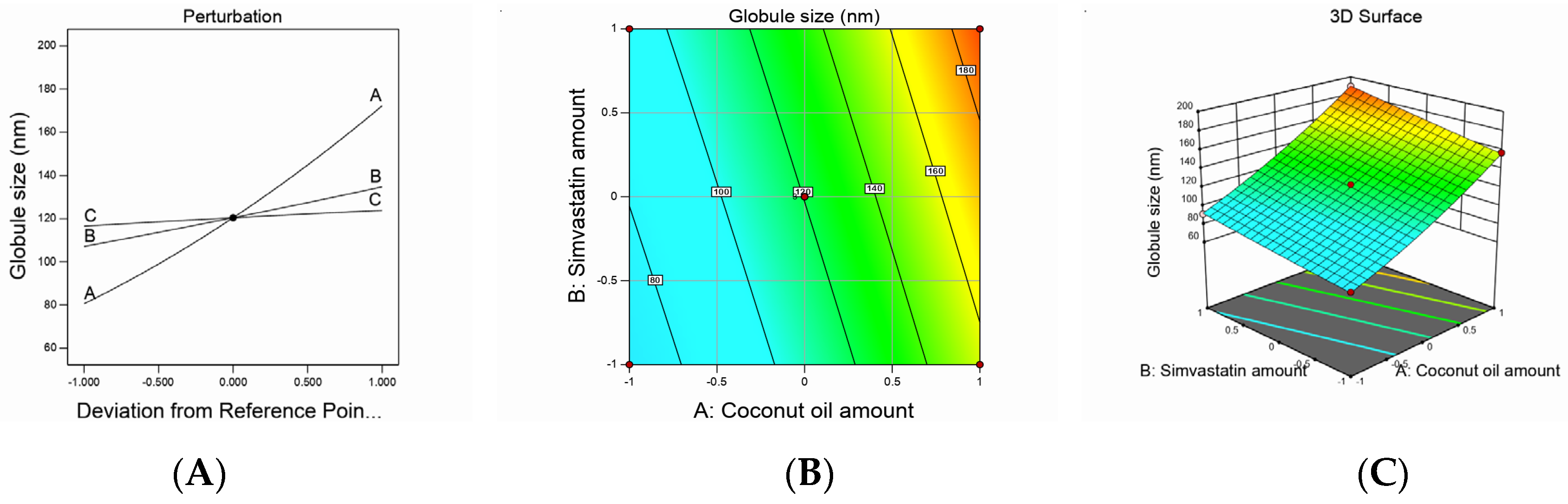
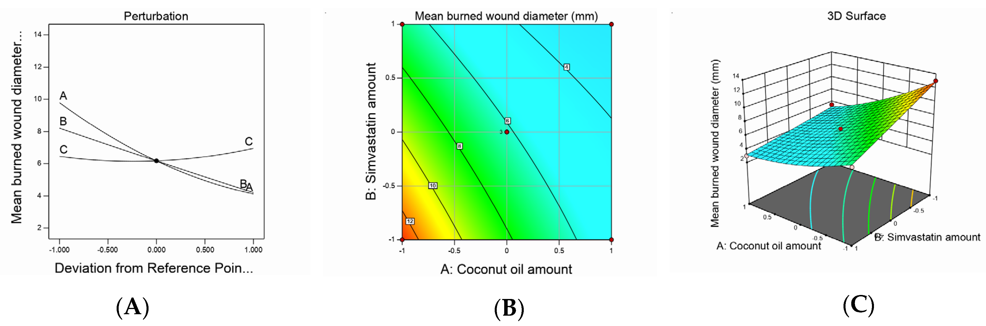
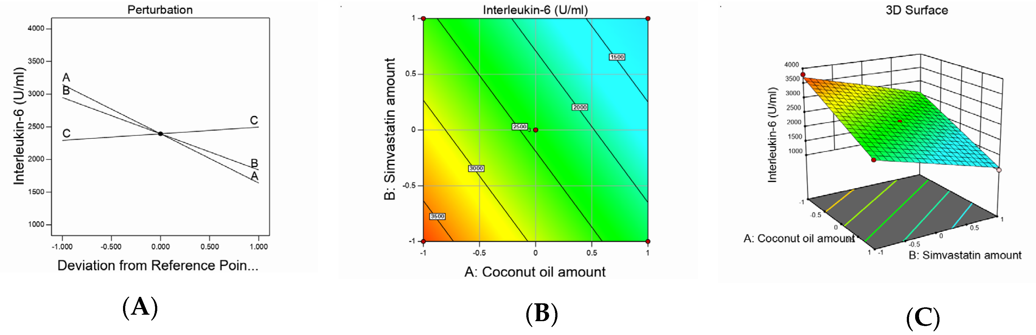
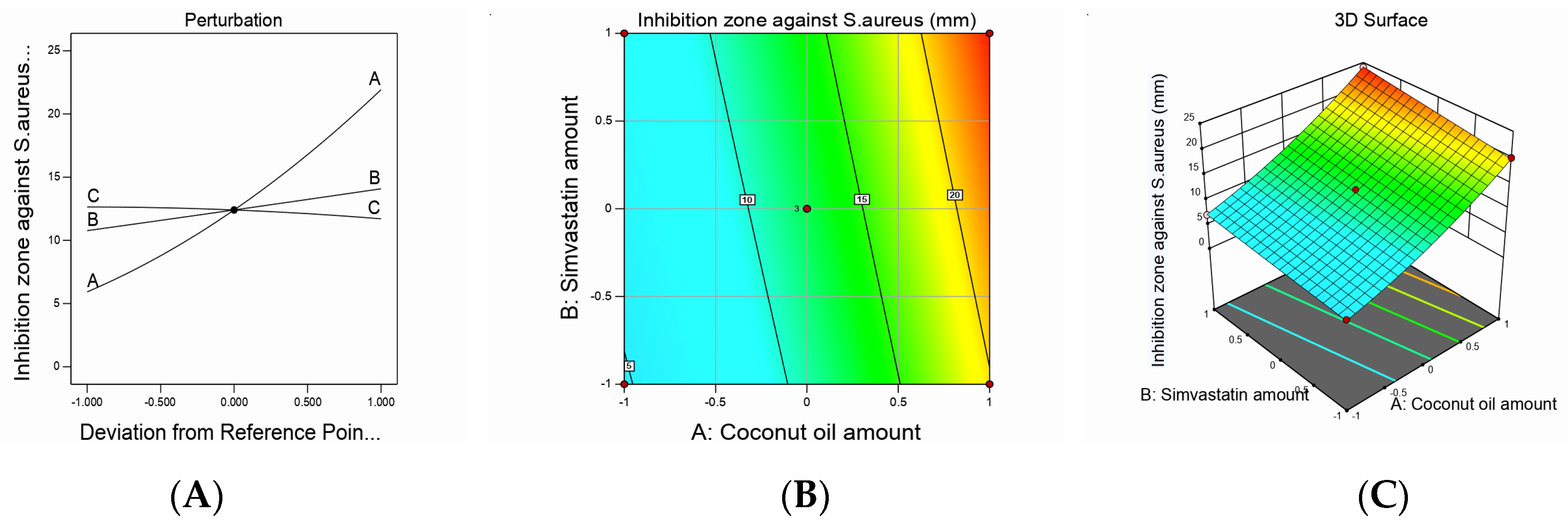
3.1. Box–Behnken Design Analysis
3.2. Formulation and Characterization of SMV Nanoemulsion
Nanoemulsion Droplet Size and Polydispersity Index
3.3. Assessment of Wound Healing
Mean Burn Wound Diameter Measurement
3.4. Interleukin-6 Measurements
3.5. Antimicrobial Activity of the Developed Nanoemulsions
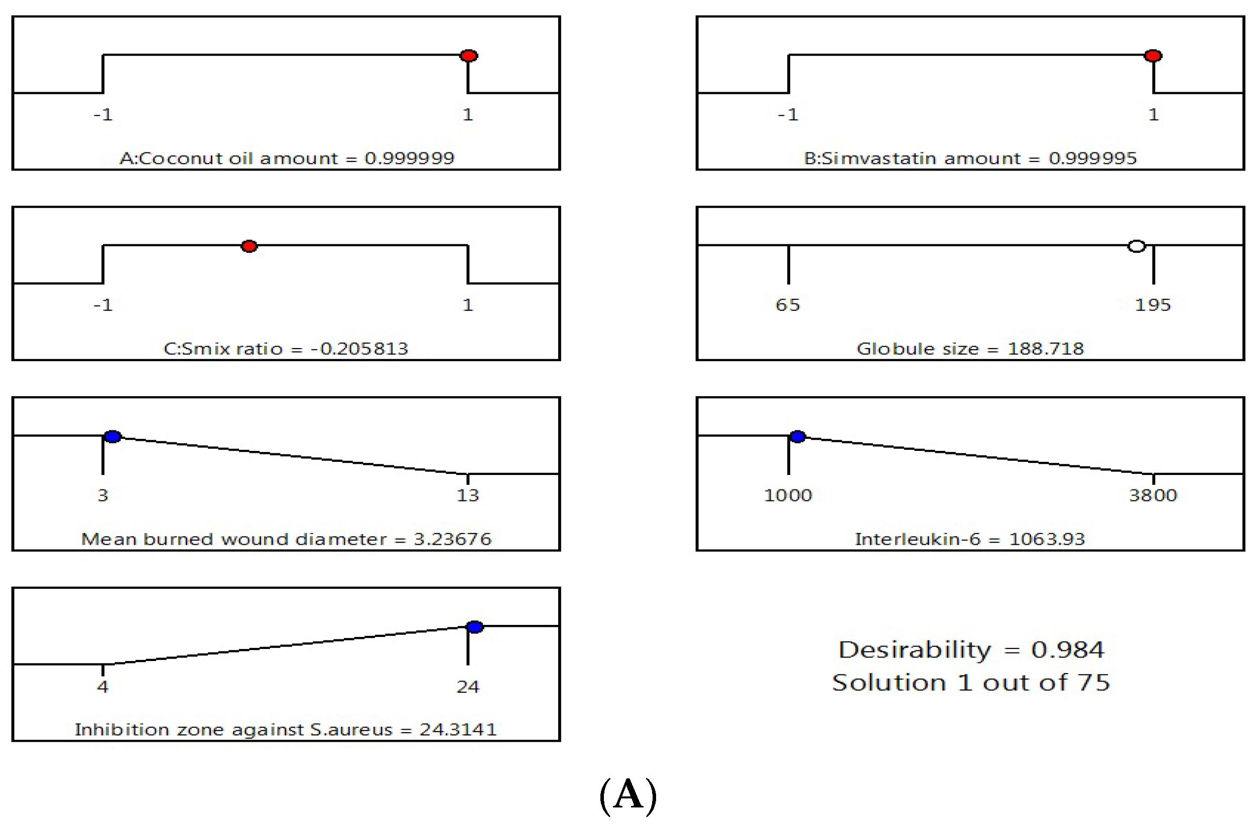
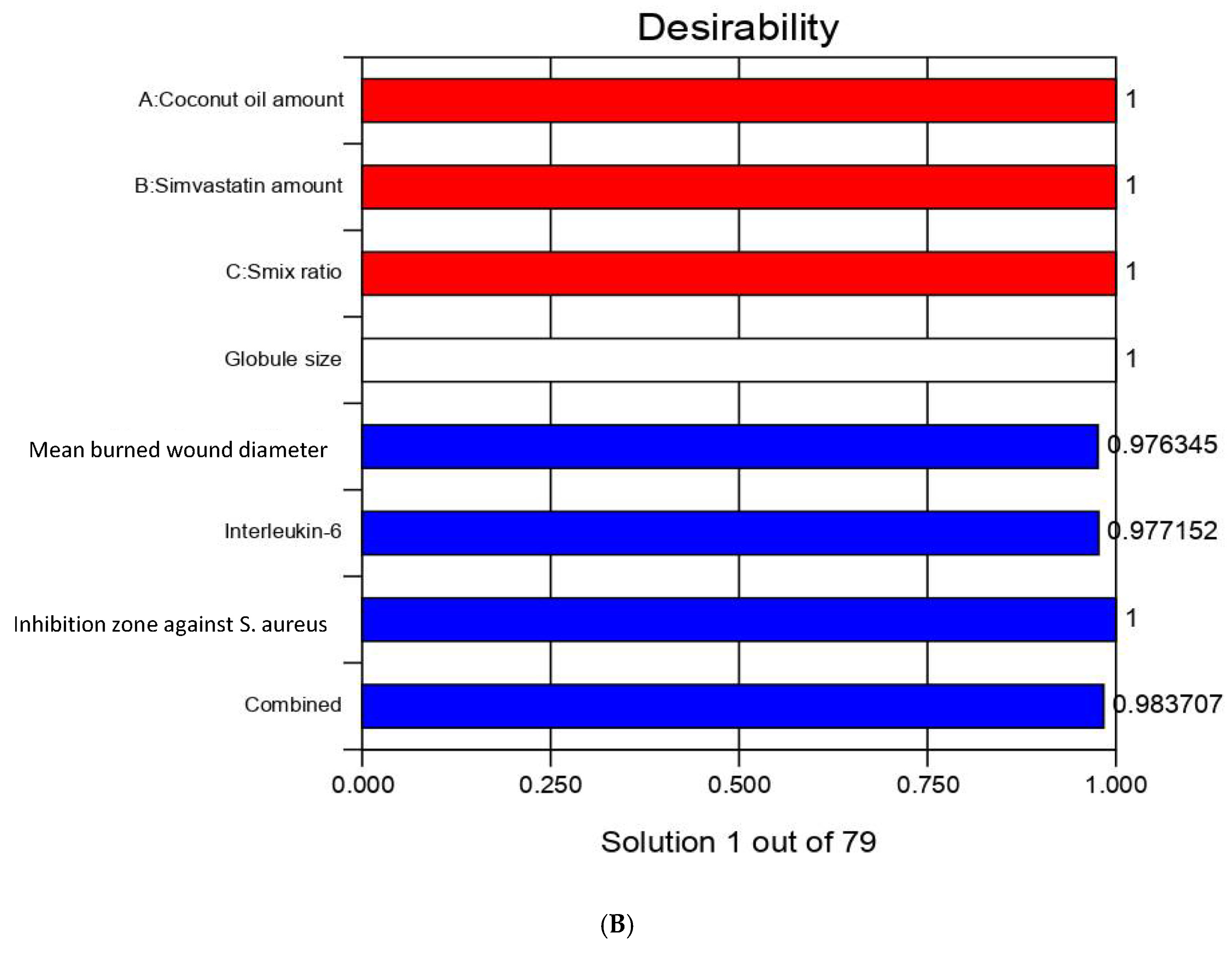
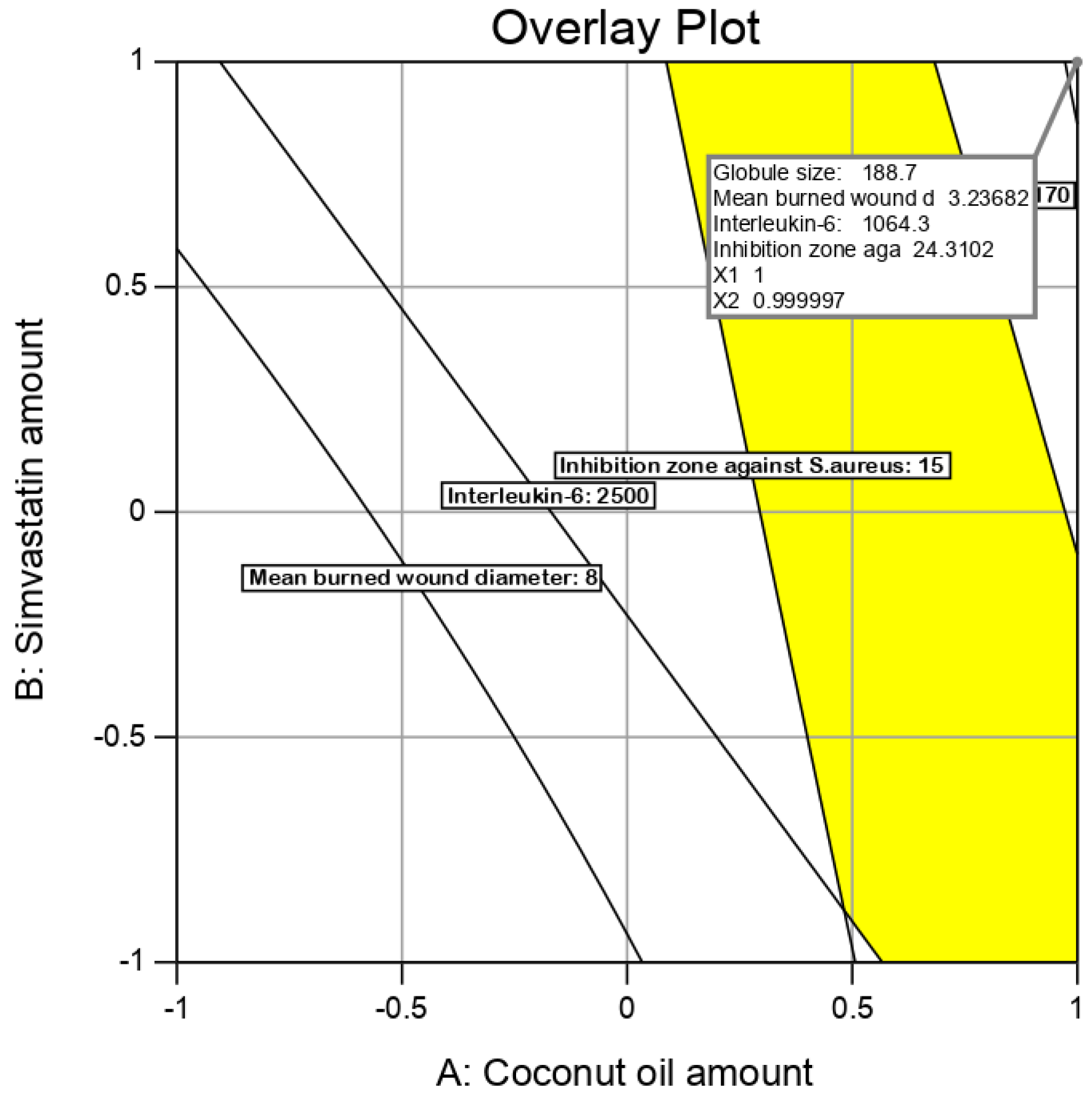
3.6. Optimization of Nanoemulsion Formulations
3.7. Checkpoint Analysis
3.8. Characterization and Evaluation of Optimum Nanoemulsion Formulation
4. Conclusions
Author Contributions
Funding
Acknowledgments
Conflicts of Interest
References
- Gould, L.; Abadir, P.; Brem, H.; Carter, M.; Conner-Kerr, T.; Davidson, J.; DiPietro, L.; Falanga, V.; Fife, C.; Gardner, S.; et al. Chronic wound repair and healing in older adults: Current status and future research. J. Am. Geriatr. Soc. 2015, 63, 427–438. [Google Scholar] [CrossRef] [PubMed]
- Boateng, J.S.; Catanzano, O. Advanced Therapeutic Dressings for Effective Wound Healing—A Review. J. Pharm. Sci. 2015, 104, 3653–3680. [Google Scholar] [CrossRef] [PubMed]
- Jahromi, M.A.M.; Zangabad, P.S.; Basri, S.M.M.; Zangabad, K.S.; Ghamarypour, A.; Aref, A.R.; Karimi, M.; Hamblin, M.R. Nanomedicine and advanced technologies for burns: Preventing infection and facilitating wound healing. Adv. Drug Deliv. Rev. 2018, 123, 33–64. [Google Scholar] [CrossRef] [PubMed]
- Fuzi, M. Editorial: The Global Challenge Posed by the Multiresistant International Clones of Bacterial Pathogens. Front. Microbiol. 2017, 8, 817. [Google Scholar] [CrossRef] [PubMed]
- Davis, S.C.; Perez, R. Cosmeceuticals and natural products: Wound healing. Clin. Derm. 2009, 27, 502–506. [Google Scholar] [CrossRef] [PubMed]
- Mayaud, L.; Carricajo, A.; Zhiri, A.; Aubert, G. Comparison of bacteriostatic and bactericidal activity of 13 essential oils against strains with varying sensitivity to antibiotics. Lett. Appl. Microbiol. 2008, 47, 167–173. [Google Scholar] [CrossRef] [PubMed]
- Sumarny, R.; Rahayu, L.; dan Sandhiutami, N. Efek Stimulansia Infus Lada Hitam (Piperis nigri fructus) pada Mencit. J. Ilmu Kefarmasian Indones. 2013, 11, 142–146. [Google Scholar]
- Nevin, K.; Rajamohan, T. Beneficial effects of virgin coconut oil on lipid parameters and in vitro LDL oxidation. Clin. Biochem. 2004, 37, 830–835. [Google Scholar] [CrossRef] [PubMed]
- Bergsson, G.; Arnfinnsson, J.; Steingrimsson, O.; Thormar, H. Killing of Gram-positive cocci by fatty acids and monoglyceridesNote. Apmis 2001, 109, 670–678. [Google Scholar] [CrossRef] [PubMed]
- Goldstein, J.L.; Brown, M.S. Regulation of the mevalonate pathway. Nat. Cell Biol. 1990, 343, 425–430. [Google Scholar] [CrossRef]
- Corsini, A.; Bellosta, S.; Baetta, R.; Fumagalli, R.; Paoletti, R.; Bernini, F. New insights into the pharmacodynamic and pharmacokinetic properties of statins. Pharm. Ther. 1999, 84, 413–428. [Google Scholar] [CrossRef]
- Pruefer, D.; Makowski, J.; Schnell, M.; Buerke, U.; Dahm, M.; Oelert, H.; Sibelius, U.; Grandel, U.; Grimminger, F.; Seeger, W.; et al. Simvastatin inhibits inflammatory properties of Staphylococcus aureus alpha-toxin. Circulation 2002, 106, 2104–2110. [Google Scholar] [CrossRef] [PubMed]
- Laufs, U.; Liao, J.K. Post-transcriptional Regulation of Endothelial Nitric Oxide Synthase mRNA Stability by Rho GTPase. J. Biol. Chem. 1998, 273, 24266–24271. [Google Scholar] [CrossRef] [PubMed]
- Naidu, B.V.; Woolley, S.M.; Farivar, A.S.; Thomas, R.; Fraga, C.; Mulligan, M.S. Simvastatin ameliorates injury in an experimental model of lung ischemia-reperfusion. J. Thorac. Cardiovasc. Surg. 2003, 126, 482–489. [Google Scholar] [CrossRef][Green Version]
- Merx, M.W.; Liehn, E.A.; Janssens, U.; Lütticken, R.; Schrader, J.; Hanrath, P.; Weber, C. HMG-CoA Reductase Inhibitor Simvastatin Profoundly Improves Survival in a Murine Model of Sepsis. Circulation 2004, 109, 2560–2565. [Google Scholar] [CrossRef]
- Wang, C.-C.; Yang, P.-W.; Yang, S.-F.; Hsieh, K.-P.; Tseng, S.-P.; Lin, Y.-C. Topical simvastatin promotes healing ofStaphylococcus aureus-contaminated cutaneous wounds. Int. Wound J. 2015, 13, 1150–1157. [Google Scholar] [CrossRef]
- Rezvanian, M.; Amin, M.C.I.M.; Ng, S.-F. Development and physicochemical characterization of alginate composite film loaded with simvastatin as a potential wound dressing. Carbohydr. Polym. 2016, 137, 295–304. [Google Scholar] [CrossRef]
- Sameh, N.; Aly, U.F.; Taleb, H.A.A.; Abdellatif, A.A. Prospective Role of Simvastatin on Wound Healing: Review of the Literature. J. Bioequivalence Bioavailab. 2018, 10, 36–42. [Google Scholar] [CrossRef]
- Thangamani, S.; Mohammad, H.; Abushahba, M.F.N.; Hamed, M.I.; Sobreira, T.J.P.; Hedrick, V.E.; Paul, L.N.; Seleem, M.N. Exploring simvastatin, an antihyperlipidemic drug, as a potential topical antibacterial agent. Sci. Rep. 2015, 5, 16407. [Google Scholar] [CrossRef]
- Crauste-Manciet, S.; Sigward, E.; Mignet, N.; Rat, P.; Dutot, M.; Muhamed, S.; Guigner, J.M.; Scherman, D.; Brossard, D. Formulation and cytotoxicity evaluation of new self-emulsifying multiple W/O/W nanoemulsions. Int. J. Nanomed. 2013, 8, 611–625. [Google Scholar] [CrossRef][Green Version]
- Esmaeili, F.; Rajabnejhad, S.; Partoazar, A.R.; Mehr, S.E.; Faridi-Majidi, R.; Sahebgharani, M.; Syedmoradi, L.; Rajabnejhad, M.R.; Amani, A. Anti-inflammatory effects of eugenol nanoemulsion as a topical delivery system. Pharm. Dev. Technol. 2015, 21, 887–893. [Google Scholar] [CrossRef] [PubMed]
- McClements, D.J. Nanoemulsions versus microemulsions: Terminology, differences, and similarities. Soft Matter 2012, 8, 1719–1729. [Google Scholar] [CrossRef]
- da Silva Marques, T.Z.; Santos-Oliveira, R.; de Oliveira de Siqueira, L.B.; da Silva Cardoso, V.; de Freitas, Z.M.F.; de Cássia da Silva Ascenção Barros, R.; Villa, A.L.V.; de Souza de Bustamante Monteiro, M.S.; Dos Santos, E.P.; Ricci-Junior, E. Development and characterization of a nanoemulsion containing propranolol for topical delivery. Int. J. Nanomed. 2018, 13, 2827–2837. [Google Scholar] [CrossRef] [PubMed]
- Singh, H.; Kumar, R.; Ahuja, N. Optimizing Drug Delivery Systems Using Systematic “Design of Experiments.” Part I: Fundamental Aspects. Crit. Rev. Ther. Drug. Carr. Syst. 2005, 22, 27–106. [Google Scholar] [CrossRef]
- Dhawan, S.; Kapil, R.; Singh, B. Formulation development and systematic optimization of solid lipid nanoparticles of quercetin for improved brain delivery. J. Pharm. Pharm. 2011, 63, 342–351. [Google Scholar] [CrossRef]
- Huang, J.; Goolcharran, C.; Ghosh, K. A Quality by Design approach to investigate tablet dissolution shift upon accelerated stability by multivariate methods. Eur. J. Pharm. Biopharm. 2011, 78, 141–150. [Google Scholar] [CrossRef]
- Dua, A.; Garg, G.; Singh, B.; Mahajan, R. Antimicrobial Properties of Methanolic Extract of Cumin (Cuminum Cyminum) Seeds. Int. J. Res. Ayurveda Pharm. 2013, 4, 104–107. [Google Scholar] [CrossRef]
- Serra, R.; Grande, R.; Butrico, L.; Rossi, A.; Settimio, U.F.; Caroleo, B.; Amato, B.; Gallelli, L.; De Franciscis, S. Chronic wound infections: The role of Pseudomonas aeruginosa and Staphylococcus aureus. Expert Rev. Anti-Infect. Ther. 2015, 13, 605–613. [Google Scholar] [CrossRef] [PubMed]
- Okur, N.Ü.; Hökenek, N.; Okur, M.E.; Ayla, Ş.; Yoltaş, A.; Siafaka, P.I.; Cevher, E. An alternative approach to wound healing field; new composite films from natural polymers for mupirocin dermal delivery. Saudi Pharm. J. 2019, 27, 738–752. [Google Scholar] [CrossRef] [PubMed]
- Lipsky, B.A.; Hoey, C. Topical Antimicrobial Therapy for Treating Chronic Wounds. Clin. Infect. Dis. 2009, 49, 1541–1549. [Google Scholar] [CrossRef]
- Okur, N.Ü.; Apaydın, Ş.; Yavaşoğlu, N.; Ülkü, K.; Yavaşoğlu, A.; Karasulu, H. Evaluation of skin permeation and anti-inflammatory and analgesic effects of new naproxen microemulsion formulations. Int. J. Pharm. 2011, 416, 136–144. [Google Scholar]
- Sakeena, M.H.F.; Elrashid, S.M.; Munavvar, A.S.; Azmin, M.N. Effects of oil and drug concentrations on droplets size of palm oil esters (POEs) nanoemulsion. J. Oleo Sci. 2011, 60, 155–158. [Google Scholar] [CrossRef]
- Okur, M.E.; Ayla, Ş.; Yozgatlı, V.; Aksu, N.B.; Yoltaş, A.; Orak, D.; Sipahi, H.; Okur, N.Ü. Evaluation of burn wound healing activity of novel fusidic acid loaded microemulsion based gel in male Wistar albino rats. Saudi Pharm. J. 2020, 28, 338–348. [Google Scholar] [CrossRef]
- Karim, F.T.; Kalam, A.; Anwar, R.; Miah, M.M.; Rahman, S.; Islam, S.M.A. Preparation and evaluation of SEDDS of simvastatin byin vivo, in vitroandex vivotechnique. Drug Dev. Ind. Pharm. 2014, 41, 1338–1342. [Google Scholar] [CrossRef]
- Ahmad, Z.; Sarmidi, M.R.; Hasham, R. Evaluation of wound closure activity of cocos nucifera oil on scratched monolayer of human dermal fibroblasts. Chem. Eng. Trans. 2017, 56, 1657–1662. [Google Scholar]
- Rego, A.C.M.D.; Filho, I.A.; Damasceno, B.P.G.L.; Egito, E.S.T.; Da Silveira, I.A.; Brandão-Neto, J.; Medeiros, A.C. Simvastatin improves the healing of infected skin wounds of rats. Acta Cir. Bras. 2007, 22, 57–63. [Google Scholar] [CrossRef] [PubMed]
- Tanaka, T.; Narazaki, M.; Kishimoto, T. IL-6 in Inflammation, Immunity, and Disease. Cold Spring Harb. Perspect. Biol. 2014, 6, a016295. [Google Scholar] [CrossRef] [PubMed]
- Kolodziejczyk, A.M.; Targosz-Korecka, M.; Szymonski, M. Nanomechanical testing of drug activities at the cellular level: Case study for endothelium-targeted drugs. Pharm. Rep. 2017, 69, 1165–1172. [Google Scholar] [CrossRef] [PubMed]
- Saggini, A.; Anogeianaki, A.; Maccauro, G.; Teté, S.; Salini, V.; Caraffa, A.; Conti, F.; Fulcheri, M.; Galzio, R.J.; Shaik-Dasthagirisaheb, Y. Cholesterol, Cytokines and Diseases. Int. J. Immunopathol. Pharm. 2011, 24, 567–581. [Google Scholar] [CrossRef] [PubMed]
- Lahera, V.; Goicoechea, M.; De Vinuesa, S.G.; Miana, M.; Heras, N.D.L.; Cachofeiro, V.; Luño, J. Endothelial dysfunction, oxidative stress and inflammation in atherosclerosis: Beneficial effects of statins. Curr. Med. Chem. 2007, 14, 243–248. [Google Scholar] [CrossRef]
- Famurewa, A.C.; Folawiyo, A.M.; Enohnyaket, E.B.; Azubuike-Osu, S.O.; Abi, I.; Obaje, S.G.; Famurewa, O.A. Beneficial role of virgin coconut oil supplementation against acute methotrexate chemotherapy-induced oxidative toxicity and inflammation in rats. Integr. Med. Res. 2018, 7, 257–263. [Google Scholar] [CrossRef]
- Serra, M.B.; Barroso, W.A.; Da Silva, N.N.; Silva, S.D.N.; Borges, A.C.R.; Abreu, I.C.; Borges, M.O.D.R. From Inflammation to Current and Alternative Therapies Involved in Wound Healing. Int. J. Inflamm. 2017, 2017, 1–17. [Google Scholar] [CrossRef]
- Shi, J.-H.; Wang, Q.; Pan, D.-Q.; Liu, T.-T.; Jiang, M. Characterization of interactions of simvastatin, pravastatin, fluvastatin, and pitavastatin with bovine serum albumin: Multiple spectroscopic and molecular docking. J. Biomol. Struct. Dyn. 2016, 35, 1529–1546. [Google Scholar] [CrossRef]
- Hanson, B.R.; Neely, M.N. Coordinate regulation of Gram-positive cell surface components. Curr. Opin. Microbiol. 2012, 15, 204–210. [Google Scholar] [CrossRef]
- Brown, S.; Maria, J.P.S.; Walker, S. Wall Teichoic Acids of Gram-Positive Bacteria. Annu. Rev. Microbiol. 2013, 67, 313–336. [Google Scholar] [CrossRef]


| Independent Variables | Levels | ||
|---|---|---|---|
| −1 | 0 | 1 | |
| A = Coconut oil amount (mg) | 125 | 175 | 225 |
| B = Simvastatin amount (mg) | 5 | 10 | 20 |
| C = S mix ratio (surfactant: cosurfactant) | 1:1 | 1:2 | 1:3 |
| Dependent variables | Constrains | ||
| Y1 = Droplet size (nm) | Minimize | ||
| Y2 = mean burned wound diameter (mm) | Minimize | ||
| Y3 = Interleukin-6 (U/mL) | Minimize | ||
| Y4 = inhibition zone against S. aureus (mm) | Maximize | ||
| A | B | C | Y1 | Y2 | Y3 | Y4 | |
|---|---|---|---|---|---|---|---|
| Run | Coconut Oil Amount | Simvastatin Amount | Smix Ratio | Globule Size (nm) | Mean Burned Wound Diameter (mm) | Interleukin-6 (U/mL) | Inhibition Zone against S. aureus (mm) |
| 1 | 1 | 0 | 1 | 174 | 4.5 | 1750 | 21 |
| 2 | −1 | 1 | 0 | 91 | 6.5 | 2450 | 7 |
| 3 | 0 | 1 | 1 | 138 | 5 | 2000 | 13 |
| 4 | 1 | −1 | 0 | 157 | 5.5 | 2200 | 20 |
| 5 | 0 | 0 | 0 | 120 | 6 | 2250 | 13 |
| 6 | −1 | 0 | −1 | 78 | 10 | 3150 | 6 |
| 7 | 0 | 0 | 0 | 118 | 6 | 2280 | 12 |
| 8 | 1 | 0 | −1 | 169 | 4.5 | 1600 | 22 |
| 9 | 0 | 1 | −1 | 130 | 5 | 1850 | 15 |
| 10 | −1 | −1 | 0 | 71 | 13 | 3800 | 5 |
| 11 | −1 | 0 | 1 | 84 | 11 | 3400 | 6 |
| 12 | 0 | −1 | −1 | 105 | 8 | 2850 | 11 |
| 13 | 0 | 0 | 0 | 123 | 6.5 | 2400 | 12 |
| 14 | 1 | 1 | 0 | 189 | 3 | 1000 | 24 |
| 15 | 0 | −1 | 1 | 110 | 9 | 3000 | 10 |
| 16 | −1 | −1 | −1 | 65 | 12.5 | 3550 | 4 |
| 17 | 1 | −1 | −1 | 152 | 5.5 | 2050 | 20 |
| 18 | −1 | 1 | 1 | 97 | 7.5 | 2600 | 6 |
| 19 | 1 | 1 | 1 | 195 | 3.5 | 1300 | 23 |
| Dependent Variables | R2 | Adjusted R2 | Predicted R2 | p-Value | F-Value | Adequate Precision |
|---|---|---|---|---|---|---|
| Y1 | 0.9990 | 0.9979 | 0.9962 | 0.0001 | 962.96 | 108.41 |
| Y2 | 0.9945 | 0.9890 | 0.9689 | 0.0001 | 181.19 | 4.73 |
| Y3 | 0.9852 | 0.9822 | 0.9761 | 0.0001 | 332.59 | 2.477 × 10⁵ |
| Y4 | 0.9976 | 0.9951 | 0.9873 | 0.0003 | 411.01 | 10.38 |
| Solution | Coconut oil Amount (mg) | SMV Amount (mg) | Smix Ratio | Droplet Size (nm) | Mean Burned Wound Diameter (mm) | Interleukin-6 (U/mL) | Inhibition Zone against S. aureus (mm) | Desirability |
|---|---|---|---|---|---|---|---|---|
| Predicated value | 225 | 20 | 1.8:1 | 188.7 | 3.2 | 1063.9 | 24.3 | 0.9840 |
| Experimental value | 225 | 20 | 1.8:1 | 186.0 ± 2.5 | 3.0 ± 0.10 | 1045.0 ± 15 | 24.0 ± 0.5 | 0.9840 |
| Factor | Optimal Value | Response Variable | Actual Value | Predicted Value | % Prediction Error a |
|---|---|---|---|---|---|
| A: Coconut oil amount (mg) | 225 | Droplet size (nm) | 186 | 188.7 | −1.45 |
| B: SMV amount (mg) | 20 | Mean burned wound diameter (mm) | 3 | 3.2 | −6.66 |
| C: Smix ratio | 1.8:1 | Interleukin-6 (U/mL) | 1045 | 1063.9 | −1.72 |
| Inhibition zone against S. aureus (mm) | 24 | 24.3 | −1.25 |
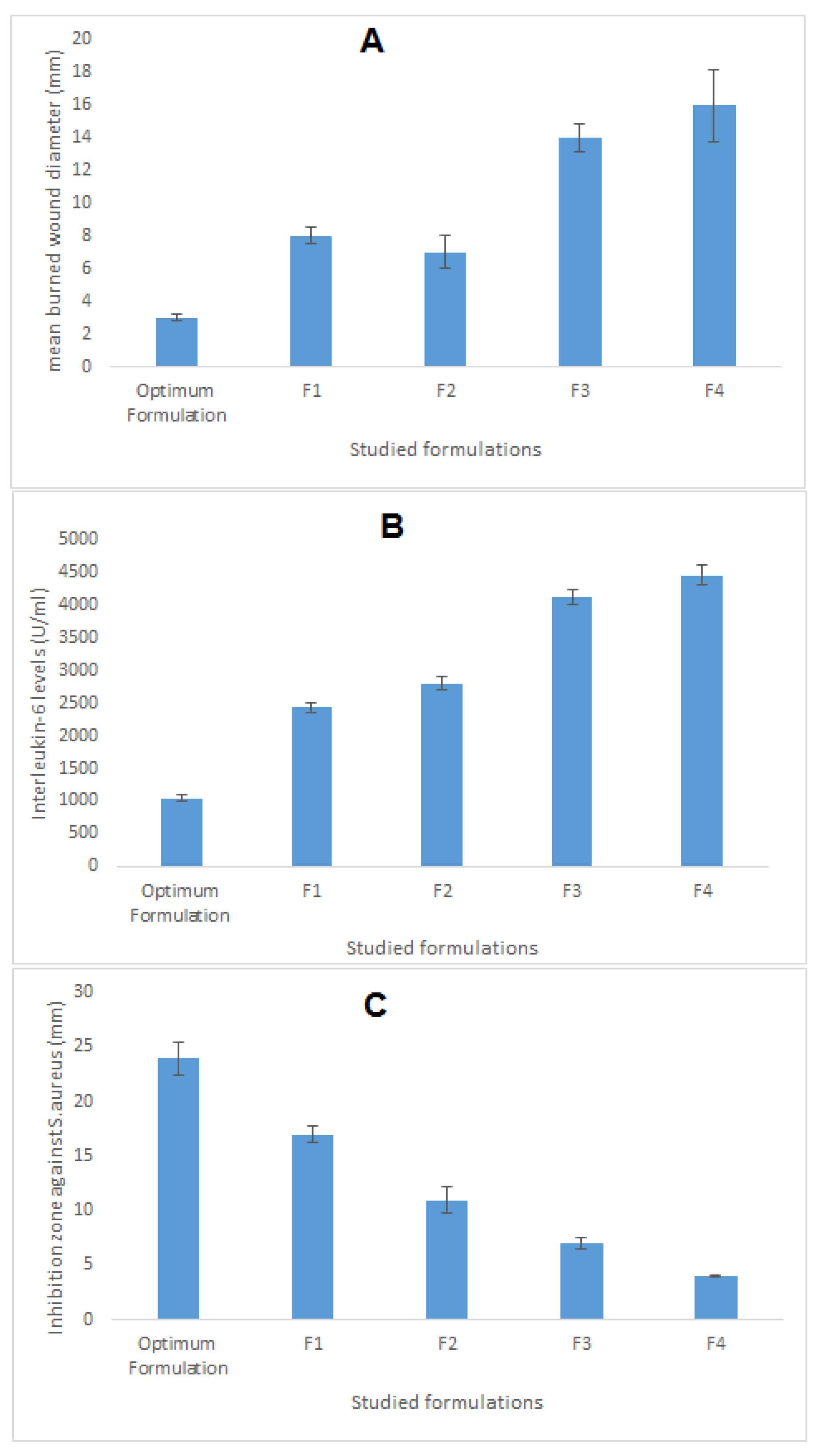
| Run | A: CnO Amount | B: SMV Amount | C: Smix Ratio | Globule Size | Mean Burned Wound Diameter | Interleukin-6 | Inhibition Zone against S. aureus |
|---|---|---|---|---|---|---|---|
| (nm) | (mm) | (U/mL) | (mm) | ||||
| Optimum formulation | 225 mg | 20 | 1.8:1 | 186 | 3 ± 0.2 | 1045 ± 55 | 24 ± 1.5 |
| F1 | 225 mg | 0 | 1.8:1 | 144 | 8 ± 0.5 | 2435 ± 76 | 17 ± 0.8 |
| F2 | Oleic acid | 20 | 1.8:1 | 198 | 7 ± 1.0 | 2800 ± 95 | 11 ± 1.2 |
| SMV aqueous dispersion | 0 | 20 | 0 | 650 | 14 ± 0.9 | 4115 ± 120 | 7 ± 0.5 |
| Normal Saline | 0 | 0 | 0 | - | 16 ± 2.2 | 4450 ± 150 | 4 ± 0.1 |
Publisher’s Note: MDPI stays neutral with regard to jurisdictional claims in published maps and institutional affiliations. |
© 2020 by the authors. Licensee MDPI, Basel, Switzerland. This article is an open access article distributed under the terms and conditions of the Creative Commons Attribution (CC BY) license (http://creativecommons.org/licenses/by/4.0/).
Share and Cite
Hosny, K.M.; Alhakamy, N.A.; Sindi, A.M.; Khallaf, R.A. Coconut Oil Nanoemulsion Loaded with a Statin Hypolipidemic Drug for Management of Burns: Formulation and In Vivo Evaluation. Pharmaceutics 2020, 12, 1061. https://doi.org/10.3390/pharmaceutics12111061
Hosny KM, Alhakamy NA, Sindi AM, Khallaf RA. Coconut Oil Nanoemulsion Loaded with a Statin Hypolipidemic Drug for Management of Burns: Formulation and In Vivo Evaluation. Pharmaceutics. 2020; 12(11):1061. https://doi.org/10.3390/pharmaceutics12111061
Chicago/Turabian StyleHosny, Khaled M., Nabil A. Alhakamy, Amal M. Sindi, and Rasha A. Khallaf. 2020. "Coconut Oil Nanoemulsion Loaded with a Statin Hypolipidemic Drug for Management of Burns: Formulation and In Vivo Evaluation" Pharmaceutics 12, no. 11: 1061. https://doi.org/10.3390/pharmaceutics12111061
APA StyleHosny, K. M., Alhakamy, N. A., Sindi, A. M., & Khallaf, R. A. (2020). Coconut Oil Nanoemulsion Loaded with a Statin Hypolipidemic Drug for Management of Burns: Formulation and In Vivo Evaluation. Pharmaceutics, 12(11), 1061. https://doi.org/10.3390/pharmaceutics12111061