Long-Lasting, Antinociceptive Effects of pH-Sensitive Niosomes Loaded with Ibuprofen in Acute and Chronic Models of Pain


 ,
, 
 ,
,  ,
,  and
and
Abstract
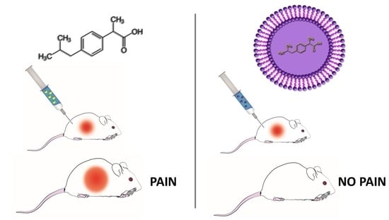
1. Introduction
2. Materials and Methods
2.1. Materials
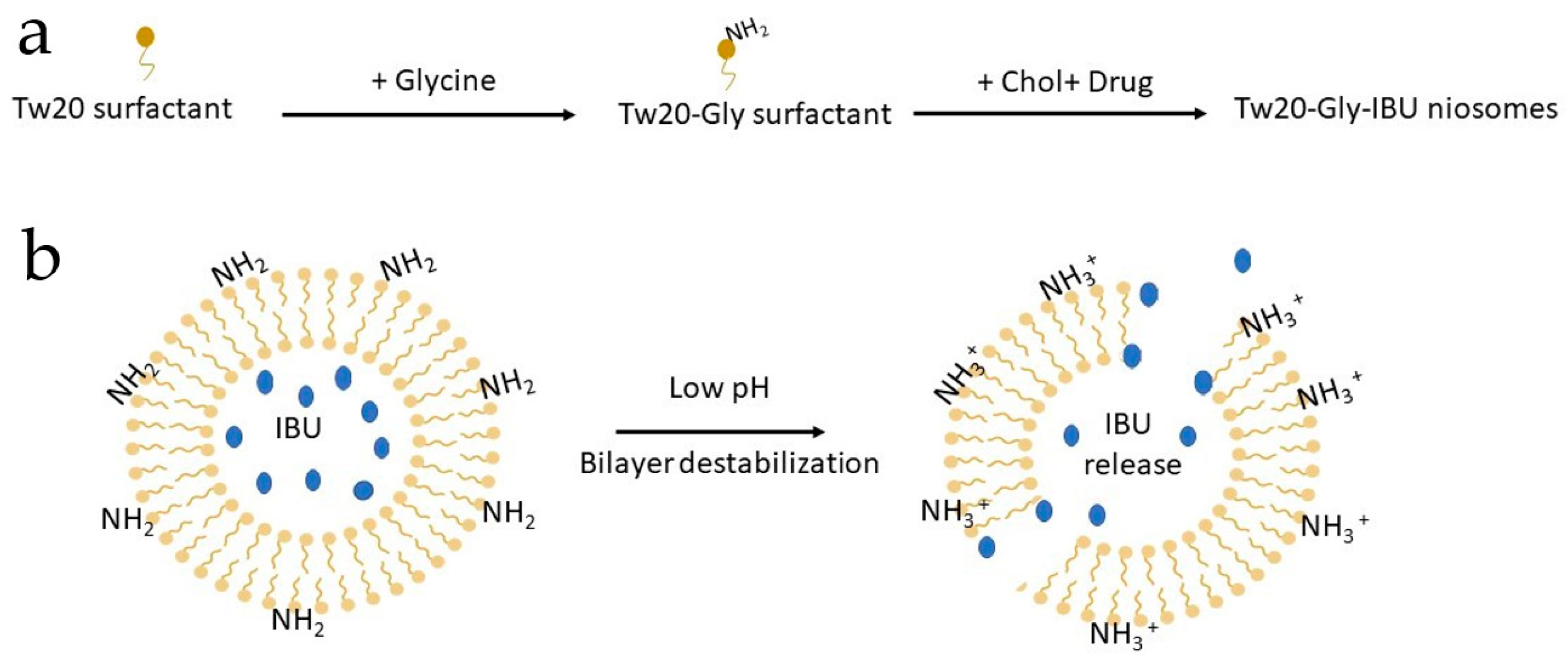
2.2. Nanovesicle Formulation and Characterization
2.3. Animals and Treatments
2.4. Writhing Test
2.5. Capsaicin-Induced Paw Licking
2.6. Zymosan-Induced Hyperalgesia
2.7. Neuropathy-Induced Allodynia and Hyperalgesia
2.8. Data Analysis and Statistics
3. Results
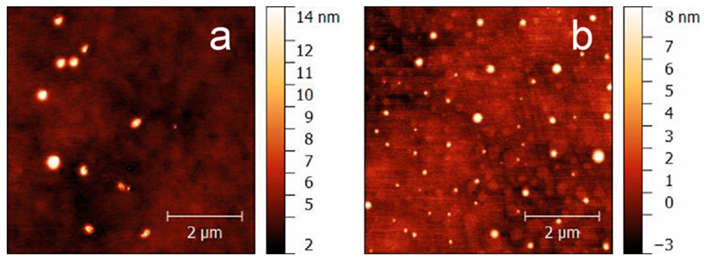
3.1. Nanovesicle Formulation and Characterization
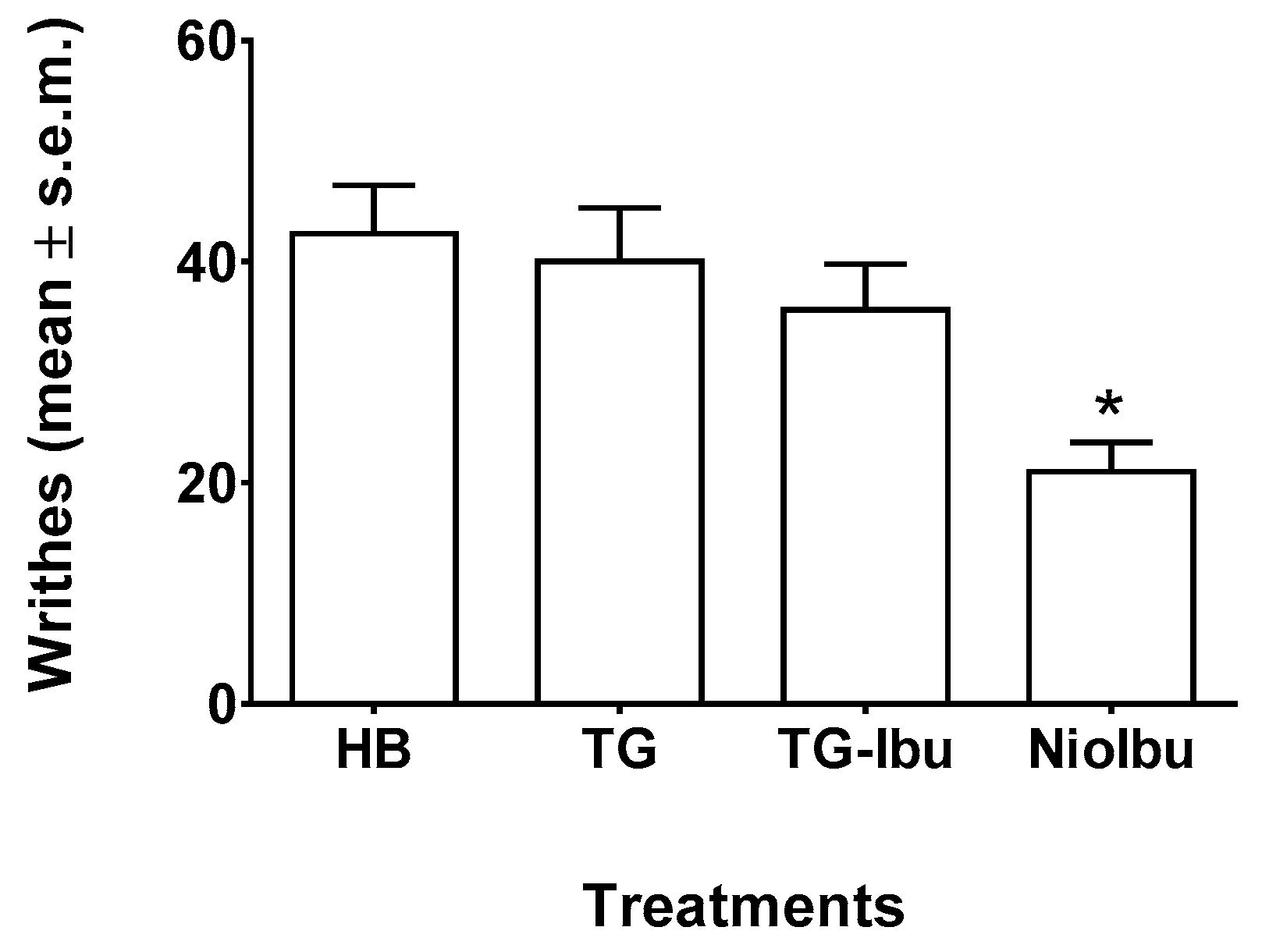
3.2. Writhing Test
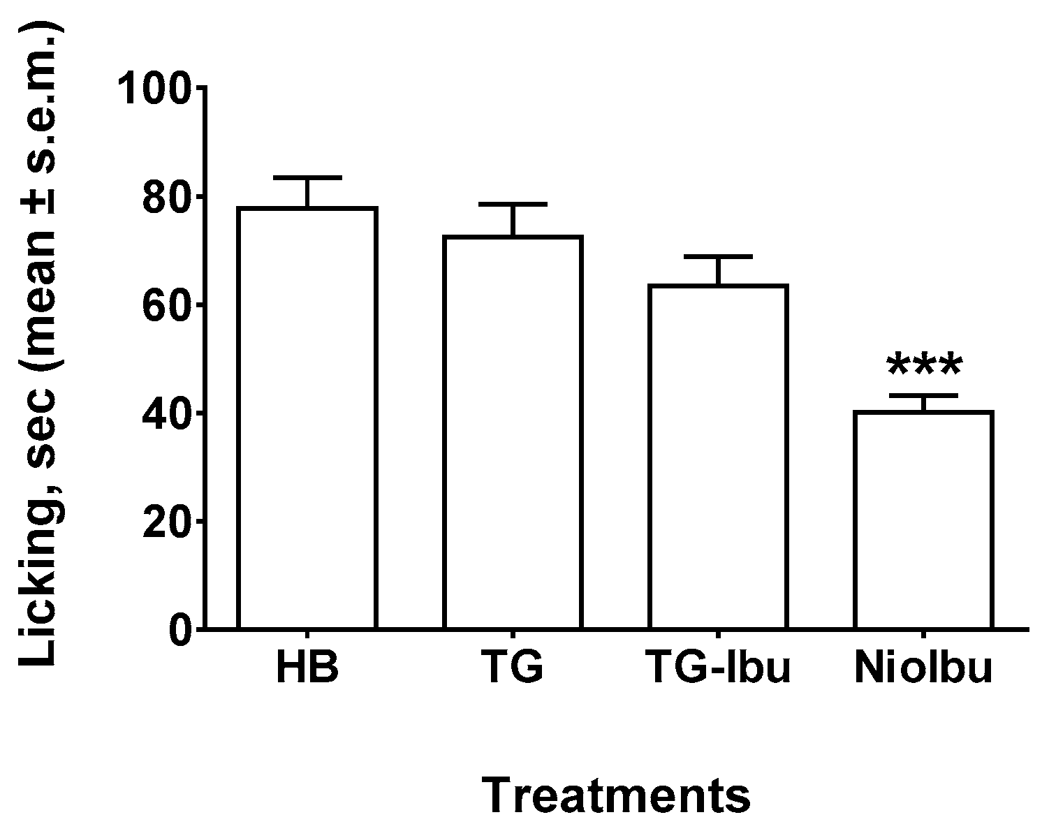
3.3. Capsaicin Test
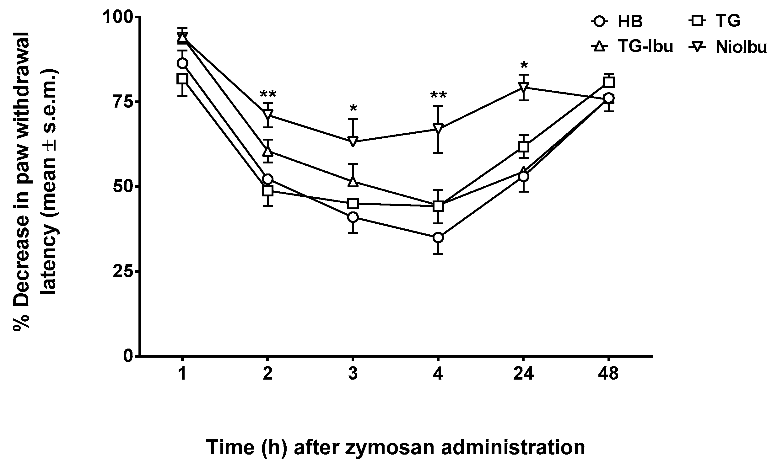
3.4. Zymosan-Induced Hyperalgesia
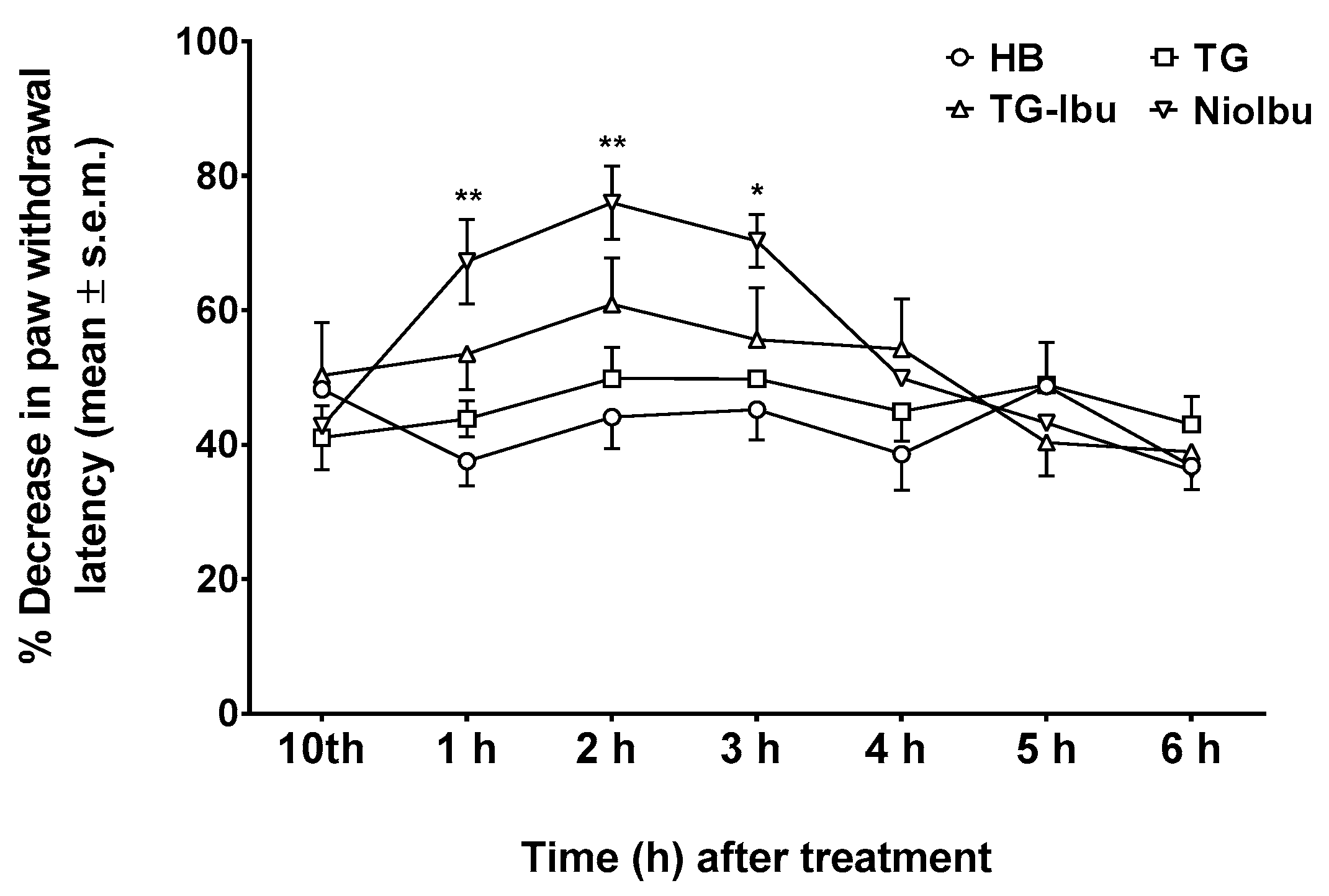
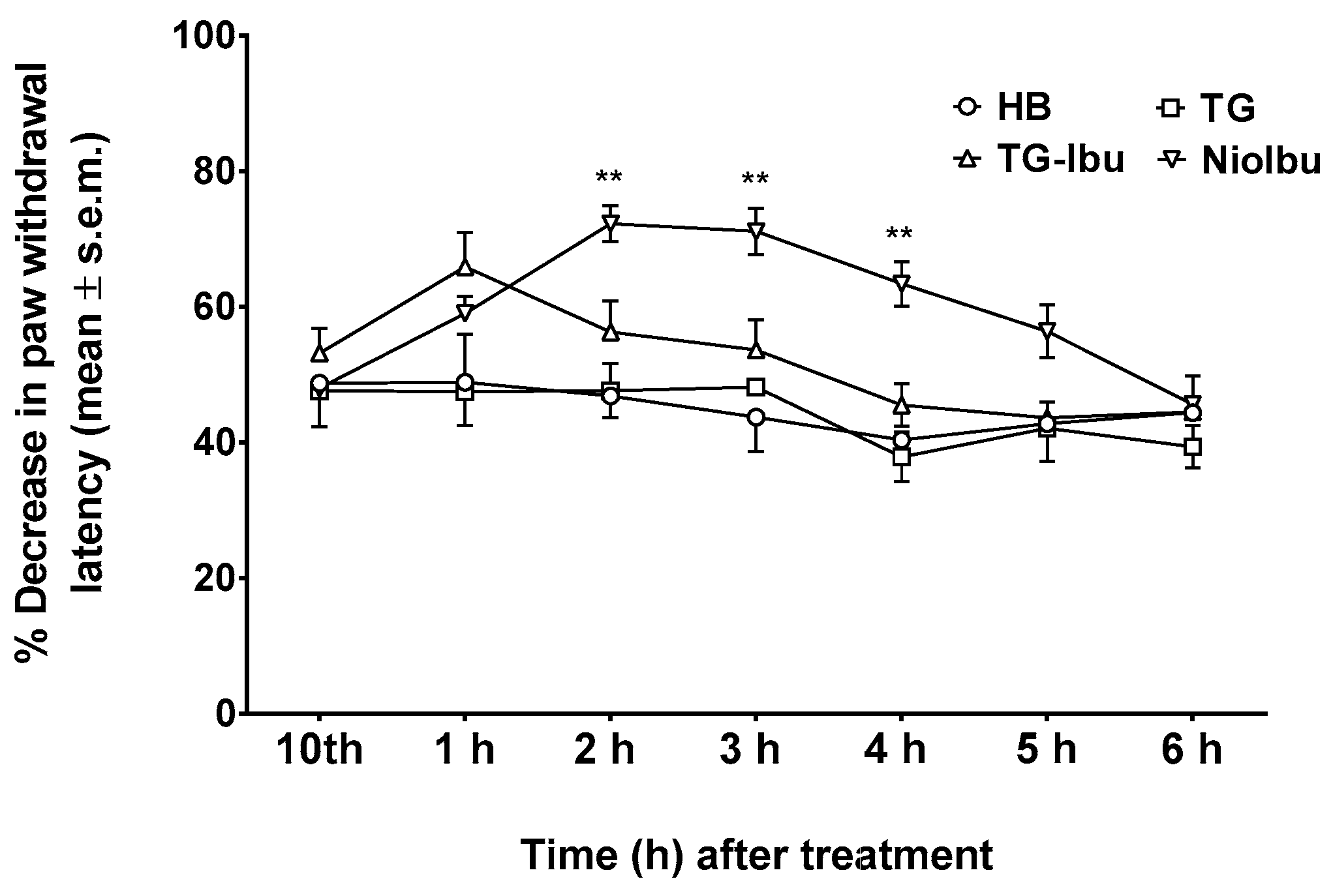
3.5. Neuropathy-Induced Allodynia and Hyperalgesia
4. Discussion
Author Contributions
Funding
Acknowledgments
Conflicts of Interest
References
- Patel, A.; Bell, M.; O’Connor, C.; Inchley, A.; Wibawa, J.; Lane, M.E. Delivery of ibuprofen to the skin. Int. J. Pharm. 2013, 457, 9–13. [Google Scholar] [CrossRef] [PubMed]
- Vane, J.R.; Botting, R.M. Anti-inflammatory drugs and their mechanism of action. Inflamm. Res. 1998, 47, 78–87. [Google Scholar] [CrossRef]
- Doherty, N.S.; Beaver, T.H.; Chan, K.Y.; Coutant, J.E.; Westrich, G.L. The role of prostaglandins in the nociceptive response induced by intraperitoneal injection of zymosan in mice. Br. J. Pharmacol. 1987, 91, 39–47. [Google Scholar] [CrossRef] [PubMed]
- Brunton, L.L.; Chabner, B.A.; Knollman, B.C. Goodman & Gilman’s the Pharmacological Basis of Therapeutics, 12th ed.; McGraw-Hill Medical: New York, NY, USA, 2011. [Google Scholar]
- Moffat, A.C.; Osselton, M.D.; Widdop, B. Clarke’s Analysis of Drugs and Poisons: In Pharmaceuticals, Body Fluids and Postmortem Material, 2; Pharmaceutical Press and American Pharmacists’ Association: London, UK, 2004; p. 1125. [Google Scholar]
- Berner, G.; Engels, B.; Vögtle-Junkert, U. Percutaneous ibuprofen therapy with Trauma-Dolgit gel: Bioequivalence studies. Drugs Exp. Clin. Res. 2004, XV, 559–564. [Google Scholar]
- Pereira-Leite, C.; Nunes, C.; Reis, S. Interaction of nonsteroidal anti-inflammatory drugs with membranes: In vitro assessment and relevance for their biological actions. Prog. Lipid Res. 2013, 52, 571–584. [Google Scholar] [CrossRef] [PubMed]
- Stoye, I.; SchrDer, K.; Müller-Goymann, C.C. Transformation of a liposomal dispersion containing ibuprofen lysinate and phospholipids into mixed micelles—Physico-chemical characterization and influence on drug permeation through excised human stratum corneum. Eur. J. Pharm. Biopharm. 1998, 46, 191–200. [Google Scholar] [CrossRef]
- Abdullah, G.Z.; Abdulkarim, M.F.; Salman, I.M.; Ameer, O.Z.; Yam, M.F.; Mutee, A.F.; Chitneni, M.; Mahdi, E.S.; Basri, M.; Sattar, M.A.; et al. In vitro permeation and in vivo anti-inflammatory and analgesic properties of nanoscaled emulsions containing ibuprofen for topical delivery. Int. J. Nanomed. 2011, 6, 387–396. [Google Scholar] [CrossRef] [PubMed]
- Santi, P.; Nicoli, S.; Colombo, G.; Bettini, R.; Artusi, M.; Rimondi, S.; Padula, C.; Rizzo, P.; Colombo, P. Post-iontophoresis transport of ibuprofen lysine across rabbit ear skin. Int. J. Pharm. 2003, 266, 69–75. [Google Scholar] [CrossRef]
- Park, E.S.; Chang, S.Y.; Hahn, M.; Chi, S.C. Enhancing effect of polyoxyethylene alkyl ethers on the skin permeation of ibuprofen. Int. J. Pharm. 2000, 209, 109–119. [Google Scholar] [CrossRef]
- Brown, M.B.; Hanpanitcharoen, M.; Martin, G.P. An in vitro investigation into the effect of glycosaminoglycans on the skin partitioning and deposition of NSAIDs. Int. J. Pharm. 2001, 225, 113–121. [Google Scholar] [CrossRef]
- Di Marzio, L.; Marianecci, C.; Petrone, M.; Rinaldi, F.; Carafa, M. Novel pH-sensitive non-ionic surfactant vesicles: Comparison between Tween 21 and Tween 20. Colloids Surf. B Biointerfaces 2011, 82, 18–24. [Google Scholar] [CrossRef] [PubMed]
- Dalmoro, A.; Bochicchio, S.; Nasibullin, SF.; Bertoncin, P.; Lamberti, G.; Barba, A.A.; Moustafine, R.I. Polymer-lipid hybrid nanoparticles as enhanced indomethacin delivery systems. Eur. J. Pharm. Sci. 2018, 121, 16–28. [Google Scholar] [CrossRef] [PubMed]
- Rinaldi, F.; Hanieh, P.N.; Chan, L.K.N.; Angeloni, L.; Passeri, D.; Rossi, M.; Wang, J.T.; Imbriano, A.; Carafa, M.; Marianecci, C. Chitosan Glutamate-Coated Niosomes: A Proposal for Nose-to-Brain Delivery. Pharmaceutics 2018, 10, 38. [Google Scholar] [CrossRef] [PubMed]
- Marianecci, C.; Di Marzio, L.; Rinaldi, F.; Celia, C.; Paolino, D.; Alhaique, F.; Esposito, S.; Carafa, M. Niosomes from 80s to present: The state of the art. Adv. Colloid Interface 2014, 205, 187–206. [Google Scholar] [CrossRef] [PubMed]
- Rajera, R.; Nagpal, K.; Singh, S.K.; Mishra, D.N. Niosomes: A controlled and novel drug delivery system. Biol. Pharm. Bull. 2011, 34, 945–953. [Google Scholar] [CrossRef]
- Edlow, D.W.; Sheldon, W.H. The pH of inflammatory exudates. Proc. Soc. Exp. Biol. Med. 1971, 137, 1328–1332. [Google Scholar] [CrossRef]
- Naghavi, M.; John, R.; Naguib, S.; Siadaty, M.S.; Grasu, R.; Kurian, K.C.; van Winkle, W.B.; Soller, B.; Litovsky, S.; Madjid, M.; et al. pH Heterogeneity of human and rabbit atherosclerotic plaques: A new insight into detection of vulnerable plaque. Atherosclerosis 2002, 164, 27–35. [Google Scholar] [CrossRef]
- Gatenby, R.A.; Gillies, R.J. Why do cancers have high aerobic glycolysis? Nat. Rev. Cancer 2004, 4, 891–899. [Google Scholar] [CrossRef]
- Rinaldi, F.; Del Favero, E.; Rondelli, V.; Pieretti, S.; Bogni, A.; Ponti, J.; Rossi, F.; Di Marzio, L.; Paolino, D.; Marianecci, C.; et al. pH-sensitive niosomes: Effects on cytotoxicity and on inflammation and pain in murine models. J. Enzym. Inhib. Med. Chem. 2017, 32, 538–546. [Google Scholar] [CrossRef]
- Marianecci, C.; Rinaldi, F.; Di Marzio, L.; Mastriota, M.; Pieretti, S.; Celia, C.; Paolino, D.; Iannone, M.; Fresta, M.; Carafa, M. Ammonium glycyrrhizinate-loaded niosomes as a potential nanotherapeutic system for anti-inflammatory activity in murine models. Int. J. Nanomed. 2014, 9, 635–651. [Google Scholar] [CrossRef]
- Carafa, M.; Marianecci, C.; Rinaldi, F.; Santucci, E.; Tampucci, S.; Monti, D. Span and Tween neutral and pH-sensitive vesicles: Characterization and in vitro skin permeation. J. Liposome Res. 2009, 19, 332–334. [Google Scholar] [CrossRef] [PubMed]
- Kilkenny, C.; Browne, W.J.; Cuthill, I.C.; Emerson, M.; Altman, D.G. Improving bioscience research reporting: The ARRIVE guidelines for reporting animal research. PLoS Biol. 2010, 8, 1000412. [Google Scholar] [CrossRef] [PubMed]
- Pieretti, S.; Di Giannuario, A.; Capasso, A.; Sorrentino, L.; Loizzo, A. Effects induced by cysteamine on chemically-induced nociception in mice. Life Sci. 1994, 54, 1091–1099. [Google Scholar] [CrossRef]
- Sakurada, T.; Katsumata, K.; Tan-No, K.; Sakurada, S.; Kisara, K. The capsaicin test in mice for evaluating tachykinin antagonists in the spinal cord. Neuropharmacology 1992, 31, 1279–1285. [Google Scholar] [CrossRef]
- Colucci, M.; Maione, F.; Bonito, M.C.; Piscopo, A.; Di Giannuario, A.; Pieretti, S. New insights of dimethyl sulphoxide effects (DMSO) on experimental in vivo models of nociception and inflammation. Pharmacol. Res. 2008, 57, 419–425. [Google Scholar] [CrossRef] [PubMed]
- Bennett, G.J.; Xie, Y.K. A peripheral mononeuropathy in rat that produces disorders of pain sensation like those seen in man. Pain 1988, 33, 87–107. [Google Scholar] [CrossRef]
- Curtis, M.J.; Bond, R.A.; Spina, D.; Ahluwalia, A.; Alexander, S.P.; Giembycz, M.A.; Gilchrist, A.; Hoyer, D.; Insel, P.A.; Izzo, A.A.; et al. Experimental design and analysis and their reporting: New guidance for publication in BJP. Br. J. Pharmacol. 2015, 172, 3461–3471. [Google Scholar] [CrossRef]
- Bélichard, P.; Landry, M.; Faye, P.; Bachvarov, D.R.; Bouthillier, J.; Pruneau, D.; Marceau, F. Inflammatory hyperalgesia induced by zymosan in the plantar tissue of the rat: Effect of kinin receptor antagonists. Immunopharmacology 2000, 46, 139–147. [Google Scholar] [CrossRef]
- Ren, K.; Dubner, R. Inflammatory Models of Pain and Hyperalgesia. ILAR J. 1999, 40, 111–118. [Google Scholar] [CrossRef]
- Lucio, M.; Lima, J.L.; Reis, S. Drug-membrane interactions: Significance for medicinal chemistry. Curr. Med. Chem. 2010, 17, 1795–1809. [Google Scholar] [CrossRef]
- Gaur, P.K.; Bajpai, M.; Mishra, S.; Verma, A. Development of ibuprofen nanoliposome for transdermal delivery: Physical characterization, in vitro/in vivo studies, and anti-inflammatory activity. Artif. Cells Nanomed. Biotechnol. 2016, 44, 370–375. [Google Scholar] [CrossRef] [PubMed]
- Bozzuto, G.; Molinari, A. Liposomes as nanomedical devices. Int. J. Nanomed. 2015, 10, 975–999. [Google Scholar] [CrossRef] [PubMed]
- Masotti, A.; Vicennati, P.; Alisi, A.; Marianecci, C.; Rinaldi, F.; Carafa, M.; Ortaggi, G. Novel Tween 20 derivatives enable the formation of efficient pH-sensitive drug delivery vehicles for human hepatoblastoma. Bioorg. Med. Chem. Lett. 2010, 20, 3021–3025. [Google Scholar] [CrossRef] [PubMed]
- Verma, S.; Singh, S.K.; Syan, N.; Mathur, P.; Valecha, V. Nanoparticle vesicular system: A versatile tool for drug delivery. J. Chem. Pharm. Res. 2010, 2, 496–509. [Google Scholar]
- Lehner, R.; Wang, X.; Wolf, M.; Hunziker, P. Designing switchable nanosystems for medical application. J. Control. Release 2012, 161, 307–316. [Google Scholar] [CrossRef]
- Das, M.K.; Palei, N.N. Sorbitan ester niosomes for topical delivery of rofecoxib. Indian J. Exp. Biol. 2011, 49, 438–445. [Google Scholar]
- Naresh, R.A.; Raja, G.K.; Pillai, N.; Udupa, N.; Chandrashekar, G. Anti-inflammatory activity of niosome encapsulated diclofenac sodium in arthritic rats. Indian J. Pharmacol. 1994, 26, 46–48. [Google Scholar]
- Shahiwala, A.; Misra, A. Studies in topical application of niosomally entrapped Nimesulide. J. Pharm. Pharm. Sci. 2002, 5, 220–225. [Google Scholar]
- Goh, J.Z.; Tang, S.N.; Zuraini, A.; Zakaria, Z.A.; Kadir, A.A.; Chiong, H.S. Enhanced anti-inflammatory effects of nanoencapsulated diclofenac. Eur. J. Inflamm. 2013, 11, 855–861. [Google Scholar] [CrossRef]
- Sankhyan, A.; Pawar, P. Recent Trends in Niosome as Vesicular Drug Delivery System. J. Appl. Pharm. Sci. 2012, 2, 20–32. [Google Scholar] [CrossRef]
- Goh, J.Z.; Tang, S.N.; Chiong, H.S.; Yong, Y.K.; Zuraini, A.; Hakim, M.N. Evaluation of antinociceptive activity of nanoliposome-encapsulated and free-form diclofenac in rats and mice. Nanomedicine 2014, 10, 297–303. [Google Scholar] [CrossRef]
- Narasimha Reddy, D.; Udupa, N. Formulation and Evaluation of Oral and Transdermal Preparations of Flurbiprofen and Piroxicam Incorporated with Different Carriers. Drug Dev. Ind. Pharm. 2008, 19, 843–852. [Google Scholar] [CrossRef]
- Joshi, S.K.; Hernandez, G.; Mikusa, J.P.; Zhu, C.Z.; Zhong, C.; Salyers, A.; Wismer, C.T.; Chandran, P.; Decker, M.W.; Honore, P. Comparison of antinociceptive actions of standard analgesics in attenuating capsaicin and nerve-injury-induced mechanical hypersensitivity. Neuroscience 2006, 143, 587–596. [Google Scholar] [CrossRef]


| Sample | Tw20 (mM) | Tw20-Gly (mM) | Chol (mM) | IBU (% p/v) |
|---|---|---|---|---|
| Nio | 3.75 | 11.25 | 7.5 | = |
| NioIbu 1% | 3.75 | 11.25 | 7.5 | 1 |
| NioIbu 3% | 3.75 | 11.25 | 7.5 | 3 |
| NioIbu 5% | 3.75 | 11.25 | 7.5 | 5 |
| NioIbu 7% | 3.75 | 11.25 | 7.5 | 7 |
| Niosomes | Diameter (nm) | AFM Diameter (nm) | ζ Potential (mV) | Polydispersity Index | Fluorescence Anisotropy (AU) | Loaded Drug Conc. (mg/mL) |
|---|---|---|---|---|---|---|
| Nio | 215.0 ± 3.0 | 152 ± 18 | −41.0 ± 1.2 | 0.160 ± 0.08 | 0.17 ± 0.01 | – |
| NioIbu 5% | 122.1 ± 19.6 | 89 ± 24 | −40.2 ± 0.1 | 0.404 ± 0.05 | 0.20 ± 0.04 | 0.37 ± 0.05 |
© 2019 by the authors. Licensee MDPI, Basel, Switzerland. This article is an open access article distributed under the terms and conditions of the Creative Commons Attribution (CC BY) license (http://creativecommons.org/licenses/by/4.0/).
Share and Cite
Marzoli, F.; Marianecci, C.; Rinaldi, F.; Passeri, D.; Rossi, M.; Minosi, P.; Carafa, M.; Pieretti, S. Long-Lasting, Antinociceptive Effects of pH-Sensitive Niosomes Loaded with Ibuprofen in Acute and Chronic Models of Pain. Pharmaceutics 2019, 11, 62. https://doi.org/10.3390/pharmaceutics11020062
Marzoli F, Marianecci C, Rinaldi F, Passeri D, Rossi M, Minosi P, Carafa M, Pieretti S. Long-Lasting, Antinociceptive Effects of pH-Sensitive Niosomes Loaded with Ibuprofen in Acute and Chronic Models of Pain. Pharmaceutics. 2019; 11(2):62. https://doi.org/10.3390/pharmaceutics11020062
Chicago/Turabian StyleMarzoli, Francesca, Carlotta Marianecci, Federica Rinaldi, Daniele Passeri, Marco Rossi, Paola Minosi, Maria Carafa, and Stefano Pieretti. 2019. "Long-Lasting, Antinociceptive Effects of pH-Sensitive Niosomes Loaded with Ibuprofen in Acute and Chronic Models of Pain" Pharmaceutics 11, no. 2: 62. https://doi.org/10.3390/pharmaceutics11020062
APA StyleMarzoli, F., Marianecci, C., Rinaldi, F., Passeri, D., Rossi, M., Minosi, P., Carafa, M., & Pieretti, S. (2019). Long-Lasting, Antinociceptive Effects of pH-Sensitive Niosomes Loaded with Ibuprofen in Acute and Chronic Models of Pain. Pharmaceutics, 11(2), 62. https://doi.org/10.3390/pharmaceutics11020062