Review

Adenovirus 36 and Obesity: An Overview

Department of Experimental Medicine, Section of Medical Pathophysiology Food Science and Endocrinology, Sapienza University of Rome, 00161 Rome, Italy
*
Author to whom correspondence should be addressed.
Viruses 2015, 7(7), 3719-3740; https://doi.org/10.3390/v7072787
Submission received: 1 May 2015 / Revised: 16 June 2015 / Accepted: 30 June 2015 / Published: 8 July 2015

Abstract

:
There is an epidemic of obesity starting about 1980 in both developed and undeveloped countries definitely associated with multiple etiologies. About 670 million people worldwide are obese. The incidence of obesity has increased in all age groups, including children. Obesity causes numerous diseases and the interaction between genetic, metabolic, social, cultural and environmental factors are possible cofactors for the development of obesity. Evidence emerging over the last 20 years supports the hypothesis that viral infections may be associated with obesity in animals and humans. The most widely studied infectious agent possibly linked to obesity is adenovirus 36 (Adv36). Adv36 causes obesity in animals. In humans, Adv36 associates with obesity both in adults and children and the prevalence of Adv36 increases in relation to the body mass index. In vivo and in vitro studies have shown that the viral E4orf1 protein (early region 4 open reading frame 1, Adv) mediates the Adv36 effect including its adipogenic potential. The Adv36 infection should therefore be considered as a possible risk factor for obesity and could be a potential new therapeutic target in addition to an original way to understand the worldwide rise of the epidemic of obesity. Here, the data indicating a possible link between viral infection and obesity with a particular emphasis to the Adv36 will be reviewed.

1. Introduction

Obesity is a disease characterized by an excess of fat mass. The incidence of obesity in recent decades has grown dramatically [1].
The prevalence of obesity doubled in adults and tripled in children during the last 20 years, even in countries with traditionally low rate of the disease. In 2014, more than 1.9 billion adults, 18 years old and older, were overweight. Of these, over 600 million were obese. The worldwide prevalence of obesity more than doubled between 1980 and 2014. (World Health Organization (WHO) data; [2]). The risks of diabetes, hypertension and dyslipidaemia increase starting from a body mass index (BMI) of about 21.0 kg/m2, thereby reducing life expectancy and greatly increasing the health and social economic burden. Obesity is clearly related to increased mortality, morbidity and disability rates. Overweight and obesity are the fifth leading risk for global deaths. Obesity is now described as a phenomenon in character epidemic [3], and according to the WHO, obesity represents “one of the major Public Health problems of our time”. Obesity is also a risk factor for numerous chronic diseases such as cardiovascular diseases and diabetes, orthopedic problems and mental disorders. The scientific community started to study the obesity epidemic looking at the possible causes with the aim to find effective treatments to reduce morbidity and mortality.
Although obesity is a multifactorial disease caused by the interaction between genetics, metabolism, social, cultural and environmental factors, viral infection has been suggested as a possible cofactor for the development of obesity [4].
Immune response depends by nutritional status and can be easily dysregulated in states of imbalanced nutrition such as obesity. This may predispose obese individuals to increased susceptibility to infection [5,6].
In the hospital setting, obese patients are more likely to develop secondary infections and complications such as sepsis, pneumonia, bacteremia, wound infections, and respiratory infections [7,8]. Obesity has been associated with increased risk of complications due to surgical site infections [9,10,11]. Obese individuals have increased risk of Helicobacter pylori [12] infection and overweight children show a three times greater risk of Neisseria meningitides [13]. Obesity is also a risk factor of severe infection and death caused by the pandemic influenza strain H1N1 [14].
Overall, these observations indicate that excessive adipose tissue expansion predisposes individuals to various infections. On the other hand, new data have been generated in the last few years suggesting infectious agents being the cause of obesity in addition to being more easily hosted in an obese individual. Among the multitude of infectious agents, adenoviruses are the human pathogens that more than others are causatively and correlatively linked with animal and human obesity, respectively, and seem to directly influence the adipose tissue [15,16].
In animals, adenovirus serotype Adv31 and Adv9 correlate with obesity and are adipogenic in animal cells culture [17,18]. An avian adenovirus (SMAM-1) and the human adenovirus type 36 (Adv36) have been associated with obesity [19] and there are reports a suggesting significant role of adenoviruses in the development of human obesity.
Here we provide an overview of the data available on the relationship between adenovirus infection and obesity.

2. Association of Viral Infections with Obesity in Animal Models
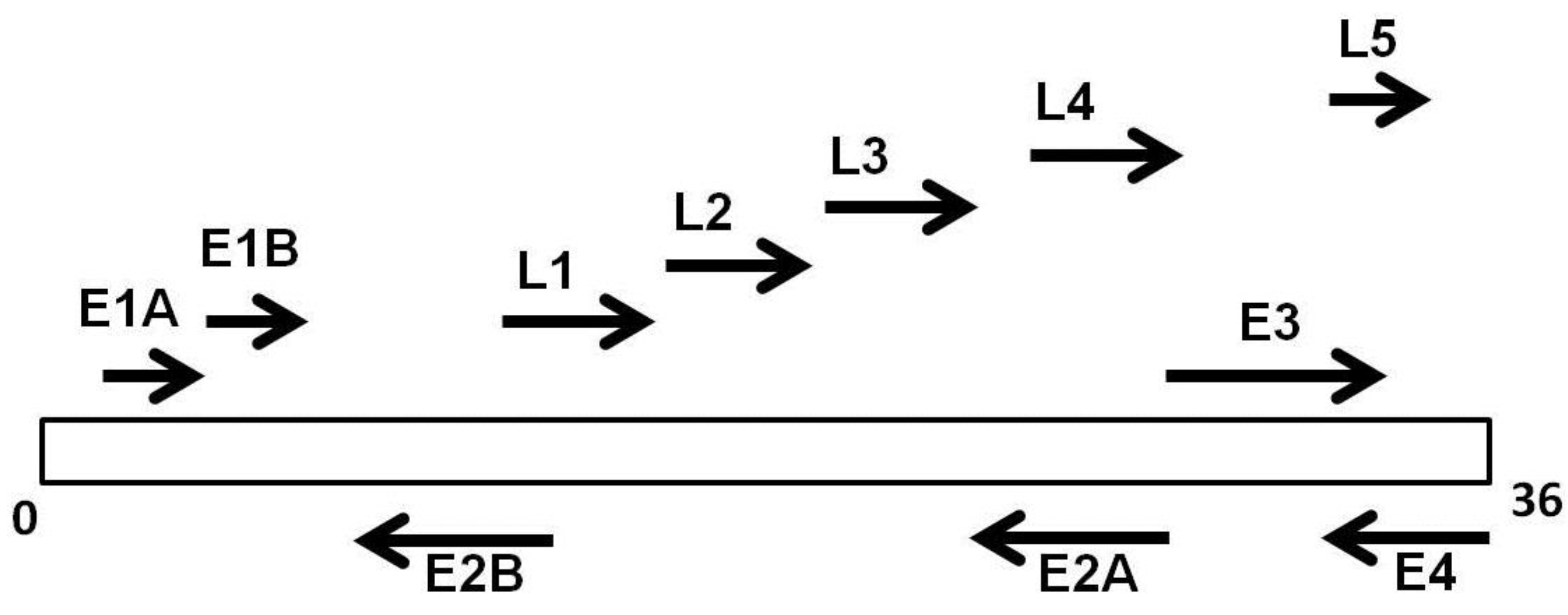
Five infectious agents were implicated in contributing to obesity, including canine distemper virus (CDV), rous associated virus (RAV)-7, borna disease virus (BDV) and adenoviruses [19]. Apart from adenoviruses, all the other viruses of this list are associated with brain damage and, in experimental animals, obesity develops via brain involvement or via direct damage of fat tissue. CDV is a paramyxovirus that infects dogs and other wild mammals; it was one of the first infectious agents identified to cause enlarged fat cells and increased body weight in mice [19]. The CDV infected animals showed twofold increase in body weight and a selective, virus-induced disruption of critical brain catecholamine pathways [20]. RAV-7 is one of the most common retroviruses affecting chickens [21]. The infected chickens are smaller than hatchmates and develop obesity, ataxia, lymphoblastoid infiltration of thyroid and pancreas, liver fat accumulation and a frank lipemia. BDV is a nonsegmented negative-stranded RNA virus that may cause obesity in various animals, including rats and chickens. Most studies suggest that BDV causes obesity by inflammation of the hypothalamus. In infected animals, hyperplasia of pancreatic islets, increase in glucose and triglyceride levels is also seen [19].
Adenoviruses are the only infective agents reported to be linked with adiposity in both experimental animal models and naturally infected humans [22].

3. Adenoviruses

Adenoviruses are medium size viruses. All human adenoviral genomes have the same general organization. The genome consists of a single linear, double-stranded DNA molecule with short inverted terminal repeats at each end. A protein, terminal protein, is covalently attached to both the 5′ ends of the genomic DNA. The viral chromosome has five early transcription units (E1A, E1B, E2, E3 and E4) (Figure 1).
Figure 1. Genomic organization of Adv36: The early proteins (E1A, E1B, E2A, E2B, E3, and E4) are involved in the regulation of replication of DNA. The late proteins (L1–L5), products of the translation of late mRNA, constitute structural capsid proteins.
Viruses 07 02787 g001
There are more than 50 immunologically distinct serotypes of adenovirus that can cause infections in humans. Serotypes are operationally defined by the ability of the antibodies induced by infection to neutralize that serotype only, and not other adenovirus. The human adenoviruses have been further classified into six subgroups (A–F) based upon hemoagglutination proprieties and genetic relatedness.
Although the epidemiological characteristics of the adenovirus serotype change, all are transmitted by direct contact, fecal-oral transmission, and sometimes by environmental transmission (contaminated water). Most of the adenovirus cause respiratory diseases, but can also cause gastroenteritis, conjunctivitis and cystitis [19,23].
The adenovirus virion contains a 36 kb genome of double-stranded DNA, which is surrounded by a non-enveloped icosahedral protein capsid. The capsid is composed of 252 capsomers: 240 hexons and 12 pentons at the vertices of the icosahedrons. Each penton consists of a penton base and a penton fiber. The fibers consist of a slender shaft with a globular head. They are involved in the process of attachment of the virus particle to the host cell.
Adv2 and Adv5 have been used for most studies of the adenovirus replication cycle. These viruses can be easily grown in the laboratory by infecting permissive cell lines such as HeLa and A549 cells.
Adv5 and Adv37 increase adiposity in animals and have been indicated as possible contributing factors to the rising problem of obesity [24], whereas human adenoviruses Adv2 and Adv31 are not adipogenic in animals. Among the over 50 human adenoviruses available, Adv36 is the most studied also because of its minimal cross-reactivity with other human adenoviruses [25,26,27]. The exact mechanisms leading to the development of obesity through these viruses are unknown. An accurate understanding of the varied etiological factors of obesity (including infecting agents) may lead to cause-specific treatment and successful management of this disease.

4. Adv36 and Obesity

4.1. In Vivo Studies

The adipogenic effect of adenoviruses has been studied since 1990, when the adipogenic role of the avian adenovirus SMAM-1 in chickens was observed for the first time [28]. SMAM-1 is antigenically similar to chicken embryo lethal orphan virus (CELO), which is common among poultry in the USA. SMAM-1 increases adiposity in experimentally infected chickens and their naive cage-mates, suggesting horizontal transmission of obesity induced by the virus [28]. Paradoxically, SMAM-1-induced adiposity in animals is associated with lower serum cholesterol and triglyceride concentrations compared to uninfected counterparts [29].
The human adenoviruses Adv5 and Adv37 increase the adiposity in animals, while other human adenoviruses like Adv2 and Adv31 are not adipogenic [30,31]. Adv36 is adipogenic in animals [30,32,33,34] and reduces significantly the concentrations of cholesterol and triglycerides compared to uninfected controls. Longitudinal studies in monkeys [33] showed a 15%–30% increase of body weight and a reduction of serum cholesterol after natural infection with Adv36. Adv36 is easily transmitted between animals [35]. After transfusion of a small amount of blood from Adv36 infected chicken to another animal, sharing the same cage, the animals became obese [35].
Pasarica et al. [34] showed that rats infected with Adv36 had an increase of weight, insulin sensitivity and glucose uptake. Furthermore, Adv36 infected non-human primates [33], had increase of weight and anti-Adv36 antibodies, but a decrease of total cholesterol. Similar results were obtained in marmosets and a decrease of total cholesterol was found in hamsters [36].
Rats inoculated with Adv36 or UV-inactivated Adv36 [37], showed 23% greater epididymal fat pad and viral mRNA and DNA were detected in liver, brain and adipose tissue. Either intranasal or intra-peritoneal routes of viral inoculation showed similar results. Adv36 stimulates preadipocyte differentiation [38] and CCAAT/enhancer-binding protein beta (C/EBPβ) expression [39]. Pasarica et al. [34] found that Adv36 up-regulated (C/EBPβ) the downstream genes CCAAT/enhancer-binding protein alfa (C/EBPα) and glycerol-3-phosphate dehydrogenase (GPDH) in infected rats, suggesting that the target of Adv36 may be C/EBPβ or genes upstream in the pathway. Adipogenesis is accelerated by progression of adipocyte proliferation and differentiation, and ultimately, cellular signaling pathways are affected [38,40]. The expression of early, intermediate, and later genes of differentiation (such as C/EBPβ) increases, followed by expression of C/EBPα and PPARγ and lipid accumulation [41]. Particularly, increases in C/EBPβ and PPARγ genes and RNA expressions related with adipogenesis [42]. These results offer further support to the up-regulation of preadipocyte differentiation as a mechanism for the adipogenic effect of Adv36.

4.2. In Vitro Studies

The infection with Adv36 accelerates differentiation and proliferation of the 3T3-L1 human preadipocytes into adipocytes [27,43,44] and increases the concentration of lipid content in fat cells.
In vitro experiments on human stem cells derived from primary adipose tissue/stromal cells (hASC) support the hypothesis that natural infection with Adv36 in hASC cell line increases adiposity. The ability to Adv36 to induce adipogenesis in this cell line can provide additional tools to investigate pathways still unknown [44]. Wang et al. suggest that the dual effects of Adv36 on lipid metabolism by reducing fatty acid oxidation and increasing de novo lipogenesis, result in fat accumulation in muscle cells that may be mediated by promoting cell death-inducing DFFA-like effector c/FSP27 expression [45]. Further research to explore the adipogenic potential of human adenoviruses is warranted.

5. Prevalence of Adv36 in Human Obesity

Six human adenoviruses have been studied in relation to obesity [29,30,31,43] and a number of human studies have shown a correlation between antibodies to the Adv36 and obesity (Table 1). Due to the unique amino acid sequences in protruding region of the hexon (loops 1 and 2) of Adv36 [46] there is a low cross-reactivity between Adv36 and other adenovirus in classical serum neutralization assays thus, the information derived from these studies are reliable. Furthermore, the Adv36 genome has a high stability, which is useful for diagnostic purposes and in the development and application of vaccines and therapeutic reagents [47]. Moreover, bioinformatics comparisons with other human adenoviruses identified significant differences, suggesting unique functions of Adv36 possibly linking Adv36 with adipose tissue [40]. In humans, the natural infection with Adv36 is diagnosed by the presence of the viral DNA in adipose tissue [44,48] or by neutralizing antibodies [49].
Analogously to animals, seropositivity to Adv36 is associated with low level of cholesterol and low triglyceride concentrations in serum of obese individuals [30].
Table 1. Studies on the association of Adv36 and obesity. BMI, body mass index; TG, triglycerides; TC, total cholesterol; LDL, low density lipoprotein; HDL, high density lipoprotein; BG, blood glucose; NAFLD, Nonalcoholic fatty liver disease; SBP, systolic blood pressure; WC, waist circumstance; NA, not available; SNA, serum neutralization assay; DXA, dual-energy X-ray absorptiometry; VEGF, vascular endothelial growth factor; MCP-1, monocyte chemoattractant protein-1; TNFα, tumor-necrosis-factor-alpha; IL-6, interleukin-6; HERITAGE, HEalth, RIsk factors, exercise Training And Genetics; PBRC, Pennington Biomedical Research Center; MET, Mechanisms of the Metabolic Syndrome in Prepubertal Youth.
First AuthorCountryParametersBMISubjectsPrevalence of Adv36Method
Atkinson, 2005 [49]USAObesity, BMI, TG, TCBMI ≥ 30360 obese and 142 non-obese adultsObese 30% Non-obese 11%SNA
Atkinson, 2005 [49]USABMI, TG, TCNA28 sets of twinsOverall 22%SNA
Trovato, 2009 [95]ITALYObesity, BMI, TG, TC, LDL,HDL, SBPBMI ≥ 3068 obese and 135non-obese adultsObese 65% Non-obese 33%SNA
Atkinson, 2010 [56]South KoreaTC, WC, SBP, BGNA83 obese or overweightchildren and one nonobese childOverall 30%SNA
Broderick, 2010 [50]USAObesityBMI ≥ 29146 obese and 147 non-obese adultsObese 34% Non-obese 39%SNA
Gabbert, 2010 [58]USAObesity, BMI, WCBMI.95 thpercentile67 obese and 57 non obese childrenObese 22% Non-obese 7%SNA
Na, 2010 [53]South CoreaObesity, BMI, TG, TC, WC,LDL, HDL, SBP, BGBMI ≥ 30259 obese and 59 nonobese childrenObese 29% Non-obese 14%SNA
Trovato, 2010 [96]ITALYBMI, TG, TC, LDL, HDL, BGNA65 NAFLD and 114 non-NAFLD adultsNAFLD 32% Non-NAFLD 46%SNA
Krishnapuram, 2011 [97]USAFasting insulin, Fasting glucose, Insulin sensitivity, HOMA,NA(1) HERITAGE Family Study (n 671) (1) HERITAGE Family Study 13%SNA
(2) PBRC Study (n 206)(2) PBRC Study 18%
(3) MET Study (n 45)(3) MET Study 22%
(4) VIVA LA FAMILIA Study (n 585)(4) VIVA LA FAMILIA Study 7.1%
Goossens, 2011 [51]NetherlandsObesity, BMINA136 obese, 281 nonobese, and 92 BMI-unknown adults5.5% were positive for Adv36 antibodies, No adenoviral DNASNA, PCR
Na, 2012 [98]South KoreaObesity, BMI, TG, TC, WC, HDL, SBP, BGBMI ≥ 25180 obese and 360 non-obese adultsObese 30% Non-obese36%
Trovato, 2012 [99]ITALYBMI, TG, TC, LDL, HDL, BGNA62 NAFLD adultsOverall 40%SNA
Almgren, 2012 [54]SwedenObesity, BMI, TG, TC, LDL, HDL, BGBMI ≥ 35; 28 ≥ BMI ≤ 25; BMI < 25424 children and 1522 nondiabetic adults, and 89 anonymous blood donors7% in 1992–1998 to 15%–20% in 2002–2009, increase in obesity prevalenceSNA and ELISA
Aldhoon-Hainerova, 2014 [55]Czech Republicanthropometric (body weight, height, BMI, WC, fat mass), blood pressure, biochemical and hormonal (lipid profile, glucose, insulin, liver enzymes, adiponectin)NA1179 Czech adolescents (85 underweight, 506 normal weight, 160 overweight and 428 obese)26.5% were positive for Adv36 antibodies (underweight: 22.3%; normal weight: 21.5%; overweight: 40.0% and obese: 28.0%)ELISA
Vander Wal, 2013 [61]USABMI, TC, HDL, LDL, TGMean BMI 33.7773 youth aged 10–17 years17 youth (23.3%; 2 boys, 15 girls) tested Ad-36 AB+ and 56 youth (76.6%; 14 boys, 42 girls) tested Ad-36 AB−.SNA
Lin, 2013 [100]MEXICOAge, sex, Body FAT, BMI, Fasting glucose, Fasting insulin Mean BMI 29.151,400 enrolled in the San Antonio Family Heart StudySeropositive subjects (14.5%) had greater adiposity at baseline, compared with seronegative subjects.SNA
Laing, 2013 [101]USADXA21 ≥ BMI ≤ 24115 females aged 18 to 19 years52% and 64% in normal-fat and high-fat groupsELISA
Vander Wal, 2013 [61]USATC, HDL, LDL, TGMean BMI 37.7773 youth aged 10-17 years17 youth (23.3%; 2 boys, 15 girls) tested Ad-36 AB+ and 56 youth (76.6%; 14 boys, 42 girls) tested Ad-36 AB–SNA
Parra-Rojas, 2013 [59]MEXICOLDL, HDL, TG, Insulin, Fasting glucose, HOMANA75 children with normal-weight and 82 with obesitySeroprevalence was 73.9%. Ad-36 seropositivity had a higher prevalence in obese children than in normal weight group 58.6 versus 41.4%ELISA
Berger, 2014 [102]USATNF-α, IL-6, VEGF, MCP-1, DXA.20 ≥ BMI ≤ 21291 children aged 9-13 years (50% female, 49% black)seropositivity [Ad36(+)] was 42%ELISA
Voss, 2014 [103]USANA20–30 kg/m(2)500 young, 18–22 yearsseropositivity [Ad36(+)] was 20.8%ELISA
Karamese, 2015 [104]TurkeyTG, TC, LDL, TNF-α, IL-6, leptinNA146 children and 130 adults27.1% and 6% in obese and non-obese children and 17.5% and 4% in obese and non-obese adultsELISA
Ergin, 2015 [105]TurkeyTC, TG, leptin Obese BMI > 30; non-obese adults with BMI < 2549 obese adults and 49 non-obese adultsseroprevalence was 12.2%, DNA was not detectedSNA, ELISA, PCR
Adv36 DNA has been isolated in few cases. Salehian et al. [48] described a patient with massive fat deposits in the thorax and abdomen arguing that the abnormal adipose tissue deposits might be caused by Adv36. Although this case seems in agreement with the increase of adipose tissue found in animals experimentally infected with Adv36, further studies are needed to understand the actual role of Adv36 in abnormal deposits and fat/lipomatosis in humans [48].
The association of natural Adv36 infection with obesity both in adults and children has been described (Table 1).
The first study of Ad36 infection in adult humans in the United States showed that about 30% of obese and 11% of non-obese had been infected, and there was a strong correlation of obesity with infection [49]. Other studies have confirmed these results (Table 1). The first study of Ad36 infection in adult humans in the United States showed that about 30% of obese and 11% of non-obese had been infected, and there was a strong correlation of obesity with infection [42]. Other studies have confirmed these results (Table 1), while few studies did not confirm this finding [50,51,52]. The average Adv36 prevalence ranged from 65% in Italy down to 6% in Belgium/Holland (Table 1). The Adv36 prevalence reported in obese children and non-obese children is 28% and 18%, respectively [53,54,55,56,57,58,59,60,61]. Two meta-analyses explored the association between Adv36 infection and obesity development. The first selected 10 studies. The authors found that Adv36 infection associates with the risk of obesity, but was not associated with abnormal metabolic markers including waist circumstance suggesting that the infection is more associated with accumulation of subcutaneous fat than that of visceral fat [62].
The second meta-analysis included 11 case-control studies, including 2508 obese subjects and 3005 control. The study identified an association between Ad36 infection and a significantly increased risk of obesity development, especially in children [63].

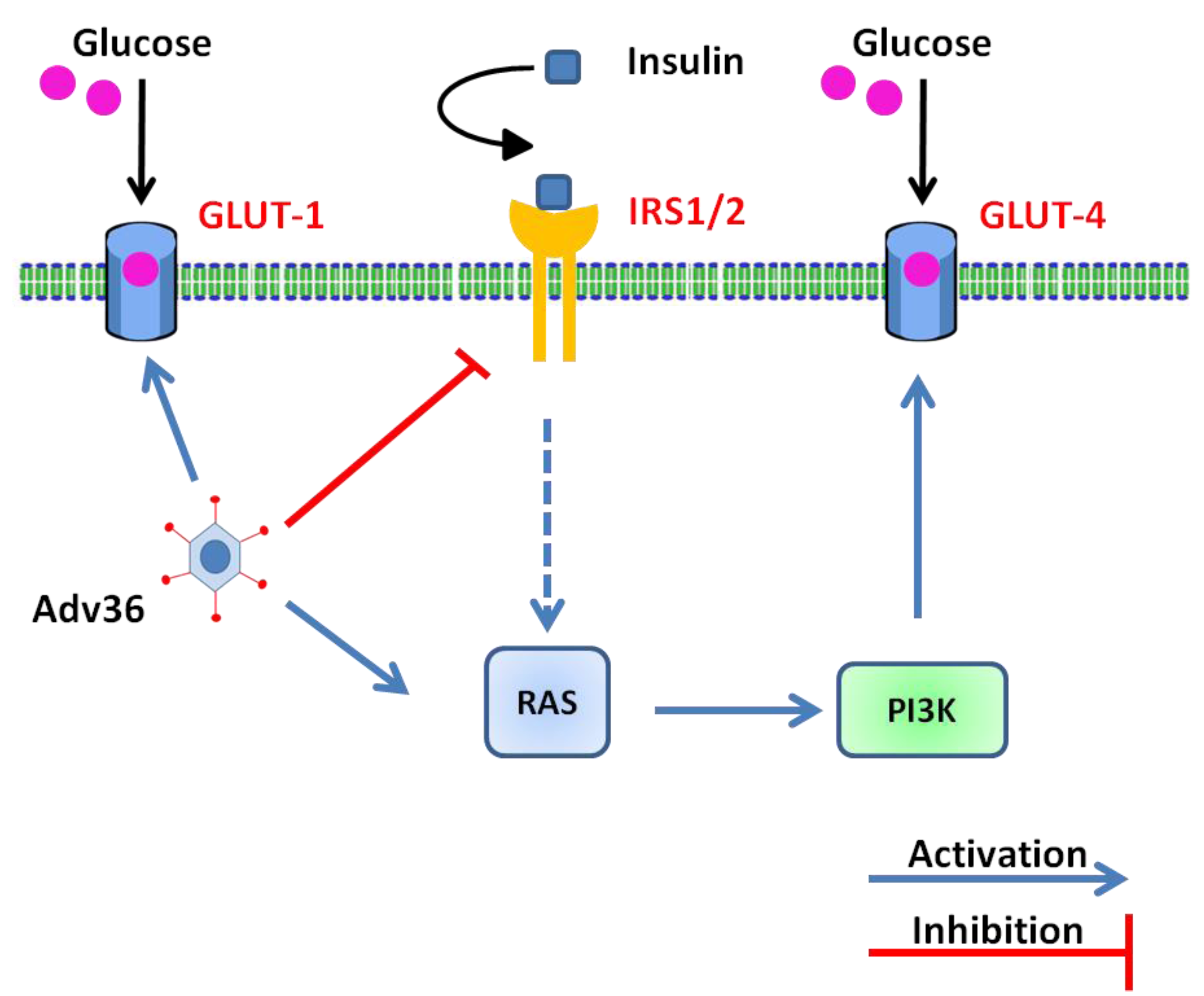
6. Molecular and Cellular Mechanisms Involved in Ad-36-Induced Glucose Uptake

Evidence emerging from animal and human studies suggests that some forms of obesity may actually lead to healthy obesity [24]. Several studies identified one Adv36 gene that mediates glucose disposal through the Ras/PI3K pathway to improve glucose uptake [64]. The glucose uptake increase may contribute to the improved glycemic control found in Adv36-infected animals [34].
In vitro, Adv36 induced adipogenic accumulation commitment, differentiation of adipocytes and increases the cellular glucose uptake [27,44].
In a work by Wang et al. [64], the authors showed that the infection with Adv36 increases glucose uptake, confirming previous data. In vitro experiments on Human Skeletal Muscle (HSKM) cells showed an increase of GLUT1 and GLUT4 gene expression mediated by Ras-activated PI 3-kinase pathway independent from insulin activation (Figure 2).
Considering the crucial role of skeletal muscle in glucose disposal, the ability to modulate its capacity to uptake glucose may have a major impact on systemic glycemic control. The property of Adv36 to improve skeletal muscle glucose uptake in an insulin-signaling independent manner is of great potential interest for the development of new drug targets in insulin resistance diabetes. Subsequently, many studies have focused on the virus E4orf1 gene that up-regulates the PI3K pathway (Figure 2) [42]. The E4orf1 gene is transcribed from the first open reading frame of the early gene of Adv36 and produces a 17 kDa protein (Figure 1) [42]. Adv36 increases cellular glucose uptake via Ras-mediated activation of the phosphatidyl inositol 3-kinase (PI3K), and improves hyperglycemia in mice, without reducing adiposity [65]. In vitro experiments showed that Adv36 significantly increases the absorption of glucose into 3T3-L1 preadipocytes and identified that the E4orf1 is “sufficient” to up-regulate the absorption of glucose [26]. The 3T3-L1 cell line expressing E4orf1 had an increased glucose uptake compared to null vector control cells. Moreover, E4orf1 up-regulated PI3K pathway and increased the Ras-molecule required to stimulates the Adv36-induced glucose uptake. Glucose uptake increases significantly in preadipocytes, adipocytes, or myoblasts derived from 3T3-L1 cells transiently transfected with E4orf1. Thus, the attractive anti-hyperglycemic effect of Adv36 could be mediated by the E4orf1 protein, which may offer a novel ligand to develop hypoglycemic drugs. Cellular uptake of glucose can be improved by up-regulation of Ras signaling in either insulin-dependent or insulin-independent diabetes. In the presence of an intact insulin signal, Ras plays a negligible role in glucose uptake. On the contrary, when the insulin signal is reduced like in obesity or diabetes, the insulin-independent Ras pathway can be useful to improve the glucose disposal. Adv36 increases cellular uptake of glucose by upregulating the Ras/Glut4pathway (Figure 2). These data improved the knowledge on the role of E4orf1 as a mediator of Adv36-induced glucose uptake [26].
Figure 2. Adv36 mediates glucose uptake independently from insulin, adapted from [64]. Adv36 up-regulates the Phosphoinositide 3-kinase (PI3K) signaling via Ras, increasing cellular glucose uptake by glucose trasporters Glut1 and Glut4 despite a down-regulation of the Insulin Receptor Substrate (IRS) signaling.
Viruses 07 02787 g002
The metabolic effects of E4orf1 are encouraging, and may provide new targets for signaling to prevent NAFLD and insulin resistance even in the presence of a high fat diet. There are limitations to these studies because all results are from in vitro experiments and should be verified in vivo. E4orf1 is not a secretory protein, and therefore does not have a cell surface receptor for cell entry. Thus, E4orf1 provides a valuable model to exogenously modulate hepatic glucose and lipid metabolism [66].
In summary, Adv36 may serve two different purposes, on the one hand, to explain the pathophysiology of certain cases of obesity and, on the other hand, to provide a potential weapon for improving insulin resistance, regardless of the consumption of fat.
Collectively, these results can provide a model to develop new agents for the treatment of hyperglycemia associated with obesity, and type 1 or type 2 diabetes [25]. Although the in vivo efficacy and safety of E4orf1 in improving hyperglycemia remain unknown, and an appropriate drug delivery system is required, Adv36 E4orf1 offers a research opportunity to develop new anti-diabetic agents with new conceptually advances for the use of a rather unconventional source, viral proteins, in the anti-diabetic drug development [25].

7. Adenovirus 36 and Immune Response

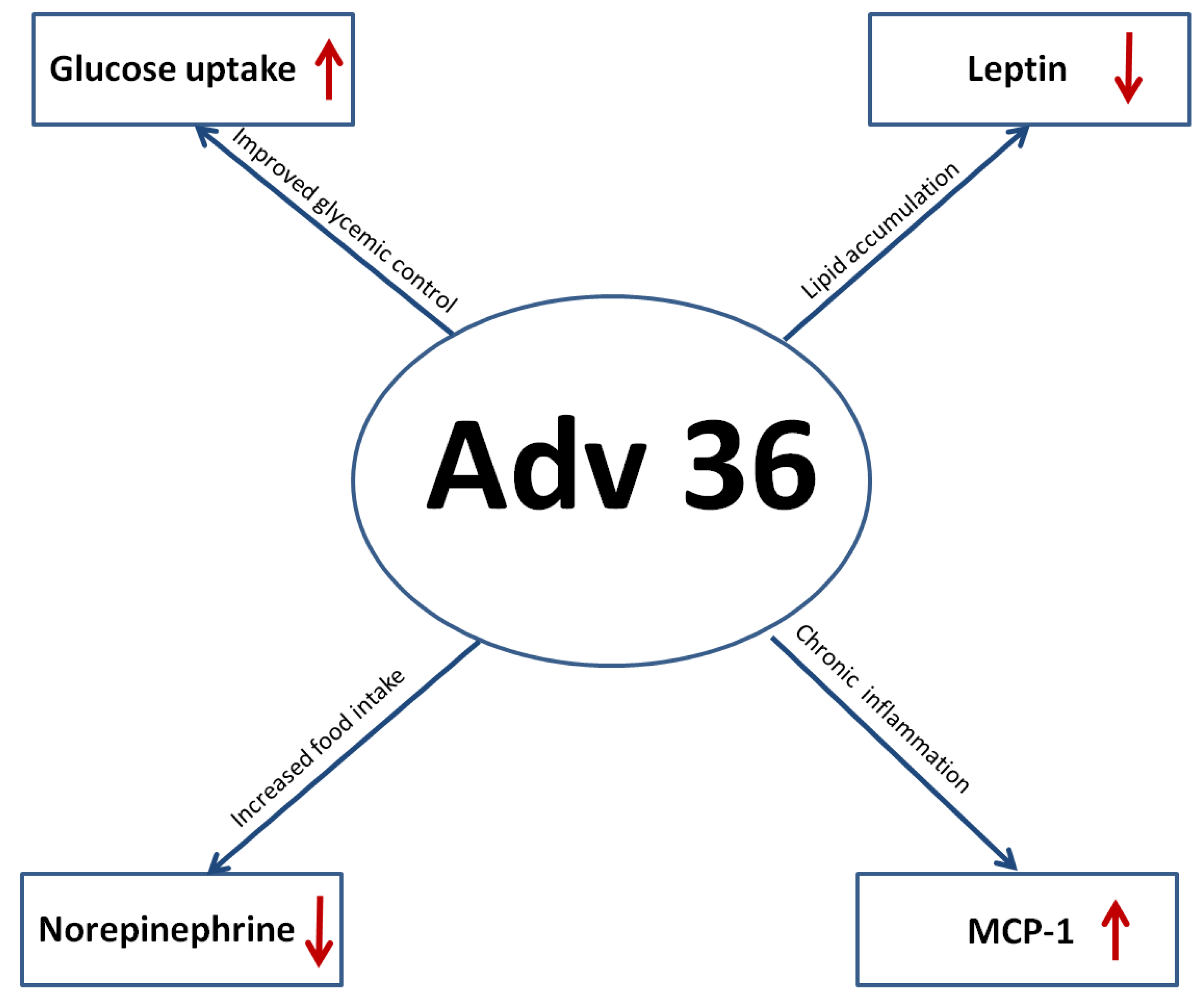
7.1. Inflammation: MCP-1 (Macrophage Chemoattractant Protein I) and Adv36

Obese subjects have altered the overall number of circulating T-cells and obesity has been associated with decreased thymic output of naive T-cells [67,68,69]. When analyzed by flow cytometry, decreased CD8 T-cell populations and increased or decreased numbers of CD4 T-cells compared with lean controls are found [70]. The inflammatory network in obesity may present different immunologic phenotypes depending on the degree of metabolic disorder. In lean individuals, adipose tissue is mainly characterized by anti-inflammatory condition, while obesity shifts towards a pro-inflammatory status. Obesity negatively impacts the ability of dendritic cells (DCs) to mature and elicit appropriate T-cell responses to a general stimulus. This may contribute to the increased susceptibility to viral infection observed in severe obesity [71]. Adv36 infection could induce obesity through inflammation, and MCP-1 may be a key regulator of adenovirus 36-induced obesity in Adv36-infected mice [72]. Thus, Adv36 could cause chronic inflammation by increasing the levels of MCP-1, activating nuclear factor κB (NF-κB), inducing the infiltration of macrophages into adipocytes, and altering lipid metabolism [72]. The correlation between obesity and inflammation has been known for decades from epidemiological and cellular studies [73,74]. Hypertrophic adipocytes may produce pro-inflammatory cytokines such as MCP-1, TNF-α, resistin, and plasminogen activator inhibitor-1 [75,76]. It was previously found that Adv36 infection triggers the inflammatory pathway in human mesenchymal stem cells, which was confirmed by microarray analysis [77].
It has been reported that MCP-1 is responsible for obesity, insulin resistance, and steatosis in MCP-1 transgenic mice and obese mice [78] and inhibition of MCP-1 ameliorates insulin resistance and hepatic steatosis [78]. MCP-1 plays a role in the recruitment of monocytes and macrophages into adipose tissue [78,79] and TNF-α increases the number of macrophages in adipose tissue [80,81]. Macrophages upregulate production of the anti-inflammatory cytokine IL-10 and downregulate synthesis of pro-inflammatory cytokines. Functionally, there are macrophages that are associated with the repair of injured tissues and the resolution of inflammation. So, it has been shown that the macrophages accumulate in the adipose tissues of obese mice mainly expressing genes associated with an M1 or “classically activated” macrophage phenotype, whereas adipose tissue macrophages from lean mice express genes associated with an M2 or “alternatively activated” macrophage phenotype [82]. Adv36 infection increased M1 macrophages migration into adipocytes by activating nuclear factor κB (NFkB), which induced the release of pro-inflammatory citokynes. In the study of Na et al. [77] on in vivo experimental infection in mice, Adv36 infection stimulates an inflammatory state due to the increased level of MCP-1 through the activation of NFkB, which in turn induces the infiltration of macrophages into adipocytes. Adv36 infection increases MCP-1, and MCP-1 might function to induce adipogenesis via MCP-1-related factors in adipocytes. Furthermore, increased MCP-1 triggers macrophage infiltration into adipocytes and inflammation may play a role in maintaining obesity [72]. Therefore, on the basis of these data, we could assume that there is a relationship between Adv36-induced obesity and inflammation.

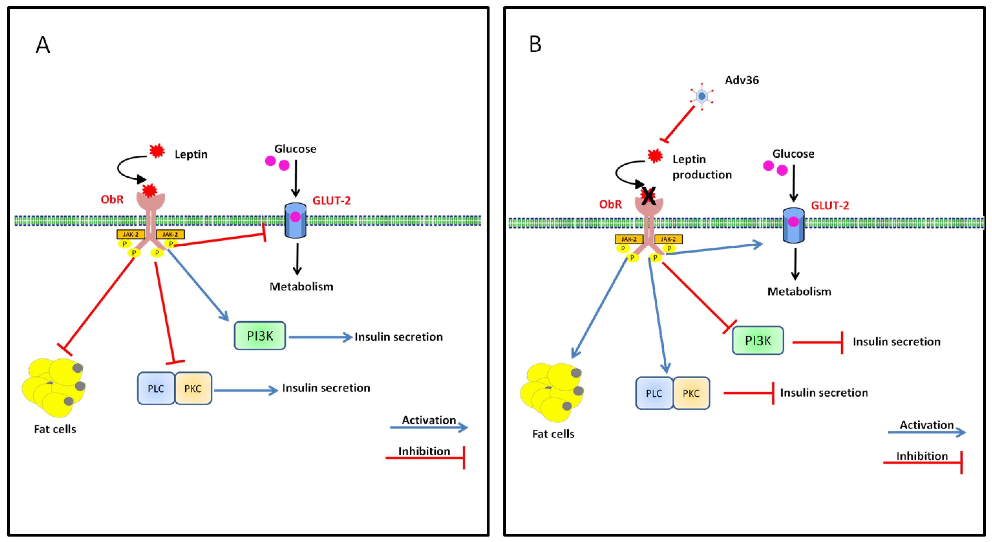
7.2. Effect of Adv36 on Leptin

Conventionally, obesity can be considered an over accumulation of white adipose tissue (WAT). Although adipocytes occupy the bulk of the volume of WAT, adipose tissue also includes many more cells types, including a diverse population of preadipocytes, macrophages, endothelial cells, fibroblasts and leukocytes [83]. Adipose tissue, traditionally thought as a passive storage for triglycerides and energy, but in the past two decades it has been linked to the production of several hormones, pro-inflammatory chemokines, adipokines and cytokines, including leptin, adiponectin, resistin, visfatin, B-cell activating factor of the Tumor Necrosis Factor (TNF) family, TNF-like weak inducer of apoptosis, a proliferation inducing ligand, TNF-α, omentin and MCP-1 [5]. Based on this concept, adipose tissue has been defined an endocrine organ. In the obese state, secretion of these adipokines is altered in correlation to the increased adipose tissue mass [84,85]. To date, adipokine modulation of immune function by leptin is the best-characterized link between obesity and immune function. Leptin plays a role in many diverse physiological processes but is primarily involved in energy homeostasis and satiety [86,87].
Leptin acts as a general signal of energy reserves and modulates food intake. Leptin levels increase proportionately to adipose mass resulting in high circulating leptin concentrations in obese individuals [88,89].
The leptin receptor is expressed by B and T lymphocytes and may directly modulate the T and B responses [90]. Leptin seems to exert its effects on immune cells through the JAK /STAT pathway. In peripheral blood mononuclear cells, leptin increases JAK2/3 and STAT3 phosphorylation, which promote proliferation and activation of T lymphocytes upon mitogen stimulation. In terms of infectious disease, the general consensus seems to be that leptin has an anti-inflammatory role, while at the same time serving a protective capacity against infections [91]. Inflammation is used as a localized, protective response to infection and changes in body weight and metabolic state are often associated with acute or chronic inflammatory processes resulting from infection [38]. The inhibition of leptin gene expression by Adv36 infection may increase lipid accumulation and obesity prevalence reported that human cells infected with Adv36 showed greater differentiation and higher levels of lipid accumulation than non-infected control cells [43] (Figure 3). Adv36 infection may increase appetite and food intake by decreasing norepinephrine levels and leptin [92], thereby increasing obesity prevalence (Figure 4).
Figure 3. Adv36 and leptin. (A) Leptin binding to its receptor (ObRb) activates the associated JAK-2 tyrosine kinase. Leptin inhibits glucose transport through GLUT-2, and activates PI3K. Additionally, Phosphoinositide 3-kinase (PI3K) activation by leptin reduces Cyclic adenosine monophosphate (cAMP) levels and activates the Protein kinase A (PKA) pathway. Leptin can also inhibit the phospholipase C (PLC)/protein kinase C (PKC) pathway; (B) Adv36 inhibits leptin production. The results are decreased insulin release and increased lipid accumulation.
Viruses 07 02787 g003
Figure 4. Proposed mechanisms underlying the effects of Adv36 in infected individuals.
Viruses 07 02787 g004

8. Proof-of-Concept of a Vaccine Using UV Inactivated Adv36

Adv36 infection is characterized by greater adiposity and inflammation. To investigate the possibility that a candidate prophylactic vaccine could protect Adv36-induced obesity and inflammation an inactivated Adv36 vaccine was tested [37]. Mice were inoculated with live Adv36, UV-inactivated Adv36, or with medium alone. Live Adv36 increased the size of the epididymal fat pad at four days post-inoculation, whereas UV-inactivated Adv36 prevented it. In another study, mice were immunized with purified and ultraviolet-irradiated virus as candidate vaccine, live Adv36 was injected into mice as a challenge test. Unvaccinated mice (control group) were immunized with phosphate-buffered saline and then challenged with live Adv36. The control group showed 17% greater body weight and 20% more epididymal fats compared with the vaccinated group. Moreover, the vaccinated group had decreased serum levels of pro-inflammatory cytokines, and infiltrated immune cells in fat tissue [93]. Therefore, the vaccine was able to protect against Adv36-increased body weight and fat as well as inflammatory states after challenge. These results could provide proof-of-concept for prophylactic vaccination against virus-induced adiposity [94].

9. Conclusions

Obesity is a multifactorial pathology and the understanding of the different contributing factors is crucial for its efficient management.
Recent studies have shown a possible correlation between obesity and Adv36 viral infections. Animals infected with Adv36 show increase of body weight and physiological changes, increased glucose absorption and decreased secretion of leptin and cholesterol (Figure 4).
The Adv36 infections should therefore be considered as a possible risk factor for obesity and this could be a potential new way to investigate on the worldwide epidemic of obesity. Further research on viruses contributing to obesity is essential. Studying the favorable effect of Adv36 on metabolic consequences of obesity may provide insight to novel treatments that improve glycemic control despite adiposity. Identifying the viral protein responsible for influencing glucose disposal may help in developing novel anti-diabetic therapeutic agents.

Acknowledgments

This study was funded by the Sapienza University of Rome, Italy.

Author contributions

E.P. drafted the first version of the manuscript, designed review and interpreted data. L.G. supervised and reviewed the manuscript. All authors read and approved the final manuscript, and agreed with the conclusions of the work.

Conflicts of Interest

The authors declare that they have no competing interests of either financial or non-financial nature regarding the work described in the present manuscript and its publication.

References

  1. Ng, M.; Fleming, T.; Robinson, M.; Thomson, B.; Graetz, N.; Margono, C.; Mullany, E.C.; Biryukov, S.; Abbafati, C.; Abera, S.F.; et al. Global, regional, and national prevalence of overweight and obesity in children and adults during 1980–2013: A systematic analysis for the global burden of disease study 2013. Lancet 2014, 384, 766–781. [Google Scholar] [CrossRef]
  2. WHO. Obesity and overweight. Available online: http://www.who.int/mediacentre/factsheets/fs311/en/ (accessed on 5 June 2015).
  3. Bray, G.A.; Bellanger, T. Epidemiology, trends, and morbidities of obesity and the metabolic syndrome. Endocrine 2006, 29, 109–117. [Google Scholar] [CrossRef]
  4. Hill, J.O.; Peters, J.C. Environmental contributions to the obesity epidemic. Science 1998, 280, 1371–1374. [Google Scholar] [CrossRef] [PubMed]
  5. Karlsson, E.A.; Beck, M.A. The burden of obesity on infectious disease. Exp. Biol. Med. 2010, 235, 1412–1424. [Google Scholar] [CrossRef] [PubMed]
  6. Koenig, S.M. Pulmonary complications of obesity. Am. J. Med. Sci. 2001, 321, 249–279. [Google Scholar] [CrossRef] [PubMed]
  7. Jubber, A.S. Respiratory complications of obesity. Int. J. Clin. Pract. 2004, 58, 573–580. [Google Scholar] [CrossRef] [PubMed]
  8. Campitelli, M.A.; Rosella, L.C.; Kwong, J.C. The association between obesity and outpatient visits for acute respiratory infections in ontario, Canada. Int. J. Obes. 2014, 38, 113–119. [Google Scholar] [CrossRef] [PubMed]
  9. Mullen, J.T.; Moorman, D.W.; Davenport, D.L. The obesity paradox: Body mass index and outcomes in patients undergoing nonbariatric general surgery. Ann. Surg. 2009, 250, 166–172. [Google Scholar] [CrossRef] [PubMed]
  10. Calle, E.E.; Thun, M.J.; Petrelli, J.M.; Rodriguez, C.; Heath, C.W., Jr. Body-mass index and mortality in a prospective cohort of U.S. Adults. N. Engl. J. Med. 1999, 341, 1097–1105. [Google Scholar] [CrossRef] [PubMed]
  11. Choban, P.S.; Flancbaum, L. The impact of obesity on surgical outcomes: A review. J. Am. Coll. Surg. 1997, 185, 593–603. [Google Scholar] [PubMed]
  12. Arslan, E.; Atilgan, H.; Yavasoglu, I. The prevalence of helicobacter pylori in obese subjects. Eur. J. Intern. Med. 2009, 20, 695–697. [Google Scholar] [CrossRef] [PubMed]
  13. Uberos, J.; Molina-Carballo, A.; Fernandez-Puentes, V.; Rodriguez-Belmonte, R.; Munoz-Hoyos, A. Overweight and obesity as risk factors for the asymptomatic carrier state of neisseria meningitidis among a paediatric population. Eur. J. Clin. Microbiol. Infect. Dis. 2010, 29, 333–334. [Google Scholar] [CrossRef] [PubMed]
  14. Morgan, O.W.; Bramley, A.; Fowlkes, A.; Freedman, D.S.; Taylor, T.H.; Gargiullo, P.; Belay, B.; Jain, S.; Cox, C.; Kamimoto, L.; et al. Morbid obesity as a risk factor for hospitalization and death due to 2009 pandemic influenza a(h1n1) disease. PLoS ONE 2010, 5, e9694. [Google Scholar] [CrossRef] [PubMed]
  15. Hegde, V.; Dhurandhar, N.V. Microbes and obesity—Interrelationship between infection, adipose tissue and the immune system. Clin. Microbiol. Infect. 2013, 19, 314–320. [Google Scholar] [CrossRef] [PubMed]
  16. Huttunen, R.; Syrjanen, J. Obesity and the risk and outcome of infection. Int. J. Obes. 2013, 37, 333–340. [Google Scholar] [CrossRef] [PubMed]
  17. Verlaeten, O.; Griffond, B.; Khuth, S.T.; Giraudon, P.; Akaoka, H.; Belin, M.F.; Fellmann, D.; Bernard, A. Down regulation of melanin concentrating hormone in virally induced obesity. Mol. Cell. Endocrinol. 2001, 181, 207–219. [Google Scholar] [CrossRef]
  18. Pasarica, M.; Dhurandhar, N.V. Infectobesity: Obesity of infectious origin. Adv. Food Nutr. Res. 2007, 52, 61–102. [Google Scholar] [PubMed]
  19. Mitra, A.K.; Clarke, K. Viral obesity: Fact or fiction? Obes. Rev. 2010, 11, 289–296. [Google Scholar] [CrossRef] [PubMed]
  20. Lyons, M.J.; Faust, I.M.; Hemmes, R.B.; Buskirk, D.R.; Hirsch, J.; Zabriskie, J.B. A virally induced obesity syndrome in mice. Science 1982, 216, 82–85. [Google Scholar] [CrossRef] [PubMed]
  21. Carter, J.K.; Ow, C.L.; Smith, R.E. Rous-associated virus type 7 induces a syndrome in chickens characterized by stunting and obesity. Infect. Immun. 1983, 39, 410–422. [Google Scholar] [PubMed]
  22. Dhurandhar, N.V. Is obesity caused by an adenovirus? Expert Rev. Anti-Infect. Ther. 2012, 10, 521–524. [Google Scholar] [CrossRef] [PubMed]
  23. Swenson, P.D.; Lowens, M.S.; Celum, C.L.; Hierholzer, J.C. Adenovirus types 2, 8, and 37 associated with genital infections in patients attending a sexually transmitted disease clinic. J. Clin. Microbiol. 1995, 33, 2728–2731. [Google Scholar] [PubMed]
  24. Dhurandhar, N.V. A framework for identification of infections that contribute to human obesity. Lancet Infect. Dis. 2011, 11, 963–969. [Google Scholar] [CrossRef]
  25. Dhurandhar, N.V. Insulin sparing action of adenovirus 36 and its e4orf1 protein. J. Diabetes Complicat. 2013, 27, 191–199. [Google Scholar] [CrossRef] [PubMed]
  26. Dhurandhar, E.J.; Dubuisson, O.; Mashtalir, N.; Krishnapuram, R.; Hegde, V.; Dhurandhar, N.V. E4orf1: A novel ligand that improves glucose disposal in cell culture. PLoS ONE 2011, 6, e23394. [Google Scholar] [CrossRef] [PubMed]
  27. Rathod, M.A.; Rogers, P.M.; Vangipuram, S.D.; McAllister, E.J.; Dhurandhar, N.V. Adipogenic cascade can be induced without adipogenic media by a human adenovirus. Obesity 2009, 17, 657–664. [Google Scholar] [CrossRef] [PubMed]
  28. Dhurandhar, N.V.; Kulkarni, P.; Ajinkya, S.M.; Sherikar, A. Effect of adenovirus infection on adiposity in chicken. Vet. Microbiol. 1992, 31, 101–107. [Google Scholar] [CrossRef]
  29. Dhurandhar, N.V.; Kulkarni, P.R.; Ajinkya, S.M.; Sherikar, A.A.; Atkinson, R.L. Association of adenovirus infection with human obesity. Obes. Res. 1997, 5, 464–469. [Google Scholar] [CrossRef] [PubMed]
  30. Dhurandhar, N.V.; Israel, B.A.; Kolesar, J.M.; Mayhew, G.F.; Cook, M.E.; Atkinson, R.L. Increased adiposity in animals due to a human virus. Int. J. Obes. Relat. Metab. Disord. 2000, 24, 989–996. [Google Scholar] [CrossRef] [PubMed]
  31. Whigham, L.D.; Israel, B.A.; Atkinson, R.L. Adipogenic potential of multiple human adenoviruses in vivo and in vitro in animals. Am. J. Physiol. Regul. Integr. Comp. Physiol. 2006, 290, R190–R194. [Google Scholar] [CrossRef] [PubMed]
  32. Dhurandhar, N.V. Infectobesity: Obesity of infectious origin. J. Nutr. 2001, 131, 2794S–2797S. [Google Scholar] [PubMed]
  33. Dhurandhar, N.V.; Whigham, L.D.; Abbott, D.H.; Schultz-Darken, N.J.; Israel, B.A.; Bradley, S.M.; Kemnitz, J.W.; Allison, D.B.; Atkinson, R.L. Human adenovirus ad-36 promotes weight gain in male rhesus and marmoset monkeys. J. Nutr. 2002, 132, 3155–3160. [Google Scholar] [PubMed]
  34. Pasarica, M.; Shin, A.C.; Yu, M.; Ou Yang, H.M.; Rathod, M.; Jen, K.L.; MohanKumar, S.; MohanKumar, P.S.; Markward, N.; Dhurandhar, N.V. Human adenovirus 36 induces adiposity, increases insulin sensitivity, and alters hypothalamic monoamines in rats. Obesity 2006, 14, 1905–1913. [Google Scholar] [CrossRef] [PubMed]
  35. Dhurandhar, N.V.; Israel, B.A.; Kolesar, J.M.; Mayhew, G.; Cook, M.E.; Atkinson, R.L. Transmissibility of adenovirus-induced adiposity in a chicken model. Int. J. Obes. Relat. Metab. Disord. 2001, 25, 990–996. [Google Scholar] [CrossRef] [PubMed]
  36. Kapila, M.; Khosla, P.; Dhurandhar, N.V. Novel short-term effects of adenovirus ad-36 on hamster lipoproteins. Int. J. Obes. Relat. Metab. Disord. 2004, 28, 1521–1527. [Google Scholar] [CrossRef] [PubMed]
  37. Pasarica, M.; Loiler, S.; Dhurandhar, N.V. Acute effect of infection by adipogenic human adenovirus ad36. Arch. Virol. 2008, 153, 2097–2102. [Google Scholar] [CrossRef] [PubMed]
  38. Vangipuram, S.D.; Sheele, J.; Atkinson, R.L.; Holland, T.C.; Dhurandhar, N.V. A human adenovirus enhances preadipocyte differentiation. Obes. Res. 2004, 12, 770–777. [Google Scholar] [CrossRef] [PubMed]
  39. Rathod, M.; Vangipuram, S.D.; Krishnan, B.; Heydari, A.R.; Holland, T.C.; Dhurandhar, N.V. Viral mrna expression but not DNA replication is required for lipogenic effect of human adenovirus ad-36 in preadipocytes. Int. J. Obes. 2007, 31, 78–86. [Google Scholar] [CrossRef] [PubMed]
  40. Arnold, J.; Janoska, M.; Kajon, A.E.; Metzgar, D.; Hudson, N.R.; Torres, S.; Harrach, B.; Seto, D.; Chodosh, J.; Jones, M.S. Genomic characterization of human adenovirus 36, a putative obesity agent. Virus Res. 2010, 149, 152–161. [Google Scholar] [CrossRef] [PubMed]
  41. Gregoire, F.M.; Smas, C.M.; Sul, H.S. Understanding adipocyte differentiation. Physiol. Rev. 1998, 78, 783–809. [Google Scholar] [PubMed]
  42. Rogers, P.M.; Fusinski, K.A.; Rathod, M.A.; Loiler, S.A.; Pasarica, M.; Shaw, M.K.; Kilroy, G.; Sutton, G.M.; McAllister, E.J.; Mashtalir, N.; et al. Human adenovirus ad-36 induces adipogenesis via its e4 orf-1 gene. Int. J. Obes. 2008, 32, 397–406. [Google Scholar] [CrossRef] [PubMed]
  43. Vangipuram, S.D.; Yu, M.; Tian, J.; Stanhope, K.L.; Pasarica, M.; Havel, P.J.; Heydari, A.R.; Dhurandhar, N.V. Adipogenic human adenovirus-36 reduces leptin expression and secretion and increases glucose uptake by fat cells. Int. J. Obes. 2007, 31, 87–96. [Google Scholar] [CrossRef] [PubMed]
  44. Pasarica, M.; Mashtalir, N.; McAllister, E.J.; Kilroy, G.E.; Koska, J.; Permana, P.; de Courten, B.; Yu, M.; Ravussin, E.; Gimble, J.M.; et al. Adipogenic human adenovirus ad-36 induces commitment, differentiation, and lipid accumulation in human adipose-derived stem cells. Stem Cells 2008, 26, 969–978. [Google Scholar] [CrossRef] [PubMed]
  45. Wang, Z.Q.; Yu, Y.; Zhang, X.H.; Floyd, E.Z.; Cefalu, W.T. Human adenovirus 36 decreases fatty acid oxidation and increases de novo lipogenesis in primary cultured human skeletal muscle cells by promoting cidec/fsp27 expression. Int. J. Obes. 2010, 34, 1355–1364. [Google Scholar] [CrossRef] [PubMed]
  46. Nam, J.H. Why is not there a match between the serological and genomic prevalence of adenovirus 36? J. Clin. Virol. 2013, 56, 370–371. [Google Scholar] [CrossRef] [PubMed]
  47. Nam, J.H.; Na, H.N.; Atkinson, R.L.; Dhurandhar, N.V. Genomic stability of adipogenic human adenovirus 36. Int. J. Obes. 2014, 38, 321–324. [Google Scholar] [CrossRef] [PubMed]
  48. Salehian, B.; Forman, S.J.; Kandeel, F.R.; Bruner, D.E.; He, J.; Atkinson, R.L. Adenovirus 36 DNA in adipose tissue of patient with unusual visceral obesity. Emerg. Infect. Dis. 2010, 16, 850–852. [Google Scholar] [CrossRef] [PubMed]
  49. Atkinson, R.L.; Dhurandhar, N.V.; Allison, D.B.; Bowen, R.L.; Israel, B.A.; Albu, J.B.; Augustus, A.S. Human adenovirus-36 is associated with increased body weight and paradoxical reduction of serum lipids. Int. J. Obes. 2005, 29, 281–286. [Google Scholar] [CrossRef] [PubMed]
  50. Broderick, M.P.; Hansen, C.J.; Irvine, M.; Metzgar, D.; Campbell, K.; Baker, C.; Russell, K.L. Adenovirus 36 seropositivity is strongly associated with race and gender, but not obesity, among us military personnel. Int. J. Obes. 2010, 34, 302–308. [Google Scholar] [CrossRef] [PubMed]
  51. Goossens, V.J.; deJager, S.A.; Grauls, G.E.; Gielen, M.; Vlietinck, R.F.; Derom, C.A.; Loos, R.J.; Rensen, S.S.; Buurman, W.A.; Greve, J.W.; et al. Lack of evidence for the role of human adenovirus-36 in obesity in a european cohort. Obesity 2011, 19, 220–221. [Google Scholar] [CrossRef] [PubMed]
  52. Bil-Lula, I.; Plonek, T.; Wozniak, M. Lack of adenovirus DNA in mediastinal adipose tissue of obese/overweight adults with cardiovascular disorders? J. Med. Virol. 2014, 86, 802–805. [Google Scholar] [CrossRef] [PubMed]
  53. Na, H.N.; Hong, Y.M.; Kim, J.; Kim, H.K.; Jo, I.; Nam, J.H. Association between human adenovirus-36 and lipid disorders in korean schoolchildren. Int. J. Obes. 2010, 34, 89–93. [Google Scholar] [CrossRef] [PubMed]
  54. Almgren, M.; Atkinson, R.; He, J.; Hilding, A.; Hagman, E.; Wolk, A.; Thorell, A.; Marcus, C.; Naslund, E.; Ostenson, C.G.; et al. Adenovirus-36 is associated with obesity in children and adults in sweden as determined by rapid elisa. PLoS ONE 2012, 7, e41652. [Google Scholar] [CrossRef] [PubMed]
  55. Aldhoon-Hainerova, I.; Zamrazilova, H.; Atkinson, R.L.; Dusatkova, L.; Sedlackova, B.; Hlavaty, P.; Lee, Z.P.; Kunesova, M.; Hainer, V. Clinical and laboratory characteristics of 1179 czech adolescents evaluated for antibodies to human adenovirus 36. Int. J. Obes. 2014, 38, 285–291. [Google Scholar] [CrossRef] [PubMed]
  56. Atkinson, R.L.; Lee, I.; Shin, H.J.; He, J. Human adenovirus-36 antibody status is associated with obesity in children. Int. J. Pediatr. Obes. 2010, 5, 157–160. [Google Scholar] [CrossRef] [PubMed]
  57. Cakmakliogullari, E.K.; Sanlidag, T.; Ersoy, B.; Akcali, S.; Var, A.; Cicek, C. Are human adenovirus-5 and 36 associated with obesity in children? J. Investig. Med. 2014, 62, 821–824. [Google Scholar] [PubMed]
  58. Gabbert, C.; Donohue, M.; Arnold, J.; Schwimmer, J.B. Adenovirus 36 and obesity in children and adolescents. Pediatrics 2010, 126, 721–726. [Google Scholar] [CrossRef] [PubMed]
  59. Parra-Rojas, I.; del Moral-Hernandez, O.; Salgado-Bernabe, A.B.; Guzman-Guzman, I.P.; Salgado-Goytia, L.; Munoz-Valle, J.F. Adenovirus-36 seropositivity and its relation with obesity and metabolic profile in children. Int. J. Endocrinol. 2013, 2013, e463194. [Google Scholar] [CrossRef] [PubMed]
  60. Tosh, A.K.; Broy-Aschenbrenner, A.; El Khatib, J.; Ge, B. Adenovirus-36 antibody status & bmi comparison among obese missouri adolescents. Mo. Med. 2012, 109, 402–403. [Google Scholar] [PubMed]
  61. Vander Wal, J.S.; Huelsing, J.; Dubuisson, O.; Dhurandhar, N.V. An observational study of the association between adenovirus 36 antibody status and weight loss among youth. Obes. Facts 2013, 6, 269–278. [Google Scholar] [CrossRef] [PubMed]
  62. Yamada, T.; Hara, K.; Kadowaki, T. Association of adenovirus 36 infection with obesity and metabolic markers in humans: A meta-analysis of observational studies. PLoS ONE 2012, 7, e42031. [Google Scholar] [CrossRef] [PubMed]
  63. Shang, Q.; Wang, H.; Song, Y.; Wei, L.; Lavebratt, C.; Zhang, F.; Gu, H. Serological data analyses show that adenovirus 36 infection is associated with obesity: A meta-analysis involving 5739 subjects. Obesity 2014, 22, 895–900. [Google Scholar] [CrossRef] [PubMed]
  64. Wang, Z.Q.; Cefalu, W.T.; Zhang, X.H.; Yu, Y.; Qin, J.; Son, L.; Rogers, P.M.; Mashtalir, N.; Bordelon, J.R.; Ye, J.; et al. Human adenovirus type 36 enhances glucose uptake in diabetic and nondiabetic human skeletal muscle cells independent of insulin signaling. Diabetes 2008, 57, 1805–1813. [Google Scholar] [CrossRef] [PubMed]
  65. Krishnapuram, R.; Dhurandhar, E.J.; Dubuisson, O.; Hegde, V.; Dhurandhar, N.V. Doxycycline-regulated 3t3-l1 preadipocyte cell line with inducible, stable expression of adenoviral e4orf1 gene: A cell model to study insulin-independent glucose disposal. PLoS ONE 2013, 8, e60651. [Google Scholar] [CrossRef] [PubMed]
  66. Dhurandhar, E.J.; Krishnapuram, R.; Hegde, V.; Dubuisson, O.; Tao, R.; Dong, X.C.; Ye, J.; Dhurandhar, N.V. E4orf1 improves lipid and glucose metabolism in hepatocytes: A template to improve steatosis & hyperglycemia. PLoS ONE 2012, 7, e47813. [Google Scholar] [PubMed]
  67. Lynch, L.A.; O’Connell, J.M.; Kwasnik, A.K.; Cawood, T.J.; O’Farrelly, C.; O’Shea, D.B. Are natural killer cells protecting the metabolically healthy obese patient? Obesity 2009, 17, 601–605. [Google Scholar] [CrossRef] [PubMed]
  68. Yang, H.; Youm, Y.H.; Vandanmagsar, B.; Rood, J.; Kumar, K.G.; Butler, A.A.; Dixit, V.D. Obesity accelerates thymic aging. Blood 2009, 114, 3803–3812. [Google Scholar] [CrossRef] [PubMed]
  69. Nieman, D.C.; Nehlsen-Cannarella, S.I.; Henson, D.A.; Butterworth, D.E.; Fagoaga, O.R.; Warren, B.J.; Rainwater, M.K. Immune response to obesity and moderate weight loss. Int. J. Obes. Relat. Metab. Disord. 1996, 20, 353–360. [Google Scholar] [PubMed]
  70. O'Rourke, R.W.; Kay, T.; Scholz, M.H.; Diggs, B.; Jobe, B.A.; Lewinsohn, D.M.; Bakke, A.C. Alterations in T-cell subset frequency in peripheral blood in obesity. Obes. Surg. 2005, 15, 1463–1468. [Google Scholar] [CrossRef] [PubMed]
  71. O’Shea, D.; Corrigan, M.; Dunne, M.R.; Jackson, R.; Woods, C.; Gaoatswe, G.; Moynagh, P.N.; O’Connell, J.; Hogan, A.E. Changes in human dendritic cell number and function in severe obesity may contribute to increased susceptibility to viral infection. Int. J. Obes. 2013, 37, 1510–1513. [Google Scholar] [CrossRef] [PubMed]
  72. Na, H.N.; Nam, J.H. Adenovirus 36 as an obesity agent maintains the obesity state by increasing mcp-1 and inducing inflammation. J. Infect. Dis. 2012, 205, 914–922. [Google Scholar] [CrossRef] [PubMed]
  73. Stienstra, R.; Duval, C.; Muller, M.; Kersten, S. Ppars, obesity, and inflammation. PPAR Res. 2007, 2007, e95974. [Google Scholar] [CrossRef] [PubMed]
  74. Tordjman, J.; Guerre-Millo, M.; Clement, K. Adipose tissue inflammation and liver pathology in human obesity. Diabetes Metab. 2008, 34, 658–663. [Google Scholar] [CrossRef]
  75. Bastard, J.P.; Maachi, M.; Lagathu, C.; Kim, M.J.; Caron, M.; Vidal, H.; Capeau, J.; Feve, B. Recent advances in the relationship between obesity, inflammation, and insulin resistance. Eur. Cytokine Netw. 2006, 17, 4–12. [Google Scholar] [PubMed]
  76. Vendrell, J.; Broch, M.; Vilarrasa, N.; Molina, A.; Gomez, J.M.; Gutierrez, C.; Simon, I.; Soler, J.; Richart, C. Resistin, adiponectin, ghrelin, leptin, and proinflammatory cytokines: Relationships in obesity. Obes. Res. 2004, 12, 962–971. [Google Scholar] [CrossRef] [PubMed]
  77. Na, H.N.; Kim, H.; Nam, J.H. Novel genes and cellular pathways related to infection with adenovirus-36 as an obesity agent in human mesenchymal stem cells. Int. J. Obes. 2012, 36, 195–200. [Google Scholar] [CrossRef] [PubMed]
  78. Kanda, H.; Tateya, S.; Tamori, Y.; Kotani, K.; Hiasa, K.; Kitazawa, R.; Kitazawa, S.; Miyachi, H.; Maeda, S.; Egashira, K.; et al. Mcp-1 contributes to macrophage infiltration into adipose tissue, insulin resistance, and hepatic steatosis in obesity. J. Clin. Investig. 2006, 116, 1494–1505. [Google Scholar] [CrossRef] [PubMed]
  79. Kirk, E.A.; Sagawa, Z.K.; McDonald, T.O.; O’Brien, K.D.; Heinecke, J.W. Monocyte chemoattractant protein deficiency fails to restrain macrophage infiltration into adipose tissue [corrected]. Diabetes 2008, 57, 1254–1261. [Google Scholar] [CrossRef] [PubMed]
  80. Fujisaka, S.; Usui, I.; Bukhari, A.; Ikutani, M.; Oya, T.; Kanatani, Y.; Tsuneyama, K.; Nagai, Y.; Takatsu, K.; Urakaze, M.; et al. Regulatory mechanisms for adipose tissue m1 and m2 macrophages in diet-induced obese mice. Diabetes 2009, 58, 2574–2582. [Google Scholar] [CrossRef] [PubMed]
  81. Aron-Wisnewsky, J.; Tordjman, J.; Poitou, C.; Darakhshan, F.; Hugol, D.; Basdevant, A.; Aissat, A.; Guerre-Millo, M.; Clement, K. Human adipose tissue macrophages: M1 and m2 cell surface markers in subcutaneous and omental depots and after weight loss. J. Clin. Endocrinol. Metab. 2009, 94, 4619–4623. [Google Scholar] [CrossRef] [PubMed]
  82. Lumeng, C.N.; Bodzin, J.L.; Saltiel, A.R. Obesity induces a phenotypic switch in adipose tissue macrophage polarization. J. Clin. Investig. 2007, 117, 175–184. [Google Scholar] [CrossRef] [PubMed]
  83. Wozniak, S.E.; Gee, L.L.; Wachtel, M.S.; Frezza, E.E. Adipose tissue: The new endocrine organ? A review article. Dig. Dis. Sci. 2009, 54, 1847–1856. [Google Scholar] [CrossRef] [PubMed]
  84. Axelsson, J.; Heimburger, O.; Lindholm, B.; Stenvinkel, P. Adipose tissue and its relation to inflammation: The role of adipokines. J. Ren. Nutr. 2005, 15, 131–136. [Google Scholar] [CrossRef] [PubMed]
  85. Bluher, M. Adipose tissue dysfunction in obesity. Exp. Clin. Endocrinol. Diabetes 2009, 117, 241–250. [Google Scholar] [CrossRef] [PubMed]
  86. Gale, S.M.; Castracane, V.D.; Mantzoros, C.S. Energy homeostasis, obesity and eating disorders: Recent advances in endocrinology. J. Nutr. 2004, 134, 295–298. [Google Scholar] [PubMed]
  87. Mancuso, P. Obesity and respiratory infections: Does excess adiposity weigh down host defense? Pulm. Pharmacol. Ther. 2013, 26, 412–419. [Google Scholar] [CrossRef] [PubMed]
  88. Myers, M.G.; Cowley, M.A.; Munzberg, H. Mechanisms of leptin action and leptin resistance. Annu. Rev. Physiol. 2008, 70, 537–556. [Google Scholar] [CrossRef] [PubMed]
  89. Morrison, C.D. Leptin resistance and the response to positive energy balance. Physiol. Behav. 2008, 94, 660–663. [Google Scholar] [CrossRef] [PubMed]
  90. Martin-Romero, C.; Santos-Alvarez, J.; Goberna, R.; Sanchez-Margalet, V. Human leptin enhances activation and proliferation of human circulating t lymphocytes. Cell. Immunol. 2000, 199, 15–24. [Google Scholar] [CrossRef] [PubMed]
  91. Iikuni, N.; Lam, Q.L.; Lu, L.; Matarese, G.; La Cava, A. Leptin and inflammation. Curr. Immunol. Rev. 2008, 4, 70–79. [Google Scholar] [CrossRef] [PubMed]
  92. Kohm, A.P.; Sanders, V.M. Norepinephrine and beta 2-adrenergic receptor stimulation regulate cd4+ t and b lymphocyte function in vitro and in vivo. Pharmacol. Rev. 2001, 53, 487–525. [Google Scholar] [PubMed]
  93. Na, H.N.; Nam, J.H. Proof-of-concept for a virus-induced obesity vaccine; vaccination against the obesity agent adenovirus 36. Int. J. Obes. 2014, 38, 1470–1474. [Google Scholar] [CrossRef] [PubMed]
  94. Na, H.N.; Kim, H.; Nam, J.H. Prophylactic and therapeutic vaccines for obesity. Clin. Exp. Vaccine Res. 2014, 3, 37–41. [Google Scholar] [CrossRef] [PubMed]
  95. Trovato, G.M.; Castro, A.; Tonzuso, A.; Garozzo, A.; Martines, G.F.; Pirri, C.; Trovato, F.; Catalano, D. Human obesity relationship with ad36 adenovirus and insulin resistance. Int. J. Obes. 2009, 33, 1402–1409. [Google Scholar] [CrossRef] [PubMed]
  96. Trovato, G.M.; Martines, G.F.; Garozzo, A.; Tonzuso, A.; Timpanaro, R.; Pirri, C.; Trovato, F.M.; Catalano, D. Ad36 adipogenic adenovirus in human non-alcoholic fatty liver disease. Liver Int. 2010, 30, 184–190. [Google Scholar] [CrossRef] [PubMed]
  97. Krishnapuram, R.; Dhurandhar, E.J.; Dubuisson, O.; Kirk-Ballard, H.; Bajpeyi, S.; Butte, N.; Sothern, M.S.; Larsen-Meyer, E.; Chalew, S.; Bennett, B.; et al. Template to improve glycemic control without reducing adiposity or dietary fat. Am. J. Physiol. Endocrinol. Metab. 2011, 300, E779–E789. [Google Scholar] [CrossRef] [PubMed]
  98. Na, H.N.; Kim, J.; Lee, H.S.; Shim, K.W.; Kimm, H.; Jee, S.H.; Jo, I.; Nam, J.H. Association of human adenovirus-36 in overweight korean adults. Int. J. Obes. 2012, 36, 281–285. [Google Scholar] [CrossRef] [PubMed]
  99. Trovato, G.M.; Martines, G.F.; Trovato, F.M.; Pirri, C.; Pace, P.; Garozzo, A.; Castro, A.; Catalano, D. Adenovirus-36 seropositivity enhances effects of nutritional intervention on obesity, bright liver, and insulin resistance. Dig. Dis. Sci. 2012, 57, 535–544. [Google Scholar] [CrossRef] [PubMed]
  100. Lin, W.Y.; Dubuisson, O.; Rubicz, R.; Liu, N.; Allison, D.B.; Curran, J.E.; Comuzzie, A.G.; Blangero, J.; Leach, C.T.; Goring, H.; et al. Long-term changes in adiposity and glycemic control are associated with past adenovirus infection. Diabetes Care 2013, 36, 701–707. [Google Scholar] [CrossRef] [PubMed]
  101. Laing, E.M.; Tripp, R.A.; Pollock, N.K.; Baile, C.A.; Della-Fera, M.A.; Rayalam, S.; Tompkins, S.M.; Keys, D.A.; Lewis, R.D. Adenovirus 36, adiposity, and bone strength in late-adolescent females. J. Bone Miner. Res. 2013, 28, 489–496. [Google Scholar] [CrossRef] [PubMed]
  102. Berger, P.K.; Pollock, N.K.; Laing, E.M.; Warden, S.J.; Hill Gallant, K.M.; Hausman, D.B.; Tripp, R.A.; McCabe, L.D.; McCabe, G.P.; Weaver, C.M.; et al. Association of adenovirus 36 infection with adiposity and inflammatory-related markers in children. J. Clin. Endocrinol. Metab. 2014, 99, 3240–3246. [Google Scholar] [CrossRef] [PubMed]
  103. Voss, J.D.; Burnett, D.G.; Olsen, C.H.; Haverkos, H.W.; Atkinson, R.L. Adenovirus 36 antibodies associated with clinical diagnosis of overweight/obesity but not bmi gain: A military cohort study. J. Clin. Endocrinol. Metab. 2014, 99, E1708–E1712. [Google Scholar] [CrossRef] [PubMed]
  104. Karamese, M.; Altoparlak, U.; Turgut, A.; Aydogdu, S.; Karamese, S.A. The relationship between adenovirus-36 seropositivity, obesity and metabolic profile in turkish children and adults. Epidemiol. Infect. 2015, 16, 1–7. [Google Scholar] [CrossRef] [PubMed]
  105. Ergin, S.; Altan, E.; Pilanci, O.; Sirekbasan, S.; Cortuk, O.; Cizmecigil, U.; Ersin, I.; Elbey, H.; Dinc, H.O.; Habip, Z.; et al. The role of adenovirus 36 as a risk factor in obesity: The first clinical study made in the fatty tissues of adults in turkey. Microb. Pathog. 2015, 80, 57–62. [Google Scholar] [CrossRef] [PubMed]

