
Domestic Dogs Exposed to Orthopoxvirus in Urban Areas of Brazil


 , , ,
, , ,  ,
,
Abstract
1. Introduction
2. Materials and Methods
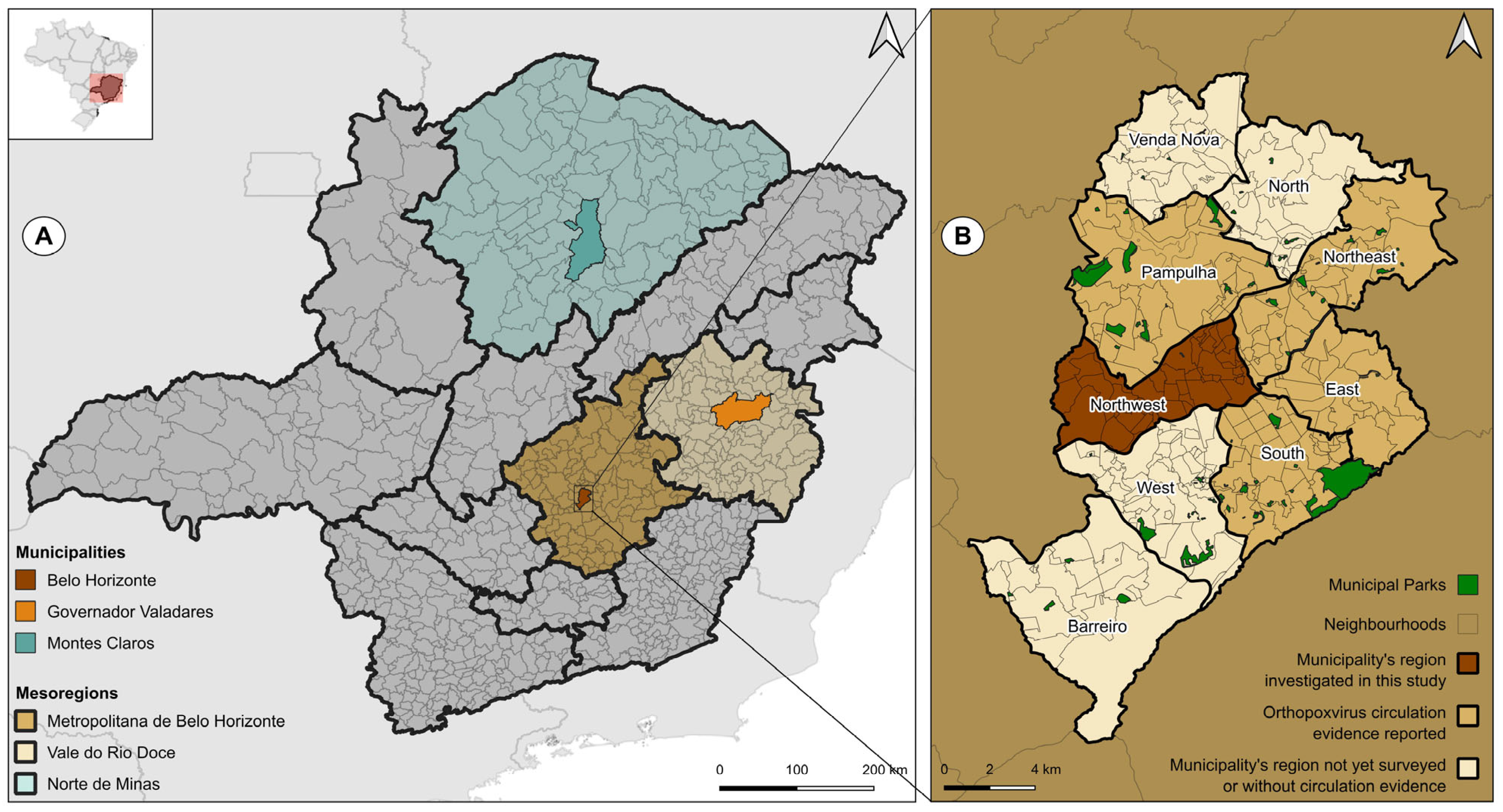
2.1. Areas of Study and Companion Animal Samples
2.2. PRNT
2.3. qPCR
3. Results
4. Discussion
Author Contributions
Funding
Institutional Review Board Statement
Informed Consent Statement
Data Availability Statement
Acknowledgments
Conflicts of Interest
References
- Morens, D.M.; Fauci, A.S. Emerging Pandemic Diseases: How We Got to COVID-19. Cell 2020, 182, 1077–1092. [Google Scholar] [CrossRef] [PubMed]
- Jones, K.E.; Patel, N.G.; Levy, M.A.; Storeygard, A.; Balk, D.; Gittleman, J.L.; Daszak, P. Global trends in emerging infectious diseases. Nature 2008, 451, 990–994. [Google Scholar] [CrossRef] [PubMed]
- Olival, K.; Hosseini, P.; Zambrana-Torrelio, C.; Ross, N.; Bogich, T.L.; Daszak, P. Host and viral traits predict zoonotic spillover from mammals. Nature 2017, 546, 646–650. [Google Scholar] [CrossRef]
- Tomori, O.; Oluwayelu, D.O. Domestic animals as potential reservoirs of zoonotic viral diseases. Annu. Rev. Anim. Biosci. 2023, 11, 33–55. [Google Scholar] [CrossRef] [PubMed]
- Wolfe, N.D.; Dunavan, C.P.; Diamond, J. Origins of major human infectious diseases. Nature 2007, 447, 279–283. [Google Scholar] [CrossRef] [PubMed]
- Ujvari, S.C. The History of the Dissemination of Microorganisms. Estud. Av. 2008, 22, 171–182. [Google Scholar] [CrossRef]
- Pearce-Duvet, J.M. The origin of human pathogens: Evaluating the role of agriculture and domestic animals in the evolution of human disease. Biol. Rev. 2006, 81, 369–382. [Google Scholar] [CrossRef]
- Bar-On, Y.M.; Phillips, R.; Milo, R. The biomass distribution on Earth. Proc. Natl. Acad. Sci. USA 2018, 115, 6506–6511. [Google Scholar] [CrossRef]
- Gilbert, M.; Nicolas, G.; Cinardi, G.; Boeckel, T.P.; Vanwambeke, S.O.; Wint, G.R.; Robinson, T.P. Data Descriptor: Global distribution data for cattle, buffaloes, horses, sheep, goats, pigs, chickens and ducks in 2010. Sci. Data 2018, 5, 180227. [Google Scholar] [CrossRef]
- Morand, S.; Mcintyre, K.M.; Baylis, M. Domesticated Animals And human infectious diseases of zoonotic origins: Domestication time matters. Infect. Genet. Evol. 2014, 24, 76–81. [Google Scholar] [CrossRef]
- Johnson, C.K.; Hitchens, P.L.; Pandit, P.S.; Rushmore, J.; Evans, T.S.; Young, C.C.; Doyle, M.M. Global shifts in mammalian population trends reveal key predictors of virus spillover risk. Proc. R. Soc. B 2020, 287, 20192736. [Google Scholar] [CrossRef] [PubMed]
- Wells, K.; Morand, S.; Wardeh, M.; Baylis, M. Distinct spread of DNA and RNA viruses among mammals amid prominent role of domestic species. Glob. Ecol. Biogeogr. 2020, 29, 470–481. [Google Scholar] [CrossRef] [PubMed]
- Desvars-Larrive, A.; Vogl, A.E.; Puspitarani, G.A.; Yang, L.; Joachim, A.; Käsbohrer, A. A One Health framework for exploring zoonotic interactions demonstrated through a case study. Nat. Commun. 2024, 15, 5650. [Google Scholar] [CrossRef] [PubMed]
- Keesing, F.; Ostfeld, R.S. Impacts of biodiversity and biodiversity loss on zoonotic diseases. Proc. Natl. Acad. Sci. USA 2021, 118, e2023540118. [Google Scholar] [CrossRef] [PubMed]
- Gamble, A.; Olarte-Castillo, X.A.; Whittaker, G.R. Backyard zoonoses: The roles of companion animals and peri-domestic wildlife. Sci. Transl. Med. 2023, 15, eadj0037. [Google Scholar] [CrossRef]
- Chomel, B.B.; Sun, A.B. Zoonoses in the Bedroom. Emerg. Infect. Dis. 2011, 17, 167. [Google Scholar] [CrossRef]
- Reperant, L.A.; Brown, I.H.; Haenen, O.L.; de Jong, M.D.; Osterhaus, A.D.M.E.; Papa, A.; Rimstad, E.; Valarcher, J.F.; Kuiken, T. Companion Animals as a Source of Viruses for Human Beings and Food Production Animals. J. Comp. Pathol. 2016, 155, S41eS53. [Google Scholar] [CrossRef]
- Forman, R.T. Urban Ecology: Science of Cities; Cambridge University Press: Cambridge, UK, 2014; ISBN 978-0-521-18824-1. [Google Scholar]
- Hughes, J.; Macdonald, D.W. A review of the interactions between free-roaming domestic dogs and wildlife. Biol. Conserv. 2013, 157, 341–351. [Google Scholar] [CrossRef]
- Orozco, L.; López-Pérez, A.M.; Zarza, H.; Suzán, G.; List, R. Dog demography and husbandry practices facilitate dog-wildlife conflict in a suburban-forest interface. Urban Ecosyst. 2022, 25, 1725–1734. [Google Scholar] [CrossRef]
- Chomel, B.B. Emerging and re-emerging zoonoses of dogs and cats. Animals 2014, 4, 434–445. [Google Scholar] [CrossRef]
- Rahman, M.T.; Sobur, M.A.; Islam, M.S.; Ievy, S.; Hossain, M.J.; El Zowalaty, M.E.; Taufiquer Rahman, A.M.M.; Ashour, H.M. Zoonotic diseases: Etiology, impact, and control. Microorganisms 2020, 8, 1405. [Google Scholar] [CrossRef] [PubMed]
- Associação Brasileira da Indústria de Produtos Para Animais de Estimação (ABINPET). 2023. Available online: https://abinpet.org.br/wp-content/uploads/2023/07/abinpet_folder_dados_mercado_2023_draft5.pdf (accessed on 29 February 2024). (In Portuguese).
- Barros, M.; Pons, D.J.; Moreno, A.; Vianna, J.; Ramos, B.; Dueñas, F.; Coccia, C.; Saavedra-Rodríguez, R.; Santibañez, A.; Medina-Vogel, G. Domestic dog and alien North American mink as reservoirs of infectious diseases in the endangered Southern river otter. Austral J. Vet. Sci. 2022, 54, 65–75. [Google Scholar] [CrossRef]
- Ellwanger, J.H.; Chies, J.A.B. The triad “dogs, conservation and zoonotic diseases”—An old and still neglected problem in Brazil. Perspect. Ecol. Conserv. 2019, 17, 157–161. [Google Scholar] [CrossRef] [PubMed]
- Dias, H.G.; Familiar-Macedo, D.; Garrido, I.O.; Dos Santos, F.B.; Pauvolid-Corrêa, A. Exposure of domestic animals to Mayaro and Oropouche viruses in urban and peri-urban areas of West-Central Brazil. One Health Outlook 2024, 6, 12. [Google Scholar] [CrossRef]
- Davila, E.; Fernández-Santos, N.A.; Estrada-Franco, J.G.; Wei, L.; Aguilar-Durán, J.A.; López-López, M.D.J.; Solís-Hernández, R.; García-Miranda, R.; Velázquez-Ramírez, D.D.; Torres-Romero, J.; et al. Domestic dogs as sentinels for West Nile virus but not Aedes-borne flaviviruses, Mexico. Emerg. Infect. Dis. 2022, 28, 1071. [Google Scholar] [CrossRef]
- Tack, D.M.; Reynolds, M.G. Zoonotic poxviruses associated with companion animals. Animals 2011, 1, 377–395. [Google Scholar] [CrossRef]
- Silva, N.I.; Oliveira, J.S.; Kroon, E.G.; Trindade, G.S.; Drumond, B.P. Here, There, and Everywhere: The Wide Host Range and Geographic Distribution of Zoonotic Orthopoxviruses. Viruses 2021, 13, 43. [Google Scholar] [CrossRef]
- MacNeill, A.L. Comparative pathology of zoonotic orthopoxviruses. Pathogens 2022, 11, 892. [Google Scholar] [CrossRef]
- Douglass, N. Borealpox (Alaskapox) virus: Will there be more emerging zoonotic orthopoxviruses? Lancet Microbe 2024, 5, 100883. [Google Scholar] [CrossRef]
- Yinka-Ogunleye, A.; Aruna, O.; Dalhat, M.; Ogoina, D.; McCollum, A.; Disu, Y.; Mamadu, I.; Akinpelu, A.; Ahmad, A.; Burga, J.; et al. Outbreak of human monkeypox in Nigeria in 2017–18: A clinical and epidemiological report. Lancet Infect. Dis. 2019, 19, 872–879. [Google Scholar] [CrossRef]
- Yinka-Ogunleye, A.; Aruna, O.; Ogoina, D.; Aworabhi, N.; Eteng, W.; Badaru, S.; Mohammed, A.; Agenyi, J.; Etebu, E.N.; Numbere, T.M. Reemergence of human monkeypox in Nigeria, 2017. Emerg. Infect. Dis. 2018, 24, 1149. [Google Scholar] [CrossRef] [PubMed]
- Ogoina, D.; Iroezindu, M.; James, H.I.; Oladokun, R.; Yinka-Ogunleye, A.; Wakama, P.; Otike-odibi, B.; Usman, L.M.; Obazee, E.; Aruna, O.; et al. Clinical Course and Outcome of Human Monkeypox in Nigeria. Clin. Infect. Dis. 2020, 7, e210–e214. [Google Scholar] [CrossRef] [PubMed]
- Nguyen, P.Y.; Ajisegiri, W.S.; Costantino, V.; Chughtai, A.A.; MacIntyre, C.R. Reemergence of human monkeypox and declining population immunity in the context of urbanization, Nigeria, 2017–2020. Emerg. Infect. Dis. 2021, 27, 1007. [Google Scholar] [CrossRef]
- Seang, S.; Burrel, S.; Todesco, E.; Leducq, V.; Monsel, G.; Pluart, D.L.; Cordevant, C.; Pourcher, V.; Palich, R. Evidence of human-to-dog transmission of monkeypox virus. Lancet 2022, 400, 658–659. [Google Scholar] [CrossRef]
- Secretaria de Estado de Saúde (SES-MG). Detecção de Monkeypox em animal em Minas Gerais. Available online: https://www.saude.mg.gov.br/lme/story/17178-nota-informativa-sobre-deteccao-de-monkeypox-em-animal-em-minas-gerais-23-8-2022 (accessed on 29 February 2024). (In Portuguese)
- Morgan, C.N.; Wendling, N.M.; Baird, N.; Kling, C.; Lopez, L.; Navarra, T.; Fischer, G.; Wynn, N.; Ayuk-Takor, L.; Darby, B.; et al. One Health Investigation into Mpox and Pets, United States. Emerg. Infect. Dis. 2024, 30, 2025–2032. [Google Scholar] [CrossRef]
- Eder, I.; Vollmar, P.; Pfeffer, M.; Naether, P.; Rodloff, A.C.; Meyer, H. Two Distinct Clinical Courses of Human Cowpox, Germany, 2015. Viruses 2017, 9, 375. [Google Scholar] [CrossRef]
- Haddadeen, C.; Ouwerkerk, M.V.; Vicek, T.; Fityan, A. A case of cowpox virus infection in the UK occurring in a domestic cat and transmitted to the adult male owner. Br. J. Dermatol. 2020, 183, e190. [Google Scholar] [CrossRef]
- Krankowska, D.C.; Wozniak, P.A.; Cybula, A.; Izdebska, J.; Suchacz, M.; Samelska, K.; Wiercińska-Drapało, A.; Szaflik, J.P. Cowpox: How dangerous could it be for humans? Case report. Int. J. Infect. Dis. 2021, 104, 239–241. [Google Scholar] [CrossRef]
- Costa, T.; Stidworthy, M.F.; Ehmann, R.; Denk, D.; Ashpole, I.; Drake, G.; Maciuca, I.; Zoeller, G.; Meyer, H.; Chantrey, J. Cowpox in zoo and wild animals in the United Kingdom. J. Comp. Pathol. 2023, 204, 39–46. [Google Scholar] [CrossRef]
- Smith, K.C.; Bennett, M.; Garret, A.D. Skin lesions caused by orthopoxvirus infection in a dog. J. Small Anim. Pract. 1999, 40, 495–497. [Google Scholar] [CrossRef]
- Pelkonen, P.M.; Tarvainen, K.; Hynninen, A.; Kallio, E.R.; Henttonen, H.; Palva, A.; Vaheri, A.; Vapalahti, O. Cowpox with Severe Generalized Eruption, Finland. Emerg. Infect. Dis. 2003, 9, 1458–1461. [Google Scholar] [CrossRef] [PubMed]
- von Bomhard, W.; Mauldin, E.A.; Breuer, W.; Pfleghaar, S.; Nitsche, A. Localized cowpox infection in a 5-month-old Rottweiler. Vet. Dermatol. 2010, 22, 111–114. [Google Scholar] [CrossRef] [PubMed]
- Stagegaard, J.; Kurth, A.; Stern, D.; Dabrowski, P.W.; Pocknell, A.; Nitsche, A.; Schrick, L. Seasonal recurrence of cowpox virus outbreaks incaptive cheetahs (Acinonyx jubatus). PLoS ONE 2017, 12, e0187089. [Google Scholar] [CrossRef] [PubMed]
- Trindade, G.S.; Guedes, M.I.C.; Drumond, B.P.; Mota, B.E.F.; Abrahao, J.S.; Lobato, Z.I.P.; Gomes, J.A.S.; Corrêa-Oliveira, R.; Nogueira, M.L.; Kroon, E.G.; et al. Zoonotic vaccinia virus: Clinical and immunological characteristics in a naturally infected patient. Clin. Infect. Dis. 2009, 48, e37–e40. [Google Scholar] [CrossRef]
- Megid, J.; Borges, I.A.; Abrahão, J.S.; Trindade, G.S.; Appolinário, C.M.; Ribeiro, M.G.; Allendorf, S.D.; Antunes, J.M.A.P.; Silva-Fernandes, A.T.; Kroon, E.G. Vaccinia virus zoonotic infection, Sao Paulo State, Brazil. Emerg. Infect. Dis. 2012, 18, 189. [Google Scholar] [CrossRef]
- Laiton-Donato, K.; Ávila-Robayo, P.; Páez-Martinez, A.; Benjumea-Nieto, P.; Usme-Ciro, J.A.; Pinzón-Nariño, N.; Giraldo, I.; Torres-Castellanos, D.; Nakazawa, Y.; Patel, N.; et al. Progressive Vaccinia Acquired through Zoonotic Transmission in a Patient with HIV/AIDS, Colombia. Emerg. Infect. Dis. 2020, 26, 601–605. [Google Scholar] [CrossRef]
- Domingos, I.J.; Oliveira, J.S.; Oliveira, D.B.; Kroon, E.G.; Costa, G.B.; Trindade, G.S. Twenty Years after Bovine Vaccinia in Brazil: Where We Are and Where Are We Going? Pathogens 2021, 10, 406. [Google Scholar] [CrossRef]
- Costa, G.B.; Almeida, L.R.A.; Cerqueira, G.R.; Mesquita, W.U.; Oliveira, J.S.; Miranda, J.B.; Saraiva-Silva, A.T.; Abrahão, J.S.; Drumond, B.P.; Kroon, E.G.; et al. Vaccinia Virus among Domestic Dogs and Wild Coatis, Brazil, 2013–2015. Emerg. Infect. Dis. 2018, 24, 2338. [Google Scholar] [CrossRef]
- Dutra, L.A.; Almeida, G.M.; Oliveira, G.P.; Abrahão, J.S.; Kroon, E.G.; Trindade, G.S. Molecular evidence of Orthopoxvirus DNA in capybara (Hydrochoerus hydrochaeris) stool samples. Arch. Virol. 2017, 162, 439–448. [Google Scholar] [CrossRef]
- Antunes, J.M.; Borges, I.A.; Trindade, G.D.; Kroon, E.G.; Cruvinel, T.M.; Peres, M.G.; Megid, J. Exposure of free-ranging capybaras (Hydrochoerus hydrochaeris) to the vaccinia virus. Transbound. Emerg. Dis. 2019, 67, 481–485. [Google Scholar] [CrossRef]
- Costa, G.B.; Miranda, J.B.; Almeida, G.G.; Oliveira, J.S.; Pinheiro, M.S.; Gonçalves, S.A.; Reis, J.K.P.; Gonçalves, R.; Ferreira, P.C.P.; Bonjardim, C.A.; et al. Detection of Vaccinia Virus in Urban Domestic Cats, Brazil. Emerg. Infect. Dis. 2017, 23, 360–362. [Google Scholar] [CrossRef] [PubMed]
- Babolin, L.; Almeida-Silva, M.J.; Potenza, M.R.; Fava, C.D.; Castro, V.; Harakava, R.; Okuda, L.H.; Rebouças, M.M.; Campos, A.E. Zoonosis associated to Rattus rattus and the impacts of the public actions to control the species. Arq. Inst. Biol. 2016, 83, e0832014. [Google Scholar] [CrossRef]
- Oliveira, J.S.; Costa, G.B.; Dutra, A.G.; Domingos, I.J.; Costa, P.S.; Silva, P.H.; Kroon, E.G.; Oliveira, D.B.; Trindade, G.S. Low prevalence of anti-Orthopoxvirus neutralizing antibodies in an urban population of Brazil. J. Med. Virol. 2023, 95, e28859. [Google Scholar] [CrossRef] [PubMed]
- Leite, J.A.; Drumond, B.P.; Trindade, G.S.; Lobato, Z.I.; da Fonseca, F.G.; dos Santos, J.R.; Madureira, M.C.; Guedes, M.I.; Ferreira, J.M.; Bonjardim, C.A.; et al. Passatempo virus, a vaccinia virus strain, Brazil. Emerg. Infect. Dis. 2005, 11, 1935–1938. [Google Scholar] [CrossRef]
- Trindade, G.S.; Lobato, Z.I.P.; Drumond, B.P.; Leite, J.A.; Trigueiro, R.C.; Guedes, M.I.M.C.; da Fonseca, F.G.; dos Santos, J.R.; Bonjardim, C.A.; Ferreira, P.C.P.; et al. Short report: Isolation of two vaccinia virus strains from a single bovine vaccinia outbreak in rural area from Brazil: Implications on the emergence of zoonotic orthopoxviruses. Am. J. Trop. Med. Hyg. 2006, 75, 486–490. [Google Scholar] [CrossRef]
- Leal, G.G.A.; Carneiro, M.; Pinheiro, A.C.; Marques, L.A.; Ker, H.G.; Reis, A.B.; Coura-Vital, W. Risk profile for Leishmania infection in dogs coming from an area of visceral leishmaniasis reemergence. Prev. Vet. Med. 2018, 150, 1–7. [Google Scholar] [CrossRef]
- Coura-Vital, W.; Ker, H.G.; Roatt, B.M.; Aguiar-Soares, R.D.O.; Leal, G.G.D.A.; Moreira, N.D.D.; Oliveira, L.A.M.; Machado, E.M.M.; Morais, M.H.F.; Correa-Oliveira, R.; et al. Evaluation of change in canine diagnosis protocol adopted by the visceral leishmaniasis control program in Brazil and a new proposal for diagnosis. PLoS ONE 2014, 9, e91009. [Google Scholar] [CrossRef]
- Instituto Brasileiro de Geografia e Estatística (IBGE). Cidades e Estados do Brasil. 2022. Available online: https://cidades.ibge.gov.br/ (accessed on 12 March 2024). (In Portuguese)
- Distância Entre Cidades. 2024. Available online: https://distanciacidades.net/ (accessed on 12 March 2024). (In Portuguese).
- Newman, F.K.; Frey, S.E.; Blevins, T.P.; Mandava, M.; Bonifacio, A., Jr.; Yan, L.; Belshe, R.B. Improved Assay To Detect Neutralizing Antibody following Vaccination with Diluted or Undiluted Vaccinia (Dryvax) Vaccine. J. Clin. Microbiol. 2003, 41, 3154–3157. [Google Scholar] [CrossRef]
- Kroon, E.G.; Abrahãao, J.S.; Trindade, G.S.; Oliveira, G.P.; Luiz, A.P.M.F.; Costa, G.B.; Lima, M.T.; Calixto, R.S.; Oliveira, D.B.; Drumond, B.P. Natural Vaccinia Virus Infection: Diagnosis, Isolation, and Characterization. Curr. Protoc. Microbiol. 2016, 42, 14A.5.1–14A.5.43. [Google Scholar] [CrossRef]
- Trindade, G.D.S.; Li, Y.; Olson, V.A.; Emerson, G.; Regnery, R.L.; da Fonseca, F.G.; Kroon, E.G.; Damon, I. Real-time PCR assay to identify variants of Vaccinia virus: Implications for the diagnosis of bovine vaccinia in Brazil. J. Virol. Methods 2008, 152, 63–71. [Google Scholar] [CrossRef]
- Taube, J.C.; Rest, E.C.; Lloyd-Smith, J.O.; Bansal, S. The global landscape of smallpox vaccination history and implications for current and future orthopoxvirus susceptibility: A modelling study. Lancet Infect. Dis. 2023, 23, 454–462. [Google Scholar] [CrossRef] [PubMed]
- Allen, T.; Murray, K.A.; Zambrana-Torrelio, C.; Morse, S.S.; Rondinini, C.; Marco, M.D.; Breit, N.; Olival, K.J.; Daszak, P. Global hotspots and correlates of emerging zoonotic diseases. Nat. Commun. 2017, 8, 1124. [Google Scholar] [CrossRef] [PubMed]
- Hassell, J.M.; Begon, M.; Ward, M.J.; Fèvre, E.M. Urbanization and Disease Emergence: Dynamics at the Wildlife–Livestock–Human Interface. TREE 2017, 32, 55–67. [Google Scholar] [CrossRef] [PubMed]
- Baxby, D.; Bennett, M.; Getty, B. Human cowpox 1969-93: A review based on 54 cases. Br. J. Dermatol. 1994, 131, 598–607. [Google Scholar] [CrossRef]
- Zelaya, C.E.; Smith, B.P.; Riser, A.P.; Hong, J.; Distler, S.; O’Connor, S.; Belay, E.; Shoeb, M.; Waltenburg, M.A.; Negron, M.E.; et al. Urban and rural mpox incidence among persons aged 15–64 years—United States, May 10–December 31, 2022. MMWR 2023, 72, 574–578. [Google Scholar] [CrossRef]
- Thornhill, J.; Gandhi, M.; Orkin, C. Mpox: The Reemergence of an Old Disease and Inequities. Annu. Rev. Med. 2024, 75, 159–175. [Google Scholar] [CrossRef]
- Oliveira, J.S.; Figueiredo, P.O.; Costa, G.B.; Assis, F.L.; Drumond, B.P.; Fonseca, F.G.; Nogueira, M.L.; Kroon, E.G.; Trindade, G.S. Vaccinia Virus Natural Infections in Brazil: The Good, the Bad, and the Ugly. Viruses 2017, 9, 340. [Google Scholar] [CrossRef]
- Peres, M.G.; Bacchiega, T.S.; Appolinário, C.M.; Vicente, A.F.; Allendorf, S.D.; Antunes, J.M.A.P.; Moreira, S.A.; Legatti, E.; Fonseca, C.R.; Pituco, E.M.; et al. Serological study of Vaccinia virus reservoirs in areas with and without official reports of outbreaks in cattle and humans in São Paulo, Brazil. Arch. Virol. 2013, 158, 2433–2441. [Google Scholar] [CrossRef]
- Peres, M.G.; Barros, C.B.; Appolinário, C.M.; Antunes, J.M.; Mioni, M.S.; Bacchiega, T.S.; Allendorf, S.D.; Vicente, A.F.; Fonseca, C.R.; Megid, J. Dogs and Opossums Positive for Vaccinia Virus during Outbreak Affecting Cattle and Humans, São Paulo State, Brazil. Emerg. Infect. Dis. 2016, 22, 271–273. [Google Scholar] [CrossRef]
- Peres, M.G.; Bacchiega, T.S.; Appolinário, C.M.; Vicente, A.F.; Mioni, M.S.R.; Ribeiro, B.L.D.; Fonseca, C.R.S.; Pelícia, V.C.; Ferreira, F.; Oliveira, G.P.; et al. Vaccinia Virus in Blood Samples of Humans, Domestic and Wild Mammals in Brazil. Viruses 2018, 10, 42. [Google Scholar] [CrossRef]
- Kile, J.C.; Panella, N.A.; Komar, N.; Chow, C.C.; MacNeil, A.; Robbins, B.; Bunning, M.L. Serologic survey of cats and dogs during an epidemic of West Nile virus infection in humans. J. Am. Veter. Med. Assoc. 2005, 226, 1349–1353. [Google Scholar] [CrossRef] [PubMed]
- Pham-Thanh, L.; Nguyen-Tien, T.; Magnusson, U.; Bui-Nghia, V.; Bui-Ngoc, A.; Le-Thanh, D.; Lundkvist, A.; Can-Xuan, M.; Thu, T.N.T.; Bich, A.B.N.; et al. Dogs as sentinels for flavivirus exposure in urban, peri-urban and rural Hanoi, Vietnam. Viruses 2021, 13, 507. [Google Scholar] [CrossRef]
- Bowser, N.H.; Anderson, N.E. Dogs (Canis familiaris) as sentinels for human infectious disease and application to Canadian populations: A systematic review. Vet. Sci. 2018, 5, 83. [Google Scholar] [CrossRef] [PubMed]
- Halliday, J.E.; Meredith, A.L.; Knobel, D.L.; Shaw, D.J.; Bronsvoort, B.M.D.C.; Cleaveland, S. A framework for evaluating animals as sentinels for infectious disease surveillance. J. R. Soc. Interface 2007, 4, 973–984. [Google Scholar] [CrossRef] [PubMed]

| City | Mesoregion | Collection Date | Sample Type | Number of Individuals |
|---|---|---|---|---|
| Belo Horizonte | Metropolitana de Belo Horizonte | 2008–2009 | Plasma | 126 |
| Montes Claros | Norte de Minas | 2020 | Serum | 42 |
| Governador Valadares | Vale do Rio Doce | 2014–2015 | Serum | 150 |
| Total: 318 |
| Municipalities | |||
|---|---|---|---|
| Belo Horizonte | Montes Claros | Governador Valadares | |
| Mesoregion | Metropolitana de Belo Horizonte | Norte de Minas | Vale do Rio Doce |
| Territory (km2) | 331,354 | 3,589,811 | 2,342,376 |
| Urbanized area (km2) | 274,04 | 73.51 | 49.93 |
| Biome | Cerrado and Atlantic Forest | Caatinga and Cerrado | Atlantic Forest |
| Human population | 2,315,560 | 414,240 | 257.171 |
| Demographic density (hab/km2) | 6988.18 | 115,39 | 109.79 |
| City | Sample | Collection Date | Sex | Age (Years) | PRNT | qPCR | ||
|---|---|---|---|---|---|---|---|---|
| Dilution Tested | % of Plaque Reduction | C11R | A56R | |||||
| Montes Claros | CA32 | 2020 | M | 3 | 1:40 | 62.8 | - | - |
| CA40 | F | 8.6 | 1:40 | 65.7 | - | - | ||
| TMV03 | M | 6 | 1:40 | 80.23 | - | - | ||
| Governador Valadares | 347 | 2014–2015 | M | 2 | 1:20 | 88.01 | - | - |
| 360 | M | 3 | 1:20 | 64.2 | - | - | ||
| Belo Horizonte | 41 | 2008 | F | 10 | 1:20 | 68.95 | - | - |
| 46 | F | 1 | 1:20 | 65.28 | - | - | ||
| 70 | M | 2 | 1:20 | 58.5 | - | - | ||
| 91 | F | 2 | 1:20 | 52.63 | - | - | ||
| 122 | F | 12 | 1:20 | 63.46 | - | - | ||
| 128 | M | 6 | 1:20 | 78.95 | - | - | ||
| 130 | F | 1 | 1:20 | 62.32 | - | - | ||
| 132 | F | 2 | 1:20 | 77.73 | - | - | ||
| 195 | M | 2 | 1:20 | 72.69 | - | - | ||
| 204 | M | 5 | 1:20 | 60.36 | - | - | ||
| 208 | F | 2 | 1:20 | 68,38 | - | - | ||
| 209 | F | 8 | 1:20 | 71.7 | - | - | ||
| 211 | M | 6 | 1:20 | 52.38 | - | - | ||
| 261 | F | 2 | 1:20 | 73.6 | - | - | ||
| 264 | F | NI * | 1:20 | 54.85 | - | - | ||
| 281 | M | 8 | 1:20 | 51.42 | - | - | ||
| Belo Horizonte | 282 | 2008 | F | 6 | 1:20 | 58.02 | - | - |
| 284 | M | 7 | 1:20 | 66.67 | - | - | ||
| 286 | M | 4 | 1:20 | 56.37 | - | - | ||
| 302 | F | 4 | 1:20 | 51.93 | - | - | ||
| 308 | F | 1 | 1:20 | 59.02 | - | - | ||
| 314 | M | 8 | 1:20 | 66,34 | - | - | ||
| 319 | M | 5 | 1:20 | 57.32 | - | - | ||
| 320 | F | 3 | 1:20 | 57.22 | - | - | ||
| 321 | F | 8 | 1:20 | 59.54 | - | - | ||
| 322 | F | 8 | 1:20 | 56.42 | - | - | ||
| 324 | F | 5 | 1:20 | 51.9 | - | - | ||
| 334 | F | 7 | 1:20 | 55.58 | - | - | ||
| 395 | F | 4 | 1:20 | 66.84 | - | - | ||
| 874 | M | 3 | 1:20 | 59.78 | - | - | ||
| 885 | F | 1 | 1:20 | 60.61 | - | - | ||
| 945 | F | 6 | 1:20 | 50 | - | - | ||
| 1147 | F | 0.8 | 1:20 | 56.58 | - | - | ||
| 1429 | F | 2 | 1:40 | 54.17 | - | - | ||
| 1436 | M | 0.7 | 1:40 | 50 | - | - | ||
| Belo Horizonte | 2063 | 2009 | M | 1 | 1:40 | 59.83 | - | - |
| 2129 | M | 2 | 1:40 | 66.54 | - | - | ||
| 2249 | F | 5 | 1:40 | 69.23 | - | - | ||
| 2273 | F | 2 | 1:40 | 57.48 | - | - | ||
| 2312 | F | 1 | 1:40 | 88.19 | - | - | ||
| Total: 45 | Mean: 62.9 | |||||||
Disclaimer/Publisher’s Note: The statements, opinions and data contained in all publications are solely those of the individual author(s) and contributor(s) and not of MDPI and/or the editor(s). MDPI and/or the editor(s) disclaim responsibility for any injury to people or property resulting from any ideas, methods, instructions or products referred to in the content. |
© 2025 by the authors. Licensee MDPI, Basel, Switzerland. This article is an open access article distributed under the terms and conditions of the Creative Commons Attribution (CC BY) license (https://creativecommons.org/licenses/by/4.0/).
Share and Cite
de Meneses, D.; Stoffella-Dutra, A.G.; Blaso, V.S.; de Almeida, I.M.; Dias, K.L.; Domingos, I.J.d.S.; Ribeiro, G.P.; Coura-Vital, W.; Reis, A.B.; Vieira, T.M.; et al. Domestic Dogs Exposed to Orthopoxvirus in Urban Areas of Brazil. Viruses 2025, 17, 131. https://doi.org/10.3390/v17010131
de Meneses D, Stoffella-Dutra AG, Blaso VS, de Almeida IM, Dias KL, Domingos IJdS, Ribeiro GP, Coura-Vital W, Reis AB, Vieira TM, et al. Domestic Dogs Exposed to Orthopoxvirus in Urban Areas of Brazil. Viruses. 2025; 17(1):131. https://doi.org/10.3390/v17010131
Chicago/Turabian Stylede Meneses, Débora, Ana G. Stoffella-Dutra, Vicenzo S. Blaso, Iara M. de Almeida, Karolina L. Dias, Iago José da S. Domingos, Gabriela P. Ribeiro, Wendel Coura-Vital, Alexandre B. Reis, Thallyta M. Vieira, and et al. 2025. "Domestic Dogs Exposed to Orthopoxvirus in Urban Areas of Brazil" Viruses 17, no. 1: 131. https://doi.org/10.3390/v17010131
APA Stylede Meneses, D., Stoffella-Dutra, A. G., Blaso, V. S., de Almeida, I. M., Dias, K. L., Domingos, I. J. d. S., Ribeiro, G. P., Coura-Vital, W., Reis, A. B., Vieira, T. M., & Trindade, G. d. S. (2025). Domestic Dogs Exposed to Orthopoxvirus in Urban Areas of Brazil. Viruses, 17(1), 131. https://doi.org/10.3390/v17010131