Article

Discovery and Genomic Characterization of a Novel Hepadnavirus from Asymptomatic Anadromous Alewife (Alosa pseudoharengus)

1
U.S. Geological Survey, Eastern Ecological Science Center, Leetown Research Laboratory, 11649 Leetown Road, Kearneysville, WV 25430, USA
2
West Virginia Cooperative Fish and Wildlife Research Unit, Davis College of Agriculture, Natural Resources & Design, West Virginia University, 1 Waterfront Pl, Morgantown, WV 26506, USA
3
U.S. Geological Survey, Western Fisheries Research Center, Seattle, WA 98115, USA
4
NJ Fish & Wildlife, Office of Fish and Wildlife Health and Forensics, 605 Pequest Rd, Oxford, NJ 07863, USA
5
Department of Fisheries, Wildlife, and Conservation Biology, College of Food, Agriculture, and Natural Resource Sciences, University of Minnesota, St. Paul, MN 55108, USA
6
Animal Disease Research and Diagnostic Laboratory, South Dakota State University, 1155 North Campus Drive, Brookings, SD 570077, USA
7
Veterinary Diagnostic Laboratory, Department of Veterinary Population Medicine, University of Minnesota, Saint Paul, MN 55455, USA
8
Department of Pathology, University of Georgia, Athens, GA 30602, USA
9
U.S. Department of Agriculture-Agricultural Research Service, National Center for Cool and Cold Water Aquaculture, Kearneysville, WV 25430, USA
*
Author to whom correspondence should be addressed.
Viruses 2024, 16(6), 824; https://doi.org/10.3390/v16060824
Submission received: 8 April 2024 / Revised: 12 May 2024 / Accepted: 18 May 2024 / Published: 22 May 2024

Abstract

:
The alewife (Alosa pseudoharengus) is an anadromous herring that inhabits waters of northeastern North America. This prey species is a critical forage for piscivorous birds, mammals, and fishes in estuarine and oceanic ecosystems. During a discovery project tailored to identify potentially emerging pathogens of this species, we obtained the full genome of a novel hepadnavirus (ApHBV) from clinically normal alewives collected from the Maurice River, Great Egg Harbor River, and Delaware River in New Jersey, USA during 2015–2018. This previously undescribed hepadnavirus contained a circular DNA genome of 3146 nucleotides. Phylogenetic analysis of the polymerase protein placed this virus in the clade of metahepadnaviruses (family: Hepadnaviridae; genus: Metahepadnavirus). There was no evidence of pathology in the internal organs of infected fish and virions were not observed in liver tissues by electron microscopy. We developed a Taqman-based quantitative (qPCR) assay and screened 182 individuals collected between 2015 and 2018 and detected additional qPCR positives (n = 6). An additional complete genome was obtained in 2018 and it has 99.4% genome nucleotide identity to the first virus. Single-nucleotide polymorphisms were observed between the two genomes, including 7/9 and 12/8 synonymous vs nonsynonymous mutations across the polymerase and surface proteins, respectively. While there was no evidence that this virus was associated with disease in this species, alewives are migratory interjurisdictional fishes of management concern. Identification of microbial agents using de novo sequencing and other advanced technologies is a critical aspect of understanding disease ecology for informed population management.

1. Introduction

The collective term “river herring” refers to the anadromous iteroparous alosine filter feeders blueback herring (Alosa aestivalis) and alewife (A. pseudoharengus). River herring have long been utilized as an easily harvested and inexpensive source of food, bait, and fertilizer. Specific use of river herring is documented as early as the arrival of the pilgrims to what is now the United States [1,2], and substantially earlier by indigenous peoples [3]. Consequently, the availability of river herring has significant economic impacts for the communities around the fishery [1,4]. Reductions in abundance were often met with reintroduction efforts and the translocation of adult fish to re-establish populations [5,6]. As with other anadromous fishes, adults migrate from marine environments into freshwater for spawning. For river herring, this occurs predominantly in eastern North American streams, lakes, and ponds, depending on environmental conditions and food availability [6,7,8]. Perhaps as a direct result of their social and economic value and habitat loss due to damming, anadromous river herring stocks have declined significantly over the past 400 years, and are now considered species of concern by the National Marine Fisheries Service (National Oceanic and Atmospheric Administration [NOAA]) [9,10] and the Atlantic States Marine Fisheries Commission [11]. However, predation, habitat quality, climate change, and disease constitute additional threats which can indirectly or directly cause population declines [12,13]. Understanding and incorporating management approaches to mitigate fish health threats to these species is critical to long-term health and population sustainability.
Hepadnaviruses are enveloped, reverse-transcribing DNA viruses of the family Hepadnaviridae. While this family was historically represented by the Avihepadnavirus and Orthohepadnavirus, de novo sequencing methods have revealed a greater diversity of other genera that include the Herpetohepadnavirus, Metahepadnavirus, and Parahepadnavirus [14]. At present, it appears that these genera are restricted to piscine or herptile hosts [14]. The avi- and orthohepadnaviruses are hepatotropic and exhibit a narrow host range. Infections are transient or chronic, and in the latter case, Orthohepadnavirus infections are associated with an increased risk of hepatocellular carcinoma. Much less is known about the tissue tropism of these more recently described hepadnaviruses, but there is evidence that they may not exhibit the same tissue restrictions [15,16]. Similarly, the disease ecology and significance in respect to host health are uncharacterized.
The white sucker hepatitis B virus (WSHBV) is a parahepadnavirus and is perhaps the best studied of the three newly recognized genera. This virus, discovered from white suckers (Catostomus commersonii), was the first reported hepadnavirus to infect fish, and phylogeographic analyses suggest that genomic diversity occurs similarly to that of well-studied hepadnaviruses [17]. While liver and skin tumors are often observed in white suckers in regions of the Great Lakes where chemical contamination and WSHBV are reported, there are no clear associations between viral infection and neoplasia [18,19,20]. It is notable that the metahepadnavirus (bluegill hepatitis B virus) was observed in a fish with an observed tumor, but, like the scenario with the WSHBV, datasets are not available to ascribe or even suggest a causative association with neoplastic disease [15]. In addition, partial genomes of fish hepadnaviruses of unknown pathogenicity have been identified in African cichlids and a number of marine fish hosts via metagenomic surveys [16,21,22]. Here, we identify a novel hepadnavirus that is most related to other described metahepadnaviruses in clinically normal migratory alewives sampled from the Great Egg Harbor and Maurice Rivers in New Jersey, USA. As alewives are a species of concern, investigation into possible pathogens is essential to support their long-term health and population sustainability.

2. Materials and Methods

2.1. Sampling

Three life stages of anadromous river herring from New Jersey, USA were examined opportunistically in this study, including spawning adult fish and young-of-the-year (age-0) fish from the river environment. A population of landlocked alewives were also included in the study to determine if viruses were detected in these isolated fish populations, which have adapted to complete their entire life cycle within a freshwater lake.

2.1.1. Spawning River Herring

Spawning adult anadromous alewives and blueback herring were collected in March and April 2015, 2016, and 2018 during the New Jersey Division of Fish and Wildlife (NJDFW) annual surveys (Figure 1). Fish were captured by gill net, set for 2 h at a time, from the Maurice River and the Great Egg Harbor River (Table 1). Following transport to shore, the fish were euthanized with an overdose of MS-222 and transported on ice to the Pequest Aquatic Animal Health Laboratory (Oxford, NJ, USA). Fish were either sampled fresh or immediately frozen for future sample collection; sample collection information is summarized in Table 1.
In 2015, spleen, kidney, brain, and gills were aseptically dissected from the 14 fish, and up to 3 fish were pooled together and processed for viral cell culture assays, further described below. In 2016 and 2018, head, kidney, spleen, and liver were aseptically removed and transferred to 2 mL Eppendorf tubes and frozen at −80 °C until shipping to the Minnesota Veterinary Diagnostic Laboratory for cell culture assays or genetic analysis (Table 2). The internal organs, including liver, heart, anterior and posterior kidney, spleen, and gastrointestinal tract, were preserved in 10% neutral-buffered formalin for histology.

2.1.2. Young-of-the-Year (Age-0) River Herring

Young-of-the-year river herring were collected in 2015 during the NJDFW annual survey from the Great Egg Harbor and Delaware Rivers (Figure 1). Fish from the Great Egg Harbor River were collected via mesh beach seine, whereas those from the Delaware River were collected by boat electrofishing. Collected fish were euthanized with an overdose of MS-222 and transported on ice to the Pequest Aquatic Animal Health laboratory for necropsy and tissue sample collection (Table 2). Spleen, kidney, gills, and brain were aseptically removed for processing by viral cell culture assays. Subsets of combined viscera were preserved for genetic analysis.

2.1.3. Landlocked Alewife

In 2015, landlocked alewives were collected in collaboration with a commercial bait shop from Lake Hopatcong located in northern New Jersey, USA (Figure 1). Fish had been recently collected at night, using lights to attract the fish. An umbrella net was deployed off a barge to capture the fish. The fish were transported to shore and held in flow-through tanks supplied with lake water for up to 72 h. Fish were transferred alive to the Pequest Aquatic Animal Health Laboratory, where the fish were euthanized with an overdose of buffered MS-222 and spleen, kidney, brain, and gills were aseptically collected for viral cell culture assays. Subsets of viscera homogenate were preserved for genetic analysis (Table 2).

2.2. Viral Screening (Cell Culture)

Spawning adult and Age 0 anadromous river herring from 2015 were screened for viruses using viral cell culture assays run at the Animal Health Diagnostic Laboratory, New Jersey Department of Agriculture (Ewing, NJ, USA). Tissue samples were processed by standard methods. Briefly, the tissue pools were homogenized in HBSS using a stomacher, suspended, incubated in antibiotic media overnight, and centrifuged to purify viral agents within the supernatant. The supernatant was inoculated onto two cell lines, the epithelioma papulosum cyprini (EPC) cell line incubated at 15 and 20 °C, and the Chinook salmon Embryo-214 (CHSE-214) cell line incubated at 15 °C. Duplicate wells within 24-well plates were inoculated for each cell line/incubation temperature. The cells were evaluated 3 times per week for two weeks, after which, if no cytopathic effects (CPEs) were noted, samples were blind passaged by re-inoculation on fresh cells for an additional two weeks. If no CPEs were noted 2 weeks after blind passage, then the samples were reported negative. The remaining supernatant that was unused for viral cell culture assays was frozen at −80 °C for further genetic analysis.

2.3. DNA Extractions and Template Preparation

Subsampled organs were preserved in RNAlater™ and DNA was extracted for High-Throughout Sequencing (HTS), Sanger sequencing, or quantitative PCR (Table 2). Samples were stored at room temperature for 24 h prior to being stored at −20 °C until extraction. All DNA was extracted using a DNeasy Blood and Tissue Kit (Qiagen, CA, USA), following manufacturer instructions.

2.4. High Throughput Sequencing-Assisted Virus Discovery

Extracted DNA from samples collected in 2015 was submitted to the University of Minnesota Genomics Centre (UMGC) for library preparation. An Illumina Nextera XT Library Preparation Kit (Illumina, CA, USA) was used in accordance with the Nextera XT Library Preparation Reference Guide (Doc# 15031942 ver. 5) for HiSeq preparation. Prepared libraries were diluted per standard protocols within the HiSeq System Denature and Dilute Libraries Guide (Doc # 15050107 v06). Pooled indexed libraries were then run for 2 × 151 cycles.

2.5. Bioinformatic Discovery of a Novel Hepadnavirus

A draft linear genome was assembled from a pooled adult alewife sample collected from the Maurice River in 2015 and presumptively identified as a novel hepadnavirus. PCR primers were designed from this genome and non-pooled samples were screened to identify PCR-positive individuals. Additionally, to increase sensitivity, qPCR primers and probes were designed to facilitate higher sampling throughput. DNA was isolated from an individual PCR-positive alewife adult (M10) to establish a complete genome of this virus. We used rolling circle amplification (RCA) to enrich circular DNA templates for increased sequencing coverage of the viral genome. In addition, we used this method to sequence an additional genome from a positive adult alewife (MR4) collected from the Maurice River in April 2018 identified via qPCR. RCA was initiated by combining template viral DNA, 500 µM Exo-resistant primers, and 1× Equiphi reaction buffer into a cocktail. The cocktail was heat denatured for 3 min at 95 °C and then immediately cooled on ice for 5 min. The cooled, denatured cocktail was then combined with a final reaction mix of 10× Equiphi reaction buffer, 100 µM DTT (dithiothreitol), dNTPs, N.F. water, and Equiphi29 polymerase (ThermoFisher Scientific, Waltham, MA, USA) and incubated at 45 °C for 3 h and 65 °C for 10 min before being immediately frozen and stored at −80 °C until sequencing. RCA products were then quantified with a Qubit 4.0 Fluorometer (Invitrogen, Carlsbad, CA, USA) and normalized as starting material for NGS library prep. An Illumina Nextera XT Library Preparation Kit (Illumina, San Diego, CA, USA) was used in accordance with the Nextera XT Library Preparation Reference Guide (Doc # 15031942 ver. 5) for MiSeq preparation. Normalization of the final library was performed with Illumina’s Bead-Based Normalization (BBN) method and pooled as described in the BBN Loading Concentrations Exceptions Table 2 (MiSeq System Denature and Dilute Libraries Guide (Doc # 15039740 ver. 10)). Pooled libraries were then run for 2 × 301 cycles and loaded with a 15% PhiX (12.5 pM) spike. In addition, primers which spanned areas of lower coverage (Primer ID 1–4; Table 3) were implemented to enhance HTS methods.
After sequencing, paired reads were quality screened and trimmed prior to de novo assembly using CLC Genomics Workbench version 22.0.2. Once complete genomes of the alewife hepadnavirus (ApHBV) were constructed, we predicted open reading frames and identified repeat regions using Geneious Prime (v.2022.2.2). Protein characteristic analyses, including isoelectric point estimates, were also conducted using Geneious Prime. All predicted proteins were queried against the PDB, Pfam-A, UniProt-SwissProt-viral70, and NCBI Conserved Domains databases via HHpred [23]. We analyzed the two alewife hepadnavirus genomes for synonymous and nonsynonymous bases using DnaSP (v.6) [24].

2.6. Quantitative PCR

We designed a TaqMan qPCR assay to facilitate screening of the virus (Primer ID 5–7; Table 3) and determine prevalence. Primers were designed using Primer3 v2.3.7, bundled in Geneious Prime. We targeted the hepatitis core conserved domain locus from sample M10. Standards were created from a synthesized 150 bp gBlockTM gene fragment (Integrated DNA technologies Inc., Coralville, IA, USA) run in a 10-fold serial dilution from 1–1 × 10⁸ copies. The reactions were run on a QuantStudio 5 (ThermoFisher, Waltham, MA, USA) with default settings. We confirmed a single 117 bp product via end-point PCR and agarose electrophoresis. In addition, melt curve analysis was performed using the TaqMan forward and reverse primers in a SYBR-based format. That assay used the TaqMan cycling conditions and profile in Table 3, but without the TaqMan probe, and SYBRTM Green to form a master mix. To confirm a single amplification peak, melt curve analysis was run with parameters of 95 °C for 15 s cooling at a rate of 1.6 °C/s, and then followed by 60 °C for 60 s and 95 °C for 15 s. A total of 32 pooled samples comprised 160 discrete specimens (5 fish per pool) and 22 individual fish samples were screened via qPCR (Table 2).

3. Results

3.1. Cell Culture Assays, Histology, and Transmission Electron Microscopy

No CPE was detected in any of the cell culture assays. Histopathologic evaluation did not detect lesions suggestive of virus infection and transmission electron microscopy revealed no hepadnavirus-like particles or any other observable virus particles. Histopathology detected notable myxozoan parasites infecting the kidney, which was the focus of a separate study [25].

3.2. Virus Prevalence in Wild Caught Herring

The qPCR assay developed here targeted a 117 bp region of the core protein coding region and was effective for the identification of virus-positive individuals. The R2 of the standard curve was 0.994 and the efficiency of the reaction was 96.3%. The limit of detection was calculated to be nine copies. Background amplification was observed in virus-negative samples, as determined by the absence of viral sequences obtained during sequencing. Given that there were no reliable means to interpolate a LoQ via CV of target amounts, we consequently set a diagnostic threshold for positive samples >10 copies, utilizing our lowest standard, consistent with a CV ≤ 35% [26]. Of the 32 pooled samples screened, six were positive via qPCR (Figure 2). ApHBV was exclusively detected in adult, migratory alewives collected in freshwater, riverine environments (31.6% positive) during the spring spawning run. Virus genome copies of virus-positive fish ranged from 15 copies to 10,228,303 copies. The median copy number was 213. Virus-positive individuals were sourced exclusively from the Great Egg Harbor River in April of 2018 (n = 1) and Maurice River in 2015 (n = 5).

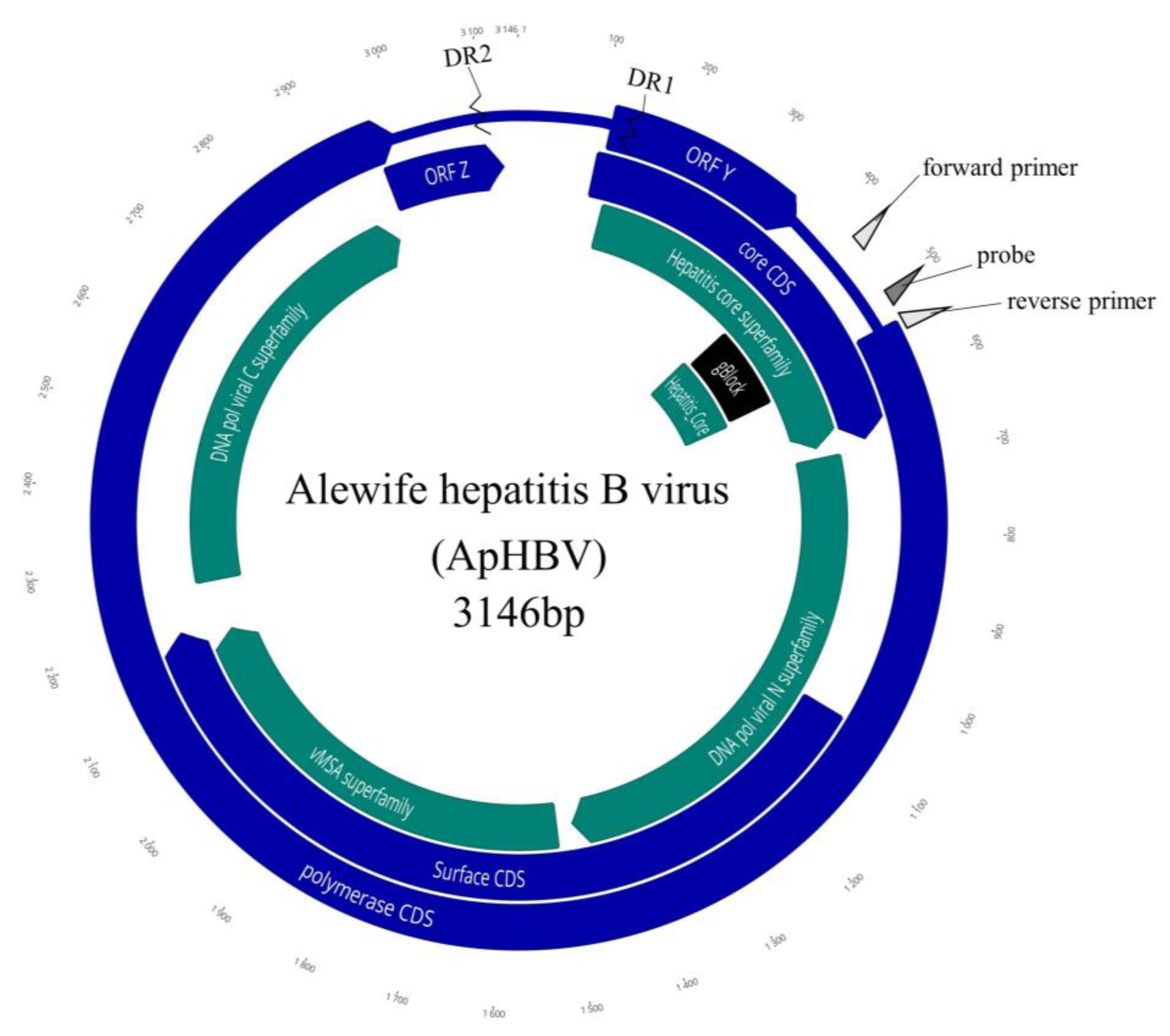
3.3. Sequencing the Viral Genome and ORF Organization

The initial assembly of pooled fish yielded a 3112 bp linear genomic contig. The complete genome of the alewife metahepadnavirus, sequenced using RCA enrichment (GenBank accession numbers OQ859058 (isolate M10) and OQ859059 (isolate MR4)), was 3146 bp (Figure 3). Genome coverage was 751 and 2786× for samples M10 and MR4, respectively. The genome size and organization were similar to that of other metahepadnaviruses. The GC content of the complete genome was 50.3%.
In silico translation identified five partially or completely overlapping reading frames (+1, +2, and +3). These corresponded to the core protein, polymerase protein, and surface protein of prototypical hepadnaviruses, in addition to a putative ORFY and ORFZ orthologous to ORFs in the bluegill hepadnavirus [15]. A hepatitis B X protein homologue common to orthohepadnaviruses was not present. The non-canonical polyadenylation signal (TATAAA) typical of hepadnaviruses was not present in the genomic sequence. Additionally, we identified a pair of 12 nt direct repeat (DR) regions, TGTTACACAGGA, located within the putative ORFZ and the core ORF that spanned 199 nt. Similar to other fish hepadnaviruses, a non-protein coding region was predated between the putative ORF Z and ORF Core.
Reading frame 1 (RF + 1) encodes the core polyprotein (nt 106–669, 564 bp) of 188 amino acids (aa) with a theoretical average molecular weight of 21041.97 daltons. The size of this ORF was in the range of other hepadnaviruses but was more similar (in size) to that of the orthohepadnaviruses. A putative ORFZ (nt 2965–3126; 162 bp) was identified that encodes a small 54 aa protein that overlaps the terminal end of the polymerase ORF. While data are unavailable to validate the expression of this protein, a 25 aa locus similar to frog virus-3 uncharacterized protein 086L (Q6GZT2) was identified in this ORF, providing evidence that this is a legitimate viral protein. We also identified the hepatitis core antigen conserved domain within this ORF (nt 402–573, 171 bp). The predicted isoelectric point of this polyprotein was 9.59 and was within the range reported for avian and mammalian hepadnaviruses (pI 9.34–10.12).
Reading frame 2 (RF +2) encodes the viral polymerase protein (nt 542 –2992; 2451 bp) of 816 aa and was similar in size to other hepadnavirus P proteins (785–902 aa). We identified ORF Y in this reading which was completely overlapped by the core ORF. This ORF is in the same reading frame as the orthologous ORF Y of the bluegill hepadnavirus and the IMDV. RF +2 also contained conserved domains that included viral DNA polymerases and reverse transcriptase (Figure 3). The overall MW of the P protein was 90,556.89 with a predicted pI of 9.21. Analysis of the predicted polymerase protein using the resistance module in the hepatitis B virus database [27] indicated that this virus is presumptively sensitive to HBV antiviral drugs that target reverse transcriptase [28]. This ORF partially overlaps the core protein ORF and completely overlaps the surface protein.
Reading frame 3 (RF + 3) of WSHBV (RF +3; nt 1059–2201; 1143 bp) was homologous to the large surface protein of hepadnaviruses. We also identified a conserved domain for major surface antigen from hepadnavirus (vMSA) in this ORF (nt 1512–2183; Figure 3).

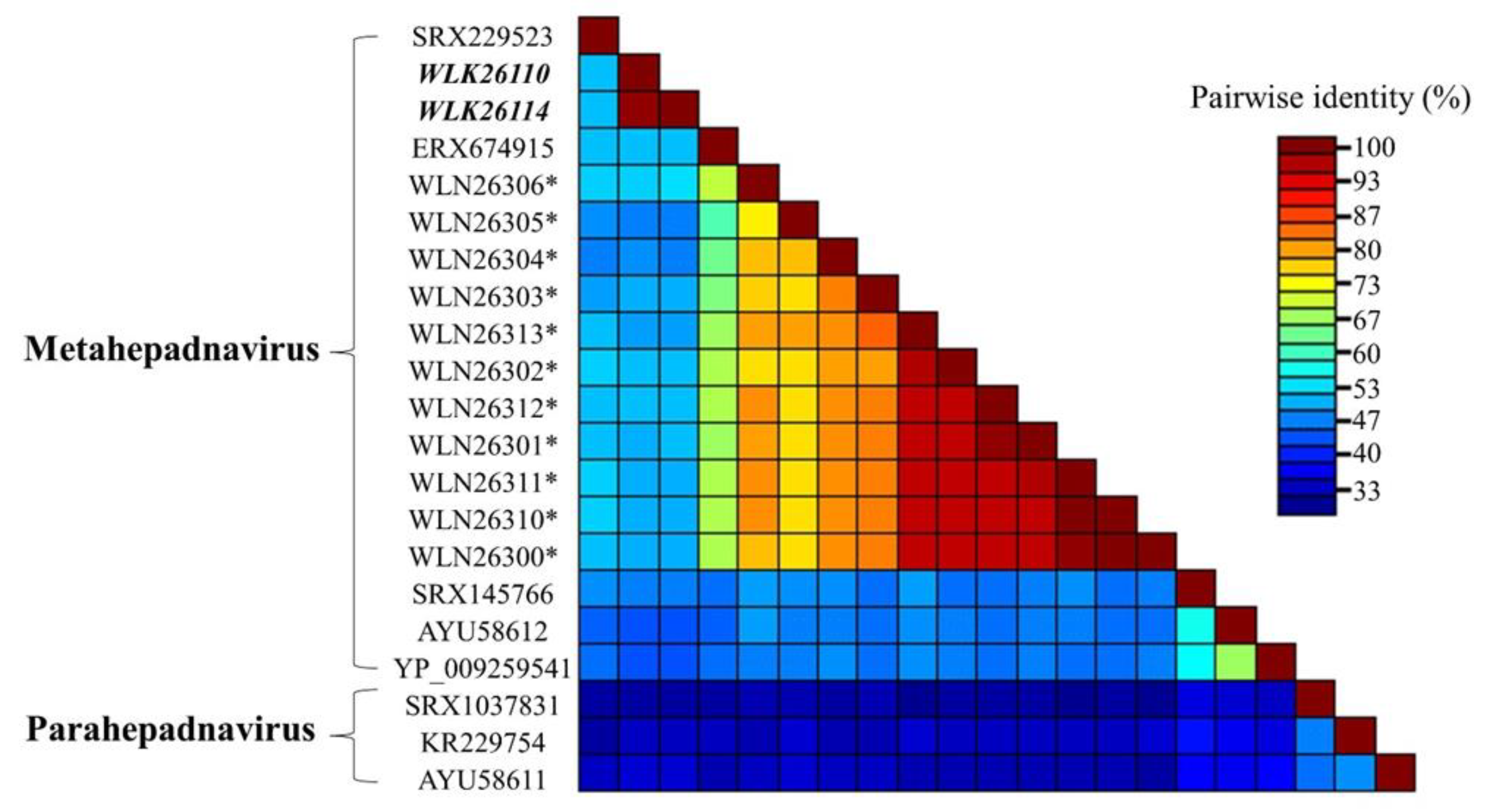
3.4. Genomic Diversity

We compared the complete viral genomes sampled from two individual fish captured during different sampling events (2015 and 2018) within the Maurice River. Pairwise alignment of these 3146 bp genomes revealed 99.4% nucleotide sequence identity (20 nt differences). Single-nucleotide polymorphisms were restricted to the polymerase and surface protein ORFs (Figure 4) observed between the two genomes that included 7/9 and 12/8 synonymous vs nonsynonymous mutations across the polymerase and surface protein open reading frames, respectively. Differences between the P protein for nucleotide and protein sequence identity were 0.96% and 0.86%, respectively. Likewise, these differences were 1.4% and 1.8% for the surface ORF for nucleotide and protein sequences, respectively.

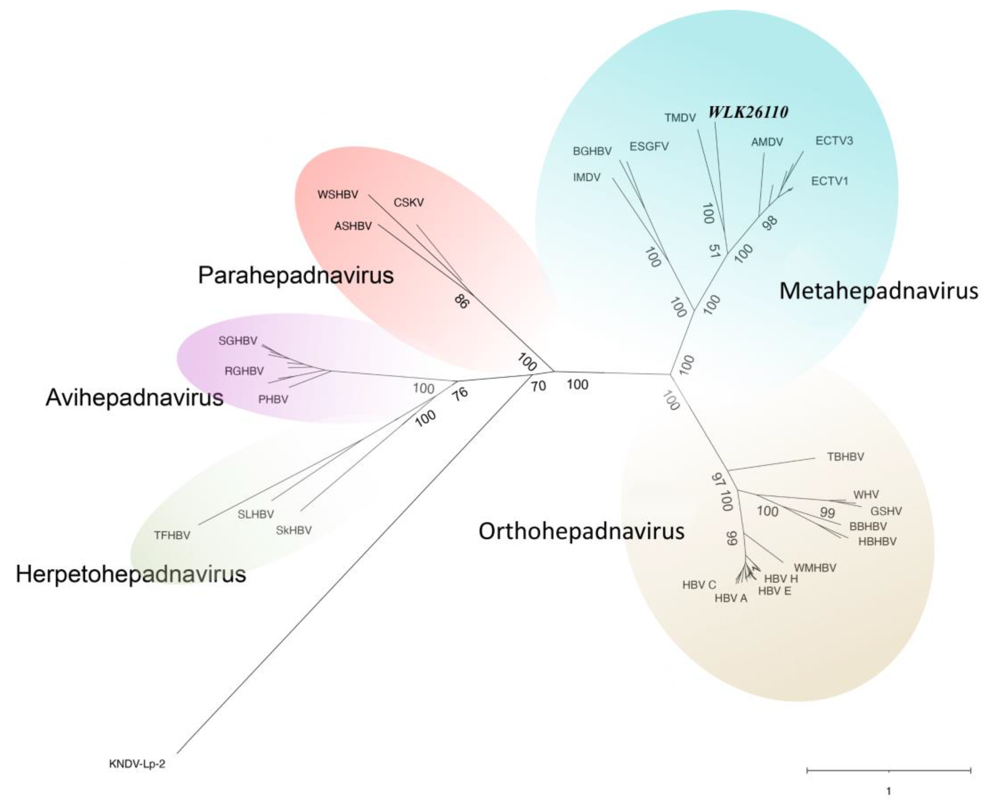
3.5. Pairwise Comparisons and Phylogenetic Analysis of P Proteins

Comparative pairwise alignment of fish meta- and parahepadnavirus P proteins revealed 31.0–52.6% amino acid identity (Figure 5; Table 4). Phylogenetic analyses of the P protein to a subset of other described hepadnaviruses (Figure 6) support the assignment of the alewife hepadnavirus (ApHBV) to the genus Metahepadnavirus ([29]; metahepadnavirus). ApHBV was observed to be most similar to the TMDV (Mexican tetra hepadnavirus), AMDV (Astatotilapia metahepadnavirus), and Ectodini metahepadnaviruses (Figure 6).

4. Discussion

Herein, a novel hepadnavirus was discovered in migratory alewife sampled from the Maurice and Great Egg Harbor Rivers, NJ, tentatively referred to here as alewife hepadnavirus (ApHBV). The closely related genomes (99.4% identity) being detected three years apart raises the possibility that it circulated endemically in the alewife population sampled. This novel virus groups most closely to the metahepadnaviruses [15,16,21]. Despite the recent global interest in viral diversity and ecology, much remains unknown about piscine hepadnaviruses, though rapid discovery is ongoing. A substantial uptick in viral discovery in fishes and other organisms in recent times has been the direct result of bioinformatic screening of short read archive databases from deep-sequencing investigations as well as purposeful metagenomic investigations designed to identify novel viruses [21,22,30,31]. Consequently, evaluations of whole genomes can be accomplished more quickly and effectively than ever, enabling apposite comparisons among specific viruses of interest.
Comparison of two complete ApHBV genomes identified 20 single-nucleotide polymorphism sites across the genomes (Figure 4). These differences led to protein coding changes in the surface (9 aa) and polymerase (8 aa) proteins, but not the core protein or predicted small ORFs. Although we did not obtain full genomes from all sampling locations, geographic variation among viral strains is possible, as was observed with WSHBV [17]. More targeted geographic sub-sampling would be necessary to discern all causes of genomic alterations. Furthermore, no conclusive phylogenetic hypothesis to the origins of hepadnaviruses exists, but the existence of synonymous HBVs among reptiles and amphibians suggests evolution driven by virus-host cospeciation [15,32,33]. Recent research suggests the potential for ancient divergence into distinct “Meta-Ortho+ and “Herpeto-Avi” lineages [34], which is not disputed by the inclusion of ApHBV among fish metahepadnaviruses (Figure 6). Two clades of the currently known fish-associated hepadnaviruses (metahepadnaviruses and parahepadnaviruses) essentially group orthogonally to one another, with KNDV-Lp-2 (killifish nackednavirus) as an outgroup to the parahepadnaviruses. On the whole, fish HBVs are genetically diverse, and many possess a pathophysiology permitting non-specific tropism, allowing the potential for more frequent host switching and mutation [16,31]. Given the divergence and diversity of previously undescribed hepadnaviruses, de novo sequencing approaches coupled with appropriate virus discovery workflows are the most appropriate means of identifying this viral “dark matter”. Recent efforts successfully detected the presence of hepadnaviruses using samples of lower pharyngeal jaw tissue or gill [31], which might support additional methods of less invasive viral screening.
While the survey efforts here represent an initial screening of geographically limited river herring populations, it is notable that virus was only detected in riverine adult alewife samples migrating to freshwater spawning grounds. Virus-positive fish were detected during multiple years from discrete sample sites more than 30 km apart. As alosines typically shoal, migrate, and spawn as part of a collective metapopulation [35], the presence of disparate virus detections merits additional inquiry to better understand its prevalence across a greater geographical range and the ecology of this virus circulating within these important fish populations. Virus-positive fish were collected as far as 30.6 km upstream from the mouth of the saltwater Delaware Bay (Maurice River; Figure 1). The 31.5% prevalence of virus among adult riverine alewives is comparable to the detection rates observed for WSHBV [20].
An additional commonality between our findings and those reported for WSHBV is that the fish sampled were collected during migratory spawning runs. Similar observations have not been reported for other hepadnaviruses identified from fish hosts. A high viral genome copy number (10,228,303 copies) was observed in only one virus-positive individual in this study, as similarly reported in WHSBV-positive white sucker [17]. Notably, the fish with the greatest number of viral genome copies was a female alewife. The sex of all fishes in the current study was not determined and sex steroid concentrations were not measured, but during this life history stage androgens are elevated in both pre-spawn male and female fishes. Of note, hepatitis B virus replication is enhanced by androgen and the androgen receptor in mice, and males are more likely to develop HBV-associated hepatocellular carcinoma [36,37]. Though other positive samples were considerably lower (less than 1000 copies), the sample number and sex data availability of virus-positive fish in the current study are insufficient to assess sex-associated differences.
Detection of ApHBV among alewife, but not syntopic migratory blueback herring, strengthens suspicions of host specificity. However, the limited sample size of adult blueback herring in this study warrants caution when making comparisons between species or evaluating host switching. Even so, recent research suggests that adaptive radiation within a geographically isolated region is associated with rapid virus diversification and interspecific transmission in African cichlids [31]. Interrelatedness and pairwise similarities of observed cichlid metahepadnaviruses (Figure 5) suggest that the viruses were widely distributed and likely diversified along with their hosts in multiple radiation events [31]. Similar species level divergence and subsequent viral divergence might also be observed in the case of river herring metahepadnaviruses. Theoretically, however, limited genetic separation among closely related species could also suggest lowered adaptive barriers to host jumping. Both river herring exhibit identifiable regional groupings across regional river populations [38], and despite hybridization across syntopic populations, there is also evidence of genetic bottlenecking across alewife populations in the northern portion of their range [39]. Potentially, bottlenecked populations could be subject to reduced fitness, posing additional threats [40].
A lack of detections of this virus may suggest its absence in Lake Hopatcong (landlocked population), or perhaps migratory spawning adult alewives are predisposed to ApHBV infections compared to other species or life history stages. The ApHBV was also not detected in juvenile alewives, or from landlocked alewife populations, despite these samples representing 90% of total fish screened by qPCR. Physiological differences between life stages could account for a lack of observed cases of ApHBV within juvenile alewives, though the simplest explanation could be a simple lack of exposure as the adult fish out-migrate before the eggs hatch [41]. Alewives migrating upriver in freshwater face different stressors than in their characteristic marine environment [6,9,42,43,44,45]. In addition, physiological changes associated with reproduction lead to seasonal stressors associated with energy repartitioning, from somatic growth to gonad and gamete development.
With respect to adult river herring, migratory behavior associated with reproduction is likely of even higher energetic cost than reaching sexual maturity [46,47], including changes to osmotic balance, development, behavior, and immune function [48]. This is particularly true when fish passage is inhibited with barriers [42,49,50,51]. Frequently, migration barriers are mitigated via fishways or fish ladders [52]. Yet, despite special considerations, it has been long understood that migrating fish still experience elevated stress and crowding when utilizing fishways [53,54]. Seasonal migration of fish introduces the opportunity for disease spread between and among different populations, which likely experience increased disease risk as a result [51,55]. Such disease transmission is not guaranteed, however, even when in a confined geographic location [56]. Other considerations include climate change-facilitated habitat degradation which reduces suitable habitat available for use by river herring [9,57]. Moreover, virulence may be substantially increased by elevated temperatures, while exceeding the optimal thermal regime may further increase alewife disease susceptibility [12,58].
Anadromous river herring stocks have declined by as much as 98% since the 1700s, due at least in part to historic overfishing and habitat loss from river damming [1,9,10]. Minimal study into potential viral pathogens or other potential diseases of anadromous river herring has been accomplished to date [13,59], and additional research is vital to improve conservation efforts. The discovery of the novel ApHBV, and other recent related discoveries [22,31], suggests there are likely to be a myriad of undiscovered hepadnaviruses within other species of fish, including other alosines and their potential hybrids. ApHBV was identified in clinically normal fish using Illumina-based sequencing of DNA in a diagnostic situation where standard tissue culture methods were not adequate for virus discovery. Further study is necessary to identify the geographical range of ApHBV and, more importantly, determine the biological significance of this virus [17,60]. While it is not immediately clear if this virus has a significant health impact, molecular method-based screening and proactive surveillance of this virus is possible even in the absence of overt pathology associated with infection. Advances in sequencing technologies have significantly increased our ability to discover novel viruses and other pathogens in diseased and clinically normal fish hosts. ApHBV provides yet another example of the enigmatic diversity of fish hepadnaviruses, and its association with an interjurisdictional species of economic and ecological importance emphasizes the necessity for continued hepadnaviral investigations.

Supplementary Materials

The following supporting information can be downloaded at https://www.mdpi.com/article/10.3390/v16060824/s1: Table S1: qPCR assay metadata and genome copy numbers. Table S2: Figure 6 Metadata.

Author Contributions

Conceptualization, L.I., J.L., T.F.F.N. and S.M.; methodology N.P., S.M. and T.F.F.N.; formal analysis, C.R., L.I. and J.L.; data curation, C.R. and J.L.; writing—original draft preparation C.R., L.I. and J.L.; writing—review and editing, C.R., J.L., N.P., S.M., T.F.F.N. and L.I. All authors have read and agreed to the published version of the manuscript.

Funding

This research was funded by the U.S. Geological Survey’s Environmental Health, Contaminant Biology Program Project Number: BA00UU1 and the Federal Aid in Sport Fish Restoration Act Projects: FW-69-R20 and FW-69-R21.

Institutional Review Board Statement

This animal study protocol was approved by the Institutional Review Board of The New Jersey Department Fish & Wildlife Project FW-69-R-18.

Informed Consent Statement

Not applicable.

Data Availability Statement

The original data presented in the study are openly available in GenBank at accession numbers OQ859058 (isolate M10) and OQ859059 (isolate MR4).

Acknowledgments

The authors thank Lakyn Sanders and Cynthia Adams for their assistance with Sanger sequencing, and Stephanie Gordon for map generation. We thank the Bureau of Marine Fisheries, New Jersey Division of Fish and Wildlife, particularly Brian Neilan and Heather Corbett, for providing fish used in this study, and Lana Castellano from the Animal Health Diagnostic Laboratory, New Jersey Department of Agriculture, for assistance with cell culture assays.

Conflicts of Interest

The authors declare no conflicts of interest.

References

  1. Belding, D.L. The Preservation of the Alewife. Trans. Am. Fish. Soc. 1920, 49, 92–104. [Google Scholar] [CrossRef]
  2. Rounsefell, G.A.; Stringer, L.D. Restoration and Management of the New England Alewife Fisheries with Special Reference to Maine. Trans. Am. Fish. Soc. 1945, 73, 394–424. [Google Scholar] [CrossRef]
  3. Vogel, V.J. The Blackout of Native American Cultural Achievements. Am. Indian Q. 1987, 11, 11–35. [Google Scholar] [CrossRef]
  4. Fenichel, E.P.; Horan, R.D.; Bence, J.R. Indirect Management of Invasive Species through Bio-Controls: A Bioeconomic Model of Salmon and Alewife in Lake Michigan. Resour. Energy Econ. 2010, 32, 500–518. [Google Scholar] [CrossRef]
  5. Havey, K.A. Restoration of Anadromous Alewives at Long Pond, Maine. Trans. Am. Fish. Soc. 1961, 90, 281–286. [Google Scholar] [CrossRef]
  6. McBride, M.C.; Hasselman, D.J.; Willis, T.V.; Palkovacs, E.P.; Bentzen, P. Influence of Stocking History on the Population Genetic Structure of Anadromous Alewife (Alosa pseudoharengus) in Maine Rivers. Conserv. Genet. 2015, 16, 1209–1223. [Google Scholar] [CrossRef]
  7. Kissil, G.W. Spawning of the Anadromous Alewife, Alosa Pseudoharengus, in Bride Lake, Connecticut. Trans. Am. Fish. Soc. 1974, 103, 312–317. [Google Scholar] [CrossRef]
  8. Walsh, H.J.; Settle, L.R.; Peters, D.S. Early Life History of Blueback Herring and Alewife in the Lower Roanoke River, North Carolina. Trans. Am. Fish. Soc. 2005, 134, 910–926. [Google Scholar] [CrossRef]
  9. Limburg, K.E.; Waldman, J.R. Dramatic Declines in North Atlantic Diadromous Fishes. BioScience 2009, 59, 955–965. [Google Scholar] [CrossRef]
  10. Hall, C.J.; Jordaan, A.; Frisk, M.G. Centuries of Anadromous Forage Fish Loss: Consequences for Ecosystem Connectivity and Productivity. BioScience 2012, 62, 723–731. [Google Scholar] [CrossRef]
  11. German, B.; Watson, J.; Best, M. River Herring Habitat Conservation Plan; Greater Atlantic Region Policy Series; Greater Atlantic Regional Fisheries Office: Gloucester, MA, USA, 2023; Volume 23. [Google Scholar]
  12. Marcogliese, D. The Impact of Climate Change on the Parasites and Infectious Diseases of Aquatic Animals. Rev. Sci. Tech. 2008, 27, 467–484. [Google Scholar] [CrossRef] [PubMed]
  13. Lovy, J.; Friend, S.E. Intestinal coccidiosis of anadromous and landlocked alewives, Alosa pseudoharengus, caused by Goussia ameliae n. sp. and G. alosii n. sp. (Apicomplexa: Eimeriidae). Int. J. Parasitol. Parasites Wildl. 2015, 4, 159–170. [Google Scholar] [CrossRef] [PubMed]
  14. Magnius, L.; Mason, W.S.; Taylor, J.; Kann, M.; Glebe, D.; Dény, P.; Sureau, C.; Norder, H.; Consortium, I.R. ICTV Virus Taxonomy Profile: Hepadnaviridae. J. Gen. Virol. 2020, 101, 571. [Google Scholar] [CrossRef] [PubMed]
  15. Dill, J.A.; Camus, A.C.; Leary, J.H.; Di Giallonardo, F.; Holmes, E.C.; Ng, T.F.F. Distinct Viral Lineages from Fish and Amphibians Reveal the Complex Evolutionary History of Hepadnaviruses. J. Virol. 2016, 90, 7920–7933. [Google Scholar] [CrossRef]
  16. Lauber, C.; Seitz, S.; Mattei, S.; Suh, A.; Beck, J.; Herstein, J.; Börold, J.; Salzburger, W.; Kaderali, L.; Briggs, J.A.; et al. Deciphering the Origin and Evolution of Hepatitis B Viruses by Means of a Family of Non-Enveloped Fish Viruses. Cell Host Microbe 2017, 22, 387–399. [Google Scholar] [CrossRef] [PubMed]
  17. Adams, C.R.; Blazer, V.S.; Sherry, J.; Cornman, R.S.; Iwanowicz, L.R. Phylogeographic Genetic Diversity in the White Sucker Hepatitis B Virus across the Great Lakes Region and Alberta, Canada. Viruses 2021, 13, 285. [Google Scholar] [CrossRef] [PubMed]
  18. Blazer, V.; Hoffman, J.; Walsh, H.; Braham, R.; Hahn, C.; Collins, P.; Jorgenson, Z.; Ledder, T. Health of White Sucker within the St. Louis River Area of Concern Associated with Habitat Usage as Assessed Using Stable Isotopes. Ecotoxicology 2014, 23, 236–251. [Google Scholar] [CrossRef] [PubMed]
  19. Blazer, V.S.; Walsh, H.; Braham, R.; Hahn, C.; Mazik, P.; McIntyre, P. Tumours in White Suckers from Lake Michigan Tributaries: Pathology and Prevalence. J. Fish Dis. 2017, 40, 377–393. [Google Scholar] [CrossRef] [PubMed]
  20. Hahn, C.M.; Iwanowicz, L.R.; Cornman, R.S.; Conway, C.M.; Winton, J.R.; Blazer, V.S. Characterization of a Novel Hepadnavirus in the White Sucker (Catostomus commersonii) from the Great Lakes Region of the United States. J. Virol. 2015, 89, 11801–11811. [Google Scholar] [CrossRef]
  21. Geoghegan, J.L.; Di Giallonardo, F.; Cousins, K.; Shi, M.; Williamson, J.E.; Holmes, E.C. Hidden Diversity and Evolution of Viruses in Market Fish. Virus Evol. 2018, 4, vey031. [Google Scholar] [CrossRef]
  22. Geoghegan, J.L.; Di Giallonardo, F.; Wille, M.; Ortiz-Baez, A.S.; Costa, V.A.; Ghaly, T.; Mifsud, J.C.; Turnbull, O.M.; Bellwood, D.R.; Williamson, J.E.; et al. Virome Composition in Marine Fish Revealed by Meta-Transcriptomics. Virus Evol. 2021, 7, veab005. [Google Scholar] [CrossRef]
  23. Zimmermann, L.; Stephens, A.; Nam, S.-Z.; Rau, D.; Kübler, J.; Lozajic, M.; Gabler, F.; Söding, J.; Lupas, A.N.; Alva, V. A Completely Reimplemented MPI Bioinformatics Toolkit with a New HHpred Server at Its Core. J. Mol. Biol. 2018, 430, 2237–2243. [Google Scholar] [CrossRef]
  24. Rozas, J.; Ferrer-Mata, A.; Sánchez-DelBarrio, J.C.; Guirao-Rico, S.; Librado, P.; Ramos-Onsins, S.E.; Sánchez-Gracia, A. DnaSP 6: DNA Sequence Polymorphism Analysis of Large Data Sets. Mol. Biol. Evol. 2017, 34, 3299–3302. [Google Scholar] [CrossRef] [PubMed]
  25. Friend, S.E.; Lewis, N.L.; Lovy, J. Myxozoan Parasites Vary in River Herring According to Life History Stage and Habitat. Parasitol. Res. 2021, 120, 3709–3723. [Google Scholar] [CrossRef]
  26. Forootan, A.; Sjöback, R.; Björkman, J.; Sjögreen, B.; Linz, L.; Kubista, M. Methods to Determine Limit of Detection and Limit of Quantification in Quantitative Real-Time PCR (qPCR). Biomol. Detect. Quantif. 2017, 12, 1–6. [Google Scholar] [CrossRef] [PubMed]
  27. Hayer, J.; Jadeau, F.; Deleage, G.; Kay, A.; Zoulim, F.; Combet, C. HBVdb: A Knowledge Database for Hepatitis B Virus. Nucleic Acids Res. 2013, 41, D566–D570. [Google Scholar] [CrossRef] [PubMed]
  28. Clark, D.N.; Hu, J. Hepatitis B Virus Reverse Transcriptase–Target of Current Antiviral Therapy and Future Drug Development. Antivir. Res. 2015, 123, 132–137. [Google Scholar] [CrossRef] [PubMed]
  29. Lefkowitz, E.J.; Dempsey, D.M.; Hendrickson, R.C.; Orton, R.J.; Siddell, S.G.; Smith, D.B. Virus Taxonomy: The Database of the International Committee on Taxonomy of Viruses (ICTV). Nucleic Acids Res. 2018, 46, D708–D717. [Google Scholar] [CrossRef] [PubMed]
  30. Filipa-Silva, A.; Parreira, R.; Martínez-Puchol, S.; Bofill-Mas, S.; Barreto Crespo, M.T.; Nunes, M. The Unexplored Virome of Two Atlantic Coast Fish: Contribution of Next-Generation Sequencing to Fish Virology. Foods 2020, 9, 1634. [Google Scholar] [CrossRef]
  31. Costa, V.A.; Ronco, F.; Mifsud, J.C.; Harvey, E.; Salzburger, W.; Holmes, E.C. Host Adaptive Radiation Is Associated with Rapid Virus Diversification and Cross-Species Transmission in African Cichlid Fishes. bioRxiv 2023. [Google Scholar] [CrossRef]
  32. Suh, A.; Brosius, J.; Schmitz, J.; Kriegs, J.O. The Genome of a Mesozoic Paleovirus Reveals the Evolution of Hepatitis B Viruses. Nat. Commun. 2013, 4, 1791. [Google Scholar] [CrossRef] [PubMed]
  33. Gilbert, C.; Meik, J.; Dashevsky, D.; Card, D.; Castoe, T.; Schaack, S. Endogenous Hepadnaviruses, Bornaviruses and Circoviruses in Snakes. Proc. R. Soc. B Biol. Sci. 2014, 281, 20141122. [Google Scholar] [CrossRef] [PubMed]
  34. Lytras, S.; Arriagada, G.; Gifford, R.J. Ancient Evolution of Hepadnaviral Paleoviruses and Their Impact on Host Genomes. Virus Evol. 2021, 7, veab012. [Google Scholar] [CrossRef] [PubMed]
  35. Ogburn, M.B.; Spires, J.; Aguilar, R.; Goodison, M.R.; Heggie, K.; Kinnebrew, E.; McBurney, W.; Richie, K.D.; Roberts, P.M.; Hines, A.H. Assessment of River Herring Spawning Runs in a Chesapeake Bay Coastal Plain Stream Using Imaging Sonar. Trans. Am. Fish. Soc. 2017, 146, 22–35. [Google Scholar] [CrossRef]
  36. Tian, Y.; Kuo, C.; Chen, W.; Ou, J.J. Enhancement of Hepatitis B Virus Replication by Androgen and Its Receptor in Mice. J. Virol. 2012, 86, 1904–1910. [Google Scholar] [CrossRef] [PubMed]
  37. Wang, S.-H.; Chen, P.-J.; Yeh, S.-H. Gender Disparity in Chronic Hepatitis B: Mechanisms of Sex Hormones. J. Gastroenterol. Hepatol. 2015, 30, 1237–1245. [Google Scholar] [CrossRef] [PubMed]
  38. Reid, K.; Palkovacs, E.P.; Hasselman, D.J.; Baetscher, D.; Kibele, J.; Gahagan, B.; Bentzen, P.; McBride, M.C.; Garza, J.C. Comprehensive Evaluation of Genetic Population Structure for Anadromous River Herring with Single Nucleotide Polymorphism Data. Fish. Res. 2018, 206, 247–258. [Google Scholar] [CrossRef]
  39. McBride, M.C.; Willis, T.V.; Bradford, R.G.; Bentzen, P. Genetic Diversity and Structure of Two Hybridizing Anadromous Fishes (Alosa pseudoharengus, Alosa aestivalis) across the Northern Portion of Their Ranges. Conserv. Genet. 2014, 15, 1281–1298. [Google Scholar] [CrossRef]
  40. Kaján, G.L.; Doszpoly, A.; Tarján, Z.L.; Vidovszky, M.Z.; Papp, T. Virus–Host Coevolution with a Focus on Animal and Human DNA Viruses. J. Mol. Evol. 2020, 88, 41–56. [Google Scholar] [CrossRef]
  41. Able, K.; Grothues, T.; Shaw, M.; VanMorter, S.; Sullivan, M.; Ambrose, D. Alewife (Alosa Pseudoharengus) Spawning and Nursery Areas in a Sentinel Estuary: Spatial and Temporal Patterns. Environ. Biol. Fishes 2020, 103, 1419–1436. [Google Scholar] [CrossRef]
  42. Hall, C.J.; Jordaan, A.; Frisk, M.G. The Historic Influence of Dams on Diadromous Fish Habitat with a Focus on River Herring and Hydrologic Longitudinal Connectivity. Landsc. Ecol. 2011, 26, 95–107. [Google Scholar] [CrossRef]
  43. Song, C.; Omalley, A.; Roy, S.G.; Barber, B.L.; Zydlewski, J.; Mo, W. Managing Dams for Energy and Fish Tradeoffs: What Does a Win-Win Solution Take? Sci. Total Environ. 2019, 669, 833–843. [Google Scholar] [CrossRef] [PubMed]
  44. Reid, K.; Carlos Garza, J.; Gephard, S.R.; Caccone, A.; Post, D.M.; Palkovacs, E.P. Restoration-Mediated Secondary Contact Leads to Introgression of Alewife Ecotypes Separated by a Colonial-Era Dam. Evol. Appl. 2020, 13, 652–664. [Google Scholar] [CrossRef] [PubMed]
  45. Alcott, D.J.; Goerig, E.; Rillahan, C.; He, P.; Castro-Santos, T. Tide Gates Form Physical and Ecological Obstacles to River Herring (Alosa Spp.) Spawning Migrations. Can. J. Fish. Aquat. Sci. 2021, 78, 869–880. [Google Scholar] [CrossRef]
  46. Lambert, Y.; Dodson, J.J. Freshwater Migration as a Determinant Factor in the Somatic Cost of Reproduction of Two Anadromous Coregonines of James Bay. Can. J. Fish. Aquat. Sci. 1990, 47, 318–334. [Google Scholar] [CrossRef]
  47. Wilson, S.M.; Taylor, J.J.; Mackie, T.A.; Patterson, D.A.; Cooke, S.J.; Willmore, W.G. Oxidative Stress in Pacific Salmon (Oncorhynchus Spp.) during Spawning Migration. Physiol. Biochem. Zool. 2014, 87, 346–352. [Google Scholar] [CrossRef] [PubMed]
  48. Schreck, C.B. Stress and Fish Reproduction: The Roles of Allostasis and Hormesis. Gen. Comp. Endocrinol. 2010, 165, 549–556. [Google Scholar] [CrossRef] [PubMed]
  49. Thorstad, E.B.; Økland, F.; Aarestrup, K.; Heggberget, T.G. Factors Affecting the Within-River Spawning Migration of Atlantic Salmon, with Emphasis on Human Impacts. Rev. Fish Biol. Fish. 2008, 18, 345–371. [Google Scholar] [CrossRef]
  50. Hershberger, P.K.; van der Leeuw, B.K.; Gregg, J.L.; Grady, C.A.; Lujan, K.M.; Gutenberger, S.K.; Purcell, M.K.; Woodson, J.C.; Winton, J.R.; Parsley, M.J. Amplification and Transport of an Endemic Fish Disease by an Introduced Species. Biol. Invasions 2010, 12, 3665–3675. [Google Scholar] [CrossRef]
  51. Altizer, S.; Bartel, R.; Han, B.A. Animal Migration and Infectious Disease Risk. Science 2011, 331, 296–302. [Google Scholar] [CrossRef]
  52. Rahel, F.J.; McLaughlin, R.L. Selective Fragmentation and the Management of Fish Movement across Anthropogenic Barriers. Ecol. Appl. 2020, 28, 2066–2081. [Google Scholar] [CrossRef] [PubMed]
  53. Dominy, C. Changes in Blood Lactic Acid Concentrations in Alewives (Alosa pseudoharengus) during Passage through a Pool and Weir Fishway. J. Fish. Board Can. 1971, 28, 1215–1217. [Google Scholar] [CrossRef]
  54. Dominy, C. Effect of Entrance-Pool Weir Elevation and Fish Density on Passage of Alewives (Alosa pseudoharengus) in a Pool and Weir Fishway. Trans. Am. Fish. Soc. 1973, 102, 398–404. [Google Scholar] [CrossRef]
  55. Zwollo, P. The Humoral Immune System of Anadromous Fish. Dev. Comp. Immunol. 2018, 80, 24–33. [Google Scholar] [CrossRef]
  56. Costa, V.A.; Bellwood, D.R.; Mifsud, J.C.; Van Brussel, K.; Geoghegan, J.L.; Holmes, E.C.; Harvey, E. Limited Cross-Species Virus Transmission in a Spatially Restricted Coral Reef Fish Community. Virus Evol. 2023, 9, vead011. [Google Scholar] [CrossRef]
  57. Lynch, P.D.; Nye, J.A.; Hare, J.A.; Stock, C.A.; Alexander, M.A.; Scott, J.D.; Curti, K.L.; Drew, K. Projected Ocean Warming Creates a Conservation Challenge for River Herring Populations. ICES J. Mar. Sci. 2015, 72, 374–387. [Google Scholar] [CrossRef]
  58. Marcos-López, M.; Gale, P.; Oidtmann, B.; Peeler, E. Assessing the Impact of Climate Change on Disease Emergence in Freshwater Fish in the United Kingdom. Transbound. Emerg. Dis. 2010, 57, 293–304. [Google Scholar] [CrossRef] [PubMed]
  59. Lovy, J.; Hutcheson, J.M. Myxobolus Mauriensis n. Sp. Infecting Rib Cartilage of Young-of-the-Year River Herring in New Jersey: Notes on Pathology, Prevalence, and Genetics. J. Parasitol. 2016, 102, 419–428. [Google Scholar] [CrossRef]
  60. Okamoto, H.; Tsuda, F.; Sakugawa, H.; Sastrosoewignjo, R.I.; Imai, M.; Miyakawa, Y.; Mayumi, M. Typing Hepatitis B Virus by Homology in Nucleotide Sequence: Comparison of Surface Antigen Subtypes. J. Gen. Virol. 1988, 69, 2575–2583. [Google Scholar] [CrossRef]
Figure 1. Collection locations of wild-caught alewife (Alosa pseudoharengus) and blueback herring (A. aestivalis) sampled between 2015 and 2018 in New Jersey, USA. Black dots correspond to locations of sampling sites. Red outline and fill denote the magnified location of New Jersey on the minimized map of the continental United States. All fish were collected live from freshwater environments.
Viruses 16 00824 g001
Figure 2. Violin plot of mean ApHBV genome copy numbers obtained from qPCR reactions associated with each sampled river herring life stage and sampling location. Gray horizontal line within the shape plot denotes the median genome copies/reaction. Dashed horizontal line designates the assay limit of quantification (LoQ; 10 copies). Labels within colored spheres identify sample locations screened where all samples screened were below the limit of detection (BD). Percentage designates the number of samples above the LOQ threshold compared to the total number screened, and (n) identifies the total number of samples screened of each life stage and species.
Viruses 16 00824 g002
Figure 3. Genome organization of the alewife hepatitis B virus (ApHBV). The complete genome is 3146 nucleotides of dsDNA and includes three partially or completely overlapping ORFs encoding for the core, polymerase, and surface proteins, respectively (indigo), with highly conserved HBV CDS (teal). The synthesized 150 bp gene fragment gBlock is labeled in black. Primers and probes used in the qPCR assay are denoted by light gray and dark gray triangles, respectively.
Viruses 16 00824 g003
Figure 4. Genome alignments of the ApHBVs. Green bars denote nucleic acid identity, with breaks corresponding to single-nucleotide polymorphisms. Predicted protein coding sequence (indigo) and HBV conserved domains (teal) are graphically depicted. Gray bars identify obtained genomes, colored squares within gray bars denote nucleotide dissimilarities among obtained genomes.
Viruses 16 00824 g004
Figure 5. Pairwise sequence alignment identity (percentage) of hepadnavirus polymerase proteins based on pairwise alignment of fish Metahepadnaviruses and Parahepadnaviruses. Accession numbers for the novel alewife hepatitis B virus are labeled in bold and italics. Label names indicate NCBI accession numbers or NCBI SRA Experiments (prefix SRX/ERX). Asterisks denote metahepadnaviruses recently identified within Lake Tanganyika cichlids.
Viruses 16 00824 g005
Figure 6. Radial phylogram depicting the relationships of the polymerase protein from 56 hepadnaviruses. Tip labels and branch support values were minimized for visual clarity. The novel alewife hepadnavirus (accession #WLK26110) is identified in bold italics and was most similar to TMDV (Mexican tetra hepadnavirus), AMDV (Astatotilapia metahepadnavirus), ECTV3, and ECTV1 (Ectodini metahepadnaviruses 3 and 1). Detailed information and metadata can be found in Supplementary Table S1.
Viruses 16 00824 g006
Table 1. River herring collected from sampled New Jersey locations, including the Maurice River (Maurice), Great Egg Harbor River (GEH), Delaware River (Delaware) and Lake Hopatcong (Hopatcong). Abbreviations: Landlocked (LL), Total length (TL), Standard Deviation (SD), not determined (ND), SP adult (Spawning adult), YOY (young of year).
Site NameLocationSpeciesLife StageSample DateWater TemperatureCollection MethodTL ± SD (mm)Weight ± SD (g)Sample Size
Maurice39.378760, −75.037418AlewifeSp adultApril 20156.5–22 °C3″ gill net301 ± 10.5252 ± 17.9n = 12
Maurice39.378760, −75.037418BluebackSp adultApril 20156.5–22 °C3″ gill net297 ± 7.8295 ± 21.6n = 2
Maurice39.378760, −75.037418AlewifeSp adultMarch–April 20169.7–17 °C3″ gill net282 ± 10NDn = 16
Maurice39.378760, −75.037418AlewifeSp adultApril–May 20185.2–13.8 °C3″ gill net277 ± 1.1214 ± 26n = 15
Maurice39.378760, −75.037418BluebackSp adultApril–May 20185.2–13.8 °C3″ gill net277 ± 0.3199 ± 11n = 3
GEH39.400229, −74.717855AlewifeSp adultApril 20184.4–15 °C3″ gill net298 ± 1.3246 ± 27n = 4
GEH39.400229, −74.717855AlewifeYOYAugust 2015ND100′ × 6′ × ¼″ mesh beach seine61.5 ± 2.4NDn = 20
GEH39.400229, −74.717855BluebackYOYAugust 2015ND100′ × 6′ × ¼″ mesh beach seine70.7 ± 4.3NDn = 40
Delaware40.153580, −74.721212BluebackYOYAugust 2015NDBoat Electrofishing65.5 ± 4.2NDn = 60
Hopatcong40.934327, −74.643549LL AlewifeJuv-AdultSeptember 2015NDBoatmounted Umbrella netNDNDn = 65
Table 2. Tissues collected and investigation methods used during disease surveillance of river herring collected from sampled New Jersey locations, including the Maurice River (Maurice), Great Egg Harbor River (GEH), Delaware River (Delaware) and Lake Hopatcong (Hopatcong). Abbreviations: Landlocked (LL), SP adult (Spawning adult), YOY (young of year), Juv (Juvenile), AK (anterior kidney), PK (posterior kidney), GI (gastrointestinal tract).
Site NameSpeciesLife StageSampling PeriodInvestigation MethodTissues Collected
MauriceAlewifeSp adultApril 2015histology, viral cell culture, qPCRAK, PK, spleen, gill
MauriceBluebackSp adultApril 2015histology, viral cell culture, qPCRAK, PK, spleen, gill
MauriceAlewifeSp adultMarch–April 2016histologyAK, PK, spleen, liver, heart, GI
MauriceAlewifeSp adultApril–May 2018histology, qPCRAK, PK, spleen, liver, heart, GI
MauriceBluebackSp adultApril–May 2018histology, qPCRAK, PK, spleen, liver, heart, GI
GEHAlewifeSp adultApril 2018histology, qPCRAK, PK, spleen, liver, heart, GI
GEHAlewifeYOYAugust 2015viral cell culture, qPCRAK, PK, spleen, gill, brain
GEHBluebackYOYAugust 2015viral cell culture, qPCRAK, PK, spleen, gill, brain
DelawareBluebackYOYAugust 2015viral cell culture, qPCRAK, PK, spleen, gill, brain
Lake HopatcongLL AlewifeJuv-AdultSeptember 2015viral cell culture, qPCRAK, PK, spleen, gill, brain
Table 3. Primer sequences and PCR protocols. For resequencing, a 25 µL reaction was conducted using a TaKaRa LA PCR™ Kit (TaKaRa Bio, Shiga, Japan) Version 2.1 consisting of 5 µL of 10× LA PCR Buffer II (Mg2+ free), 1 µL dNTP Mixture, 0.375 µL (10 µM) each of F (Primer ID 1 or 3) and R primers (Primer ID 2 or 4), 15.75 µL of nuclease-free water, 0.5 µL of TaKaRa LA Taq, and 1 µL DNA template. For qPCR, a 20 µL reaction was conducted using 0.7 µL (10 µM) each of F, R, and Probes (Primer ID 5–7), 10 µL TaqMan™ Universal PCR Master Mix (ThermoFisher Scientific, Waltham, MA, USA), 3.9 µL nuclease-free water, and 1 µL DNA template.
Primer IDPrimer NameSequence (5′ → 3′)PurposePCR Conditions
Resequencing PCR
1ApHBV802 F5′-TTACAGCTACAGGGCATCAA-3′Resequencing reaction 1 F30 cycles of 10 s at 98 °C, 60 s at 60 °C, and 5 min at 68 °C. Final hold at 4 °C
2ApHBV2914 R5′-CAAAACAGCAGATGCGATAC-3′Resequencing reaction 1 R
3ApHBVGapF5′-CACGCGGTTTAGTGCTAACG-3′Resequencing reaction 2 F
4ApHBVGapR5′-GCAAGCCCAGTGAAACCAAG-3′Resequencing reaction 2 R
Quantitative PCR
5ApHBV438_191F5′-CTTGGTTTCACTGGGCTTG-3′qPCR F Primer2 min at 50 °C, 10 min at 95 °C, followed by 40 cycles of 15 s at 95 °C, 60 s at 60 °C, and 15 s at 95 °C
6ApHBV_555R5′-AGAATGGGAGCATTCGGTGG-3′qPCR R Primer
7ApHBV probe5′-/56-FAM/CTGGACGCA/ZEN/GACCCCAGCAG/3IABkFQ/-3′qPCR Probe
Table 4. Polymerase protein pairwise sequence alignment identity (percentage) comparison to sample M10. Accession # indicate NCBI accession numbers, asterisks denote sequences obtained from NCBI SRA experiments in lieu of accession numbers. Novel alewife hepatitis B virus accession numbers are identified with bold and italics.
Accession #HostTissue SourceVirus% Similarity (M10)
WLK26110Alosa pseudoharengusVisceraAlewife hepatitis B virus-
WLK26114Alosa pseudoharengusLiverAlewife hepatitis B virus99.1
WLN26306Julidochromis dickfeldiLower pharengeal jawLamprologini metahepadnavirus52.6
WLN26302Cyathopharynx furciferLower pharengeal jawEctodini metahepadnavirus 151.1
SRX229523 *Astyanax mexicanusEyes-surfaceMexican tetra hepadnavirus51.0
WLN26312Ophthalmotilapia ventralisLower pharengeal jawEctodini metahepadnavirus 150.8
ERX674915 *Astatotilapia sp.UnknownAstatotilapia metahepadnavirus50.6
WLN26311Cyathopharynx furciferLower pharengeal jawEctodini metahepadnavirus 350.5
WLN26301Simochromis diagrammaLower pharengeal jawSimochromis diagramma metahepadnavirus50.4
WLN26310Xenotilapia sp.Lower pharengeal jawXenotilapia metahepadnavirus50.4
WLN26300Lamprologus lemairiilower pharyngeal jawLamprologini metahepadnavirus50.3
WLN26303Aulonocranus dewindtiLower pharengeal jawEctodini metahepadnavirus 149.8
WLN26313Callochromis pleurospilusGillEctodini metahepadnavirus 248.8
WLN26304Enantiopus melanogenysLower pharengeal jawEctodini metahepadnavirus 247.7
WLN26305Ophthalmotilapia ventralisGillEctodini metahepadnavirus 347.0
SRX145766 *Boulengerochromis microlepisLower pharengeal jawBoulengerochromis microlepis metahepadnavirus47.0
AYU58612Hyporhamphus australisGillsEastern sea garfish hepatitis B virus43.7
YP_009259541Lepomis macrochirusLipBluegill hepatitis B virus43.1
AYU58611Pagrus auratusLiverAustralasian snapper hepatitis B virus34.4
KR229754Catostomas commersoniLiverWhite Sucker hepatitis B virus33.3
SRX1037831 *Oncorynchus kisutchKidneyCoho Salmon parahepadnavirus31.0
Disclaimer/Publisher’s Note: The statements, opinions and data contained in all publications are solely those of the individual author(s) and contributor(s) and not of MDPI and/or the editor(s). MDPI and/or the editor(s) disclaim responsibility for any injury to people or property resulting from any ideas, methods, instructions or products referred to in the content.

