
Genome-Wide Analysis and Identification of UDP Glycosyltransferases Responsive to Chinese Wheat Mosaic Virus Resistance in Nicotiana benthamiana

Abstract
1. Introduction
2. Materials and Method
2.1. Identification and Bioinformatics Analysis of NbUGTs
2.2. Analysis of Conserved Motifs, Gene Structure, and Conserved Domains of NbUGTs
2.3. Prediction of Cis-Acting Elements in the Putative Promoter Regions
2.4. Plant Culture and Virus Inoculation
2.5. Plasmid Construction
2.6. RNA Extraction and Quantitative Reverse-Transcription PCR (qRT-PCR) Assay
2.7. Western Blot (WB) Assay
2.8. Confocal Fluorescence Microscope Observation
2.9. Chloroplast Extraction
3. Results
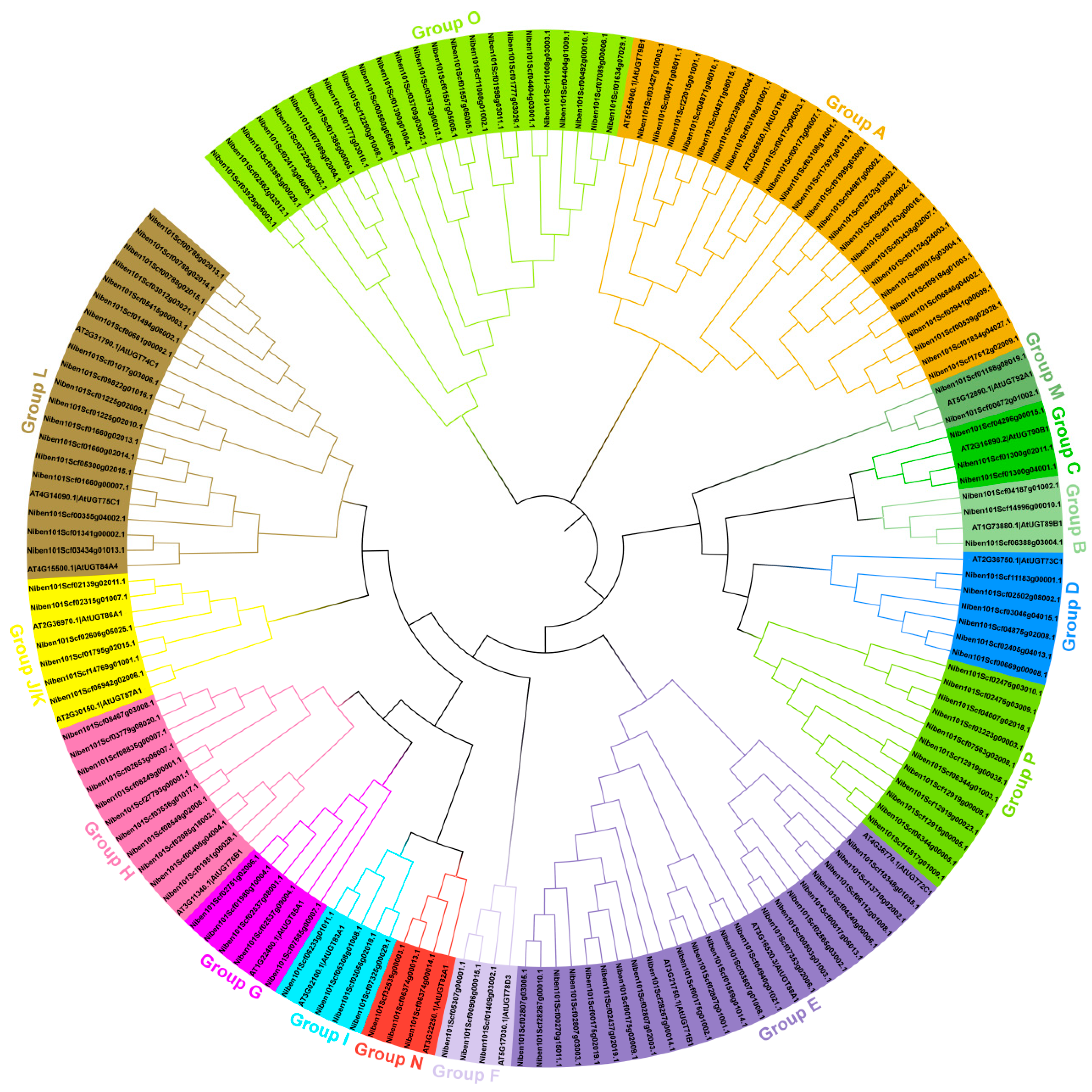
3.1. Identification and Phylogenetic Analysis of NbUGTs
3.2. Prediction of the Subcellular Localization and Physicochemical Properties of NbUGTs
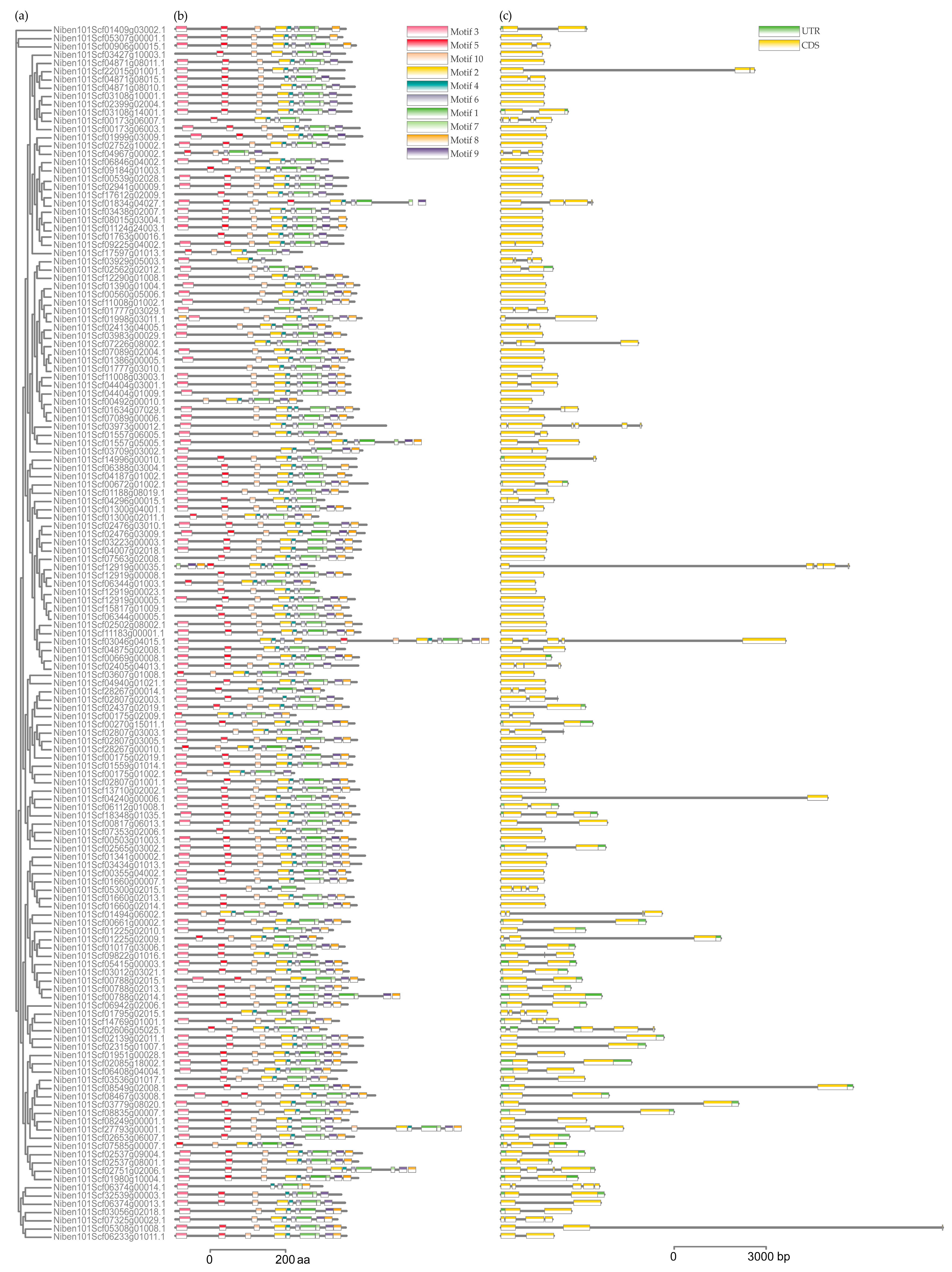
3.3. Gene Structure and Conserved Motifs Analysis of NbUGTs
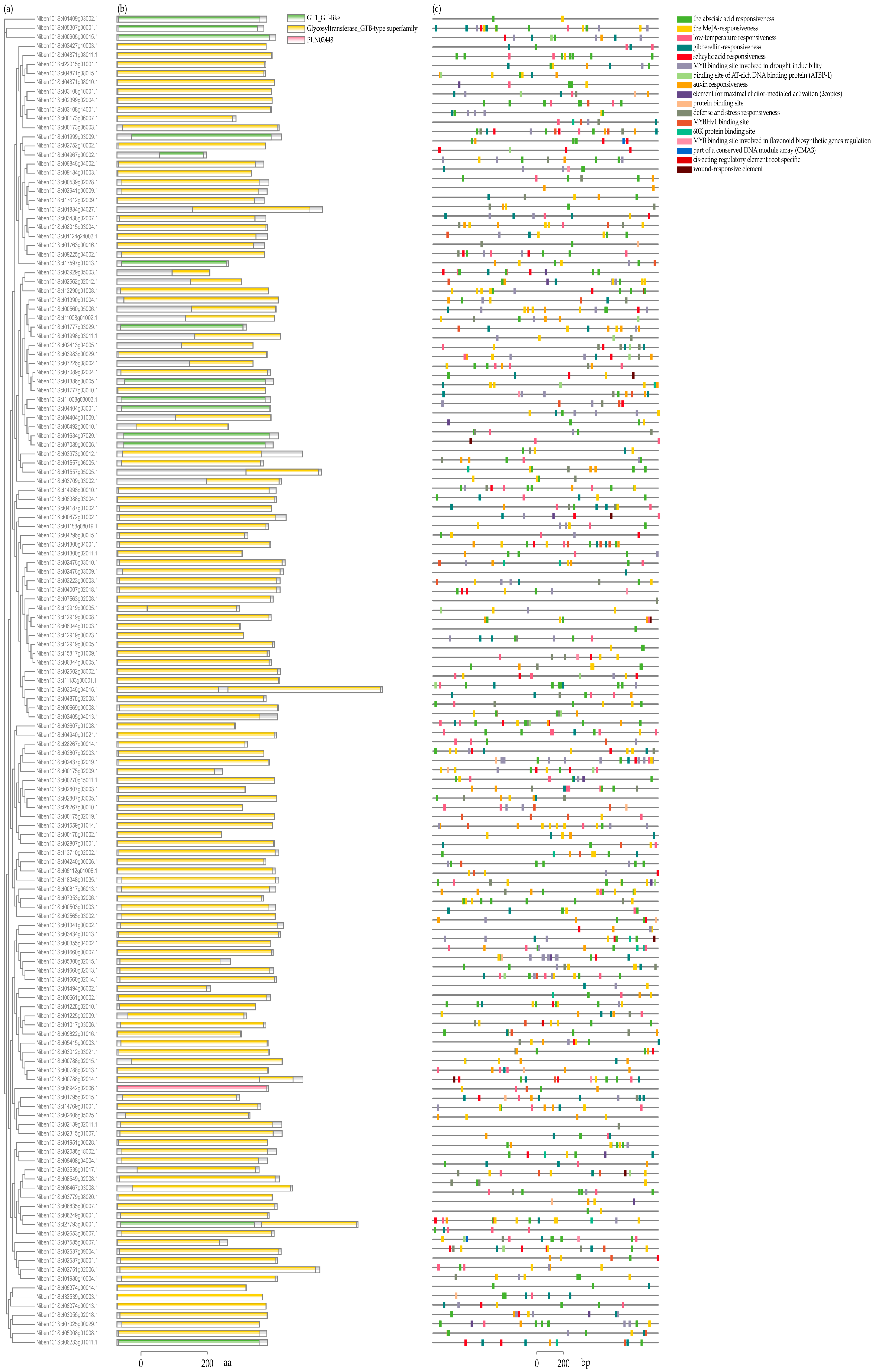
3.4. Identification of Conserved Domains and Cis-Acting Elements in the Putative Promoter of NbUGTs
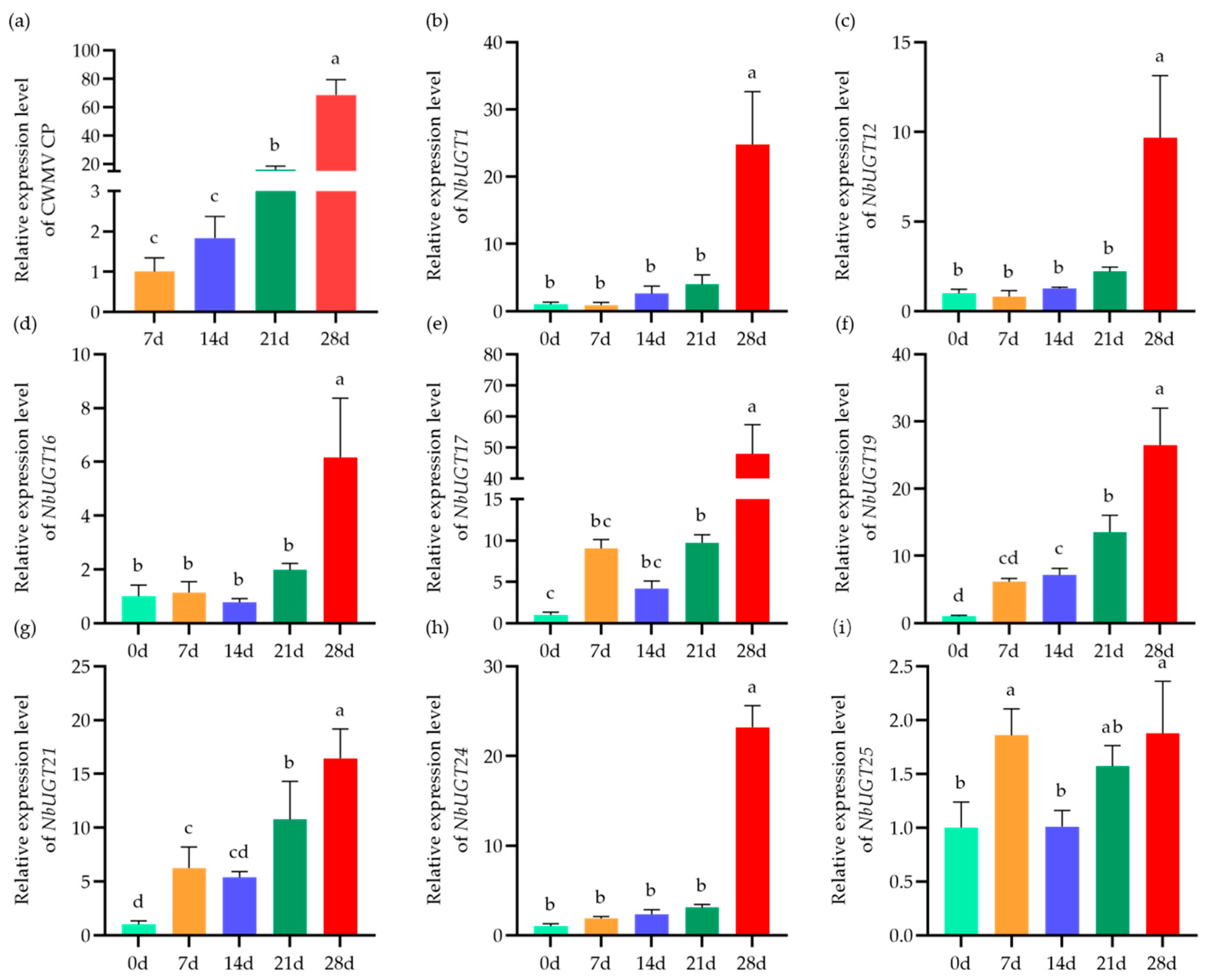
3.5. Expression Patterns of NbUGTs during CWMV Infection
3.6. Subcellular Localization of NbUGT12, NbUGT16, and NbUGT17
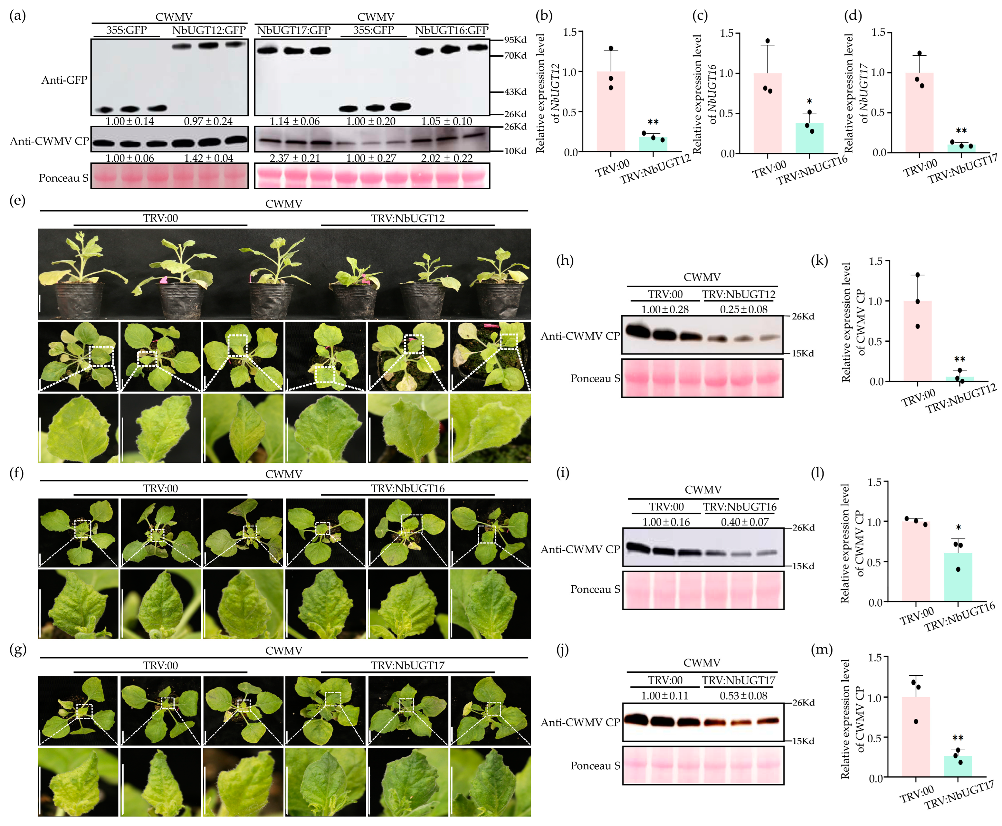
3.7. NbUGT12, NbUGT16, and NbUGT17 Positively Regulate CWMV Infection in N. benthamiana
4. Discussion
5. Conclusions
Supplementary Materials
Author Contributions
Funding
Institutional Review Board Statement
Informed Consent Statement
Data Availability Statement
Conflicts of Interest
References
- Caputi, L.; Malnoy, M.; Goremykin, V.; Nikiforova, S.; Martens, S. A genome-wide phylogenetic reconstruction of family 1 UDP-glycosyltransferases revealed the expansion of the family during the adaptation of plants to life on land. Plant J. 2012, 69, 1030–1042. [Google Scholar] [CrossRef] [PubMed]
- Tiwari, P.; Sangwan, R.S.; Sangwan, N.S. Plant secondary metabolism linked glycosyltransferases: An update on expanding knowledge and scopes. Biotechnol. Adv. 2016, 34, 714–739. [Google Scholar] [CrossRef] [PubMed]
- Lim, E.K.; Bowles, D.J. A class of plant glycosyltransferases involved in cellular homeostasis. EMBO J. 2004, 23, 2912–2955. [Google Scholar] [CrossRef] [PubMed]
- Rehman, H.M.; Nawaz, M.A.; Shah, Z.H.; Ludwig-Müller, J.; Chung, G.; Ahmad, M.Q.; Yang, S.H.; Lee, S.I. Comparative genomic and transcriptomic analyses of Family-1 UDP glycosyltransferase in three Brassica species and Arabidopsis indicates stress-responsive regulation. Sci. Rep. 2018, 8, 1875. [Google Scholar] [CrossRef] [PubMed]
- Song, C.; Härtl, K.; McGraphery, K.; Hoffmann, T.; Schwab, W. Attractive but Toxic: Emerging Roles of Glycosidically Bound Volatiles and Glycosyltransferases Involved in Their Formation. Mol. Plant 2018, 11, 1225–1236. [Google Scholar] [CrossRef] [PubMed]
- Lairson, L.L.; Henrissat, B.; Davies, G.J.; Withers, S.G. Glycosyltransferases: Structures, functions, and mechanisms. Annu. Rev. Biochem. 2008, 77, 521–555. [Google Scholar] [CrossRef]
- Xiao, X.; Lu, Q.; Liu, R.; Gong, J.; Gong, W.; Liu, A.; Ge, Q.; Li, J.; Shang, H.; Li, P.; et al. Genome-wide characterization of the UDP-glycosyltransferase gene family in upland cotton. 3 Biotech 2019, 9, 453. [Google Scholar] [CrossRef]
- Ross, J.; Li, Y.; Lim, E.; Bowles, D.J. Higher plant glycosyltransferases. Genome Biol. 2001, 2, 3004.1–3004.6. [Google Scholar] [CrossRef]
- Vogt, T.; Jones, P. Glycosyltransferases in plant natural product synthesis: Characterization of a supergene family. Trends Plant Sci. 2000, 5, 380–386. [Google Scholar] [CrossRef]
- Bowles, D.; Isayenkova, J.; Lim, E.K.; Poppenberger, B. Glycosyltransferases: Managers of small molecules. Curr. Opin. Plant Biol. 2005, 8, 254–263. [Google Scholar] [CrossRef] [PubMed]
- Zhang, P.; Zhang, Z.; Zhang, L.; Wang, J.; Wu, C. Glycosyltransferase GT1 family: Phylogenetic distribution, substrates coverage, and representative structural features. Comput. Struct. Biotechnol. J. 2020, 18, 1383–1390. [Google Scholar] [CrossRef] [PubMed]
- Li, Y.; Baldauf, S.; Lim, E.K.; Bowles, D.J. Phylogenetic analysis of the UDP-glycosyltransferase multigene family of Arabidopsis thaliana. J. Biol. Chem. 2001, 276, 4338–4343. [Google Scholar] [CrossRef]
- Gachon, C.M.; Langlois-Meurinne, M.; Saindrenan, P. Plant secondary metabolism glycosyltransferases: The emerging functional analysis. Trends Plant Sci. 2005, 10, 542–549. [Google Scholar] [CrossRef] [PubMed]
- Nagatoshi, M.; Terasaka, K.; Nagatsu, A.; Mizukami, H. Iridoid-specific glucosyltransferase from Gardenia jasminoides. J. Biol. Chem. 2011, 286, 32866–32874. [Google Scholar] [CrossRef] [PubMed]
- Gharabli, H.; Della Gala, V.; Welner, D.H. The function of UDP-glycosyltransferases in plants and their possible use in crop protection. Biotechnol. Adv. 2023, 67, 108182. [Google Scholar] [CrossRef] [PubMed]
- Cheng, Y.; Zhang, J.; Shao, Y.; Xu, Y.; Ge, H.; Yu, B.; Wang, W. Enzyme-Catalyzed Glycosylation of Curcumin and Its Analogues by Glycosyltransferases from Bacillus subtilis ATCC 6633. Catalysts 2019, 9, 734. [Google Scholar] [CrossRef]
- Osmani, S.A.; Bak, S.; Imberty, A.; Olsen, C.E.; Møller, B.L. Catalytic key amino acids and UDP-sugar donor specificity of a plant glucuronosyltransferase, UGT94B1: Molecular modeling substantiated by site-specific mutagenesis and biochemical analyses. Plant Physiol. 2008, 148, 1295–1308. [Google Scholar] [CrossRef]
- Rahimi, S.; Kim, J.; Mijakovic, I.; Jung, K.H.; Choi, G.; Kim, S.C.; Kim, Y.J. Triterpenoid-biosynthetic UDP-glycosyltransferases from plants. Biotechnol. Adv. 2019, 37, 107394. [Google Scholar] [CrossRef]
- Wang, D.; Wang, J.; Shi, Y.; Li, R.; Fan, F.; Huang, Y.; Li, W.; Chen, N.; Huang, L.; Dai, Z.; et al. Elucidation of the complete biosynthetic pathway of the main triterpene glycosylation products of Panax notoginseng using a synthetic biology platform. Metab. Eng. 2020, 61, 131–140. [Google Scholar] [CrossRef]
- Zhang, Z.; Zhuo, X.; Yan, X.; Zhang, Q. Comparative Genomic and Transcriptomic Analyses of Family-1 UDP Glycosyltransferase in Prunus Mume. Int. J. Mol. Sci. 2018, 19, 3382. [Google Scholar] [CrossRef]
- Boachon, B.; Gamir, J.; Pastor, V.; Erb, M.; Dean, J.V.; Flors, V.; Mauch-Mani, B. Role of two UDP-Glycosyltransferases from the L group of arabidopsis in resistance against pseudomonas syringae. Eur. J. Plant Pathol. 2014, 139, 707–720. [Google Scholar] [CrossRef]
- Song, J.T.; Koo, Y.J.; Seo, H.S.; Kim, M.C.; Choi, Y.D.; Kim, J.H. Overexpression of AtSGT1, an Arabidopsis salicylic acid glucosyltransferase, leads to increased susceptibility to Pseudomonas syringae. Phytochemistry 2008, 69, 1128–1134. [Google Scholar] [CrossRef] [PubMed]
- Tezuka, D.; Matsuura, H.; Saburi, W.; Mori, H.; Imai, R. A Ubiquitously Expressed UDP-Glucosyltransferase, UGT74J1, Controls Basal Salicylic Acid Levels in Rice. Plants 2021, 10, 1875. [Google Scholar] [CrossRef] [PubMed]
- Chen, L.; Wang, W.S.; Wang, T.; Meng, X.F.; Chen, T.T.; Huang, X.X.; Li, Y.J.; Hou, B.K. Methyl Salicylate Glucosylation Regulates Plant Defense Signaling and Systemic Acquired Resistance. Plant Physiol. 2019, 180, 2167–2181. [Google Scholar] [CrossRef] [PubMed]
- Lim, C.E.; Choi, J.N.; Kim, I.A.; Lee, S.A.; Hwang, Y.S.; Lee, C.H.; Lim, J. Improved resistance to oxidative stress by a loss-of-function mutation in the Arabidopsis UGT71C1 gene. Mol. Cells 2008, 25, 368–375. [Google Scholar] [CrossRef] [PubMed]
- Campos, L.; López-Gresa, M.P.; Fuertes, D.; Bellés, J.M.; Rodrigo, I.; Lisón, P. Tomato glycosyltransferase Twi1 plays a role in flavonoid glycosylation and defence against virus. BMC Plant Biol. 2019, 19, 450. [Google Scholar] [CrossRef] [PubMed]
- Diao, A.; Chen, J.; Ye, R.; Zheng, T.; Yu, S.; Antoniw, J.F.; Adams, M.J. Complete sequence and genome properties of Chinese wheat mosaic virus, a new furovirus from China. J. Gen. Virol. 1999, 80, 1141–1145. [Google Scholar] [CrossRef]
- Chen, J. Occurrence of fungally transmitted wheat mosaic viruses in China. Ann. Appl. Biol. 1993, 123, 55–61. [Google Scholar]
- Ye, R.; Zheng, T.; Chen, J.; Diao, A.; Adams, M.J.; Yu, S.; Antoniw, F. Characterization and partial sequence of a new furovirus of wheat in China. Plant Pathol. 1999, 48, 379–387. [Google Scholar] [CrossRef]
- Guo, L.M.; He, J.; Li, J.; Chen, J.P.; Zhang, H.M. Chinese wheat mosaic virus: A long-term threat to wheat in China. J. Integr. Agric. 2019, 18, 821–829. [Google Scholar] [CrossRef]
- Kanyuka, K.; Ward, E.; Adams, M.J. Polymyxa graminis and the cereal viruses it transmits: A research challenge. Mol. Plant Pathol. 2003, 4, 393–406. [Google Scholar] [CrossRef] [PubMed]
- Yang, J.; Zhang, F.; Xie, L.; Song, X.J.; Li, J.; Chen, J.P.; Zhang, H.M. Functional identification of two minor capsid proteins from Chinese wheat mosaic virus using its infectious full-length cDNA clones. J. Gen. Virol. 2016, 97, 2441–2450. [Google Scholar] [CrossRef]
- Chong, J.; Baltz, R.; Schmitt, C.; Beffa, R.; Fritig, B.; Saindrenan, P. Downregulation of a pathogen-responsive tobacco UDP-Glc: Phenylpropanoid glucosyltransferase reduces scopoletin glucoside accumulation, enhances oxidative stress, and weakens virus resistance. Plant Cell 2002, 14, 1093–1107. [Google Scholar] [CrossRef] [PubMed]
- Matros, A.; Mock, H.P. Ectopic Expression of a UDP-Glucose: Phenylpropanoid Glucosyltransferase Leads to Increased Resistance of Transgenic Tobacco Plants against Infection with Potato Virus Y. Plant Cell Physiol. 2004, 45, 1185–1193. [Google Scholar] [CrossRef] [PubMed]
- Kumar, S.; Stecher, G.; Li, M.; Knyaz, C.; Tamura, K. MEGA X: Molecular Evolutionary Genetics Analysis across Computing Platforms. Mol. Biol. Evol. 2018, 35, 1547–1549. [Google Scholar] [CrossRef]
- Wilkins, M.R.; Gasteiger, E.; Bairoch, A.; Sanchez, J.C.; Williams, K.L.; Appel, R.D.; Hochstrasser, D.F. Protein identification and analysis tools in the ExPASy server. Methods Mol. Biol. 1999, 112, 531–552. [Google Scholar] [PubMed]
- Bailey, T.L.; Boden, M.; Buske, F.A.; Frith, M.; Grant, C.E.; Clementi, L.; Ren, J.; Li, W.W.; Noble, W.S. MEME SUITE: Tools for motif discovery and searching. Nucleic Acids Res. 2009, 37, W202–W208. [Google Scholar] [CrossRef] [PubMed]
- Lescot, M.; Déhais, P.; Thijs, G.; Marchal, K.; Moreau, Y.; Van de Peer, Y.; Rouzé, P.; Rombauts, S. PlantCARE, a database of plant cis-acting regulatory elements and a portal to tools for in silico analysis of promoter sequences. Nucleic Acids Res. 2002, 30, 325–327. [Google Scholar] [CrossRef]
- Li, J.; Feng, H.; Liu, S.; Liu, P.; Chen, X.; Yang, J.; He, L.; Yang, J.; Chen, J. Phosphorylated viral protein evades plant immunity through interfering the function of RNA-binding protein. PLoS Pathog. 2022, 18, e1010412. [Google Scholar] [CrossRef]
- Zhang, T.; Shi, C.; Hu, H.; Zhang, Z.; Wang, Z.; Chen, Z.; Feng, H.; Liu, P.; Guo, J.; Lu, Q.; et al. N6-methyladenosine RNA modification promotes viral genomic RNA stability and infection. Nat. Commun. 2022, 13, 6576. [Google Scholar] [CrossRef]
- Yang, J.; Chen, L.; Zhang, J.; Liu, P.; Chen, M.; Chen, Z.; Zhong, K.; Liu, J.; Chen, J.; Yang, J. TaTHI2 interacts with Ca2+-dependent protein kinase TaCPK5 to suppress virus infection by regulating ROS accumulation. Plant Biotechnol. J. 2023. [Google Scholar] [CrossRef]
- Liu, C.; Talbot, N.J.; Chen, X.L. Protein glycosylation during infection by plant pathogenic fungi. New Phytol. 2021, 230, 1329–1335. [Google Scholar] [CrossRef]
- Shumilina, J.; Kusnetsova, A.; Tsarev, A.; Janse van Rensburg, H.C.; Medvedev, S.; Demidchik, V.; Van den Ende, W.; Frolov, A. Glycation of Plant Proteins: Regulatory Roles and Interplay with Sugar Signalling? Int. J. Mol. Sci. 2019, 20, 2366. [Google Scholar] [CrossRef]
- Wenjin, Z.; Sheng, W.; Jian, Y.; Chuanzhi, K.; Luqi, H.; Lanping, G. Glycosylation of plant secondary metabolites: Regulating from chaos to harmony. Environ. Exp. Bot. 2021, 194, 104703. [Google Scholar]
- Yonekura-Sakakibara, K.; Hanada, K. An evolutionary view of functional diversity in family 1 glycosyltransferases. Plant J. 2011, 66, 182–193. [Google Scholar] [CrossRef]
- Chang, A.; Singh, S.; Phillips, G.N., Jr.; Thorson, J.S. Glycosyltransferase structural biology and its role in the design of catalysts for glycosylation. Curr. Opin. Biotechnol. 2011, 22, 800–808. [Google Scholar] [CrossRef]
- Vrielink, A.; Rüger, W.; Driessen, H.P.; Freemont, P.S. Crystal structure of the DNA modifying enzyme beta-glucosyltransferase in the presence and absence of the substrate uridine diphosphoglucose. Embo J. 1994, 13, 3413–3422. [Google Scholar] [CrossRef]
- Blanchard, S.; Thorson, J.S. Enzymatic tools for engineering natural product glycosylation. Curr. Opin. Chem. Biol. 2006, 10, 263–271. [Google Scholar] [CrossRef] [PubMed]
- Mackenzie, P.I.; Owens, I.S.; Burchell, B.; Bock, K.W.; Bairoch, A.; Bélanger, A.; Fournel-Gigleux, S.; Green, M.; Hum, D.W.; Iyanagi, T.; et al. The UDP glycosyltransferase gene superfamily: Recommended nomenclature update based on evolutionary divergence. Pharmacogenetics 1997, 7, 255–269. [Google Scholar] [CrossRef] [PubMed]
- Meech, R.; Mackenzie, P.I. Structure and function of uridine diphosphate glucuronosyltransferases. Clin. Exp. Pharmacol. Physiol. 1997, 24, 907–915. [Google Scholar] [CrossRef] [PubMed]
- Chen, Y.; Fu, M.; Li, H.; Wang, L.; Liu, R.; Liu, Z. Genome-wide characterization of the UDP-glycosyltransferase gene family reveals their potential roles in leaf senescence in cotton. Int. J. Biol. Macromol. 2022, 222, 2648–2660. [Google Scholar] [CrossRef]
- Li, Y.; Li, P.; Wang, Y.; Dong, R.; Yu, H.; Hou, B. Genome-wide identification and phylogenetic analysis of Family-1 UDP glycosyltransferases in maize (Zea mays). Planta 2014, 239, 1265–1279. [Google Scholar] [CrossRef]
- Yu, G.; Chen, Q.; Chen, F.; Liu, H.; Lin, J.; Chen, R.; Ren, C.; Wei, J.; Zhang, Y.; Yang, F.; et al. Glutathione Promotes Degradation and Metabolism of Residual Fungicides by Inducing UDP-Glycosyltransferase Genes in Tomato. Front. Plant Sci. 2022, 13, 893508. [Google Scholar] [CrossRef]
- He, Y.; Ahmad, D.; Zhang, X.; Zhang, Y.; Wu, L.; Jiang, P.; Ma, H. Genome-wide analysis of family-1 UDP glycosyltransferases (UGT) and identification of UGT genes for FHB resistance in wheat (Triticum aestivum L.). BMC Plant Biol. 2018, 18, 67. [Google Scholar] [CrossRef]
- Dauda, W.P.; Shanmugam, V.; Tyagi, A.; Solanke, A.U.; Kumar, V.; Krishnan, S.G.; Bashyal, B.M.; Aggarwal, R. Genome-Wide Identification and Characterisation of Cytokinin-O-Glucosyltransferase (CGT) Genes of Rice Specific to Potential Pathogens. Plants 2022, 11, 917. [Google Scholar] [CrossRef]
- Elasad, M.; Wei, H.; Wang, H.; Su, J.; Ondati, E.; Yu, S. Genome-Wide Analysis and Characterization of the TRX Gene Family in Upland Cotton. Trop. Plant Biol. 2018, 11, 119–130. [Google Scholar] [CrossRef]
- Dooner, H.K.; Nelson, O.E. Controlling element-induced alterations in UDPglucose: Flavonoid glucosyltransferase, the enzyme specified by the bronze locus in maize. Proc. Natl. Acad. Sci. USA 1977, 74, 5623–5627. [Google Scholar] [CrossRef] [PubMed]
- Ao, B.; Han, Y.; Wang, S.; Wu, F.; Zhang, J. Genome-Wide Analysis and Profile of UDP-Glycosyltransferases Family in Alfalfa (Medicago sativa L.) under Drought Stress. Int. J. Mol. Sci. 2022, 23, 7243. [Google Scholar] [CrossRef]
- Dong, L.; Tang, Z.; Yang, T.; Hao, F.; Deng, X. Genome-Wide Analysis of UGT Genes in Petunia and Identification of PhUGT51 Involved in the Regulation of Salt Resistance. Plants 2022, 11, 2434. [Google Scholar] [CrossRef]
- He, Q.; Yin, H.; Jiang, J.; Bai, Y.; Chen, N.; Liu, S.; Zhuang, Y.; Liu, T. Fermentative Production of Phenolic Glucosides by Escherichia coli with an Engineered Glucosyltransferase from Rhodiola sachalinensis. J. Agric. Food Chem. 2017, 65, 4691–4697. [Google Scholar] [CrossRef]
- Zheng, S.W.; Chen, Z.F.; Liu, T.T.; Zhao, Z.Y.; Li, T.M.; Xing, G.M. Identification and Characterization of the Tomato UGT Gene Family and Effects of GAME 17 Overexpression on Plants and Growth and Development under High-CO2 Conditions. Agronomy 2022, 12, 1998. [Google Scholar] [CrossRef]
- Song, C.; Hong, X.; Zhao, S.; Liu, J.; Schulenburg, K.; Huang, F.C.; Franz-Oberdorf, K.; Schwab, W. Glucosylation of 4-Hydroxy-2,5-Dimethyl-3(2H)-Furanone, the Key Strawberry Flavor Compound in Strawberry Fruit. Plant Physiol. 2016, 171, 139–151. [Google Scholar] [CrossRef] [PubMed]
- Su, X.; Shen, G.; Di, S.; Dixon, R.A.; Pang, Y. Characterization of UGT716A1 as a Multi-substrate UDP: Flavonoid Glucosyltransferase Gene in Ginkgo biloba. Front. Plant Sci. 2017, 8, 2085. [Google Scholar] [CrossRef] [PubMed]
- Yang, C.; Li, C.; Wei, W.; Wei, Y.; Liu, Q.; Zhao, G.; Yue, J.; Yan, X.; Wang, P.; Zhou, Z. The unprecedented diversity of UGT94-family UDP-glycosyltransferases in Panax plants and their contribution to ginsenoside biosynthesis. Sci. Rep. 2020, 10, 15394. [Google Scholar] [CrossRef] [PubMed]
- Holmes, E.C.; Chen, Y.C.; Mudgett, M.B.; Sattely, E.S. Arabidopsis UGT76B1 glycosylates N-hydroxy-pipecolic acid and inactivates systemic acquired resistance in tomato. Plant Cell 2021, 33, 750–765. [Google Scholar] [CrossRef]
- Huang, X.X.; Wang, Y.; Lin, J.S.; Chen, L.; Li, Y.J.; Liu, Q.; Wang, G.F.; Xu, F.; Liu, L.; Hou, B.K. The novel pathogen-responsive glycosyltransferase UGT73C7 mediates the redirection of phenylpropanoid metabolism and promotes SNC1-dependent Arabidopsis immunity. Plant J. 2021, 107, 149–165. [Google Scholar] [CrossRef] [PubMed]
- Dare, A.P.; Yauk, Y.K.; Tomes, S.; McGhie, T.K.; Rebstock, R.S.; Cooney, J.M.; Atkinson, R.G. Silencing a phloretin-specific glycosyltransferase perturbs both general phenylpropanoid biosynthesis and plant development. Plant J. 2017, 91, 237–250. [Google Scholar] [CrossRef] [PubMed]
- Hettwer, K.; Böttcher, C.; Frolov, A.; Mittasch, J.; Albert, A.; von Roepenack-Lahaye, E.; Strack, D.; Milkowski, C. Dynamic metabolic changes in seeds and seedlings of Brassica napus (oilseed rape) suppressing UGT84A9 reveal plasticity and molecular regulation of the phenylpropanoid pathway. Phytochemistry 2016, 124, 46–57. [Google Scholar] [CrossRef] [PubMed]
- Saxe, H.J.; Horibe, T.; Balan, B.; Butterfield, T.S.; Feinberg, N.G.; Zabaneh, C.M.; Jacobson, A.E.; Dandekar, A.M. Two UGT84A Family Glycosyltransferases Regulate Phenol, Flavonoid, and Tannin Metabolism in Juglans regia (English Walnut). Front. Plant Sci. 2021, 12, 626483. [Google Scholar] [CrossRef]
- Bowles, D.; Lim, E.K.; Poppenberger, B.; Vaistij, F.E. Glycosyltransferases of lipophilic small molecules. Annu. Rev. Plant Biol. 2006, 57, 567–597. [Google Scholar] [CrossRef]
- Fukuchi-Mizutani, M.; Okuhara, H.; Fukui, Y.; Nakao, M.; Katsumoto, Y.; Yonekura-Sakakibara, K.; Kusumi, T.; Hase, T.; Tanaka, Y. Biochemical and molecular characterization of a novel UDP-glucose: Anthocyanin 3′-O-glucosyltransferase, a key enzyme for blue anthocyanin biosynthesis, from gentian. Plant Physiol. 2003, 132, 1652–1663. [Google Scholar] [CrossRef]
- Dong, N.Q.; Lin, H.X. Contribution of phenylpropanoid metabolism to plant development and plant-environment interactions. J. Integr. Plant Biol. 2021, 63, 180–209. [Google Scholar] [CrossRef]
- Muro-Villanueva, F.; Mao, X.; Chapple, C. Linking phenylpropanoid metabolism, lignin deposition, and plant growth inhibition. Curr. Opin. Biotechnol. 2019, 56, 202–208. [Google Scholar] [CrossRef]
- Wang, T.; Ma, Y.Q.; Huang, X.X.; Mu, T.J.; Li, Y.J.; Li, X.K.; Liu, X.; Hou, B.K. Overexpression of OsUGT3 enhances drought and salt tolerance through modulating ABA synthesis and scavenging ROS in rice. Environ. Exp. Bot. 2021, 192, 104653. [Google Scholar] [CrossRef]
- Haroth, S.; Feussner, K.; Kelly, A.A.; Zienkiewicz, K.; Shaikhqasem, A.; Herrfurth, C.; Feussner, I. The glycosyltransferase UGT76E1 significantly contributes to 12-O-glucopyranosyl-jasmonic acid formation in wounded Arabidopsis thaliana leaves. J. Biol. Chem. 2019, 294, 9858–9872. [Google Scholar] [CrossRef] [PubMed]
- Mohnike, L.; Rekhter, D.; Huang, W.; Feussner, K.; Tian, H.; Herrfurth, C.; Zhang, Y.; Feussner, I. The glycosyltransferase UGT76B1 modulates N-hydroxy-pipecolic acid homeostasis and plant immunity. Plant Cell 2021, 33, 735–749. [Google Scholar] [CrossRef]
- White, R.F. Acetylsalicylic acid (aspirin) induces resistance to tobacco mosaic virus in tobacco. Virology 1979, 99, 410–412. [Google Scholar] [CrossRef]
- An, C.; Mou, Z. Salicylic acid and its function in plant immunity. J. Integr. Plant Biol. 2011, 53, 412–428. [Google Scholar] [CrossRef]
- Fu, Z.Q.; Dong, X. Systemic acquired resistance: Turning local infection into global defense. Annu. Rev. Plant Biol. 2013, 64, 839–863. [Google Scholar] [CrossRef] [PubMed]
- Ryals, J.; Uknes, S.; Ward, E. Systemic Acquired Resistance. Plant Physiol. 1994, 104, 1109–1112. [Google Scholar] [CrossRef] [PubMed]
- Liu, L.; Sonbol, F.M.; Huot, B.; Gu, Y.; Withers, J.; Mwimba, M.; Yao, J.; He, S.Y.; Dong, X. Salicylic acid receptors activate jasmonic acid signalling through a non-canonical pathway to promote effector-triggered immunity. Nat. Commun. 2016, 7, 13099. [Google Scholar] [CrossRef] [PubMed]
- Vos, I.A.; Moritz, L.; Pieterse, C.M.; Van Wees, S.C. Impact of hormonal crosstalk on plant resistance and fitness under multi-attacker conditions. Front. Plant Sci. 2015, 6, 639. [Google Scholar] [CrossRef] [PubMed]
- Van Butselaar, T.; Van den Ackerveken, G. Salicylic Acid Steers the Growth-Immunity Tradeoff. Trends Plant Sci. 2020, 25, 566–576. [Google Scholar] [CrossRef] [PubMed]


| Gene Stable ID/Locus Name | MW (kDa) | PL | pI | CDS Length/bp | IN | SL |
|---|---|---|---|---|---|---|
| Niben101Scf00173g06003.1 | 55.25088 | 489 | 6.01 | 1470 | 0 | Cytoplasmic PlasmaMembrane |
| Niben101Scf00173g06007.1 | 40.97301 | 359 | 6.94 | 1080 | 4 | Cytoplasmic |
| Niben101Scf00175g01002.1 | 36.15161 | 315 | 5.61 | 948 | 0 | PlasmaMembrane |
| Niben101Scf00175g02009.1 | 35.93907 | 319 | 4.92 | 960 | 1 | Cytoplasmic |
| Niben101Scf00175g02019.1 | 53.26710 | 475 | 5.16 | 1428 | 1 | Cytoplasmic |
| Niben101Scf00270g15011.1 | 53.74701 | 475 | 5.49 | 1428 | 1 | PlasmaMembrane |
| Niben101Scf00355g04002.1 | 52.24085 | 464 | 5.32 | 1395 | 0 | Cytoplasmic |
| Niben101Scf00492g00010.1 | 38.22123 | 336 | 7.09 | 1011 | 0 | Cytoplasmic |
| Niben101Scf00503g01003.1 | 52.50974 | 478 | 5.43 | 1437 | 0 | Cytoplasmic |
| Niben101Scf00539g02028.1 | 52.55631 | 458 | 7.43 | 1377 | 0 | Cytoplasmic |
| Niben101Scf00560g05006.1 | 53.88507 | 480 | 6.77 | 1443 | 0 | PlasmaMembrane |
| Niben101Scf00661g00002.1 | 51.93085 | 463 | 6.93 | 1392 | 1 | Chloroplast Cytoplasmic |
| Niben101Scf00669g00008.1 | 55.45511 | 487 | 5.77 | 1464 | 0 | Cytoplasmic |
| Niben101Scf00672g01002.1 | 57.90343 | 510 | 6.37 | 1533 | 1 | Cytoplasmic PlasmaMembrane |
| Niben101Scf00788g02013.1 | 51.60161 | 457 | 5.37 | 1374 | 1 | Cytoplasmic |
| Niben101Scf00788g02014.1 | 63.31325 | 561 | 5.77 | 1686 | 1 | Cytoplasmic |
| Niben101Scf00788g02015.1 | 56.78464 | 500 | 5.53 | 1503 | 1 | PlasmaMembrane Cytoplasmic Chloroplast |
| Niben101Scf00817g06013.1 | 53.06923 | 479 | 5.68 | 1440 | 1 | Cytoplasmic |
| Niben101Scf00906g00015.1 | 53.08143 | 479 | 6.50 | 1440 | 1 | Chloroplast Cytoplasmic |
| Niben101Scf01017g03006.1 | 50.54148 | 449 | 4.69 | 1350 | 1 | Cytoplasmic |
| Niben101Scf01124g24003.1 | 51.36213 | 454 | 5.38 | 1365 | 0 | Cytoplasmic |
| Niben101Scf01188g08019.1 | 50.64306 | 457 | 6.37 | 1374 | 1 | Cytoplasmic |
| Niben101Scf01225g02009.1 | 43.94214 | 390 | 6.48 | 1173 | 2 | Cytoplasmic |
| Niben101Scf01225g02010.1 | 46.67563 | 418 | 4.83 | 1257 | 1 | Cytoplasmic |
| Niben101Scf01300g02011.1 | 43.07863 | 379 | 6.63 | 1140 | 0 | Chloroplast |
| Niben101Scf01300g04001.1 | 52.21414 | 464 | 5.02 | 1395 | 0 | Cytoplasmic |
| Niben101Scf01341g00002.1 | 56.11798 | 503 | 6.08 | 1512 | 0 | Cytoplasmic |
| Niben101Scf01386g00005.1 | 53.27002 | 472 | 6.08 | 1419 | 0 | Cytoplasmic PlasmaMembrane |
| Niben101Scf01390g01004.1 | 55.40395 | 488 | 6.45 | 1467 | 0 | PlasmaMembrane Cytoplasmic |
| Niben101Scf01409g03002.1 | 49.66975 | 452 | 4.88 | 1359 | 1 | Cytoplasmic |
| Niben101Scf01494g06002.1 | 32.08006 | 282 | 5.25 | 849 | 3 | Cytoplasmic |
| Niben101Scf01557g05005.1 | 69.25793 | 616 | 6.18 | 1851 | 1 | Cytoplasmic |
| Niben101Scf01557g06005.1 | 49.85595 | 441 | 6.53 | 1326 | 1 | Cytoplasmic |
| Niben101Scf01559g01014.1 | 52.66307 | 469 | 5.86 | 1410 | 0 | Cytoplasmic |
| Niben101Scf01634g07029.1 | 55.20606 | 487 | 7.86 | 1464 | 1 | Cytoplasmic Mitochondrial |
| Niben101Scf01660g00007.1 | 52.94962 | 471 | 5.15 | 1416 | 0 | Cytoplasmic |
| Niben101Scf01660g02013.1 | 53.50339 | 473 | 6.01 | 1422 | 0 | Cytoplasmic PlasmaMembrane |
| Niben101Scf01660g02014.1 | 54.15285 | 481 | 5.61 | 1446 | 0 | PlasmaMembrane |
| Niben101Scf01763g00016.1 | 50.55943 | 445 | 5.84 | 1338 | 0 | Cytoplasmic |
| Niben101Scf01777g03010.1 | 50.47786 | 448 | 6.63 | 1347 | 0 | Chloroplast Cytoplasmic |
| Niben101Scf01777g03029.1 | 43.98762 | 390 | 7.11 | 1173 | 2 | Cytoplasmic Mitochondrial |
| Niben101Scf01795g02015.1 | 41.70329 | 370 | 6.58 | 1113 | 3 | Cytoplasmic Nuclear |
| Niben101Scf01834g04027.1 | 70.89713 | 619 | 6.19 | 1860 | 3 | Cytoplasmic |
| Niben101Scf01951g00028.1 | 51.00416 | 454 | 5.93 | 1365 | 1 | PlasmaMembrane Cytoplasmic |
| Niben101Scf01980g10004.1 | 54.34053 | 485 | 5.23 | 1458 | 1 | Cytoplasmic |
| Niben101Scf01998g03011.1 | 56.29511 | 494 | 6.37 | 1485 | 1 | Cytoplasmic |
| Niben101Scf01999g03009.1 | 56.56114 | 496 | 5.49 | 1491 | 0 | Cytoplasmic |
| Niben101Scf02085g18002.1 | 54.48497 | 481 | 5.11 | 1446 | 1 | Cytoplasmic |
| Niben101Scf02139g02011.1 | 56.47553 | 497 | 6.47 | 1494 | 1 | Cytoplasmic |
| Niben101Scf02315g01007.1 | 56.55771 | 498 | 6.35 | 1497 | 1 | Cytoplasmic |
| Niben101Scf02399g02004.1 | 53.54442 | 468 | 4.94 | 1407 | 0 | Cytoplasmic |
| Niben101Scf02405g04013.1 | 55.28666 | 485 | 7.44 | 1458 | 3 | Cytoplasmic |
| Niben101Scf02413g04005.1 | 47.04465 | 411 | 6.62 | 1236 | 1 | Cytoplasmic |
| Niben101Scf02437g02019.1 | 51.83977 | 460 | 5.37 | 1383 | 1 | OuterMembrane Cytoplasmic |
| Niben101Scf02476g03009.1 | 56.22729 | 502 | 5.14 | 1509 | 0 | Cytoplasmic OuterMembrane |
| Niben101Scf02476g03010.1 | 57.12081 | 507 | 6.15 | 1524 | 0 | Cytoplasmic |
| Niben101Scf02502g08002.1 | 54.26894 | 494 | 6.38 | 1485 | 0 | OuterMembrane Cytoplasmic |
| Niben101Scf02537g08001.1 | 54.44675 | 485 | 5.42 | 1458 | 1 | Cytoplasmic |
| Niben101Scf02537g09004.1 | 55.60343 | 495 | 5.72 | 1488 | 1 | Cytoplasmic |
| Niben101Scf02562g02012.1 | 42.27469 | 376 | 5.59 | 1131 | 1 | Cytoplasmic |
| Niben101Scf02565g03002.1 | 53.48191 | 478 | 5.63 | 1437 | 1 | Cytoplasmic |
| Niben101Scf02606g05025.1 | 45.87567 | 401 | 5.95 | 1206 | 5 | PlasmaMembrane |
| Niben101Scf02653g06007.1 | 53.47655 | 474 | 5.28 | 1425 | 1 | PlasmaMembrane Cytoplasmic |
| Niben101Scf02751g02006.1 | 69.02013 | 612 | 5.37 | 1839 | 3 | Cytoplasmic |
| Niben101Scf02752g10002.1 | 50.66152 | 449 | 6.34 | 1350 | 0 | PlasmaMembrane Cytoplasmic Chloroplast |
| Niben101Scf02807g01001.1 | 53.25338 | 475 | 5.37 | 1428 | 0 | Chloroplast |
| Niben101Scf02807g02003.1 | 49.35174 | 443 | 5.11 | 1332 | 2 | Cytoplasmic |
| Niben101Scf02807g03003.1 | 43.19847 | 387 | 5.21 | 1164 | 2 | Cytoplasmic Chloroplast |
| Niben101Scf02807g03005.1 | 53.50874 | 482 | 5.50 | 1449 | 0 | Cytoplasmic |
| Niben101Scf02941g00009.1 | 51.88057 | 453 | 7.60 | 1362 | 0 | Cytoplasmic Mitochondrial Nuclear |
| Niben101Scf03012g03021.1 | 51.94987 | 460 | 5.27 | 1383 | 1 | Cytoplasmic |
| Niben101Scf03046g04015.1 | 91.45533 | 801 | 6.10 | 2406 | 4 | Cytoplasmic |
| Niben101Scf03056g02018.1 | 49.93344 | 454 | 5.11 | 1365 | 1 | PlasmaMembrane |
| Niben101Scf03108g10001.1 | 53.36225 | 466 | 5.09 | 1401 | 0 | Cytoplasmic |
| Niben101Scf03108g14001.1 | 53.57633 | 467 | 6.12 | 1404 | 1 | Cytoplasmic |
| Niben101Scf03223g00003.1 | 55.78762 | 492 | 6.43 | 1479 | 0 | Cytoplasmic |
| Niben101Scf03427g10003.1 | 50.63953 | 450 | 6.14 | 1353 | 0 | Cytoplasmic |
| Niben101Scf03434g01013.1 | 55.40669 | 493 | 5.77 | 1482 | 0 | Cytoplasmic |
| Niben101Scf03438g02007.1 | 50.72439 | 449 | 6.63 | 1350 | 0 | Cytoplasmic |
| Niben101Scf03536g01017.1 | 48.24982 | 429 | 6.51 | 1290 | 1 | Cytoplasmic |
| Niben101Scf03607g01008.1 | 40.13816 | 358 | 5.06 | 1077 | 0 | Cytoplasmic |
| Niben101Scf03709g03002.1 | 55.30632 | 496 | 5.40 | 1491 | 1 | Cytoplasmic |
| Niben101Scf03779g08020.1 | 53.47131 | 470 | 5.46 | 1413 | 1 | Cytoplasmic |
| Niben101Scf03929g05003.1 | 31.39917 | 280 | 5.54 | 843 | 3 | Cytoplasmic |
| Niben101Scf03973g00012.1 | 62.75820 | 559 | 7.52 | 1680 | 5 | Mitochondrial Cytoplasmic |
| Niben101Scf03983g00029.1 | 51.55136 | 453 | 6.14 | 1362 | 0 | Cytoplasmic |
| Niben101Scf04007g02018.1 | 55.76853 | 492 | 6.33 | 1479 | 0 | Cytoplasmic |
| Niben101Scf04187g01002.1 | 51.39185 | 467 | 6.53 | 1404 | 0 | Cytoplasmic |
| Niben101Scf04240g00006.1 | 49.98282 | 449 | 6.17 | 1350 | 1 | Mitochondrial Chloroplast Cytoplasmic |
| Niben101Scf04296g00015.1 | 44.93744 | 395 | 7.50 | 1188 | 1 | PlasmaMembrane |
| Niben101Scf04404g01009.1 | 52.33412 | 465 | 8.97 | 1398 | 0 | Chloroplast Mitochondrial |
| Niben101Scf04404g03001.1 | 52.62037 | 464 | 9.76 | 1395 | 1 | PlasmaMembrane |
| Niben101Scf04871g08010.1 | 52.96204 | 476 | 5.68 | 1431 | 0 | Cytoplasmic Chloroplast |
| Niben101Scf04871g08011.1 | 52.55898 | 468 | 6.30 | 1407 | 0 | Cytoplasmic |
| Niben101Scf04871g08015.1 | 49.54030 | 448 | 6.70 | 1347 | 1 | Cytoplasmic Chloroplast |
| Niben101Scf04875g02008.1 | 50.66160 | 450 | 4.83 | 1353 | 1 | Cytoplasmic |
| Niben101Scf04940g01021.1 | 53.47241 | 481 | 5.54 | 1446 | 0 | PlasmaMembrane |
| Niben101Scf04967g00002.1 | 30.55219 | 270 | 7.13 | 813 | 2 | Cytoplasmic |
| Niben101Scf05300g02015.1 | 39.04028 | 342 | 4.95 | 1029 | 3 | PlasmaMembrane |
| Niben101Scf05307g00001.1 | 48.83847 | 443 | 5.93 | 1332 | 0 | Cytoplasmic Chloroplast PlasmaMembrane |
| Niben101Scf05308g01008.1 | 50.29414 | 452 | 5.46 | 1359 | 2 | Cytoplasmic |
| Niben101Scf05415g00003.1 | 51.39926 | 456 | 5.46 | 1371 | 1 | PlasmaMembrane Cytoplasmic |
| Niben101Scf06112g01008.1 | 53.47046 | 477 | 5.97 | 1434 | 1 | Cytoplasmic |
| Niben101Scf06233g01011.1 | 50.64607 | 454 | 6.34 | 1365 | 1 | PlasmaMembrane |
| Niben101Scf06344g00005.1 | 52.51495 | 466 | 5.31 | 1401 | 0 | Cytoplasmic |
| Niben101Scf06344g01003.1 | 41.46452 | 372 | 4.80 | 1119 | 0 | Cytoplasmic |
| Niben101Scf06374g00013.1 | 50.73411 | 450 | 6.50 | 1353 | 1 | PlasmaMembrane |
| Niben101Scf06374g00014.1 | 43.93319 | 390 | 6.65 | 1173 | 4 | PlasmaMembrane |
| Niben101Scf06388g03004.1 | 53.42717 | 481 | 6.25 | 1446 | 0 | Cytoplasmic |
| Niben101Scf06408g04004.1 | 51.19207 | 454 | 4.87 | 1365 | 1 | Cytoplasmic Nuclear |
| Niben101Scf06846g04002.1 | 49.89490 | 443 | 6.31 | 1332 | 0 | Cytoplasmic |
| Niben101Scf06942g02006.1 | 51.59511 | 457 | 6.20 | 1374 | 1 | Cytoplasmic |
| Niben101Scf07089g00006.1 | 53.24665 | 471 | 8.53 | 1416 | 0 | Cytoplasmic Mitochondrial |
| Niben101Scf07089g02004.1 | 52.01572 | 463 | 5.61 | 1392 | 0 | Chloroplast PlasmaMembrane Cytoplasmic |
| Niben101Scf07226g08002.1 | 46.71687 | 411 | 6.61 | 1236 | 3 | PlasmaMembrane |
| Niben101Scf07325g00029.1 | 47.18517 | 430 | 5.98 | 1293 | 3 | PlasmaMembrane |
| Niben101Scf07353g02006.1 | 49.38242 | 442 | 6.55 | 1329 | 0 | Cytoplasmic Mitochondrial |
| Niben101Scf07563g02008.1 | 52.72471 | 471 | 6.09 | 1416 | 0 | Cytoplasmic |
| Niben101Scf07585g00007.1 | 37.81300 | 334 | 6.03 | 1005 | 2 | PlasmaMembrane Cytoplasmic |
| Niben101Scf08015g03004.1 | 51.62843 | 454 | 5.53 | 1365 | 0 | Cytoplasmic PlasmaMembrane |
| Niben101Scf08249g00001.1 | 52.34963 | 459 | 6.36 | 1380 | 1 | Nuclear Cytoplasmic PlasmaMembrane |
| Niben101Scf08467g03008.1 | 60.48347 | 530 | 5.98 | 1593 | 1 | Cytoplasmic Nuclear |
| Niben101Scf08549g02008.1 | 55.34183 | 490 | 5.31 | 1473 | 1 | PlasmaMembrane |
| Niben101Scf08835g00007.1 | 54.52800 | 483 | 6.48 | 1452 | 1 | PlasmaMembrane Cytoplasmic Mitochondrial |
| Niben101Scf09184g01003.1 | 46.08446 | 405 | 6.05 | 1218 | 0 | Cytoplasmic |
| Niben101Scf09225g04002.1 | 50.10597 | 446 | 6.23 | 1341 | 1 | Cytoplasmic |
| Niben101Scf09822g01016.1 | 42.25115 | 376 | 5.79 | 1131 | 1 | Cytoplasmic |
| Niben101Scf11008g01002.1 | 53.83543 | 475 | 6.30 | 1428 | 0 | Cytoplasmic |
| Niben101Scf11008g03003.1 | 52.19999 | 464 | 8.58 | 1395 | 1 | PlasmaMembrane |
| Niben101Scf11183g00001.1 | 55.52863 | 491 | 6.48 | 1476 | 0 | Cytoplasmic PlasmaMembrane |
| Niben101Scf12290g01008.1 | 52.29987 | 458 | 6.16 | 1377 | 0 | PlasmaMembrane |
| Niben101Scf12919g00005.1 | 53.55560 | 476 | 6.43 | 1431 | 0 | Cytoplasmic |
| Niben101Scf12919g00008.1 | 51.96516 | 465 | 4.83 | 1398 | 0 | Cytoplasmic |
| Niben101Scf12919g00023.1 | 43.26769 | 381 | 6.20 | 1146 | 0 | Cytoplasmic |
| Niben101Scf12919g00035.1 | 41.70371 | 369 | 7.23 | 1110 | 5 | Cytoplasmic Chloroplast |
| Niben101Scf13710g02002.1 | 54.38508 | 488 | 5.78 | 1467 | 0 | Cytoplasmic |
| Niben101Scf14769g01001.1 | 48.72355 | 434 | 7.28 | 1305 | 3 | Cytoplasmic Mitochondrial |
| Niben101Scf14996g00010.1 | 53.55044 | 480 | 6.47 | 1443 | 1 | PlasmaMembrane |
| Niben101Scf15817g01009.1 | 51.89515 | 460 | 5.21 | 1383 | 0 | Cytoplasmic |
| Niben101Scf17597g01013.1 | 37.80270 | 336 | 5.01 | 1011 | 0 | Cytoplasmic Chloroplast |
| Niben101Scf17612g02009.1 | 50.56254 | 444 | 6.22 | 1335 | 0 | Cytoplasmic |
| Niben101Scf18348g01035.1 | 54.45771 | 488 | 5.11 | 1467 | 2 | PlasmaMembrane Cytoplasmic |
| Niben101Scf22015g01001.1 | 49.64530 | 449 | 6.96 | 1350 | 2 | Chloroplast Cytoplasmic |
| Niben101Scf27793g00001.1 | 82.62507 | 727 | 5.34 | 2184 | 2 | Cytoplasmic PlasmaMembrane |
| Niben101Scf28267g00010.1 | 42.14358 | 379 | 5.01 | 1140 | 0 | Cytoplasmic |
| Niben101Scf28267g00014.1 | 44.16199 | 394 | 6.58 | 1185 | 2 | Cytoplasmic |
| Niben101Scf32539g00003.1 | 49.55187 | 440 | 6.52 | 1323 | 1 | PlasmaMembrane |
Disclaimer/Publisher’s Note: The statements, opinions and data contained in all publications are solely those of the individual author(s) and contributor(s) and not of MDPI and/or the editor(s). MDPI and/or the editor(s) disclaim responsibility for any injury to people or property resulting from any ideas, methods, instructions or products referred to in the content. |
© 2024 by the authors. Licensee MDPI, Basel, Switzerland. This article is an open access article distributed under the terms and conditions of the Creative Commons Attribution (CC BY) license (https://creativecommons.org/licenses/by/4.0/).
Share and Cite
Wang, X.; Yang, J.; Hu, H.; Yuan, T.; Zhao, Y.; Liu, Y.; Li, W.; Liu, J. Genome-Wide Analysis and Identification of UDP Glycosyltransferases Responsive to Chinese Wheat Mosaic Virus Resistance in Nicotiana benthamiana. Viruses 2024, 16, 489. https://doi.org/10.3390/v16040489
Wang X, Yang J, Hu H, Yuan T, Zhao Y, Liu Y, Li W, Liu J. Genome-Wide Analysis and Identification of UDP Glycosyltransferases Responsive to Chinese Wheat Mosaic Virus Resistance in Nicotiana benthamiana. Viruses. 2024; 16(4):489. https://doi.org/10.3390/v16040489
Chicago/Turabian StyleWang, Xia, Jin Yang, Haichao Hu, Tangyu Yuan, Yingjie Zhao, Ying Liu, Wei Li, and Jiaqian Liu. 2024. "Genome-Wide Analysis and Identification of UDP Glycosyltransferases Responsive to Chinese Wheat Mosaic Virus Resistance in Nicotiana benthamiana" Viruses 16, no. 4: 489. https://doi.org/10.3390/v16040489
APA StyleWang, X., Yang, J., Hu, H., Yuan, T., Zhao, Y., Liu, Y., Li, W., & Liu, J. (2024). Genome-Wide Analysis and Identification of UDP Glycosyltransferases Responsive to Chinese Wheat Mosaic Virus Resistance in Nicotiana benthamiana. Viruses, 16(4), 489. https://doi.org/10.3390/v16040489