Review

A Balancing Act: The Viral–Host Battle over RNA Binding Proteins

by
Yahaira Bermudez
,
David Hatfield
and
Mandy Muller
*
Department of Microbiology, University of Massachusetts, Amherst, MA 01003, USA
*
Author to whom correspondence should be addressed.
Viruses 2024, 16(3), 474; https://doi.org/10.3390/v16030474
Submission received: 15 December 2023 / Revised: 16 March 2024 / Accepted: 18 March 2024 / Published: 20 March 2024
(This article belongs to the Special Issue Regulation of the Virus Lifecycle by Cellular RNA-Binding Proteins)

Abstract

:
A defining feature of a productive viral infection is the co-opting of host cell resources for viral replication. Despite the host repertoire of molecular functions and biological counter measures, viruses still subvert host defenses to take control of cellular factors such as RNA binding proteins (RBPs). RBPs are involved in virtually all steps of mRNA life, forming ribonucleoprotein complexes (mRNPs) in a highly ordered and regulated process to control RNA fate and stability in the cell. As such, the hallmark of the viral takeover of a cell is the reshaping of RNA fate to modulate host gene expression and evade immune responses by altering RBP interactions. Here, we provide an extensive review of work in this area, particularly on the duality of the formation of RNP complexes that can be either pro- or antiviral. Overall, in this review, we highlight the various ways viruses co-opt RBPs to regulate RNA stability and modulate the outcome of infection by gathering novel insights gained from research studies in this field.

1. Introduction

Viruses rely on intricate hijacking mechanisms to seize control of the host gene expression machinery for their own replication. Given the importance of RNA binding proteins (RBPs) in processes such as splicing, stability, localization, degradation, export, and translation, these proteins are at the center of this battle to control the gene expression resources [1,2,3]. Ribonucleoprotein complexes (RNPs) are assembled on mRNAs, and they reshape the fate of transcripts as these complexes are recruited via sequence motifs or sequence-independent secondary and tertiary structures [1,4]. Thus, the mRNA–protein interface can vary widely from forming stable RNP complexes to transient interactions. In infected cells, this is further complicated by the presence of viral proteins that may disrupt any number of these post-transcriptional processes. Furthermore, membrane-less organelles containing RNAs and RBPs are formed as compartmentalized densities that serve as important sites of host post-transcriptional regulation but can also be co-opted as viral replication factories [5,6,7]. While an extensive amount of work in the field has shown the importance of RBPs in the regulation of gene expression, and as a particular crux of targeting during viral infection [8,9,10,11], one important gap remains to be explored: how can the same RNP complexes be pro-viral in some contexts while antiviral in others? Studies portraying the impact of RNA–protein interactions during viral infection are an exciting avenue of research and will likely yield important information on transcript regulation. In this review, we will explore the complex interplay between RNA and proteins and their role(s) in RNA stability during viral infections, shedding light on new avenues to explore RNA fate and RBP modulation during viral infections in the future.

2. RNA Elements and Viral Infection

One strategy that viruses can use to redistribute gene expression resources is to induce broad RNA degradation events to support the needs of active viral translation. These RNA decay events are orchestrated directly by virally encoded endonucleases. Interestingly, recent work has begun to reveal RNA elements that are incorporated into select mRNAs that can drastically impact RNA stability in the face of these viral-induced decay events. Some of these RNA elements serve as target sites that recruit viral nucleases, while others can provide protective measures against viral-induced decay. For example, during Kaposi Sarcoma-Associated Herpesvirus (KSHV) lytic replication, the virally encoded endonuclease SOX causes widespread decay of the mRNA transcriptome [12]. However, a fraction of the transcriptome is spared from SOX-mediated degradation, which likely ensures the cell viability and proper progression of viral replication. Thus, it appears that SOX targets are carefully selected and in fact, the Gaglia group has recently mapped the SOX targeting motif, which is preferentially targeted during KSHV infection [13]. This targeting motif does not rely solely on sequence but rather on several key conserved residues alongside a potentially conserved structural motif [13,14]. Similarly, during Influenza A virus infection, the frameshift product PA-X functions as an endonuclease and contributes to a global viral-induced host shutoff. But how does PA-X specifically locate the right transcripts to degrade? Recently, Gaucherand et al. determined that PA-X targets GCUG tetramers in hairpin loops within transcripts for cleavage [15]. This specific element is widely abundant in the host genome but not in the viral genome, thus serving as a highly specific target to differentiate host mRNAs from viral ones [16]. Therefore, it emerges that using a viral nuclease with a wide range of host transcripts is an effective way to reallocate resources toward viral needs. Additionally, the extent of the mRNA target pool appears to be critical as a means to regulate viral infection. However, how the host fights back against the viral takeover and whether certain RNA elements could potentially resist viral nuclease targeting has recently been investigated [17,18,19]. In the context of KSHV infection, it was discovered that the transcript for Interleukin-6 (IL-6) encodes an RNA element within its 3’ UTR that can provide protection from SOX [17,18]. This RNA element of about 200 nucleotides was termed the Sox-Resistant Element (SRE) [18]. The SRE appears to fold into a stem-loop structure that modulates mRNAs’ susceptibility to the viral endonuclease and likely serves as a scaffold for the recruitment of RBPs [18]. Several RBPs were identified as specifically binding to this RNA element and forming a complex that prevents SOX-mediated decay [18,19]. Intriguingly, this “protective” element appears to dominate over the SOX targeting motif, as a transcript that contains both the targeting motif and the protective elements will remain unaffected by SOX [17,18,19]. This highlights that the stability and fate of an mRNA in the face of viral infection is controlled by the complex balance of its RBP landscape. Overall, the RNP environment on individual transcripts may serve as specific markers in the context of viral-induced decay and could provide clues to understand how viruses can distinguish between host and viral genes.

3. Host–Viral RBP Role during Viral Infections

Evolutionary pressure and the need to fit through their host translational machinery has driven viruses to mimic features of host transcripts to facilitate the hijacking of resources and escape immune detection [20,21,22]. By mimicking host features, especially altering their own transcripts, viruses also facilitate the hijacking of host RBPs and create RNP complexes that are indistinguishable from host RNPs [23,24]. For instance, during Dengue virus (DENV) infection, the receptor for activated C kinase 1 (RACK1)—a core component of the 40S ribosomal subunit—was shown to specifically interact with host factors SERBP1 and Vigilin to promote viral replication [25]. The authors denoted RACK1 as a platform to bind and recruit SERBP1 and Vigilin to the 40S ribosomal subunit to then collectively connect viral RNAs to the translation machinery to facilitate DENV infection [25]. Outside of infection, the ribosomal form of RACK1 has been demonstrated to help bridge mRNAs to polysomes, to recruit translational initiation factors, and contribute to translational quality control [25,26,27,28,29]. Yet, during DENV infection, RACK1 is co-opted to promote viral replication and acts as a binding platform at the 40S ribosomal subunit for the recruitment of DENV host-dependency factors [25]. Therefore, by hijacking key host RBPs, viruses ensure the progression of infection [30,31,32]. However, not all cellular RBPs favor viral replication, and mimicking host mRNAs can be a double-edge sword as some RBPs can have potent antiviral effects. For example, the host factor Shiftless (SHFL) is a RBP that has been shown to restrict the gene expression of a number of viruses from inhibiting HIV frameshifts to manipulating cytoplasmic RNP granules [11,33,34]. Another example is the RNA binding protein polypyrimidine tract-binding protein 1 (PTBP1), which was shown by Qin et al. to target and degrade the viral nucleocapsid (N) protein by activating the MARCH8-NDP52 autophagosome pathway in porcine epidemic diarrhea virus (PEDV) [35]. Collectively, these studies illustrate the profound impact cellular RBPs can have on the outcome of an infection and the duality in their functions. By further understanding the mechanisms by which cellular factors modulate processes in a pro-viral or antiviral way, we will continue to better decipher the complex landscape of RNA–protein interactions during viral infections.

4. RBP Modulation of RNA Stability in Subcellular Localizations during Viral Infections

The physical separation between the processes of transcription and translation enables RNA–protein complexes to adapt and diversify throughout the life of an mRNA, a factor that is contingent on its subcellular localization. However, during viral infections, viral RNAs (vRNAs) compete with cellular RNAs to take over resources from the cell. Viruses, by hijacking cellular RBPs, can disrupt the RBPs’ normal localization and their function. To date, several studies have investigated the mechanisms behind RBP re-localization in response to cellular environmental changes, particularly during viral infection [36,37,38,39]. In this section, we will review how RBPs regulate RNA stability between cellular compartments during viral infections.

4.1. Nuclear and Cytoplasmic Regulation
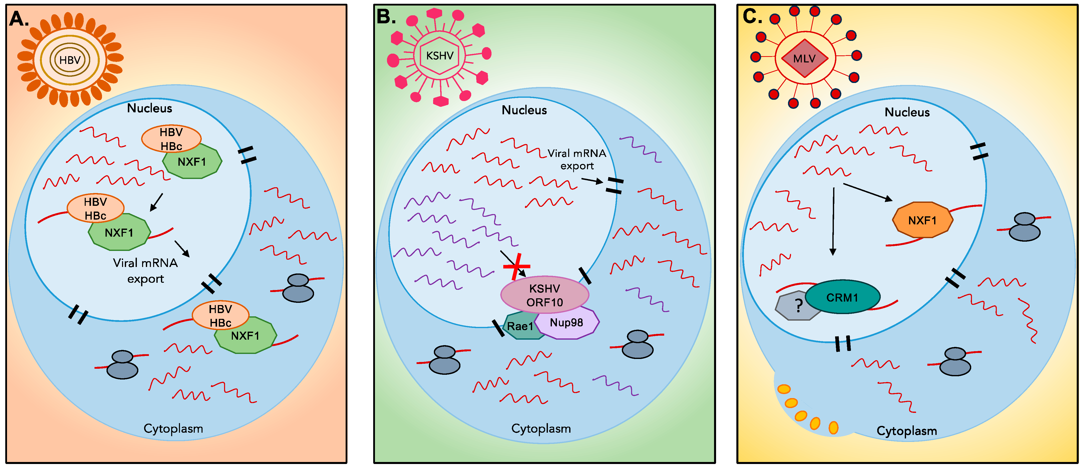
RNA fate is highly linked to its subcellular localization, a process largely driven by the pattern of RBPs on transcripts [40,41,42]. In the context of viral infection, the dynamic process of nuclear–cytoplasmic shuttling can be drastically altered [30,43]. For example, in the context of gamma-herpesvirus infection where host shutoff leads to widespread mRNA decay, many RBPs could potentially become a target to facilitate viral replication [43]. Using TMT labelling coupled to LC/MS-MS, Gilbertson et al. were able to track the subcellular localization of RBPs and showed that numerous RNA binding proteins undergo a change in their localization in response to viral-induced decay [43]. Given the scale of RNA decay triggered by these viruses, it is perhaps unsurprising that many RBPs would lose their target mRNA and be released, many of them finding their way back to the nucleus [43]. This potentially creates a measurable cellular response “sensing” of RNA decay in the cytoplasm, and the message can be relayed to the nucleus through the shuttling of RBPs [43,44,45,46,47]. According to Gilbertson et al., this creates a feedback mechanism, akin to informing the transcription machinery of the state of RNA decay in the cytoplasm and resulting in a halt of host transcription [43,47]. Therefore, trafficking of RBPs between cellular compartments during events of stress such as viral-induced RNA decay could be fundamental to relaying information about cellular mRNA abundance within the cell [41,43,47]. Another study by Garcia-Moreno et al. observed similar dynamics of RBP in response to Sindbis Virus (SINV) infection [30]. The authors used a system-wide approach known as RNA-interactome capture (RIC) to determine the distribution of RBPs in cells infected with SINV [30]. In the context of drastic loss of cellular RNAs and high abundance of viral RNAs, they observed a remodeling of the RBP interactome [30]. In particular, it was observed that many key gene expression regulators such as UPF1 (helicase, nonsense-mediated decay pathway), SRPK1 (alternative splicing, RNA export and stability), GEMIN5 (mediates assembly of small nuclear RNPs), TRIM25 and TRIM56 (E3 ubiquitin ligases), PPIA (regulation, signaling, apoptosis), and FAM98A (form the tRNA ligase complex) were re-localized to viral factories [30]. These proteins were observed to colocalize with viral RNAs, suggesting a role in facilitating viral replication during SINV infection. However, it remains unclear what the cell response is to drastic loss of host RNA and RBP re-localization during SINV infection and how remodeling of viral–host protein interactions could contribute to alter RBP function and localization during SINV infection.
It is becoming clear that RBP function and localization is pivotal during viral infections and can help viruses co-opt resources. Many questions continue to mount surrounding the mechanisms that regulate the shuttling of RBPs during viral infection, especially during viral-induced RNA decay. With the advent of novel protein labeling methods, we anticipate that we will learn more about the dynamics of these processes in the coming years.

4.2. Nuclear Export

Once mature, mRNAs are transported to the cytoplasm through nuclear pore complexes (NPCs) embedded in the nuclear envelope [48,49,50,51]. Transport of mRNAs is mediated by mRNP complexes composed of shuttling proteins, commonly the NXF1-based bulk export, and a more specialized CRM1-based export for unspliced mRNA [49,52]. Several studies have shown that viruses target the nuclear export machinery, which results in the downregulation of host gene expression and reduction in host antiviral responses [51,53,54,55]. For instance, Hepatitis B Virus (HBV) core protein HBc interacts with NXF1 [56]. This interaction is suggested to mediate the shuttling of the Hepatitis B core/capsid protein (HBc)–NXF1 complex, and facilitate the export of HBV transcripts (Figure 1) [56]. During KSHV infection, Gong et al. identified the interaction of the viral protein ORF10 with nuclear pore proteins (Nup98) and export factors (Rae1), which results in blocked mRNA export and the nuclear accumulation of transcripts (Figure 1) [55]. However, this export inhibition is not total but instead relies on transcript selectivity based on an unidentified RNA element located at their 3’UTR [55], which likely ensures proper export of viral mRNAs. This inhibition contributes to the global “host shutoff” phenotype induced by herpesviruses and helps with ensuring easier access to the translational machinery for viral transcripts [55,57]. Meanwhile, in Murine Leukemia Virus (MLV), both nuclear export routes (NXF1 and CRM1) are proposed to be exploited by the virus [58]. Mougel et al. show that MLV full-length RNA interacts with NXF1, allowing export of viral RNAs destined to undergo translation (Figure 1) [58]. Simultaneously, export of MLV RNA by CRM1 marked transcripts for viral packaging in the cytoplasm [58]. The use of these two export pathways by MLV highlights the ability of the viral RNA to assemble two different mRNP complexes and control RNA fate. Overall, many of the components of the nuclear export pathway have been identified as viral targets but the precise mechanisms by which their hijacking is coordinated are still not well understood. Ongoing and future studies will likely shed light on the processes that regulate the fate of viral and cellular mRNAs through the recruitment of RNA–protein complexes.

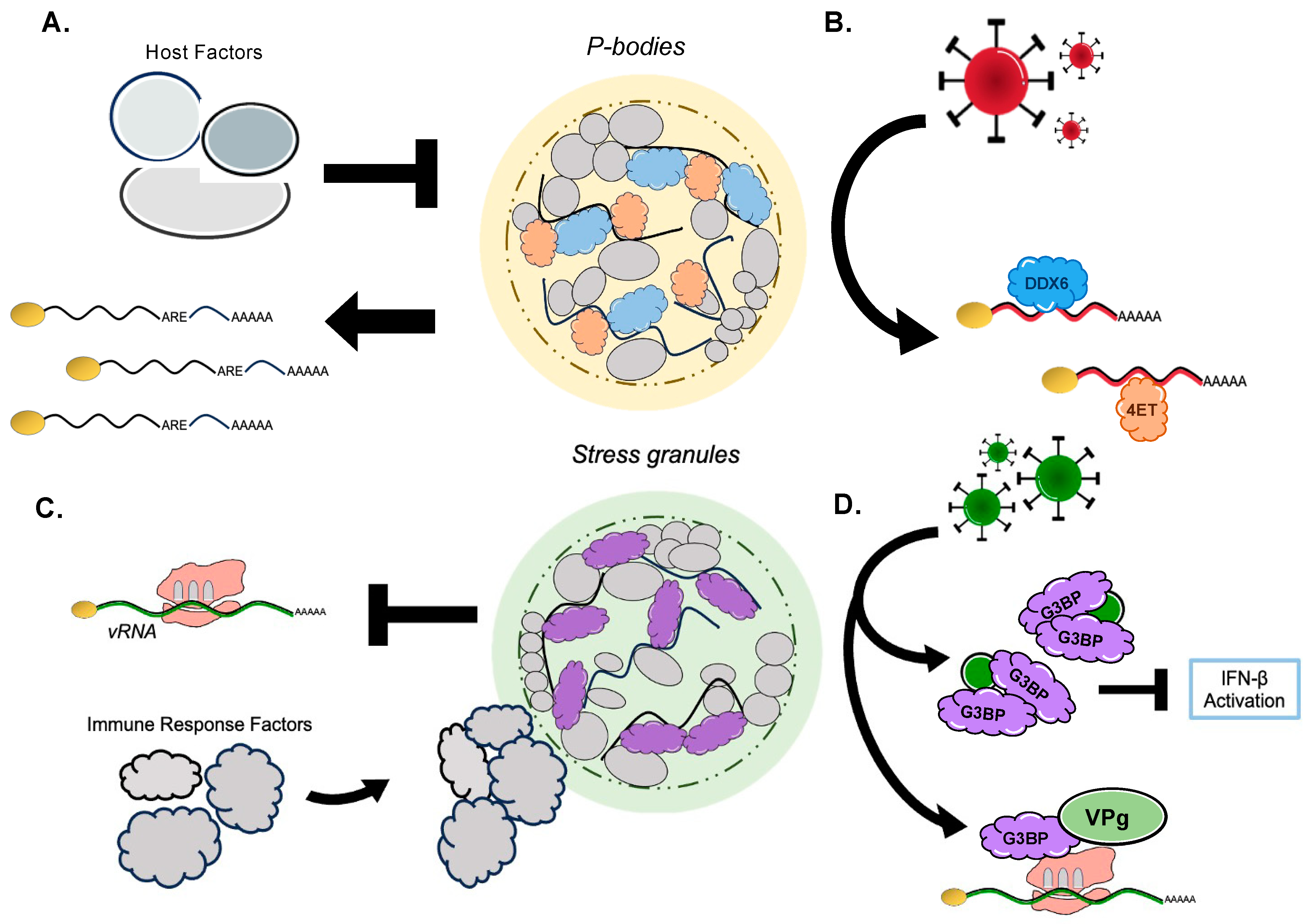
5. RNA Granules: A Nexus in the Viral–Host Struggle over RNA

RNA granules are biomolecular condensates of RNA and protein that exist within the cytoplasm. While these densities are often described to undergo liquid–liquid phase separation (LLPS), the story is more complex [59]. A better description of LLPS helps to better illustrate what these foci are: sequestered conglomerates of biomolecules that are separated from the surrounding cytoplasmic matrix without any phospholipid barrier [59]. However, this distinction can be quite difficult to define in such a dynamic system [60,61,62]. Granules are not simply defined as viscous liquids but are more precisely described as viscoelastic densities [63,64,65,66,67,68]. This distinction implies a duality and fluctuation of solid and liquid elements, and active exchange with the environment. Two classifications of cytoplasmic granules have emerged as important regulatory mechanisms within cells and both heavily rely on complex and dynamic interactions between RBP and RNA: processing bodies (P-bodies) and stress granules (SGs) [69,70]. P-bodies are often comprised of mRNA and RBPs such as DDX6, ET-4, PAT1B, LSM14A, EDC4, DCP1/DCP2, and CPEB1 [71,72,73,74,75,76,77,78,79,80,81,82]. Along with ribonucleic acids, SGs often contain proteins such as G3BP1/G3BP2, TIA-1, TIAR, Atx2, eIF2, eIF3, eIF4A, eIF4B, eIF4E, eIF4G, and eIF5 [69,82,83,84,85,86,87,88,89]. Some granules, such as P-bodies, are constitutively expressed in cells, and treatment with translation elongation inhibitors, like cycloheximide, will lead to cytoplasmic loss of the granules [70,71,90]. Conversely, stress granule formation must be induced by treatment with a translation terminator such as puromycin [70,91] or via cellular stressors like viral agents [5]. P-bodies are thought to be sites of RNA metabolism and regulation within the cytoplasm and have been implicated in mRNA decay, translation repression, nonsense-mediated decay (NMD), as well as mRNA silencing. They have commonly been identified as sites of mRNA decay due to several constituents’ ties to transcript degradation, namely DDX6, DCP1, DCP2, Xrn1, EDC3, and EDC4, among others. However, they can also serve as compartments to temporarily sequester some transcripts from the translation pool to control pathway activation/deactivation or for energetic conservation [70]. On the other hand, it has been well characterized that SGs form upon viral infection, and these cytoplasmic granules are often targeted for downregulation by viruses.
Due to their dynamic nature, diversity in composition, and temporal/environmental sensitivity, RNA granules’ functionality varies broadly from mRNA storage, degradation, and triage to assisting in cellular processes such as energetic conservation, the stress response, or the inflammatory response. This multitude of processes can readily be taken advantage of by viruses. Viruses and the host wage constant battles over the formation and regulation of RNA granules by manipulating RBPs, in a means to ultimately influence gene expression and lead to the arrest of certain cellular pathways to control the fate of RNA (Figure 2).

5.1. Viral Factors Exercise Control over P-Body RBPs to Promote Viral Replication

Given their extensive role in regulating mRNA fate, P-bodies are prime targets for viruses that need to co-opt these pathways for their own replication. It has previously been shown that viruses such as Kaposi Sarcoma-Associated Herpesvirus (KSHV), Hepatitis C Virus (HCV), and West Nile virus (WNV) all utilize P-body constituents such as DDX6, Lsm-1, DDX3, Ago2, Xrn1, and others to promote viral RNA and protein stability [8,92,93,94,95,96,97,98,99]. Recently, an investigation of Enterovirus 71 (EV71) showed how the virus induces a loss of P-bodies within human cells, which Fan et al. demonstrated stems from blocking the formation of de novo P-bodies [100]. The group further determined that certain scaffold proteins (i.e., DDX6 and 4E-T, among others) of P-bodies affected viral mRNA transcript levels [100]. The absence of DDX6 and 4E-T leads to decreased viral transcript levels compared to when both proteins were expressed [100]. Thus, they hypothesized that virally induced P-body loss reallocated and repurposed key P-body components for RNA stability and expression [100]. The group’s final model highlighted that EV71’s protease 2C facilitates host RBP interactions with viral mRNA to promote viral gene expression, which ultimately leads to the blockage in the formation of P-bodies [100]. It emerges that viruses are masters at seizing control of P-bodies through their interactions with host RBPs and use them to promote virion production. This also potentially suggests that the regulation of host gene expression could be suppressed to further remodel the host environment, making it more favorable for viral replication.

5.2. Hosts Manipulate P-Body RBPs to Alter RNA Availability/Degradation to Combat Viral Infection

It has been widely reported that P-bodies often house transcripts with AU-rich elements (AREs), located in their 3’ UTR for the purposes of degradation or translational repression [101,102,103,104,105,106]. Many ARE-bearing transcripts encode for chemokines and cytokines [107,108]. Several groups have demonstrated that following P-body loss, an increase in ARE-mRNAs follows [101,102,103,104,105,106]. These results suggest that a decrease in P-body counts may in fact play an antiviral role with the increased expression of these effectors. However, it remains unclear to what extent does the host or virus individually contribute to the loss of P-bodies during infection. Are P-bodies altered to stimulate the host inflammatory response to mount an antiviral response or to enhance an environmental favorability for viral agents? A host RBP that we mentioned previously in this review—Shiftless (SHFL)—is a broad-acting and potent antiviral factor [11,33,109,110,111,112,113,114] with known roles in regulating cytoplasmic granules. In the context of KSHV, SHFL has been shown to restrict P-body foci [11]. The exact subcellular mechanism through which SHFL accomplishes this P-body loss remains unclear. SHFL localizes to P-bodies [115] and its overexpression causes the loss of P-body formation [11]. The SHFL-mediated depletion of P-bodies could subsequently alter the expression levels of certain ARE-mRNAs that are often included in these granules, which likely contribute to SHFL overall ability to restrict viral agents. Outside of the context of infection, cellular loss of P-bodies typically occurs through two methods: 1. The depletion of core protein components, or 2. The expression/phosphorylation of the 68-amino-acid microprotein—the non-annotated P-body dissociating polypeptide (or “NBDY”) [69,86,116]. Losses of certain proteins such as LSm14a [73,77], DDX6 [75,77], or EDC4 have all been shown to lead to losses in P-bodies [72]. Otherwise, the expression and phosphorylation of the polypeptide NBDY has been shown to cause the loss of P-bodies, by potentially disrupting the electrostatic networks of these granules [117,118]. However, it has also been posited that P-body regulation may also release many decapping and endonucleolytic enzymes, leading to a translation suppression [119]. Therefore, it is possible that during viral infection, the host attempts to control the cytoplasmic gene expression environment by disassembling P-bodies. Viral agents often seek control over P-body RBPs as a tool to influence RNA expression within the cell. All the while, the host attempts to antagonize viral replication through a similar method: utilizing P-body-associated RBPs to alter RNA expression for antiviral pathways.

5.3. Viruses Influence Stress Granule RBPs to Suppress Host Immune Response Transcripts and Positively Regulate Viral RNA Fate

Numerous viruses have been shown to affect SGs: Japanese Encephalitis Virus (JEV) has been shown to alter the localization of SG marker G3BP1 [120]; Dengue virus (DENV), West Nile virus (WNV), Murine Respirovirus (SeV), and the Zika virus (ZIKV) have all been shown to utilize viral biomolecules to sequester SG critical proteins to block granule formation [9,121,122]. Recently, the betacoronavirus, severe acute respiratory syndrome coronavirus 2 (SARS-CoV-2), has been shown to have a similar effect on SG abundance and the re-purposing of the foci’s components [123]. The SARS-CoV-2 nucleocapsid protein (NP) has also been found to interact with the structurally critical SG proteins G3BP1/G3BP2 [123]. Liu et al. observed that this NP-G3BP interaction limits the degree to which SGs can form in response to viral infection, which in turn limits the host’s ability to stall translation and slow viral replication [123]. The group went on to observe that SARS-CoV-2’s NP sequestration of G3BPs not only resulted in the loss of SGs but also in the downregulation of IFN-β transcript levels and the RIG-I pathway (a critical piece of the innate immune response) as a whole [123]. This differs from a normal immune response, where G3BP1 typically influences the RIG-I pathway and IRF3 becomes phosphorylated, ultimately causing an increase in IFN-β transcription [124]. The researchers were then able to demonstrate how this virus exercises control over SG RBPs, such as G3BP1, to ultimately increase the propensity for viral mRNAs and translation. Given the increase in viral mRNAs in the absence of G3BP1, the authors concluded that the NP-G3BP1 interactions could help facilitate viral replication [124]. A genomic intermediate of the SARS-CoV-2 genome appears to exist as dsDNA, which has an observable affinity for G3BP1; mechanically, this was hypothesized to be how the virus isolates this RBP from the host’s antiviral system [124]. Additionally, this SG component has been shown to be necessary for both murine norovirus (MNV) replication as well as Norwalk norovirus replication (a replicon used to model human norovirus, HuNoV) [125]. Following the knockout of G3BP1 within cells, both MNV and HuNoV were observed to have significantly low transcript levels and were no longer tied to cytotoxic outcomes; cells were seen to be virally resistant [125]. Interestingly, Hosmillo et al. were able to uncover that G3BP1 RNA binding domains directly impacted viral replication; the loss of these domains resulted in less viral yield [125]. In addition, Hosmillo and colleagues discovered that G3BP1 interacts with the viral protein VPg and helps facilitate the loading of ribosomes and polysomes onto norovirus RNA [125]. It thus emerges that SGs are fascinating focal points for where the host and virus compete over RBPs to gain an advantage over the other.

5.4. Host Agents Orchestrate Stress Granule RBPs to Effectively Quench Viral Replication

Stress granules (SGs) have been widely characterized as antiviral subcellular compartments. In the case of the α-coronavirus porcine epidemic diarrhea virus (PEDV), SGs form and eventually are lost toward the latter stages (i.e., around 36 h) of infection [126]. In the study, Sun et al. identified that the overexpression of G3BP1 resulted in significantly decreased levels of viral mRNA [126]. They further discovered that PEDV encodes a protease that cleaves G3BP1 in the late stages of infection, which causes this loss [126]. They were able to pinpoint the specific cleavage sites that viral caspase-8 targets down to two aspartic acid residues [126]. Interestingly, robust SGs with cleavage-resistant G3BP1 stringently limited viral transcripts as well as overall viral titers, indicating that SGs can efficiently limit viral transcription and translation [126]. Studies like these demonstrate the capability and antiviral functionality of SGs and how the host attempts to use RBPs like G3BP1 to restrict viral agents.

5.5. Granule Functionality/Effects Are Convoluted; Both Virus and Host Wield SG RBPs for Their Own Benefit

The above sections detailing the great virus and host struggle over P-body and SG RBPs already outline how ambiguous the functionality of granules can be. One other fascinating viral case study that depicts this murkiness can be seen in the context of Viral Hemorrhagic Septicemia Virus (VHSV), a member of Rhabdoviridae, which appears to trigger the formation of stress granules [127]. Interestingly, G3BP1 was shown to be redistributed to viral replication complexes and proven to be essential for efficient virion production [127]. Even though viral infection results in SG formation, VHSV still influences G3BP1 to facilitate its own replication [127]. However, SGs also seem to play an antiviral role within the system. G3BP1 knockdown led to an increase in IFN signaling and a decrease in VHSV protein levels and viral titers [127]. This suggests that G3BP1 and SGs can limit viral replication to an extent, likely via translational arrest and sequestration [127]. VHSV utilizes SG RBPs such as G3BP1 to enhance replication, yet the host also exercises some control over these RBPs to simultaneously limit viral replication [127]. This case of viral infection and SG dynamics captures the convoluted subcellular environment and depicts how RNA granules exist at the axis where pro-viral and pro-host states teeter. Have viral agents robustly conquered this host defense and adapted to largely benefit from granules? Will host mechanisms adapt or can they be shaped to repurpose the likes of P-bodies and SGs to effectively combat viral replication?

6. Conclusions and Future Perspectives

RNA binding proteins comprise the subcellular foundation for host survival and viral replicative strategies. While this review covers many of the most recent findings of how both sides of this conflict attempt to take advantage of these proteins, new studies continue to unveil different viral case studies and mechanisms of exploit. mRNP complexes present an exciting avenue of study to better understand viral replication and takeover. As proper gene expression requires fine-tuned RNA processing, this multifaceted quality control system helps to regulate the cell homeostasis. It can prove to be disastrous when certain viral agents target one juncture of RNA processing and critical information is lost. Certain host factors, such as NXF1 and CRM1, critically regulate nuclear egress of transcripts, which can largely impact host survival dependent upon whether the virus or host controls these export pathways. Interestingly, RBPs can also help regulate the dynamics of certain RNA granules, which can significantly impact gene expression. Through P-bodies and stress granules, RBPs influence mRNA sequestration, decay, and release into the translational pool.
Furthermore, from the studies reviewed here, we can continue to interrogate how cellular pathways are affected by viral takeover, but also how the host responds to the viral attack. As mRNA abundance and availability can be drastically altered, cellular feedback response pathways can potentially serve to relay stress signals during viral infection. This area is still ripe for further study since it is still unclear which cellular factors are involved and to what extent communication is carried out in the cell between cellular compartments. Moreover, more studies to discern RNA–protein interactions when RBP expression levels are altered during viral infections are imperative to shed light onto the unknown biological significance of RBP activity. This will reveal crucial information on RBP localization, RBP multifunctionality, and their response to target availability. Studies in these areas could prove exceptionally fruitful, providing a deeper understanding of these processes, which could lead to the discovery of new antiviral targets and the development of therapeutic agents.
Regarding the RBPs associated with RNA granules, many factors and mechanisms are yet to be discovered. For viral agents, such as EV71, that lead to P-body loss, it remains to be seen mechanistically how the virus utilizes and recruits RBPs like DDX6 to provide stability and promote viral gene expression. Further characterization also ought to be conducted on exactly how the host wields P-bodies and their biomolecules specifically to alter its own gene expression to ultimately combat viral gene expression. For stress granule RBPs, how prevalent is this viral hijacking strategy? What other viral agents have evolved mechanisms to disrupt SG formation for purposes other than a lack of transcript sequestration? Overall, the characterization of RNA granule functionality during viral infection has emerged as an exciting frontier for better understanding virus–host interactions at the subcellular level.

Funding

M.M. is supported by NIH grant R35GM138043; Y.B. was supported by a training grant (T32 GM139789) and a Smith Spaulding Fellowship; D.H. is supported by the United States Air Force. The views expressed in this submission are those of the authors and do not reflect the official policy or position of the United States Air Force, Department of Defense, or the U.S. Government.

Conflicts of Interest

The authors declare no conflicts of interest.

References

  1. Singh, G.; Pratt, G.; Yeo, G.W.; Moore, M.J. The Clothes Make the mRNA: Past and Present Trends in mRNP Fashion. Annu. Rev. Biochem. 2015, 84, 325–354. [Google Scholar] [CrossRef]
  2. Mitchell, S.F.; Parker, R. Principles and properties of eukaryotic mRNPs. Mol. Cell 2014, 54, 547–558. [Google Scholar] [CrossRef]
  3. Dreyfuss, G.; Kim, V.N.; Kataoka, N. Messenger-RNA-binding proteins and the messages they carry. Nat. Rev. Mol. Cell Biol. 2002, 3, 195–205. [Google Scholar] [CrossRef] [PubMed]
  4. Corley, M.; Burns, M.C.; Yeo, G.W. How RNA-Binding Proteins Interact with RNA: Molecules and Mechanisms. Mol. Cell 2020, 78, 9–29. [Google Scholar] [CrossRef]
  5. Lloyd, R.E. Regulation of stress granules and P-bodies during RNA virus infection. Wiley Interdiscip. Rev. RNA 2013, 4, 317–331. [Google Scholar] [CrossRef]
  6. Sagan, S.M.; Weber, S.C. Let’s phase it: Viruses are master architects of biomolecular condensates. Trends Biochem. Sci. 2023, 48, 229–243. [Google Scholar] [CrossRef]
  7. Roden, C.; Gladfelter, A.S. RNA contributions to the form and function of biomolecular condensates. Nat. Rev. Mol. Cell Biol. 2021, 22, 183–195. [Google Scholar] [CrossRef]
  8. Chahar, H.S.; Chen, S.; Manjunath, N. P-body components LSM1, GW182, DDX3, DDX6 and XRN1 are recruited to WNV replication sites and positively regulate viral replication. Virology 2013, 436, 1–7. [Google Scholar] [CrossRef] [PubMed]
  9. Iseni, F.; Garcin, D.; Nishio, M.; Kedersha, N.; Anderson, P.; Kolakofsky, D. Sendai virus trailer RNA binds TIAR, a cellular protein involved in virus-induced apoptosis. EMBO J. 2002, 21, 5141–5150. [Google Scholar] [CrossRef] [PubMed]
  10. Cao, L.; Liu, S.; Li, Y.; Yang, G.; Luo, Y.; Li, S.; Du, H.; Zhao, Y.; Wang, D.; Chen, J.; et al. The Nuclear Matrix Protein SAFA Surveils Viral RNA and Facilitates Immunity by Activating Antiviral Enhancers and Super-enhancers. Cell Host Microbe 2019, 26, doi. [Google Scholar] [CrossRef]
  11. Rodriguez, W.; Mehrmann, T.; Hatfield, D.; Muller, M. Shiftless Restricts Viral Gene Expression and Influences RNA Granule Formation during Kaposi’s Sarcoma-Associated Herpesvirus Lytic Replication. J. Virol. 2022, 96, e0146922. [Google Scholar] [CrossRef] [PubMed]
  12. Glaunsinger, B.; Ganem, D. Lytic KSHV infection inhibits host gene expression by accelerating global mRNA turnover. Mol. Cell 2004, 13, 713–723. [Google Scholar] [CrossRef] [PubMed]
  13. Gaglia, M.M.; Rycroft, C.H.; Glaunsinger, B.A. Transcriptome-Wide Cleavage Site Mapping on Cellular mRNAs Reveals Features Underlying Sequence-Specific Cleavage by the Viral Ribonuclease SOX. PLoS Pathog. 2015, 11, e1005305. [Google Scholar] [CrossRef] [PubMed]
  14. Mendez, A.S.; Vogt, C.; Bohne, J.; Glaunsinger, B.A. Site specific target binding controls RNA cleavage efficiency by the Kaposi’s sarcoma-associated herpesvirus endonuclease SOX. Nucleic Acids Res. 2018, 46, 11968–11979. [Google Scholar] [CrossRef]
  15. Gaucherand, L.; Iyer, A.; Gilabert, I.; Rycroft, C.H.; Gaglia, M.M. Cut site preference allows influenza A virus PA-X to discriminate between host and viral mRNAs. Nat. Microbiol. 2023, 8, 1304–1317. [Google Scholar] [CrossRef]
  16. Gaucherand, L.; Porter, B.K.; Levene, R.E.; Price, E.L.; Schmaling, S.K.; Rycroft, C.H.; Kevorkian, Y.; McCormick, C.; Khaperskyy, D.A.; Gaglia, M.M. The Influenza A Virus Endoribonuclease PA-X Usurps Host mRNA Processing Machinery to Limit Host Gene Expression. Cell Rep. 2019, 27, 776–792.e7. [Google Scholar] [CrossRef] [PubMed]
  17. Hutin, S.; Lee, Y.; Glaunsinger, B.A. An RNA element in human interleukin 6 confers escape from degradation by the gammaherpesvirus SOX protein. J. Virol. 2013, 87, 4672–4682. [Google Scholar] [CrossRef]
  18. Muller, M.; Hutin, S.; Marigold, O.; Li, K.H.; Burlingame, A.; Glaunsinger, B.A. A ribonucleoprotein complex protects the interleukin-6 mRNA from degradation by distinct herpesviral endonucleases. PLoS Pathog. 2015, 11, e1004899. [Google Scholar] [CrossRef]
  19. Muller, M.; Glaunsinger, B.A. Nuclease escape elements protect messenger RNA against cleavage by multiple viral endonucleases. PLoS Pathog. 2017, 13, e1006593. [Google Scholar] [CrossRef]
  20. Greenbaum, B.D.; Levine, A.J.; Bhanot, G.; Rabadan, R. Patterns of evolution and host gene mimicry in influenza and other RNA viruses. PLoS Pathog. 2008, 4, e1000079. [Google Scholar] [CrossRef]
  21. Vijaykrishna, D.; Mukerji, R.; Smith, G.J. RNA Virus Reassortment: An Evolutionary Mechanism for Host Jumps and Immune Evasion. PLoS Pathog. 2015, 11, e1004902. [Google Scholar] [CrossRef]
  22. Smyth, R.P.; Negroni, M.; Lever, A.M.; Mak, J.; Kenyon, J.C. RNA Structure-A Neglected Puppet Master for the Evolution of Virus and Host Immunity. Front. Immunol. 2018, 9, 2097. [Google Scholar] [CrossRef]
  23. Garcia-Moreno, M.; Jarvelin, A.I.; Castello, A. Unconventional RNA-binding proteins step into the virus-host battlefront. Wiley Interdiscip. Rev. RNA 2018, 9, e1498. [Google Scholar] [CrossRef] [PubMed]
  24. Lisy, S.; Rothamel, K.; Ascano, M. RNA Binding Proteins as Pioneer Determinants of Infection: Protective, Proviral, or Both? Viruses 2021, 13, 2172. [Google Scholar] [CrossRef] [PubMed]
  25. Brugier, A.; Hafirrassou, M.L.; Pourcelot, M.; Baldaccini, M.; Kril, V.; Couture, L.; Kummerer, B.M.; Gallois-Montbrun, S.; Bonnet-Madin, L.; Vidalain, P.O.; et al. RACK1 Associates with RNA-Binding Proteins Vigilin and SERBP1 to Facilitate Dengue Virus Replication. J. Virol. 2022, 96, e0196221. [Google Scholar] [CrossRef] [PubMed]
  26. Adams, D.R.; Ron, D.; Kiely, P.A. RACK1, A multifaceted scaffolding protein: Structure and function. Cell Commun. Signal. 2011, 9, 22. [Google Scholar] [CrossRef] [PubMed]
  27. Baum, S.; Bittins, M.; Frey, S.; Seedorf, M. Asc1p, a WD40-domain containing adaptor protein, is required for the interaction of the RNA-binding protein Scp160p with polysomes. Biochem. J. 2004, 380, 823–830. [Google Scholar] [CrossRef] [PubMed]
  28. Sundaramoorthy, E.; Leonard, M.; Mak, R.; Liao, J.; Fulzele, A.; Bennett, E.J. ZNF598 and RACK1 Regulate Mammalian Ribosome-Associated Quality Control Function by Mediating Regulatory 40S Ribosomal Ubiquitylation. Mol. Cell 2017, 65, 751–760.e4. [Google Scholar] [CrossRef] [PubMed]
  29. Ceci, M.; Gaviraghi, C.; Gorrini, C.; Sala, L.A.; Offenhauser, N.; Marchisio, P.C.; Biffo, S. Release of eIF6 (p27BBP) from the 60S subunit allows 80S ribosome assembly. Nature 2003, 426, 579–584. [Google Scholar] [CrossRef] [PubMed]
  30. Garcia-Moreno, M.; Noerenberg, M.; Ni, S.; Jarvelin, A.I.; Gonzalez-Almela, E.; Lenz, C.E.; Bach-Pages, M.; Cox, V.; Avolio, R.; Davis, T.; et al. System-wide Profiling of RNA-Binding Proteins Uncovers Key Regulators of Virus Infection. Mol. Cell 2019, 74, 196–211.e11. [Google Scholar] [CrossRef]
  31. Takamatsu, Y.; Krahling, V.; Kolesnikova, L.; Halwe, S.; Lier, C.; Baumeister, S.; Noda, T.; Biedenkopf, N.; Becker, S. Serine-Arginine Protein Kinase 1 Regulates Ebola Virus Transcription. mBio 2020, 11, e02565-19. [Google Scholar] [CrossRef]
  32. Merino, V.F.; Yan, Y.; Ordonez, A.A.; Bullen, C.K.; Lee, A.; Saeki, H.; Ray, K.; Huang, T.; Jain, S.K.; Pomper, M.G. Nucleolin mediates SARS-CoV-2 replication and viral-induced apoptosis of host cells. Antivir. Res. 2023, 211, 105550. [Google Scholar] [CrossRef] [PubMed]
  33. Wang, X.; Xuan, Y.; Han, Y.; Ding, X.; Ye, K.; Yang, F.; Gao, P.; Goff, S.P.; Gao, G. Regulation of HIV-1 Gag-Pol Expression by Shiftless, an Inhibitor of Programmed -1 Ribosomal Frameshifting. Cell 2019, 176, 625–635.e14. [Google Scholar] [CrossRef] [PubMed]
  34. Rodriguez, W.; Muller, M. Shiftless, a Critical Piece of the Innate Immune Response to Viral Infection. Viruses 2022, 14, 1338. [Google Scholar] [CrossRef] [PubMed]
  35. Qin, W.; Kong, N.; Zhang, Y.; Wang, C.; Dong, S.; Zhai, H.; Zhai, X.; Yang, X.; Ye, C.; Ye, M.; et al. PTBP1 suppresses porcine epidemic diarrhea virus replication via inducing protein degradation and IFN production. J. Biol. Chem. 2023, 299, 104987. [Google Scholar] [CrossRef] [PubMed]
  36. Davis, Z.H.; Verschueren, E.; Jang, G.M.; Kleffman, K.; Johnson, J.R.; Park, J.; Von Dollen, J.; Maher, M.C.; Johnson, T.; Newton, W.; et al. Global mapping of herpesvirus-host protein complexes reveals a transcription strategy for late genes. Mol. Cell 2015, 57, 349–360. [Google Scholar] [CrossRef] [PubMed]
  37. Trendel, J.; Schwarzl, T.; Horos, R.; Prakash, A.; Bateman, A.; Hentze, M.W.; Krijgsveld, J. The Human RNA-Binding Proteome and Its Dynamics during Translational Arrest. Cell 2019, 176, 391–403.e19. [Google Scholar] [CrossRef]
  38. Chen, T.; Yang, H.; Liu, P.; Hamiti, M.; Zhang, X.; Xu, Y.; Quan, W.; Zhang, Y.; Yu, W.; Jiao, L.; et al. Splicing factor SF3B3, a NS5-binding protein, restricts ZIKV infection by targeting GCH1. Virol. Sin. 2023, 38, 222–232. [Google Scholar] [CrossRef]
  39. Schneider-Lunitz, V.; Ruiz-Orera, J.; Hubner, N.; van Heesch, S. Multifunctional RNA-binding proteins influence mRNA abundance and translational efficiency of distinct sets of target genes. PLoS Comput. Biol. 2021, 17, e1009658. [Google Scholar] [CrossRef]
  40. Mayya, V.K.; Duchaine, T.F. Ciphers and Executioners: How 3′-Untranslated Regions Determine the Fate of Messenger RNAs. Front. Genet. 2019, 10, 6. [Google Scholar] [CrossRef]
  41. Timmers, H.T.M.; Tora, L. Transcript Buffering: A Balancing Act between mRNA Synthesis and mRNA Degradation. Mol. Cell 2018, 72, 10–17. [Google Scholar] [CrossRef]
  42. Chin, A.; Lecuyer, E. RNA localization: Making its way to the center stage. Biochim. Biophys. Acta Gen. Subj. 2017, 1861, 2956–2970. [Google Scholar] [CrossRef]
  43. Gilbertson, S.; Federspiel, J.D.; Hartenian, E.; Cristea, I.M.; Glaunsinger, B. Changes in mRNA abundance drive shuttling of RNA binding proteins, linking cytoplasmic RNA degradation to transcription. eLife 2018, 7, e37663. [Google Scholar] [CrossRef] [PubMed]
  44. Lee, Y.J.; Glaunsinger, B.A. Aberrant herpesvirus-induced polyadenylation correlates with cellular messenger RNA destruction. PLoS Biol. 2009, 7, e1000107. [Google Scholar] [CrossRef]
  45. Park, R.; El-Guindy, A.; Heston, L.; Lin, S.F.; Yu, K.P.; Nagy, M.; Borah, S.; Delecluse, H.J.; Steitz, J.; Miller, G. Nuclear translocation and regulation of intranuclear distribution of cytoplasmic poly(A)-binding protein are distinct processes mediated by two Epstein Barr virus proteins. PLoS ONE 2014, 9, e92593. [Google Scholar] [CrossRef] [PubMed]
  46. Richner, J.M.; Clyde, K.; Pezda, A.C.; Cheng, B.Y.; Wang, T.; Kumar, G.R.; Covarrubias, S.; Coscoy, L.; Glaunsinger, B. Global mRNA degradation during lytic gammaherpesvirus infection contributes to establishment of viral latency. PLoS Pathog. 2011, 7, e1002150. [Google Scholar] [CrossRef] [PubMed]
  47. Hartenian, E.; Glaunsinger, B.A. Feedback to the central dogma: Cytoplasmic mRNA decay and transcription are interdependent processes. Crit. Rev. Biochem. Mol. Biol. 2019, 54, 385–398. [Google Scholar] [CrossRef]
  48. Sandri-Goldin, R.M. Nuclear export of herpes virus RNA. Curr. Top. Microbiol. Immunol. 2001, 259, 2–23. [Google Scholar] [PubMed]
  49. Carmody, S.R.; Wente, S.R. mRNA nuclear export at a glance. J. Cell Sci. 2009, 122, 1933–1937. [Google Scholar] [CrossRef] [PubMed]
  50. Wente, S.R.; Rout, M.P. The nuclear pore complex and nuclear transport. Cold Spring Harb. Perspect. Biol. 2010, 2, a000562. [Google Scholar] [CrossRef]
  51. Guo, J.; Zhu, Y.; Ma, X.; Shang, G.; Liu, B.; Zhang, K. Virus Infection and mRNA Nuclear Export. Int. J. Mol. Sci. 2023, 24, 12593. [Google Scholar] [CrossRef]
  52. Stewart, M. Polyadenylation and nuclear export of mRNAs. J. Biol. Chem. 2019, 294, 2977–2987. [Google Scholar] [CrossRef]
  53. Vogt, C.; Bohne, J. The KSHV RNA regulator ORF57: Target specificity and its role in the viral life cycle. Wiley Interdiscip. Rev. RNA 2016, 7, 173–185. [Google Scholar] [CrossRef]
  54. Gales, J.P.; Kubina, J.; Geldreich, A.; Dimitrova, M. Strength in Diversity: Nuclear Export of Viral RNAs. Viruses 2020, 12, 1014. [Google Scholar] [CrossRef]
  55. Gong, D.; Kim, Y.H.; Xiao, Y.; Du, Y.; Xie, Y.; Lee, K.K.; Feng, J.; Farhat, N.; Zhao, D.; Shu, S.; et al. A Herpesvirus Protein Selectively Inhibits Cellular mRNA Nuclear Export. Cell Host Microbe 2016, 20, 642–653. [Google Scholar] [CrossRef]
  56. Li, H.C.; Huang, E.Y.; Su, P.Y.; Wu, S.Y.; Yang, C.C.; Lin, Y.S.; Chang, W.C.; Shih, C. Nuclear export and import of human hepatitis B virus capsid protein and particles. PLoS Pathog. 2010, 6, e1001162. [Google Scholar] [CrossRef]
  57. Pardamean, C.I.; Wu, T.T. Inhibition of Host Gene Expression by KSHV: Sabotaging mRNA Stability and Nuclear Export. Front. Cell Infect. Microbiol. 2021, 11, 648055. [Google Scholar] [CrossRef] [PubMed]
  58. Mougel, M.; Akkawi, C.; Chamontin, C.; Feuillard, J.; Pessel-Vivares, L.; Socol, M.; Laine, S. NXF1 and CRM1 nuclear export pathways orchestrate nuclear export, translation and packaging of murine leukaemia retrovirus unspliced RNA. RNA Biol. 2020, 17, 528–538. [Google Scholar] [CrossRef]
  59. Wang, B.; Zhang, L.; Dai, T.; Qin, Z.; Lu, H.; Zhang, L.; Zhou, F. Liquid-liquid phase separation in human health and diseases. Signal Transduct. Target. Ther. 2021, 6, 290. [Google Scholar] [CrossRef] [PubMed]
  60. Dundr, M.; Hebert, M.D.; Karpova, T.S.; Stanek, D.; Xu, H.; Shpargel, K.B.; Meier, U.T.; Neugebauer, K.M.; Matera, A.G.; Misteli, T. In vivo kinetics of Cajal body components. J. Cell Biol. 2004, 164, 831–842. [Google Scholar] [CrossRef]
  61. Phair, R.D.; Misteli, T. High mobility of proteins in the mammalian cell nucleus. Nature 2000, 404, 604–609. [Google Scholar] [CrossRef] [PubMed]
  62. Weidtkamp-Peters, S.; Lenser, T.; Negorev, D.; Gerstner, N.; Hofmann, T.G.; Schwanitz, G.; Hoischen, C.; Maul, G.; Dittrich, P.; Hemmerich, P. Dynamics of component exchange at PML nuclear bodies. J. Cell Sci. 2008, 121, 2731–2743. [Google Scholar] [CrossRef] [PubMed]
  63. Mittag, T.; Pappu, R.V. A conceptual framework for understanding phase separation and addressing open questions and challenges. Mol. Cell 2022, 82, 2201–2214. [Google Scholar] [CrossRef] [PubMed]
  64. Alshareedah, I.; Moosa, M.M.; Pham, M.; Potoyan, D.A.; Banerjee, P.R. Programmable viscoelasticity in protein-RNA condensates with disordered sticker-spacer polypeptides. Nat. Commun. 2021, 12, 6620. [Google Scholar] [CrossRef] [PubMed]
  65. Bergeron-Sandoval, L.P.; Kumar, S.; Heris, H.K.; Chang, C.L.A.; Cornell, C.E.; Keller, S.L.; Francois, P.; Hendricks, A.G.; Ehrlicher, A.J.; Pappu, R.V.; et al. Endocytic proteins with prion-like domains form viscoelastic condensates that enable membrane remodeling. Proc. Natl. Acad. Sci. USA 2021, 118, e2113789118. [Google Scholar] [CrossRef] [PubMed]
  66. Ghosh, A.; Kota, D.; Zhou, H.X. Shear relaxation governs fusion dynamics of biomolecular condensates. Nat. Commun. 2021, 12, 5995. [Google Scholar] [CrossRef] [PubMed]
  67. Jawerth, L.; Fischer-Friedrich, E.; Saha, S.; Wang, J.; Franzmann, T.; Zhang, X.; Sachweh, J.; Ruer, M.; Ijavi, M.; Saha, S.; et al. Protein condensates as aging Maxwell fluids. Science 2020, 370, 1317–1323. [Google Scholar] [CrossRef]
  68. Shen, Y.; Ruggeri, F.S.; Vigolo, D.; Kamada, A.; Qamar, S.; Levin, A.; Iserman, C.; Alberti, S.; George-Hyslop, P.S.; Knowles, T.P.J. Biomolecular condensates undergo a generic shear-mediated liquid-to-solid transition. Nat. Nanotechnol. 2020, 15, 841–847. [Google Scholar] [CrossRef]
  69. White, J.P.; Lloyd, R.E. Regulation of stress granules in virus systems. Trends Microbiol. 2012, 20, 175–183. [Google Scholar] [CrossRef]
  70. Ivanov, P.; Kedersha, N.; Anderson, P. Stress Granules and Processing Bodies in Translational Control. Cold Spring Harb. Perspect. Biol. 2019, 11, a032813. [Google Scholar] [CrossRef] [PubMed]
  71. Andrei, M.A.; Ingelfinger, D.; Heintzmann, R.; Achsel, T.; Rivera-Pomar, R.; Luhrmann, R. A role for eIF4E and eIF4E-transporter in targeting mRNPs to mammalian processing bodies. RNA 2005, 11, 717–727. [Google Scholar] [CrossRef] [PubMed]
  72. Yu, J.H.; Yang, W.H.; Gulick, T.; Bloch, K.D.; Bloch, D.B. Ge-1 is a central component of the mammalian cytoplasmic mRNA processing body. RNA 2005, 11, 1795–1802. [Google Scholar] [CrossRef] [PubMed]
  73. Yang, W.H.; Yu, J.H.; Gulick, T.; Bloch, K.D.; Bloch, D.B. RNA-associated protein 55 (RAP55) localizes to mRNA processing bodies and stress granules. RNA 2006, 12, 547–554. [Google Scholar] [CrossRef] [PubMed]
  74. Wilczynska, A.; Aigueperse, C.; Kress, M.; Dautry, F.; Weil, D. The translational regulator CPEB1 provides a link between dcp1 bodies and stress granules. J. Cell Sci. 2005, 118, 981–992. [Google Scholar] [CrossRef] [PubMed]
  75. Ozgur, S.; Chekulaeva, M.; Stoecklin, G. Human Pat1b connects deadenylation with mRNA decapping and controls the assembly of processing bodies. Mol. Cell Biol. 2010, 30, 4308–4323. [Google Scholar] [CrossRef] [PubMed]
  76. Marnef, A.; Maldonado, M.; Bugaut, A.; Balasubramanian, S.; Kress, M.; Weil, D.; Standart, N. Distinct functions of maternal and somatic Pat1 protein paralogs. RNA 2010, 16, 2094–2107. [Google Scholar] [CrossRef]
  77. Ayache, J.; Benard, M.; Ernoult-Lange, M.; Minshall, N.; Standart, N.; Kress, M.; Weil, D. P-body assembly requires DDX6 repression complexes rather than decay or Ataxin2/2L complexes. Mol. Biol. Cell 2015, 26, 2579–2595. [Google Scholar] [CrossRef]
  78. Eulalio, A.; Behm-Ansmant, I.; Schweizer, D.; Izaurralde, E. P-body formation is a consequence, not the cause, of RNA-mediated gene silencing. Mol. Cell Biol. 2007, 27, 3970–3981. [Google Scholar] [CrossRef]
  79. Davidson, A.; Parton, R.M.; Rabouille, C.; Weil, T.T.; Davis, I. Localized Translation of gurken/TGF-alpha mRNA during Axis Specification Is Controlled by Access to Orb/CPEB on Processing Bodies. Cell Rep. 2016, 14, 2451–2462. [Google Scholar] [CrossRef]
  80. Fan, S.J.; Marchand, V.; Ephrussi, A. Drosophila Ge-1 promotes P body formation and oskar mRNA localization. PLoS ONE 2011, 6, e20612. [Google Scholar] [CrossRef]
  81. Eystathioy, T.; Jakymiw, A.; Chan, E.K.; Seraphin, B.; Cougot, N.; Fritzler, M.J. The GW182 protein colocalizes with mRNA degradation associated proteins hDcp1 and hLSm4 in cytoplasmic GW bodies. RNA 2003, 9, 1171–1173. [Google Scholar] [CrossRef]
  82. Sharma, N.R.; Zheng, Z.M. RNA Granules in Antiviral Innate Immunity: A Kaposi’s Sarcoma-Associated Herpesvirus Journey. Front. Microbiol. 2021, 12, 794431. [Google Scholar] [CrossRef]
  83. Buchan, J.R.; Muhlrad, D.; Parker, R. P bodies promote stress granule assembly in Saccharomyces cerevisiae. J. Cell Biol. 2008, 183, 441–455. [Google Scholar] [CrossRef] [PubMed]
  84. Gilks, N.; Kedersha, N.; Ayodele, M.; Shen, L.; Stoecklin, G.; Dember, L.M.; Anderson, P. Stress granule assembly is mediated by prion-like aggregation of TIA-1. Mol. Biol. Cell 2004, 15, 5383–5398. [Google Scholar] [CrossRef] [PubMed]
  85. Solomon, S.; Xu, Y.; Wang, B.; David, M.D.; Schubert, P.; Kennedy, D.; Schrader, J.W. Distinct structural features of caprin-1 mediate its interaction with G3BP-1 and its induction of phosphorylation of eukaryotic translation initiation factor 2alpha, entry to cytoplasmic stress granules, and selective interaction with a subset of mRNAs. Mol. Cell Biol. 2007, 27, 2324–2342. [Google Scholar] [CrossRef] [PubMed]
  86. Kedersha, N.; Chen, S.; Gilks, N.; Li, W.; Miller, I.J.; Stahl, J.; Anderson, P. Evidence that ternary complex (eIF2-GTP-tRNA(i)(Met))-deficient preinitiation complexes are core constituents of mammalian stress granules. Mol. Biol. Cell 2002, 13, 195–210. [Google Scholar] [CrossRef]
  87. Mazroui, R.; Sukarieh, R.; Bordeleau, M.E.; Kaufman, R.J.; Northcote, P.; Tanaka, J.; Gallouzi, I.; Pelletier, J. Inhibition of ribosome recruitment induces stress granule formation independently of eukaryotic initiation factor 2alpha phosphorylation. Mol. Biol. Cell 2006, 17, 4212–4219. [Google Scholar] [CrossRef] [PubMed]
  88. Bolster, D.R.; Kubica, N.; Crozier, S.J.; Williamson, D.L.; Farrell, P.A.; Kimball, S.R.; Jefferson, L.S. Immediate response of mammalian target of rapamycin (mTOR)-mediated signalling following acute resistance exercise in rat skeletal muscle. J. Physiol. 2003, 553, 213–220. [Google Scholar] [CrossRef] [PubMed]
  89. Kedersha, N.L.; Gupta, M.; Li, W.; Miller, I.; Anderson, P. RNA-binding proteins TIA-1 and TIAR link the phosphorylation of eIF-2 alpha to the assembly of mammalian stress granules. J. Cell Biol. 1999, 147, 1431–1442. [Google Scholar] [CrossRef] [PubMed]
  90. Hirose, T.; Ninomiya, K.; Nakagawa, S.; Yamazaki, T. A guide to membraneless organelles and their various roles in gene regulation. Nat. Rev. Mol. Cell Biol. 2023, 24, 288–304. [Google Scholar] [CrossRef]
  91. Kedersha, N.; Cho, M.R.; Li, W.; Yacono, P.W.; Chen, S.; Gilks, N.; Golan, D.E.; Anderson, P. Dynamic shuttling of TIA-1 accompanies the recruitment of mRNA to mammalian stress granules. J. Cell Biol. 2000, 151, 1257–1268. [Google Scholar] [CrossRef]
  92. Robinson, C.A.; Singh, G.K.; Kleer, M.; Katsademas, T.; Castle, E.L.; Boudreau, B.Q.; Corcoran, J.A. Kaposi’s sarcoma-associated herpesvirus (KSHV) utilizes the NDP52/CALCOCO2 selective autophagy receptor to disassemble processing bodies. PLoS Pathog. 2023, 19, e1011080. [Google Scholar] [CrossRef]
  93. Sharma, N.R.; Majerciak, V.; Kruhlak, M.J.; Yu, L.; Kang, J.G.; Yang, A.; Gu, S.; Fritzler, M.J.; Zheng, Z.M. KSHV RNA-binding protein ORF57 inhibits P-body formation to promote viral multiplication by interaction with Ago2 and GW182. Nucleic Acids Res. 2019, 47, 9368–9385. [Google Scholar] [CrossRef] [PubMed]
  94. Ariumi, Y.; Kuroki, M.; Kushima, Y.; Osugi, K.; Hijikata, M.; Maki, M.; Ikeda, M.; Kato, N. Hepatitis C virus hijacks P-body and stress granule components around lipid droplets. J. Virol. 2011, 85, 6882–6892. [Google Scholar] [CrossRef]
  95. Pager, C.T.; Schutz, S.; Abraham, T.M.; Luo, G.; Sarnow, P. Modulation of hepatitis C virus RNA abundance and virus release by dispersion of processing bodies and enrichment of stress granules. Virology 2013, 435, 472–484. [Google Scholar] [CrossRef] [PubMed]
  96. Perez-Vilaro, G.; Scheller, N.; Saludes, V.; Diez, J. Hepatitis C virus infection alters P-body composition but is independent of P-body granules. J. Virol. 2012, 86, 8740–8749. [Google Scholar] [CrossRef]
  97. Ariumi, Y.; Kuroki, M.; Abe, K.; Dansako, H.; Ikeda, M.; Wakita, T.; Kato, N. DDX3 DEAD-box RNA helicase is required for hepatitis C virus RNA replication. J. Virol. 2007, 81, 13922–13926. [Google Scholar] [CrossRef]
  98. Jangra, R.K.; Yi, M.; Lemon, S.M. DDX6 (Rck/p54) is required for efficient hepatitis C virus replication but not for internal ribosome entry site-directed translation. J. Virol. 2010, 84, 6810–6824. [Google Scholar] [CrossRef]
  99. Scheller, N.; Mina, L.B.; Galao, R.P.; Chari, A.; Gimenez-Barcons, M.; Noueiry, A.; Fischer, U.; Meyerhans, A.; Diez, J. Translation and replication of hepatitis C virus genomic RNA depends on ancient cellular proteins that control mRNA fates. Proc. Natl. Acad. Sci. USA 2009, 106, 13517–13522. [Google Scholar] [CrossRef] [PubMed]
  100. Fan, S.; Xu, Z.; Liu, P.; Qin, Y.; Chen, M. Enterovirus 71 2A Protease Inhibits P-Body Formation To Promote Viral RNA Synthesis. J. Virol. 2021, 95, e0092221. [Google Scholar] [CrossRef]
  101. Corcoran, J.A.; Johnston, B.P.; McCormick, C. Viral activation of MK2-hsp27-p115RhoGEF-RhoA signaling axis causes cytoskeletal rearrangements, p-body disruption and ARE-mRNA stabilization. PLoS Pathog. 2015, 11, e1004597. [Google Scholar] [CrossRef]
  102. Blanco, F.F.; Sanduja, S.; Deane, N.G.; Blackshear, P.J.; Dixon, D.A. Transforming growth factor beta regulates P-body formation through induction of the mRNA decay factor tristetraprolin. Mol. Cell Biol. 2014, 34, 180–195. [Google Scholar] [CrossRef]
  103. Franks, T.M.; Lykke-Andersen, J. TTP and BRF proteins nucleate processing body formation to silence mRNAs with AU-rich elements. Genes. Dev. 2007, 21, 719–735. [Google Scholar] [CrossRef]
  104. Vindry, C.; Marnef, A.; Broomhead, H.; Twyffels, L.; Ozgur, S.; Stoecklin, G.; Llorian, M.; Smith, C.W.; Mata, J.; Weil, D.; et al. Dual RNA Processing Roles of Pat1b via Cytoplasmic Lsm1-7 and Nuclear Lsm2-8 Complexes. Cell Rep. 2017, 20, 1187–1200. [Google Scholar] [CrossRef]
  105. Hubstenberger, A.; Courel, M.; Benard, M.; Souquere, S.; Ernoult-Lange, M.; Chouaib, R.; Yi, Z.; Morlot, J.B.; Munier, A.; Fradet, M.; et al. P-Body Purification Reveals the Condensation of Repressed mRNA Regulons. Mol. Cell 2017, 68, 144–157.e5. [Google Scholar] [CrossRef]
  106. Corcoran, J.A.; Khaperskyy, D.A.; Johnston, B.P.; King, C.A.; Cyr, D.P.; Olsthoorn, A.V.; McCormick, C. Kaposi’s sarcoma-associated herpesvirus G-protein-coupled receptor prevents AU-rich-element-mediated mRNA decay. J. Virol. 2012, 86, 8859–8871. [Google Scholar] [CrossRef]
  107. Bakheet, T.; Williams, B.R.; Khabar, K.S. ARED 3.0: The large and diverse AU-rich transcriptome. Nucleic Acids Res. 2006, 34, D111–D114. [Google Scholar] [CrossRef] [PubMed]
  108. Chen, C.Y.; Shyu, A.B. AU-rich elements: Characterization and importance in mRNA degradation. Trends Biochem. Sci. 1995, 20, 465–470. [Google Scholar] [CrossRef] [PubMed]
  109. Xiong, W.; Contreras, D.; Irudayam, J.I.; Ali, A.; Yang, O.O.; Arumugaswami, V. C19ORF66 is an interferon-stimulated gene (ISG) which Inhibits human immunodeficiency virus-1. BioRxiv 2016, 050310. [Google Scholar] [CrossRef]
  110. Suzuki, Y.; Chin, W.X.; Han, Q.; Ichiyama, K.; Lee, C.H.; Eyo, Z.W.; Ebina, H.; Takahashi, H.; Takahashi, C.; Tan, B.H.; et al. Characterization of RyDEN (C19orf66) as an Interferon-Stimulated Cellular Inhibitor against Dengue Virus Replication. PLoS Pathog. 2016, 12, e1005357. [Google Scholar] [CrossRef]
  111. Kinast, V.; Plociennikowska, A.; Anggakusuma; Bracht, T.; Todt, D.; Brown, R.J.P.; Boldanova, T.; Zhang, Y.; Bruggemann, Y.; Friesland, M.; et al. C19orf66 is an interferon-induced inhibitor of HCV replication that restricts formation of the viral replication organelle. J. Hepatol. 2020, 73, 549–558. [Google Scholar] [CrossRef]
  112. Wu, Y.; Yang, X.; Yao, Z.; Dong, X.; Zhang, D.; Hu, Y.; Zhang, S.; Lin, J.; Chen, J.; An, S.; et al. C19orf66 interrupts Zika virus replication by inducing lysosomal degradation of viral NS3. PLoS Negl. Trop. Dis. 2020, 14, e0008083. [Google Scholar] [CrossRef]
  113. Hanners, N.W.; Mar, K.B.; Boys, I.N.; Eitson, J.L.; De La Cruz-Rivera, P.C.; Richardson, R.B.; Fan, W.; Wight-Carter, M.; Schoggins, J.W. Shiftless inhibits flavivirus replication in vitro and is neuroprotective in a mouse model of Zika virus pathogenesis. Proc. Natl. Acad. Sci. USA 2021, 118, e2111266118. [Google Scholar] [CrossRef]
  114. Yu, D.; Zhao, Y.; Pan, J.; Yang, X.; Liang, Z.; Xie, S.; Cao, R. C19orf66 Inhibits Japanese Encephalitis Virus Replication by Targeting -1 PRF and the NS3 Protein. Virol. Sin. 2021, 36, 1443–1455. [Google Scholar] [CrossRef]
  115. Balinsky, C.A.; Schmeisser, H.; Wells, A.I.; Ganesan, S.; Jin, T.; Singh, K.; Zoon, K.C. IRAV (FLJ11286), an Interferon-Stimulated Gene with Antiviral Activity against Dengue Virus, Interacts with MOV10. J. Virol. 2017, 91, e01606-16. [Google Scholar] [CrossRef]
  116. Luo, Y.; Na, Z.; Slavoff, S.A. P-Bodies: Composition, Properties, and Functions. Biochemistry 2018, 57, 2424–2431. [Google Scholar] [CrossRef]
  117. Na, Z.; Luo, Y.; Schofield, J.A.; Smelyansky, S.; Khitun, A.; Muthukumar, S.; Valkov, E.; Simon, M.D.; Slavoff, S.A. The NBDY Microprotein Regulates Cellular RNA Decapping. Biochemistry 2020, 59, 4131–4142. [Google Scholar] [CrossRef] [PubMed]
  118. Na, Z.; Luo, Y.; Cui, D.S.; Khitun, A.; Smelyansky, S.; Loria, J.P.; Slavoff, S.A. Phosphorylation of a Human Microprotein Promotes Dissociation of Biomolecular Condensates. J. Am. Chem. Soc. 2021, 143, 12675–12687. [Google Scholar] [CrossRef] [PubMed]
  119. Bloch, D.B.; Sinow, C.O.; Sauer, A.J.; Corman, B.H.P. Assembly and regulation of the mammalian mRNA processing body. PLoS ONE 2023, 18, e0282496. [Google Scholar] [CrossRef] [PubMed]
  120. Katoh, H.; Okamoto, T.; Fukuhara, T.; Kambara, H.; Morita, E.; Mori, Y.; Kamitani, W.; Matsuura, Y. Japanese encephalitis virus core protein inhibits stress granule formation through an interaction with Caprin-1 and facilitates viral propagation. J. Virol. 2013, 87, 489–502. [Google Scholar] [CrossRef] [PubMed]
  121. Emara, M.M.; Brinton, M.A. Interaction of TIA-1/TIAR with West Nile and dengue virus products in infected cells interferes with stress granule formation and processing body assembly. Proc. Natl. Acad. Sci. USA 2007, 104, 9041–9046. [Google Scholar] [CrossRef] [PubMed]
  122. Bonenfant, G.; Williams, N.; Netzband, R.; Schwarz, M.C.; Evans, M.J.; Pager, C.T. Zika Virus Subverts Stress Granules To Promote and Restrict Viral Gene Expression. J. Virol. 2019, 93, e00520-19. [Google Scholar] [CrossRef] [PubMed]
  123. Liu, H.; Bai, Y.; Zhang, X.; Gao, T.; Liu, Y.; Li, E.; Wang, X.; Cao, Z.; Zhu, L.; Dong, Q.; et al. SARS-CoV-2 N Protein Antagonizes Stress Granule Assembly and IFN Production by Interacting with G3BPs to Facilitate Viral Replication. J. Virol. 2022, 96, e0041222. [Google Scholar] [CrossRef] [PubMed]
  124. Yang, W.; Ru, Y.; Ren, J.; Bai, J.; Wei, J.; Fu, S.; Liu, X.; Li, D.; Zheng, H. G3BP1 inhibits RNA virus replication by positively regulating RIG-I-mediated cellular antiviral response. Cell Death Dis. 2019, 10, 946. [Google Scholar] [CrossRef]
  125. Hosmillo, M.; Lu, J.; McAllaster, M.R.; Eaglesham, J.B.; Wang, X.; Emmott, E.; Domingues, P.; Chaudhry, Y.; Fitzmaurice, T.J.; Tung, M.K.; et al. Noroviruses subvert the core stress granule component G3BP1 to promote viral VPg-dependent translation. eLife 2019, 8, e46681. [Google Scholar] [CrossRef]
  126. Sun, L.; Chen, H.; Ming, X.; Bo, Z.; Shin, H.J.; Jung, Y.S.; Qian, Y. Porcine Epidemic Diarrhea Virus Infection Induces Caspase-8-Mediated G3BP1 Cleavage and Subverts Stress Granules To Promote Viral Replication. J. Virol. 2021, 95, e02344-20. [Google Scholar] [CrossRef]
  127. Ramnani, B.; Powell, S.; Shetty, A.G.; Manivannan, P.; Hibbard, B.R.; Leaman, D.W.; Malathi, K. Viral Hemorrhagic Septicemia Virus Activates Integrated Stress Response Pathway and Induces Stress Granules to Regulate Virus Replication. Viruses 2023, 15, 466. [Google Scholar] [CrossRef]
Figure 1. Viral manipulation of nuclear export pathways: (A) HBV viral protein HBc binds export proteins NXF1 to facilitate export of viral transcripts (red). (B) KSHV viral protein ORF10 binds export proteins NXF1-NXT1 to block export of cellular transcripts (purple) promoting export of viral transcripts (red). (C) MLV viral transcripts interact with NXF1 protein to promote their transport from the nucleus into the translation machinery, while other viral transcripts interact with CRM1 which marks them for further viral packaging.
Viruses 16 00474 g001
Figure 2. RNA granule functionality in the context of viral infection. (A) Host factors, such as SHFL or NBDY, result in P-body loss, which can impact the availability of ARE-mRNA transcript levels. (B) Viral agents like KSHV, HCV, WNV, and EV71 recruit P-body RBPs to promote viral RNA stability and translation. (C) Stress granule induction helps to slow viral translation, while serving as a scaffold to mount an immune response and combat infection. (D) Viruses, such as SARS-CoV-2, MNV, and HuNoV, have been observed to re- purpose G3BP1 leading to a blockage for certain anti-viral pathways. G3BP1 may also be repurposed during infection to promote stability for viral translation complexes.
Viruses 16 00474 g002
Disclaimer/Publisher’s Note: The statements, opinions and data contained in all publications are solely those of the individual author(s) and contributor(s) and not of MDPI and/or the editor(s). MDPI and/or the editor(s) disclaim responsibility for any injury to people or property resulting from any ideas, methods, instructions or products referred to in the content.

