Article

Stratification of COVID-19 Severity Using SeptiCyte RAPID, a Novel Host Immune Response Test

1
Biology Department, Foch Hospital, 92150 Suresnes, France
2
Radiology Department, Foch Hospital, 92150 Suresnes, France
3
Internal Medicine Department, Foch Hospital, 92150 Suresnes, France
4
Emergency Department, Foch Hospital, 92150 Suresnes, France
5
Intensive Care Unit, Foch Hospital, 92150 Suresnes, France
6
Immunexpress, Inc., Seattle, WA 98109, USA
7
Institut National de la Santé et de la Recherche Médicale (INSERM), UMR-S1176, 94270 Le Kremlin-Bicêtre, France
*
Author to whom correspondence should be addressed.
Viruses 2023, 15(2), 419; https://doi.org/10.3390/v15020419
Submission received: 6 January 2023 / Revised: 20 January 2023 / Accepted: 30 January 2023 / Published: 2 February 2023
(This article belongs to the Section SARS-CoV-2 and COVID-19)

Abstract

:
SeptiCyte® RAPID is a gene expression assay measuring the relative expression levels of host response genes PLA2G7 and PLAC8, indicative of a dysregulated immune response during sepsis. As severe forms of COVID-19 may be considered viral sepsis, we evaluated SeptiCyte RAPID in a series of 94 patients admitted to Foch Hospital (Suresnes, France) with proven SARS-CoV-2 infection. EDTA blood was collected in the emergency department (ED) in 67 cases, in the intensive care unit (ICU) in 23 cases and in conventional units in 4 cases. SeptiScore (0–15 scale) increased with COVID-19 severity. Patients in ICU had the highest SeptiScores, producing values comparable to 8 patients with culture-confirmed bacterial sepsis. Receiver operating characteristic (ROC) curve analysis had an area under the curve (AUC) of 0.81 for discriminating patients requiring ICU admission from patients who were immediately discharged or from patients requiring hospitalization in conventional units. SeptiScores increased with the extent of the lung injury. For 68 patients, a chest computed tomography (CT) scan was performed within 24 h of COVID-19 diagnosis. SeptiScore >7 suggested lung injury ≥50% (AUC = 0.86). SeptiCyte RAPID was compared to other biomarkers for discriminating Critical + Severe COVID-19 in ICU, versus Moderate + Mild COVID-19 not in ICU. The mean AUC for SeptiCyte RAPID was superior to that of any individual biomarker or combination thereof. In contrast to C-reactive protein (CRP), correlation of SeptiScore with lung injury was not impacted by treatment with anti-inflammatory agents. SeptiCyte RAPID can be a useful tool to identify patients with severe forms of COVID-19 in ED, as well as during follow-up.

1. Introduction

Sepsis is a complex immuno-pathological disorder characterized by a pro-inflammatory response that may be followed by an anti-inflammatory, immunosuppressive state, or possibly a cycling between these states over several weeks [1]. Sepsis-3 now defines sepsis as “a life-threatening organ dysfunction caused by a dysregulated host immune response to infection” [2]. It is now well-known that SARS-CoV-2 infection can progress to a severe disease characterized by a dysregulated immune response and a maladaptive cytokine release [3], leading to multiple complications including acute respiratory distress syndrome (ARDS), myocarditis, acute kidney and liver failure and coagulopathy [4]. Therefore, there are important similarities of this severe disease progression to sepsis [5,6,7,8]. As such, Beltran-Garcia et al. [9] reviewed the common features of COVID-19 and sepsis and discussed the possible use of anti-inflammatory therapeutics in the treatment of SARS-CoV-2 disease. In a large meta-analysis of 24,983 patients with COVID-19, Abate et al. [10] reported that approximately one-third of these patients were admitted to the Intensive Care Unit (ICU); of these, 68% suffered complications, 37% were deemed to have become septic, and 39% died.
Since the start of the COVID-19 pandemic in 2020, the disease has evolved in several waves, corresponding with the emergence of different variants of the SARS-CoV-2 virus. The original lineage-B ancestral strain (Wuhan-Hu-1) gave rise to the WHO-designated variants of concern (VOC) alpha (B.1.1.7), beta (B.1.351), gamma (P.1), delta (B.1.617.2), and finally omicron (B.1.1.529) [11]. Because of the multiplicity of mutations presented by omicron, this VOC exhibited enhanced transmissibility and immune-evasive properties [12,13]. More recently, novel recombinants have been detected, most likely arising from different variants replicating simultaneously in immunocompromised or unvaccinated individuals [14,15]. As of January 2023, the highly transmissible and immune-evasive XBB1.5 recombinant is becoming the dominant strain worldwide. In public health terms, the appearance of each new variant with immune-evasive properties is characterized by rapid and massive contamination of patients, who arrive in large numbers in the emergency department (ED).
Severity of the disease is evaluated by different biological parameters such as lymphopenia, blood oxygen saturation, C-reactive protein (CRP) levels and D-dimer levels [16]. In addition, chest computed tomography (CT) has been proposed as a diagnostic method to identify patients at risk of poor evolution [17]. Because of huge influxes of patients at the same time in the ED, laboratory and radiology departments can be overwhelmed, due to this massive activity and possibly the absence of medical workers, themselves affected by the disease, leading to a delay in the triage of the patients. Therefore, a test that would be able to determine easily and quickly the clinical severity would be welcome.
SeptiCyte® RAPID (Immunexpress, Seattle, WA, USA) is a gene expression assay using reverse transcription quantitative polymerase chain reaction (RT-qPCR) to measure the relative expression levels of host response genes (PLA2G7 and PLAC8) that are indicative of a dysregulated immune response during sepsis. SeptiCyte RAPID is used in conjunction with clinical assessments, vital signs and laboratory findings as an aid to differentiate infection-positive (sepsis) from infection-negative systemic inflammation in patients suspected of sepsis [18]. SeptiCyte RAPID generates a score (SeptiScore®) that falls within discrete Interpretation bands based on the increasing likelihood of infection-positive systemic inflammation. Analysis of public next-generation sequencing (NGS) datasets (e.g., [19]) suggested that SeptiCyte RAPID is able to discriminate between milder and more severe COVID-19 cases.
Here, we report on a prospective study that sought to evaluate the performance of the SeptiCyte RAPID assay in patients with laboratory-confirmed COVID-19 with different degrees of severity, and analyze whether this assay could be used as a triaging tool for patients requiring hospitalization and potentially ICU care. A preliminary version of this work has been presented in abstract form [20].

2. Materials and Methods

2.1. Ethic Committee Approval

This study was conducted under a blanket protocol for COVID-related research at the Foch Hospital (“Information réutilisation des données personnelles et des échantillons biologiques à des fins de recherche V2–9 avril 2020”), which had been established and approved by the local ethics committee of the Foch Hospital (IRB00012437). The study was performed in agreement with French ethical laws, such that the patients and their families were informed that their biological samples and data used for routine care could be used in an anonymous manner unless they expressed their opposition. No patients enrolled in this study, or their families, expressed opposition to the use of their biological samples and data, which according to the French ethical laws and Foch Hospital ethics committee constitutes informed consent.

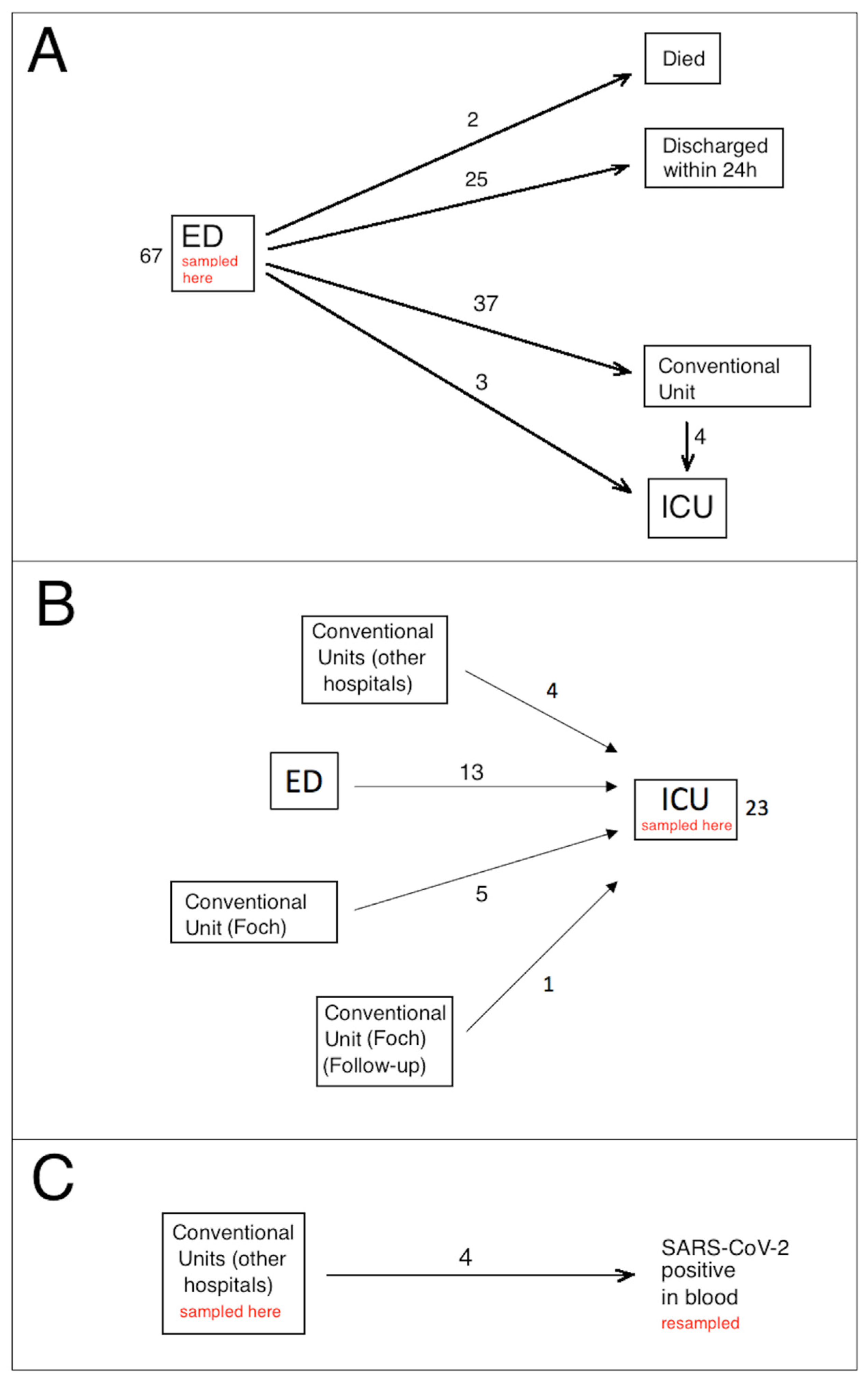
2.2. Patient Recruitment

The study was performed between 12 January and 15 July 2021, when the SARS-CoV-2 B.1.1.7 variant was present in France. Ninety-four patients (44 females, 50 males, median age 65 years) were included in the study. All enrolled patients were confirmed by RT-qPCR to be nasopharyngeal swab positive for SARS-CoV-2 infection. Figure 1 presents a diagram of the flow of the patients in this study through the ED, conventional units and ICU, indicating the hospital locations where blood sampling for SeptiCyte measurement occurred. A ‘conventional unit’ is defined as a general hospital ward, with extra precautions and isolations taken to minimize SARS-CoV-2 transmission.

2.3. Collection of Blood Samples

K3-EDTA whole blood samples, collected for complete blood count (CBC) and subsequently tested with SeptiCyte RAPID, were collected on admission in the ED in 67 cases (Figure 1A), in ICU for 23 patients (Figure 1B), and in conventional units for 4 patients (Figure 1C). Of the 67 patients tested in the ED, 25 were discharged within 24 h, because they had a moderate clinical form, did not require oxygen therapy, and had no biological signs of marked inflammation. None of these 25 patients required another hospitalization for COVID-19 and all were alive at least one month after their admission to the ED. Thirty-three patients were transferred from ED to conventional units for a median time of 8 days (range 2 to 22 days), four were first transferred from ED to conventional units, but then required admission to ICU some days later (median time 6 days, range 1–12 days), and three were immediately transferred from ED to ICU. Two older patients (85 and 93 years old) died in the ED.
Twenty-three patients were tested in ICU (Figure 1B). Of these, 13 were admitted first to the ED (within 24 h) but required high levels (>15 L/min) of oxygenotherapy and therefore were transferred to ICU as soon as possible, with nasopharyngeal swab RT-qPCR and blood sample collection occurring after ICU admission. Another group of 9 patients in ICU came from conventional units, either from Foch Hospital (5 cases) or from other hospitals (4 cases). Median length of stay in conventional units before ICU admission was 3 days (range 2 to 5 days). For the last case (labeled “Follow-up” in Figure 1B), the SeptiScore was determined during ICU stay, because the patient was tested during a follow-up for COVID-19 by RT-qPCR, and was still positive 35 days after the initial diagnosis of COVID-19. Of the 23 patients tested in ICU (Figure 1B), 22/23 were treated by corticotherapy and 11/23 were treated by antibiotherapy at the time of the SeptiScore determination. A blood culture was taken on the day SeptiCyte RAPID testing was performed in 20 cases, and was positive in 2 cases (Klebsiella pneumoniae and Pseudomonas aeruginosa). Similarly, cytobacteriological examination of the urine was taken from 12 patients, and was positive in 2 cases (Klebsiella pneumoniae and Enterobacter cloacae). On the day of SeptiCyte RAPID testing, the SOFA score was available for 20 patients and the median value was 2 (range: 0–12). For 10 ICU patients, follow-up samples were analyzed during ICU hospitalization, spaced approximately one week apart.
Lastly (Figure 1C), for 4 patients hospitalized in conventional units, a sample was analyzed during their hospitalization (median time: 7 days, range: 3–15 days). These patients were initially hospitalized because they had a positive RT-qPCR on nasopharyngeal swab, but on the day when the SeptiScore was analyzed, SARS-CoV-2 viral sequences were also found in their blood. Testing for SARS-CoV-2 in blood samples was not performed for all the patients, but only for patients in whom the use of convalescent plasma was envisaged.
For comparison, we also quantified the SeptiScore in 8 patients from ICU with culture proven bacterial sepsis as per the Sepsis-3 criteria [2], who were SARS-CoV-2 negative.

2.4. Processing of Samples

Diagnosis of SARS-CoV-2 infection was made using either the Alinity M SARS-CoV-2 RT-PCR assay (Abbott Molecular, Des Plaines, IL, USA) or the Xpert® Xpress SARS-CoV-2 (Cepheid Europe SAS, Maurens-Scopont, France).
Complete blood counts (CBC) were performed on a DxH 800 instrument (Beckman-Coulter, Villepinte, France) and CD16-positive monocyte quantification was performed using the CytoDiff™ reagent (Beckman-Coulter) as previously described [21]. Blood gas analysis, including lactate measurement, was performed on a GEM 4000 instrument (Werfen, Le Pré-Saint-Gervais, France). CRP and D-dimer were measured on Alinity IC (Abbott Diagnostics, Rungis, France) and ACL 700 (Werfen) systems, respectively. All measurements were made at approximately the same time the blood samples were collected for CBC and SeptiCyte RAPID testing.
SeptiCyte RAPID testing of eligible patients was performed on qualified Biocartis Idylla instruments (Biocartis, Mechelen, Belgium) installed with SeptiCyte RAPID assay software. Residual EDTA blood samples from CBC were diluted within several hours of collection with phosphate-buffered saline (PBS; 1 vol blood to 2.75 vol PBS), and 900 µL of each diluted sample was put into a SeptiCyte RAPID cartridge and automatically processed.
The SeptiCyte RAPID test is completely self-contained within single use cartridges that are designed to run on the Biocartis Idylla system. The entire assay process from blood sample extraction to final test result is achieved on-cartridge, with fully integrated sample preparation followed by reverse transcription and quantitative real-time polymerase chain reaction (RT-qPCR) for the detection and quantification of changes in expression of the immune host response genes PLAC8 and PLA2G7.
The test result (SeptiScore) is calculated based on the difference of cycle of quantification (Cq) values in RT-qPCR for the target genes. SeptiScores range from 0–15 and higher scores are associated with increasing sepsis probabilities. A sample processing control (SPC) is included in each cartridge to confirm that adequate nucleic acid extraction and amplification has occurred.

2.5. Radiologic Evaluation

COVID-19 disease severity was assessed as the percentage of specific lung abnormalities or damage as observed by a radiologist on a chest CT scan: (1) peripheral, bilateral ground glass opacity (GGO) with or without consolidation or visible intra-lobular lines (crazy paving), or (2) multifocal GGO of rounded shape with or without consolidation or visible intra-lobular lines. Patients were then classified as “absence”, “mild”, “moderate”, “extensive”, “severe”, or “critical” based on 0%, present but <10%, 10–25%, 25–50%, 50–75%, or >75% lung damage respectively. Three patients were indicated as having “non-specific” CT scan results, and were not considered further in the classification totals.

2.6. Statistical Analyses

Statistical analyses were performed using a combination of publicly available and custom scripts written in the R programming language [22]. The pROC package in R [23] was used for AUC calculations, and some calculations were verified with JLABROC4 (http://www.rad.jhmi.edu/jeng/javarad/roc/JROCFITi.html; accessed 22 April 2022). Pearson’s chi-squared test (stats package in R) was used to calculate p-values for categorical variables. Welch’s two-sided t-test or one-way ANOVA (stats package in R) was used to calculate p-values for numeric variables. In the case of ANOVA, variables were assumed to be normally distributed. Summary tables were then generated using the gtsummary package in R.
Combinatorial regression analysis, in which values of multiple clinical parameters (SeptiScore, CRP, D-dimer, lactate, monocytes, CD16-positive monocytes) were combined, was performed with a custom R script. Missing values in the clinical dataset were imputed prior to the combinatorial analysis, with the R package Amelia [24].

3. Results

The main characteristics of the patients in this study are summarized in Table 1. Obesity, characterized by a body mass index (BMI) >30, was not reported in this table because this parameter was available only for 46 patients. Additional risk factors (including BMI when available) and treatments of the patients during their hospitalization are indicated in Supplementary Table S1. The distribution of SARS-CoV-2 -positive patients across COVID-19 severity categories and hospital locations is indicated in Supplementary Table S2.
In the 94-patient cohort, there were 17 patients with pre-existing comorbidities involving lung conditions (some patients with multiple conditions): cystic fibrosis (n = 1), asthma (n = 4), chronic obstructive pulmonary disease (COPD; n = 7), lung cancer (n = 3), lung transplantation (n = 4), non-specific interstitial pneumopathy (n = 1). The SeptiScores for these patients as a group were compared to the SeptiScores of the remaining 77 patients in the cohort, using the Kolmogorov-Smirnov test. No significant difference was found between the two groups (D = 0.293, p = 0.243).

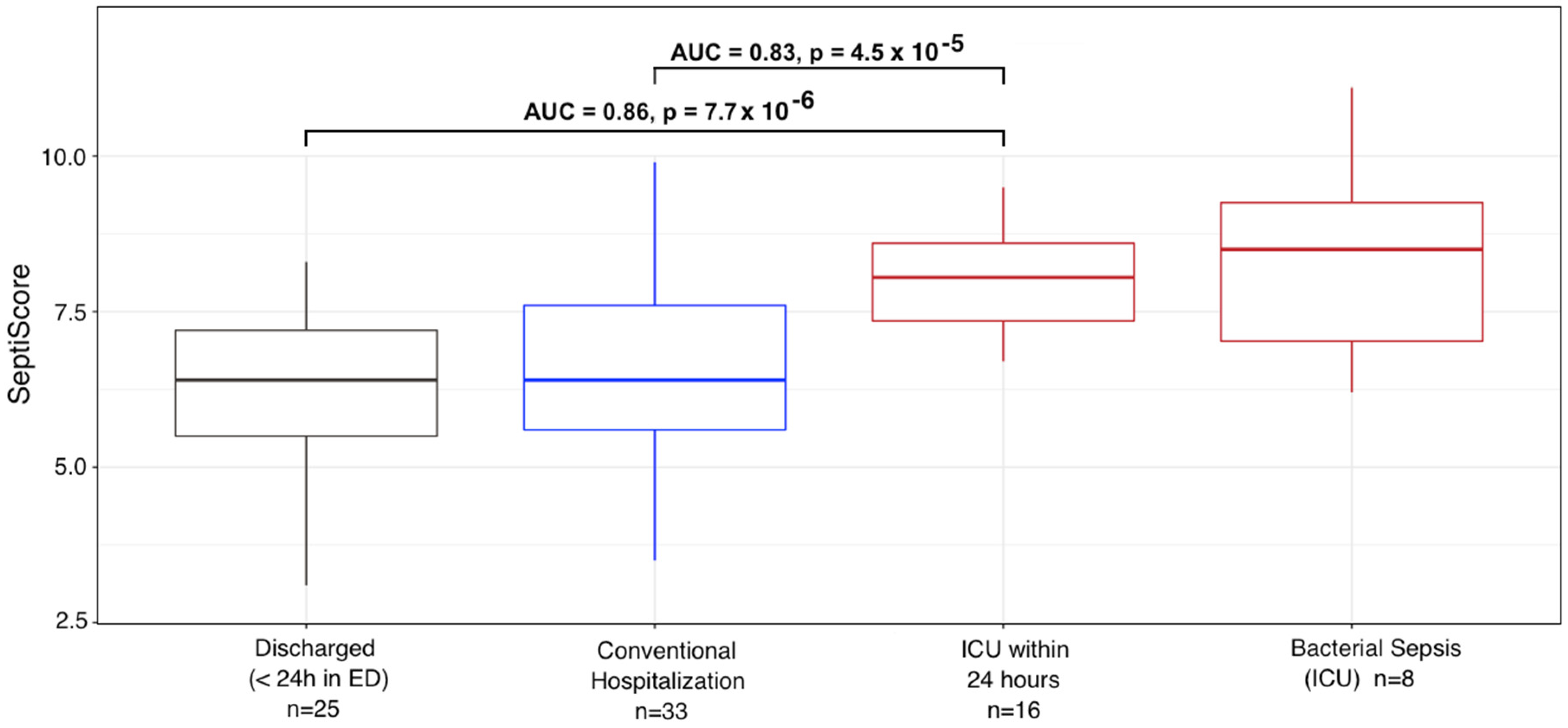
3.1. Variations of SeptiScore and Other Biological Markers According to Patient Group

Variations of the SeptiScore were analyzed in comparison to other well-recognized biological markers of severity of SARS-CoV-2 infection (variations in leucocyte differential, including CD16-positive monocytes, CRP, D-dimer and lactate). As can be seen in Table 2 and Figure 2 the SeptiScore increased with the severity of the disease. In considering the entire 94 patient cohort, using samples taken at admission, SeptiScore was significantly correlated with CRP (Pearson sample correlation coefficient r = 0.513; p < 0.0001), lactate (r = 0.321; p = 0.014) lymphocyte count (r = −0.354; p = 0.0008) and monocyte count (r = −0.219; p = 0.038).
SeptiScores were significantly higher in patients requiring ICU hospitalization, compared to patients who were immediately discharged or transferred to conventional units (p = 0.0004, Kruskal-Wallis test). SeptiScores for the ICU patients were similar in value to scores for 8 SARS-CoV-2 -negative patients having culture-confirmed bacterial sepsis (Figure 2).
A receiver operating characteristic (ROC) curve analysis was performed on the different biological parameters recorded during the study, in order to identify patients requiring ICU hospitalization. An AUC > 0.7 was considered to be relevant as a discriminating parameter [25]. Using data from the earliest measured values of the parameters, only two biological parameters, SeptiScore and CRP, fulfilled this criteria, with an AUC of 0.78 and 0.79, and a binary cut-off of 6.6 and 100 mg/L, respectively.

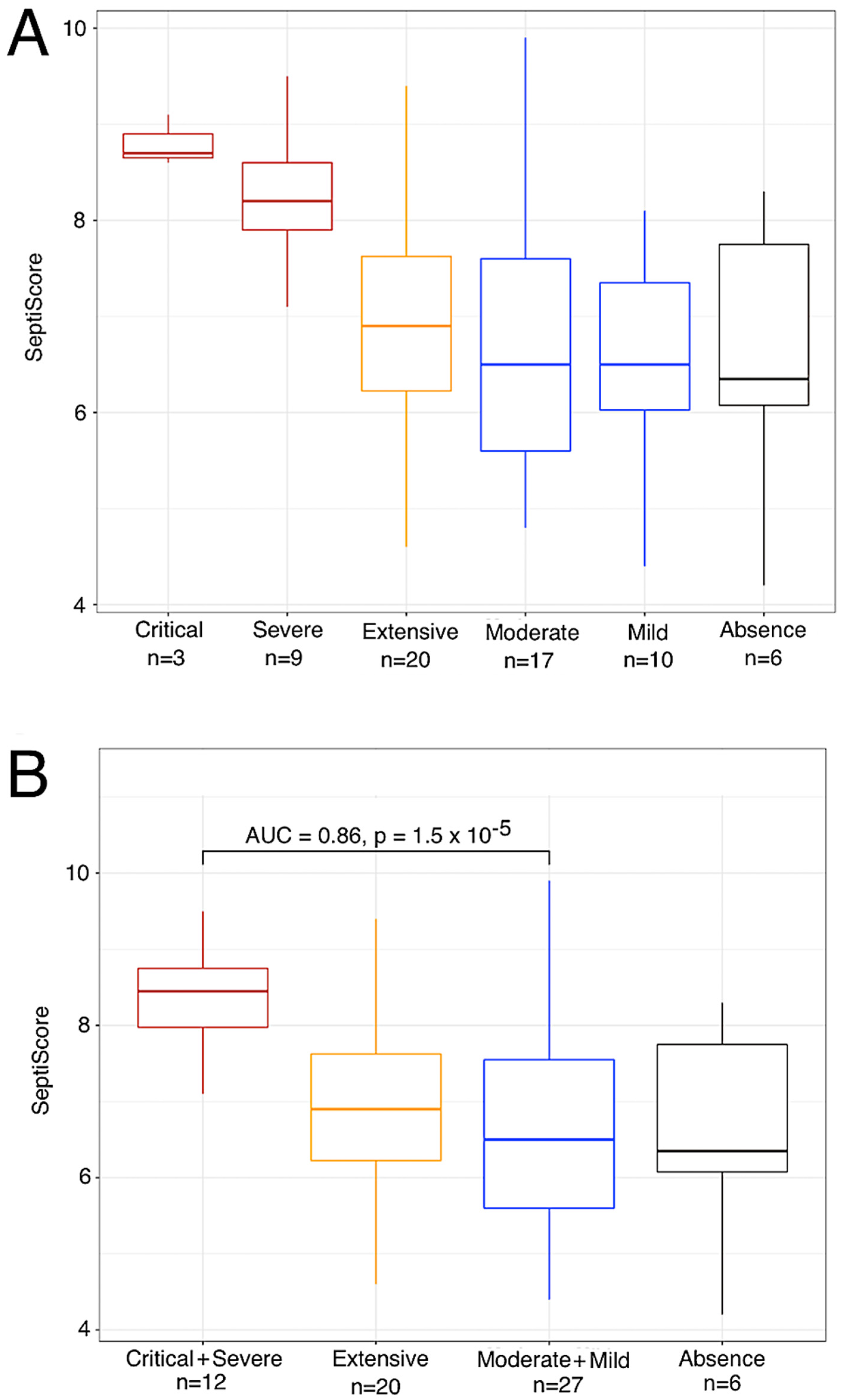
3.2. Variation in SeptiScore, According to Lung Injury Observed in CT Scan

For 68 patients, a CT scan was performed within 24 h after admission to the hospital. Three of the patients had non-specific presentations in CT scan, and were not considered further. Six had an absence of lung damage, while the remaining patients could be classified as having mild (n = 10), moderate (n = 17), extensive (n = 20), severe (n = 9) or critical (n = 3) extents of lung damage. As can be seen in Figure 3, the SeptiScore increased with the extent of the lung injury, the highest SeptiScore values being observed in patients with severe or critical lung injuries. Following from this analysis, patients were divided into two groups: those with severe or critical (≥50%) lung injury (n = 12), vs. those with specific but <50% lung injury (n = 53), as defined by chest CT scan. ROC curve analysis gave AUC = 0.86, with a SeptiScore cut-off of 7 providing a good discrimination between these two groups. CRP, lymphocytes, monocytes, CD16-positive monocytes and lactate were also discriminatory for patients with ≥50% versus <50% lung injury (Table 3).
Interestingly, SeptiScores and CT scans were determined three times for two patients while they were in the ICU. Each time, both patients had lung injury ≥50% and their median SeptiScore was 8.4 (range 6 to 9.7), while their median CRP had decreased to 2.5 mg/L, presumably because of their treatments with corticosteroids and tocilizumab.
We have demonstrated in Figure 3 that the SeptiScore correlates with the severity of lung damage in chest CT scans, which serves as a proxy for severity of COVID-19 disease. It is true that pre-existing lung-related comorbidities such as COPD produce lung damage that is evident on CT scans, and 17 patients in our cohort had preexisting lung-related comorbidities, including 7 with COPD. However, the correlations we observed between SeptiCyte scores and lung injuries are restricted to lung injuries attributable to COVID-19, particularly concerning ground glass opacity (GGO), which is commonly found in COVID-19 disease and not in COBP [26,27].

3.3. Elevated SeptiScore Is Predictive of ICU Admission in Patients with Extensive Lung Injury

All the patients except for one with lung injury ≥50% were admitted to ICU. In addition, among the 17 patients with extensive lung injury (between 25–50%) whose age was less than 80 years, 6 were admitted to ICU, either immediately after hospital admission (3 cases) or after some days in conventional units (3 cases). For patients with extensive lung injury and age <80 years, SeptiScores were significantly higher in those requiring ICU care (n = 6) versus those who did not require this level of care (n = 11), with median SeptiScore values of 7.6 versus 6.6, respectively (p = 0.014 by Welch’s two sample t-test). In contrast, CRP levels were not significantly different for these patients, with median levels of 83 versus 114 mg/L, respectively (p = 0.71 by Welch’s two sample t-test). The SeptiScore AUC was 0.86, and an optimal SeptiScore cut-off for this discrimination was 6.7.

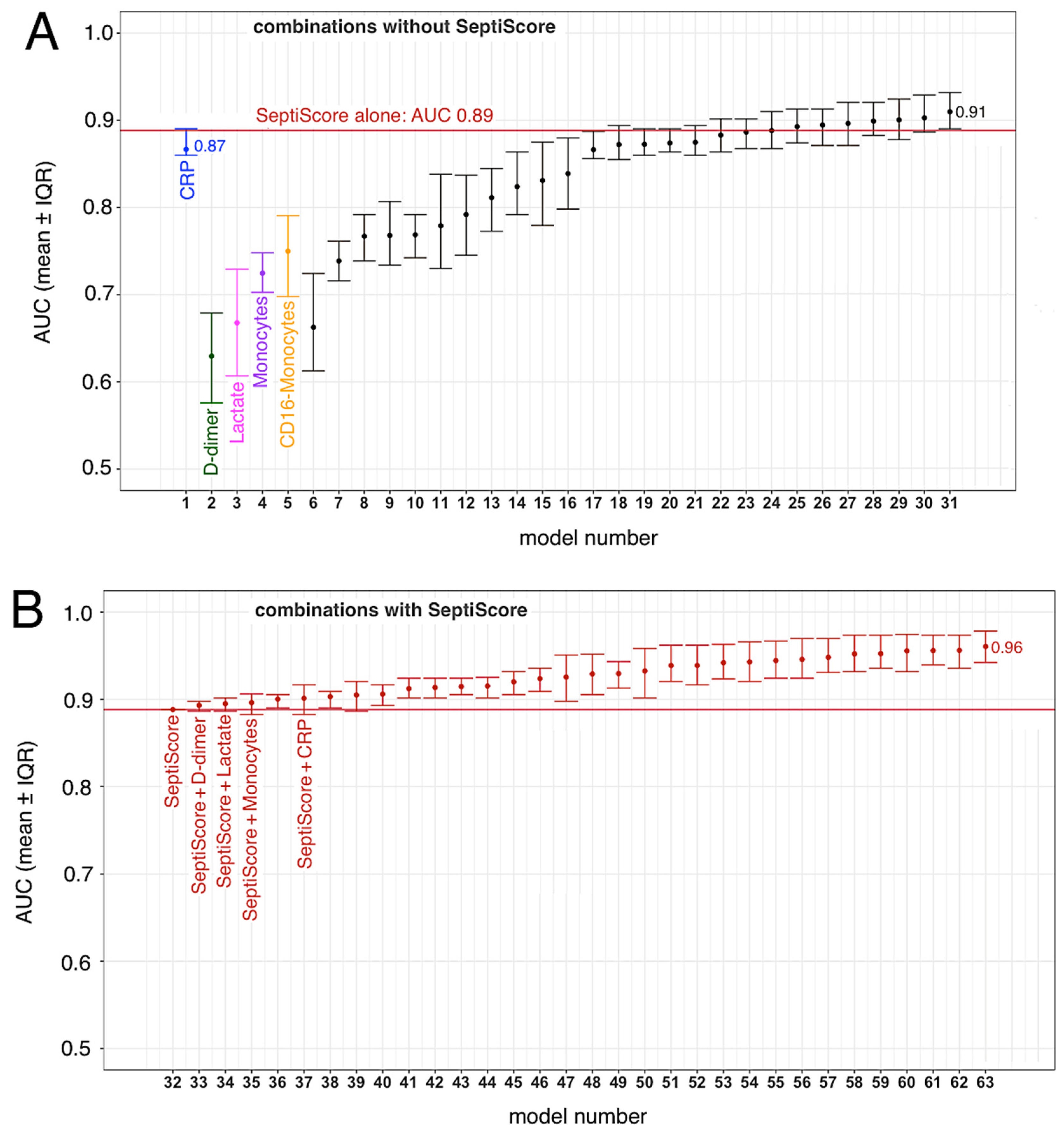
3.4. SeptiCyte RAPID, Compared to/Combined with Other Clinical Variables

We conducted ROC curve analyses to compare SeptiCyte RAPID to other single biomarkers, specifically CRP, D-dimer, lactate, monocytes, and CD16-positive monocytes, for discriminating (critical + severe) vs. (moderate + mild) cases. These biomarkers have all been used previously by clinicians and investigators to assess the severity of COVID-19 clinical trajectories [21,28,29,30,31,32].
In these analyses, we compared critical + severe cases in ICU (n = 11) versus moderate + mild cases not in ICU (n = 24), with only the first SeptiCyte RAPID measurement considered. The complete combinatorial analysis is presented in Supplementary Table S3. We found that the mean AUC for SeptiCyte RAPID was greater than that of any individual biomarker and of most combinations thereof (Figure 4A).
The single biomarker that came closest to SeptiCyte RAPID in performance was CRP (AUC 0.87, versus SeptiScore AUC 0.89). In this regard, however, we note that SeptiCyte RAPID remains discriminatory (i.e., sensitive to lung injury) during the follow-up period, when CRP has been artificially reduced due to treatment with corticosteroids and tocilizumab (see Section 4).
Combining the SeptiScore with one or more of these other biomarkers provided an additional improvement in AUC beyond the use of the SeptiScore alone (Figure 4B). Any of the logistic regression models containing SeptiScore with clinical parameters (Panel B) performed better than all of the singleton biomarker models, and better than most models that did not contain SeptiScore (compare panels B and A).

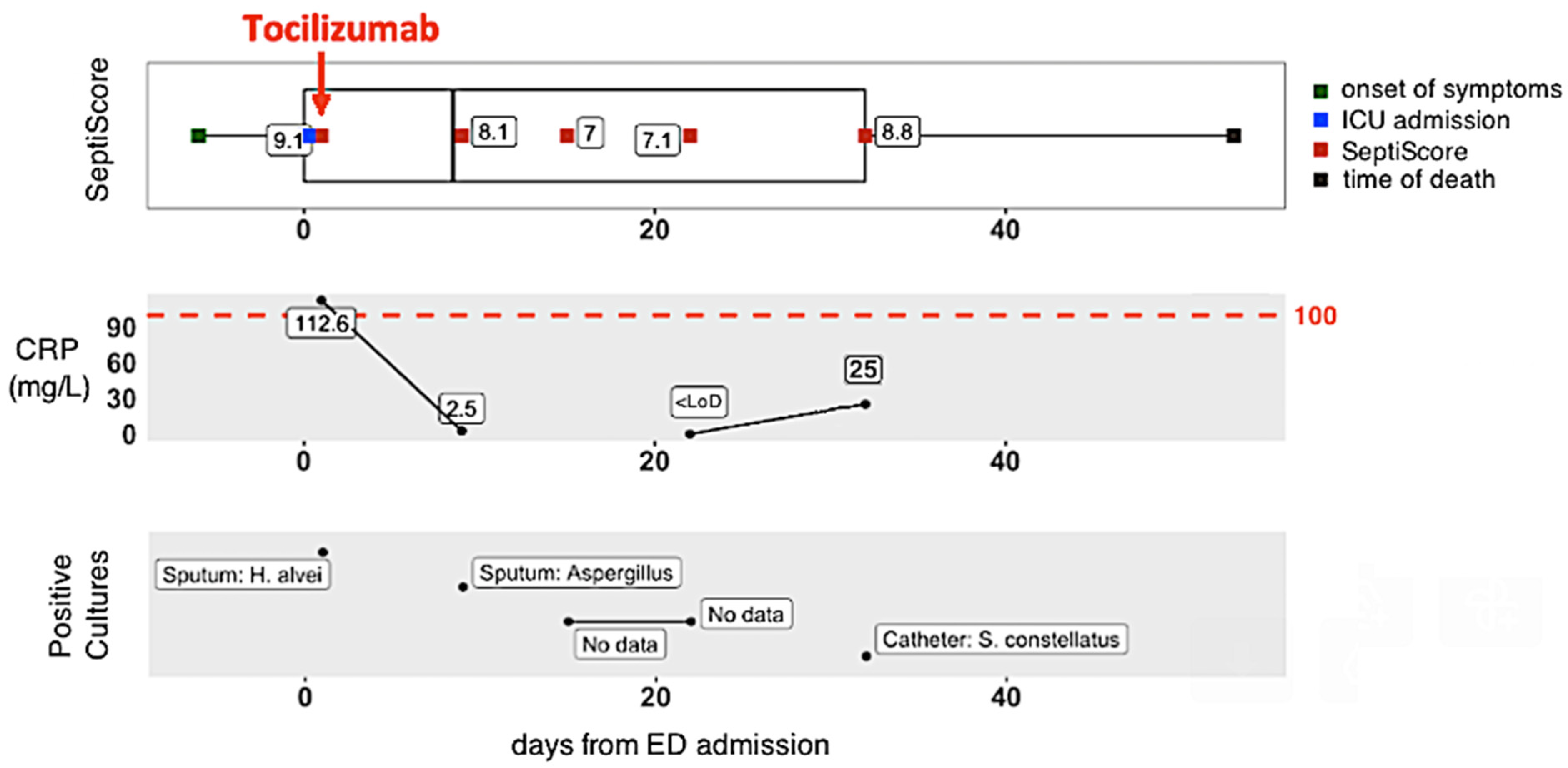
3.5. Longitudinal Monitoring of Patient Trajectories

Finally, we consider the use of SeptiCyte RAPID, as a component of the longitudinal monitoring of COVID-19 patients during their transit through hospital. Figure 5 presents the clinical trajectory of one such patient, who presented to ED, was transferred immediately to ICU, suffered a critical level of lung damage (>75%), received intubation with mechanical ventilation, and unfortunately died. An important point to note about this patient’s trajectory is that an initially high CRP level (112.5 mg/L) was greatly reduced (to 2.5 mg/L) by tocilizumab + corticosteroids. However, the SeptiScore, initially 9.1, remained high throughout the patient’s trajectory (8.1, 7.0, 7.1, 8.8 upon repeat testing).

4. Discussion

Infection by SARS-CoV-2, initially described in China in 2019, rapidly spread throughout the world and has evolved in several waves, corresponding with the appearance of new variants. These waves are characterized by the influx of patients to emergency departments, which are quickly saturated. Therefore, it is important to have a rapid diagnosis which identifies patients with mild forms of the disease who can immediately be discharged, versus those who require hospitalization, or even admission to intensive care.
In addition, identification of potential risk factors that could potentially predict the disease course may be of great utility for healthcare professionals to efficiently triage patients, personalize treatment, monitor clinical progress, and allocate proper resources at all levels of care to mitigate morbidity and mortality. Different risk factors, which can be divided into demographic, clinical, hematological, biochemical, and radiographic factors, have been described so far to evaluate COVID-19 disease severity [6]. However, although certain factors clearly identify patients at high risk, none can be considered a gold standard.
At this time, in addition to the clinical examination and hypoxemia, evaluation of disease severity is based on hematological tests (severity of the lymphopenia is considered as a risk factor), biochemical tests to evaluate the inflammatory state (mainly CRP), and CT scans [33], with the patient’s age and comorbidities also taken into account. However, hospital central laboratories and imaging departments may be quickly overwhelmed by the importance of the requests, potentially leading to delays in the return of results, and consequently to emergency department overcrowding. Therefore, it is important to have tests allowing a rapid assessment of the severity of patient infections.
In this study we have evaluated the performance of a new host immune response test, SeptiCyte RAPID, in discriminating COVID-19 severity in confirmed SARS-CoV-2 positive patients who were admitted to the ED. The study cohort reflected the real-world situation at this stage of the pandemic: it consisted of a heterogeneous group of non-acute and acutely ill patients with varying phases of an inflammatory response as well as a range of comorbidities.
SeptiCyte RAPID was found to have utility for diagnosing severe or critical COVID-19 and the subsequent risk of ICU admission in SARS-CoV-2 positive patients. At time of presentation, SeptiCyte RAPID differentiated critical + severe vs. moderate + mild COVID-19 with AUC 0.86 (Figure 3B). If the critical + severe cohort was restricted to those measured in the ICU vs. moderate + mild measured outside the ICU, then the AUC increases to 0.89 (Figure 4). In patients requiring ICU hospitalization, the SeptiScore was similar to values observed in patients with culture-confirmed bacterial sepsis.
Additionally, the SeptiScore discriminated patients with lung injury above 50% on CT scan (AUC = 0.88 for critical + severe vs. absence + mild + moderate + extensive), and particularly could identify patients with extensive lung injury (25–50%) requiring ICU admission. Lastly, during the follow-up of the patients, the SeptiScore remained elevated in patients with lung injury above 50%, whereas other biological markers such as CRP decreased, likely due to anti-inflammatory treatments (corticosteroids, tocilizumab) received by the patients.
We note that SeptiCyte RAPID was initially validated for use in patients exhibiting ≥2 SIRS criteria and suspected of sepsis, with bacterial infection being the most common etiology [18]. Optimal application of the test for discriminating COVID-19 severities, or for guiding the ICU admission decision for COVID-19 patients, could involve minor adjustment to the predefined assay cutoffs used by the SeptiCyte RAPID software. Testing of additional COVID-19 patients may provide further insight on this point.
In comparing SeptiCyte and CRP, several points should be further emphasized. The study cohort was heavily treated with both corticosteroids (59/94 = 63%) and tocilizumab (24/94 = 26%), with some patients receiving both therapies (22/94 = 23%). It is well-known that CRP is negatively modulated by both corticosteroids and tocilizumab [34,35,36,37,38,39]. Thus, in treated patients, CRP may no longer be a useful indicator of progression to more severe states, e.g., secondary bacterial infections [36,40], whereas SeptiScore values were not impacted, consistent with its original function as an indicator of sepsis. The patient trajectory described in Figure 5 illustrates this point: an initially high CRP level was greatly reduced by tocilizumab + corticosteroids, but the SeptiScore remained high throughout the patient’s trajectory. In this particular case, while CRP may have been an accurate indicator of inflammatory state, it was clearly unable to indicate the underlying condition that eventually led to the patient’s death. In contrast, the SeptiScore remained high and at an alert level.
Another point to note is that CRP is an integral component of the “acute phase response” which is an early and relatively non-specific part of the innate immune response to various stressors, including trauma and infection by both bacteria and viruses [41,42]. The two genes of SeptiCyte RAPID (PLAC8 and PLA2G7) are not in the acute phase response pathway, so it is perhaps unsurprising that they behave differently in response to anti-inflammatory treatments.
One strength of our study is that test results were returned quickly in real time, typically within 1–2 h of sample collection, in a near-patient setting (even in the ED). An additional strength of the study is that it is representative of the real-world situation that occurred in our institution over the study period. However, it is clear that is also a limitation, since the patient population was quite heterogeneous in terms of pre-existing conditions and comorbidities. This study design limitation could lead to multiple variables contributing to an elevation of the SeptiCyte RAPID score. A further limitation is that most of the patients admitted to ICU had blood drawn after ICU admission. Consequently, the SeptiScore was perhaps different (possibly higher) when patients were in the ED. This question will need to be addressed in subsequent studies. A final limitation of our study is that we were unable to follow patients after discharge from hospital, to determine if they ultimately exhibited any post-acute sequelae of COVID-19 disease, i.e., signs or symptoms of “long COVID” [43,44,45]. It remains to be established whether SeptiCyte RAPID would have clinical utility in helping to diagnose this condition.

5. Conclusions

This study supports the potential use of SeptiCyte RAPID in the risk stratification of COVID-19 patients, as measured by clinical severity assessment based on chest CT scans and/or ICU admission. With a quick availability from whole blood (potentially within 1 h) and with minimal user interaction, the assay may be helpful in stratifying or triaging patients during their transit through hospital. In contrast, hematological and biochemical markers as well as CT scans can take several hours, especially in the event of a massive influx of patients to the hospital. Secondly, small hospitals do not always have an imaging department available 24 h per day, and the SeptiScore result provides potentially useful information in the event of lung damage greater than 50%. Thirdly, it can be useful for the follow-up of patients, particularly patients in the ICU, who are difficult to transport to the imaging department, and for whom the interventional treatments may have normalized the usual biochemical and/or hematological parameters (such as CRP). The SeptiCyte RAPID test could also constitute an approach to evaluate lung damage. Moreover, the evolution of patients with COVID-19 in intensive care is often marked by the appearance of bacterial complications, for which the SeptiScore has shown potential value (Figure 5). SARS-CoV-2 is becoming endemic [46,47] and yet continues to evolve rapidly, raising the possibility that new variants with unknown properties will be generated unpredictably over the foreseeable future. Thus, there will be a continuing need for tools such as SeptiCyte RAPID to help clinicians evaluate the potential severity of COVID-19 disease trajectories, particularly in vulnerable individuals.

Supplementary Materials

The following supporting information can be downloaded at https://www.mdpi.com/article/10.3390/v15020419/s1, Table S1: Patient comorbidities and treatments; Table S2: Distribution of SARS-CoV-2 (+) patients across COVID-19 severity categories and hospital locations; Table S3: Logistic Regression models (see Figure 4A,B in main text).

Author Contributions

Conceptualization: M.V.; methodology: M.V., V.G. and J.T.K.; investigation: M.V.; data acquisition, V.G., F.M., F.A., M.-C.B., B.Z., F.P., T.P., E.F. and M.V.; data curation: M.V. and K.N.; custom software: K.N.; visualization: K.N., T.D.Y. and M.V.; formal analysis: M.V., V.G., K.N. and T.D.Y.; writing—original draft preparation: M.V. and T.D.Y.; writing—review and editing: M.V., B.Z., F.M., T.P., E.F., T.D.Y., K.N. and J.T.K.; supervision: M.V.; project administration: M.V.; funding acquisition: M.V. and T.D.Y. All authors have read and agreed to the published version of the manuscript.

Funding

This research was funded in part by contract # 75A50120C00125 from the DRIVe Solving Sepsis program of the Biomedical Advanced Research and Development Authority (BARDA), part of the US HHS Office of the Assistant Secretary for Preparedness and Response.

Institutional Review Board Statement

This study was conducted under a blanket protocol for COVID-related research at the Foch Hospital (“Information réutilisation des données personnelles et des échantillons biologiques à des fins de recherche V2–9 avril 2020”), which had been established and approved by the local ethics committee of the Foch Hospital (IRB00012437).

Informed Consent Statement

The study was performed in agreement with French ethical laws, such that the patients and their families were informed that their biological samples and data used for routine care could be used in an anonymous manner unless they expressed their opposition. No patients enrolled in this study, or their families, expressed opposition to the use of their biological samples and data, which according to the French ethical laws and Foch Hospital ethics committee constitutes informed consent.

Data Availability Statement

Data will be provided, upon reasonable request to the corresponding author.

Acknowledgments

We thank Biocartis for provision of the Idylla platform to the Foch Hospital site, and Julie Gaiffas and David Favy of Biocartis for liason support. We also thank Immunexpress for continued financial support over the course of this project. We thank Dayle Sampson for initial R coding for analysis leading to Figure 4.

Conflicts of Interest

T.D.Y., K.N. and J.T.K. are current or former employees and shareholders in Immunexpress. The other authors declare no competing interests. The DRIVe Solving Sepsis program of the Biomedical Advanced Research and Development Authority (BARDA) had no role in the design of the study; in the collection, analyses, or interpretation of data; in the writing of the manuscript; or in the decision to publish the results.

Abbreviations

AHT, arterial hypertension; ARDS, acute respiratory distress syndrome; AUC, area under ROC curve; BMI, body mass index; CD16, Fc receptor with low affinity for IgG, encoded by genes FCGR3A and FCGR3B; CI, confidence interval; COPD, chronic obstructive pulmonary disease; COVID-19, coronavirus disease 2019; CRP, C-reactive protein; CT, computed tomography; ECMO, extracorporeal membrane oxygenation; ED, Emergency Department; EDTA, ethylene diamine tetraacetic acid; F, female; GGO, ground glass opacity; ICU, Intensive Care Unit; M, male; NIH, United States National Institutes of Health; NGS, next-generation sequencing; NA, not available; PCT, procalcitonin; PLA2G7, Phospholipase A2 Group VII gene; PLAC8, Placenta Associated 8 gene; ROC, receiver operating characteristic [curve]; RT-qPCR, reverse transcription–quantitative polymerase chain reaction; SARS-CoV-2, severe acute respiratory syndrome coronavirus 2; SPC, sample processing control; WHO, World Health Organization.

References

  1. Hotchkiss, R.S.; Monneret, G.; Payen, D. Sepsis-induced immunosuppression: From cellular dysfunctions to immunotherapy. Nat. Rev. Immunol. 2013, 13, 862–874. [Google Scholar] [CrossRef]
  2. Singer, M.; Deutschman, C.S.; Seymour, C.W.; Shankar-Hari, M.; Annane, D.; Bauer, M.; Bellomo, R.; Bernard, G.R.; Chiche, J.-D.; Coopersmith, C.M.; et al. The Third International Consensus Definitions for Sepsis and Septic Shock (Sepsis-3). JAMA 2016, 315, 801–810. [Google Scholar] [CrossRef]
  3. Bohn, M.K.; Hall, A.; Sepiashvili, L.; Jung, B.; Steele, S.; Adeli, K. Pathophysiology of COVID-19: Mechanisms Underlying Disease Severity and Progression. Physiology 2020, 35, 288–301. [Google Scholar] [CrossRef] [PubMed]
  4. Berlin, D.A.; Gulick, R.M.; Martinez, F.J. Severe COVID-19. N. Engl. J. Med. 2020, 383, 2451–2460. [Google Scholar] [CrossRef]
  5. Shappell, C.N.; Klompas, M.M.; Kanjilal, S.M.; Chan, C.M.; Rhee, C.M. Prevalence, Clinical Characteristics, and Outcomes of Sepsis Caused by Severe Acute Respiratory Syndrome Coronavirus 2 Versus Other Pathogens in Hospitalized Patients with COVID-19. Crit. Care Explor. 2022, 4, e0703. [Google Scholar] [CrossRef]
  6. Bruse, N.; Kooistra, E.J.; Jansen, A.; van Amstel, R.B.E.; de Keizer, N.F.; Kennedy, J.N.; Seymour, C.; van Vught, L.A.; Pickkers, P.; Kox, M. Clinical sepsis phenotypes in critically ill COVID-19 patients. Crit. Care 2022, 26, 1–6. [Google Scholar] [CrossRef]
  7. Vincent, J.-L. COVID-19: It’s all about sepsis. Futur. Microbiol. 2021, 16, 131–133. [Google Scholar] [CrossRef] [PubMed]
  8. Olwal, C.O.; Nganyewo, N.N.; Tapela, K.; Djomkam, L.A.; Owoicho, O.; Bediako, Y.; Duodu, S. Parallels in Sepsis and COVID-19 Conditions: Implications for Managing Severe COVID-19. Front. Immunol. 2021, 12, 602848. [Google Scholar] [CrossRef]
  9. Beltrán-García, J.; Osca-Verdegal, R.; Pallardó, F.V.; Ferreres, J.; Rodríguez, M.; Mulet, S.; Ferrando-Sánchez, C.; Carbonell, N.; García-Giménez, J.L. Sepsis and Coronavirus Disease 2019: Common Features and Anti-Inflammatory Therapeutic Approaches. Crit. Care Med. 2020, 48, 1841–1844. [Google Scholar] [CrossRef] [PubMed]
  10. Abate, S.M.; Ali, S.A.; Mantfardo, B.; Basu, B. Rate of Intensive Care Unit admission and outcomes among patients with coronavirus: A systematic review and Meta-analysis. PLoS ONE 2020, 15, e0235653. [Google Scholar] [CrossRef]
  11. Hirabara, S.M.; Serdan, T.D.A.; Gorjao, R.; Masi, L.N.; Pithon-Curi, T.C.; Covas, D.T.; Curi, R.; Durigon, E.L. SARS-CoV-2 Variants: Differences and Potential of Immune Evasion. Front. Cell. Infect. Microbiol. 2022, 11, 781429. [Google Scholar] [CrossRef] [PubMed]
  12. Jung, C.; Kmiec, D.; Koepke, L.; Zech, F.; Jacob, T.; Sparrer, K.M.J.; Kirchhoff, F. Omicron: What Makes the Latest SARS-CoV-2 Variant of Concern So Concerning? J. Virol. 2022, 96, e0207721. [Google Scholar] [CrossRef] [PubMed]
  13. Chakraborty, C.; Bhattacharya, M.; Sharma, A.R.; Dhama, K.; Agoramoorthy, G. A comprehensive analysis of the mutational landscape of the newly emerging Omicron (B.1.1.529) variant and comparison of mutations with VOCs and VOIs. Geroscience 2022, 44, 2393–2425. [Google Scholar] [CrossRef]
  14. Dhawan, M.; Saied, A.A.; Mitra, S.; Alhumaydhi, F.A.; Bin Emran, T.; Wilairatana, P. Omicron variant (B.1.1.529) and its sublineages: What do we know so far amid the emergence of recombinant variants of SARS-CoV-2? Biomed. Pharmacother. 2022, 154, 113522. [Google Scholar] [CrossRef] [PubMed]
  15. Ou, J.; Lan, W.; Wu, X.; Zhao, T.; Duan, B.; Yang, P.; Ren, Y.; Quan, L.; Zhao, W.; Seto, D.; et al. Tracking SARS-CoV-2 Omicron diverse spike gene mutations identifies multiple inter-variant recombination events. Signal Transduct. Target. Ther. 2022, 7, 1–9. [Google Scholar] [CrossRef] [PubMed]
  16. Marin, B.G.; Aghagoli, G.; Lavine, K.; Yang, L.; Siff, E.J.; Chiang, S.S.; Salazar-Mather, T.P.; Dumenco, L.; Savaria, M.C.; Aung, S.N.; et al. Predictors of COVID-19 severity: A literature review. Rev. Med. Virol. 2021, 31, 1–10. [Google Scholar] [CrossRef] [PubMed]
  17. Ye, Z.; Zhang, Y.; Wang, Y.; Huang, Z.; Song, B. Chest CT manifestations of new coronavirus disease 2019 (COVID-19): A pictorial review. Eur. Radiol. 2020, 30, 4381–4389. [Google Scholar] [CrossRef]
  18. Brandon, R.; Kirk, J.; Yager, T.; Cermelli, S.; Davis, R.; Sampson, D.; Sillekens, P.; Keuleers, I.; Vanhoey, T. Clinical performance of a rapid sepsis test on a near-patient molecular testing platform. Abstract # P481, ISICEM 2020 - 40th International Symposium on Intensive Care and Emergency Medicine, Brussels, Belgium. Crit Care 2020, 24 (Suppl 1), 87. [Google Scholar] [CrossRef]
  19. Schulte-Schrepping, J.; Reusch, N.; Paclik, D.; Baßler, K.; Schlickeiser, S.; Zhang, B.; Krämer, B.; Krammer, T.; Brumhard, S.; Bonaguro, L.; et al. Severe COVID-19 Is Marked by a Dysregulated Myeloid Cell Compartment. Cell 2020, 182, 1419–1440. [Google Scholar] [CrossRef]
  20. Gravrand, V.; Mellot, F.; Ackermann, F.; Ballester, M.-C.; Zuber, B.; Kirk, J.; Yager, T.; Navalkar, K.; Pascreau, T.; Farfour, E.; et al. SeptitiCyte RAPID in COVID-19 Severity Stratification and Triage. Abstract # 04729, ECCMID 2022. In Proceedings of the European Congress of Clinical Microbiology and Infectious Diseases, Lisbon, Portugal, 23–26 April 2022; Available online: https://2022.eccmid.org/ (accessed on 6 January 2023).
  21. Vasse, M.; Zuber, B.; Goubeau, L.; Ballester, M.-C.; Roumier, M.; Delcominette, F.; Habarou, F.; Jolly, E.; Ackermann, F.; Cerf, C.; et al. A low level of CD16pos monocytes in SARS-CoV-2 infected patients is a marker of severity. Clin. Chem. Lab. Med. 2021, 59, 1315–1322. [Google Scholar] [CrossRef]
  22. R Core Team. R: A Language and Environment for Statistical Computing; R Foundation for Statistical Computing: Vienna, Austria, 2013; Available online: http://www.R-project.org/ (accessed on 1 November 2021)ISBN 3-900051-07-0.
  23. Robin, X.; Turck, N.; Hainard, A.; Tiberti, N.; Lisacek, F.; Sanchez, J.-C.; Müller, M. pROC: An open-source package for R and S+ to analyze and compare ROC curves. BMC Bioinform. 2011, 12, 77. [Google Scholar] [CrossRef] [PubMed]
  24. Honaker, J.; King, G.; Blackwell, M. AmeliaII: A Program for Missing Data. J. Stat. Softw. 2011, 45, 1–47. [Google Scholar] [CrossRef]
  25. Mandrekar, J.N. Receiver Operating Characteristic Curve in Diagnostic Test Assessment. J. Thorac. Oncol. 2010, 5, 1315–1316. [Google Scholar] [CrossRef] [PubMed]
  26. Alves, A.F.F.; Miranda, J.R.A.; Reis, F.; Oliveira, A.A.; Souza, S.A.S.; Fortaleza, C.M.C.B.; Tanni, S.E.; Castro, J.T.S.; Pina, D.R. Automatic algorithm for quantifying lung involvement in patients with chronic obstructive pulmonary disease, infection with SARS-CoV-2, paracoccidioidomycosis and no lung disease patients. PLoS ONE 2021, 16, e0251783. [Google Scholar] [CrossRef] [PubMed]
  27. Salehi, S.; Abedi, A.; Balakrishnan, S.; Gholamrezanezhad, A. Coronavirus Disease 2019 (COVID-19): A Systematic Review of Imaging Findings in 919 Patients. Am. J. Roentgenol. 2020, 215, 87–93. [Google Scholar] [CrossRef]
  28. Ali, N. Elevated level of C-reactive protein may be an early marker to predict risk for severity of COVID-19. J. Med Virol. 2020, 92, 2409–2411. [Google Scholar] [CrossRef]
  29. Varikasuvu, S.R.; Varshney, S.; Dutt, N.; Munikumar, M.; Asfahan, S.; Kulkarni, P.P.; Gupta, P. D-dimer, disease severity, and deaths (3D-study) in patients with COVID-19: A systematic review and meta-analysis of 100 studies. Sci. Rep. 2021, 11, 1–11. [Google Scholar] [CrossRef]
  30. Correia, B.S.B.; Ferreira, V.G.; Piagge, P.M.F.D.; Almeida, M.B.; Assunção, N.A.; Raimundo, J.R.S.; Fonseca, F.L.A.; Carrilho, E.; Cardoso, D.R. 1H qNMR-Based Metabolomics Discrimination of Covid-19 Severity. J. Proteome Res. 2022, 21, 1640–1653. [Google Scholar] [CrossRef]
  31. Knoll, R.; Schultze, J.L.; Schulte-Schrepping, J. Monocytes and Macrophages in COVID-19. Front. Immunol. 2021, 12. [Google Scholar] [CrossRef]
  32. Payen, D.; Cravat, M.; Maadadi, H.; Didelot, C.; Prosic, L.; Dupuis, C.; Losser, M.-R.; Bittencourt, M.D.C. A Longitudinal Study of Immune Cells in Severe COVID-19 Patients. Front. Immunol. 2020, 11, 580250. [Google Scholar] [CrossRef]
  33. Francone, M.; Iafrate, F.; Masci, G.M.; Coco, S.; Cilia, F.; Manganaro, L.; Panebianco, V.; Andreoli, C.; Colaiacomo, M.C.; Zingaropoli, M.A.; et al. Chest CT score in COVID-19 patients: Correlation with disease severity and short-term prognosis. Eur. Radiol. 2020, 30, 6808–6817. [Google Scholar] [CrossRef]
  34. Keske, Ş.; Tekin, S.; Sait, B.; Irkören, P.; Kapmaz, M.; Çimen, C.; Uğur, S.; Çelebi, I.; Bakır, V.O.; Palaoğlu, E.; et al. Appropriate use of tocilizumab in COVID-19 infection. Int. J. Infect. Dis. 2020, 99, 338–343. [Google Scholar] [CrossRef] [PubMed]
  35. Zeng, J.; Xie, M.-H.; Yang, J.; Chao, S.-W.; Xu, E.-L. Clinical efficacy of tocilizumab treatment in severe and critical COVID-19 patients. World J. Clin. Cases 2020, 8, 3763–3773. [Google Scholar] [CrossRef]
  36. Hofmaenner, D.A.; Garcia, P.D.W.; Ganter, C.C.; Brugger, S.D.; Buehler, P.K.; David, S. What every intensivist should know about Tocilizumab. Crit. Care 2021, 25, 262. [Google Scholar] [CrossRef] [PubMed]
  37. Hashimoto, S.; Yoshizaki, K.; Uno, K.; Kitajima, H.; Arai, T.; Tamura, Y.; Morishita, H.; Matsuoka, H.; Han, Y.; Minamoto, S.; et al. Prompt Reduction in CRP, IL-6, IFN-γ, IP-10, and MCP-1 and a Relatively Low Basal Ratio of Ferritin/CRP Is Possibly Associated with the Efficacy of Tocilizumab Monotherapy in Severely to Critically Ill Patients with COVID-19. Front. Med. 2021, 8, 734838. [Google Scholar] [CrossRef] [PubMed]
  38. Kooistra, E.J.; van Berkel, M.; van Kempen, N.F.; van Latum, C.R.M.; Bruse, N.; Frenzel, T.; Berg, M.J.W.V.D.; Schouten, J.A.; Kox, M.; Pickkers, P. Dexamethasone and tocilizumab treatment considerably reduces the value of C-reactive protein and procalcitonin to detect secondary bacterial infections in COVID-19 patients. Crit. Care 2021, 25, 281. [Google Scholar] [CrossRef] [PubMed]
  39. Hafez, W.; Ziade, M.A.; Arya, A.; Saleh, H.; Abdelshakor, M.; Alla, O.F.; Agrawal, P.; Ali, S.; Rao, S.R.; Gupta, S.; et al. Treatment Outcomes of Tocilizumab in Critically-Ill COVID-19 Patients, Single-Centre Retrospective Study. Antibiotics 2022, 11, 241. [Google Scholar] [CrossRef] [PubMed]
  40. Bari, S.F.; Khan, A.; Lawson, T. C reactive protein may not be reliable as a marker of severe bacterial infection in patients receiving tocilizumab. BMJ Case Rep. 2013, 2013, bcr2013010423. [Google Scholar] [CrossRef]
  41. Jain, S.; Gautam, V.; Naseem, S. Acute-phase proteins: As diagnostic tool. J. Pharm. Bioallied Sci. 2011, 3, 118–127. [Google Scholar] [CrossRef]
  42. Perez, L. Acute phase protein response to viral infection and vaccination. Arch. Biochem. Biophys. 2019, 671, 196–202. [Google Scholar] [CrossRef]
  43. Di Gennaro, F.; Belati, A.; Tulone, O.; Diella, L.; Bavaro, D.F.; Bonica, R.; Genna, V.; Smith, L.; Trott, M.; Bruyere, O.; et al. Incidence of long COVID-19 in people with previous SARS-Cov2 infection: A systematic review and meta-analysis of 120,970 patients. Intern. Emerg. Med. 2022, 1–9. [Google Scholar] [CrossRef]
  44. Lopez-Leon, S.; Wegman-Ostrosky, T.; Perelman, C.; Sepulveda, R.; Rebolledo, P.A.; Cuapio, A.; Villapol, S. More than 50 long-term effects of COVID-19: A systematic review and meta-analysis. Sci. Rep. 2021, 11, 16144. [Google Scholar] [CrossRef] [PubMed]
  45. Veronese, N.; Bonica, R.; Cotugno, S.; Tulone, O.; Camporeale, M.; Smith, L.; Trott, M.; Bruyere, O.; Mirarchi, L.; Rizzo, G.; et al. Interventions for Improving Long COVID-19 Symptomatology: A Systematic Review. Viruses 2022, 14, 1863. [Google Scholar] [CrossRef]
  46. Veldhoen, M.; Simas, J.P. Endemic SARS-CoV-2 will maintain post-pandemic immunity. Nat. Rev. Immunol. 2021, 21, 131–132. [Google Scholar] [CrossRef] [PubMed]
  47. Mullin, S.; Wyk, B.V.; Asher, J.L.; Compton, S.R.; Allore, H.G.; Zeiss, C.J. Modeling pandemic to endemic patterns of SARS-CoV-2 transmission using parameters estimated from animal model data. PNAS Nexus 2022, 1, pgac096. [Google Scholar] [CrossRef] [PubMed]
Figure 1. Diagram of the flow of the patients in this study. (A) Sixty-seven patients sampled in ED. (B) Twenty-three patients sampled in ICU. (C) Four patients sampled by nasopharyngeal swab in the conventional unit, with subsequent SARS-CoV-2 (+) result from blood.
Viruses 15 00419 g001
Figure 2. Variation of the SeptiScore in COVID-19 patients determined within 24 h after hospital admission according to the location of the patients. Comparison with SeptiScores of patients with culture-confirmed bacterial sepsis. COVID patients: Group 1—discharged within 24 h (n = 25, Figure 1A); Group 2—hospitalization in conventional unit, without subsequent admission to ICU (n = 33, Figure 1A); Group 3—transferred to ICU within 24 h (n = 16); Group 4—bacterial sepsis in ICU (n = 8).
Viruses 15 00419 g002
Figure 3. SeptiCyte RAPID scores, stratified by COVID-19 severity as measured by chest CT scans within 24 h of SeptiCyte RAPID testing. (A) Stratification into critical, severe, extensive, moderate, mild, and absence groups based on chest CT scans. (B) Stratification with critical + severe combined, and moderate + mild combined.
Viruses 15 00419 g003
Figure 4. Combining SeptiScore with other clinical variables. The variables CRP, D-dimer, lactate, monocytes, CD16-positive monocytes, and SeptiScore were examined alone and in combination. The patients included in this analysis were as follows: Critical + Severe in ICU (n = 11) versus Moderate + Mild not in ICU (n = 24), with only the first SeptiCyte RAPID measurement considered. The identities of the regression models are given in Supplementary Table S3. Error bars indicate the interquartile range of AUCs for 100 imputation replicates. (A) Combinations without SeptiScore. (B) Combinations with SeptiScore.
Viruses 15 00419 g004
Figure 5. Hospital trajectory of patient with “critical” lung damage (male, 65 years, angor, dyslipidemia, >75% lung injury (critical), intubated with mechanical ventilation, died). In the CRP panel, the limit of detection (LoD) = 1 mg/L.
Viruses 15 00419 g005
Table 1. Main characteristics of COVID-19 patients in this study, according to the department where the sample for SeptiScore measurement was collected and patient follow-up occurred. Range is specified in [ ] square brackets, and percentage is specified in ( ) parentheses. Abbreviation: NA, not applicable. The p-values for categorical variables were calculated with Pearson’s chi-squared test, while the p-values for the numeric variables were calculated by one-way ANOVA.
Emergency Department (Figure 1A) (n = 67)ICU
(Figure 1B)
Conventional Unit,
Viremia
(Figure 1C)
Bacterial
Sepsis (ICU)
p-Value
Follow-UpDischarged
(<24 h
in ED)
Conventional
Hospitalization
Delayed ICU AdmissionImmediate ICU AdmissionDeceased
in ED
n25334322348
Sex (F/M)15/1015/182/21/22/08/151/32/60.4
Median age (years)60
[20–84]
72
[23–96]
66
[63–66]
76
[69–78]
89
[85–93]
57
[23–78]
56
[38–67]
62
[35–90]
0.001
Median time between COVID-19 diagnosis and SeptiCyte result, days 10
[0–12]
0
[0–7]
0.5
[0–16]
003
[0–25]
8.5
[1–10]
NA
<0.001
Median time between onset of symptoms and
SeptiCyte result, days (symptomatic patients)
5 (n = 21)
[1–12]
7 (n = 31)
[0–30]
6.5 (n = 4)
[4–9]
7 (n = 3)
[4–10]
NA10.5
[4–42]
14
[10–21]
3
[1–10]

<0.001
Diabetes, n (%)6 (24.0)11 (33.3)1 (25.0)2 (66.7)1 (50)10 (43.5)1 (25.0)5 (62.5)0.3
Arterial Hypertension (AHT),
n (%)
9 (36.0)13 (39.4)2 (50.0)3 (100)1 (50)14 (60.9)0 (0)3 (37.5)0.2
Diabetes and AHT,
n (%)
4 (16.0)4 (12.1)1 (25)2 (66.7)1 (50)8 (34.8)0 (0)3 (37.5)0.07
CT scan performed,
n (%)
12 (48)30 (90.1)4 (100)2 (66.7)1 (50)15 (65.2)4 (100)0(0)<0.001
Alive, n (%)25 (100)27 (81.8)4 (100)3 (100)0 (0)15 (65.2)4 (100)7 (87.5]<0.001
Median length of hospitalization, days0 (100)7
[2–22]
21
[6–37]
20
[7–38]
1
[1–1]
24
[6–74]
10
[3–13]
10
[6–34]
<0.001
Median delay to death (days) 2no cases8
[5–17]
no casesno cases1
[1–1]
24
[7–55]
no cases 7
[7–7]

0.005
1 Positive COVID diagnosis is equated with a (+) RT-qPCR test result for SARS-CoV-2. 2 Time between the first day of hospitalization and death.
Table 2. Comparison of the SeptiCyte score with conventional biological parameters and the hospital locations. Values expressed as median and [range]. Abbreviations: CRP, C-reactive protein; NA, not available. The p-values were calculated by one-way ANOVA, which used 102 out of 131 samples.
Emergency Department (Figure 1A)ICU
(Figure 1B)
Conventional Unit, Viremia
(Figure 1C)
Bacterial
Sepsis (ICU)
p-Value
Follow-UpDischarged (<24 h in ED)Conventional
Hospitalization
Delayed ICU AdmissionImmediate ICU AdmissionDeceased
in ED
n25334322348
SeptiCyte score (0–15 scale)6.4
[3.1–8.3]
6.4
[3.5–9.9]
8
[6.4–8.6]
8.1
[7.7–8.2]
7.2
[6.5–7.9]
7.6
[5.3–9.5]
6.2
[4.8–6.7]
8.5
[2.9–11.1]
0.003
White blood cell (×109/L)5
[1.9–5.5]
7.1
[2–24]
5.3
[3.5–8.1]
9.4
[7.5–10.7]
12.6
[9.8–15.3]
8.2
[2.2–39.7]
6.3
[4.8–7.1]
13.3
[4.4–24.3]
0.020
Platelets
(×109/L)
197
[97–708]
198
[86–588]
180
[117–311]
316
[136–421]
178
[139–217]
238
[68–433]
182
[167–242]
180
[17–330]
0.7
Neutrophils
(×109/L)
3.4
[1.1–11.6]
5.5
[1.1–22.8]
4.1
[2.2–5.7]
6.8
[6.8–9.5]
10.6
[8.6–12.7]
6.8
[1.8–33.4]
4.3
[3.6–6]
8.6
[3.1–18.0]
0.04
Lymphocytes (×109/L)0.9
[0.4–3.6]
0.8
[0.2–2.2]
1.1
[0.4–1.4]
0.7
[0.5–1.4]
1.1
[0.5–1.6]
0.65
[0.2–0.8]
0.8
[0.7–1.7]
1
[0.3–2.0]
0.3
Monocytes
(×109/L)
0.6
[0–1.2]
0.6
[0.1–1.5]
0.4
[0.2–1.2]
0.5
[0.2–1.1]
0.8
[0.6–0.9]
0.5
[0.1–2.1]
0.6
[0.3–0.9]
0.8
[0.1–1.4]
>0.9
CD16POS monocytes (×109/L)0.07
[0–0.38]
0.059
[0.003–0.29]
0.033
[0.022–0.058]
0.053
[0.005–0.094]
0.0640.020
[0.005–0.12]
0.058
[0.044–0.074]
0.094
[0.004–0.16]
0.7
CRP (mg/L)22
[1–279]
95
[1–287]
166
[84–181]
173
[105–296]
111
[86–137]
161
[23–270]
47
[2–154]
196
(159–233)
<0.001
D-dimer (mg/L)0.79
[0.22–3.80]
0.66
[0.28–2.21]
1.07
[0.43–92.86]
1.59
[0.54–1.81]
6.9610.95
[0.43–16.25]
1.29
[0.83–1.74]
NA0.02
Lactate (mmol/L)0.8
[0.6–1.2]
1.1
[0.9–5.3]
1.6
[1.5–1.7]
2.6
[1.1–2.6]
4.2
[3.2–5.1]
1.5
[0.8–2.5]
1.81.3
(0.6–8.0)
0.01
Table 3. AUC and cut-off values of the different biological parameters tested for the discrimination of patients with lung injury <50% (absence + mild + moderate + extensive) versus ≥50% (severe + critical) on initial CT scan. The first measured timepoint data were used for parameter values. Abbreviations: AUC, area under the curve; CRP, C-reactive protein.
N, lower
Damage
(<50%)
N, higher
Damage
(≥50%)
p-Value
(t-Test)
AUCCut-OffSensitivity (%)Specificity (%)
SeptiScore53120.000130.860≥7100.0 61.1
CRP (mg/L)49110.000320.836>154.181.878.4
Lymphocytes (109/L)52110.0250.786≤0.781.862.3
Monocytes (109/L)52110.0160.758≤0.581.856.6
Monocytes CD16POS (109/L)43100.0460.773≤0.0378066.7
Lactate (mmol/L)35110.1140.745˃1.281.866.7
Disclaimer/Publisher’s Note: The statements, opinions and data contained in all publications are solely those of the individual author(s) and contributor(s) and not of MDPI and/or the editor(s). MDPI and/or the editor(s) disclaim responsibility for any injury to people or property resulting from any ideas, methods, instructions or products referred to in the content.

