Review

LAG-3 Contribution to T Cell Downmodulation during Acute Respiratory Viral Infections

by
Linmar Rodríguez-Guilarte
1,
Mario A. Ramírez
1,
Catalina A. Andrade
1 and
Alexis M. Kalergis
1,2,*
1
Millennium Institute of Immunology and Immunotherapy, Departamento de Genética Molecular y Microbiología, Facultad de Ciencias Biológicas, Pontificia Universidad Católica de Chile, Santiago 8331150, Chile
2
Departamento de Endocrinología, Facultad de Medicina, Pontificia Universidad Católica de Chile, Santiago 8331150, Chile
*
Author to whom correspondence should be addressed.
Viruses 2023, 15(1), 147; https://doi.org/10.3390/v15010147
Submission received: 11 October 2022 / Revised: 20 December 2022 / Accepted: 23 December 2022 / Published: 3 January 2023
(This article belongs to the Special Issue Intrinsic Antiviral Factors 2022)

Abstract

:
LAG-3 is a type I transmembrane protein expressed on immune cells, such as activated T cells, and binds to MHC class II with high affinity. LAG-3 is an inhibitory receptor, and its multiple biological activities on T cell activation and effector functions play a regulatory role in the immune response. Immunotherapies directed at immune checkpoints, including LAG-3, have become a promising strategy for controlling malignant tumors and chronic viral diseases. Several studies have suggested an association between the expression of LAG-3 with an inadequate immune response during respiratory viral infections and the susceptibility to reinfections, which might be a consequence of the inhibition of T cell effector functions. However, important information relative to therapeutic potential during acute viral lower respiratory tract infections and the mechanism of action of the LAG-3 checkpoint remains to be characterized. In this article, we discuss the contribution of LAG-3 to the impairment of T cells during viral respiratory infections. Understanding the host immune response to respiratory infections is crucial for developing effective vaccines and therapies.

1. Introduction

Lymphocyte activation gene 3 (LAG-3) or CD223 is an inhibitory receptor highly expressed on T cells, which is essential for downregulating T cell function, controlling autoreactivity, and decreasing inflammation during chronic infection or cancer disease [1,2,3,4,5,6]. Furthermore, there are cases where LAG-3 is responsible for mediating T cell dysfunction, known as cell exhaustion or depletion, which are T cells characterized by a progressive loss of effector functions that can prevent optimal control of infections and tumors [2]. T cell dysfunction has also been reported during acute infections, including respiratory viral infections [7,8,9,10]. These exhausted T cells lack early Interleukin-2 (IL-2) production capacity and display reduced proliferation, followed by defects in the production of Tumor necrosis factor-α (TNF-α), Interferon-γ (IFN-γ), and Granzyme B (GrzB) [1,2,3,4]. In addition, virus- or target-specific suppressed T cells show a characteristic phenotype expressing multiple inhibitory receptors, including LAG-3, Programmed cell death protein 1 (PD-1), T cell immunoglobulin and mucin-domain containing-3 (TIM-3), Cytotoxic T-lymphocyte antigen 4 (CTLA-4), T cell immunoglobulin, and immunoreceptor tyrosine-based inhibitory motif (ITIM) [3,4]. These inhibitory receptors regulate T cell responses by directly inhibiting effector T cell activation, promoting regulatory T cell suppressive function, or modulating antigen-presenting cell (APC) function to reduce T cell activation [11]. The expression of the inhibitory receptor repertoire changes according to the activation state of the T cell and the tissue microenvironment, thereby exerting tight control over the T cell response that is important for the functioning of the immune system [1,2,3,4].
Continuous exposure of the human respiratory tract to the environment makes it more susceptible to viral infections [12]. The severity of the illness associated with respiratory viral infections depends on several factors, including immunomodulation, which is crucial for virus containment [12]. During the course of a viral infection, innate immunity triggers pro-inflammatory responses in the early stage of infection, and adaptive immunity has a critical role in eliminating viral pathogens during the later stages of infection [13,14]. Interestingly, during the last phase of infection, the T cells regulate the inflammatory response and promote the development of immunological memory [15]. The viral clearance during this stage is mediated by CD8+ T cell subsets, which might be activated in a CD4+ T cell-dependent or independent manner [16,17,18]. Therefore, the correct functioning of CD8+ T cells is essential for the elimination of pathogenic viruses during an infection.
CD8+ T cells exert their effector function by eliminating intracellular pathogens by releasing perforin and GrzB that lyse infected cells or by upregulating molecules that induce cellular death [18,19,20]. Once a respiratory virus is cleared, normal pulmonary homeostasis must be restored [21]. Downmodulation of T cell function likely represents a regulatory mechanism to restore the normal pulmonary state by reducing the activity or survival of virus-specific cytotoxic CD8+ T cells [21,22]. However, T cell-mediated inflammation has also been shown to contribute to lung damage and clinical disease during acute respiratory infections, such as human metapneumovirus (hMPV), human respiratory syncytial virus (hRSV), influenza virus, and severe acute respiratory syndrome coronavirus 2 (SARS-CoV-2) [23,24,25,26,27]. These are the most prevalent respiratory viruses that cause acute lower respiratory tract infections (ALRTI); they affect millions of people of different age groups annually, especially infants, young children, the elderly, and immunocompromised patients [28,29]. Therefore, knowing how these respiratory viruses modulate the CD8+ T cells is essential.
Additionally, virus-specific memory CD8+ T cells can protect against a secondary viral infection by reducing viral lung loads and killing virus-infected cells [15,27,30]. Interestingly, LAG-3 plays an essential role as a negative regulator of CD8+ T cell function by suppressing cytotoxic activity [31]. However, a sustained expression of LAG-3 can affect the development of memory responses [7,32]. The failure to generate a quality memory CD8+ T cell response, which is caused by the continuous expression of LAG-3, might explain the ability of respiratory viruses to reinfect people despite minimal antigenic drift [7,8,32,33]. Despite this complexity, the functional synergy that PD-1 and LAG-3 receptors usually exert can be used to restore immune functions [7]. Immunotherapy directed at immune checkpoints, such as LAG-3, has arisen as a promising strategy for controlling malignant tumors and chronic viral diseases [34]. However, important information relative to the therapeutic potential during ALRTI and the mechanism of action for these checkpoint receptors remains to be characterized [3,4]. In this article, we discuss in detail the current literature that describes the biology of LAG-3, the relevance of this inhibitory receptor in T cell immunomodulation during ALRTIs caused by the most prevalent respiratory viruses, and the therapeutic potential of this molecule during viral infections.

2. General Characteristics of LAG-3

LAG-3 is a type I transmembrane protein that was first identified in 1990, and is expressed on the surface of various immune cells, including T cells [35]. This receptor can regulate T cell activity by inhibiting the activation and effector functions of these cells [6]. LAG-3 activation promotes a suppressive immune response, reducing cytokine and granzyme production in effector T cells and promoting differentiation of regulatory T cells (Tregs) [36,37,38,39]. In this section, we will further describe the characteristics of the LAG-3 receptor in T cells.

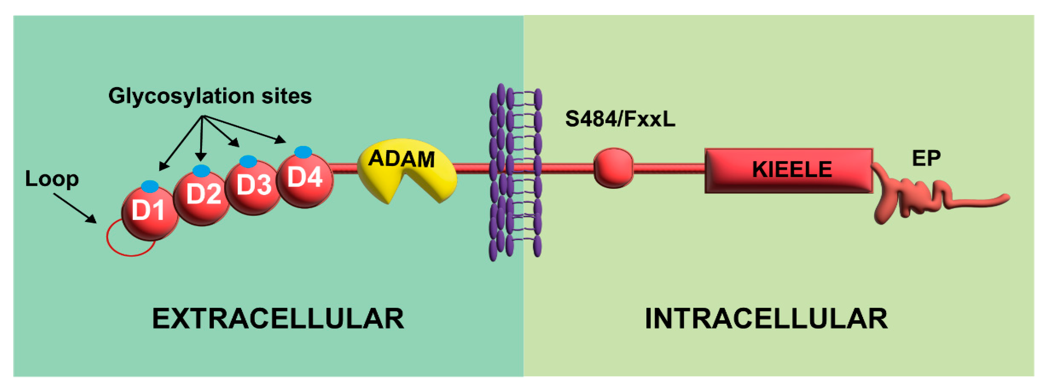
2.1. Molecular Organization

The LAG-3 gene includes eight exons, encoding a transmembrane protein of 498 amino acids [35]. LAG-3 possesses four immunoglobulin (Ig)-like domains (D1 to D4) in the extracellular region with multiple glycosylation sites (Figure 1) [35]. In addition, LAG-3 possesses two unique structural features in the extracellular region [35,40]. First, a proline-rich amino acid loop within D1 allows LAG-3 dimerization and mediates the interaction between LAG-3 and Major Histocompatibility Complex Class II (MHC-II). This likely disrupts the interaction of the glycoprotein called cluster of differentiation 4 (CD4) with MHC-II [40]. Second, LAG-3 is more susceptible to a disintegrin and metalloproteinase (ADAM) shedding since it has a longer connecting peptide between D4 and the transmembrane region, allowing the formation of a soluble ligand (sLAG-3) [40,41].
The cytoplasmic tail of LAG-3 has three conserved motifs (Figure 1) [42,43,44]. The first contains a putative serine phosphorylation site (S484/FxxL motif), and no specific function has been attributed to it apart from its negative association with IL-2 production [42]. The second motif, KIEELE, corresponds to a highly conserved short sequence not found in other proteins, which is required for LAG-3 to downregulate T cell function [43]. However, KIEELE-independent intrinsic mechanisms have been shown to mediate the inhibitory signal of LAG-3 in T cells [42]. The third motif is a proline dipeptide (EP motif) that is critical for LAG-3 trafficking to the cell surface after stimulation and for its colocalization with CD3/TCR, CD4, and CD8 (Figure 1) [44]. After clarifying the structure of LAG-3, it is necessary to know the ligands for this receptor and the inhibition mechanisms of activated T cells that will be discussed below.

2.2. Expression of LAG-3 and Its Ligands

LAG-3 and its ligands are expressed in various immune cells, such as T cells, B cells, and dendritic cells (DCs) [37,45,46]. Several characteristics of this receptor have been described in different cell types. In T cells, this receptor negatively regulates homeostatic activation, proliferation, and expansion [47,48]. LAG-3 has also been identified as a marker for a subset of IL-10-producing Treg cells [37]. The expression of LAG-3 on Treg cells induces their suppressive function, reducing the proliferative capacity of effector cells [37]. On the other hand, in B cells, LAG-3 expression is dependent on T cells and plays a regulatory role [46]. A subset of LAG-3+ regulatory B cells has been described in plasma cells that produce IL-10, suppressing B cell activity [49]. LAG-3 is also expressed in different subtypes of DCs, contributing to immune homeostasis regulation and maturation of DCs [47,50].
MHC-II molecules, transmembrane αβ heterodimer antigen-presenting proteins expressed on APCs, are the primary canonical ligand binding to LAG-3 with a high affinity [39,51,52]. MHC-II binds to the loop of the D1 domain of LAG-3 and transduces inhibitory signals through its cytoplasmic domain (Table 1) [52]. Four ligands have been described other than MHC-II: Galectin-3 (Gal-3), Fibrinogen-like protein 1 (FGL-1), Liver Sinusoidal Endothelial Cell lectin (LSECtin), and α-synuclein (α-syn) [53,54]. Gal-3 is a 31 kDa β-galactoside-binding lectin expressed in different cells, such as neutrophils, macrophages, mast cells, and sensory neurons in several tissues, including the lung epithelium and endothelium (Table 1) [55,56]. Gal-3 interacts with LAG-3 through the glycosylation sites on the extracellular domain, thus exerting its regulatory function on CD8+ T cells [55]. This interaction downregulates T cell proliferation and their ability to produce IL-2 and IFN-γ (Figure 2A) [55]. FGL-1 is a 68 kDa protein member of the fibrinogen-associated protein 1 (FREP) family and contains two homodimers connected by disulfide bonds; it is expressed by some neoplastic cells and secreted by hepatocytes (Table 1) [57,58]. The interaction occurs by binding D1 and D2 domains of LAG-3 with FGL-1, leading to reduced IL-2 levels in the tumor microenvironment (Figure 2A) [54]. LSECtin is a type II integral membrane protein of approximately 40 kDa that regulates CD8+ T cell function in tumor environments by inhibiting their cytotoxic function [59]. LSECtin is expressed in liver sinusoidal endothelial cells, endothelial cells of the lymph nodes, and melanoma cells [59,60]. This molecule is a lectin with carbohydrate-recognition domains that may bind to glycosylated sites of LAG-3 (Table 1) [61]. This interaction with LAG-3 inhibits the secretion of IFN-γ in effector T cells (Figure 2A) [60]. Lastly, α-syn is a neuronal protein 140 amino acids long in presynaptic nerve terminals [62]. In patients with Parkinson’s disease and other neurodegenerative disorders, preformed fibrils (PFF) of α-syn could preferentially bind to the D1 domain of LAG-3 with high affinity, promoting the loss of dopaminergic neurons and behavioral deficits (Table 1) [63].
Table 1. LAG-3 binding ligands distribution and function.
LigandLAG-3
Binding Site
Cell and Tissue
Distribution
FunctionReference
MHC-IILoop of the D1 domain.APCs (macrophages, DCs, and B cells).Disrupt the interaction of CD4+ with MHC-II.[39,52]
Gal-3Glycosylation sites.Macrophages, monocytes, DCs, eosinophils, mast cells, NK, activated T and B cells, epithelial cells, endothelial cells, and sensory neurons.Inhibit the CD8+ T cell cytotoxic function.[55,56]
FGL-1D1 and D2 domains.Neoplastic cells and hepatocytes.Reduce IL-2 levels in the tumor microenvironment.[54]
LSECtinGlycosylation sites.Liver sinusoidal endothelial cells, endothelial cells of the lymph nodes, and melanoma cells.Inhibit the cytotoxic functions of NK and CD8+ T cells in tumors. Inhibit IFN-γ secretion in effector T cells.[59,60,61]
α-synD1 domain.Presynaptic nerve terminals.Enhance the loss of dopaminergic neurons and behavioral deficits.[63]

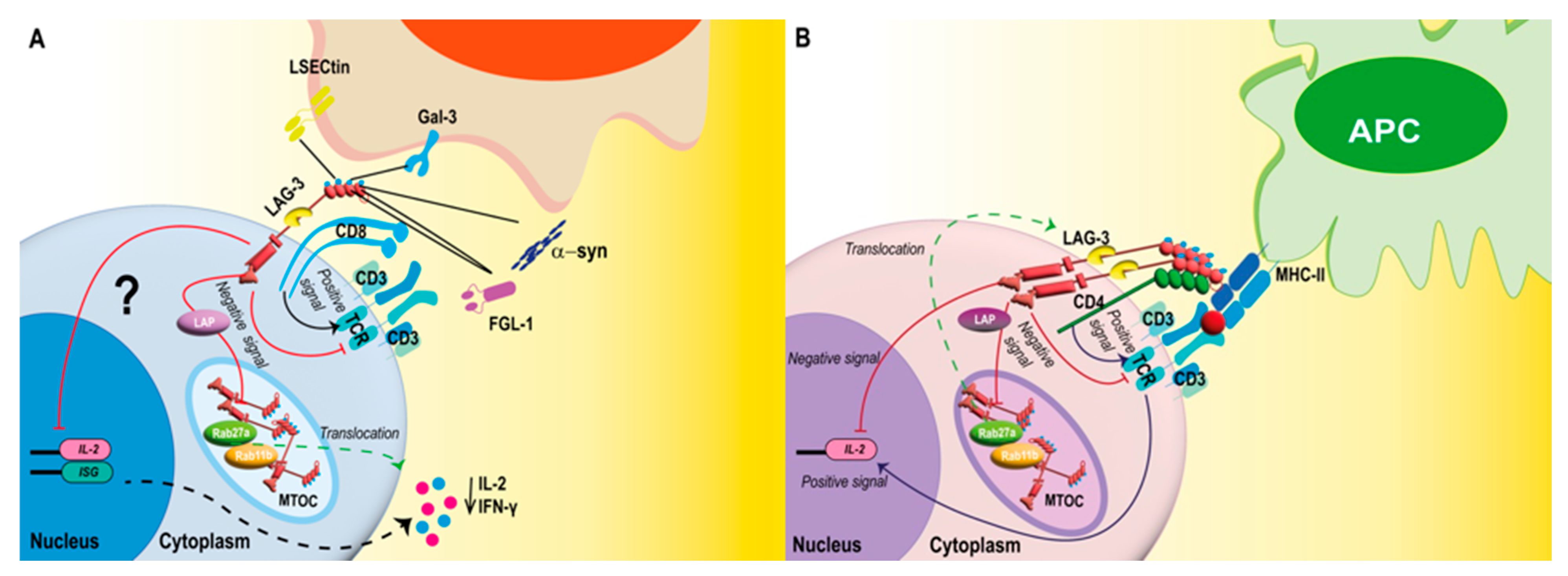
2.3. LAG-3 Signaling

T cell activation upon antigen recognition via the T cell receptor (TCR) is controlled by antigen-independent signals through stimulatory or inhibitory receptors that optimize immune responses [5,64,65]. LAG-3 colocalizes with other molecules, such as CD3, CD4, or CD8, in cholesterol-rich raft areas during the early steps of antigen recognition (Figure 2A,B) [5]. This colocalization allows the association of LAG-3 with CD3 in the TCR complex, as well as LAG-3 cross-linking during immunological synapse assembly, which downregulates signal transduction activity [5]. Moreover, in the cytoplasm, LAG-3 colocalizes with the endosomal recycling compartment protein (Rab11b) and the lysosomal secretory pathway marker (Rab27a), which could suggest that LAG-3 tends to be continuously recycled, facilitating a rapid translocation to the membrane by the pathway lysosomal secretory pathway through the microtubule-organizing center (MTOC) after T cell activation (Figure 2B) [66]. The amount of expression of LAG-3 on the cell surface is strongly associated with its inhibitory activity [42].
The cytoplasmic tail of the LAG-3 plays a critical role in the mechanisms of T cell function inhibition [42,43]. The KIEELE motif was initially identified as the crucial driver of LAG-3 signal transduction [43]. However, recent studies using an in vitro T cell activation system with LAG-3-MHC-II blocking mAbs suggested that the proximal region of the membrane containing the FxxL motif, including the residues F475 and L478, is the main responsible for inhibiting T cell activation by directly suppressing the secretion of IL-2 or the response to this cytokine (Figure 2B) [42]. The similarity between the FxxL motif and the YxxL motif, which are located in immunoreceptor tyrosine-based inhibitory motifs (ITIM), means that phosphotyrosine-independent signaling adaptor proteins might also be involved in LAG-3-induced inhibitory signaling [42]. Other inhibitory signals are associated with the recruitment and colocalization of LAG-3 on the cell surface and depend on the EP motif (Figure 2B) [42].
Interestingly, an intracellular factor for the EP motif was shown to recruit LAG-3-associated protein (LAP) directly, where LAP assembles LAG-3 into lipid rafts induced after TCR signaling (Figure 2B) [5]. LAP mediates LAG-3 transport and colocalization to the cell surface, which can enhance the dimerization/oligomerization of LAG-3, a process that is necessary for MHC-II/LAG-3 binding (Figure 2B), but not for the binding to other ligands [5,67]. Recent studies have described how, during the immunological synapse for CD4+ and CD8+ T cells, LAG-3 associates with the TCR-CD3 complex and interacts through the EP located in the cytoplasmic domain, leading to a pH reduction with a phosphorylation decrease on the tyrosine kinase ZAP70, which reduces T cell activation (Figure 2A,B) [68]. Even though the differential function elicited by LAG-3 on CD4+ and CD8+ T cells has not yet been the subject of many studies, it is necessary to know the mechanisms by which LAG-3 participates in the inhibition of CD8+ cells because these cells do not recognize the main LAG-3 ligand, MHC-II [69]. Diverse mechanisms have been proposed to study this differential function, such as identifying alternative LAG-3 ligands, including LSECtin, Gal-3, α-syn, and FGL-1 binding to LAG-3 in CD8+ T cells and the co-distribution of LAG-3 and CD8 coreceptor during early T cell activation events (Figure 2A) [70,71].
LAG-3 expression is regulated at the cell surface, partly by cleavage of the extracellular domain by TCR-induced disintegrins and metalloproteases (ADAM10 and ADAM17) [41]. Cleavage gives rise to a 52 kDa soluble form of sLAG-3 [72]; sLAG-3 mediates the activation of APCs, thus activating and promoting the production and proliferation of CD8+ T lymphocytes, favoring a cellular response characterized by a Th1 profile [73,74].

2.4. LAG-3 Mediates Immunometabolic Programming of T Cells

The metabolic programs of T cells match their functional demands [75]. A specific metabolic profile is necessary for T cells to maintain their effector functions and eliminate pathogens [76,77]. Naïve T cells predominantly rely on mitochondrial oxidative phosphorylation to generate ATP, whereas an activated T cell displays robust aerobic glycolysis to maximize macromolecule synthesis and energy [76]. Memory T cells revert to oxidative and fatty acid metabolism, resembling the metabolic phenotype of naïve T cells [76]. Interestingly, these metabolic processes can be regulated through cell signaling, gene transcription, and protein synthesis, as well as an inadequate supply of nutrients and oxygen or the accumulation of metabolic intermediaries [76]. In CD4+ T cells, LAG-3 maintains the metabolic and mitochondrial quiescence of naïve CD4+ T cells, limiting oxygen consumption and respiratory capacity and preventing excessive proliferation [78]. LAG-3 deficiency leads to the activation of the signal transducer and activator of transcription 5 (STAT5), leading to increased glycolytic capacity and effector function after activation [78]. Furthermore, upregulation of STAT5 expression reduces CD4+ T cell dependence on IL-7 for survival and metabolism [78]. Other evidence shows that metabolic reprogramming of diabetogenic CD4+ T cells using a competitive inhibitor of the limiting enzyme of glycolysis 6-phosphofructo-2-kinase/fructose-2,6-bisphosphatase 3 (PFKFB3) increases the expression of LAG-3 and PD-1 [79]. On the other hand, LAG-3 deficiency alters APC metabolism, and cell metabolism shifts from oxidative phosphorylation to energy production via the glycolytic pathway, altering APC cytokine secretion through an increase in TNF-α and a reduction in IL-10 production [80].
Currently, there is limited information on the metabolic consequences of LAG-3 on CD8+ T cells. However, the generation of long-term memory can be affected by the high glycolytic rate in T cells since the increase in glycolytic activity leads to a state of terminal differentiation in these cells [81]. In this sense, the expression of LAG-3 may play an essential role in CD8+ T cell memory responses by maintaining cellular metabolism in oxidative phosphorylation, as occurs with APC [80].

3. Respiratory Viral Infections Modify the Expression of LAG-3

As mentioned above, LAG-3 plays an essential role in negatively regulating the activation of effector CD8+ T cells [82]. In this line, respiratory viruses can modulate the expression of LAG-3 to their advantage to prevent the activation of CD8+ T cells [7]. The up-regulation of LAG-3 during viral infection may generate an incomplete cytotoxic response that impairs viral clearance and immunological memory [7,8,32,83,84]. This section will discuss the effects of increasing the expression of LAG-3 on CD8+ T cells during acute infections caused by the most prevalent respiratory viruses, such as SARS-CoV-2, influenza virus, hMPV, and hRSV.

3.1. Severe Acute Respiratory Syndrome Coronavirus 2

SARS-CoV-2 is the viral pathogen responsible for the 2019 pandemic and the etiological agent for Coronavirus disease 2019 (COVID-19) [85]. Since then, SARS-CoV-2 has become one of the most prevalent respiratory viruses, affecting people of all ages [86,87,88,89,90,91]. Patients infected with this virus commonly show symptoms that include fever, cough, and headache; in more severe cases, the patients can develop pneumonia and acute respiratory distress syndrome (ARDS) [90,91]. Additionally, SARS-CoV-2 infection can reach various organs, such as the kidney, heart, and brain, and cause damage to all of them [90,91].
Samples from severe SARS-CoV-2 patients have demonstrated an increase of innate immune cells, such as neutrophils and NK cells, while observing a downmodulation in T cells [85,90,92]. The reduced T cell response might be associated with the lack of long-lasting immunity that lymphocytes are usually responsible for achieving [85,90,92]. Other human lung tissue samples and nasopharyngeal lavage have shown an overexpression of inhibitory receptors on T cells, such as LAG-3 [9,93,94,95,96,97,98]. This observation could explain the significantly reduced numbers of T cells in severely ill patients. Severe COVID-19 patients have been reported to have reduced MHC-II expression in various APCs [99]. This reduction, accompanied by increased LAG3 expression on the surface of T cells, may cause an imbalance in the interactions between these two molecules. This may reflect a failure in the proper interaction between innate and adaptive cells and result in aberrant expression of cytotoxic cytokines [99]. This alteration could contribute to the immunopathology of SARS-CoV-2 [99].
Interestingly, patients with severe SARS-CoV-2-infection promote the expression of LAG-3 in T cells is regulated through the genes activated downstream of the IFN-I signaling [99,100]. An in vitro study performed with cells from critically ill COVID-19 patients with a predominant IFN-I response and T cells revealed two mutually antagonistic modules of ISG regulators, in which SP140 is a bidirectional regulator for LAG-3 and TIGIT under IFN-I responses [100]. The transcription factor SP140 promotes an increase in the expression of LAG-3 but decreases TIGIT expression in response to interferon [100]. Lymphopenia and expression of inhibitory receptors in the T cells of patients who are severely ill with SARS-CoV-2 suggest that the loss of function in these cells can worsen pathology by interfering with the eradication of the virus.

3.2. Influenza Virus

The influenza virus is a respiratory virus that causes considerable morbidity and mortality in humans every year, leading to symptoms such as fever, cough, and pneumonia in the more severe cases [29]. The cases with the highest rates of influenza-related mortality occur in the elderly and those with underlying medical conditions [101]. In addition, it has been observed that influenza virus infection produces excessive inflammation that can cause severe damage to the host [102].
Since LAG-3 plays a regulatory role in immune responses [83,84,103], it can be suggested that LAG-3 may have a role during the period of infection with the influenza virus. It has been shown that CD4+ T cells that recognize influenza virus hemagglutinin protein also express LAG-3 [84,104]. This observation suggests that recognition of this protein can induce the expression of LAG-3. Interestingly, Th1 cells with high expression of LAG-3 can suppress CD8+ T lymphocytes and restrict lung inflammation without affecting the clearance of this virus [83]. Furthermore, it has been observed that the blockade of LAG-3 promotes the activation of CD8+ T cells stimulated with peptides from the influenza virus [7]. These results suggest that the influenza virus hemagglutinin might positively modulate the effect of LAG-3, leading to impairment of CD8+ T cells activation. However, further studies are required to understand the mechanism behind this modulation over LAG-3.

3.3. Human Metapneumovirus

An important cause of ALRTI in infants, hMPV is the second most frequently identified pathogen after hRSV in lower respiratory diseases, including bronchiolitis, pneumonia, and croup [105,106,107]. One distinctive aspect of this virus is that it can cause multiple reinfections throughout the infant’s life [108,109].
In vivo studies have shown that the depletion of CD8+ T cells may increase reinfections cases that are caused by hMPV [7]. These cells lose the ability to secrete the cytokines necessary to eliminate hMPV [7,8,33]. In addition, increased LAG-3 expression has been described in pulmonary CD8+ T cells that leads to mouse susceptibility to hMPV reinfection [32]. Interestingly, LAG-3 blockade can restore cytokine production, suggesting the crucial involvement of LAG-3 during the infection with hMPV [7]. Furthermore, experiments with PD-1 deficient mice demonstrated that using LAG-3 blockade during infection with hMPV can restore CD8+ T cell function, but increases pulmonary pathology [7]. These studies suggest that, during the infection with hMPV, the expression of LAG-3 increases in pulmonary CD8+ T cells to control the inflammatory response elicited against the virus, displaying a protective role during this infection.

3.4. Human Respiratory Syncytial Virus

In children under two years of age, hRSV is the principal cause of ALRTI, leading to cough, fever, wheezing, bronchiolitis, and pneumonia in more severe cases [110,111,112,113,114]. The hRSV belongs to the Pneumoviridae family, the same family as the hMPV [115]. Even though there is no information about the expression of LAG-3 during hRSV infection, a role could be suggested based on what is known for hMPV. As mentioned previously, hMPV infection causes an increase in LAG-3 expression, possibly as a protective regulatory response [7,8,33]. Based on this information, it could be suggested that during infection with hRSV, the expression of LAG-3 also increases to inhibit the activation of CD8+ T cells. However, the response of CD8+ T cells is robust during hRSV infection [116,117], which indicates that LAG-3 possibly behaves differently during the hRSV infection, decreasing the activity of LAG-3.

4. Therapeutic Perspectives Targeting LAG-3

LAG-3 has been described as an essential next-generation immune checkpoint receptor proposed as a therapeutic target against cancer, autoimmune and inflammatory diseases, parasitic infections, and acute and chronic viral infections [10,118,119,120,121]. Research on the use of the LAG-3 receptor has focused on immunotherapies, especially for treating different types of cancer [119,122]. Several formulations have been explored, including soluble dimeric LAG-3 as an adjuvant, agonist antibodies that block the interaction between this receptor and its ligands, antibody-mediated LAG-3 cell depletion in autoimmunity, and finally, the use of small molecule GSK-3 to modulate LAG-3 expression [123]. Clinical trials have shown that LAG-3 blockade as a cancer treatment activates APC to promote DCs proliferation and enhances regulatory T cell immunosuppression and antigen cross-presentation to CD8+ T cells, promoting an anti-tumor state [124]. Therefore, LAG-3 blockade has been used in these pathologies as monotherapy or in combination with other agonists, such as anti-PD1, showing better results [125]. However, it has also been described that using LAG-3 inhibitors can help overcome immune exhaustion during persistent inflammatory states, such as cancer and viral infections [126]. In this case, research has mainly focused on treating LAG-3-blocking antibodies during chronic viral infections. However, an in vivo study evaluated and compared the role of LAG-3 in both chronic and acute viral infections, resulting in a similar intrinsic cellular effect of LAG-3 in both cases, thus supporting the use of LAG-3 blockers to enhance CD8+ T cell responses [121]. However, it does not generate the same results under other circumstances, as demonstrated by a study in a mouse model infected with lymphocytic choriomeningitis virus, where LAG-3 blockade failed to either rescue CD8+ T cell cytokine production or to affect viral titers [127].
Similarly, in a PD-1-deficient mouse model, it was possible to observe that using an antibody against LAG-3 resulted in restoring CD8+ T cell effector functions [7,121,127]. However, it was also found that the restored effector function of CD8+ T cell increased the lung immunopathology when mice were infected with hMPV, suggesting that LAG-3 contributes to the protection against the development of the immunopathology during hMPV shedding [7,121,127]. Phase I clinical trials have highlighted the role of sLAG-3, known as IMP321, as an adjuvant in a trivalent influenza vaccine that demonstrated safety, tolerability, and immunogenicity [128]. In addition, IMP321 has been described to enhance the response of Th1 influenza-specific CD4+ cells by increasing the secretion of IFN-γ, the prototypical Th1 effector cytokine, TNF-α, an inflammatory cytokine, and IL-2, which is involved in the development of memory T cells [128]. More recently, based on studies showing a transient increase in the expression of this receptor during immune activation as part of COVID-19, the use of LAG-3 blockers (alone or in combination with other checkpoint inhibitors) during the early or late phase of SARS-CoV-2 infection has been proposed as an effective therapeutic measure for treatment [9,97]. However, additional studies are required to support this strategy conclusively. Considering the current pandemic and the lack of treatments against other viruses such as hRSV or hMPV, exploring the use of inhibitors for this receptor as a therapy against acute respiratory diseases would be important.

5. Concluding Remarks

LAG-3 is an immune checkpoint receptor that can have beneficial effects, serving as an adjuvant for vaccines and immune treatments, contributing to the generation of Th1-type antiviral responses, and suppressing excessive cytotoxic activity. However, the role of LAG-3 in ALRTI is still controversial. In addition, LAG-3 can contribute to reducing an exacerbated inflammatory response and also quickly suppress the response of CD8+ T cells. CD8+ T cells play a fundamental role in viral eradication and the generation of immunological memory. LAG-3 expression can affect the function of these cells, which can interfere with viral eradication and prevent the generation of long-lasting memory responses. On the other hand, blockade or genetic ablation of LAG-3 improves viral eradication and could contribute to the generation of longer-lasting memory responses, implying a protective response against reinfections.
In contrast, the generation of a more robust immune response involves the release of pro-inflammatory cytokines such as TNF-α and IFN-γ, in addition to GrzB and Perforin, which can cause tissue damage and increase immunopathology. Based on these features, it is likely that LAG-3 expression could work as a biomarker to predict the outcome of acute infections. Its use as a marker for immune cell activation or inhibition during acute infections may be advantageous for considering treatment options. In addition, it is essential to evaluate the effect of LAG-3 targeted immunotherapy for treating malignancies or enhancing the effectiveness of vaccines and antiviral treatments. Studies designed to approach this question became important because treatment with anti-LAG-3 can alter humoral and cellular immune responses. Further studies are needed to evaluate the potential therapeutic implications of sLAG-3 or LAG-3 blockade in viral ALRTIs and their possible use as vaccine adjuvants.

Author Contributions

L.R.-G., M.A.R., C.A.A. and A.M.K., conceptualization, original draft, review, editing, and revision. A.M.K. reviewed the manuscript and approved the version to be published. All authors have read and agreed to the published version of the manuscript.

Funding

This research was funded by the Millennium Institute on Immunology and Immunotherapy, grant number P09/016-F, ACE 210015, and ICN09_016. CORFO grant #13CTI-21526/P4 and P5; Biomedical Research Consortium CTU06 (AK). COPEC-UC2019.R.1169. COPEC-UC2020.E.1; ANID TDP 210015 A.K. and L.R-G.; ANID scholarship #21210336 for M.A.R and #21210662 for C.A.A.

Data Availability Statement

Not applicable.

Acknowledgments

The authors are grateful to students and researchers at the Biomedical Molecular Immunology Laboratory of the Millennium Institute on Immunology and Immunotherapy for providing guidance and suggesting additional references to incorporate into this review article.

Conflicts of Interest

The authors declare no conflict of interest.

References

  1. Xia, A.-L.; Wang, J.-C.; Yang, K.; Ji, D.; Huang, Z.-M.; Xu, Y. Genomic and epigenomic perspectives of T-cell exhaustion in cancer. Brief. Funct. Genom. 2019, 18, 113–118. [Google Scholar] [CrossRef] [PubMed]
  2. Kurachi, M. CD8+ T cell exhaustion. Semin. Immunopathol. 2019, 41, 327–337. [Google Scholar] [CrossRef] [PubMed]
  3. Attanasio, J.; Wherry, E.J. Costimulatory and Coinhibitory Receptor Pathways in Infectious Disease. Immunity 2016, 44, 1052–1068. [Google Scholar] [CrossRef] [PubMed] [Green Version]
  4. Sharpe, A.H.; Wherry, E.J.; Ahmed, R.; Freeman, G.J. The function of programmed cell death 1 and its ligands in regulating autoimmunity and infection. Nat. Immunol. 2007, 8, 239–245. [Google Scholar] [CrossRef]
  5. Maçon-Lemaître, L.; Triebel, F. The negative regulatory function of the lymphocyte-activation gene-3 co-receptor (CD223) on human T cells. Immunology 2005, 115, 170–178. [Google Scholar] [CrossRef]
  6. Chocarro, L.; Blanco, E.; Zuazo, M.; Arasanz, H.; Bocanegra, A.; Fernández-Rubio, L.; Morente, P.; Fernández-Hinojal, G.; Echaide, M.; Garnica, M.; et al. Understanding LAG-3 Signaling. Int. J. Mol. Sci. 2021, 22, 5282. [Google Scholar] [CrossRef]
  7. Erickson, J.J.; Rogers, M.C.; Tollefson, S.J.; Boyd, K.L.; Williams, J.V. Multiple Inhibitory Pathways Contribute to Lung CD8+ T Cell Impairment and Protect against Immunopathology during Acute Viral Respiratory Infection. J. Immunol. 2016, 197, 233–243. [Google Scholar] [CrossRef] [Green Version]
  8. Erickson, J.J.; Lu, P.; Wen, S.; Hastings, A.K.; Gilchuk, P.; Joyce, S.; Shyr, Y.; Williams, J.V. Acute Viral Respiratory Infection Rapidly Induces a CD8+ T Cell Exhaustion-like Phenotype. J. Immunol. 2015, 195, 4319–4330. [Google Scholar] [CrossRef] [Green Version]
  9. Herrmann, M.; Schulte, S.; Wildner, N.H.; Wittner, M.; Brehm, T.T.; Ramharter, M.; Woost, R.; Lohse, A.W.; Jacobs, T.; Schulze Zur Wiesch, J. Analysis of Co-inhibitory Receptor Expression in COVID-19 Infection Compared to Acute Plasmodium falciparum Malaria: LAG-3 and TIM-3 Correlate with T Cell Activation and Course of Disease. Front. Immunol. 2020, 11, 1870. [Google Scholar] [CrossRef]
  10. Butler, N.S.; Moebius, J.; Pewe, L.L.; Traore, B.; Doumbo, O.K.; Tygrett, L.T.; Waldschmidt, T.J.; Crompton, P.D.; Harty, J.T. Therapeutic blockade of PD-L1 and LAG-3 rapidly clears established blood-stage Plasmodium infection. Nat. Immunol. 2011, 13, 188–195. [Google Scholar] [CrossRef]
  11. Odorizzi, P.M.; Wherry, E.J. Inhibitory receptors on lymphocytes: Insights from infections. J. Immunol. 2012, 188, 2957–2965. [Google Scholar] [CrossRef] [PubMed] [Green Version]
  12. Subbarao, K.; Mahanty, S. Respiratory Virus Infections: Understanding COVID-19. Immunity 2020, 52, 905–909. [Google Scholar] [CrossRef] [PubMed]
  13. Canedo-Marroquín, G.; Acevedo-Acevedo, O.; Rey-Jurado, E.; Saavedra, J.M.; Lay, M.K.; Bueno, S.M.; Riedel, C.A.; Kalergis, A.M. Modulation of Host Immunity by Human Respiratory Syncytial Virus Virulence Factors: A Synergic Inhibition of Both Innate and Adaptive Immunity. Front. Cell. Infect. Microbiol. 2017, 7, 367. [Google Scholar] [CrossRef] [PubMed] [Green Version]
  14. Andrade, C.A.; Pacheco, G.A.; Gálvez, N.M.S.; Soto, J.A.; Bueno, S.M.; Kalergis, A.M. Innate Immune Components that Regulate the Pathogenesis and Resolution of hRSV and hMPV Infections. Viruses 2020, 12, 637. [Google Scholar] [CrossRef] [PubMed]
  15. Schmidt, M.E.; Varga, S.M. The CD8 T Cell Response to Respiratory Virus Infections. Front. Immunol. 2018, 9, 678. [Google Scholar] [CrossRef] [Green Version]
  16. Belz, G.T.; Wodarz, D.; Diaz, G.; Nowak, M.A.; Doherty, P.C. Compromised influenza virus-specific CD8+-T-cell memory in CD4+-T-cell-deficient mice. J. Virol. 2002, 76, 12388–12393. [Google Scholar] [CrossRef] [Green Version]
  17. Janssen, E.M.; Lemmens, E.E.; Wolfe, T.; Christen, U.; von Herrath, M.G.; Schoenberger, S.P. CD4+ T cells are required for secondary expansion and memory in CD8+ T lymphocytes. Nature 2003, 421, 852–856. [Google Scholar] [CrossRef]
  18. Retamal-Díaz, A.; Covián, C.; Pacheco, G.A.; Castiglione-Matamala, A.T.; Bueno, S.M.; González, P.A.; Kalergis, A.M. Contribution of Resident Memory CD8+ T Cells to Protective Immunity Against Respiratory Syncytial Virus and Their Impact on Vaccine Design. Pathogens 2019, 8, 147. [Google Scholar] [CrossRef] [Green Version]
  19. Knudson, C.J.; Weiss, K.A.; Hartwig, S.M.; Varga, S.M. The Pulmonary Localization of Virus-Specific T Lymphocytes Is Governed by the Tissue Tropism of Infection. J. Virol. 2014, 88, 9010–9016. [Google Scholar] [CrossRef] [Green Version]
  20. Jozwik, A.; Habibi, M.S.; Paras, A.; Zhu, J.; Guvenel, A.; Dhariwal, J.; Almond, M.; Wong, E.H.C.; Sykes, A.; Maybeno, M.; et al. RSV-specific airway resident memory CD8+ T cells and differential disease severity after experimental human infection. Nat. Commun. 2015, 6, 10224. [Google Scholar] [CrossRef]
  21. Braciale, T.J.; Sun, J.; Kim, T.S. Regulating the adaptive immune response to respiratory virus infection. Nat. Rev. Immunol. 2012, 12, 295–305. [Google Scholar] [CrossRef] [PubMed]
  22. David, P.; Drabczyk-Pluta, M.; Pastille, E.; Knuschke, T.; Werner, T.; Honke, N.; Megger, D.A.; Akhmetzyanova, I.; Shaabani, N.; Eyking-Singer, A.; et al. Combination immunotherapy with anti-PD-L1 antibody and depletion of regulatory T cells during acute viral infections results in improved virus control but lethal immunopathology. PLoS Pathog. 2020, 16, e1008340. [Google Scholar] [CrossRef] [Green Version]
  23. Zenarruzabeitia, O.; Astarloa-Pando, G.; Terrén, I.; Orrantia, A.; Pérez-Garay, R.; Seijas-Betolaza, I.; Nieto-Arana, J.; Imaz-Ayo, N.; Pérez-Fernández, S.; Arana-Arri, E.; et al. T Cell Activation, Highly Armed Cytotoxic Cells and a Shift in Monocytes CD300 Receptors Expression Is Characteristic of Patients with Severe COVID-19. Front. Immunol. 2021, 12, 655934. [Google Scholar] [CrossRef] [PubMed]
  24. Goplen, N.P.; Wu, Y.; Son, Y.M.; Li, C.; Wang, Z.; Cheon, I.S.; Jiang, L.; Zhu, B.; Ayasoufi, K.; Chini, E.N.; et al. Tissue-resident CD8+ T cells drive age-associated chronic lung sequelae after viral pneumonia. Sci. Immunol. 2020, 5, eabc4557. [Google Scholar] [CrossRef] [PubMed]
  25. Graham, B.S.; Bunton, L.A.; Wright, P.F.; Karzon, D.T. Role of T lymphocyte subsets in the pathogenesis of primary infection and rechallenge with respiratory syncytial virus in mice. J. Clin. Investig. 1991, 88, 1026–1033. [Google Scholar] [CrossRef]
  26. González, A.E.; Lay, M.K.; Jara, E.L.; Espinoza, J.A.; Gómez, R.S.; Soto, J.; Rivera, C.A.; Abarca, K.; Bueno, S.M.; Riedel, C.A.; et al. Aberrant T cell immunity triggered by human Respiratory Syncytial Virus and human Metapneumovirus infection. Virulence 2017, 8, 685–704. [Google Scholar] [CrossRef]
  27. Schmidt, M.E.; Knudson, C.J.; Hartwig, S.M.; Pewe, L.L.; Meyerholz, D.K.; Langlois, R.A.; Harty, J.T.; Varga, S.M. Memory CD8 T cells mediate severe immunopathology following respiratory syncytial virus infection. PLoS Pathog. 2018, 14, e1006810. [Google Scholar] [CrossRef]
  28. Troeger, C.; Forouzanfar, M.; Rao, P.C.; Khalil, I.; Brown, A.; Swartz, S.; Fullman, N.; Mosser, J.; Thompson, R.L.; Reiner, R.C., Jr.; et al. Estimates of the global, regional, and national morbidity, mortality, and aetiologies of lower respiratory tract infections in 195 countries: A systematic analysis for the Global Burden of Disease Study 2015. Lancet Infect. Dis. 2017, 17, 1133–1161. [Google Scholar] [CrossRef] [Green Version]
  29. Lafond, K.E.; Porter, R.M.; Whaley, M.J.; Suizan, Z.; Ran, Z.; Aleem, M.A.; Thapa, B.; Sar, B.; Proschle, V.S.; Peng, Z.; et al. Global burden of influenza-associated lower respiratory tract infections and hospitalizations among adults: A systematic review and meta-analysis. PLoS Med. 2021, 18, e1003550. [Google Scholar] [CrossRef]
  30. Lee, S.; Stokes, K.L.; Currier, M.G.; Sakamoto, K.; Lukacs, N.W.; Celis, E.; Moore, M.L. Vaccine-elicited CD8+ T cells protect against respiratory syncytial virus strain A2-line19F-induced pathogenesis in BALB/c mice. J. Virol. 2012, 86, 13016–13024. [Google Scholar] [CrossRef]
  31. Tian, X.; Zhang, A.; Qiu, C.; Wang, W.; Yang, Y.; Qiu, C.; Liu, A.; Zhu, L.; Yuan, S.; Hu, H.; et al. The upregulation of LAG-3 on T cells defines a subpopulation with functional exhaustion and correlates with disease progression in HIV-infected subjects. J. Immunol. 2015, 194, 3873–3882. [Google Scholar] [CrossRef] [PubMed] [Green Version]
  32. Erickson, J.J.; Rogers, M.C.; Hastings, A.K.; Tollefson, S.J.; Williams, J.V. Programmed death-1 impairs secondary effector lung CD8+ T cells during respiratory virus reinfection. J. Immunol. 2014, 193, 5108–5117. [Google Scholar] [CrossRef] [PubMed] [Green Version]
  33. Erickson, J.J.; Gilchuk, P.; Hastings, A.K.; Tollefson, S.J.; Johnson, M.; Downing, M.B.; Boyd, K.L.; Johnson, J.E.; Kim, A.S.; Joyce, S.; et al. Viral acute lower respiratory infections impair CD8+ T cells through PD-1. J. Clin. Investig. 2012, 122, 2967–2982. [Google Scholar] [CrossRef] [PubMed] [Green Version]
  34. Chocarro, L.; Bocanegra, A.; Blanco, E.; Fernández-Rubio, L.; Arasanz, H.; Echaide, M.; Garnica, M.; Ramos, P.; Piñeiro-Hermida, S.; Vera, R.; et al. Cutting-Edge: Preclinical and Clinical Development of the First Approved Lag-3 Inhibitor. Cells 2022, 11, 2351. [Google Scholar] [CrossRef] [PubMed]
  35. Triebel, F.; Jitsukawa, S.; Baixeras, E.; Roman-Roman, S.; Genevee, C.; Viegas-Pequignot, E.; Hercend, T. LAG-3, a novel lymphocyte activation gene closely related to CD4. J. Exp. Med. 1990, 171, 1393–1405. [Google Scholar] [CrossRef] [Green Version]
  36. Durham, N.M.; Nirschl, C.J.; Jackson, C.M.; Elias, J.; Kochel, C.M.; Anders, R.A.; Drake, C.G. Lymphocyte Activation Gene 3 (LAG-3) modulates the ability of CD4 T-cells to be suppressed in vivo. PLoS ONE 2014, 9, e109080. [Google Scholar] [CrossRef] [Green Version]
  37. Huang, C.-T.; Workman, C.J.; Flies, D.; Pan, X.; Marson, A.L.; Zhou, G.; Hipkiss, E.L.; Ravi, S.; Kowalski, J.; Levitsky, H.I.; et al. Role of LAG-3 in regulatory T cells. Immunity 2004, 21, 503–513. [Google Scholar] [CrossRef] [Green Version]
  38. Naik, S.; Larsen, S.B.; Gomez, N.C.; Alaverdyan, K.; Sendoel, A.; Yuan, S.; Polak, L.; Kulukian, A.; Chai, S.; Fuchs, E. Inflammatory memory sensitizes skin epithelial stem cells to tissue damage. Nature 2017, 550, 475–480. [Google Scholar] [CrossRef] [Green Version]
  39. Workman, C.J.; Cauley, L.S.; Kim, I.-J.; Blackman, M.A.; Woodland, D.L.; Vignali, D.A.A. Lymphocyte activation gene-3 (CD223) regulates the size of the expanding T cell population following antigen activation in vivo. J. Immunol. 2004, 172, 5450–5455. [Google Scholar] [CrossRef] [Green Version]
  40. Li, N.; Workman, C.J.; Martin, S.M.; Vignali, D.A.A. Biochemical analysis of the regulatory T cell protein lymphocyte activation gene-3 (LAG-3; CD223). J. Immunol. 2004, 173, 6806–6812. [Google Scholar] [CrossRef]
  41. Li, N.; Wang, Y.; Forbes, K.; Vignali, K.M.; Heale, B.S.; Saftig, P.; Hartmann, D.; Black, R.A.; Rossi, J.J.; Blobel, C.P.; et al. Metalloproteases regulate T-cell proliferation and effector function via LAG-3. EMBO J. 2007, 26, 494–504. [Google Scholar] [CrossRef] [PubMed] [Green Version]
  42. Maeda, T.K.; Sugiura, D.; Okazaki, I.-M.; Maruhashi, T.; Okazaki, T. Atypical motifs in the cytoplasmic region of the inhibitory immune co-receptor LAG-3 inhibit T cell activation. J. Biol. Chem. 2019, 294, 6017–6026. [Google Scholar] [CrossRef] [PubMed]
  43. Workman, C.J.; Dugger, K.J.; Vignali, D.A.A. Cutting edge: Molecular analysis of the negative regulatory function of lymphocyte activation gene-3. J. Immunol. 2002, 169, 5392–5395. [Google Scholar] [CrossRef] [Green Version]
  44. Bae, J.; Lee, S.J.; Park, C.-G.; Lee, Y.S.; Chun, T. Trafficking of LAG-3 to the surface on activated T cells via its cytoplasmic domain and protein kinase C signaling. J. Immunol. 2014, 193, 3101–3112. [Google Scholar] [CrossRef] [Green Version]
  45. Blackburn, S.D.; Shin, H.; Haining, W.N.; Zou, T.; Workman, C.J.; Polley, A.; Betts, M.R.; Freeman, G.J.; Vignali, D.A.; Wherry, E.J. Coregulation of CD8+ T cell exhaustion by multiple inhibitory receptors during chronic viral infection. Nat. Immunol. 2009, 10, 29–37. [Google Scholar] [CrossRef] [Green Version]
  46. Kisielow, M.; Kisielow, J.; Capoferri-Sollami, G.; Karjalainen, K. Expression of lymphocyte activation gene 3 (LAG-3) on B cells is induced by T cells. Eur. J. Immunol. 2005, 35, 2081–2088. [Google Scholar] [CrossRef] [PubMed]
  47. Workman, C.J.; Wang, Y.; El Kasmi, K.C.; Pardoll, D.M.; Murray, P.J.; Drake, C.G.; Vignali, D.A. LAG-3 regulates plasmacytoid dendritic cell homeostasis. J. Immunol. 2009, 182, 1885–1891. [Google Scholar] [CrossRef] [Green Version]
  48. Zhang, Q.; Chikina, M.; Szymczak-Workman, A.L.; Horne, W.; Kolls, J.K.; Vignali, K.M.; Normolle, D.; Bettini, M.; Workman, C.J.; Vignali, D.A.A. LAG3 limits regulatory T cell proliferation and function in autoimmune diabetes. Sci. Immunol. 2017, 2, eaah4569. [Google Scholar] [CrossRef] [Green Version]
  49. Lino, A.C.; Dang, V.D.; Lampropoulou, V.; Welle, A.; Joedicke, J.; Pohar, J.; Simon, Q.; Thalmensi, J.; Baures, A.; Flühler, V.; et al. LAG-3 Inhibitory Receptor Expression Identifies Immunosuppressive Natural Regulatory Plasma Cells. Immunity 2018, 49, 120–133.e9. [Google Scholar] [CrossRef]
  50. Andreae, S.; Piras, F.; Burdin, N.; Triebel, F. Maturation and activation of dendritic cells induced by lymphocyte activation gene-3 (CD223). J. Immunol. 2002, 168, 3874–3880. [Google Scholar] [CrossRef]
  51. Huard, B.; Mastrangeli, R.; Prigent, P.; Bruniquel, D.; Donini, S.; El-Tayar, N.; Maigret, B.; Dréano, M.; Triebel, F. Characterization of the major histocompatibility complex class II binding site on LAG-3 protein. Proc. Natl. Acad. Sci. USA 1997, 94, 5744–5749. [Google Scholar] [CrossRef] [PubMed] [Green Version]
  52. Workman, C.J.; Vignali, D.A.A. The CD4-related molecule, LAG-3 (CD223), regulates the expansion of activated T cells. Eur. J. Immunol. 2003, 33, 970–979. [Google Scholar] [CrossRef] [PubMed]
  53. Souri, Z.; Wierenga, A.P.A.; Kroes, W.G.M.; van der Velden, P.A.; Verdijk, R.M.; Eikmans, M.; Luyten, G.P.M.; Jager, M.J. LAG3 and Its Ligands Show Increased Expression in High-Risk Uveal Melanoma. Cancers 2021, 13, 4445. [Google Scholar] [CrossRef]
  54. Shi, A.-P.; Tang, X.-Y.; Xiong, Y.-L.; Zheng, K.-F.; Liu, Y.-J.; Shi, X.-G.; Lv, Y.; Jiang, T.; Ma, N.; Zhao, J.B. Immune Checkpoint LAG3 and Its Ligand FGL1 in Cancer. Front. Immunol. 2021, 12, 785091. [Google Scholar] [CrossRef] [PubMed]
  55. Kouo, T.; Huang, L.; Pucsek, A.B.; Cao, M.; Solt, S.; Armstrong, T.; Jaffee, E. Galectin-3 Shapes Antitumor Immune Responses by Suppressing CD8+ T Cells via LAG-3 and Inhibiting Expansion of Plasmacytoid Dendritic Cells. Cancer Immunol. Res. 2015, 3, 412–423. [Google Scholar] [CrossRef] [PubMed] [Green Version]
  56. Dumic, J.; Dabelic, S.; Flögel, M. Galectin-3: An open-ended story. Biochim. Biophys. Acta 2006, 1760, 616–635. [Google Scholar] [CrossRef]
  57. Nayeb-Hashemi, H.; Desai, A.; Demchev, V.; Bronson, R.T.; Hornick, J.L.; Cohen, D.E.; Ukomadu, C. Targeted disruption of fibrinogen like protein-1 accelerates hepatocellular carcinoma development. Biochem. Biophys. Res. Commun. 2015, 465, 167–173. [Google Scholar] [CrossRef] [Green Version]
  58. Hara, H.; Yoshimura, H.; Uchida, S.; Toyoda, Y.; Aoki, M.; Sakai, Y.; Morimoto, S.; Shiokawa, K. Molecular cloning and functional expression analysis of a cDNA for human hepassocin, a liver-specific protein with hepatocyte mitogenic activity. Biochim. Biophys. Acta 2001, 1520, 45–53. [Google Scholar] [CrossRef]
  59. Liu, W.; Tang, L.; Zhang, G.; Wei, H.; Cui, Y.; Guo, L.; Gou, Z.; Chen, X.; Jiang, D.; Zhu, Y.; et al. Characterization of a novel C-type lectin-like gene, LSECtin: Demonstration of carbohydrate binding and expression in sinusoidal endothelial cells of liver and lymph node. J. Biol. Chem. 2004, 279, 18748–18758. [Google Scholar] [CrossRef] [Green Version]
  60. Xu, F.; Liu, J.; Liu, D.; Liu, B.; Wang, M.; Hu, Z.; Du, X.; Tang, L.; He, F. LSECtin expressed on melanoma cells promotes tumor progression by inhibiting antitumor T-cell responses. Cancer Res. 2014, 74, 3418–3428. [Google Scholar] [CrossRef]
  61. Maruhashi, T.; Sugiura, D.; Okazaki, I.-M.; Okazaki, T. LAG-3: From molecular functions to clinical applications. J. Immunother. Cancer 2020, 8, e001014. [Google Scholar] [CrossRef]
  62. Burré, J.; Sharma, M.; Südhof, T.C. Cell Biology and Pathophysiology of α-Synuclein. Cold Spring Harb. Perspect. Med. 2018, 8, a024091. [Google Scholar] [CrossRef] [PubMed]
  63. Mao, X.; Ou, M.T.; Karuppagounder, S.S.; Kam, T.-I.; Yin, X.; Xiong, Y.; Ge, P.; Umanah, G.E.; Brahmachari, S.; Shin, J.H.; et al. Pathological α-synuclein transmission initiated by binding lymphocyte-activation gene 3. Science 2016, 353, aah3374. [Google Scholar] [CrossRef] [PubMed] [Green Version]
  64. Mizuno, R.; Sugiura, D.; Shimizu, K.; Maruhashi, T.; Watada, M.; Okazaki, I.-M.; Okazaki, T. PD-1 Primarily Targets TCR Signal in the Inhibition of Functional T Cell Activation. Front. Immunol. 2019, 10, 630. [Google Scholar] [CrossRef] [PubMed] [Green Version]
  65. Smith-Garvin, J.E.; Koretzky, G.A.; Jordan, M.S. T cell activation. Annu. Rev. Immunol. 2009, 27, 591–619. [Google Scholar] [CrossRef]
  66. Woo, S.-R.; Li, N.; Bruno, T.C.; Forbes, K.; Brown, S.; Workman, C.; Drake, C.G.; Vignali, D.A. Differential subcellular localization of the regulatory T-cell protein LAG-3 and the coreceptor CD4. Eur. J. Immunol. 2010, 40, 1768–1777. [Google Scholar] [CrossRef] [Green Version]
  67. Iouzalen, N.; Andreae, S.; Hannier, S.; Triebel, F. LAP, a lymphocyte activation gene-3 (LAG-3)-associated protein that binds to a repeated EP motif in the intracellular region of LAG-3, may participate in the down-regulation of the CD3/TCR activation pathway. Eur. J. Immunol. 2001, 31, 2885–2891. [Google Scholar] [CrossRef]
  68. Guy, C.; Mitrea, D.M.; Chou, P.-C.; Temirov, J.; Vignali, K.M.; Liu, X.; Zhang, H.; Kriwacki, R.; Bruchez, M.P.; Watkins, S.C.; et al. LAG3 associates with TCR-CD3 complexes and suppresses signaling by driving co-receptor-Lck dissociation. Nat. Immunol. 2022, 23, 757–767. [Google Scholar] [CrossRef]
  69. Graydon, C.G.; Balasko, A.L.; Fowke, K.R. Roles, function and relevance of LAG3 in HIV infection. PLoS Pathog. 2019, 15, e1007429. [Google Scholar] [CrossRef] [Green Version]
  70. Graydon, C.G.; Mohideen, S.; Fowke, K.R. LAG3′s Enigmatic Mechanism of Action. Front. Immunol. 2020, 11, 615317. [Google Scholar] [CrossRef]
  71. Hannier, S.; Triebel, F. The MHC class II ligand lymphocyte activation gene-3 is co-distributed with CD8 and CD3-TCR molecules after their engagement by mAb or peptide-MHC class I complexes. Int. Immunol. 1999, 11, 1745–1752. [Google Scholar] [CrossRef] [PubMed]
  72. Prigent, P.; El Mir, S.; Dréano, M.; Triebel, F. Lymphocyte activation gene-3 induces tumor regression and antitumor immune responses. Eur. J. Immunol. 1999, 29, 3867–3876. [Google Scholar] [CrossRef]
  73. Casati, C.; Camisaschi, C.; Rini, F.; Arienti, F.; Rivoltini, L.; Triebel, F.; Parmiani, G.; Castelli, C. Soluble human LAG-3 molecule amplifies the in vitro generation of type 1 tumor-specific immunity. Cancer Res. 2006, 66, 4450–4460. [Google Scholar] [CrossRef] [PubMed] [Green Version]
  74. Demeure, C.E.; Wolfers, J.; Martin-Garcia, N.; Gaulard, P.; Triebel, F. T Lymphocytes infiltrating various tumour types express the MHC class II ligand lymphocyte activation gene-3 (LAG-3): Role of LAG-3/MHC class II interactions in cell-cell contacts. Eur. J. Cancer 2001, 37, 1709–1718. [Google Scholar] [CrossRef]
  75. Balyan, R.; Gautam, N.; Gascoigne, N.R.J. The Ups and Downs of Metabolism during the Lifespan of a T Cell. Int. J. Mol. Sci. 2020, 21, 7972. [Google Scholar] [CrossRef]
  76. Marchingo, J.M.; Cantrell, D.A. Protein synthesis, degradation, and energy metabolism in T cell immunity. Cell. Mol. Immunol. 2022, 19, 303–315. [Google Scholar] [CrossRef] [PubMed]
  77. Munford, H.; Dimeloe, S. Intrinsic and Extrinsic Determinants of T Cell Metabolism in Health and Disease. Front. Mol. Biosci. 2019, 6, 118. [Google Scholar] [CrossRef]
  78. Previte, D.M.; Martins, C.P.; O’Connor, E.C.; Marre, M.L.; Coudriet, G.M.; Beck, N.W.; Menk, A.V.; Wright, R.H.; Tse, H.M.; Delgoffe, G.M.; et al. Lymphocyte Activation Gene-3 Maintains Mitochondrial and Metabolic Quiescence in Naive CD4+ T Cells. Cell Rep. 2019, 27, 129–141.e4. [Google Scholar] [CrossRef] [Green Version]
  79. Martins, C.P.; New, L.A.; O’Connor, E.C.; Previte, D.M.; Cargill, K.R.; Tse, I.L.; Sims-Lucas, S.; Piganelli, J.D. Glycolysis Inhibition Induces Functional and Metabolic Exhaustion of CD4+ T Cells in Type 1 Diabetes. Front. Immunol. 2021, 12, 669456. [Google Scholar] [CrossRef]
  80. Garcia Cruz, D.; Giri, R.R.; Gamiotea Turro, D.; Balsbaugh, J.L.; Adler, A.J.; Rodriguez, A. Lymphocyte Activation Gene-3 Regulates Dendritic Cell Metabolic Programing and T Cell Priming Function. J. Immunol. 2021, 207, 2374–2384. [Google Scholar] [CrossRef]
  81. Sukumar, M.; Liu, J.; Ji, Y.; Subramanian, M.; Crompton, J.G.; Yu, Z.; Roychoudhuri, R.; Palmer, D.C.; Muranski, P.; Karoly, E.D.; et al. Inhibiting glycolytic metabolism enhances CD8+ T cell memory and antitumor function. J. Clin. Investig. 2013, 123, 4479–4488. [Google Scholar] [CrossRef] [PubMed]
  82. Grosso, J.F.; Kelleher, C.C.; Harris, T.J.; Maris, C.H.; Hipkiss, E.L.; De Marzo, A.; Anders, R.; Netto, G.; Getnet, D.; Bruno, T.C.; et al. LAG-3 regulates CD8+ T cell accumulation and effector function in murine self- and tumor-tolerance systems. J. Clin. Investig. 2007, 117, 3383–3392. [Google Scholar] [CrossRef] [PubMed] [Green Version]
  83. Dutta, A.; Hung, C.-Y.; Hsieh, Y.-C.; Chang, C.-S.; Chen, T.-A.; Huang, Y.-L.; Lin, Y.-C.; Chen, T.-C.; Lin, C.-Y.; Huang, C.-T. LAG-3 marks effector to regulatory evolution of Th1 immunity in influenza. J. Immunol. 2020, 1, 204. Available online: http://www.jimmunol.org/content/204/1_Supplement/93.3.abstract (accessed on 1 May 2020). [CrossRef]
  84. Dutta, A.; Huang, C.-T.; Chen, T.-C.; Lin, C.-Y.; Lin, Y.-C.; Chang, C.-S.; He, Y.-C.; Huang, Y.-L. LAG-3 expressing antigen-specific CD4+ T cells attenuate lung inflammation during acute influenza virus infection. J. Immunol. 2016, 196 (Suppl. S1), 148.1. Available online: http://www.jimmunol.org/content/196/1_Supplement/148.1.abstract (accessed on 1 May 2020). [CrossRef]
  85. Schultze, J.L.; Aschenbrenner, A.C. COVID-19 and the human innate immune system. Cell 2021, 184, 1671–1692. [Google Scholar] [CrossRef] [PubMed]
  86. Doolan, D.L.; Kozlakidis, Z.; Zhang, Z.; Paessler, S.; Su, L.; Yokota, Y.T.; Shioda, T.; Rodriguez-Palacios, A.; Kaynar, A.M.; Ahmed, R.; et al. Editorial: Coronavirus Disease (COVID-19): Pathophysiology, Epidemiology, Clinical Management and Public Health Response. Front. Public Health 2021, 9, 807159. [Google Scholar] [CrossRef] [PubMed]
  87. Melo-González, F.; Soto, J.A.; González, L.A.; Fernández, J.; Duarte, L.F.; Schultz, B.M.; Gálvez, N.M.S.; Pacheco, G.A.; Ríos, M.; Vázquez, Y.; et al. Recognition of Variants of Concern by Antibodies and T Cells Induced by a SARS-CoV-2 Inactivated Vaccine. Front. Immunol. 2021, 12, 747830. [Google Scholar] [CrossRef]
  88. Covián, C.; Retamal-Díaz, A.; Bueno, S.M.; Kalergis, A.M. Could BCG Vaccination Induce Protective Trained Immunity for SARS-CoV-2? Front. Immunol. 2020, 11, 970. [Google Scholar] [CrossRef]
  89. Duarte, L.F.; Gálvez, N.M.S.; Iturriaga, C.; Melo-González, F.; Soto, J.A.; Schultz, B.M.; Urzúa, M.; González, L.A.; Vázquez, Y.; Ríos, M.; et al. Immune Profile and Clinical Outcome of Breakthrough Cases After Vaccination with an Inactivated SARS-CoV-2 Vaccine. Front. Immunol. 2021, 12, 742914. [Google Scholar] [CrossRef]
  90. Ayittey, F.K.; Chiwero, N.B.; Dhar, B.K.; Tettey, E.L.; Saptoro, A. Epidemiology, clinical characteristics and treatment of SARS-CoV-2 infection in children: A narrative review. Int. J. Clin. Pract. 2021, 75, e15012. [Google Scholar] [CrossRef]
  91. Dong, E.; Du, H.; Gardner, L. An interactive web-based dashboard to track COVID-19 in real time. Lancet. Infect. Diseases. 2020, 20, 533–534. [Google Scholar] [CrossRef] [PubMed]
  92. Canedo-Marroquín, G.; Saavedra, F.; Andrade, C.A.; Berrios, R.V.; Rodríguez-Guilarte, L.; Opazo, M.C.; Riedel, C.A.; Kalergis, A.M. SARS-CoV-2: Immune Response Elicited by Infection and Development of Vaccines and Treatments. Front. Immunol. 2020, 11, 569760. [Google Scholar] [CrossRef] [PubMed]
  93. Zheng, M.; Gao, Y.; Wang, G.; Song, G.; Liu, S.; Sun, D.; Xu, Y.; Tian, Z. Functional exhaustion of antiviral lymphocytes in COVID-19 patients. Cell. Mol. Immunol. 2020, 17, 533–535. [Google Scholar] [CrossRef] [PubMed] [Green Version]
  94. Zheng, H.Y.; Zhang, M.; Yang, C.X.; Zhang, N.; Wang, X.C.; Yang, X.P.; Dong, X.Q.; Zheng, Y.T. Elevated exhaustion levels and reduced functional diversity of T cells in peripheral blood may predict severe progression in COVID-19 patients. Cell. Mol. Immunol. 2020, 17, 541–543. [Google Scholar] [CrossRef] [PubMed]
  95. Zhang, Q.; Meng, Y.; Wang, K.; Zhang, X.; Chen, W.; Sheng, J.; Qiu, Y.; Diao, H.; Li, L. Inflammation and Antiviral Immune Response Associated with Severe Progression of COVID-19. Front. Immunol. 2021, 18, 631226. [Google Scholar] [CrossRef]
  96. Rendeiro, A.F.; Casano, J.; Vorkas, C.K.; Singh, H.; Morales, A.; DeSimone, R.A.; Ellsworth, G.B.; Soave, R.; Kapadia, S.N.; Saito, K.; et al. Profiling of immune dysfunction in COVID-19 patients allows early prediction of disease progression. Life Sci. Alliance 2021, 4, e202000955. [Google Scholar] [CrossRef] [PubMed]
  97. Saheb Sharif-Askari, N.; Saheb Sharif-Askari, F.; Mdkhana, B.; Al Heialy, S.; Alsafar, H.S.; Hamoudi, R.; Hamid, Q.; Halwani, R. Enhanced expression of immune checkpoint receptors during SARS-CoV-2 viral infection. Mol. Ther. Methods Clin. Dev. 2021, 20, 109–121. [Google Scholar] [CrossRef] [PubMed]
  98. Kong, Y.; Wang, Y.; Wu, X.; Han, J.; Li, G.; Hua, M.; Han, K.; Zhang, H.; Li, A.; Zeng, H. Storm of soluble immune checkpoints associated with disease severity of COVID-19. Signal Transduct. Target. Ther. 2020, 5, 192. [Google Scholar] [CrossRef]
  99. Unterman, A.; Sumida, T.S.; Nouri, N.; Yan, X.; Zhao, A.Y.; Gasque, V.; Schupp, J.C.; Asashima, H.; Liu, Y.; Cosme, C., Jr.; et al. Single-cell multi-omics reveals dyssynchrony of the innate and adaptive immune system in progressive COVID-19. Nat. Commun. 2022, 13, 440. [Google Scholar] [CrossRef]
  100. Sumida, T.S.; Dulberg, S.; Schupp, J.C.; Lincoln, M.R.; Stillwell, H.A.; Axisa, P.P.; Comi, M.; Unterman, A.; Kaminski, N.; Madi, A.; et al. Type I interferon transcriptional network regulates expression of coinhibitory receptors in human T cells. Nat. Immunol. 2022, 23, 632–642. [Google Scholar] [CrossRef]
  101. Paget, J.; Spreeuwenberg, P.; Charu, V.; Taylor, R.J.; Iuliano, A.D.; Bresee, J.; Simonsen, L.; Viboud, C.; Global Seasonal Influenza-associated Mortality Collaborator Network and GLaMOR Collaborating Teams. Global mortality associated with seasonal influenza epidemics: New burden estimates and predictors from the GLaMOR Project. J. Glob. Health. 2019, 9, 20421. [Google Scholar] [CrossRef]
  102. Yang, Y.; Tang, H. Aberrant coagulation causes a hyper-inflammatory response in severe influenza pneumonia. Cell. Mol. Immunol. 2016, 13, 432–442. [Google Scholar] [CrossRef] [PubMed] [Green Version]
  103. Huang, C.-T.; Dutta, A.; Hung, C.-Y.; Chen, T.-C.; Lin, C.-Y.; Lin, Y.-C. LAG-3+ induced regulatory T cells confer infectious tolerance with suppression of IFN-γ response decoupled from reserved proliferation. J. Immunol. 2019, 1, 202. Available online: http://www.jimmunol.org/content/202/1_Supplement/57.16.abstract (accessed on 1 May 2019). [CrossRef]
  104. Knowlden, Z.A.G.; Richards, K.A.; Moritzky, S.A.; Sant, A.J. Peptide Epitope Hot Spots of CD4 T Cell Recognition within Influenza Hemagglutinin During the Primary Response to Infection. Pathogens 2019, 8, 220. [Google Scholar] [CrossRef] [Green Version]
  105. Yan, X.-L.; Li, Y.-N.; Tang, Y.-J.; Xie, Z.-P.; Gao, H.-C.; Yang, X.-M.; Li, Y.-M.; Liu, L.-J.; Duan, Z.-J. Clinical characteristics and viral load of respiratory syncytial virus and human metapneumovirus in children hospitaled for acute lower respiratory tract infection. J. Med. Virol. 2017, 89, 589–597. [Google Scholar] [CrossRef]
  106. Furuta, T.; Hasegawa, S.; Mizutani, M.; Iwai, T.; Ohbuchi, N.; Kawano, S.; Tashiro, N.; Uchida, M.; Hasegawa, M.; Motoyama, M.; et al. Burden of Human Metapneumovirus and Respiratory Syncytial Virus Infections in Asthmatic Children. Pediatr. Infect. Dis. J. 2018, 37, 1107–1111. [Google Scholar] [CrossRef] [Green Version]
  107. Gálvez, N.M.S.; Andrade, C.A.; Pacheco, G.A.; Soto, J.A.; Stranger, V.; Rivera, T.; Vásquez, A.E.; Kalergis, A.M. Host Components That Modulate the Disease Caused by hMPV. Viruses 2021, 13, 519. [Google Scholar] [CrossRef]
  108. Ebihara, T.; Endo, R.; Ishiguro, N.; Nakayama, T.; Sawada, H.; Kikuta, H. Early reinfection with human metapneumovirus in an infant. J. Clin. Microbiol. 2004, 42, 5944–5946. [Google Scholar] [CrossRef] [Green Version]
  109. Céspedes, P.F.; Palavecino, C.E.; Kalergis, A.M.; Bueno, S.M. Modulation of Host Immunity by the Human Metapneumovirus. Clin. Microbiol. Rev. 2016, 29, 795–818. [Google Scholar] [CrossRef] [Green Version]
  110. Kenmoe, S.; Kengne-Nde, C.; Ebogo-Belobo, J.T.; Mbaga, D.S.; Fatawou Modiyinji, A.; Njouom, R. Systematic review and meta-analysis of the prevalence of common respiratory viruses in children < 2 years with bronchiolitis in the pre-COVID-19 pandemic era. PLoS ONE 2020, 15, e0242302. [Google Scholar]
  111. Collins, P.L.; Graham, B.S. Viral and host factors in human respiratory syncytial virus pathogenesis. J. Virol. 2008, 82, 2040–2055. [Google Scholar] [CrossRef]
  112. Carvajal, J.J.; Avellaneda, A.M.; Salazar-Ardiles, C.; Maya, J.E.; Kalergis, A.M.; Lay, M.K. Host Components Contributing to Respiratory Syncytial Virus Pathogenesis. Front. Immunol. 2019, 10, 2152. [Google Scholar] [CrossRef]
  113. Kalergis, A.M.; Soto, J.A.; Gálvez, N.M.S.; Andrade, C.A.; Fernandez, A.; Bohmwald, K.; Bueno, S.M. Pharmacological management of human respiratory syncytial virus infection. Expert Opin. Pharmacother. 2020, 21, 2293–2303. [Google Scholar] [CrossRef]
  114. Bohmwald, K.; Espinoza, J.A.; Rey-Jurado, E.; Gómez, R.S.; González, P.A.; Bueno, S.M.; Riedel, C.A.; Kalergis, A.M. Human Respiratory Syncytial Virus: Infection and Pathology. Semin. Respir. Crit. Care Med. 2016, 37, 522–537. [Google Scholar] [CrossRef]
  115. Maes, P.; Amarasinghe, G.K.; Ayllón, M.A.; Basler, C.F.; Bavari, S.; Blasdell, K.R.; Briese, T.; Brown, P.A.; Bukreyev, A.; Balkema-Buschmann, A.; et al. Taxonomy of the order Mononegavirales: Second update 2018. Arch. Virol. 2019, 164, 1233–1244. [Google Scholar] [CrossRef] [Green Version]
  116. Heidema, J.; Lukens, M.V.; van Maren, W.W.; van Dijk, M.E.; Otten, H.G.; van Vught, A.J.; van der Werff, D.B.; van Gestel, S.J.; Semple, M.G.; Smyth, R.L.; et al. CD8+ T cell responses in bronchoalveolar lavage fluid and peripheral blood mononuclear cells of infants with severe primary respiratory syncytial virus infections. J. Immunol. 2007, 179, 8410–8417. [Google Scholar] [CrossRef] [Green Version]
  117. Lukens, M.V.; van de Pol, A.C.; Coenjaerts, F.E.; Jansen, N.J.; Kamp, V.M.; Kimpen, J.L.; Rossen, J.W.; Ulfman, L.H.; Tacke, C.E.; Viveen, M.C.; et al. A systemic neutrophil response precedes robust CD8+ T-cell activation during natural respiratory syncytial virus infection in infants. J. Virol. 2010, 84, 2374–2383. [Google Scholar] [CrossRef] [Green Version]
  118. Ruffo, E.; Wu, R.C.; Bruno, T.C.; Workman, C.J.; Vignali, D.A.A. Lymphocyte-activation gene 3 (LAG3): The next immune checkpoint receptor. Semin. Immunol. 2019, 42, 101305. [Google Scholar] [CrossRef]
  119. Long, L.; Zhang, X.; Chen, F.; Pan, Q.; Phiphatwatchara, P.; Zeng, Y.; Chen, H. The promising immune checkpoint LAG-3: From tumor microenvironment to cancer immunotherapy. Genes Cancer 2018, 5–6, 176–189. [Google Scholar] [CrossRef] [Green Version]
  120. Angin, M.; Brignone, C.; Triebel, F. A LAG-3-Specific Agonist Antibody for the Treatment of T Cell-Induced Autoimmune Diseases. J. Immunol. 2020, 204, 810–818. [Google Scholar] [CrossRef]
  121. Cook, K.D.; Whitmire, J.K. LAG-3 Confers a Competitive Disadvantage upon Antiviral CD8+ T Cell Responses. J. Immunol. 2016, 197, 119–127. [Google Scholar] [CrossRef]
  122. Shan, C.; Li, X.; Zhang, J. Progress of immune checkpoint LAG-3 in immunotherapy. Oncol. Lett. 2020, 20, 207. [Google Scholar] [CrossRef]
  123. Burnell, S.E.A.; Capitani, L.; MacLachlan, B.J.; Mason, G.H.; Gallimore, A.M.; Godkin, A. Seven mysteries of LAG-3: A multi-faceted immune receptor of increasing complexity. Immunother. Adv. 2022, 2, ltab025. [Google Scholar] [CrossRef]
  124. Andrews, L.P.; Marciscano, A.E.; Drake, C.G.; Vignali, D.A. LAG3 (CD223) as a cancer immunotherapy target. Immunol. Rev. 2017, 276, 80–96. [Google Scholar] [CrossRef] [Green Version]
  125. Lichtenegger, F.S.; Rothe, M.; Schnorfeil, F.M.; Deiser, K.; Krupka, C.; Augsberger, C.; Schlüter, M.; Neitz, J.; Subklewe, M. Targeting LAG-3 and PD-1 to Enhance T Cell Activation by Antigen-Presenting Cells. Front. Immunol. 2018, 9, 385. [Google Scholar] [CrossRef] [Green Version]
  126. Anderson, A.C.; Joller, N.; Kuchroo, V.K. Lag-3, Tim-3, and TIGIT: Co-inhibitory Receptors with Specialized Functions in Immune Regulation. Immunity 2016, 44, 989–1004. [Google Scholar] [CrossRef] [Green Version]
  127. Richter, K.; Agnellini, P.; Oxenius, A. On the role of the inhibitory receptor LAG-3 in acute and chronic LCMV infection. Int. Immunol. 2010, 22, 13–23. [Google Scholar] [CrossRef]
  128. Brignone, C.; Grygar, C.; Marcu, M.; Perrin, G.; Triebel, F. IMP321 (sLAG-3) safety and T cell response potentiation using an influenza vaccine as a model antigen: A single-blind phase I study. Vaccine 2007, 25, 4641–4650. [Google Scholar] [CrossRef]
Figure 1. Extracellular and intracellular structure of LAG-3. The receptor LAG-3 comprises four extracellular domains, D1–D4, with D4 located closer to the extracellular membrane and D1 more distal. Additionally, the extracellular domains possess multiple glycosylation sites and a loop in D1 that promotes interaction with MHC-II. An extracellular domain can undergo proteolytic cleavage by ADAM. The intracellular domain contains a serine phosphorylation site (S484/FxxL motif), a unique amino acid sequence (KIEELE), and a glutamic acid and EP repeat.
Viruses 15 00147 g001
Figure 2. LAG-3 binds to various ligands activating an inhibitory signaling pathway in T cells. LAG-3 colocalizes in the membrane with the CD3/TCR complex and CD8 (A) or CD4 co-receptors (B). In CD8+ T cells, LAG-3 recognition occurs by alternative ligands (Gal-3, FGL-1, LSECtin, and α-syn). Gal-3 and LSECtin bind to LAG-3 through glycosylation sites, whereas FGL-1 binds to LAG-3 through the D1 and D2 domains, and α-syn binds to LAG-3 through the D1 domain (A). In these cells, LAG-3 downregulates the TCR leading to decrease secretion of IL-2 and IFN-γ (A). In CD4+ T cells, LAG-3 binds to its primary ligand, MHC-II. After the immunological synapse takes place, the CD4 co-receptors send positive signals to the nucleus that lead to the activation of Il-2 and the expression of this cytokine (B). In contrast, LAG-3 sends negative signals through its cytoplasmic domains. In both cases (A,B), the EP motifs recruit the LAP protein, which mediates LAG-3 transport and colocalization to the cell surface, enhancing the dimerization/oligomerization of LAG-3. This process is necessary for MHC-II/LAG-3 binding (B). The KIEELE cytoplasmic domain of LAG-3 is responsible for the inhibition of the first steps of the TCR pathway. KIEELE possesses a lysine residue that prevents the activation of transcription factors, including the IL-2 transcription factor (B).
Viruses 15 00147 g002
Disclaimer/Publisher’s Note: The statements, opinions and data contained in all publications are solely those of the individual author(s) and contributor(s) and not of MDPI and/or the editor(s). MDPI and/or the editor(s) disclaim responsibility for any injury to people or property resulting from any ideas, methods, instructions or products referred to in the content.

