Article

Virulent Phages Isolated from a Smear-Ripened Cheese Are Also Detected in Reservoirs of the Cheese Factory

1
Université Paris-Saclay, INRAE, AgroParisTech, UMR SayFood, 91120 Palaiseau, France
2
Université Paris-Saclay, INRAE, AgroParisTech, Micalis Institute, 78352 Jouy-en-Josas, France
3
Université Paris-Saclay, INRAE, MaIAGE, 78350 Jouy-en-Josas, France
4
Université Paris-Saclay, INRAE, BioinfOmics, MIGALE Bioinformatics Facility, 78350 Jouy-en-Josas, France
5
Université Paris-Saclay, INRAE, PROSE, 92761 Antony, France
*
Author to whom correspondence should be addressed.
Viruses 2022, 14(8), 1620; https://doi.org/10.3390/v14081620
Submission received: 3 June 2022 / Revised: 20 July 2022 / Accepted: 22 July 2022 / Published: 25 July 2022
(This article belongs to the Special Issue Roles and Applications of Phages in the Food Industry and Agriculture)

Abstract

:
Smear-ripened cheeses host complex microbial communities that play a crucial role in the ripening process. Although bacteriophages have been frequently isolated from dairy products, their diversity and ecological role in such this type of cheese remain underexplored. In order to fill this gap, the main objective of this study was to isolate and characterize bacteriophages from the rind of a smear-ripened cheese. Thus, viral particles extracted from the cheese rind were tested through a spot assay against a collection of bacteria isolated from the same cheese and identified by sequencing the full-length small subunit ribosomal RNA gene. In total, five virulent bacteriophages infecting Brevibacterium aurantiacum, Glutamicibacter arilaitensis, Leuconostoc falkenbergense and Psychrobacter aquimaris species were obtained. All exhibit a narrow host range, being only able to infect a few cheese-rind isolates within the same species. The complete genome of each phage was sequenced using both Nanopore and Illumina technologies, assembled and annotated. A sequence comparison with known phages revealed that four of them may represent at least new genera. The distribution of the five virulent phages into the dairy-plant environment was also investigated by PCR, and three potential reservoirs were identified. This work provides new knowledge on the cheese rind viral community and an overview of the distribution of phages within a cheese factory.

1. Introduction

Cheese is a fermented food hosting a complex microbial ecosystem, comprising bacteria (mainly Firmicutes, Actinobacteria and Proteobacteria), yeasts and molds in varying proportions [1]. These microorganisms can naturally originate from raw milk or colonize the facility environment. They are thus commonly referred to as the “house” microbiota, which is specific to each dairy plant [2]. They can also be intentionally added during the cheese production process via the use of commercial starters, ripening cultures or through back slopping procedures [3,4]. The controlled succession of these microorganisms all along the production process is key in obtaining a final product meeting the expectations of the consumer in terms of visual appearance, organoleptic qualities and safety.
In dairy plants, equipment is carefully washed to avoid the formation of biofilms and, moving forward, logic is widely applied to avoid cross-contaminations. At the industrial scale, raw milk is often pasteurized at the beginning of the process [5] to eliminate undesirable microorganisms before starter cultures’ inoculation. Acidifying starter cultures, composed of lactic acid bacteria (LAB), such as Lactococcus lactis, Streptococcus thermophilus or Lactobacillus species, provides a fast acidification of the milk that helps the coagulation process and reduces the growth of acid-sensitive bacteria [6], in particular pathogens and spoilage microorganisms. They also participate in the overall degradation of milk constituents and production of aroma compounds, e.g., through their proteolytic activities and amino acids’ catabolism [7]. The combination of all the technological factors and microbial inhibition activities presented above, known as the hurdle technology [8], ensures achieving a safe product with acceptable shelf life [3]. However, a recurrent agent is still often beyond the control of these measures: the bacteriophages. Bacteriophages, or phages, are viruses infecting bacteria to replicate and represent a key player in the dynamics of many microbial ecosystems [9,10,11]. In fermented foods, and dairy products in particular, phages are frequently isolated after a fermentation failure [12]. As many phages are able to infect acidifying starter cultures (e.g., Lactococcus lactis, Streptococcus thermophilus, Lactobacillus delbrueckii) [13], their detrimental impact on milk acidification is very well-described in the literature [14,15,16]. Phage issues concern every size of plants, every kind of dairy product [17,18,19,20], and may result in important economic losses.
Among dairy products, smear-ripened cheeses are of special interest because of their singular production process, involving several washing steps with a saline solution sometimes mixed with alcoholic beverages (wine, beer or liquors). Smear-ripened cheeses possess a typical viscous, red-orange smear on their surface which is mainly composed of bacteria and yeasts [21,22]. The bacteria observed in the rind of such a type of cheese are diverse and comprise coryneform bacteria, staphylococci and various Gram-negative bacteria [23,24,25]. Regarding viral diversity, a metaviromic study conducted on the surface of a smear-ripened cheese revealed the presence of a wide diversity of phage sequence fragments [26]. Host predictions suggested that these phages may target several typical bacteria of smear-ripened cheeses’ community. Recently, the isolation of phages infecting Brevibacterium aurantiacum from failed productions of a Canadian smear-ripened cheese was also reported [27]. Together, these results support the need for a deeper exploration of the viral community in smear-ripened cheese ecosystems.
In this study, we extracted viral particles from a French smear-ripened cheese and used it to infect a collection of bacteria isolated from the same cheese. Twelve phages-host combinations were obtained which ultimately allowed for isolating five virulent bacteriophages. Their characterization included morphological characteristics, genome sequencing, host range evaluation and infection capacity at different temperatures. Samples were also collected from the dairy plant producing the studied cheese and analyzed in order to identify their potential reservoirs.

2. Material and Methods

2.1. Sampling Procedure

Three soft smear-ripened cheeses of the same type, from the same dairy plant, and produced at the same date were purchased in a local food store in November 2019 and directly processed at the lab as triplicates. The rind was gently separated from the core using sterile knives (thickness ∼2–3 mm), mixed using a sterile spatula and used further for microbial counts, bacterial isolation and extraction of viral particles.

2.2. Microbiological Analysis

2.2.1. Enumeration and Isolation of Microorganisms

Bacteria and yeasts were enumerated by plating suitable dilutions (10−4 to 10−7) of one gram of cheese rind mixed in 9 mL of physiological water (9 g/L NaCl) on three different media as described in [26]. Brain Heart Infusion Agar (BHI, Biokar Diagnostics) supplemented with 50 mg/L amphotericin (Sigma Aldrich, Saint-Louis, MO, USA) was used to count total aerobic bacteria after 48 h of incubation at 28 °C. Man, Rogosa and Sharpe Agar (MRS, Biokar Diagnostics, Allonne, France) supplemented with 50 mg/L amphotericin was used to count lactic acid bacteria after 48 h of incubation at 30 °C under anaerobic conditions. Yeasts were counted on Yeast Extract Glucose Chloramphenicol (YEGC, Biokar Diagnostics, Allonne, France) after 48 h of incubation at 28 °C.
For bacterial isolation, appropriate dilutions (based on the enumeration) were plated on 14 cm diameter Petri dishes containing different growth media. Aerobic bacteria were isolated on rich non-selective BHI medium after incubation at 28 °C for 48 h. Lactic acid bacteria were isolated on MRS medium after incubation at 30 °C for 48 h under anaerobic conditions. Halophilic and halotolerant bacteria were isolated on Marine Agar medium (MA, Difco, BD, Franklin Lakes, NJ, USA). A condition with a final NaCl concentration at 40 g/L (instead of 20 g/L initially) was also tested. MA plates were incubated at three different temperatures (10, 15 and 28 °C) for 48 h to one week. All media were supplemented with 50 mg/L amphotericin to avoid the growth of yeasts and filamentous fungi. For each medium, an initial selection of apparently different morphotypes was performed based on colony morphology (shape and color after 48 h light exposure). A representative of each morphotype was then purified by restriking twice on a new plate. One colony was finally picked and grown in liquid medium (BHI broth, Marine broth or MRS broth) for 24 h before identification and finally stored at −80 °C in glycerol (20% final concentration).

2.2.2. Identification of Bacterial Isolates

For each isolate, genomic DNA extraction was performed as follows: 1–2 mL of an overnight culture in the appropriate broth medium (the same as that used for the isolation step) were centrifuged for 5 min at 5000× g and 4 °C. After removing the supernatant, the bacterial pellet was resuspended in 300 µL of TE buffer (10 mM Tris-HCl pH 8.0, 1 mM EDTA, Sigma-Aldrich, Saint-Louis, MO, USA), and 200 mg zirconium beads (BioSpec, Bartlesville, OK, USA), with a 50/50 ratio of 0.1 and 0.5 mm diameters, were added to the tube. Seventy-five µL of the lysozyme-lyticase mix (40 mg/mL and 100 U/mL, respectively, Sigma-Aldrich) were added, and the tube was incubated for 30 min at 37 °C. Forty µL of proteinase K (14 mg/mL, Amresco, VWR, Radnor, PA, USA) and 100 µL of sodium dodecyl sulfate (200 g/L, Sigma-Aldrich) were added, and the tube was incubated for 30 min at 55 °C. After cooling on ice, 500 µL of phenol-chloroform-isoamylic alcohol (25:24:1, pH 8, Sigma-Aldrich, Saint-Louis, MO, USA) were added, and the tube was shaken in a Precellys Evolution homogenizer (Bertin Instruments, Montigny-le-Bretonneux, France) for two 45 s mixing steps at a speed of 9500× g. The tube was cooled on ice for 5 min between mixing sequences. After centrifugation at 11,000× g for 15 min at room temperature, the aqueous phase was transferred to a Phase Lock Gel tube (Eppendorf, Montesson, France); 500 µL of phenol-chloroform-isoamylic alcohol were added, and the tube was gently mixed. After centrifugation at 11,000× g for 5 min at room temperature, the aqueous phase (approximately 700 µL) was transferred to a new Phase Lock Gel tube; 500 µL of chloroform (Sigma-Aldrich, Saint-Louis, MO, USA) were added, and the tube was gently mixed. After centrifugation at 11,000× g for 5 min at room temperature, the aqueous phase was recovered in a 2 mL tube, mixed with 2 µL of RNase A (20 mg/mL; Sigma-Aldrich, Saint-Louis, MO, USA) and incubated for 30 min at 37 °C. DNA was precipitated by adding 1200 µL (i.e., twice the aqueous phase volume) of absolute ethanol (Carlo Erba Reagents, Val-de-Reuil, France) and 60 µL (10% of the aqueous phase volume) of sodium acetate (3 M, pH 5.2, Sigma-Aldrich), followed by an incubation period of 30 min at −20 °C. The DNA was recovered by centrifugation at 11,000× g for 15 min at 4 °C. The DNA pellet was subsequently washed twice with 1 mL of either 80% or 70% ethanol (v/v) with a centrifugation step at 11,000× g for 5 min at 4 °C. The pellet was then dried for 30 min at 42 °C and dissolved in 100 to 200 µL of molecular biology grade water.
The small subunit ribosomal RNA gene was amplified using FS1A (5′-AGAGTTTGATCCTGGCTCAG-3′) and FS5H (5′-AAGGAGGTGATCCAGCCGCA-3′) universal primers [28], and the Q5 Hot Start High Fidelity DNA Polymerase (New England BioLabs, Ipswich, MA, USA). Thermal cycling conditions were applied as follows: (i) 5 min at 95 °C for initial denaturation, (ii) 30 cycles of 30 s at 95 °C for denaturation, 30 s at 57 °C for primer annealing, 30 s at 72 °C for elongation, and (iii) 5 min at 72 °C to ensure final elongation. DNA amplicons (expected size of 1500 bp) were assessed on 1.5% w/v agarose gel and sent for Sanger sequencing to Eurofins Genomics (Köln, Germany). Raw sequences were cleaned under Chromas version 2.6.6, and trimmed sequences were finally compared to the EzBiocloud database using the associated identification tool [29].

2.3. Isolation of Bacteriophages, Purification and Concentration

Extraction of the viral fraction from the cheese rind was performed according to protocol P4 detailed in [26] comprising a filtration step and a chloroform treatment. To enhance the chances to isolate phages, an enrichment step was performed as follows. Aerobic bacteria isolated from the cheese rind, including Brevibacterium aurantiacum, Glutamicibacter arilaitensis, Psychrobacter aquimaris and Psychrobacter cibarius, were grown overnight in pure cultures in 20 mL of BHI broth supplemented with 10 mM MgSO4 and 1 mM CaCl2, at 28 °C under agitation at 160 rpm. Pseudoalteromonas nigrifaciens was grown in similar conditions, Marine broth (MB) replacing the BHI broth. Leuconostoc mesenteroides and Leuconostoc falkenbergense were grown in hermetic tubes with 9 mL of MRS broth supplemented with 10 mM MgSO4 and 1 mM CaCl2, at 28 °C without agitation. One hundred µL of overnight cultures were transferred in fresh medium (20 mL for BHI, 9 mL for MRS) and mixed with 10 µL of the viral fraction to be enriched. The growth conditions were the same except for the temperature of incubation which was lowered to 23 °C for cultures in BHI and MB, as we determined that 28 °C was suboptimal for phage infection in these media.
After centrifugation at 5000× g for 10 min at 4 °C, the supernatant was filtrated using 0.22 µm polyethersulfone syringe filters (Sartorius, Göttingen, Germany). Then, 100 µL of the filtrate were used to infect the tested-bacterial isolate through a double-layer spot assay [30]. Double-layer plates were prepared as follows. According to the bacterium tested, the sublayer was made of BHI, MRS or MB containing agar (1.5 %), MgSO4 (10 mM) and CaCl2 (1 mM). Thirty to one hundred µL of overnight culture of the tested bacterial isolate were added to 5 mL of molten top agarose made of BHI, MRS or Marine broth mixed with 0.3% agarose (MP Biomedicals, Irvine, CA, USA) and supplemented with 10 mM MgSO4 and 1 mM CaCl2 that were poured on the respective agar plates. The double layer plates were allowed to dry before spotting the enriched phages and were then incubated overnight at 23 °C (28 °C and under anaerobic conditions for MRS). Lysis zones were picked and resuspended in 100 µL of sodium-magnesium (SM) buffer (50 mM Tris ph7.5, 10 mM MgSO4, 100 mM NaCl, 1 mM CaCl2). Phages were streaked on the same bacterial isolate, and plaques were picked. This step was repeated twice to obtain pure phage stocks. Phage titration was performed using a classical double layer plaque assay [30]. The protocol is close to the spot assay used for phage isolation. One hundred µL of serial dilutions of the phage stock were mixed with the bacterial host and top agarose before pouring on the appropriate agar-medium. After overnight incubation, lysis plaques were enumerated. Phage stocks were stored at 4 °C.
In order to obtain large phage stocks, 100 µL of the pure phages (104 to 106 PFU/mL, depending on phages) were mixed with thirty to one hundred µL of an overnight culture of the propagation host (depending on the strain) and poured on a new double layer plate to obtain confluent lysis. Five ml of SM buffer were poured onto the plates, which were then incubated for one hour at room temperature. The buffer and the top agarose layer were then harvested with a sterile spreader and transferred to a 50 mL tube. After centrifugation for 10 min at 5000× g at 4 °C, the supernatant was filtrated. Phage titration was performed as described above. Phage stocks (at least 108 PFU/mL) were stored at 4 °C until use.

2.4. Transmission Electronic Microscopy (TEM)

Two milliliters of high titer phage stocks (minimal concentration of 108 PFU/mL) were centrifuged for 1 h at 20,000× g and 4 °C. Phage pellets were separately washed and centrifuged for 1 h at 20,000× g and 4 °C two times in ammonium acetate (AA) buffer (0.1 M Ammonium Acetate pH 7) before being resuspended in 100 µL of AA buffer. Ten microliters of each phage suspensions were spotted onto a Formwar carbon coated copper grid. Particles were allowed to adsorb to the carbon layer for 5 min, and the excess of liquid was removed. Ten microliters of a staining uranyl acetate solution (1%) were then spotted to the grid for 10 s, and the excess of liquid was removed again. The grid was imaged at 80 kV in a Hitachi HT7700 transmission electron microscope. The dimensions of each phage were determined by averaging the measurements of five separate particles using ImageJ software [31].

2.5. Viral Genomic DNA Extraction and Sequencing

Phage stocks at a minimal concentration of 108 PFU/mL were used for DNA extraction following the protocol described in [32] with slight modifications. The DNeasy Blood & Tissue Kit from Qiagen (Hilden, Germany) was replaced by the NucleoSpin Tissue kit from Macherey-Nagel (Hoerdt, France). Final concentrations were measured with a Qubit fluorometer (Thermo Fisher Scientific, Waltham, MA, USA), and the DNA integrity was analyzed on a TapeStation (Agilent Technologies, Santa Clara, CA, USA) using Genomic DNA Screen Tape. Phage DNA was further sequenced using both Illumina and Nanopore technologies.
Regarding Illumina technology, library preparation and sequencing were handled by Eurofins Genomics (Konstanz, Germany). A minimum of 5 million of 150 bp paired-end reads were produced for each phage using a NovaSeq platform (Illumina, San Diego, CA, USA).
For Nanopore sequencing, barcoded genomic DNA sequencing was performed according to the specifications of the Native Barcoding protocol version “NBE_9065_v109_revV_14Aug2019” (Oxford Nanopore Technologies, Oxford, UK). A total of 1–1.5 μg purified DNA from each phage were used to prepare sequencing libraries using the standard Ligation Sequencing Kit SQK-LSK109 (Oxford Nanopore Technologies, Oxford, UK), increasing the DNA repair and end-prep incubation times to 30 min. Sequencing was conducted on MinION Mk1C with FLO-MIN106 (R 9.4.1) flowcells (Oxford Nanopore Technologies, Oxford, UK).

2.6. Phage Genome Assembly

A first draft assembly was constructed from Nanopore reads as described below. The quality of Nanopore reads was evaluated using FastQC (v0.11.8; https://www.bioinformatics.babraham.ac.uk/projects/fastqc/, accessed on 19 April 2022), MultiQC (v1.8) [33] and MinionQC [34]. Porechop (v0.2.3) was used to remove remaining barcodes [35]. After a subsampling of trimmed long reads by Trycycler [36] (v0.4.2; --count 8), genome assembly was performed with three different assemblers, namely Unicycler [37] (v0.4.4; with --long option), Flye [38] (v2.7.1; with --nano-raw--genome-size --plasmids options) and Raven [39] (v1.3.0; default parameters). Then, cleaned contigs of each sample were clustered with Trycycler according to expected size by combining intermediate assemblies of the three tools cited above. We kept the contigs clustered by Trycycler after a manual curation according to the most highly represented clusters and the expected genome size. In accordance with the manual, we then reconciled selected clusters with Trycycler reconcile using cleaned Nanopore reads, aligned sequences with Trycycler msa, partitioned reads with Trycycler partition and finally produced a consensus with Trycycler consensus. Contigs were first polished using trimmed Nanopore reads with Medaka (https://github.com/nanoporetech/medaka, accessed on 19 April 2022) (v1.0.3; -m r941_min_high_g360 option). Then, short Illumina reads were used to polish again the draft assembly with Pilon (v1.23) [40]. For this, reads were first trimmed with Trimmomatic v0.39 [41] (options: ILLUMINACLIP:TruSeq3-PE.fa:2:30:10, LEADING:3, TRAILING:3, SLIDINGWINDOW:4:20, MINLEN:125). The final quality of the assembled contigs was assessed with QUAST v5.0.2 [42].
A second-draft assembly was produced from trimmed Illumina reads only and assembled with SPAdes (v3.13.1) with the --only-assembler option and increasing kmer values -k 21,33,55,77,99,127 [43]. PhageTerm [44] was used to predict the genomic termini and phage packaging strategy using both trimmed Illumina reads and corresponding assembled contigs. Automatically rearranged contigs were thus obtained and aligned with the contigs originating from the first draft assembly. When necessary, this second assembly was used to correct the first assembly manually and produce the final phage genomes.

2.7. Structural and Functional Annotation

For each polished genome, open reading frames (ORFs) were predicted with the RAST server [45] with the following parameters: Domain = Viruses, Genetic code = 11, RAST annotation scheme = RASTtk. Afterwards, each ORF was manually annotated using a combination of the following tools: (i) HHpred against the PDB-mmCIF70_12_Oct database [46], (ii) Blast [47] against the nr/nt database from the National Center for Biotechnology Information (NCBI)) and the Conserved Domains Database (CDD) [48,49], (iii) PHROGs [50] and (iv) Virfam [51]. The presence of virulence genes was searched by comparing all the coding DNA sequences (CDS) to the Virulence Factor DataBase [52] by BlastX (thresholds: 90% identity, 60% coverage). The presence of antibiotic resistance genes were searched using ResFinder [53] using the same thresholds.

2.8. Genome-Based Classification of Phages

Assembled genomes were compared to the nr/nt NCBI database using BLASTn (https://blast.ncbi.nlm.nih.gov/Blast.cgi, accessed on 21 April 2022), with and without specifying the “Viruses” option in the search. According to the International Committee on Taxonomy of Viruses (ICTV), two phages were considered to respectively belong to the same species or genus if the genome were reciprocally more than 95% or 70% identical at the nucleotide level over their (almost) full genome length [54]. These taxonomic values were calculated using BLASTn multiplying % identity by % coverage, which is one of the tools proposed in [54]. In order to evaluate putative higher phage taxonomic affiliation (subfamily or family ranks), we used the ViPTree web server [55] (https://www.genome.jp/viptree, accessed on 22 April 2022) which is a proteome-based clustering tool that generates “proteomic trees” of phage genome sequences based on genome-wide similarities computed by tBLASTx. Finally, comparative genomics were performed between the newly sequenced phages and their closest relative(s) with Easyfig (v2.2.5) [56]. The shed of red lines precisely connects regions of adjacent phages that have tBLASTx identity from 30% to 100% over at least 100 or 150 bp depending on phage genomes.

2.9. Host Range Determination

To assess the host range of the newly isolated phages, we used a set of bacterial strains and isolates listed in Table 1. Briefly, we tested the sensitivity of each phage against (i) up to 13 isolates from the washed-rind cheese (see Section 2.2.2) belonging to the same bacterial species as the propagation strain where possible (randomly selected), (ii) collection strains isolated form other dairy products or different environments and (iii) collection strains belonging to the same genus but different species as the propagation strain. Bacterial sensitivity was assessed through a spot assay experiment, as described in [57].

2.10. Effect of Temperature on Phage Infection

Serial dilutions for each phage (from pure stocks to 10−8) were performed in SM Buffer and spotted (5 µL) on five double-agar plates containing a pure culture of the appropriate recipient strain (G. arilaitensis G65 for Volaire, G. arilaitensis G51 for Montesquieu, B. aurantiacum B67 for Rousseau, L. falkenbergense 91 for Diderot or Psychrobacter aquimaris 87 for D’Alembert). Plates were incubated for 24 to 48 h either at 12 °C, 16 °C, 20 °C, the original temperature of isolation (25 °C for Voltaire, Montesquieu, Rousseau and D’Alembert; 28 °C for Diderot) or 30 °C. For each temperature, the phage titer was determined by counting lysis plaques at the lowest possible dilution and then compared to that obtained at the phage isolation temperature (25 °C or 28 °C).

2.11. Identification of Phage Reservoirs in a Dairy Plant

Five sample types were collected in the production unit producing the studied surface ripened cheese: milk after inoculation with acidification ferments, salting tables (after cleaning), cheese turning line (after cleaning), and two washing solutions. For each sample type, three replicates were collected at weekly intervals during May 2021, that is, one-and-a-half years after the first viral extractions from the cheese rind used for the isolation of phages.
A virome of each sample was prepared using a procedure adapted to each sample type. Liquid samples (milk and the two washing solutions, 50 mL each) were centrifuged at 300× g for 10 min at 4 °C, and the supernatant was filtrated on 0.22 µm polyethersulfone syringe filters (Sartorius). Wipes from the solid surfaces (salting tables and cheese turning lines) were placed in flasks containing 100 mL of SM buffer and agitated overnight at 4 °C. They were then manually and aseptically wrung, and the liquid was filtered on 0.22 µm polyethersulfone syringe filters (Sartorius). After filtration, phage precipitations were performed by adding 10% (w/v) polyethylene glycol 8000 (Sigma Aldrich) and 1 M NaCl. After an overnight incubation at 4 °C, precipitates were centrifuged at 6000× g for 60 min at 4 °C. The pellet was resuspended in 2 mL of SM buffer and stored at 4 °C.
To search for phages in the collected samples from the dairy plant, spot assays were performed. For this, we selected as recipients the bacterial hosts sensitive to the five phages isolated from the cheese rind. Briefly, 10 µL of the viral extract from the production sample were spotted on a double-agar plate containing a pure culture of either G. arilaitensis G65, G. arilaitensis G51, B. aurantiacum B67, L. falkenbergense 91 or Psychrobacter aquimaris 87 bacterial isolates (Table 1). In cases where no lysis was detected by direct plating, a phage enrichment was performed by adding 10 µL of the viral extract from the production sample to the tested strain in 20 mL of the appropriate broth medium and incubation overnight at 23 °C. The culture supernatant was then filtered and used for the spot-testing.
When present, plaques were picked with a sterile loop and resuspended in 30 µL of SM buffer. Then, PCR amplification using diagnostic primers for each of our 5 characterized phages (Table 2) and Sanger sequencing (Eurofins Genomics) was conducted to test whether the phage detected in the sample corresponded to the one isolated from the cheese rind. Thermal cycling conditions were applied as follows: (i) 5 min at 95 °C for initial denaturation, (ii) 30 cycles of 30 s at 95 °C for denaturation, 30 s at the appropriate annealing temperature, 30 s at 72 °C for elongation and (iii) 5 min at 72 °C to ensure final elongation. DNA amplicons were assessed on 1.5% w/v agarose gel and sent for Sanger sequencing to Eurofins Genomics (Köln, Germany). Raw sequence reads were cleaned under Chromas version 2.6.6 and aligned to the targeted sequences from isolated phage genomes.

3. Results

3.1. Construction of a Bacterial Collection from the Cheese Surface

Enumeration of total viable counts on BHI, lactic acid bacteria on MRS and yeasts on YEGC gave 5.1 × 10⁹ CFU/g, 1.7 × 10⁷ CFU/g and 2.2 × 10⁸ CFU/g of the cheese rind, respectively, was conducted. From the three sampled cheeses, 203 bacterial strains were isolated on four different media and selected on the basis of colony morphology in order to maximize the final diversity present in the collection. After purification, bacteria were identified according to the full-length sequence of the SSU rRNA gene. Most isolates belonged to six species, i.e., Glutamicibacter arilaitensis (78 isolates), Psychrobacter cibarius (46), Psychrobacter aquimaris (25), Leuconostoc falkengergense (19), Leuconostoc mesenteroides (10) and Halomonas nigrificans (9). Remaining bacterial isolates corresponded to sub-dominant species such as Brevibacterium aurantiacum (2), Staphylococcus equorum and Vibrio hibernica. This collection of bacterial isolates was used to isolate bacteriophages from the same samples.

3.2. Five Bacteriophages Isolated from the Cheese Surface

We explored the presence of bacteriophages in the rind of the same cheese used for bacterial sampling by screening the bacterial collection using a classical double-layer spot assay (see Methods).
For three of the six most frequently isolated species, no phages were isolated, namely Psychrobacter cibarius, Leuconostoc mesenteroides and Halomonas nigrificans. Four virulent bacteriophages could be isolated, however, from the three other dominating species: two Glutamicibacter arilaitensis infecting phages, Voltaire (infecting isolate G65) and Montesquieu (infecting isolate G51), one Leuconostoc falkenbergense infecting phage, Diderot (infecting isolate 91) and one Psychrobacter aquimaris infecting phage, D’Alembert (infecting isolate 87). Interestingly, we could also isolate a phage infecting the sub-dominant species Brevibacterium aurantiacum, Rousseau (on isolate B67).

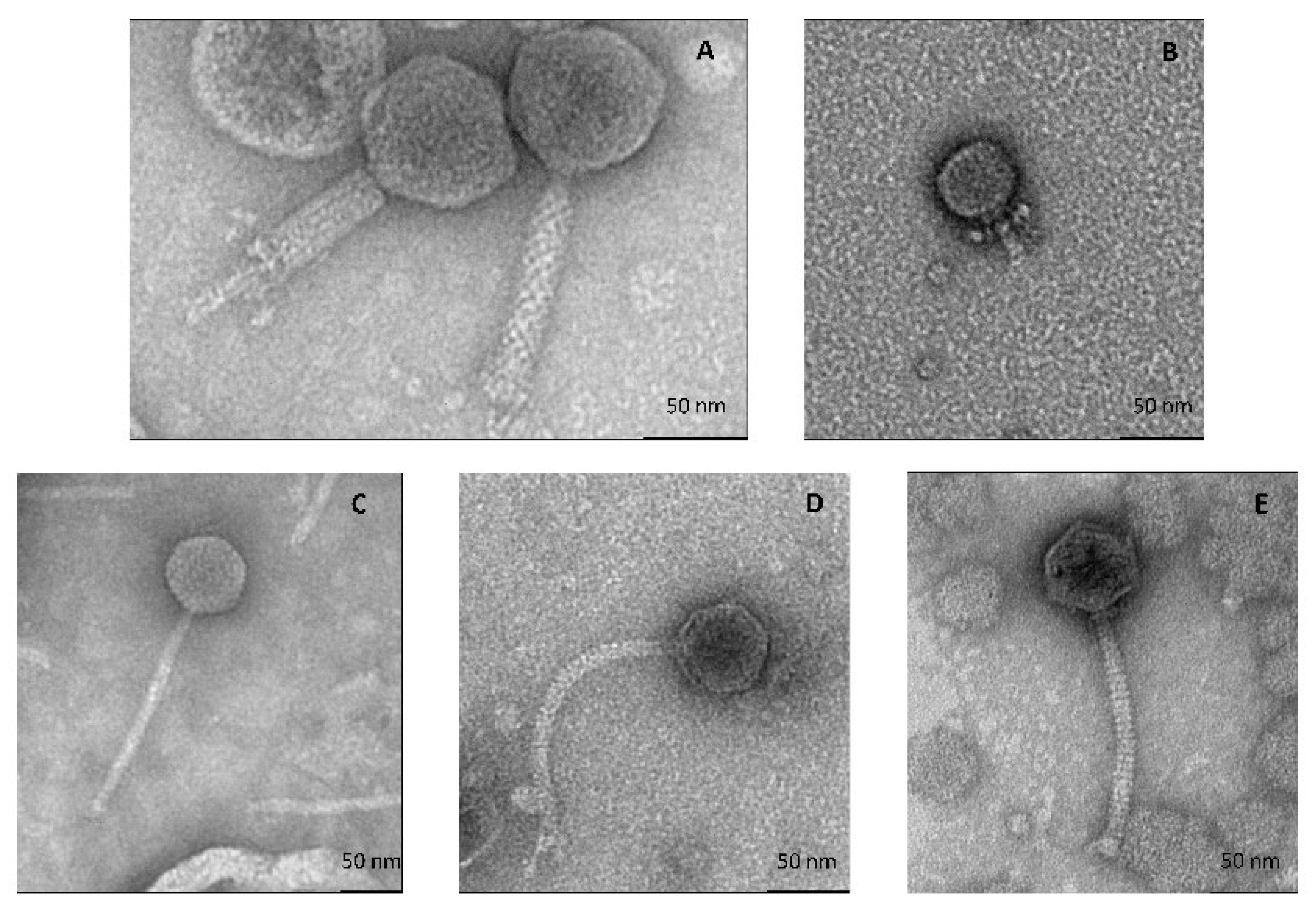
3.2.1. All Cultivable Phages Are Tailed Phages

Electron micrographs of these phages showed that they were all tailed and therefore belonged to the Caudoviricetes class (Figure 1). Their main morphological characteristics are summarized in Table 3. D’Alembert is a myophage (long, contractile tail); Voltaire is a podophage (i.e., it has a short tail), while Rousseau, Diderot and Montesquieu are siphophages (long non-contractile tail).

3.2.2. All Five Phages Have Narrow Host Ranges

Each phage host spectrum was evaluated by spot testing using three distinct groups of strains or isolates, namely: (i) isolates of the same species as the propagation strain obtained from the same cheese, (ii) collection strains belonging to the same species as the propagation strain but obtained from other sources (other dairy products or various environments) and (iii) collection strains belonging to the same genus but different species as the propagation strain (Table 4).
The five newly isolated phages all had a narrow host range, being able to infect only one to seven isolates of a single species and all originating from the studied cheese. Interestingly, the infection profiles of Voltaire and Montesquieu phages, both infecting G. arilaitensis, were completely different (Table S1).

3.2.3. Effect of Temperature on Phage Infection

Phage infections were performed at different incubation temperatures ranging from 12 °C to 30 °C (Figure S1). The infection success of Psychrobacter phage D’Alembert and Glutamicibacter phage Montesquieu was barely constant from 12 to 25 °C but dropped at 30 °C (almost 2 and 4 logs for D’Alembert and Montesquieu, respectively). A moderate reduction in the titer was observed for Glutamicibacter phage Voltaire (almost 1 log) at 25 and 30 °C compared to lower temperatures, and, on the contrary, at 12 °C for Leuconostoc phage Diderot compared to higher temperatures. Brevibacterium phage Rousseau infection was not affected by the temperature within the tested range.

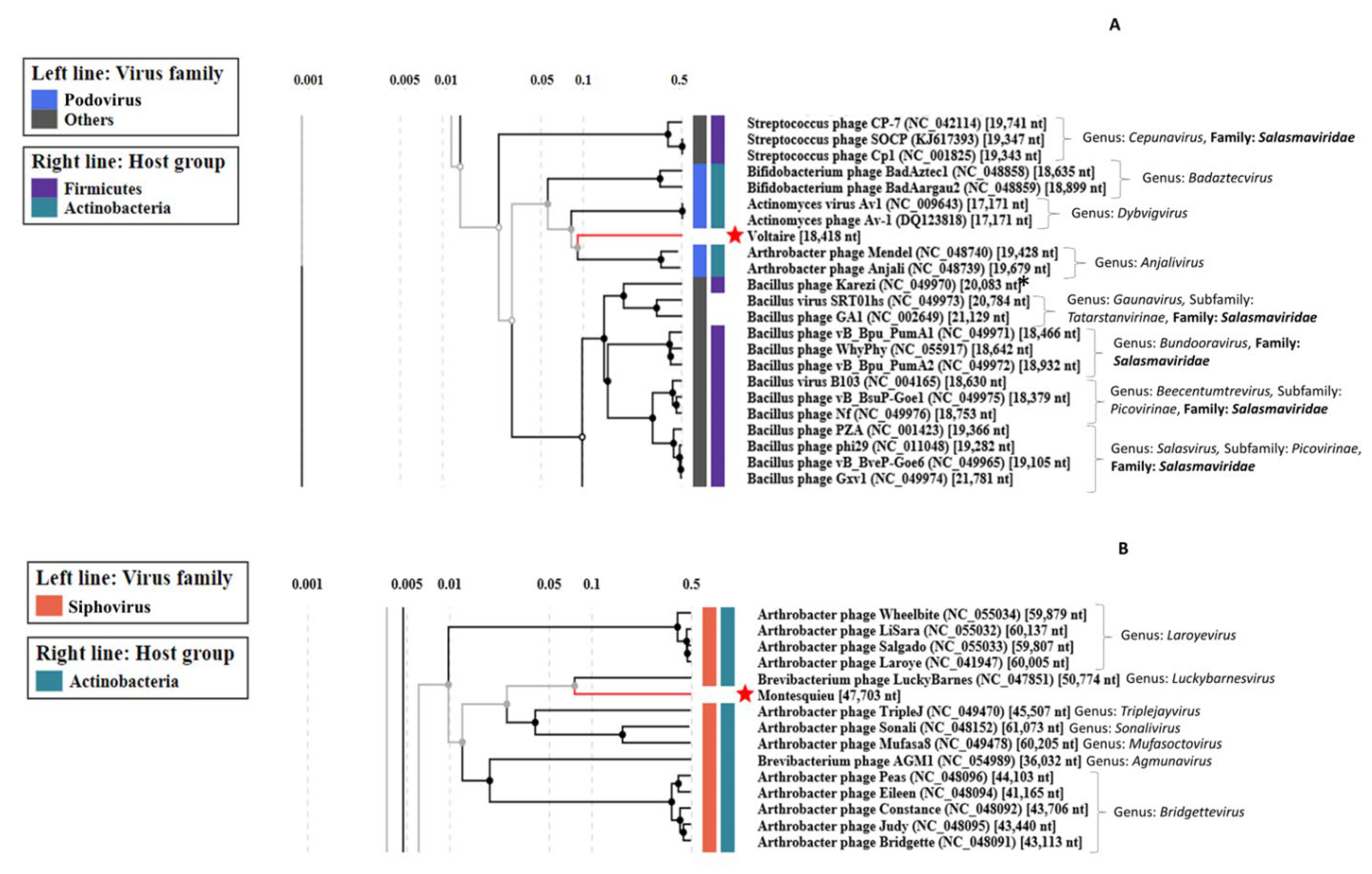
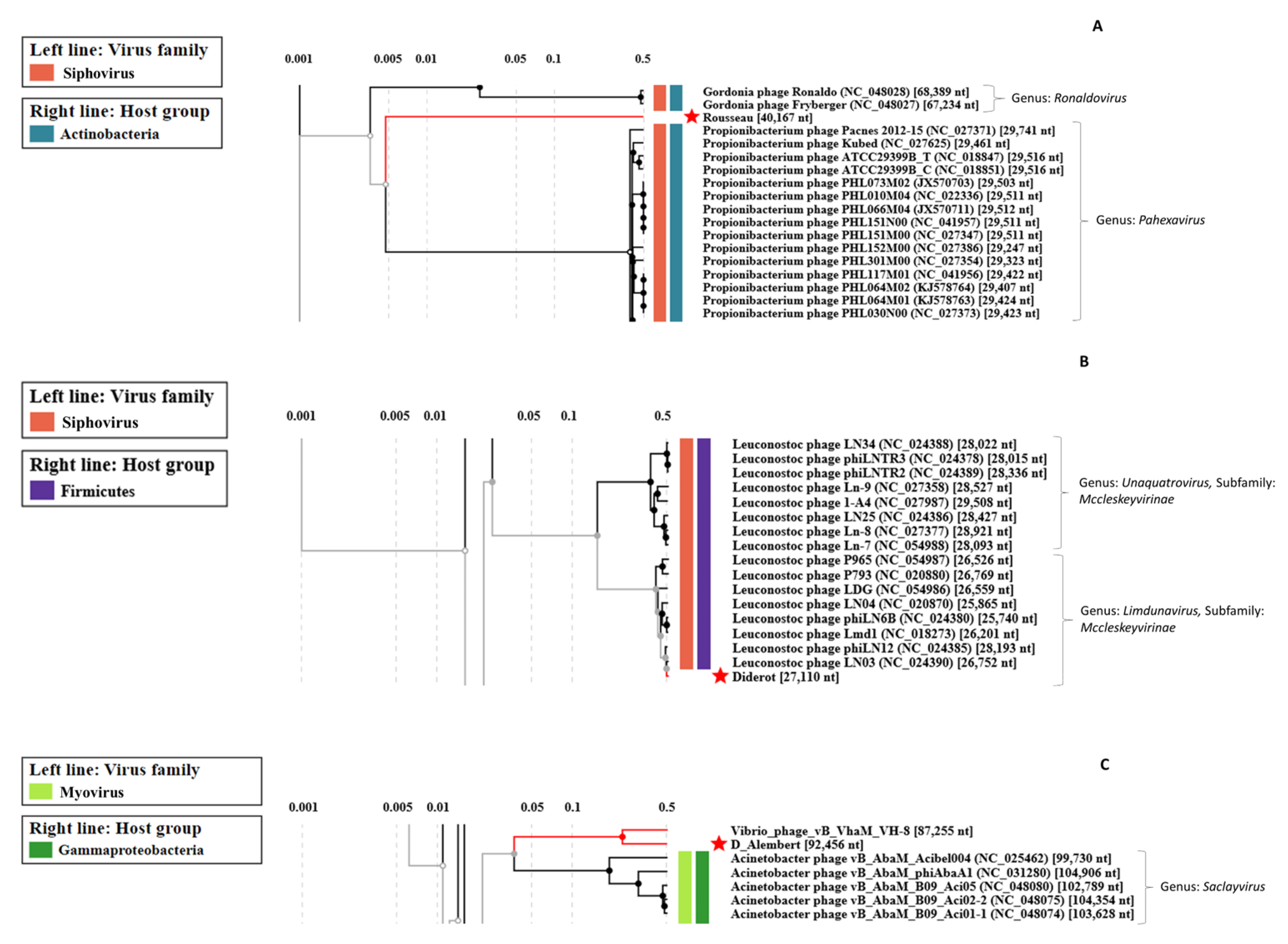
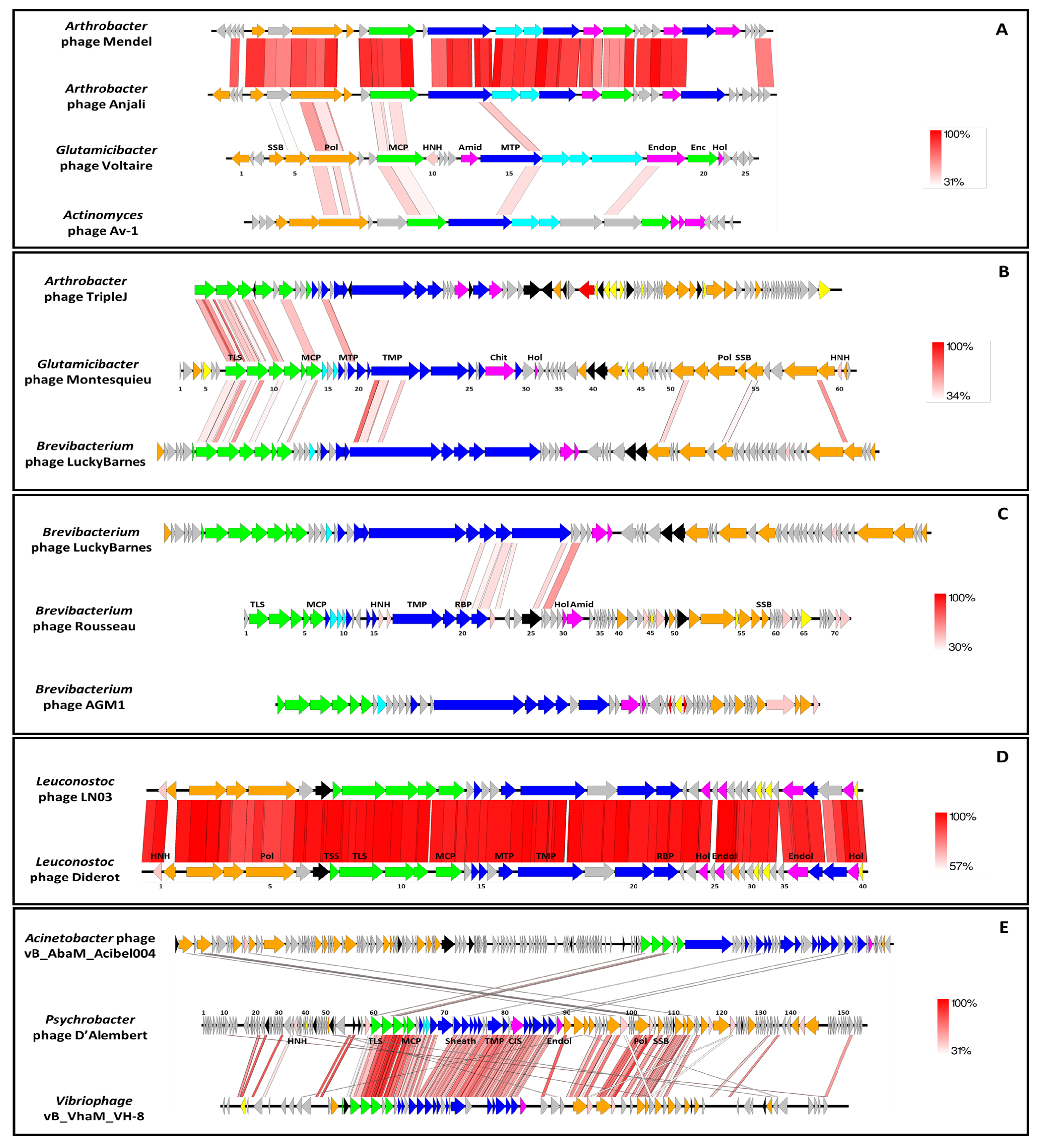
3.2.4. Uncovering Three Completely New Phage Genomes

The genomes of the five phages isolated from the cheese rind were sequenced using both Nanopore and Illumina technologies. The main sequencing and assembly information are summarized in Table 5. Briefly, the number of raw Nanopore reads obtained was comprised between 15 k and 1.7 M, and the number of raw Illumina reads between 5.5 M and 8.9 M. As expected, Nanopore sequencing produced long reads, from 3074 to 9872 bases on average, which facilitated complete genome assembly.
Genome sizes ranged from 18 kb for Glutamicibacter phage Voltaire to 92 kb for Psychrobacter phage D’Alembert. Gene density was high (1.6 genes/kb in average), as is usual for phage genomes, although we noticed that Montesquieu had a slightly lower gene density (1.3 genes/kb). For Glutamicibacter phage Montesquieu, the assembly of Nanopore reads did not produce a unique contig.
The completeness of the sequenced genomes was further investigated (Table 6). Voltaire was complete as indicated by the presence of 176 bp inverted terminal repeats, and D’Alembert as well, with 5.7 kb-long direct terminal repeats (DTRs). Interestingly, D’Alembert DTRs were found thanks to PhageTerm, whereas, in the single contig generated upon the SPAdes assembly from Illumina reads, one of the repeats was missing. Regarding Rousseau and Montesquieu, Illumina assembly displayed a single contig with 127 bp artefactual DTRs, indicating completeness as well. Finally, Diderot was not assembled into a single contig from Illumina reads, but its assembly from Nanopore reads produced a single 27.1 kb contig, highly similar to a known phage over its total length, suggesting genome completeness (see below). Overall, both assemblies gave complementary information and allowed for conclusions on the completeness of 4 out of 5 phage genomes and suggested completion for the last one. With respect to encapsidation modes, PhageTerm predicted Rousseau and Diderot as cos phages with 3′ extensions, Montesquieu as a pac phage, and D’Alembert uses long DTRs.
Each genome was further characterized by performing manual annotation of each ORF. We used the ViPTree webserver to perform comparisons of global protein content of our phages with those available as of March 2022 in this interface (Figure 2 and Figure 3). Then, each phage genome was aligned to one or two close relatives with Easyfig (Figure 4). A table gathering the eleven auxiliary metabolic genes (AMGs) detected can be found in Supplementary Materials (Table S2). Here, AMGs are encoded by all phages but Voltaire. Finally, no virulence genes nor antibiotic resistance genes were detected on the five newly assembled genomes.
Glutamicibacter phage Voltaire encodes 26 ORFs, of which 14 had function predictions. It has no AMGs but possesses three genes coding for enzymes involved in lysis: an amidase (VOLT_14), an endopeptidase (VOLT_19) and a holin (VOLT_21). It represents the first completely new phage genome uncovered by the study. According to ViPTree, the closest phages to Voltaire are Mendel and Anjali, two small and closely related podophages infecting Arthrobacter (Figure 2A). Voltaire is more distantly related to Actinomyces podophage Av-1, sharing weak homologies with the polymerase B (VOLT_6), major capsid protein (VOLT_9) and major tail protein (MTP, VOLT_15) (Figure 4A). The ViPTree positioning leads to proposing that Glutamicibacter phage Voltaire may represent a new genus within the Salasmaviridae family.
Glutamicibacter phage Montesquieu comprises 62 ORFs of which 36 have function predictions. It possesses two predicted AMGs, an ABC transporter (MONT_40) and an aminocyclopropane-1-carboxylate deaminase (MONT_41). No significant BLASTn hits were obtained by comparing the Montesquieu sequence to the NCBI database (nr and viruses), making this latter the second new phage characterized in the study. According to the ViPTree analysis, the closest phages to Montesquieu are a clade of siphophages infecting Brevibacterium or Arthrobacter, from which Montesquieu probably represents a new genus (Figure 2B). The genomic comparison of Montesquieu with its two closest ViPTree neighbors, Arthrobacter phage TripleJ and Brevibacterium phage LuckyBarnes showed a similar genetic organization but little sequence identity at the protein level, mostly within the structural module (Figure 4B). Montesquieu owns proteins related to the tail but no sheath protein, which is consistent with the siphophage morphotype observed in TEM.
Brevibacterium phage Rousseau has 71 ORFs of which 41 have functional predictions. It possesses three AMGs: a putative glutaminyl cyclase (ROUS_25), a thioredoxin-like protein (ROUS_48) and an S-adenosylmethionine-dependent methyltransferase (ROUS_51). With no homologues in the NCBI database, Rousseau is the third completely new phage uncovered by the study and represents a new genus. According to ViPTree, Rousseau is only very distantly related to phages infecting Propionibacterium (Figure 3A). A comparison of the genetic maps of Rousseau and AGM1, a phage previously isolated from a smear-ripened cheese wash solution, shows similar organizations despite their lack of genetic relatedness (Figure 4C). The Rousseau tail module supports its siphophage morphological characteristics observed in TEM.
Psychrobacter phage D’Alembert comprises 158 ORFs of which 56 have functional predictions. It has several AMGs, namely: a nucleoside triphosphate pyrophosphohydrolase (DAL_25), an Ntn_hydrolase-like protein (DAL_45), an S-adenosylmethionine-dependent methyltransferase (DAL_53), a putative chaperonin (DAL_56), an endolytic peptidoglycan transglycosylase (DAL_57), a putative antitoxin (DAL_112) and a thioredoxin glutathione reductase (DAL_125). It represents a new phage genus, sharing a third of its genome with Vibrio phage vB_VhaM_VH-8 (84% nt identity). According to ViPTree, both phages, D’Alembert and VH-8, are distantly related to myophages infecting Acinetobacter (Figure 3C). Genomic comparison confirmed that D’Alembert is closer to VH-8 than to Acinetobacter phage vB_AbaM_Acibel004, with which it showed only little gene synteny and sequence homology for a few proteins (Figure 4E). The fact that a sheath protein is encoded on the D’Alembert genome supports the direct myophage morphotype observation performed using TEM.
Leuconostoc phage Diderot has 40 ORFs of which 29 have a functional prediction. It shares a very strong nucleotidic identity with Leuconostoc siphophage LN03 (98% identity and 98% coverage for both genomes). Both Diderot and LN03 harbor an AMG coding for a ribonuclease Z (DID_7 and LN03_7). A remarkable difference among the genomes is located on LN03_2 and DID_2: although both proteins are predicted endodeoxyribonucleases, they share less than 30% protein identity (Figure 4D), suggesting a recent exchange. Based on ICTV taxonomic criteria, Diderot belongs to the same species as LN03, the Limdunavirus genus and the Mccleskeyvirinae subfamily, which is confirmed by ViPTree analysis (Figure 3B).

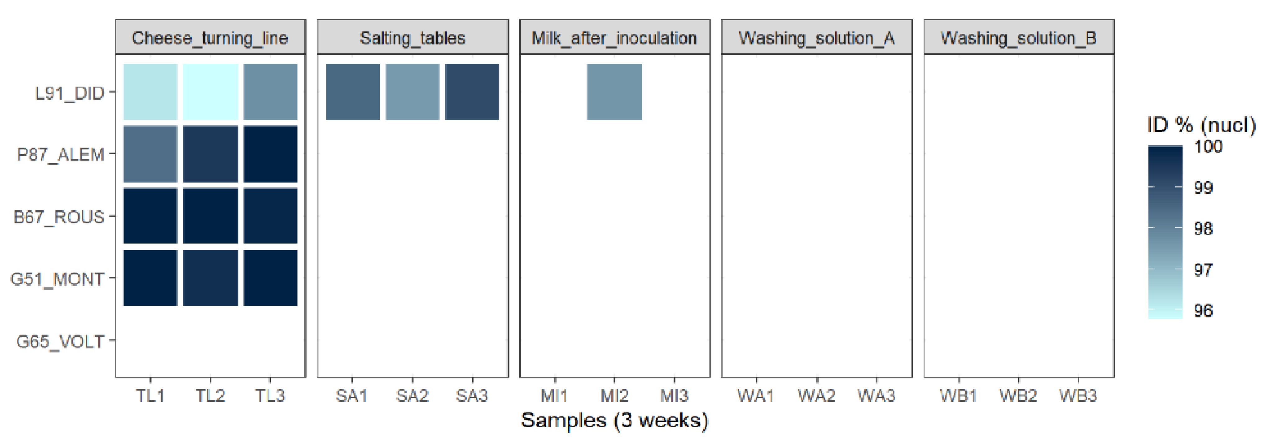
3.2.5. Most of the Identified Phages Are Also Present in the Dairy Plant

In order to identify potential reservoirs of the five phages studied, the dairy plant—producing the cheese from which the phages were isolated—was investigated. The viral fractions resulting from each sample were first tested on five indicator strains with no enrichment step (each strain is sensitive to one of the five isolated phages described above) through a spot assay. If no plaques were observed, a second spot assay on the same strains was performed after enrichment (see Methods). The results are summarized in Figure 5.
Samples obtained from the two washing solutions did not produce lysis plaques using any of the tested strains. However, confluent lysis or clear lysis plaques were obtained using samples from milk, salting tables and the cheese turning line. More precisely, the cheese turning line represented the main reservoir for virulent phages infecting bacteria growing on the rind of this cheese. Indeed, confluent lysis spots were detected for four of the five recipient strains used in the assay (all but G. arilaitensis G65, the propagation strain of phage Voltaire) after infection with these samples. Furthermore, this result was observed three times at a one-week interval, revealing the persistence of the corresponding phages’ species on the cheese turning line despite several cleaning cycles.
Salting tables represented a second, more restricted but persistent reservoir for dairy phages infecting L. falkenbergense. Lysis plaques (rather than confluent lysis zones) were repeatedly observed with L. falkenbergense 91 as the tested strain. This strain was also sensitive to phages coming from one of the three milk samples, indicating that inoculated milk can occasionally contain virulent phages.
Overall, the second spot assay (after enrichment) allowed for the observation of plaques for the same samples as the first one. In order to determine if the phages detected in the potential reservoirs through this experiment were indeed related to the ones previously isolated from the cheese surface, specific primers were designed for each phage (Table 2) and used to amplify and sequence the genetic material present in the different lysis zones or plaques. The sequences amplified from lysis zones detected using G. arilaitensis G51, B. aurantiacum B67 and P. aquimaris 87 indicator strains presented high nucleotidic identity (from 98.4% to 100%) with phages Montesquieu, Rousseau and D’Alembert, respectively (Figure 5). This suggested that these phages, or their close relatives, were still present and infectious in the dairy plant one-and-a-half years after isolation from the cheese surface. Regarding the lysis plaques obtained with L. falkenbergense 91, a PCR product of the size expected for Diderot was also observed, but, depending on the sample, its sequence was not exactly identical to Diderot (ranging from 95.78 to 99.1%) (Figure 5). This may indicate that multiple phages capable of infecting this strain co-exist or evolve in the dairy plant.

4. Discussion

In this study, we first isolated and identified a collection of bacterial isolates from the rind of a French smear-ripened cheese to use it in a second phase for the isolation of phages from the same cheese. The biodiversity of the bacteria isolated during this research, comprising six main species, is typical of this kind of cheese. Indeed, the bacterial community of the surface of washed-rind cheeses generally comprises several distinct groups such as non-starter lactic acid bacteria (e.g., Leuconostoc spp.), staphylococci (e.g., S. xylosus, S. equorum), coryneform bacteria (e.g., Glutamicibacter arilaitensis, Brevibacterium aurantiacum, Corynebacterium variabile, Microbacterium gubbeenense) and Gram-negative bacteria (e.g., Alcaligenes faecalis, Halomonas spp., Psychrobacter spp., Hafnia alvei, Proteus spp., Vibrio spp., Pseudoalteromonas spp.) [24,58,59,60].
In contrast, only two isolates of Brevibacterium aurantiacum, which is generally added as a ripening culture in washed-rind cheeses produced worldwide [61], were obtained, indicating its low ability to outcompete the resident microbiota in this particular cheese. This trend was already observed for several commercial smear starter strains [62,63] and is widely discussed in the literature [64,65], although the reasons explaining their lack of fitness is not fully understood yet. One reason may be the presence of phages infecting such species in cheese. Indeed, one was successfully isolated in this study from a French smear-ripened cheese (Brevibacterium phage Rousseau), and a collection of sixteen phages (represented by Brevibacterium phage AGM1) was also recently isolated from similar Canadian products or their production environment [27].
Unlike B. aurantiacum, Glutamicibacter arilaitensis (formerly Arthrobacter arialitensis) represented the most frequently isolated species in the bacterial collection established from the studied cheese. This yellow-pigmented bacteria is one of the major bacterial species found at the surface of smear-ripened cheeses [66,67]. It can be either deliberately inoculated as a ripening culture or naturally present in cheese, and previous work indicated the possible co-existence of multiple strains of G. arilaitensis in a single cheese product [62]. Interestingly, two genetically different phages (Voltaire and Montesquieu) with non-overlapping host ranges were isolated in this study from the same cheese rind. Whether the observed phage sensitivity could be related to the co-existence of distinct strains of G. arilaitensis within the studied cheese remains to be elucidated. Indeed, phages have already been identified as a key biotic factor favoring the maintenance of intra-species diversity in undefined starter cultures [68,69]. Another explanation would be that two populations of the same strain are present, differing only by their phage resistance profiles in terms of CRISPR diversity as suggested in [70].
Four out of the five newly described phages, namely Glutamicibacter phage Voltaire, Glutamicibacter phage Montesquieu, Brevibacterium phage Rousseau and Psychrobacter phage d’Alembert, shared only little sequence homology with previously sequenced phages. With the increasing number of phage genomes available, genome-based taxonomy is now used for phage classification. Specific requirements, including sequence identity thresholds for species and genus levels, have been proposed for rank-based demarcation of tailed phages [54]. Based on these criteria, the four above-mentioned phages would represent at least four new genera. This result illustrates the under-representation of phages infecting cheese-rind bacteria in public databases. Therefore, the cheese rind should be considered as an attractive environment for the discovery of new phages with potential interest for the cheese industry and ferment producers.
AMGs were found in four of the five studied phages. These genes encode proteins similar to those used in the host metabolism and are supposed to boost metabolic steps that might be bottlenecks in the phage reproduction process [71]. They are mainly implied in the protein and nucleic acid metabolism. Among them, two were, to our knowledge, never described within phage genomes until now. Phage Rousseau ROUS_25 protein is a putative glutaminyl cyclase, named QC for short (HHpred likelyhood probability 99.19% to PDB QC 3NOL). Glutaminyl cyclases are well characterized in eukaryotic organisms. This enzyme catalyzes the cyclization of N-terminal glutamine residues to the pyroglutamate of various proteins [72]. QC were more recently found as well in bacteria, and their function appears essential for Porphyromonas gingivalis growth [73,74]. The QC function in phage Rousseau remains to be established; it may help virion proteins to be more resistant against host proteolytic activity. Interestingly, a distant homolog of this protein is encoded in the genome of several Brevibacterium species (45% amino-acid identity). It is also similar to a Brevibacterium iodinum phage gene (Lucky Barnes, accession YP_009792202.1), which is annotated as “minor tail protein”. The second new AMG is present in Glutamicibacter phage Montesquieu, and encodes an aminocyclopropane-1-carboxylate deaminase (ACCD, MONT_41), just downstream from an ABC transporter. This cassette may contribute to improved amino-acid import and/or synthesis. One AMG present in the D’Alembert genome encoded a putative antitoxin (DAL_112) and might be considered as a host takeover function. Indeed, it is related to a Staphylococcus aureus antitoxin (HHpred likelihood probability 98.56% to PDB 6L8G), and antitoxins encoded by phages can protect themselves against host-produced toxins [75,76].
The host range of the five newly isolated phages is narrow and limited to a few sensitive isolates, which were exclusively obtained from the same tested cheese. All tested collection strains (not originating from that particular cheese), even the ones belonging to the same species as the indicator strain, were resistant. Similar results were reported for Propionibacterium freudenreichii phages isolated from Swiss hard cheese [18], and it is assumed that most phages possess a narrow host range [77]. Thus, with the aim of isolating bacteriophages from a given food sample, one should privilege building a specific collection of bacterial isolates from the same sample (sharing the same ecological niche), and, then, use it to search for phages through spot assays with or without enrichment. This approach may, however, favor the isolation of virulents at the expense of temperate phages due to superinfection immunity. Indeed, temperate phages originate from, or generate, bacterial lysogens, which, when isolated in the same environment and used as indicators, will prevent phage growth and plaque detection [78].
Looking for the origin of the isolated phages, we investigated five different types of samples collected in the cheesemaking plant producing the studied washed-rind cheese as potential reservoirs. Four phages (Rousseau, Montesquieu, Diderot and D’Alembert) were directly detected on the cheese turning line and one on the salting tables (Diderot). For Diderot, PCR-sequencing results revealed some nucleotide variance suggesting the co-occurrence of several closely related phages within the dairy plant, but further experiments are required to confirm this observation. The fact that the non-enriched viral fraction allows for the observation of plaques as well as enriched ones indicates that the level of contamination is non-negligible. Interestingly, in such samples, the positive detection of the different phages was observed three times at weekly intervals, indicating the persistence of these phages on working surfaces of the cheese plant despite regular cleaning procedures. Furthermore, the samples from the dairy plant were obtained almost 18 months after the isolation of phages from the cheese surface. This result indicates a long-term persistence of the four phages within the production environment and especially on manufacturing surfaces. On the other hand, milk after inoculation or the washing solutions should not be considered as major reservoirs since no targeted phages were repeatedly detected in such samples. Phage contamination in dairy plants has already been observed for a long time, but most studies were focused on phages infecting LAB starters. According to the literature, the most probable sources of dairy phages are the starter cultures themselves, as some strains carry prophages that can evolve towards virulence later during the process [79], milk, whey, airborne particles and processing surfaces [15,19,80,81,82]. Previous studies conducted on cheesemaking facilities producing Gubbeen [83], or fresh, bloomy-rind and washed-rind cheeses [2], revealed that dominant bacterial and fungal taxa present on cheese, and mainly the non-inoculated ones, are also contaminating processing surfaces. Our study therefore suggests that the same applies for four bacteriophages infecting the rind bacteria.

5. Conclusions

Virulent bacteriophages infecting four of the main bacterial species living on the rind of a smear-ripened cheese were isolated and characterized. This provides the formal evidence that a diverse viral community co-occurs in this ecosystem along with the well-described bacterial and fungal communities, as previously suggested by viral metagenomics data. The low genomic relatedness of most of the newly isolated phages with currently known phages underlines the lack of knowledge regarding the viral fraction of the cheese ecosystem. Microbial communities of the cheese surface being largely involved in the typical sensory attributes and quality of the final products, further understanding about the role of such entities on cheese microbial ecology and finally their impact on cheese ripening would now be desirable.

Supplementary Materials

The following supporting information can be downloaded at: www.mdpi.com/article/10.3390/v14081620/s1, Table S1: Sensitive/resistant strains to Voltaire and Montesquieu; Table S2: AMGs encoded by each phage; Figure S1: Effect of temperature on infection efficiency.

Author Contributions

Conceptualization, T.P., M.-A.P. and E.D.-B.; Data curation, T.P.; Formal analysis, T.P., J.L., C.M. and O.R.; Funding acquisition, E.D.-B.; Investigation, T.P. and C.F.; Supervision, M.-A.P. and E.D.-B.; Visualization, T.P., J.L.; Writing—original draft preparation, T.P.; Writing—review and editing, J.L., M.-A.P. and E.D.-B. All authors have read and agreed to the published version of the manuscript.

Funding

T.P. is the recipient of a doctoral fellowship from the French Ministry of Higher Education, Research and Innovation (MESRI) and the MICA department of the French National Research Institute for Agriculture, Food and Environment (INRAE).

Institutional Review Board Statement

Not applicable.

Informed Consent Statement

Not applicable.

Data Availability Statement

The genomic data for this study have been deposited in the European Nucleotide Archive (ENA) at EMBL-EBI under accession number PRJEB48484. Assembled/annotated genome sequences of the five isolated phages were precisely deposited under the accession numbers OV696617 (Voltaire), OV696619 (Montesquieu), OV696620 (D’Alembert), OV696621 (Diderot) and OV696622 (Rousseau).

Acknowledgments

We thank Nicolas Ginet (CNRS, France) for sharing the protocol for the preparation of phages for the TEM observations, Christine Longin from the MIMA2 facilities (UMR 1313 GABI, INRAE) for the TEM observations, and our undergraduate students Thomas Clerc and Ines Pedros for testing the host range through spot assays.

Conflicts of Interest

The authors declare no conflict of interest.

References

  1. Irlinger, F.; Layec, S.; Hélinck, S.; Dugat-Bony, E. Cheese Rind Microbial Communities: Diversity, Composition and Origin. FEMS Microbiol. Lett. 2015, 362, 1–11. [Google Scholar] [CrossRef] [PubMed] [Green Version]
  2. Bokulich, N.A.; Mills, D.A. Facility-Specific “House” Microbiome Drives Microbial Landscapes of Artisan Cheesemaking Plants. Appl. Environ. Microbiol. 2013, 79, 5214–5223. [Google Scholar] [CrossRef] [Green Version]
  3. Montel, M.-C.; Buchin, S.; Mallet, A.; Delbes-Paus, C.; Vuitton, D.A.; Desmasures, N.; Berthier, F. Traditional Cheeses: Rich and Diverse Microbiota with Associated Benefits. Int. J. Food Microbiol. 2014, 177, 136–154. [Google Scholar] [CrossRef]
  4. Penland, M.; Falentin, H.; Parayre, S.; Pawtowski, A.; Maillard, M.-B.; Thierry, A.; Mounier, J.; Coton, M.; Deutsch, S.-M. Linking Pélardon Artisanal Goat Cheese Microbial Communities to Aroma Compounds during Cheese-Making and Ripening. Int. J. Food Microbiol. 2021, 345, 109130. [Google Scholar] [CrossRef]
  5. Panthi, R.R.; Jordan, K.N.; Kelly, A.L.; Sheehan, J.J. Selection and Treatment of Milk for Cheesemaking. In Cheese, 4th ed.; McSweeney, P.L.H., Fox, P.F., Cotter, P.D., Everett, D.W., Eds.; Academic Press: San Diego, CA, USA, 2017; Chapter 2; pp. 23–50. ISBN 978-0-12-417012-4. [Google Scholar]
  6. Parente, E.; Cogan, T.M.; Powell, I.B. Starter Cultures: General Aspects. In Cheese, 4th ed.; McSweeney, P.L.H., Fox, P.F., Cotter, P.D., Everett, D.W., Eds.; Academic Press: San Diego, CA, USA, 2017; Chapter 8; pp. 201–226. ISBN 978-0-12-417012-4. [Google Scholar]
  7. Helinck, S.; Le Bars, D.; Moreau, D.; Yvon, M. Ability of Thermophilic Lactic Acid Bacteria to Produce Aroma Compounds from Amino Acids. Appl. Environ. Microbiol. 2004, 70, 3855–3861. [Google Scholar] [CrossRef] [PubMed] [Green Version]
  8. Leistner, L.; Gorris, L.G.M. Food Preservation by Hurdle Technology. Trends Food Sci. Technol. 1995, 6, 41–46. [Google Scholar] [CrossRef]
  9. Brum, J.R.; Ignacio-Espinoza, J.C.; Roux, S.; Doulcier, G.; Acinas, S.G.; Alberti, A.; Chaffron, S.; Cruaud, C.; de Vargas, C.; Gasol, J.M.; et al. Patterns and Ecological Drivers of Ocean Viral Communities. Science 2015, 348, 1261498. [Google Scholar] [CrossRef] [PubMed] [Green Version]
  10. Shapiro, O.H.; Kushmaro, A.; Brenner, A. Bacteriophage Predation Regulates Microbial Abundance and Diversity in a Full-Scale Bioreactor Treating Industrial Wastewater. ISME J. 2010, 4, 327–336. [Google Scholar] [CrossRef] [PubMed] [Green Version]
  11. Sutton, T.D.S.; Hill, C. Gut Bacteriophage: Current Understanding and Challenges. Front. Endocrinol. 2019, 10, 784. [Google Scholar] [CrossRef] [PubMed]
  12. Paillet, T.; Dugat-Bony, E. Bacteriophage Ecology of Fermented Foods: Anything New under the Sun? Curr. Opin. Food Sci. 2021, 40, 102–111. [Google Scholar] [CrossRef]
  13. Brüssow, H. Phages of Dairy Bacteria. Annu. Rev. Microbiol. 2001, 55, 283–303. [Google Scholar] [CrossRef] [PubMed]
  14. Brüssow, H.; Kutter, E. Phage Ecology. In Bacteriophages; Kutter, E., Sulakvelidze, A., Eds.; CRC Press: Boca Raton, FL, USA, 2004; ISBN 978-0-8493-1336-3. [Google Scholar]
  15. Garneau, J.E.; Moineau, S. Bacteriophages of Lactic Acid Bacteria and Their Impact on Milk Fermentations. Microb. Cell Fact 2011, 10, S20. [Google Scholar] [CrossRef] [PubMed] [Green Version]
  16. Marcó, M.B.; Moineau, S.; Quiberoni, A. Bacteriophages and Dairy Fermentations. Bacteriophage 2012, 2, 149–158. [Google Scholar] [CrossRef] [PubMed] [Green Version]
  17. Brüssow, H.; Fremont, M.; Bruttin, A.; Sidoti, J.; Constable, A.; Fryder, V. Detection and Classification of Streptococcus Thermophilus Bacteriophages Isolated from Industrial Milk Fermentation. Appl. Environ. Microbiol. 1994, 60, 4537–4543. [Google Scholar] [CrossRef] [PubMed] [Green Version]
  18. Gautier, M.; Rouault, A.; Sommer, P.; Briandet, R. Occurrence of Propionibacterium Freudenreichii Bacteriophages in Swiss Cheese. Appl. Environ. Microbiol. 1995, 61, 2572–2576. [Google Scholar] [CrossRef] [PubMed] [Green Version]
  19. Madera, C.; Monjardín, C.; Suárez, J.E. Milk Contamination and Resistance to Processing Conditions Determine the Fate of Lactococcus Lactis Bacteriophages in Dairies. Appl. Environ. Microbiol. 2004, 70, 7365–7371. [Google Scholar] [CrossRef] [Green Version]
  20. Wagner, N.; Brinks, E.; Samtlebe, M.; Hinrichs, J.; Atamer, Z.; Kot, W.; Franz, C.M.A.P.; Neve, H.; Heller, K.J. Whey Powders Are a Rich Source and Excellent Storage Matrix for Dairy Bacteriophages. Int. J. Food Microbiol. 2017, 241, 308–317. [Google Scholar] [CrossRef]
  21. Bockelmann, W. Cheese | Smear-Ripened Cheeses. In Encyclopedia of Dairy Sciences, 2nd ed.; Fuquay, J.W., Ed.; Academic Press: San Diego, CA, USA, 2011; pp. 753–766. ISBN 978-0-12-374407-4. [Google Scholar]
  22. Mounier, J.; Coton, M.; Irlinger, F.; Landaud, S.; Bonnarme, P. Smear-Ripened Cheeses. In Cheese, 4th ed.; McSweeney, P.L.H., Fox, P.F., Cotter, P.D., Everett, D.W., Eds.; Academic Press: San Diego, CA, USA, 2017; Chapter 38; pp. 955–996. ISBN 978-0-12-417012-4. [Google Scholar]
  23. Dugat-Bony, E.; GARNIER, L.; Denonfoux, J.; Ferreira, S.; Sarthou, A.-S.; Bonnarme, P.; Irlinger, F. Highlighting the Microbial Diversity of 12 French Cheese Varieties. Int. J. Food Microbiol. 2016, 238, 265–273. [Google Scholar] [CrossRef]
  24. Irlinger, F.; Monnet, C. Temporal Differences in Microbial Composition of Époisses Cheese Rinds during Ripening and Storage. J. Dairy Sci. 2021, 104, 7500–7508. [Google Scholar] [CrossRef]
  25. Mounier, J.; Gelsomino, R.; Goerges, S.; Vancanneyt, M.; Vandemeulebroecke, K.; Hoste, B.; Scherer, S.; Swings, J.; Fitzgerald, G.F.; Cogan, T.M. Surface Microflora of Four Smear-Ripened Cheeses. Appl. Environ. Microbiol. 2005, 71, 6489–6500. [Google Scholar] [CrossRef] [Green Version]
  26. Dugat-Bony, E.; Lossouarn, J.; De Paepe, M.; Sarthou, A.-S.; Fedala, Y.; Petit, M.-A.; Chaillou, S. Viral Metagenomic Analysis of the Cheese Surface: A Comparative Study of Rapid Procedures for Extracting Viral Particles. Food Microbiol. 2020, 85, 103278. [Google Scholar] [CrossRef] [PubMed]
  27. De Melo, A.G.; Rousseau, G.M.; Tremblay, D.M.; Labrie, S.J.; Moineau, S. DNA Tandem Repeats Contribute to the Genetic Diversity of Brevibacterium Aurantiacum Phages. Environ. Microbiol. 2020, 22, 3413–3428. [Google Scholar] [CrossRef] [PubMed]
  28. Edwards, U.; Rogall, T.; Blöcker, H.; Emde, M.; Böttger, E.C. Isolation and Direct Complete Nucleotide Determination of Entire Genes. Characterization of a Gene Coding for 16S Ribosomal RNA. Nucleic Acids Res. 1989, 17, 7843–7853. [Google Scholar] [CrossRef] [PubMed] [Green Version]
  29. Yoon, S.-H.; Ha, S.-M.; Kwon, S.; Lim, J.; Kim, Y.; Seo, H.; Chun, J. Introducing EzBioCloud: A Taxonomically United Database of 16S RRNA Gene Sequences and Whole-Genome Assemblies. Int. J. Syst. Evol. Microbiol. 2017, 67, 1613–1617. [Google Scholar] [CrossRef] [PubMed]
  30. Jurczak-Kurek, A.; Gąsior, T.; Nejman-Faleńczyk, B.; Bloch, S.; Dydecka, A.; Topka, G.; Necel, A.; Jakubowska-Deredas, M.; Narajczyk, M.; Richert, M.; et al. Biodiversity of Bacteriophages: Morphological and Biological Properties of a Large Group of Phages Isolated from Urban Sewage. Sci. Rep. 2016, 6, 34338. [Google Scholar] [CrossRef]
  31. Schneider, C.A.; Rasband, W.S.; Eliceiri, K.W. NIH Image to ImageJ: 25 Years of Image Analysis. Nat. Methods 2012, 9, 671–675. [Google Scholar] [CrossRef]
  32. Jakočiūnė, D.; Moodley, A. A Rapid Bacteriophage DNA Extraction Method. Methods Protoc. 2018, 1, 27. [Google Scholar] [CrossRef] [Green Version]
  33. Ewels, P.; Magnusson, M.; Lundin, S.; Käller, M. MultiQC: Summarize Analysis Results for Multiple Tools and Samples in a Single Report. Bioinformatics 2016, 32, 3047–3048. [Google Scholar] [CrossRef] [PubMed] [Green Version]
  34. Lanfear, R.; Schalamun, M.; Kainer, D.; Wang, W.; Schwessinger, B. MinIONQC: Fast and Simple Quality Control for MinION Sequencing Data. Bioinformatics 2019, 35, 523–525. [Google Scholar] [CrossRef] [Green Version]
  35. Wick, R.R.; Judd, L.M.; Gorrie, C.L.; Holt, K.E. Completing Bacterial Genome Assemblies with Multiplex MinION Sequencing. Microb. Genom. 2017, 3, e000132. [Google Scholar] [CrossRef]
  36. Wick, R.R.; Judd, L.M.; Cerdeira, L.T.; Hawkey, J.; Méric, G.; Vezina, B.; Wyres, K.L.; Holt, K.E. Trycycler: Consensus Long-Read Assemblies for Bacterial Genomes. Genome Biol. 2021, 22, 266. [Google Scholar] [CrossRef]
  37. Wick, R.R.; Judd, L.M.; Gorrie, C.L.; Holt, K.E. Unicycler: Resolving Bacterial Genome Assemblies from Short and Long Sequencing Reads. PLoS Comput. Biol. 2017, 13, e1005595. [Google Scholar] [CrossRef] [PubMed] [Green Version]
  38. Kolmogorov, M.; Yuan, J.; Lin, Y.; Pevzner, P.A. Assembly of Long, Error-Prone Reads Using Repeat Graphs. Nat. Biotechnol. 2019, 37, 540–546. [Google Scholar] [CrossRef] [PubMed]
  39. Vaser, R.; Šikić, M. Time- and Memory-Efficient Genome Assembly with Raven. Nat. Comput. Sci. 2021, 1, 332–336. [Google Scholar] [CrossRef]
  40. Walker, B.J.; Abeel, T.; Shea, T.; Priest, M.; Abouelliel, A.; Sakthikumar, S.; Cuomo, C.A.; Zeng, Q.; Wortman, J.; Young, S.K.; et al. Pilon: An Integrated Tool for Comprehensive Microbial Variant Detection and Genome Assembly Improvement. PLoS ONE 2014, 9, e112963. [Google Scholar] [CrossRef] [PubMed]
  41. Bolger, A.M.; Lohse, M.; Usadel, B. Trimmomatic: A Flexible Trimmer for Illumina Sequence Data. Bioinformatics 2014, 30, 2114–2120. [Google Scholar] [CrossRef] [PubMed] [Green Version]
  42. Mikheenko, A.; Prjibelski, A.; Saveliev, V.; Antipov, D.; Gurevich, A. Versatile Genome Assembly Evaluation with QUAST-LG. Bioinformatics 2018, 34, i142–i150. [Google Scholar] [CrossRef]
  43. Bankevich, A.; Nurk, S.; Antipov, D.; Gurevich, A.A.; Dvorkin, M.; Kulikov, A.S.; Lesin, V.M.; Nikolenko, S.I.; Pham, S.; Prjibelski, A.D.; et al. SPAdes: A New Genome Assembly Algorithm and Its Applications to Single-Cell Sequencing. J. Comput. Biol. 2012, 19, 455–477. [Google Scholar] [CrossRef] [Green Version]
  44. Garneau, J.R.; Depardieu, F.; Fortier, L.-C.; Bikard, D.; Monot, M. PhageTerm: A Tool for Fast and Accurate Determination of Phage Termini and Packaging Mechanism Using next-Generation Sequencing Data. Sci Rep 2017, 7, 8292. [Google Scholar] [CrossRef]
  45. Aziz, R.K.; Bartels, D.; Best, A.A.; DeJongh, M.; Disz, T.; Edwards, R.A.; Formsma, K.; Gerdes, S.; Glass, E.M.; Kubal, M.; et al. The RAST Server: Rapid Annotations Using Subsystems Technology. BMC Genom. 2008, 9, 75. [Google Scholar] [CrossRef] [Green Version]
  46. Zimmermann, L.; Stephens, A.; Nam, S.-Z.; Rau, D.; Kübler, J.; Lozajic, M.; Gabler, F.; Söding, J.; Lupas, A.N.; Alva, V. A Completely Reimplemented MPI Bioinformatics Toolkit with a New HHpred Server at Its Core. J. Mol. Biol. 2018, 430, 2237–2243. [Google Scholar] [CrossRef] [PubMed]
  47. Altschul, S.F.; Gish, W.; Miller, W.; Myers, E.W.; Lipman, D.J. Basic Local Alignment Search Tool. J. Mol. Biol. 1990, 215, 403–410. [Google Scholar] [CrossRef]
  48. Lu, S.; Wang, J.; Chitsaz, F.; Derbyshire, M.K.; Geer, R.C.; Gonzales, N.R.; Gwadz, M.; Hurwitz, D.I.; Marchler, G.H.; Song, J.S.; et al. CDD/SPARCLE: The Conserved Domain Database in 2020. Nucleic Acids Res. 2020, 48, D265–D268. [Google Scholar] [CrossRef] [PubMed] [Green Version]
  49. Marchler-Bauer, A.; Bryant, S.H. CD-Search: Protein Domain Annotations on the Fly. Nucleic Acids Res. 2004, 32, W327–W331. [Google Scholar] [CrossRef]
  50. Terzian, P.; Olo Ndela, E.; Galiez, C.; Lossouarn, J.; Pérez Bucio, R.E.; Mom, R.; Toussaint, A.; Petit, M.-A.; Enault, F. PHROG: Families of Prokaryotic Virus Proteins Clustered Using Remote Homology. NAR Genom. Bioinform. 2021, 3, lqab067. [Google Scholar] [CrossRef] [PubMed]
  51. Lopes, A.; Tavares, P.; Petit, M.-A.; Guérois, R.; Zinn-Justin, S. Automated Classification of Tailed Bacteriophages According to Their Neck Organization. BMC Genom. 2014, 15, 1027. [Google Scholar] [CrossRef] [Green Version]
  52. Chen, L.; Yang, J.; Yu, J.; Yao, Z.; Sun, L.; Shen, Y.; Jin, Q. VFDB: A Reference Database for Bacterial Virulence Factors. Nucleic Acids Res. 2005, 33, D325–D328. [Google Scholar] [CrossRef] [Green Version]
  53. Florensa, A.F.; Kaas, R.S.; Clausen, P.T.L.C.; Aytan-Aktug, D.; Aarestrup, F.M.Y. ResFinder—An Open Online Resource for Identification of Antimicrobial Resistance Genes in next-Generation Sequencing Data and Prediction of Phenotypes from Genotypes. Microb. Genom. 2022, 8, 000748. [Google Scholar] [CrossRef] [PubMed]
  54. Turner, D.; Kropinski, A.M.; Adriaenssens, E.M. A Roadmap for Genome-Based Phage Taxonomy. Viruses 2021, 13, 506. [Google Scholar] [CrossRef]
  55. Nishimura, Y.; Yoshida, T.; Kuronishi, M.; Uehara, H.; Ogata, H.; Goto, S. ViPTree: The Viral Proteomic Tree Server. Bioinformatics 2017, 33, 2379–2380. [Google Scholar] [CrossRef] [PubMed]
  56. Sullivan, M.B.; Weitz, J.S.; Wilhelm, S. Viral Ecology Comes of Age. Environ. Microbiol. Rep. 2017, 9, 33–35. [Google Scholar] [CrossRef] [PubMed]
  57. Xie, Y.; Wahab, L.; Gill, J.J. Development and Validation of a Microtiter Plate-Based Assay for Determination of Bacteriophage Host Range and Virulence. Viruses 2018, 10, 189. [Google Scholar] [CrossRef] [PubMed] [Green Version]
  58. Cogan, T.M.; Chamba, J.-F.; Hohenegger, M.; Guéguen, M.; Ward, A.C.; Gelsomino, R.; Jamet, E.; Swings, J.; Irlinger, F.; Larpin, S.; et al. Biodiversity of the Surface Microbial Consortia from Limburger, Reblochon, Livarot, Tilsit, and Gubbeen Cheeses. In Cheese and Microbes; Donnelly, C.W., Ed.; American Society of Microbiology: Washington, DC, USA, 2014; pp. 219–250. ISBN 978-1-55581-586-8. [Google Scholar]
  59. Larpin-Laborde, S.; Imran, M.; Bonaïti, C.; Bora, N.; Gelsomino, R.; Goerges, S.; Irlinger, F.; Goodfellow, M.; Ward, A.C.; Vancanneyt, M.; et al. Surface Microbial Consortia from Livarot, a French Smear-Ripened Cheese. Can. J. Microbiol. 2011, 57, 651–660. [Google Scholar] [CrossRef] [PubMed]
  60. Quigley, L.; O’Sullivan, O.; Beresford, T.P.; Ross, R.P.; Fitzgerald, G.F.; Cotter, P.D. High-Throughput Sequencing for Detection of Subpopulations of Bacteria Not Previously Associated with Artisanal Cheeses. Appl. Environ. Microbiol. 2012, 78, 5717–5723. [Google Scholar] [CrossRef] [PubMed] [Green Version]
  61. Irlinger, F.; Helinck, S.; Jany, J.L. Secondary and Adjunct Cultures. In Cheese, 4th ed.; McSweeney, P.L.H., Fox, P.F., Cotter, P.D., Everett, D.W., Eds.; Academic Press: San Diego, CA, USA, 2017; Chapter 11; pp. 273–300. ISBN 978-0-12-417012-4. [Google Scholar]
  62. Feurer, C.; Vallaeys, T.; Corrieu, G.; Irlinger, F. Does Smearing Inoculum Reflect the Bacterial Composition of the Smear at the End of the Ripening of a French Soft, Red-Smear Cheese? J. Dairy Sci. 2004, 87, 3189–3197. [Google Scholar] [CrossRef]
  63. Goerges, S.; Mounier, J.; Rea, M.C.; Gelsomino, R.; Heise, V.; Beduhn, R.; Cogan, T.M.; Vancanneyt, M.; Scherer, S. Commercial Ripening Starter Microorganisms Inoculated into Cheese Milk Do Not Successfully Establish Themselves in the Resident Microbial Ripening Consortia of a South German Red Smear Cheese. Appl. Env. Microbiol. 2008, 74, 2210–2217. [Google Scholar] [CrossRef] [PubMed] [Green Version]
  64. Bertuzzi, A.S.; Walsh, A.M.; Sheehan, J.J.; Cotter, P.D.; Crispie, F.; McSweeney, P.L.H.; Kilcawley, K.N.; Rea, M.C. Omics-Based Insights into Flavor Development and Microbial Succession within Surface-Ripened Cheese. mSystems 2018, 3, e00211-17. [Google Scholar] [CrossRef] [Green Version]
  65. Pham, N.-P.; Layec, S.; Dugat-Bony, E.; Vidal, M.; Irlinger, F.; Monnet, C. Comparative Genomic Analysis of Brevibacterium Strains: Insights into Key Genetic Determinants Involved in Adaptation to the Cheese Habitat. BMC Genom. 2017, 18, 955. [Google Scholar] [CrossRef] [PubMed] [Green Version]
  66. Irlinger, F.; Bimet, F.; Delettre, J.; Lefèvre, M.; Grimont, P.A.D.Y. Arthrobacter Bergerei Sp. Nov. and Arthrobacter Arilaitensis Sp. Nov., Novel Coryneform Species Isolated from the Surfaces of Cheeses. Int. J. Syst. Evol. Microbiol. 2005, 55, 457–462. [Google Scholar] [CrossRef] [Green Version]
  67. Monnet, C.; Loux, V.; Gibrat, J.-F.; Spinnler, E.; Barbe, V.; Vacherie, B.; Gavory, F.; Gourbeyre, E.; Siguier, P.; Chandler, M.; et al. The Arthrobacter Arilaitensis Re117 Genome Sequence Reveals Its Genetic Adaptation to the Surface of Cheese. PLoS ONE 2010, 5, e15489. [Google Scholar] [CrossRef] [PubMed] [Green Version]
  68. Erkus, O.; de Jager, V.C.; Spus, M.; van Alen-Boerrigter, I.J.; van Rijswijck, I.M.; Hazelwood, L.; Janssen, P.W.; van Hijum, S.A.; Kleerebezem, M.; Smid, E.J. Multifactorial Diversity Sustains Microbial Community Stability. ISME J. 2013, 7, 2126–2136. [Google Scholar] [CrossRef] [PubMed]
  69. Spus, M.; Li, M.; Alexeeva, S.; Wolkers-Rooijackers, J.C.M.; Zwietering, M.H.; Abee, T.; Smid, E.J. Strain Diversity and Phage Resistance in Complex Dairy Starter Cultures. J. Dairy Sci. 2015, 98, 5173–5182. [Google Scholar] [CrossRef] [Green Version]
  70. Somerville, V.; Berthoud, H.; Schmidt, R.S.; Bachmann, H.-P.; Meng, Y.H.; Fuchsmann, P.; von Ah, U.; Engel, P. Functional Strain Redundancy and Persistent Phage Infection in Swiss Hard Cheese Starter Cultures. ISME J. 2022, 16, 388–399. [Google Scholar] [CrossRef] [PubMed]
  71. Thompson, L.R.; Zeng, Q.; Kelly, L.; Huang, K.H.; Singer, A.U.; Stubbe, J.; Chisholm, S.W. Phage Auxiliary Metabolic Genes and the Redirection of Cyanobacterial Host Carbon Metabolism. Proc. Natl. Acad. Sci. USA 2011, 108, E757–E764. [Google Scholar] [CrossRef] [PubMed] [Green Version]
  72. Booth, R.E.; Lovell, S.C.; Misquitta, S.A.; Bateman, R.C. Human Glutaminyl Cyclase and Bacterial Zinc Aminopeptidase Share a Common Fold and Active Site. BMC Biol. 2004, 2, 2. [Google Scholar] [CrossRef] [PubMed] [Green Version]
  73. Taudte, N.; Linnert, M.; Rahfeld, J.-U.; Piechotta, A.; Ramsbeck, D.; Buchholz, M.; Kolenko, P.; Parthier, C.; Houston, J.A.; Veillard, F.; et al. Mammalian-like Type II Glutaminyl Cyclases in Porphyromonas Gingivalis and Other Oral Pathogenic Bacteria as Targets for Treatment of Periodontitis. J. Biol. Chem. 2021, 296, 100263. [Google Scholar] [CrossRef] [PubMed]
  74. Lamers, S.; Feng, Q.; Cheng, Y.; Yu, S.; Sun, B.; Lukman, M.; Jiang, J.; Ruiz-Carrillo, D. Structural and Kinetic Characterization of Porphyromonas Gingivalis Glutaminyl Cyclase. Biol Chem 2021, 402, 759–768. [Google Scholar] [CrossRef]
  75. Blower, T.R.; Evans, T.J.; Przybilski, R.; Fineran, P.C.; Salmond, G.P.C. Viral Evasion of a Bacterial Suicide System by RNA–Based Molecular Mimicry Enables Infectious Altruism. PLoS Genet. 2012, 8, e1003023. [Google Scholar] [CrossRef] [PubMed]
  76. Otsuka, Y.; Yonesaki, T. Dmd of Bacteriophage T4 Functions as an Antitoxin against Escherichia Coli LsoA and RnlA Toxins. Mol. Microbiol. 2012, 83, 669–681. [Google Scholar] [CrossRef]
  77. Weinbauer, M.G. Ecology of Prokaryotic Viruses. FEMS Microbiol. Rev. 2004, 28, 127–181. [Google Scholar] [CrossRef] [Green Version]
  78. Abedon, S.T. Prophages Preventing Phage Superinfection. In Bacteriophages as Drivers of Evolution: An Evolutionary Ecological Perspective; Abedon, S.T., Ed.; Springer International Publishing: Cham, Switzerland, 2022; pp. 179–191. ISBN 978-3-030-94309-7. [Google Scholar]
  79. Chopin, A.; Bolotin, A.; Sorokin, A.; Ehrlich, S.D.; Chopin, M.-C. Analysis of Six Prophages in Lactococcus Lactis IL1403: Different Genetic Structure of Temperate and Virulent Phage Populations. Nucleic Acids Res. 2001, 29, 644–651. [Google Scholar] [CrossRef] [PubMed]
  80. Murphy, J.; Mahony, J.; Fitzgerald, G.F.; van Sinderen, D. Bacteriophages Infecting Lactic Acid Bacteria. In Cheese, 4th ed.; McSweeney, P.L.H., Fox, P.F., Cotter, P.D., Everett, D.W., Eds.; Academic Press: San Diego, CA, USA, 2017; Chapter 10; pp. 249–272. ISBN 978-0-12-417012-4. [Google Scholar]
  81. Neve, H.; Kemper, U.; Geis, A.; Heller, K.J. Monitoring and characterization of lactococcal bacteriophages in a dairy plant. Kiel. Milchwirtsch. Forsch. 1994, 46, 167–178. [Google Scholar]
  82. Verreault, D.; Gendron, L.; Rousseau, G.M.; Veillette, M.; Massé, D.; Lindsley, W.G.; Moineau, S.; Duchaine, C. Detection of Airborne Lactococcal Bacteriophages in Cheese Manufacturing Plants. Appl. Environ. Microbiol. 2011, 77, 491–497. [Google Scholar] [CrossRef] [PubMed] [Green Version]
  83. Mounier, J.; Goerges, S.; Gelsomino, R.; Vancanneyt, M.; Vandemeulebroecke, K.; Hoste, B.; Brennan, N.M.; Scherer, S.; Swings, J.; Fitzgerald, G.F.; et al. Sources of the Adventitious Microflora of a Smear-Ripened Cheese. J. Appl. Microbiol. 2006, 101, 668–681. [Google Scholar] [CrossRef] [PubMed]
Figure 1. Transmission electron micrographs of 5 phages isolated from cheese rind. (A), Psychrobacter phage D’Alembert (contracted form at left), (B) Glutamicibacter phage Voltaire, (C) Glutamicibacter phage Montesquieu, (D) Brevibacterium phage Rousseau and (E) Leuconostoc phage Diderot.
Viruses 14 01620 g001
Figure 2. Screenshots of proteomic trees of (A) Voltaire and (B) Montesquieu and related phages, computed with ViPTree. *: Genus: Karezivirus, Subfamily: Tatarstanvirinae, Family: Salasmaviridae. Red stars indicate the position of the newly sequenced phage genomes.
Viruses 14 01620 g002
Figure 3. Screenshots of proteomic trees of (A) Rousseau, (B) Diderot, (C) D’Alembert and related phages, computed with ViPTree. Red stars indicate the position of the newly sequenced phage genomes.
Viruses 14 01620 g003
Figure 4. Schematic representation of the phage genomes and comparisons to their closest relatives. (A) Glutamicibacter phage Voltaire. (B) Glutamicibacter phage Montesquieu. (C) Brevibacterium phage Rousseau. (D) Leuconostoc phage Diderot. (E) Psychrobacter phage D’Alembert. Each line represents a phage genome, and each arrow represents an ORF. Red shade lines and percentages indicate tBLASTx identity between two genes. A minimum BLAST hit length of 100 nt (150 for d’Alembert) and with at least 30% tBLASTx identity were set. Gene functions are color-coded and detailed (yellow: transcriptional regulation, orange: DNA metabolism, green: DNA packaging and head, light blue: head to tail, dark blue: tail, pink: HNH endonuclease, fuchsia: lysis, black: auxiliary metabolic genes, grey: hypothetical proteins). List of abbreviations: Amid = amidase; Chit = chitinase; CIS = Contractile Injection System; Enc = encapsidation protein; Endop = endopeptidase; Endol = endolysin; HNH = HNH homing endonuclease; Hol = holin; MCP = Major Capsid Protein; MTP = Major Tail Protein; Pol = polymerase; RBP = Receptor-Binding Protein; SSB = Single-Strand Binding protein; TLS = Terminase Large Subunit; TMP = Tail tape Measure Protein; TSS = Terminase Small Subunit.
Viruses 14 01620 g004
Figure 5. Sensitivity of indicator strains to phages present in different samples collected within the dairy plant.
Viruses 14 01620 g005
Table 1. Bacterial hosts tested for their sensitivity to isolated phages.
Strain or IsolateIsolation SourceTested Phages
Glutamicibacter arilaitensis G16Studied cheeseVoltaire and Montesquieu
Glutamicibacter arilaitensis G26Studied cheese
Glutamicibacter arilaitensis G33Studied cheese
Glutamicibacter arilaitensis G43Studied cheese
Glutamicibacter arilaitensis G51Studied cheese
Glutamicibacter arilaitensis G52Studied cheese
Glutamicibacter arilaitensis G53Studied cheese
Glutamicibacter arilaitensis G65Studied cheese
Glutamicibacter arilaitensis G119Studied cheese
Glutamicibacter arilaitensis G135Studied cheese
Glutamicibacter arilaitensis G183Studied cheese
Glutamicibacter arilaitensis G186Studied cheese
Glutamicibacter arilaitensis G201Studied cheese
Glutamicibacter arilaitensis DSM 16368Reblochon cheese
Glutamicibacter bergerei DSM 16367Camembert cheese
Glutamicibacter nicotianae DSM 20123Air of tobacco warehouses
Glutamicibacter uratoxydans DSM 20647Humus soil
Brevibacterium aurantiacum B20Studied cheeseRousseau
Brevibacterium aurantiacum B67Studied cheese
Brevibacterium aurantiacum 2M23Cheese
Brevibacterium aurantiacum FME9Cheese
Brevibacterium aurantiacum FME34Cheese
Brevibacterium aurantiacum FME43Cheese
Brevibacterium aurantiacum FME45Cheese
Brevibacterium aurantiacum FME48Cheese
Brevibacterium aurantiacum FME49Cheese
Brevibacterium aurantiacum ATCC 9174Cheese
Brevibacterium aurantiacum ATCC 9175 (DSM 20426)Camembert cheese
Brevibacterium aurantiacum 25Camembert cheese
Brevibacterium aurantiacum 299Camembert cheese
Brevibacterium aurantiacum B3Langres cheese
Brevibacterium aurantiacum CAM-4Camembert cheese
Brevibacterium aurantiacum CAM 12CCamembert cheese
Brevibacterium casei CIP 102111 (DSM 20657)Cheese
Brevibacterium epidermidis NCDO 2286T (DSM 20660)Skin
Brevibacterium iodinum ATCC 49514T (DSM 20626)Skin
Brevibacterium linens ATCC 9172 (DSM 20425)Cheese
Brevibacterium sandarakinum DSM 22082Wall surface
Leuconostoc falkenbergense 90Studied cheeseDiderot
Leuconostoc falkenbergense 91Studied cheese
Leuconostoc falkenbergense 92Studied cheese
Leuconostoc falkenbergense 93Studied cheese
Leuconostoc falkenbergense 96Studied cheese
Leuconostoc falkenbergense 98Studied cheese
Leuconostoc falkenbergense 99Studied cheese
Leuconostoc falkenbergense 114Studied cheese
Leuconostoc falkenbergense 116Studied cheese
Leuconostoc mesenteroides 88Studied cheese
Leuconostoc mesenteroides 89Studied cheese
Leuconostoc mesenteroides 95Studied cheese
Leuconostoc mesenteroides 97Studied cheese
Leuconostoc mesenteroides 101Studied cheese
Leuconostoc mesenteroides 102Studied cheese
Leuconostoc mesenteroides 107Studied cheese
Leuconostoc mesenteroides 108Studied cheese
Leuconostoc mesenteroides 113Studied cheese
Leuconostoc mesenteroides 115Studied cheese
Leuconostoc citreum MSE2Milk
Leuconostoc lactis NCW1Cheese
Leuconostoc mesenteroides ssp. cremoris DSM 20346Cheese
Leuconostoc pseudomesenteroides MSE7Cheese
Psychrobacter aquimaris 15Studied cheeseD’Alembert
Psychrobacter aquimaris 54Studied cheese
Psychrobacter aquimaris 59Studied cheese
Psychrobacter aquimaris 60Studied cheese
Psychrobacter aquimaris 69Studied cheese
Psychrobacter aquimaris 87Studied cheese
Psychrobacter aquimaris 124Studied cheese
Psychrobacter aquimaris 129Studied cheese
Psychrobacter aquimaris 184Studied cheese
Psychrobacter aquimaris 200Studied cheese
Psychrobacter cibarius 132Studied cheese
Psychrobacter cibarius 139Studied cheese
Psychrobacter cibarius 140Studied cheese
Psychrobacter cibarius 157Studied cheese
Psychrobacter cibarius 158Studied cheese
Psychrobacter cibarius 160Studied cheese
Psychrobacter cibarius 165Studied cheese
Psychrobacter cibarius 171Studied cheese
Psychrobacter cibarius 181Studied cheese
Psychrobacter cibarius 198Studied cheese
Psychrobacter aquimaris ER15 174 BHI7Saint-Nectaire cheese
Psychrobacter celer DSM 23510Munster cheese
Psychrobacter cibarius DSM 16327Epoisses cheese
Psychrobacter faecalisLivarot cheese
Psychrobacter namhaensis 1439Camembert cheese
Table 2. PCR primers targeting specific phage genes.
PhagePrimerTargeted CDSProductSequence (5′-3′)Annealing Temperature (°C)Amplicon Size (bp)
VoltaireVOLT_FVOLT_18Pre-neck proteinactacctaccctgcccctaa57705
VOLT_Rttcgttgaccagcacacaag
RousseauROUS_FROUS_20Receptor-binding proteinggcggttcggagggtattag57877
ROUS_Rgaaccaaaccttcatcgcca
DiderotDID_FDID_20Tail tape measure proteinaaaactgctgtgactcgtgg57931
DID_Rcaccaaacacgccagagaaa
D’AlembertALEM_FDAL_18RNA ligasetggtactaatgcaggtatcggt57714
ALEM_Rtcaacctcaaagcccatctct
MontesquieuMONT_FMONT_53DNA polymerase Itgacggcaagttcaatcagc57683
MONT_Rgctggttcggagtagtgtct
Table 3. Morphologic characteristics of the isolated phages.
PhageCapsid Size (nm ± SD 1)Tail Size (nm ± SD)MorphotypePlaque Morphology
D’Alembert88 ± 2113 ± 2.6myophageClear, small
Voltaire47 ± 1.130 ± 3.8podophageClear, small
Montesquieu64 ± 1.8184 ± 5.5siphophageClear, large
Rousseau62 ± 5.5177 ± 15.6siphophageClear, large
Diderot57 ± 4.3141 ± 0.9siphophageClear, large
1 SD = Standard deviation.
Table 4. Host spectrum of the 5 tested phages.
Phage Sensitive Isolates/Tested Isolates (Same Species as the Host)Sensitive Species/Tested Species
(Same Genus but Different Species as the Host)
Propagation StrainIsolated from the Studied CheeseFrom Other Sources
D’AlembertPsychrobacter aquimaris 873/100/50/4
VoltaireGlutamicibacter arilaitensis G652/130/10/3
MontesquieuGlutamicibacter arilaitensis G517/130/10/3
RousseauBrevibacterium aurantiacum B671/20/160/5
DiderotLeuconostoc falkenbergense 917/90/10/4
Table 5. Global metrics around sequencing and assembly steps.
PhageRaw Reads CountAverage Size of Reads (bp)Number of ContigsGenome Size (kb)Number of ORFsTerminal Repeat Size (bp)Best Blast Hit 2
IlluminaNanoporeIlluminaNanopore
Voltaire8.9 × 10663,4942 × 1504320118.426176Brevibacterium phage Cantare (83.33% id 1% cov)
Montesquieu 15.6 × 10⁶-2 × 150-147.762-Arthrobacter phage TripleJ (75.25% id 2% cov)
Rousseau5.5 × 10615,6492 × 1503576140.271-Siphoviridae sp. Isolate ctmmc7 (75.54% id 0% cov)
Diderot6.9 × 1061,736,1252 × 1503074127.140-Leuconostoc phage LN03 (98.20% id 98% cov)
D’Alembert7.1 × 106115,7632 × 1509872192.51585719Vibrio phage vB_VhaM_VH-8 (83.95% id 34% cov)
1 As explained in Section 2, Montesquieu genome assembly was obtained from Illumina reads only. 2 Accession numbers for each related phage: Brevibacterium phage Cantare: MK016493; Siphoviridae sp. Isolate ctmmc7: BK019734; Leuconostoc phage PhiLN03: NC_024390; Vibrio phage vB_VhaM_VH-8: MN497415; Arthrobacter phage TripleJ: MN234178.
Table 6. Completeness and encapsidation strategy for the five phages.
Illumina Only AssemblyFinal assembly
Size in bpTerminal Repeat PhageTerm Prediction: Boundaries and Encapsidation StrategySize in bp after PolishingTerminal Repeat
Voltaire18,300/Redundant, permuted and unknown 118,418176 bps ITR
Montesquieu47,703127 bp DTR, assembly artefact removedHeadful (pac)47,576/
Rousseau40,294127 bp DTR, assembly artefact removedCos (3′)40,167/
Diderot//Cos (3′)27,116/
D’Alembert86,864127 bp DTR, assembly artefact removed5719 bps DTR (long)92,4565719 bps DTR
1 phi29-like phage packaging strategy not predictable using PhageTerm [44].
Publisher’s Note: MDPI stays neutral with regard to jurisdictional claims in published maps and institutional affiliations.

