Article

Off-Target Effect of Activation of NF-κB by HIV Latency Reversal Agents on Transposable Elements Expression

by
Gislaine Curty
1,
Luis P. Iniguez
2,
Marcelo A. Soares
1,
Douglas F. Nixon
2 and
Miguel de Mulder Rougvie
2,3,*,†
1
Oncovirology Program, Instituto Nacional de Cancer, Rio de Janeiro 20231-050, Brazil
2
Division of Infectious Diseases, Weill Cornell Medicine, New York, NY 10065, USA
3
Department of Genetic Medicine, Weill Cornell Medicine, New York, NY 10065, USA
*
Author to whom correspondence should be addressed.
Current address: 1300 York Ave., Room W-405, New York, NY 10065, USA.
Viruses 2022, 14(7), 1571; https://doi.org/10.3390/v14071571
Submission received: 27 May 2022 / Revised: 14 July 2022 / Accepted: 18 July 2022 / Published: 20 July 2022
(This article belongs to the Special Issue Endogenous Retroviruses)

Abstract

:
Many drugs have been evaluated to reactivate HIV-1 from cellular reservoirs, but the off-target effects of these latency reversal agents (LRA) remain poorly defined. Transposable elements (TEs) are reactivated during HIV-1 infection, but studies of potential off-target drug effects on TE expression have been limited. We analyzed the differential expression of TEs induced by canonical and non-canonical NF-κB signaling. We evaluated the effect of PKC agonists (Bryostatin and Ingenol B) on the expression of TEs in memory CD4+ T cells. Ingenol B induced 38 differentially expressed TEs (17 HERV (45%) and 21 L1 (55%)). Interestingly, TE expression in effector memory CD4+ T cells was more affected by Bryostatin compared to other memory T-cell subsets, with 121 (107 upregulated and 14 downregulated) differentially expressed (DE) TEs. Of these, 31% (n = 37) were HERVs, and 69% (n = 84) were LINE-1 (L1). AZD5582 induced 753 DE TEs (406 HERV (54%) and 347 L1 (46%)). Together, our findings show that canonical and non-canonical NF-κB signaling activation leads to retroelement expressions as an off-target effect. Furthermore, our data highlights the importance of exploring the interaction between LRAs and the expression of retroelements in the context of HIV-1 eradication strategies.

1. Introduction

According to data from UNAIDS, over 37 million people were living with HIV-1 (PLWH) worldwide in 2020 [1]. Antiretroviral therapy (ART) is the principal approach implemented to control and suppress HIV-1 to undetectable levels to prevent AIDS [2,3]. However, HIV-1 is able to infect and develop a latent infection in CD4+ T cells, establishing a stable viral reservoir in memory CD4+ T cells. This is the main challenge for current HIV-1 cure strategies [4,5,6]. Therapeutic strategies are focused on how to eliminate the HIV-1 reservoir, one approach is known as the “shock and kill strategy”, in which latency reversal agents (LRA) reactivate HIV-1 from cellular reservoirs (shock) for elimination by ART treatment (kill) [7,8]. However, LRAs are not specific for HIV-1 reactivation, and they can also impact on the expression of other genes such as transposable elements (TE) [9,10,11].
Over 40% of the human genome is composed of ancient TE. They are divided into retroelements and transposons. Retroelements are endogenous sequences which replicate through an RNA intermediate, and are divided into those with long terminal repeat (LTR) or without (non-LTR) [12]. LTR retrotransposons are old virus sequences integrated in the genome and they compose about 8% of the human genome and are known as human endogenous retroviruses (HERV) [12,13]. Non-LTR retrotransposons represent about 33% of the genome and are divided into short interspersed nuclear elements (SINE) and long counterparts (LINE or L1) [12]. While SINEs are elements which depend on L1 expression for their replication, L1s are autonomous elements able to replicate into the human genome. In addition, HERV and L1 expression in cells are associated with viral infections and human diseases, such as HIV-1 and cancer [14,15,16,17,18,19].
Differential HERV and L1 expression has been reported during LRA treatment [9,10]. However, the effect of newer LRAs such as bryostatin-1 and ingenol-3-hexanoate (Ingenol B) on TE expression has not been investigated. Both induce gene expression though PKC-NF-κB signaling and are able to induce HIV expression [20,21,22]. They promote phosphorylation and inactivation of IκB proteins, allowing for NF-κB translocation to the nucleus and the initiation of gene transcription. In addition, phorbol 12-myristate 13-acetate (PMA), another agonist of PKC used to activate T cells, can also stimulate HIV-1 expression through PKC–NF-κB signaling [23,24]. In addition, a new class of LRAs known as mimetics of the second mitochondrial-derived activator of caspases (SMACm—SMAC mimetics) are also able to induce HIV-1 expression through non-canonical NF-κB signaling [21,25]. They are able to induce HIV latency reversion alone or combined with other LRAs [26]. SMAC mimetics antagonize the inhibitor of apoptosis proteins (IAPs) such as AZD5582, which binds to the BIR3 domains cIAP1, cIAP2 and XIAP and induces apoptosis [27]. The cIAP1/cIAP2 degradation induces non-canonical NF-κB signaling activation in resting CD4+ T cells from PLWH on ART and in latently infected cells from models of HIV-1 latency. In addition, the activation of non-canonical NF-κB signaling by AZD5582 induces HIV-1 RNA expression in the blood of ART-suppressed bone marrow–liver–thymus in humanized mice [25]. Although several studies have explored the effects of LRAs on HIV-1 reactivation, little is known about their impact on TE expression. In this study, we evaluated the off-target effects of two PKC agonists (Bryostatin-1 and Ingenol B) and a SMAC mimetic (AZD5582) on the expression of TEs in memory CD4+ T-cell subsets.

2. Materials and Methods

2.1. RNA-seq Datasets

In this study, we used publicly available RNA-seq datasets to analyze the impact of TE expression in T-cell subsets treated with two different PKC LRAs. Datasets were downloaded from the Sequence Read Archive (SRA) with the following accession numbers: GSE142774 and GSE94150. We selected studies where LRAs (Bryostatin, Ingenol-B or AZD5582) were used ex vivo on memory CD4+ T cells obtained from PLWH. In GSE142774, resting CD4+ T cells obtained from HIV-1-infected and ART-suppressed donors were treated ex vivo with PKC agonists (Ingenol B) and SMACm (AZD5582). The activity of Ingenol B (25 nM), AZD5582 (100 nM) or DMSO (0.05%) on memory CD4+ T cells was evaluated, and cells were harvested at 2, 6, and 24 h after exposure. RNA was isolated from the harvested cells, processed into stranded mRNA libraries and then sequenced using an Illumina Hiseq 4000 sequencer using a paired-end 50 bp × 50 bp run [25]. In GSE94150, CD4+ T cells were isolated from PLWH who had been on successful ART for over 36 months with an undetectable viral load (≤50 copies/mL). T cells were sorted into three T-cell subpopulations: central memory (CM), effector memory (EM) and transition memory (TM) cells. The ex vivo effects of PKC agonists (Bryostatin and PMA) were assessed for their ability to induce viral transcription in sorted memory CD4+ T-cell subsets following a 24 h stimulation with 10 nM Bryostatin and 100 ng/mL PMA. Total RNA was isolated, converted into cDNA, and libraries were generated using the Nextera XT DNA library preparation kit and sequenced on an Illumina HiSeq 2500 [28]. In short, these are ex vivo studies using memory CD4+ T-cell subsets obtained from ART-suppressed HIV-infected individuals. We used RNA-seq data obtained from CD4+ T-cell subsets treated for the same 24 h period with an array of LRAs: Bryostatin, Ingenol B and AZD5582. DMSO and PMA were used for drug control comparison. Following LRA treatment, RNA was isolated, and libraries were prepared and sequenced on the Illumina platform.

2.2. Bioinformatic Analyses

FASTQ files from RNA datasets were downloaded in the GEO and aligned to the human genome (hg38) using Bowtie2 (very-sensitive-local-k 100—score-min L, 0, 1.6 for multi-mappings) [29,30]. The Bowtie2 output was used in the Telescope software. Telescope was used to define and quantify accurate retrotransposon expression on samples using HERV and L1 annotation (retro.hg38.v1, available on https://github.com/mlbendall/telescope_annotation_db/tree/master/builds, accessed on 19 July 2022) [31]. Host gene quantification was performed using Salmon v1.3.0 through gencode v.33 plus the HIV-1 genome (K03455) [32]. Telescope and Salmon outputs were used to calculate retrotransposons differentially expressed in treated vs. untreated CD4+ T cells using DESeq2 using the Wald test [33]. Retrotransposons with adjusted p-values < 0.05 and absolute (log2FoldChange) > 1.5 were considered differentially expressed and visualized using Volcano plots with Bioconductor EnhancedVolcano (https://github.com/kevinblighe/EnhancedVolcano, accessed on 19 July 2022) and by the R ggplot2 package. Unsupervised hierarchical analysis using retroelements abundance data was performed using Bray–Curtis dissimilarity and average linkage metrics by the R vegan package. In addition, Venn and upset plots were performed using the Venn diagram and UpSetR packages in R to show all relationships between sets of data from differentially expressed retroelements. The most expressed HERV and L1 were shown by bar plots. HERV families were defined according to the retro.hg38.v1 annotation and shown by pie charts. Neighboring host genes from differentially expressed retroelements and whether the TEs were noncoding or coding were defined using Telescope meta annotations (https://raw.githubusercontent.com/LIniguez/Telescope_MetaAnnotations/main/TE_annotation.v2.0.tsv, accessed on 19 July 2022). Over-representation analysis (ORA) was also performed with WebGestal (available at http://webgestalt.org/, accessed on 19 July 2022) using gene ontology biological processes as a functional database.

3. Results

In order to characterize the profile of TE expression in CD4+ T cells upon distinct LRA treatments, we first evaluated the off-target ex vivo effect of canonical NF-κB activation induced by PKC signaling from Ingenol B and Bryostatin in CD4+ T cells obtained from aviremic HIV-1 participants. We found a total of 38 differentially expressed (23 upregulated and 15 downregulated) transposable elements in memory CD4+ T cells treated with Ingenol B (Figure 1A). Of these, 21 (55.3%) were assigned to L1 sequences and 17 (44.7%) to HERV sequences. Among the most differentially expressed HERV loci we found: MER41_14q24.2a, ERV316A3_2q14.1d, PRIMA41_19q13.2b, PRIMA41_19q13.2a, HERV4_4q12, HERVP71A_3p26.1, HERV30_6q22.31, ERVLB4_11p15.5d, HERVK11D_2q11.2 and HERVH_11q24.1a (Figure 1B). Regarding DE L1 loci, we detected: L1FLnI_12p12.1q, L1FLnI_1q32.2m, L1FLnI_12p12.1r, L1FLnI_12p12.1s, L1FLnI_Xq22.1la, L1FLnI_4q25l, L1FLnI_13q22.1c, L1FLnI_5p15.33b, L1FLnI_5q35.3b and L1FLnI_13q22.1d (Figure 1C).
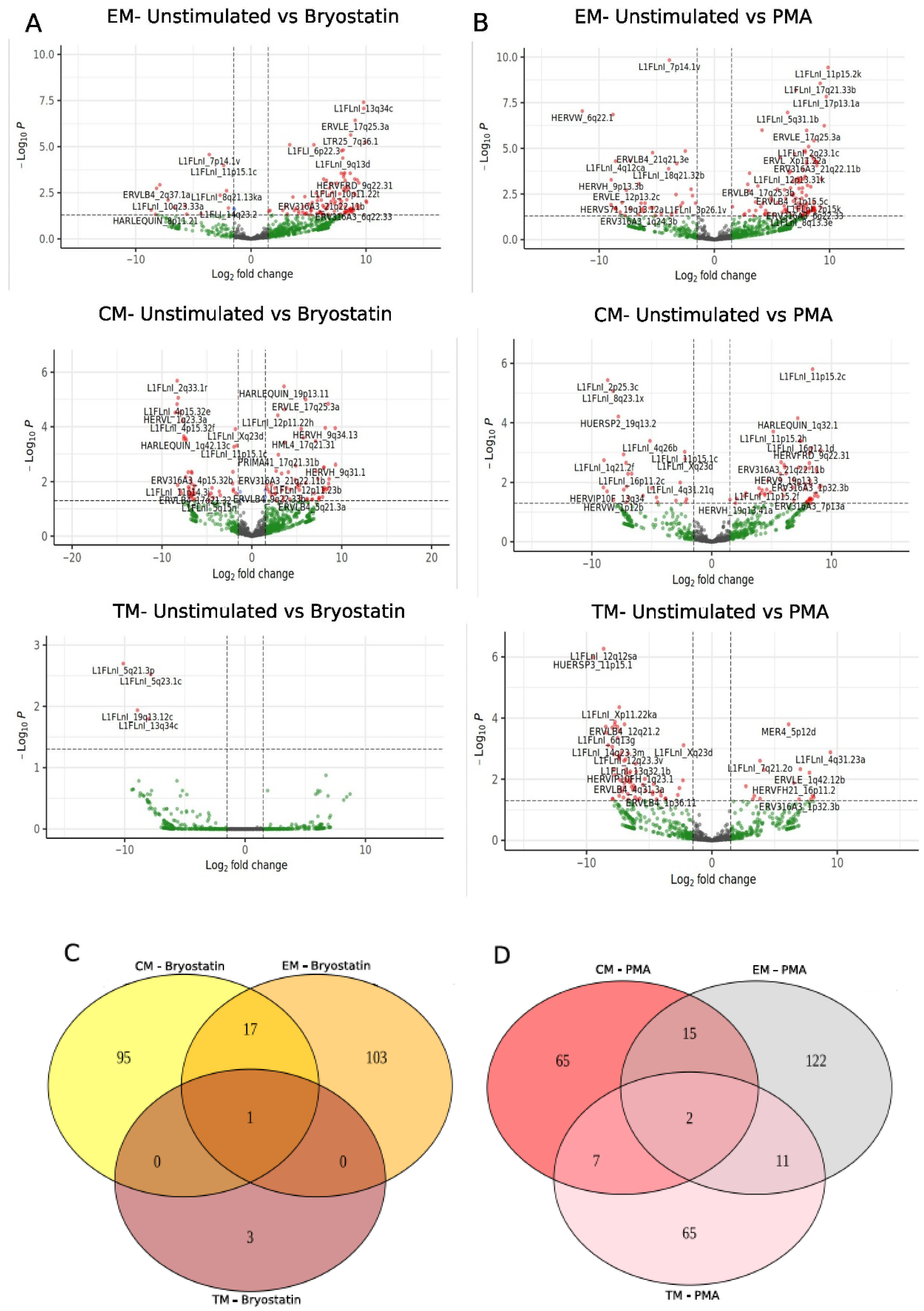
We also evaluated the effect of PKC activation induced by Bryostatin on the expression of TEs in central memory (CM), transition memory (TM) and effector memory (EM) CD4+ T-cell subsets. Interestingly, EMs were the most impacted cell population by Bryostatin compared to other CM and TM subsets. We found a total of 121 (14 upregulated and 107 downregulated) differentially expressed TEs in the Bryostatin treatment (Figure 2A). Of these, 30.6% (n = 37) were HERVs and 69.4% (n = 84) were L1 sequences. The most upregulated TEs induced by Bryostatin treatment were: L1FLnI_2p25.3c, HARLEQUIN_8p11.21, ERVLB4_2q37.1a, HUERSP3_1p36.13, L1FLnI_10q23.33a. The most downregulated TEs were L1FLnI_12p13.31k, L1FLnI_7q34b, L1FLnI_Xp11.21ec, L1FLnI_11p11.2c and L1FLnI_17p13.1a. The number of differentially expressed TEs following Bryostatin treatment was 113 and 4 in CM and TM cell populations, respectively (Figure 2A). Of the 113 differentially expressed TEs in CM, we found 49 up- and 64 downregulated in Bryostatin-treated cells. Of these, 35.4% (n = 40) were HERVs and 64.6% (n = 73) were L1 sequences. Regarding differentially expressed TEs in TM cells, we only detected upregulated L1 sequences induced by Bryostatin treatment (L1FLnI_5q21.3p, L1FLnI_19q13.12c, L1FLnI_13q34c and L1FLnI_5q23.1c). Additionally, we also evaluated the effect of phorbol myristate acetate (PMA) on T-cell subsets (Figure 2B). PMA is known to promote elevated levels of T cells’ activation through the PKC activation signal pathway and HIV expression. In EM T cells, a total of 150 differentially expressed TEs (112 up- and 38 downregulated) were detected following PMA treatment (Figure 2B). Of those, 67.3% (n = 101) were L1 sequences and 32.7% (n = 49) were HERV sequences. In CM T cells, we found 89 differentially expressed TEs (63 up- and 26 downregulated sequences) (Figure 2B). Of these, 35 were HERVs and 54 were L1 sequences. In TM T cells we found 85 differentially expressed TEs upon PMA treatment (70 up- and 15 downregulated to PMA treatment), of which 27 (31.8%) were HERVs and 58 (68.2%) were L1 sequences (Figure 2B).
Most of the differentially expressed TEs were exclusively regulated for each memory T-cell subset tested; 103, 95 and 3 TEs were exclusively modulated in EM, CM and TM cells treated with Bryostatin, respectively (Figure 2C). Most interestingly, 17 DE TEs (5 HERVs and 12 L1) such as ERV316A3_21q22.11b, ERVLE_17q25.3a, HUERSP1_Xp22.2, MER101_18q21.1b and MER41_21q22.11b were common to CM and EM; and one L1, localized on chromosome 13 (L1FLnI_13q34c), was common to all T-cell subsets. In addition, 15, 7 and 11 differentially expressed TEs were found commonly regulated between CM and EM, CM and TM, and EM and TM, respectively (Figure 2D). In addition, two DE TEs (HUERSP2_19q13.2 and L1FLnI_Xq23d) were common to all T-cell subsets. Among all DE TEs, 65, 65 and 122 of them were exclusively regulated in TM, CM and EM cells treated with PMA, respectively.
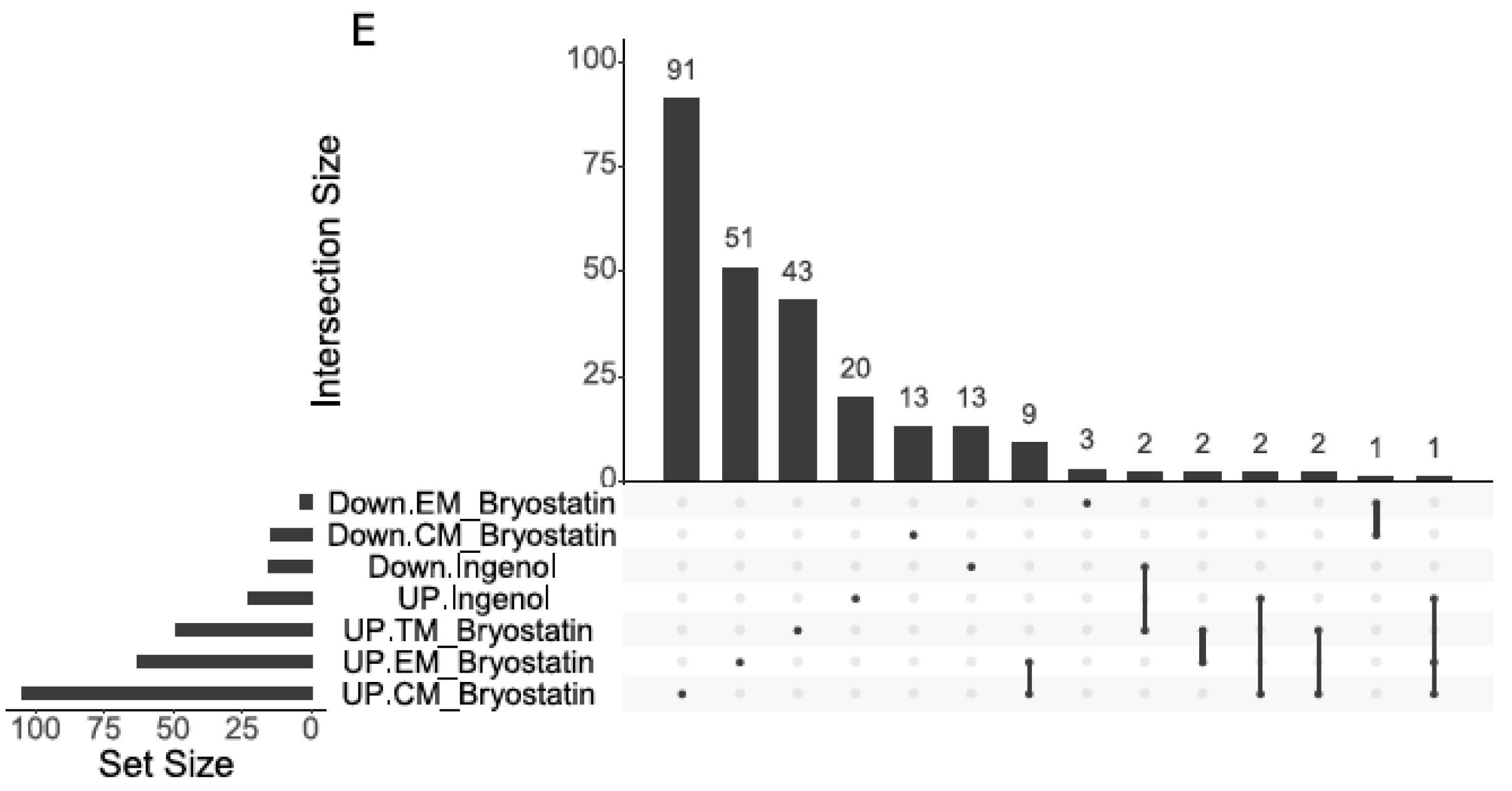
Next, we compared the effect on TE expression caused by different LRA PKC agonists (Bryostatin and Ingenol B) in treated memory CD4+ T-cell subsets. We found L1FLnI_Xq22.1la to be commonly upregulated in EM and CM T cells treated with Bryostatin and in general memory T cells treated with Ingenol B (Figure 2E). Furthermore, some differentially expressed TEs were exclusively regulated by LRA treatment according to each memory CD4+ T-cell subset. A total of 39, 8 and 2 TEs were found exclusively upregulated in CM, EM and TM cells treated with Bryostatin, respectively. In memory CD4+ T cells we found 20 TEs exclusively expressed by Ingenol B treatment. In addition, HERV loci from full-length HERV families were found exclusively up-regulated in memory CD4+ T cells treated with Ingenol B HERV9_1p36.23(HERVW), HML3_1p36.23 (HERVK) and in CM cells treated with Bryostatin HERV-K (HML2_14q11.2, HML3_5p15.33b, HML3_Xq13.3) (Figure 2E). Together, these data show that PKC agonists lead to differential TE expression in each memory CD4+ T cell.
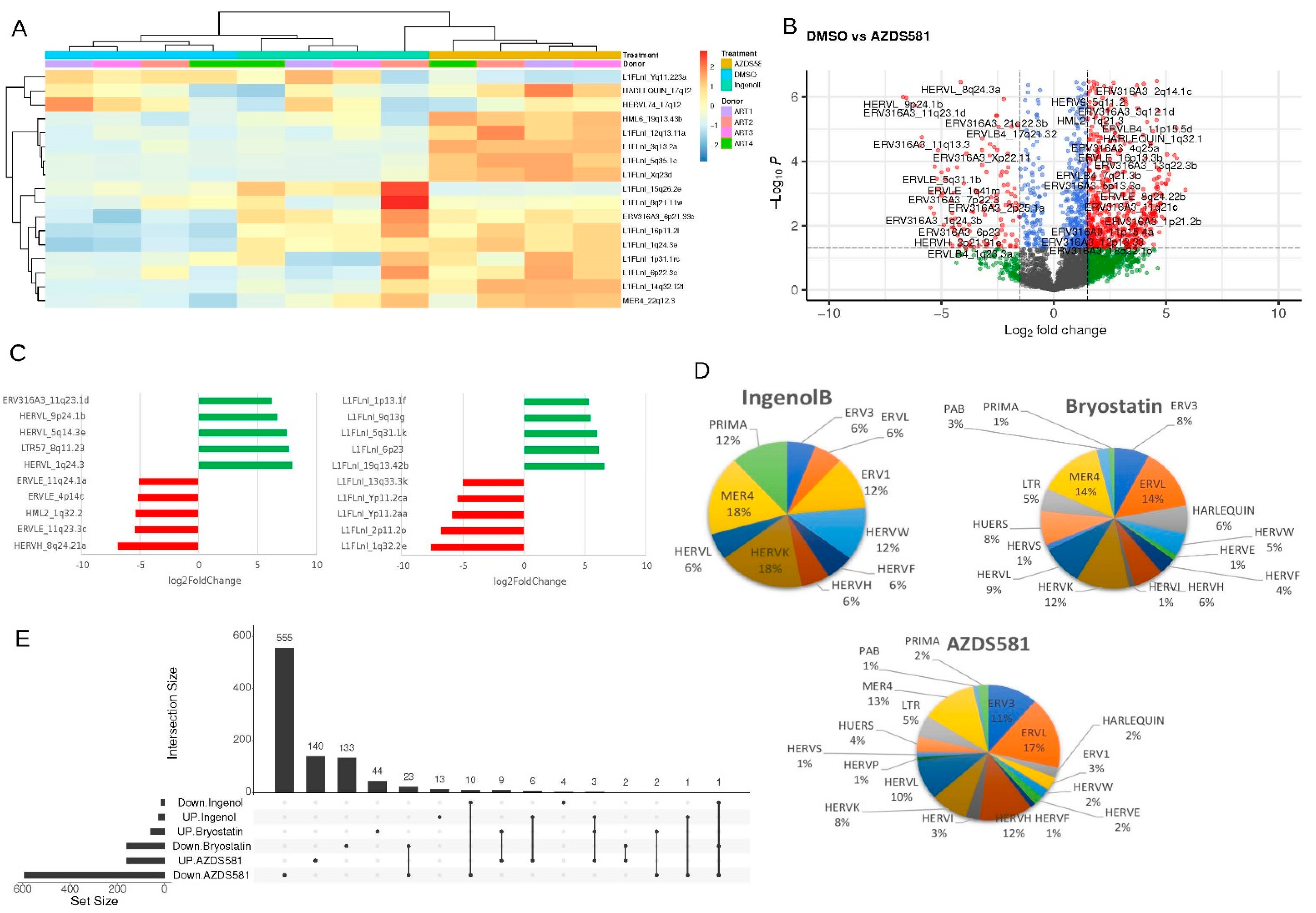
We also analyzed TE expression in noncanonical NF-κB activation from memory CD4+ T cells treated with AZD5582 and compared those with Ingenol B and Bryostatin treatment, which are also able to induce canonical NF-kB activation. In an unsupervised analysis we found three clusters from TE loci expressed in cells treated with DMSO, Ingenol B and AZD5582 (Figure 3A). We found 753 differentially expressed TEs (406 HERV and 347 L1) in AZD5582 (Figure 3B). Two full-length HERVs, such as HERVL (9p24.1b and 5q14.3e loci) and L1 sequences localized on chromosome 1 (L1FLnI_1p13.1f), 9 (L1FLnI_9q13g), 5 (L1FLnI_5q31.1k), 6 (L1FLnI_6p23) and 19 (L1FLnI_19q13.42b) were upregulated by AZD5582 treatment (Figure 3C). In contrast, HERVK (HML2_1q32.2) and HERVH (HERVH_8q24.21a) were the most downregulated loci in cells treated with AZD5582 (Figure 3C). Comparison between the HERV families regulated between the AZD5582 and Ingenol B treatment shows that the Harlequin, HERVE, HERVI, HERVL, HERVS, HUERS, LTR and PAB families were exclusively regulated in cells treated with AZD5582 (Figure 3D). On the other hand, the ERV1 and HERVP families were differently regulated between the AZD5582 and Bryostatin treatment (Figure 3D).
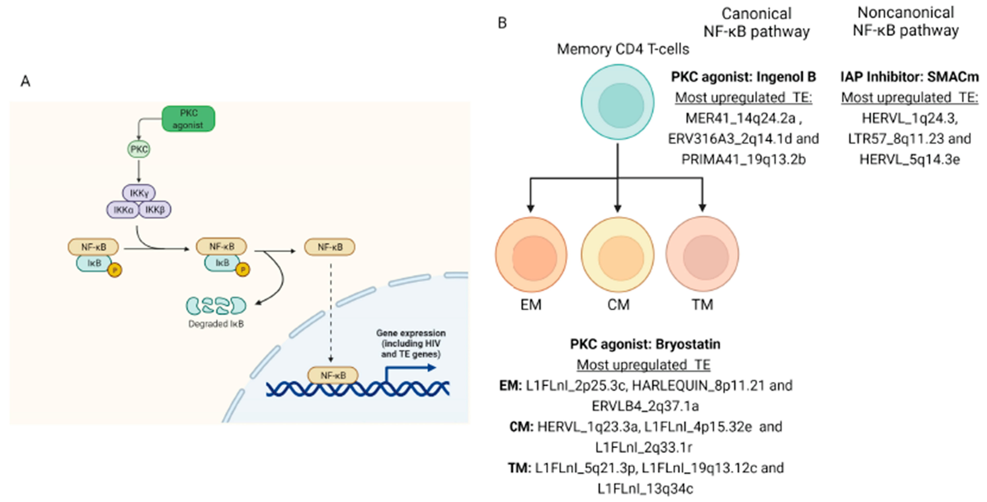
In addition, we found three L1 loci, localized in the chromosomes 16, 17 and X, commonly upregulated by treatments (Figure 3E). Additionally, 140 TEs (66 HERV and 74 L1 loci), 13 TEs (7 HERV and 6 L1 loci) and 44 TEs (15 HERV and 29 L1 loci) were upregulated exclusively by AZD5582, Ingenol B and Bryostatin treatment. The retroelements’ neighboring genes and the either coding and noncoding DE TE data were also defined to each type of treatment (Supplementary Figure S1). There were no significant enriched pathways of the neighboring genes from DE TE (Supplementary Figure S1). Altogether, these data show that LRAs can regulate the differential expression of retroelements through canonical and non-canonical NF-κB activation (Figure 4).

4. Discussion

The HIV genome can be stabilized into cellular reservoirs through latent infection and can persist for decades in the human body. Thus, many drugs have been evaluated to reactivate and eliminate HIV-1 from reservoirs but their transcriptional reactivation is non-specific, and off-target effects have been reported [9,10,11,34,35]. In this study, we analyzed the off-target effects on the expression of retroelements in memory T cells treated with Bryostatin, Ingenol B and AZD5582, which are able to induce NF-κB signaling pathways.
Bryostatin-1 and Ingenol B lead to HIV-1 reactivation in vivo and ex vivo. Bryostatin-1 has also been assessed in clinical trials to treat Alzheimer and cancer [36,37]. Both drugs activate the PKC-NF-κB signaling pathway and induce gene expression. The phosphorylation and inactivation of IκB proteins leads to the translocation of NF-κB to the nucleus and induces HIV-1 and endogenous gene transcription [21]. Herein, we found a total of 38 differentially expressed transposable elements in memory CD4+ T cells treated with Ingenol B. The MER4 (14q24.2a loci), ERV3 (2q14.1d loci) and PRIMA (19q13.2b and 19q13.2a loci) families were the most upregulated HERVs by Ingenol B in T cells. Additionally, L1 sequences from chromosome 1 (1q32.2m), 12 (12p12.1q, 12p12.1r and 12p12.1s) and X (Xq22.1la) were also found to be the most upregulated in treated cells. The expression of transposable elements might induce immune responses, genomic instability, tumorigenesis and can be used as biomarkers, which could suggest a functional effect role in memory T cells treated with LRA [15,38,39]. Memory CD4+ T cells are classified into central, transitional and effector memory cells, and each of them have a distinct cell phenotype, function and survival properties [40,41,42]. Although all memory CD4+ T-cell subsets have been shown to contribute to HIV-1 reservoirs, EM cells have the largest proportion of HIV-1 intact genomes and show the highest latency reversal phenotype compared to either CM or TM subsets [28,43]. Here, we found differential TE regulation in memory CD4+ T-cell subsets (CM, TM and EM) through PKC activation mediated by Bryostatin. EM cells were the most impacted cell population compared to other memory CD4+ T-cell subsets. A total of 103, 95 and 3 TEs were exclusively modulated in EM, CM and TM cells treated with Bryostatin, respectively. In addition, classical latency reversal attempts are achieved through T-cell activation, thus we also evaluated the effect of TE expression in memory T-cell subsets treated with PMA, a positive control of T-cell activation mediated by PKC activation [23,24]. Similar to Bryostatin, the EM subpopulation was the most affected cell subset, showing a total of 150 differentially expressed TEs (112 up- and 38 downregulated) following PMA treatment. Together, our data show a distinct TE expression pattern in each memory CD4+ T-cell subset, suggesting a differential molecular regulation from the EM-cell subset treated with LRA. Similarly, a study analyzing the transcriptional profiling of host genes from CM, TM and EM cells has shown a distinct response to Bryostatin, which might be associated with signaling pathways specific to each subset [28]. That same study has also shown the expression of several genes associated with greater LRA responsiveness in EM cells, such as PKC activation pathway genes, NF-κB and the nuclear factor of activated T-cell (NFAT) pathway genes as well as AP-1 and E2F transcription factor target and proinflammatory pathway genes [28].
We also compared the effect of two LRA PKC agonists (Bryostatin and Ingenol B) on the regulation of TE expression in memory T cells. We found L1 sequences located in chromosome X (L1FLnI_Xq22.1la) to be commonly upregulated in EM and CM T cells treated with Bryostatin and, in general, in memory T cells treated with Ingenol B. Prior studies have shown how L1 sequences integrated into the X chromosome are different to those integrated in autosomes. The X chromosome is enriched with young L1 sequences (inserted <100 million years ago) which are involved in X inactivation [44,45,46,47,48]. We also found a total of 39, 8 and 2 TEs exclusively upregulated in CM, EM and TM cells treated with Bryostatin, respectively. In addition, 20 TEs were exclusively expressed in memory T cells treated with Ingenol B. Furthermore, full-length HERV loci were found exclusively upregulated in T cells treated with Bryostatin (HERV-K (HML2_14q11.2, HML3_5p15.33b and HML3_Xq13.3 loci) and with Ingenol B (HERVW (HERV9_1p36.23 locus) and HERVK (HML3_1p36.23 locus). Interestingly, full-length HERV elements such as HERV-K and HERV-W display an integrated complete genome (~9.5 kb) with two long terminal repeats (LTRs) that flank three structural viral genes (gag, pol and env) encoding Env, PR, RT and Gag proteins, which have been shown to be able to modulate cellular pathways, stimulate immune responses and reduce the infectivity of HIV-1 [19,39,49,50,51,52]. These findings show that PKC activator drugs lead to differential TE expression in each memory CD4+ T-cell subset, which can lead to a distinct cellular phenotype depending on the LRA treatment.
Moreover, we also analyzed differential TE expression in noncanonical NF-kB activation induced by AZD5582. SMACm, such as AZD5582, is a new class of LRAs that leads to cIAP1/cIAP2 degradation and induces non-canonical NF-κB signaling activation and apoptosis. AZD5582 potently activates the NFκB pathway, but does not induce apoptosis in non-tumor cells and shows a modest off-target effect in host gene expression in CD4+ T cells treated ex vivo when compared with a stabilized Ingenol-B derivative (GSK445A) [53,54,55]. Herein, we found 753 differentially expressed TEs (406 HERV and 347 L1) in AZD5582-treated cells. In addition, comparison between the HERV families shows that Harlequin, HERVE, HERVI, HERVL, HERVS, HUERS, LTR, PAB, ERV1 and HERVP were exclusively regulated in AZD5582-treated cells. These results suggest a putative TE-related molecular marker that could be used to detect HIV-1 infected cells following NF-κB activation. Finally, taken together, we show that LRAs can regulate the differential expression of retroelements through canonical and non-canonical NF-κB activation, suggesting that they can lead to distinct cellular phenotypes, which might be used as biomarkers and/or immunotherapeutic targets to eliminate specific memory CD4+ T-cell subsets involved in HIV-1 persistence.

Supplementary Materials

The following supporting information can be downloaded at: https://www.mdpi.com/article/10.3390/v14071571/s1, Figure S1: Proportion of coding and noncoding differentially expressed retro elements (A) and enriched neighboring gene analysis (B) for each type of treatment.

Author Contributions

G.C. and M.d.M.R. designed the study and wrote the manuscript. G.C. and L.P.I. performed the data analysis. M.A.S. and D.F.N. helped with study design and wrote the manuscript. All authors have read and agreed to the published version of the manuscript.

Funding

The work was supported in part by US National Institutes of Health (NIH) grant AI152929 (MMR) and UM1AI164559 (DFN). Its contents are solely the responsibility of the authors and do not necessarily represent the official views of the NIH. MMR is supported in part by the Department of Medicine, Fund for the Future program at Weill Cornell Medicine sponsored by the Kellen Foundation.

Institutional Review Board Statement

Not applicable.

Informed Consent Statement

Not applicable.

Data Availability Statement

Datasets were downloaded from the Sequence Read Archive (SRA) with the following accession numbers: GSE142774 and GSE94150.

Conflicts of Interest

The authors declare that the research was conducted in the absence of any commercial or financial relationships that could be construed as a potential conflict of interest.

References

  1. UNAIDS Epidemiological Estimates. Available online: https://aidsinfo.unaids.org (accessed on 17 April 2022).
  2. Saag, M.S.; Gandhi, R.T.; Hoy, J.F.; Landovitz, R.J.; Thompson, M.A.; Sax, P.E.; Smith, D.M.; Benson, C.A.; Buchbinder, S.P.; del Rio, C.; et al. Antiretroviral Drugs for Treatment and Prevention of HIV Infection in Adults. JAMA 2020, 324, 1651. [Google Scholar] [CrossRef] [PubMed]
  3. Eggleton, J.S.; Nagalli, S. Highly Active Antiretroviral Therapy (HAART). Available online: www.ncbi.nlm.nih.gov/books/NBK554533/ (accessed on 17 April 2022).
  4. Vanhamel, J.; Bruggemans, A.; Debyser, Z. Establishment of latent HIV-1 reservoirs: What do we really know? J. Virus Erad. 2019, 5, 3–9. [Google Scholar] [CrossRef]
  5. Cohn, L.B.; Chomont, N.; Deeks, S.G. The Biology of the HIV-1 Latent Reservoir and Implications for Cure Strategies. Cell Host Microbe 2020, 27, 519–530. [Google Scholar] [CrossRef] [PubMed]
  6. Deeks, S.G.; Archin, N.; Cannon, P.; Collins, S.; Jones, R.B.; de Jong, M.A.W.P.; Lambotte, O.; Lamplough, R.; Ndung’u, T.; Sugarman, J.; et al. Research priorities for an HIV cure: International AIDS Society Global Scientific Strategy 2021. Nat. Med. 2021, 27, 2085–2098. [Google Scholar] [CrossRef]
  7. Kim, Y.; Anderson, J.L.; Lewin, S.R. Getting the “Kill” into “Shock and Kill”: Strategies to Eliminate Latent HIV. Cell Host Microbe 2018, 23, 14–26. [Google Scholar] [CrossRef] [Green Version]
  8. Sadowski, I.; Hashemi, F.B. Strategies to eradicate HIV from infected patients: Elimination of latent provirus reservoirs. Cell. Mol. Life Sci. 2019, 76, 3583–3600. [Google Scholar] [CrossRef] [Green Version]
  9. Curty, G.; Iñiguez, L.P.; Nixon, D.F.; Soares, M.A.; de Mulder Rougvie, M. Hallmarks of Retroelement Expression in T-Cells Treated With HDAC Inhibitors. Front. Virol. 2021, 1, 756635. [Google Scholar] [CrossRef]
  10. Díaz-Carballo, D.; Saka, S.; Acikelli, A.H.; Homp, E.; Erwes, J.; Demmig, R.; Klein, J.; Schröer, K.; Malak, S.; D’Souza, F.; et al. Enhanced antitumoral activity of TLR7 agonists via activation of human endogenous retroviruses by HDAC inhibitors. Commun. Biol. 2021, 4, 276. [Google Scholar] [CrossRef]
  11. White, C.H.; Beliakova-Bethell, N.; Lada, S.M.; Breen, M.S.; Hurst, T.P.; Spina, C.A.; Richman, D.D.; Frater, J.; Magiorkinis, G.; Woelk, C.H. Transcriptional Modulation of Human Endogenous Retroviruses in Primary CD4+ T Cells Following Vorinostat Treatment. Front. Immunol. 2018, 9, 603. [Google Scholar] [CrossRef]
  12. Bannert, N.; Kurth, R. Retroelements and the human genome: New perspectives on an old relation. Proc. Natl. Acad. Sci. USA 2004, 101, 14572–14579. [Google Scholar] [CrossRef] [Green Version]
  13. Garcia-Montojo, M.; Doucet-O’Hare, T.; Henderson, L.; Nath, A. Human endogenous retrovirus-K (HML-2): A comprehensive review. Crit. Rev. Microbiol. 2018, 44, 715–738. [Google Scholar] [CrossRef] [PubMed]
  14. van der Kuyl, A.C. HIV infection and HERV expression: A review. Retrovirology 2012, 9, 6. [Google Scholar] [CrossRef] [PubMed] [Green Version]
  15. Curty, G.; Marston, J.L.; de Mulder Rougvie, M.; Leal, F.E.; Nixon, D.F.; Soares, M.A. Human Endogenous Retrovirus K in Cancer: A Potential Biomarker and Immunotherapeutic Target. Viruses 2020, 12, 726. [Google Scholar] [CrossRef] [PubMed]
  16. Jones, R.B.; Garrison, K.E.; Mujib, S.; Mihajlovic, V.; Aidarus, N.; Hunter, D.V.; Martin, E.; John, V.M.; Zhan, W.; Faruk, N.F.; et al. HERV-K-specific T cells eliminate diverse HIV-1/2 and SIV primary isolates. J. Clin. Investig. 2012, 122, 4473–4489. [Google Scholar] [CrossRef] [PubMed] [Green Version]
  17. Johanning, G.L.; Malouf, G.G.; Zheng, X.; Esteva, F.J.; Weinstein, J.N.; Wang-Johanning, F.; Su, X. Expression of human endogenous retrovirus-K is strongly associated with the basal-like breast cancer phenotype. Sci. Rep. 2017, 7, 41960. [Google Scholar] [CrossRef] [Green Version]
  18. Bannert, N.; Hofmann, H.; Block, A.; Hohn, O. HERVs New Role in Cancer: From Accused Perpetrators to Cheerful Protectors. Front. Microbiol. 2018, 9, 178. [Google Scholar] [CrossRef]
  19. Monde, K.; Contreras-Galindo, R.; Kaplan, M.H.; Markovitz, D.M.; Ono, A. Human Endogenous Retrovirus K Gag Coassembles with HIV-1 Gag and Reduces the Release Efficiency and Infectivity of HIV-1. J. Virol. 2012, 86, 11194–11208. [Google Scholar] [CrossRef] [Green Version]
  20. Okoye, A.A.; Fromentin, R.; Takata, H.; Brehm, J.H.; Fukazawa, Y.; Randall, B.; Pardons, M.; Tai, V.; Tang, J.; Smedley, J.; et al. The ingenol-based protein kinase C agonist GSK445A is a potent inducer of HIV and SIV RNA transcription. PLOS Pathog. 2022, 18, e1010245. [Google Scholar] [CrossRef]
  21. Wong, L.M.; Jiang, G. NF-κB sub-pathways and HIV cure: A revisit. EBioMedicine 2021, 63, 103159. [Google Scholar] [CrossRef]
  22. Díaz, L.; Martínez-Bonet, M.; Sánchez, J.; Fernández-Pineda, A.; Jiménez, J.L.; Muñoz, E.; Moreno, S.; Álvarez, S.; Muñoz-Fernández, M.Á. Bryostatin activates HIV-1 latent expression in human astrocytes through a PKC and NF-ĸB-dependent mechanism. Sci. Rep. 2015, 5, 12442. [Google Scholar] [CrossRef] [Green Version]
  23. Tong-Starkesen, S.E.; Luciw, P.A.; Peterlin, B.M. Signaling through T lymphocyte surface proteins, TCR/CD3 and CD28, activates the HIV-1 long terminal repeat. J. Immunol. 1989, 142, 702–707. [Google Scholar] [PubMed]
  24. Steffan, N.M.; Bren, G.D.; Frantz, B.; Tocci, M.J.; O’Neill, E.A.; Paya, C. V Regulation of IkB alpha phosphorylation by PKC- and Ca(2+)-dependent signal transduction pathways. J. Immunol. 1995, 155, 4685–4691. [Google Scholar] [PubMed]
  25. Nixon, C.C.; Mavigner, M.; Sampey, G.C.; Brooks, A.D.; Spagnuolo, R.A.; Irlbeck, D.M.; Mattingly, C.; Ho, P.T.; Schoof, N.; Cammon, C.G.; et al. Systemic HIV and SIV latency reversal via non-canonical NF-κB signalling in vivo. Nature 2020, 578, 160–165. [Google Scholar] [CrossRef] [PubMed]
  26. Falcinelli, S.D.; Peterson, J.J.; Turner, A.-M.W.; Irlbeck, D.; Read, J.; Raines, S.L.M.; James, K.S.; Sutton, C.; Sanchez, A.; Emery, A.; et al. Combined noncanonical NF-κB agonism and targeted BET bromodomain inhibition reverse HIV latency ex vivo. J. Clin. Investig. 2022, 132, e157281. [Google Scholar] [CrossRef]
  27. Hennessy, E.J.; Adam, A.; Aquila, B.M.; Castriotta, L.M.; Cook, D.; Hattersley, M.; Hird, A.W.; Huntington, C.; Kamhi, V.M.; Laing, N.M.; et al. Discovery of a Novel Class of Dimeric Smac Mimetics as Potent IAP Antagonists Resulting in a Clinical Candidate for the Treatment of Cancer (AZD5582). J. Med. Chem. 2013, 56, 9897–9919. [Google Scholar] [CrossRef]
  28. Kulpa, D.A.; Talla, A.; Brehm, J.H.; Ribeiro, S.P.; Yuan, S.; Bebin-Blackwell, A.-G.; Miller, M.; Barnard, R.; Deeks, S.G.; Hazuda, D.; et al. Differentiation into an Effector Memory Phenotype Potentiates HIV-1 Latency Reversal in CD4 + T Cells. J. Virol. 2019, 93, e00969-19. [Google Scholar] [CrossRef] [Green Version]
  29. Langmead, B.; Salzberg, S.L. Fast gapped-read alignment with Bowtie 2. Nat. Methods 2012, 9, 357–359. [Google Scholar] [CrossRef] [Green Version]
  30. Kim, D.; Paggi, J.M.; Park, C.; Bennett, C.; Salzberg, S.L. Graph-based genome alignment and genotyping with HISAT2 and HISAT-genotype. Nat. Biotechnol. 2019, 37, 907–915. [Google Scholar] [CrossRef]
  31. Bendall, M.L.; de Mulder, M.; Iñiguez, L.P.; Lecanda-Sánchez, A.; Pérez-Losada, M.; Ostrowski, M.A.; Jones, R.B.; Mulder, L.C.F.; Reyes-Terán, G.; Crandall, K.A.; et al. Telescope: Characterization of the retrotranscriptome by accurate estimation of transposable element expression. PLoS Comput. Biol. 2019, 15, e1006453. [Google Scholar] [CrossRef] [Green Version]
  32. Kovaka, S.; Zimin, A.V.; Pertea, G.M.; Razaghi, R.; Salzberg, S.L.; Pertea, M. Transcriptome assembly from long-read RNA-seq alignments with StringTie2. Genome Biol. 2019, 20, 278. [Google Scholar] [CrossRef] [Green Version]
  33. Love, M.I.; Huber, W.; Anders, S. Moderated estimation of fold change and dispersion for RNA-seq data with DESeq2. Genome Biol. 2014, 15, e02804-20. [Google Scholar] [CrossRef] [PubMed] [Green Version]
  34. Beliakova-Bethell, N.; Mukim, A.; White, C.H.; Deshmukh, S.; Abewe, H.; Richman, D.D.; Spina, C.A. Histone deacetylase inhibitors induce complex host responses that contribute to differential potencies of these compounds in HIV reactivation. J. Biol. Chem. 2019, 294, 5576–5589. [Google Scholar] [CrossRef] [PubMed] [Green Version]
  35. Maxwell, J.W.; Falcinelli, S.D.; Nefedov, A.; Dorfmeier, C.; Wu, G.; Dewey, M.; Webber, A.L.; Archin, N.M.; Margolis, D.M.; Hazuda, D.J.; et al. Cellular Gene Modulation of HIV-Infected CD4 T Cells in Response to Serial Treatment with the Histone Deacetylase Inhibitor Vorinostat. J. Virol. 2020, 94, e00351-20. [Google Scholar] [CrossRef] [PubMed] [Green Version]
  36. Nelson, T.J.; Sun, M.-K.; Lim, C.; Sen, A.; Khan, T.; Chirila, F.V.; Alkon, D.L. Bryostatin Effects on Cognitive Function and PKCɛ in Alzheimer’s Disease Phase IIa and Expanded Access Trials. J. Alzheimer’s Dis. 2017, 58, 521–535. [Google Scholar] [CrossRef] [Green Version]
  37. Kollár, P.; Rajchard, J.; Balounová, Z.; Pazourek, J. Marine natural products: Bryostatins in preclinical and clinical studies. Pharm. Biol. 2014, 52, 237–242. [Google Scholar] [CrossRef] [PubMed] [Green Version]
  38. Zhang, X.; Zhang, R.; Yu, J. New Understanding of the Relevant Role of LINE-1 Retrotransposition in Human Disease and Immune Modulation. Front. Cell Dev. Biol. 2020, 8, 657. [Google Scholar] [CrossRef]
  39. Garrison, K.E.; Jones, R.B.; Meiklejohn, D.A.; Anwar, N.; Ndhlovu, L.C.; Chapman, J.M.; Erickson, A.L.; Agrawal, A.; Spotts, G.; Hecht, F.M.; et al. T Cell Responses to Human Endogenous Retroviruses in HIV-1 Infection. PLoS Pathog. 2007, 3, e165. [Google Scholar] [CrossRef]
  40. Jameson, S.C.; Masopust, D. Understanding Subset Diversity in T Cell Memory. Immunity 2018, 48, 214–226. [Google Scholar] [CrossRef] [Green Version]
  41. Fromentin, R.; Chomont, N. HIV persistence in subsets of CD4+ T cells: 50 shades of reservoirs. Semin. Immunol. 2021, 51, 101438. [Google Scholar] [CrossRef]
  42. Kwon, K.J.; Timmons, A.E.; Sengupta, S.; Simonetti, F.R.; Zhang, H.; Hoh, R.; Deeks, S.G.; Siliciano, J.D.; Siliciano, R.F. Different human resting memory CD4+ T cell subsets show similar low inducibility of latent HIV-1 proviruses. Sci. Transl. Med. 2020, 12, eaax6795. [Google Scholar] [CrossRef]
  43. Hiener, B.; Horsburgh, B.A.; Eden, J.-S.; Barton, K.; Schlub, T.E.; Lee, E.; von Stockenstrom, S.; Odevall, L.; Milush, J.M.; Liegler, T.; et al. Identification of Genetically Intact HIV-1 Proviruses in Specific CD4+ T Cells from Effectively Treated Participants. Cell Rep. 2017, 21, 813–822. [Google Scholar] [CrossRef] [PubMed] [Green Version]
  44. Bailey, J.A.; Carrel, L.; Chakravarti, A.; Eichler, E.E. Molecular evidence for a relationship between LINE-1 elements and X chromosome inactivation: The Lyon repeat hypothesis. Proc. Natl. Acad. Sci. USA 2000, 97, 6634–6639. [Google Scholar] [CrossRef] [PubMed] [Green Version]
  45. Chow, J.C.; Ciaudo, C.; Fazzari, M.J.; Mise, N.; Servant, N.; Glass, J.L.; Attreed, M.; Avner, P.; Wutz, A.; Barillot, E.; et al. LINE-1 Activity in Facultative Heterochromatin Formation during X Chromosome Inactivation. Cell 2010, 141, 956–969. [Google Scholar] [CrossRef] [PubMed] [Green Version]
  46. Lyon, M.F. LINE-1 elements and X chromosome inactivation: A function for “junk” DNA? Proc. Natl. Acad. Sci. USA 2000, 97, 6248–6249. [Google Scholar] [CrossRef] [Green Version]
  47. Lyon, M.F. Do LINEs Have a Role in X-Chromosome Inactivation? J. Biomed. Biotechnol. 2006, 2006, 059746. [Google Scholar] [CrossRef]
  48. Gunter, C. Get in LINE for silencing. Nat. Rev. Genet. 2010, 11, 528–529. [Google Scholar] [CrossRef]
  49. Attermann, A.S.; Bjerregaard, A.M.; Saini, S.K.; Grønbæk, K.; Hadrup, S.R. Human endogenous retroviruses and their implication for immunotherapeutics of cancer. Ann. Oncol. 2018, 29, 2183–2191. [Google Scholar] [CrossRef]
  50. Rycaj, K.; Plummer, J.B.; Yin, B.; Li, M.; Garza, J.; Radvanyi, L.; Ramondetta, L.M.; Lin, K.; Johanning, G.L.; Tang, D.G.; et al. Cytotoxicity of Human Endogenous Retrovirus K-Specific T Cells toward Autologous Ovarian Cancer Cells. Clin. Cancer Res. 2015, 21, 471–483. [Google Scholar] [CrossRef] [Green Version]
  51. Doorbar, J.; Egawa, N.; Griffin, H.; Kranjec, C.; Murakami, I. Human papillomavirus molecular biology and disease association. Rev. Med. Virol. 2015, 25 (Suppl. 1), 2–23. [Google Scholar] [CrossRef] [Green Version]
  52. Denne, M.; Sauter, M.; Armbruester, V.; Licht, J.D.; Roemer, K.; Mueller-Lantzsch, N. Physical and Functional Interactions of Human Endogenous Retrovirus Proteins Np9 and Rec with the Promyelocytic Leukemia Zinc Finger Protein. J. Virol. 2007, 81, 5607–5616. [Google Scholar] [CrossRef] [Green Version]
  53. Fulda, S.; Vucic, D. Targeting IAP proteins for therapeutic intervention in cancer. Nat. Rev. Drug Discov. 2012, 11, 109–124. [Google Scholar] [CrossRef] [PubMed]
  54. Pache, L.; Dutra, M.S.; Spivak, A.M.; Marlett, J.M.; Murry, J.P.; Hwang, Y.; Maestre, A.M.; Manganaro, L.; Vamos, M.; Teriete, P.; et al. BIRC2/cIAP1 Is a Negative Regulator of HIV-1 Transcription and Can Be Targeted by Smac Mimetics to Promote Reversal of Viral Latency. Cell Host Microbe 2015, 18, 345–353. [Google Scholar] [CrossRef] [PubMed] [Green Version]
  55. Sampey, G.C.; Irlbeck, D.M.; Browne, E.P.; Kanke, M.; McAllister, A.B.; Ferris, R.G.; Brehm, J.H.; Favre, D.; Routy, J.-P.; Jones, C.D.; et al. The SMAC Mimetic AZD5582 is a Potent HIV Latency Reversing Agent. bioRxiv 2018, 312447. [Google Scholar] [CrossRef] [Green Version]
Figure 1. Differential expression of transposable elements in CD4 T cells treated with Ingenol B. (A) Volcano Plot of the comparison Ingenol B vs. DMSO; Top differentially expressed HERV (B,C) LINE-1 loci.
Viruses 14 01571 g001
Figure 2. Differential expression of transposable elements in CD4+ T cells treated with Bryostatin and PMA. Volcano plots for each memory CD4+ T-cell subset are shown for Bryostatin (A) and PMA treatment (B). Venn plots depict commonly and exclusively regulated TEs for Bryostatin (C) and PMA (D). (E) UpSet plot for up- and downregulated TEs in memory T CD4+ treated with Bryostatin and Ingenol B.
Viruses 14 01571 g002aViruses 14 01571 g002b
Figure 3. Canonical and noncanonical NF-κB activation from memory CD4+ T cells. (A) Unsupervised heatmap of most expressed TE by each LRA treatment (Ingenol B or AZDS581) with relative abundance ≥1%. Labels from donors and kind of treatment are also plotted. (B) Volcano plot of differential TE expressed by DMSO vs. AZDS581 treatment. Red dots are significantly regulated TE in the AZDS581 treated cells. (C) Bar plot of the most differential HERV and L1 expressed showed upregulated (green bar) and downregulated (red bar) TEs by AZDS581 treatment. (D) Pie chart shows HERV families regulated in AZD5582, Ingenol B and Bryostatin treatment. (E) UpSet plot shows up- and downregulated TEs by each treatment.
Viruses 14 01571 g003
Figure 4. TE expression by PKC LRA. (A) Canonical NF-κB pathways lead to TE expression in the PKC activation during HIV shock and kill strategy and (B) most differential TE expressed in memory CD4+ T cells.
Viruses 14 01571 g004
Publisher’s Note: MDPI stays neutral with regard to jurisdictional claims in published maps and institutional affiliations.