Article

Serological Evidence of Exposure to Onyong-Nyong and Chikungunya Viruses in Febrile Patients of Rural Taita-Taveta County and Urban Kibera Informal Settlement in Nairobi, Kenya

1
KAVI Institute of Clinical Research, University of Nairobi, POB 19676, Nairobi 00202, Kenya
2
Department of Medical Microbiology, University of Nairobi, POB 19676, Nairobi 00202, Kenya
3
Department of Virology, University of Helsinki, 00014 Helsinki, Finland
4
Department of Veterinary Biosciences, University of Helsinki, 00014 Helsinki, Finland
5
HUS Diagnostic Center, HUSLAB, Virology and Immunology, Helsinki University Hospital, 00029 Helsinki, Finland
6
Department of Geosciences and Geography, University of Helsinki, 00014 Helsinki, Finland
7
Department of Clinical Microbiology, Umeå University, 90185 SE Umeå, Sweden
*
Author to whom correspondence should be addressed.
Viruses 2022, 14(6), 1286; https://doi.org/10.3390/v14061286
Submission received: 13 April 2022 / Revised: 28 May 2022 / Accepted: 8 June 2022 / Published: 13 June 2022
(This article belongs to the Section Human Virology and Viral Diseases)

Abstract

:
Several alphaviruses, such as chikungunya (CHIKV) and Onyong-nyong (ONNV), are endemic in Kenya and often cause outbreaks in different parts of the country. We assessed the seroprevalence of alphaviruses in patients with acute febrile illness in two geographically distant areas in Kenya with no previous record of alphavirus outbreaks. Blood samples were collected from febrile patients in health facilities located in the rural Taita-Taveta County in 2016 and urban Kibera informal settlement in Nairobi in 2017 and tested for CHIKV IgG and IgM antibodies using an in-house immunofluorescence assay (IFA) and a commercial ELISA test, respectively. A subset of CHIKV IgG or IgM antibody-positive samples were further analyzed using plaque reduction neutralization tests (PRNT) for CHIKV, ONNV, and Sindbis virus. Out of 537 patients, 4 (0.7%) and 28 (5.2%) had alphavirus IgM and IgG antibodies, respectively, confirmed on PRNT. We show evidence of previous and current exposure to alphaviruses based on serological testing in areas with no recorded history of outbreaks.

1. Introduction

Alphaviruses are enveloped RNA viruses and form the only genus in the Togaviridae family [1]. In humans, alphaviruses are mainly transmitted through mosquito vectors [2,3]. They are classified into two sub-groups based on where they were first detected. The ‘Old world’ or Eurasian–African–Australasian sub-group, which is associated with fever, rash, and arthralgia includes well-described pathogens, such as chikungunya (CHIKV), Onyong-nyong (ONNV), Sindbis (SINV), and Ross River virus and the ‘New world’ or American sub-group, which is associated with encephalitis, includes Western, Eastern, and Venezuelan equine encephalitis viruses [2,3]. Alphaviruses are also grouped into eight serocomplexes based on antigenic properties. CHIKV and ONNV belong to the Semliki Forest virus complex while SINV belongs to the Western equine encephalitis complex [2,3].
In Kenya, CHIKV and ONNV are the most commonly reported alphaviruses in humans [4,5]. Both viruses are known to exist in sylvatic cycles in Africa but the wild host species are not well defined in Kenya [6]. CHIKV has been causing sporadic outbreaks in Africa for decades and has grown into global prominence over the last two decades becoming a global public health concern [7,8,9]. CHIKV is transmitted by Aedes (A.) aegypti and A. albopictus mosquitoes. One of the factors that fueled the spread of CHIKV across the globe is a mutation (A226V) in the E1 glycoprotein that enhanced its transmission by A. albopictus [10]. This mutation occurred during an outbreak that started in coastal Kenya in 2004 and spread to the Indian Ocean islands, India, and Southeast Asia [9]. Since then, two other CHIKV outbreaks have been documented in Kenya, one in 2016 that occurred in Mandera in the country’s north-eastern tip and another in late 2017 to 2018 in Mombasa, coastal Kenya [11,12]. CHIKV causes fever, severe joint pain, muscle pains, headache, nausea, fatigue, and rash [13], and is transmitted in urban cycles between humans by Aedes mosquitoes [2,14]. ONNV was first described in an outbreak that occurred in Uganda in 1959, and thereafter it has been detected in various African countries, including Kenya [4,5]. It is associated with fever, polyarthralgia, rash, and lymphadenopathy, and is clinically indistinguishable from CHIKV [15,16]. ONNV is transmitted by Anopheles mosquito vectors [17] and, therefore, tends to occur in areas where malaria is also common [18,19] CHIKV and ONNV are genetically and antigenically closely related. They belong to the Semliki Forest serocomplex and are cross-reactive in serological assays used in laboratory diagnostics [20,21].
SINV was first detected in an eponymous district in northern Egypt in 1952. Its spread is amplified by infected migratory birds flying from northern Europe to Australia. It is transmitted by several mosquito genera including Culex, Culiseta, and Aedes. The virus causes manifestations similar to other arthritogenic alphaviruses, presenting with fever, arthralgia and maculopapular rash [22,23,24]. In Kenya, SINV has been isolated in mosquito vectors but not in humans [25]. A few studies report a low seroprevalence of SINV in areas near the Great Rift Valley lakes and coastal Kenya which are known to be along the flyways for migratory birds [4,24,25].
Several studies show that CHIKV and ONNV are important causes of febrile disease in Kenya [19,26]. The seroprevalence reported for CHIKV and ONNV varies depending on the study population, geographic location and diagnostic assays used. Many of these studies have reported higher alphavirus seroprevalence in western Kenya than elsewhere in the country [4,5,19,27,28,29]. One study that included samples from patients with a febrile rash illness between 2008 and 2014 detected alphavirus (CHIKV or ONNV) antibodies in two individuals in Nairobi (n = 179), and one in Taita-Taveta (n = 35) [30]. In this study, we aimed to assess the prevalence and previous exposure to CHIKV, ONNV and other alphaviruses in patients with acute febrile illness in rural (Taita-Taveta) and urban (Kibera, Nairobi) locations in Kenya, where limited previous data were available, and where no epidemic activity or outbreaks have been reported.

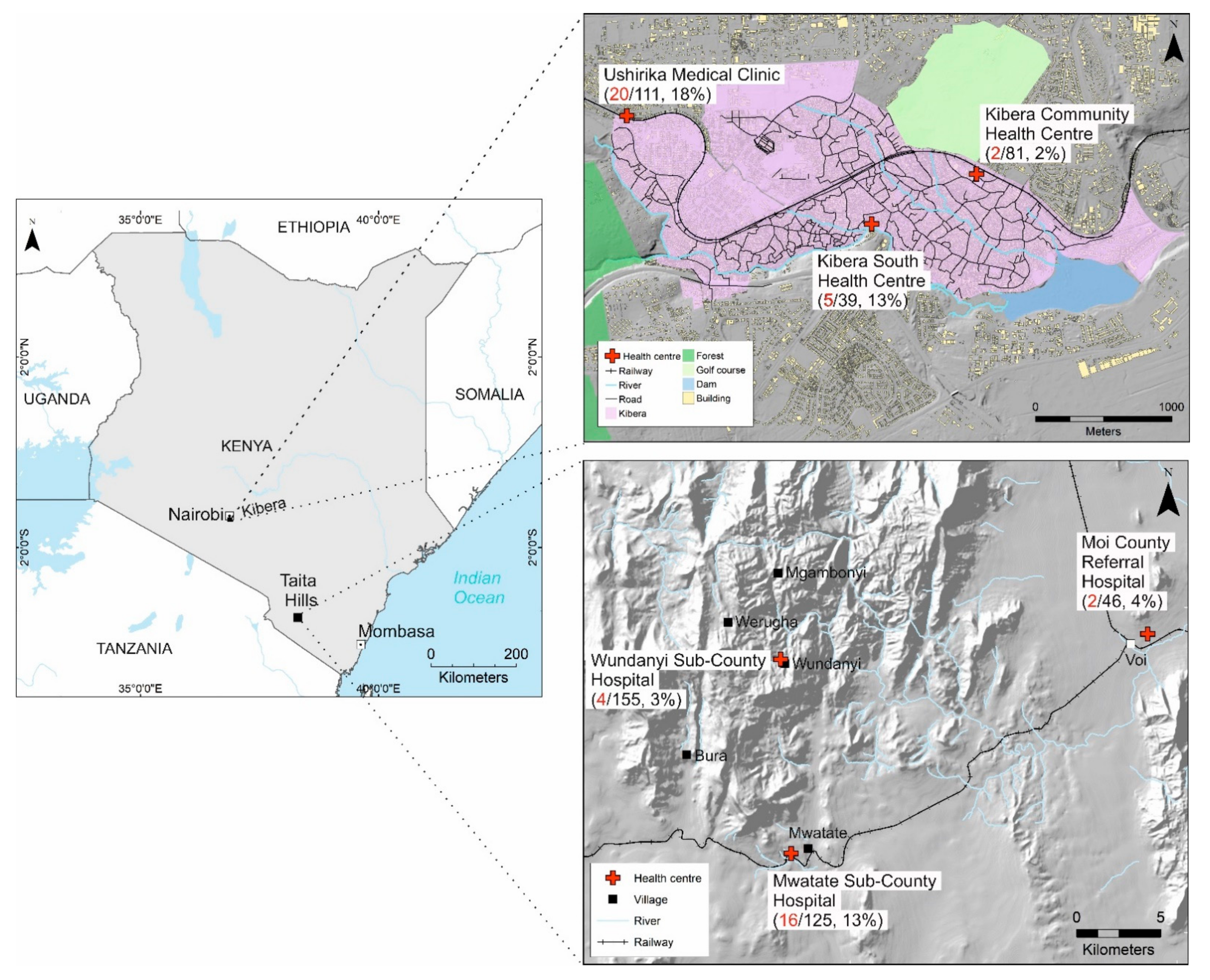
2. Materials and Methods

Study design: This was a cross-sectional study. We analyzed samples that had been previously tested for flavivirus infection [31]. The samples were collected from febrile patients in two geographically disparate regions in Kenya (Figure 1). Taita-Taveta County is located in rural southern Kenya, an ecologically diverse area with a rapidly growing population [32] and little previous research on mosquito-borne viruses in humans [31]. Taita-Taveta County has an elevation range of 400 m in the lowest areas to 2200 m in Taita hills. Kibera slum in Nairobi is the largest informal settlement in Kenya and has a high population density with an estimated 250,000 residents [33]. In Taita-Taveta, serum samples were collected between April and August 2016, and in Kibera slums, plasma samples were collected between February and June 2017. We collected samples and pertinent medical data from patients presenting to six hospitals with acute febrile illness. Samples were stored at −20 °C at hospital labs for one to three weeks before shipment to the KAVI Institute of Clinical Research Lab in Nairobi for storage at −80 °C. All samples were later shipped on dry ice to the University of Helsinki, Finland. A subset of samples was later shipped to Umeå University, Sweden for confirmatory alphavirus plaque reduction neutralization assays.
IgG and IgM antibody testing: Samples were tested for CHIKV IgG antibodies using an in-house IFA modified for CHIKV [36]. Briefly, CHIKV Ross strain-infected Vero E6 cells were detached with trypsin and mixed with uninfected Vero E6 for use as background control. The cells were then washed with phosphate-buffered saline, spotted on glass slides and air dried, fixed with acetone and stored at −70 °C before use. In initial IgG IFA screening, serum or plasma samples were diluted 1:20 in phosphate-buffered saline (PBS), added to pre-spotted slides, and incubated at 37 °C for 30 min in a moist chamber. The slides were washed three times with PBS and incubated at 37 °C for 30 min with FITC-anti-human IgG conjugate (Jackson Immuno Research Laboratories, West Grove, PA, USA) diluted 1:100 in PBS, then washed again three times with PBS and once with distilled water, and examined using a fluorescence microscope. Positive samples were retested as a dilution series in PBS from 1:10 to 1:2560 (1:10, 1:40, 1:160, 1:640, 1:2560). The reciprocal of the last positive dilution was recorded as the IFA IgG titer. All 557 samples were also tested for CHIKV IgM antibodies using a commercial ELISA assay (Chikungunya virus (CHIKV) ELISA incl. IgG/RF absorbent, Euroimmun, Lübeck, Germany) as per the manufacturer’s instructions.
Plaque Reduction Neutralization Tests: A subset of CHIKV IgG and/or IgM seropositive samples (n = 58) and eleven IgM/IgG negative samples were tested using plaque reduction neutralization test (PRNT) for CHIKV, ONNV, and SINV antibodies. The 20 seropositive samples were excluded from PRNT due to insufficient volume. The PRNT was based on a method described previously [5]. In brief, Vero B4 (African green monkey kidney) cells were seeded in 96-well plates (15,000 cells/well). Then, 50 μL of threefold serial dilutions of the serum samples (diluted from 1:40, 1:80, 1:160 to 1:320) was mixed with 50 μL of the respective virus (50 pfu), incubated for 1 h at room temperature, thereafter the mixture was transferred to 12-well plates containing a confluent monolayer of Vero B4 cells. After additional incubation for 1 h at 37 °C in a carbon dioxide incubator, the cells were overlaid with 100 μL of Dulbecco’s modified Eagle medium containing 2% carboxymethylcellulose (CMC; Sigma Life Science, St. Louis, MO, USA). After 3 days, the CMC overlay was removed, and the cells were fixed with 4% paraformaldehyde solution before being stained with crystal violet. Visible plaques were counted and the titers are expressed as the reciprocal of the serum dilution that showed 80% of plaque reduction. Samples were considered as PRNT positive if the PRNT titer was >40, positive for ONNV if ONNV titers were at least two-fold higher than CHIKV titers, and CHIKV positive if CHIKV titers were at least four-fold higher than ONNV titers [5]. The threshold was lower for ONNV than CHIKV because of the unique one-way cross-reactivity between CHIKV and ONNV—CHIKV antibodies are more likely to cross-react with ONNV antigens than ONNV antibodies with CHIKV antigens [37]. The virus strains used were CHIKV LR2006_OPY1 (GenBank EU224268.1), ONNV IbH 12628 and SINV Lovanger strain (GenBank KF737350.1).
Virus Isolation: For PCR-positive samples, we attempted virus isolation in Vero E6 (ATCC CRL-1586) and C6/36 A. albopictus cells (ATCC CRL-1660), as described previously [31].
Statistical analysis: Due to serological cross-reactivity among alphaviruses, we interpreted antibodies detected through IFA and ELISA against CHIKV to be alphavirus antibodies. To analyze the prevalence of alphavirus antibodies on PRNT, we excluded 20 samples that were positive for CHIKV IgG antibodies on IFA or CHIKV-IgM antibodies on ELISA but had not been tested on PRNT due to insufficient sample volume. We analyzed data using IBM SPSS Statistics (Version 22, IBM Corporation, Armonk, NY, USA). We tested for associations between demographic or clinical characteristics and alphavirus seropositivity using Fisher’s Exact Test (FET) with the level of significance set at <0.05 and odds ratios (OR) at a confidence interval (CI) of 95%.

3. Results

3.1. Participant Characteristics

In this study, a total of 557 samples were analyzed including 326 serum samples collected in Taita-Taveta in 2016, and 231 plasma samples collected in Kibera in 2017. Participant characteristics were reported in a previous study on flavivirus infection [31] and are summarized in Table 1.

3.2. CHIKV IgG IFA and IgM ELISA Antibody Assay Results

Out of 557 serum/plasma samples that were tested for CHIKV IgG antibodies on IFA, 49 (8.8%) were positive. IgG seroprevalence was 6.7% in samples from Taita-Taveta and 11.7% in samples from Kibera; p = 0.049 (FET).
Out of 553 serum/plasma samples that were tested for CHIKV IgM antibodies on ELISA, 33 (5.9%) were positive; 5.5% and 6.6% IgM seropositivity for Taita-Taveta and Kibera, respectively. The difference was not statistically significant; p = 0.59 FET. Four samples were positive for both CHIKV IgM and IgG.

3.3. Plaque Reduction Neutralization Assay Results

The 69 samples were tested on PRNT for CHIKV, ONNV, and SINV neutralizing antibodies. Of these 69 samples, 31 were positive for CHIKV IgG (IFA), 23 were positive for CHIKV IgM (ELISA), 4 were positive for both CHIKV IgG and IgM, and 11 were negative for CHIKV antibodies on IFA/ELISA. Then, 24 out of 31 (77%) of tested CHIKV IgG IFA positive samples were confirmed by PRNT, showing high concordance between PRNT and CHIKV IgG IFA. Only 1 out of 24 CHIKV IgM ELISA samples was positive in PRNT showing poor concordance between CHIKV IgM ELISA and PRNT results. Three of four samples that had both CHIKV IgM and IgG antibodies also tested positive on PRNT. Then, 1 out of 11 samples that were negative for both IgG and IgM antibodies was positive on PRNT. This sample was from a 19-year-old patient with fever for one day. Overall, 30 samples were found positive in PRNT, most (25/30) had higher titers for ONNV compared to CHIKV (Supplementary Table S1). Overall, 15/30 samples were positive for ONNV, 1/30 for CHIKV and 14/30 for both CHIKV and ONNV. None of the tested samples were positive for SINV. See Table 2.
Due to insufficient volume, 20 samples that had either CHIKV IgG (14) or CHIKV IgM (6) were not tested by PRNT. When these 20 samples were excluded from the analysis, the prevalence of alphavirus IgM antibody verified by PRNT was 4/537 (0.7%); 3/228 (1.3%) in Kibera and 1/309 (0.3%) in Taita-Taveta. The prevalence of alphavirus IgG antibodies verified by PRNT was 28/537 (5.2%); 21/228 (9.2%) in Kibera, and 7/309 (2.3%) in Taita-Taveta.

3.4. Factors Associated with Alphavirus Seropositivity

CHIKV IgG antibody seroprevalence (by PRNT) was higher in Kibera (9.2%) than in Taita Taveta (2.3%); p < 0.001 (FET). The prevalence was also significantly higher in adults (9.5%) than in children (1.1%); p < 0.001 (FET). IgG seropositive patients were also more likely to report joint pain than seronegative patients (8.5% vs. 3.3%, p = 0.015, FET). CHIKV IgG seroprevalence was higher in patients who had a history of travel to western Kenya (11.3% vs. 4.4%, p = 0.032, FET), as shown in Table 3.
In Taita-Taveta, 54 patients had a history of travel within 30 days before sample collection. Over two-thirds of the patients (37/54, 69%) had traveled to the coast (mainly Mombasa County). In Kibera, 88 patients had a history of travel, 56/88 (64%) having traveled to western Kenya (mainly Kisumu, Kakamega and Siaya Counties). Patients in Kibera were more likely to report travel to western Kenya than those in Taita-Taveta (OR = 11.3, CI 5.4–23.4; p < 0.001). History of travel to any part of Kenya was not significantly associated with arbovirus infection in either children or adults but travel to western Kenya was associated with risk of infection. Travel to western Kenya was also significantly associated with CHIKV IgM ELISA seropositivity (15.6% vs. 4.7%, OR: 3.8 (CI 1.69–8.30); p = 0.002, FET).

4. Discussion

Alphaviruses, especially CHIKV and ONNV, are an important cause of febrile illness in eastern Africa and have the potential to cause large outbreaks. Distinguishing ONNV and CHIKV from patient samples requires molecular detection methods that are not often feasible in clinical settings. Due to this cross-reactivity and the fact that CHIKV and ONNV have been documented to co-circulate [4,5], the epidemiological situation and health impact of the individual viruses is currently unclear.
In this study, we examined febrile patients from non-outbreak settings in two geographical areas for exposure to alphaviruses using a set of serological tests. Only four patients (0.7%) were positive for IgM antibody on ELISA confirmed by PRNT. This indicates that acute alphavirus infection was low in the study population and/or patients were sampled before antibodies were detectable. The poor correlation between PRNT and IgM ELISA could also be because neutralizing CHIKV IgM starts to appear. The median duration of fever in patients in this study was 2 days whereas IgM and IgG antibodies are usually detected after ~3–8 days and 4–10 days, respectively [38,39]. One sample was repeatedly negative on CHIKV IgG IFA and IgM ELISA but positive on PRNT. This was a sample from a 19-year-old patient with fever for one day and it is possible that early IgM antibody response is not detected as sensitively in the IgM-ELISA as with PRNT. Alternatively, PRNT may have detected antibody response against an epitope not present in the IgM ELISA assay.
Given the low prevalence of acute alphavirus infection, the two study areas appear to have been largely spared from the CHIKV outbreaks that occurred in coastal Kenya and Mandera in 2016, and in Mombasa from late 2017 to mid-2018 [11,12], whose timing approximated with our study sampling in Taita-Taveta in 2016, and Kibera in 2017.
The serological findings in this study represent a relatively high IgG seroprevalence with low IgM and the alphavirus IgG antibody seroprevalence of 6.7% for Taita-Taveta and 11.6% for Kibera is significant considering that these locations have no history of recorded alphavirus outbreaks. Notably, alphavirus seropositivity was higher among participants with a history of travel to western Kenya where CHIKV and ONNV infection is common [40]. Patients from Kibera were more likely to have traveled to western Kenya than those in Taita-Taveta. Alphavirus IgG seroprevalence in Nairobi city was nearly two-fold that of the more rural Taita-Taveta, but lower when compared to reports from coastal and western Kenya where previous studies detected alphavirus IgG seroprevalence as high as 26% by ELISA [5,40]. Since the primary aim of the study was to investigate the viral causes of acute febrile illness, only recent travel history was documented. Thus, the effect of possible earlier travel history that may have been a predictor of existing IgG antibodies cannot be confirmed. This may be especially crucial in the study area of Kibera, Nairobi, where relatively high rates of migration and travel are known to occur. Vector distribution in the two areas also supports the contribution of travel to alphavirus infections in Nairobi. Culex mosquitoes are the most abundant mosquito genus in Nairobi with infrequent reports of Aedes, mainly in a few pockets of forested areas, and even less frequent reports of Anopheles mosquitoes. Taita-Taveta has more mosquito vector diversity with Culex mosquitoes been the most abundant, Aedes frequently reported, and Anopheles mosquitoes more frequent than in Nairobi [41,42,43,44].
The results from confirmatory PRNT testing showed that none of the patients had neutralizing antibodies against SINV and that most of the PRNT-positive samples preferentially neutralized ONNV over CHIKV. This may be due to actual ONNV infection or cross-reactivity of CHIKV antibodies with ONNV as has been reported before [37,45]. It could also be due to sequential infection with both viruses in the past. Although we did not detect any SINV antibodies in this study, previous studies have reported very low SINV seroprevalence in humans [4,27]. Additionally, some studies have confirmed the circulation of SINV in mosquito vectors in Kenya, especially along the Great Rift Valley lakes and coastal region, which are located along the flyways for migratory birds [25,46,47].
Seroprevalence of alphavirus IgG antibodies was higher in adults than children. This is expected since exposure occurs over time. Participants with exposure to alphaviruses were also more likely to report joint pains than those not exposed to alphaviruses. Alphaviruses are known to cause chronic joint pain that may last for months to years [47,48].
When viewed together with our previous report on flaviviruses on the same samples [31], none of the samples had acute infection with both alphaviruses and flaviviruses while two samples had IgG antibodies for both virus groups, indicating that the transmission patterns for alphaviruses and flaviviruses are different in the study areas.
One limitation in this study is that the performance of our CHIKV IgG IFA assay has not been evaluated against any commercial ELISA. It is an in-house assay that is readily available and easy to carry-out. We considered it a reliable low-cost option for screening a large number of samples, before confirmation with PRNT. In our experience, it is comparable to CHIKV ELISA and has been used for diagnosis [46]. Similar IFAs have also been shown to have similar accuracy to ELISA [49,50].
Although all confirmed recent alphavirus outbreaks in Kenya have been caused by CHIKV, the risk of ONNV outbreak exists as the virus may be circulating unnoticed in both urban and rural areas, as indicated by previous studies [4,28,30]. The detection of higher ONNV antibody titers than CHIKV is in line with a previous study in which PRNT was used to distinguish ONNV from CHIKV in endemic coastal Kenya [5]. It is known that both CHIKV and ONNV can exist in sylvatic cycles in Africa [6,51], but the factors affecting their emergence and outbreaks are currently not well described.
More ONNV and CHIKV typing data from different areas of Kenya are needed for building a better understanding of their distribution and separate impacts on human health, such as long-term sequelae of alphavirus-associated arthralgia. The current understanding of ONNV and its impact on human health may be underestimated.

Supplementary Materials

The following supporting information can be downloaded at: https://www.mdpi.com/article/10.3390/v14061286/s1, Table S1: Characteristics of samples that tested positive for alphavirus antibodies on Plaque Reduction Neutralization Tests.

Author Contributions

Conceptualization, M.M.M., E.M.K., D.M., O.A., O.V. and E.H.; methodology, E.M.K., T.S., M.E.; A.J.J., E.M.K.; software, T.S.; validation, E.M.K., Y.-D.G. and E.H.; formal analysis, M.M.M., E.M.K., E.H.; investigation, M.M.M., E.M.K., T.S., R.U., J.O., A.J.J., H.A., Y.-D.G., M.E., D.M., O.A., O.V. and E.H.; resources, D.M., O.A., M.E. and E.H.; data curation, M.M.M. and T.S.; writing—original draft preparation, M.M.M., E.M.K., Y.-D.G., M.E., E.H., D.M. and E.H.; writing—review and editing, M.M.M., E.M.K., T.S., R.U., J.O., D.M., A.J.J., H.A., Y.-D.G., M.E., O.A., O.V. and E.H.; visualization, R.U.; supervision, D.M., M.E., O.A., O.V. and E.H.; project administration, D.M., O.A., O.V. and E.H.; funding acquisition, M.M.M., E.M.K., O.V., E.H. All authors have read and agreed to the published version of the manuscript.

Funding

This work was funded by, the Paulo Foundation to EH, Academy of Finland (318726) to OV, Jane and Aatos Erkko Foundation to OV, Nessling Foundation to EMK, Centre for International Mobility (TM-16-1 0283) to MM and Swedish Research Council grant (2017-05607) to ME. MM is supported by a Career Development Fellowship Grant (number TMA2017CDF-1865) under the EDCTP2 program which is supported by the European Union. The funders had no role in the study design, data collection, analysis, decision to publish, or preparation of the manuscript.

Institutional Review Board Statement

The study was conducted in accordance with the Declaration of Helsinki, and approved by the Kenyatta National Hospital—University of Nairobi Ethics and Research Committee (protocol code P707/11/2015 on 8 March 2016).

Informed Consent Statement

Informed consent was obtained from all subjects (or their legally authorized guardians) involved in the study.

Data Availability Statement

The data presented in this study are available on request from the corresponding author.

Acknowledgments

We thank the staff of Moi County Referral Hospital (Voi), Wundanyi and Mwatate Sub-County Hospitals, Ushirika Medical Clinic, Amref Kibera Community Health Centre, Kibera South Health Centre, KAVI Institute of Clinical Research and Helsinki University Hospital laboratory (HUSLAB, Finland). We also thank the Nairobi and Taita-Taveta County Health Departments and the Taita Research Station of the University of Helsinki and its director Petri Pellikka for their support.

Conflicts of Interest

The authors declare no conflict of interest. The funders had no role in the design of the study; in the collection, analyses, or interpretation of data; in the writing of the manuscript, or in the decision to publish the results.

References

  1. ICTV. ICTV Master Species List 2021.v1. 2022. Available online: https://talk.ictvonline.org/files/master-species-lists/m/msl/13425 (accessed on 12 April 2022).
  2. Azar, S.R.; Campos, R.K.; Bergren, N.A.; Camargos, V.N.; Rossi, S.L. Epidemic Alphaviruses: Ecology, Emergence and Outbreaks. Microorganisms 2020, 8, 1167. [Google Scholar] [CrossRef] [PubMed]
  3. Chen, R.; Mukhopadhyay, S.; Merits, A.; Bolling, B.; Nasar, F.; Coffey, L.L.; Powers, A.; Weaver, S.C.; Ictv Report, C. ICTV Virus Taxonomy Profile: Togaviridae. J. Gen. Virol. 2018, 99, 761–762. [Google Scholar] [CrossRef] [PubMed]
  4. Tigoi, C.; Lwande, O.; Orindi, B.; Irura, Z.; Ongus, J.; Sang, R. Seroepidemiology of selected arboviruses in febrile patients visiting selected health facilities in the lake/river basin areas of Lake Baringo, Lake Naivasha, and Tana River, Kenya. Vector Borne Zoonotic Dis. 2015, 15, 124–132. [Google Scholar] [CrossRef] [PubMed] [Green Version]
  5. LaBeaud, A.D.; Banda, T.; Brichard, J.; Muchiri, E.M.; Mungai, P.L.; Mutuku, F.M.; Borland, E.; Gildengorin, G.; Pfeil, S.; Teng, C.Y.; et al. High rates of o’nyong nyong and Chikungunya virus transmission in coastal Kenya. PLoS Negl. Trop. Dis. 2015, 9, e0003436. [Google Scholar] [CrossRef] [PubMed]
  6. Raulino, R.; Thaurignac, G.; Butel, C.; Villabona-Arenas, C.J.; Foe, T.; Loul, S.; Ndimbo-Kumugo, S.P.; Mbala-Kingebeni, P.; Makiala-Mandanda, S.; Ahuka-Mundeke, S.; et al. Multiplex detection of antibodies to Chikungunya, O’nyong-nyong, Zika, Dengue, West Nile and Usutu viruses in diverse non-human primate species from Cameroon and the Democratic Republic of Congo. PLoS Negl. Trop. Dis. 2021, 15, e0009028. [Google Scholar] [CrossRef] [PubMed]
  7. Lwande, O.W.; Obanda, V.; Bucht, G.; Mosomtai, G.; Otieno, V.; Ahlm, C.; Evander, M. Global emergence of Alphaviruses that cause arthritis in humans. Infect. Ecol. Epidemiol. 2015, 5, 29853. [Google Scholar] [CrossRef] [Green Version]
  8. Moizéis, R.N.C.; Fernandes, T.; Guedes, P.; Pereira, H.W.B.; Lanza, D.C.F.; Azevedo, J.W.V.; Galvão, J.M.A.; Fernandes, J.V. Chikungunya fever: A threat to global public health. Pathog. Glob. Health 2018, 112, 182–194. [Google Scholar] [CrossRef]
  9. Zeller, H.; Van Bortel, W.; Sudre, B. Chikungunya: Its History in Africa and Asia and Its Spread to New Regions in 2013–2014. J. Infect. Dis. 2016, 214, S436–S440. [Google Scholar] [CrossRef] [PubMed]
  10. Schuffenecker, I.; Iteman, I.; Michault, A.; Murri, S.; Frangeul, L.; Vaney, M.C.; Lavenir, R.; Pardigon, N.; Reynes, J.M.; Pettinelli, F.; et al. Genome microevolution of chikungunya viruses causing the Indian Ocean outbreak. PLoS Med. 2006, 3, e263. [Google Scholar] [CrossRef] [PubMed] [Green Version]
  11. Maljkovic Berry, I.; Eyase, F.; Pollett, S.; Konongoi, S.L.; Joyce, M.G.; Figueroa, K.; Ofula, V.; Koka, H.; Koskei, E.; Nyunja, A.; et al. Global Outbreaks and Origins of a Chikungunya Virus Variant Carrying Mutations Which May Increase Fitness for Aedes aegypti: Revelations from the 2016 Mandera, Kenya Outbreak. Am. J. Trop. Med. Hyg. 2019, 100, 1249–1257. [Google Scholar] [CrossRef]
  12. Eyase, F.; Langat, S.; Berry, I.M.; Mulwa, F.; Nyunja, A.; Mutisya, J.; Owaka, S.; Limbaso, S.; Ofula, V.; Koka, H.; et al. Emergence of a novel chikungunya virus strain bearing the E1:V80A substitution, out of the Mombasa, Kenya 2017–2018 outbreak. PLoS ONE 2020, 15, e0241754. [Google Scholar] [CrossRef] [PubMed]
  13. Silva, M.M.O.; Tauro, L.B.; Kikuti, M.; Anjos, R.O.; Santos, V.C.; Gonçalves, T.S.F.; Paploski, I.A.D.; Moreira, P.S.S.; Nascimento, L.C.J.; Campos, G.S.; et al. Concomitant Transmission of Dengue, Chikungunya, and Zika Viruses in Brazil: Clinical and Epidemiological Findings From Surveillance for Acute Febrile Illness. Clin. Infect. Dis. Off. Publ. Infect. Dis. Soc. Am. 2019, 69, 1353–1359. [Google Scholar] [CrossRef] [PubMed]
  14. Forrester, N.L.; Palacios, G.; Tesh, R.B.; Savji, N.; Guzman, H.; Sherman, M.; Weaver, S.C.; Lipkin, W.I. Genome-scale phylogeny of the alphavirus genus suggests a marine origin. J. Virol. 2012, 86, 2729–2738. [Google Scholar] [CrossRef] [PubMed] [Green Version]
  15. Rezza, G.; Chen, R.; Weaver, S.C. O’nyong-nyong fever: A neglected mosquito-borne viral disease. Pathog. Glob. Health 2017, 111, 271–275. [Google Scholar] [CrossRef] [PubMed]
  16. Lanciotti, R.S.; Ludwig, M.L.; Rwaguma, E.B.; Lutwama, J.J.; Kram, T.M.; Karabatsos, N.; Cropp, B.C.; Miller, B.R. Emergence of epidemic O’nyong-nyong fever in Uganda after a 35-year absence: Genetic characterization of the virus. Virology 1998, 252, 258–268. [Google Scholar] [CrossRef] [Green Version]
  17. Johnson, B.K.; Gichogo, A.; Gitau, G.; Patel, N.; Ademba, G.; Kirui, R.; Highton, R.B.; Smith, D.H. Recovery of o’nyong-nyong virus from Anopheles funestus in Western Kenya. Trans. R. Soc. Trop. Med. Hyg. 1981, 75, 239–241. [Google Scholar] [CrossRef]
  18. Gopal, S.; Ma, Y.; Xin, C.; Pitts, J.; Were, L. Characterizing the Spatial Determinants and Prevention of Malaria in Kenya. Int. J. Environ. Res. Public Health 2019, 16, 5078. [Google Scholar] [CrossRef] [Green Version]
  19. Hortion, J.; Mutuku, F.M.; Eyherabide, A.L.; Vu, D.M.; Boothroyd, D.B.; Grossi-Soyster, E.N.; King, C.H.; Ndenga, B.A.; LaBeaud, A.D. Acute Flavivirus and Alphavirus Infections among Children in Two Different Areas of Kenya, 2015. Am. J. Trop. Med. Hyg. 2019, 100, 170–173. [Google Scholar] [CrossRef] [Green Version]
  20. Powers, A.M.; Brault, A.C.; Tesh, R.B.; Weaver, S.C. Re-emergence of Chikungunya and O’nyong-nyong viruses: Evidence for distinct geographical lineages and distant evolutionary relationships. J. Gen. Virol. 2000, 81, 471–479. [Google Scholar] [CrossRef]
  21. Henss, L.; Yue, C.; Kandler, J.; Faddy, H.M.; Simmons, G.; Panning, M.; Lewis-Ximenez, L.L.; Baylis, S.A.; Schnierle, B.S. Establishment of an Alphavirus-Specific Neutralization Assay to Distinguish Infections with Different Members of the Semliki Forest complex. Viruses 2019, 11, 82. [Google Scholar] [CrossRef] [Green Version]
  22. Ling, J.; Smura, T.; Lundström, J.O.; Pettersson, J.H.; Sironen, T.; Vapalahti, O.; Lundkvist, Å.; Hesson, J.C. Introduction and Dispersal of Sindbis Virus from Central Africa to Europe. J. Virol. 2019, 93, e00620-19. [Google Scholar] [CrossRef] [Green Version]
  23. Adouchief, S.; Smura, T.; Sane, J.; Vapalahti, O.; Kurkela, S. Sindbis virus as a human pathogen-epidemiology, clinical picture and pathogenesis. Rev. Med. Virol. 2016, 26, 221–241. [Google Scholar] [CrossRef] [PubMed]
  24. Nyaruaba, R.; Mwaliko, C.; Mwau, M.; Mousa, S.; Wei, H. Arboviruses in the East African Community partner states: A review of medically important mosquito-borne Arboviruses. Pathog. Glob. Health 2019, 113, 209–228. [Google Scholar] [CrossRef]
  25. Ochieng, C.; Lutomiah, J.; Makio, A.; Koka, H.; Chepkorir, E.; Yalwala, S.; Mutisya, J.; Musila, L.; Khamadi, S.; Richardson, J.; et al. Mosquito-borne arbovirus surveillance at selected sites in diverse ecological zones of Kenya; 2007–2012. Virol. J. 2013, 10, 140. [Google Scholar] [CrossRef] [Green Version]
  26. Nyamwaya, D.K.; Otiende, M.; Omuoyo, D.O.; Githinji, G.; Karanja, H.K.; Gitonga, J.N.; de Laurent, Z.R.; Otieno, J.R.; Sang, R.; Kamau, E.; et al. Endemic chikungunya fever in Kenyan children: A prospective cohort study. BMC Infect. Dis. 2021, 21, 186. [Google Scholar] [CrossRef] [PubMed]
  27. Geser, A.; Henderson, B.E.; Christensen, S. A multipurpose serological survey in Kenya. 2. Results of arbovirus serological tests. Bull. World Health Organ. 1970, 43, 539–552. Available online: https://pubmed.ncbi.nlm.nih.gov/5313066/ (accessed on 3 January 2022). [PubMed]
  28. Grossi-Soyster, E.N.; Cook, E.A.J.; de Glanville, W.A.; Thomas, L.F.; Krystosik, A.R.; Lee, J.; Wamae, C.N.; Kariuki, S.; Fèvre, E.M.; LaBeaud, A.D. Serological and spatial analysis of alphavirus and flavivirus prevalence and risk factors in a rural community in western Kenya. PLoS Negl. Trop. Dis. 2017, 11, e0005998. [Google Scholar] [CrossRef] [Green Version]
  29. Fritzell, C.; Rousset, D.; Adde, A.; Kazanji, M.; Van Kerkhove, M.D.; Flamand, C. Current challenges and implications for dengue, chikungunya and Zika seroprevalence studies worldwide: A scoping review. PLoS Negl. Trop. Dis. 2018, 12, e0006533. [Google Scholar] [CrossRef] [Green Version]
  30. Kimata, Y.; Borus, P.; Nzunza, R.; Ofula, V.; Chepkorir, E.; Waihenya, R.; Sang, R. Serological Evidence of Chikungunya Virus Infection Among Suspected Measles Cases in Selected Regions of Kenya: 2008–2014. Vector Borne Zoonotic Dis. 2020, 20, 903–909. [Google Scholar] [CrossRef]
  31. Masika, M.M.; Korhonen, E.; Smura, T.; Uusitalo, R.; Vapalahti, K.; Mwaengo, D.; Jääskeläinen, A.J.; Anzala, O.; Vapalahti, O.; Huhtamo, E. Detection of dengue virus type 2 of Indian origin in acute febrile patients in rural Kenya. PLoS Negl. Trop. Dis. 2020, 14, e0008099. [Google Scholar] [CrossRef] [Green Version]
  32. Abera, T.A.; Vuorinne, I.; Munyao, M.; Pellikka, P.; Heiskanen, J. Taita Taveta County, Kenya—2020 Land cover map and reference database. 2021. Available online: https://data.mendeley.com/datasets/xv24ngy2dz (accessed on 6 January 2022). [CrossRef]
  33. UN-HABITAT. Informal Settlements’ Vulnerability Mapping in Kenya: The Case of Kibera. 2020. Available online: https://data.unhabitat.org/documents/GUO-UN-Habitat::the-case-of-kibera-slum-mapping-report/about (accessed on 12 April 2022).
  34. Kounkuey Design Initiative. Kounkuey Design Initiative—Open Data. 2015. Available online: https://www.kounkuey.org/about/open_data (accessed on 28 February 2022).
  35. Clark, B.J.F.; Pellikka, P.K.E. The development of a land use change detection methodology for mapping the Taita Hills, South-East Kenya. In Proceedings of the 31st International Symposium of Remote Sensing of the Environment, St. Petersburg, Russia, 20–24 June 2005. [Google Scholar]
  36. Kallio-Kokko, H.; Laakkonen, J.; Rizzoli, A.; Tagliapietra, V.; Cattadori, I.; Perkins, S.E.; Hudson, P.J.; Cristofolini, A.; Versini, W.; Vapalahti, O.; et al. Hantavirus and arenavirus antibody prevalence in rodents and humans in Trentino, Northern Italy. Epidemiol. Infect. 2006, 134, 830–836. [Google Scholar] [CrossRef]
  37. Hozé, N.; Diarra, I.; Sangaré, A.K.; Pastorino, B.; Pezzi, L.; Kouriba, B.; Sagara, I.; Dabo, A.; Djimdé, A.; Thera, M.A.; et al. Model-based assessment of Chikungunya and O’nyong-nyong virus circulation in Mali in a serological cross-reactivity context. Nat. Commun. 2021, 12, 6735. [Google Scholar] [CrossRef]
  38. Chua, C.-L.; Sam, I.C.; Chiam, C.-W.; Chan, Y.-F. The neutralizing role of IgM during early Chikungunya virus infection. PLoS ONE 2017, 12, e0171989. [Google Scholar] [CrossRef] [Green Version]
  39. Suhrbier, A.; Jaffar-Bandjee, M.-C.; Gasque, P. Arthritogenic alphaviruses—An overview. Nat. Rev. Rheumatol. 2012, 8, 420–429. [Google Scholar] [CrossRef]
  40. Inziani, M.; Adungo, F.; Awando, J.; Kihoro, R.; Inoue, S.; Morita, K.; Obimbo, E.; Onyango, F.; Mwau, M. Seroprevalence of yellow fever, dengue, West Nile and chikungunya viruses in children in Teso South Sub-County, Western Kenya. Int. J. Infect. Dis. IJID Off. Publ. Int. Soc. Infect. Dis. 2020, 91, 104–110. [Google Scholar] [CrossRef] [Green Version]
  41. Kasili, S.; Odemba, N.; Ngere, F.G.; Kamanza, J.B.; Muema, A.M.; Kutima, H.L. Entomological assessment of the potential for malaria transmission in Kibera slum of Nairobi, Kenya. J. Vector Borne Dis. 2009, 46, 273–279. [Google Scholar]
  42. Eastwood, G.; Sang, R.C.; Lutomiah, J.; Tunge, P.; Weaver, S.C. Sylvatic Mosquito Diversity in Kenya-Considering Enzootic Ecology of Arboviruses in an Era of Deforestation. Insects 2020, 11, 342. [Google Scholar] [CrossRef]
  43. Karungu, S.; Atoni, E.; Ogalo, J.; Mwaliko, C.; Agwanda, B.; Yuan, Z.; Hu, X. Mosquitoes of Etiological Concern in Kenya and Possible Control Strategies. Insects 2019, 10, 173. [Google Scholar] [CrossRef] [Green Version]
  44. Munyao, V.; Karisa, J.; Munyao, C.M.; Ngari, M.; Menza, N.; Peshu, N.; Rono, M.; Mbogo, C.; Mwangangi, J. Surveillance of Culicine Mosquitoes in Six Villages of Taita-Taveta County, Kenya, With Host Determinations From Blood-Fed Females. J. Med. Entomol. 2020, 57, 1972–1982. [Google Scholar] [CrossRef]
  45. Clements, T.L.; Rossi, C.A.; Irish, A.K.; Kibuuka, H.; Eller, L.A.; Robb, M.L.; Kataaha, P.; Michael, N.L.; Hensley, L.E.; Schoepp, R.J. Chikungunya and O’nyong-nyong Viruses in Uganda: Implications for Diagnostics. Open Forum Infect. Dis. 2019, 6, ofz001. [Google Scholar] [CrossRef]
  46. Jääskeläinen, A.J.; Kareinen, L.; Smura, T.; Kallio-Kokko, H.; Vapalahti, O. Chikungunya virus infections in Finnish travellers 2009–2019. Infect. Ecol. Epidemiol. 2020, 10, 1798096. [Google Scholar] [CrossRef]
  47. Silva, J.V.J., Jr.; Ludwig-Begall, L.F.; Oliveira-Filho, E.F.D.; Oliveira, R.A.S.; Durães-Carvalho, R.; Lopes, T.R.R.; Silva, D.E.A.; Gil, L.H.V.G. A scoping review of Chikungunya virus infection: Epidemiology, clinical characteristics, viral co-circulation complications, and control. Acta Trop. 2018, 188, 213–224. [Google Scholar] [CrossRef]
  48. Lemant, J.; Boisson, V.; Winer, A.; Thibault, L.; Andre, H.; Tixier, F.; Lemercier, M.; Antok, E.; Cresta, M.P.; Grivard, P.; et al. Serious acute chikungunya virus infection requiring intensive care during the Reunion Island outbreak in 2005–2006. Crit. Care Med. 2008, 36, 2536–2541. [Google Scholar] [CrossRef]
  49. Harring, J.; Granger, D.; Jespersen, D.; Theel, E. Comparison of the euroimmun Chikungunya Immunoglobulin (Ig)M and IgG Enzyme Immunoassays (Eias) to a Commercially Available Immunofluorescence Assay. Open Forum Infect. Dis. 2015, 2, 235. [Google Scholar] [CrossRef] [Green Version]
  50. De Salazar, P.M.; Valadere, A.M.; Goodman, C.H.; Johnson, B.W. Evaluation of three commercially-available chikungunya virus immunoglobulin G immunoassays. Rev. Panam. Salud Publica 2017, 41, e62. [Google Scholar] [CrossRef]
  51. Valentine, M.J.; Murdock, C.C.; Kelly, P.J. Sylvatic cycles of arboviruses in non-human primates. Parasites Vectors 2019, 12, 463. [Google Scholar] [CrossRef]
Figure 1. A map of Kenya showing the study sites in Taita-Taveta County and Kibera slum, Nairobi. Indicated alongside the site names is the number of samples per site (denominator) and prevalence of chikungunya IgG antibodies by immunofluorescence assay (red text and percentage). Geographic information system (GIS) data for Kibera was obtained from Kounkuey Design Initiative [34]. Hillshade for Kibera was created in GIS software from digital elevation model (DEM) 5 m and for Taita-Taveta, hillshade was created from DEM 20 m [35].
Viruses 14 01286 g001
Table 1. Characteristics of study participants.
ParameterTaita-TavetaKiberaTotal (%)
n326 (59%)231 (41%)557
Gender (n = 547)
  Male146 (46%)115 (50%)261 (48%)
  Female172 (54%)114 (50%)286 (52%)
Age (n= 546)
  Mean (Standard deviation (SD))26.8 (22.1)16.2 (15.3)22.4 (20.2)
  Median (Interquartile range (IQR))20.6 (39.5)10.2 (24.4)16.4 (33.6)
  Range6 months–85 years2 months–77 years2 months–85 years
Age Groups (n = 545)
  0–5 years69 (22%)87 (39%)156 (29%)
  5–17 years81 (25%)45 (20%)126 (23%)
  18 years and above169 (53%)94 (41%)263 (48%)
Education Level in adults (n = 245)
  None15 (10%)0 (0%)15 (6%)
  Primary school 77 (50%)27 (30%)104 (42%)
  Secondary school29 (19%)36 (40%)65 (27%)
  Tertiary level34 (22%)27 (30%)61 (25%)
History of travel (n = 552)
  Any travel outside the study area54 (17%)88 (38%)142 (26%)
  Travel to Nyanza or western Kenya9 (3%)56 (24%)65 (12%)
  Travel to coastal Kenya37 (11%)2 (1%)39 (7%)
Contact with animals (n = 557)
  Contact with goats134 (41%)25 (11%)159 (29%)
  Contact with cattle96 (29%)26 (11%)122 (22%)
  Contact with chicken190 (58%)43 (19%)233 (42%)
  Contact with cats61 (19%)87 (38%)148 (27%)
  Contact with rodents217 (67%)127 (55%)344 (62%)
  Contact with bats94 (29%)6 (3%)100 (18%)
Signs and symptoms (n = 557)
  Body Temperature in °C (Mean/SD)38.8 (0.56)38.4 (0.67)38.6 (0.63)
  Median duration of fever (range)2 (1–7) days2 (1–14)2 (1–14) days
  Joint pain139 (43%)70 (30%)209 (38%)
  Myalgia123 (38%)65 (28%)188 (34%)
  Headache53 (16%)108 (47%)161 (29%)
  Cough44 (13%)95 (41%)139 (25%)
  Vomiting53 (16%)68 (29%)121 (22%)
  Diarrhoea29 (9%)47 (20%)76 (14%)
  Rash18 (6%)16 (7%)34 (6%)
Table 2. Alphavirus plaque reduction neutralization test results.
Sample CharacteristicsnCHIKV and ONNV PositiveCHIKV PositiveONNV PositiveSINV PositivePositive for Any AlphavirusNegative
CHIKV IgG positive (IFA)3112 (39%)0 (0%)13 (42%)0 (0%)25 (81%)6 (19%)
CHIKV IgM positive (ELISA)230 (0%)1 (4%)0 (0%)0 (0%)1 (4%)22 (96%)
Both CHIKV IgG and IgM positive42 (50%)0 (0%)1 (25%)0 (0%)3 (75%)1 (25%)
Negative110 (0%)0 (0%)1 (9%)0 (0%)1 (9%)10 (91%)
TOTAL6914 (20%)1 (1%)15 (22%)0 (0%)30 (43%)39 (57%)
Table 3. Association between patient characteristics and alphavirus IgG antibodies on Plaque Reduction Neutralization assay (PRNT).
CharacteristicAlphavirus IgG Antibody Positive (PRNT)Odds Ratio
(95% CI)
p Value (Fisher’s Exact Test)
Age Group
  Adults24/253 (9.5%)9.5 (2.83–31.96)<0.001
  Children (<18 years)3/275 (1.1%)
Gender
  Female15/274 (5.5%)1.1 (0.49–2.29)1.000
  Male13/253 (5.1%)
Facility Location
  Kibera 21/228 (9.2%)4.3 (1.83–10.48)<0.001
  Taita-Taveta7/309 (2.3%)
History of Travel
  Yes9/137 (6.6%)1.5 (0.64–3.36)0.369
  No18/395 (4.6%)
Travel to Western Kenya
  Yes 7/62 (11.3%)2.8 (1.12–6.77)0.032
  No21/475 (4.4%)
Travel to Coastal Kenya
  Yes 1/38 (2.6%)0.5 (0.06–3.58)0.712
  No27/499 (5.4%)
Joint Pain
  Yes17/201 (8.5%)2.7 (1.25–5.95)0.015
  No11/336 (3.3%)
Myalgia
  Yes12/180 (6.7%)1.5 (0.70–3.29)0.307
  No16/357 (4.5%)
Headache
  Yes16/157 (10.2%)3.5 (1.61–7.54)0.002
  No12/380 (3.2%)
Cough
  Yes5/136 (3.7%)0.6 (0.23–1.68)0.503
  No25/401 (6.2%)
Contact with goats
  Yes4/148 (2.7%)0.4 (0.14–1.24)0.130
  No24/389 (6.2%)
Contact with cattle
  Yes4/117 (3.4%)0.6 (0.20–1.72)0.480
  No24/420 (5.7%)
Contact with rodents
  Yes15/331 (4.5%)0.7 (0.33–1.51)0.426
  No13/206 (6.3%)
Contact with bats
  Yes3/91 (3.3%)0.6 (0.17–1.94)0.450
  No25/446 (5.6%)
Any animal contact
  Yes19/419 (4.5%)0.6 (0.25–1.31)0.238
  No9/118 (7.6%)
Publisher’s Note: MDPI stays neutral with regard to jurisdictional claims in published maps and institutional affiliations.

