Article

Robust AAV Genotyping Based on Genetic Distances in Rep Gene That Are Maintained by Ubiquitous Recombination

by
Marina I. Beloukhova
1,*,
Alexander N. Lukashev
2,
Pavel Y. Volchkov
3,4,
Andrey A. Zamyatnin, Jr.
1,5,6,7 and
Andrei A. Deviatkin
3,4,8,*
1
Institute of Molecular Medicine, Sechenov First Moscow State Medical University, 119991 Moscow, Russia
2
Martsinovsky Institute of Medical Parasitology, Tropical and Vector-Borne Diseases, First Moscow State Medical University (Sechenov University), 119991 Moscow, Russia
3
Genome Engineering Lab, Moscow Institute of Physics and Technology (National Research University), 141700 Dolgoprudniy, Russia
4
The National Medical Research Center for Endocrinology, 117036 Moscow, Russia
5
Belozersky Institute of Physico-Chemical Biology, Lomonosov Moscow State University, 119992 Moscow, Russia
6
Department of Biotechnology, Sirius University of Science and Technology, 1 Olympic Ave, 354340 Sochi, Russia
7
Department of Immunology, Faculty of Health and Medical Sciences, University of Surrey, Guildford GU2 7XH, UK
8
Laboratory of Postgenomic Technologies, Izmerov Research Institute of Occupational Health, 105275 Moscow, Russia
*
Authors to whom correspondence should be addressed.
Viruses 2022, 14(5), 1038; https://doi.org/10.3390/v14051038
Submission received: 31 March 2022 / Revised: 4 May 2022 / Accepted: 9 May 2022 / Published: 13 May 2022
(This article belongs to the Special Issue Advances in Parvovirus Research 2022)

Abstract

:
Adeno-associated viruses (AAVs) are a convenient tool for gene therapy delivery. According to the current classification, they are divided into the species AAV A and AAV B within the genus Dependoparvovirus. Historically AAVs were also subdivided on the intraspecies level into 13 serotypes, which differ in tissue tropism and targeted gene delivery capacity. Serotype, however, is not a universal taxonomic category, and their assignment is not always robust. Cross-reactivity has been shown, indicating that classification could not rely on the results of serological tests alone. Moreover, since the isolation of AAV4, all subsequent AAVs were subdivided into serotypes based primarily on genetic differences and phylogenetic reconstructions. An increased interest in the use of AAV as a gene delivery tool justifies the need to improve the existing classification. Here, we suggest genotype-based AAV classification below the species level based on the rep gene. A robust threshold was established as 10% nt differences within the 1248 nt genome fragment, with 4 distinct AAV genotypes identified. This distinct sub-species structure is maintained by ubiquitous recombination within, but not between, rep genes of the suggested genotypes.

1. Introduction

1.1. AAV Biology

Adeno-associated viruses (AAVs) are non-enveloped particles with a size of 18–26 nm. Sixty protein molecules form an icosahedral capsid. The genome is represented by a linear single-stranded DNA of approximately 4.7 thousand bases.
The AAV genome contains two open reading frames (ORFs): (1) rep, which encodes four overlapping proteins necessary for the regulation of viral DNA replication: Rep78, Rep68, Rep52, and Rep40 [1,2]; and (2) cap encoding viral capsid proteins: VP1, VP2 and VP3 [3,4]. The genome is flanked by inverted terminal repeats (ITRs) [5], which form a T-shaped self-complementary secondary structure with a free 3′-hydroxyl that acts as a replication primer [6,7].
For effective replication and reproduction in host cells, AAV requires co-infection with an auxiliary virus, e.g., adenovirus (hence the name adeno-associated), although AAV replication is also possible with herpesvirus, cytomegalovirus, and papillomavirus co-infection [8].
The best-known hosts of AAVs are primates and humans [9,10], although these viruses have been found in other animals [11,12,13]. AAVs do not cause a significant immune response or any notable pathology in the host cells. Consequently, they are not considered pathogenic [4]. Notably, over 90% of the adult population is seropositive to AAVs, i.e., are likely asymptomatic carriers [14]. The prevalence of AAV in distinct tissues varies between 37 and 72% [15].

1.2. AAV Gene Therapy

AAVs are a convenient tool for gene therapy [16,17]. Since wild AAVs persist in the form of episomes (integration into the host genome at AAVS1 19q13.3-qter site is extremely rare [18] and absent in vectors), they are safer compared to retroviruses, which insert into the host genome randomly, leading to oncogenesis [19]. AAVs also have reduced immunogenicity compared to adenoviruses, further supporting their utility as gene therapy vectors [20].
Results of preclinical and clinical studies have demonstrated that AAV vectors are safe and effective tools in gene therapy for a range of diseases including cystic fibrosis, hemophilia B, arthritis, hereditary emphysema, muscular dystrophy, Parkinson’s disease, Alzheimer’s disease, prostate cancer, malignant melanoma, epilepsy, and others [16,21,22,23]. To date, three AAV-vector-based products have been approved for use in medical practice: Glybera (familial lipoprotein lipase deficiency) [24], Luxturna (hereditary retinal dystrophy) [25] and Zolgensma (spinal muscular atrophy) [26].

1.3. AAV Classification

Current AAV classification is based on the phenotype (the shape of the virion), replication peculiarities, and the host range [27]. According to these criteria, AAVs are represented by two species—AAV A and AAV B. Besides AAVs, there are eight other species within the genus dependovirus (ICTV: https://talk.ictvonline.org/taxonomy/ accessed 25 June 2021).
Additionally, AAVs are subdivided on the intraspecies level into serotypes. The first reports of serologically distinct AAVs date back to 1966, when the first three serotypes were identified [28].
Further, any newly identified AAV serologically distinct from known types were assigned a new serotype in chronological order. To date, 13 AAV serotypes are known. However, there are significant ambiguities and controversies in the properties and definitions of the AAV serotypes. For instance, AAV4 was identified based on the reaction with antiserum [29], whereas AAV5 was assigned based on the DNA structure differences identified by restriction analysis and blot-hybridization [30,31]. This protocol could correspond to the identification of a new genotype, which is consistent with the later assignment of AAV5 to a separate species within the dependovirus genus by the International Committee on Taxonomy of Viruses [32].
AAV6 was assigned a new serotype based on genetic differences from the complete genomes of AAV2 and AAV3 (82% identity), as well as AAV4 (75–78% identity) [33]. However, its cap genes were 96% similar to AAV1, and most likely AAV6 is a variant of AAV1. Despite this, AAV serotype 6 is still in use.
In 2002, during an investigation of the asymptomatic presence of AAV in primate tissues, Gao et al. identified AAV7 and AAV8 [34]. The authors used signature regions—a fragment of genomic sequence at positions 2886–3143 nt unique to each AAV type (previously identified by Rutledge et al. [33])—to define distinct virus types, and named the newly isolated viruses according to the differences in these regions. The rep and cap nucleotide sequences and the predicted amino acid sequence comparisons for AAV1-8 (with the exception of AAV5 as obviously incongruent) revealed differences, primarily in the region encoding capsid proteins. AAV7 was shown to have a 63% to 85% similarity to the amino acid sequences and 68% to 84% of the nucleotide sequences of other AAV serotypes; similar results were obtained for AAV8. The serological difference of AAV7, AAV8, and other serotypes was also established based on the absence of neutralization by any antiserum other than their own (anti-AAV7 and anti-AAV8, respectively).
Later, Gao et al. identified 11 phylogenetic groups based on phylogenetic analysis of cap sequences—so-called ‘clades’—from A to F, consisting of phylogenetically similar representatives from three or more sources, and five groups of clones (phylogenetically similar representatives from less than three sources) [35].
AAV10 and AAV11 were also identified based on the signature region differences and characterized according to the cap sequence [36]. The serological analysis confirmed that AAV10 and AAV11 were serologically distinct from AAV2.
Similarly, AAV12 was identified based on the rep and cap sequence differences [37]. Finally, AAV(VR-942) was isolated in 2008 and had a high degree of amino acid sequence similarity of rep with AAV4 (98%) and AAV3 (93%), as well as 93% identity in VP1 as compared to AAV3. Despite this, AAV(VR-942) has a distinct pattern of cellular receptor interaction and was suggested as a new serotype, AAV13 (the name is currently present only in the corresponding GenBank entry); it should be noted that serological studies were not conducted for AAV13 [38].
Thus, since the isolation of AAV4, all subsequent AAVs were subdivided into different serotypes based primarily on genetic differences and phylogenetic reconstructions. In addition to the established 13 serotypes, one of which comprises a separate virus species (AAV5), more than 100 novel genetic variants (so-called serovars [39]) are not assigned to any taxonomic unit below the species level. It is unlikely that they will be all tested serologically, and the genetic boundaries of types are not robust.
Here, we analyzed all available AAV-A sequences to test the possibility of systematically distinguishing AAVs according to genomic sequence data and to suggest a classification based on genotypes with a measurable robust threshold.

2. Materials and Methods

2.1. Sequence Selection for Analysis

All sequences with a length limited to 4000–5100 nt, which approximately corresponds to the full genome, available as of June, 2021, for Dependoparvovirus isolates were retrieved from the GenBank database (artificial sequences were excluded). Since this study aimed to characterize genetic relationships at the intraspecies level, only sequences of the species AAV A were selected. For this purpose, a phylogenetic tree for the retrieved sequences was constructed using neighbor-joining, implemented in MEGA [40].

2.2. Recombination Analysis

The selected sequences were aligned using MAFFT online [41]. Multiple recombination events were analyzed by the RDP4 software [42] (RDP4 output is available at https://github.com/AndreiDeviatkin/repo/blob/main/AAV.rdp, accessed on 11 May 2022). A more comprehensive recombination screening was carried out using pairwise distance deviation matrices (PDDM) and pairwise distance correspondence plots (PDCP) as described earlier [43] with an online Shiny web application (https://v-julia.shinyapps.io/recdplot_app/, accessed on 25 June 2021). Similarity plots for selected sequences were generated using Simplot 3.5.1 [44].

2.3. Nucleotide and Predicted Amino Acid Sequences Analysis

The analysis of nucleotide and inferred amino acid sequence differences was carried out in R software.

3. Results

3.1. The Dataset

A total of 272 Dependoparvovirus sequences (4000–5100 nt length) were retrieved from GenBank. Of them, 105 sequences belonged to the AAV A species based on phylogenetic analysis (Figure S1). The dataset included all previously characterized genotypes, with the exception of AAV9, for which only VP1 coding sequence is available, as well as other unclassified variants available in the database. Two sequences (MK163941.1 and MK163942.1) were significantly different from the others and thus excluded from the dataset.

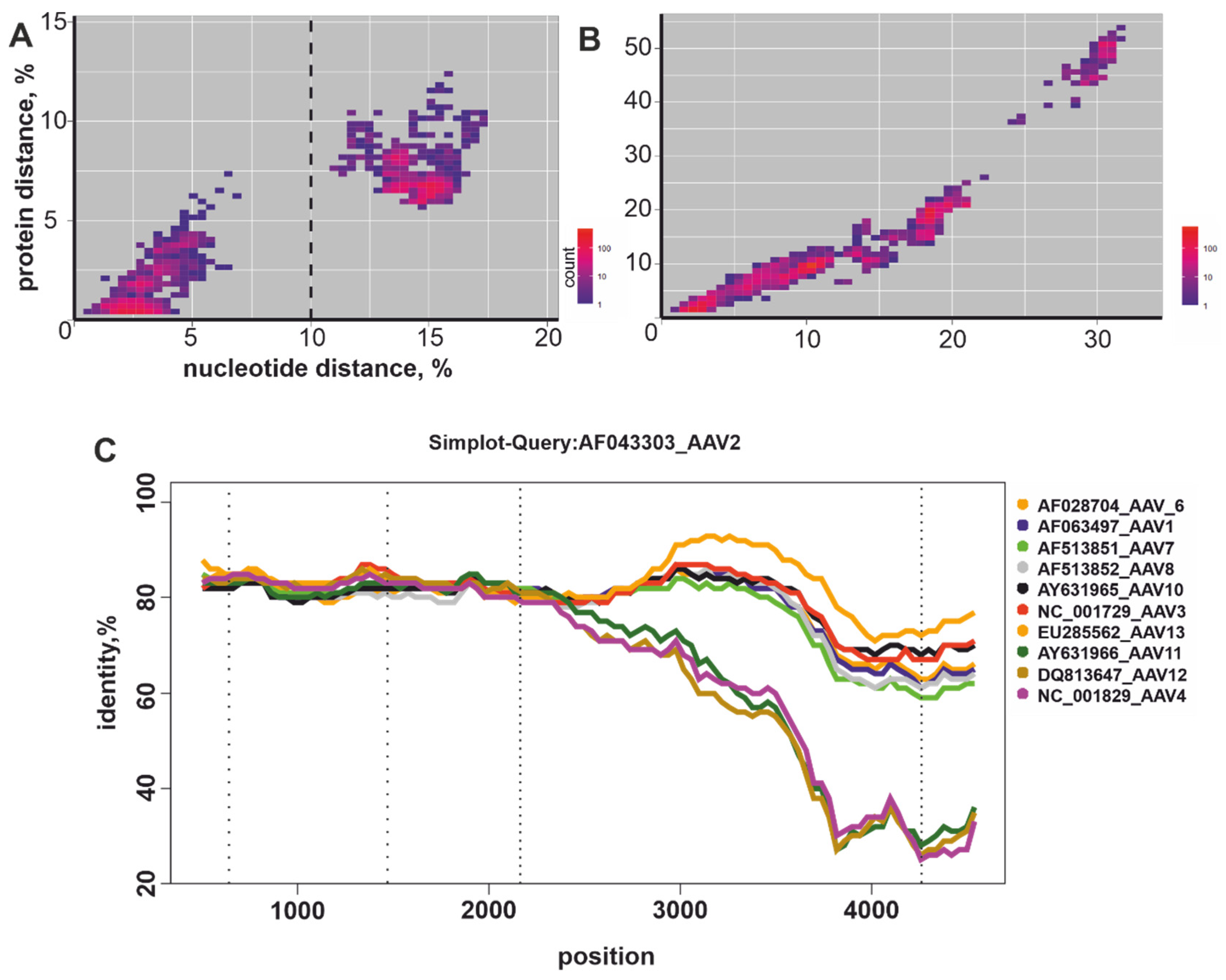
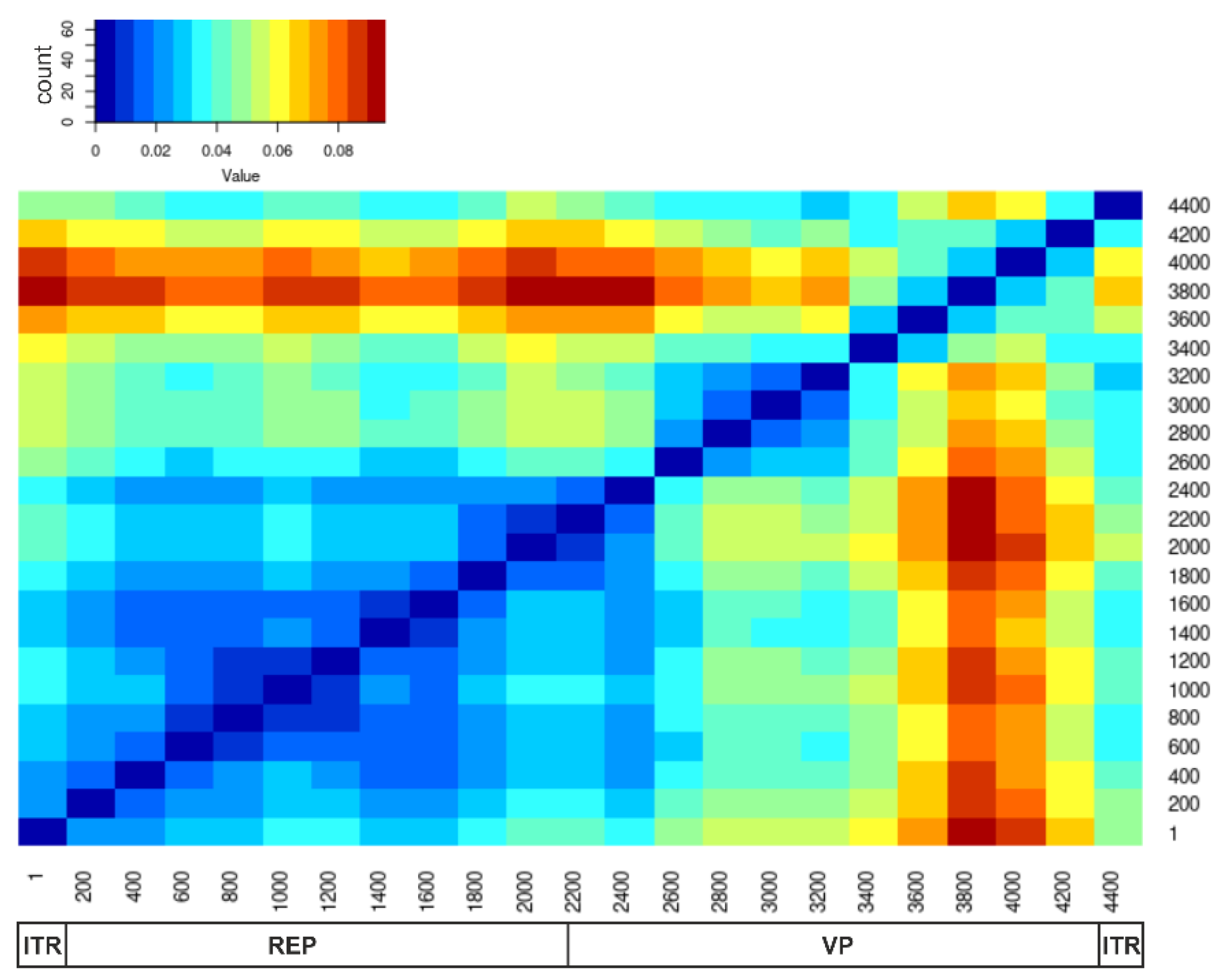
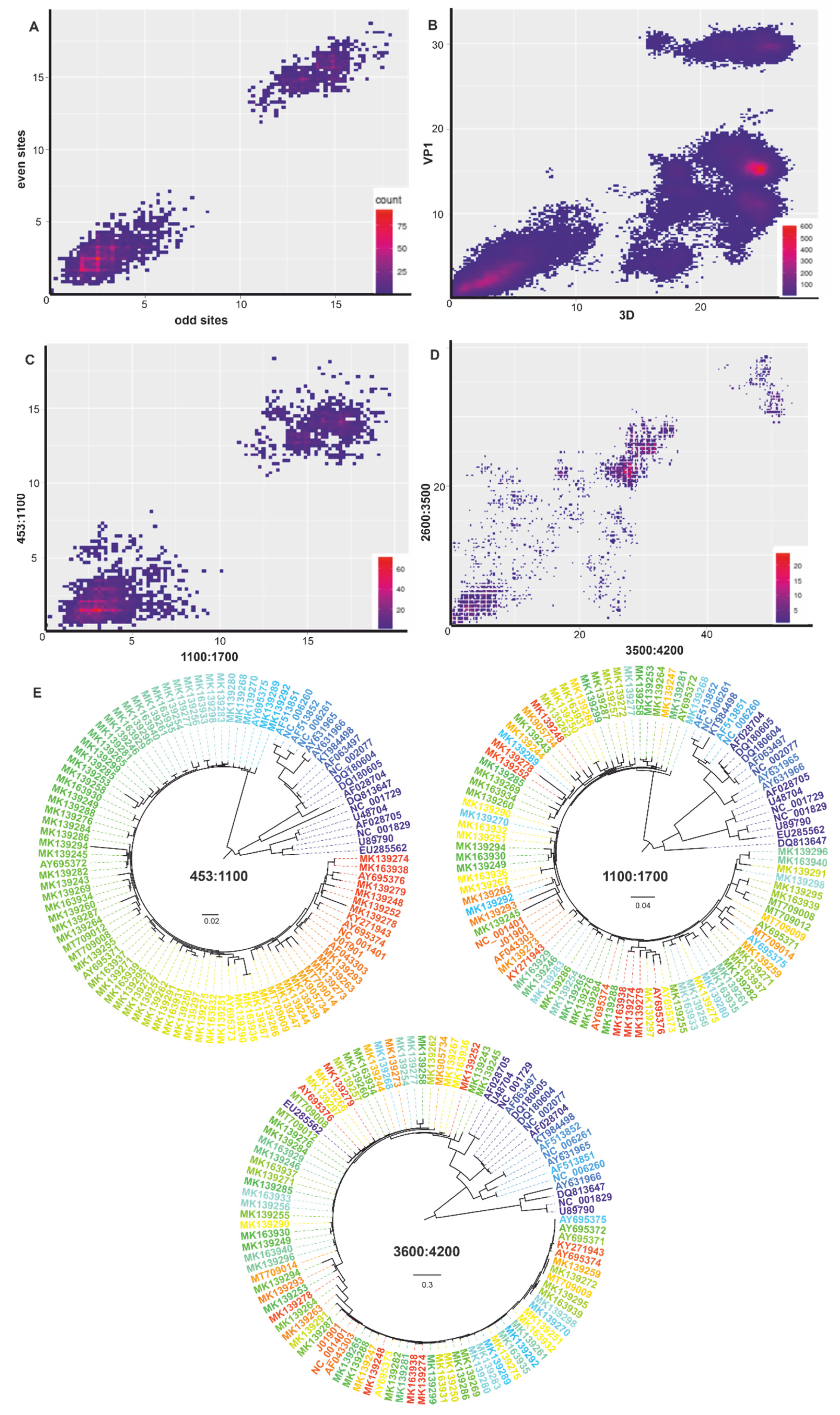
3.2. Recombination Analysis

Recombination occurs more frequently in some fragments of the virus genome than in others. The root mean square error (RMSE) of all pairwise distances for two genomic regions from the regression line reflects the extent of phylogenetic inconsistency between these fragments of the genome. A high RMSE (indicated by red in Figure 1) indicates more frequent recombination between the corresponding regions of the genome. A pairwise distance deviation matrix (PDDM) was built for all possible pairs of genomic regions using a sliding window (500 nt). Based on the PDDM, a region covering genome positions 200–1700 nt was identified as the region with the lowest relative incidence of recombination, which approximately corresponds to the coding sequence of Rep proteins (Figure 1).
The PDCP and phylogenetic reconstruction confirmed that recombination in this genome region occurred only between closely related viruses that differed by less than 10% in their nucleotide sequence (Figure 2C, compare to control Figure 2A). Despite ubiquitous recombination within four phylogenetic clades formed by such closely related viruses, there were no signs of recombination between them (Figure 2E). This pattern contrasted with recombination both within and between these four clades when comparing rep and cap genes on the phylogenetic trees (Figure 2E) and signs of recombination between viruses that differed by over 20% nucleotide sequences in cap (Figure 2D). Since complete genomic sequences were not available for all known AAV types (for AAV10-13 only rep + cap complete coding sequences were accessible), alignment positions 453–1700 (genome positions 470–1700 in the reference sequence #NC_001401) were selected for further analysis. Therefore, the region of rep between positions 453 and 1700 was indeed devoid of long-distance recombination and was thus used for further analysis.

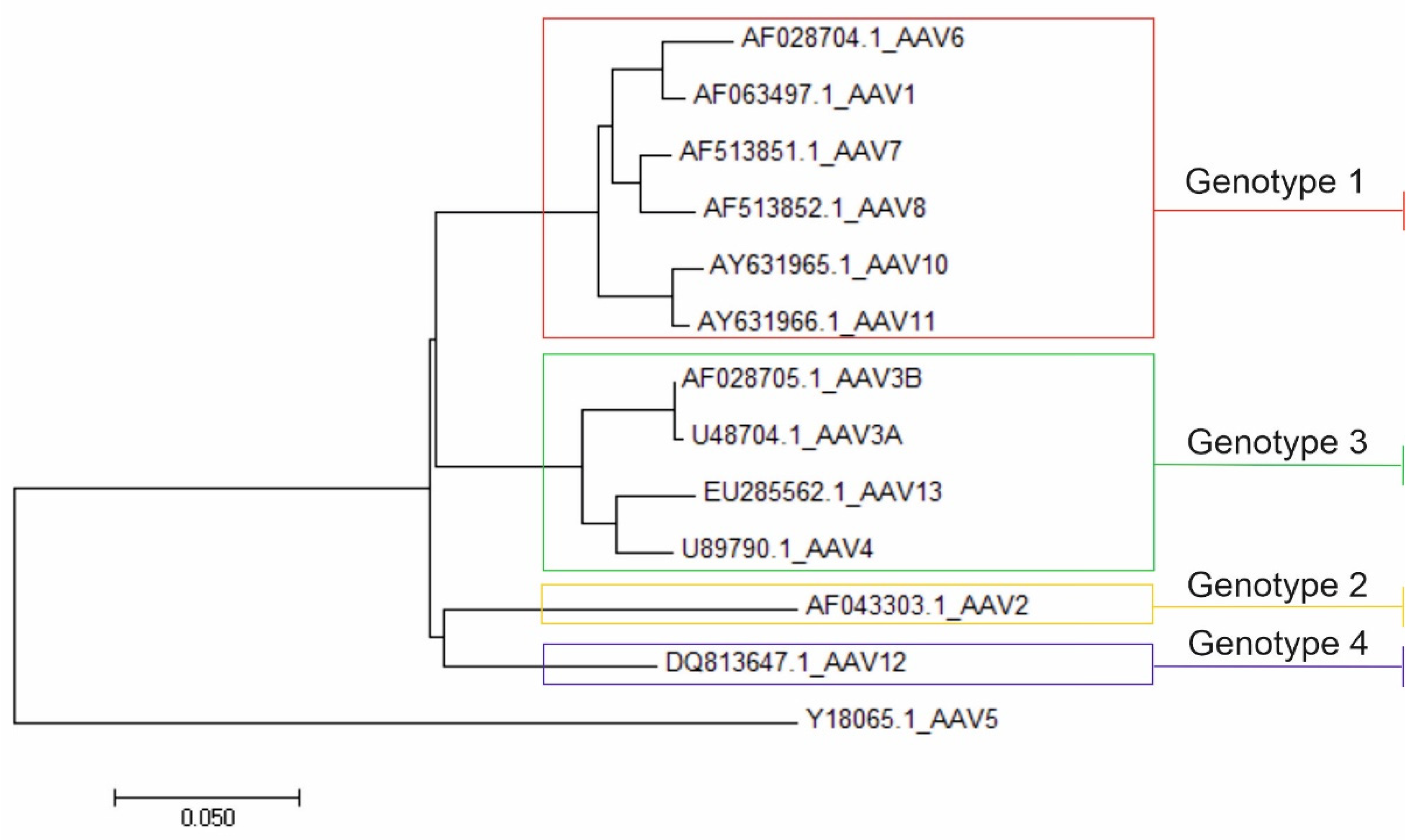
3.3. Nucleotide and Amino Acid Pairwise Distance Analysis

Nucleotide or protein sequences may be preferable for distinguishing virus taxa on different levels and in distinct virus groups. A plot of pairwise nucleotide and amino acid distances (Figure 3A) showed that all pairs of viruses formed two clearly distinguishable clouds in the rep gene. Out of a total of 5253 virus pairs, 3558 pairs differed by up to 7.5% nucleotide and up to 8% amino acid sequence, whereas 1695 virus pairs were different in more than 11% nucleotide and 6% amino acid positions. An artificial threshold of 10% nt could be thus suggested for robust AAV subdivision within the species level. At a protein sequence level, there was an overlap between distances within the four phylogenetic groups (below 7.5%) and between them (above 5.5%). Thus, the amino acid sequence is less suitable for the assignment of rep genotypes. However, no conclusions could be made according to the plot of pairwise nucleotide and amino acid distances of the cap gene (Figure 3B). The robust separation between AAV4, AAV11, AAV12 and all other serotypes (pairs differing in above 32.5% amino acids in 2250–4400 nt genomic region) was caused by a recombination event that led to the fact that fragments of AAV4, AAV11 and AAV12 capsids have an independent origin from AAV1, AAV2, AAV3, AAV6, AAV7, AAV8, AAV10, AAV13 capsids (Figure 3C).
Analysis of the individual AAV pairs in both plot areas showed that some of them are currently registered as distinct serotypes, but differ by less than 10% of the rep nucleotide sequence and thus may be assigned to the same rep genotype (Table 1).
There were no virus pairs in the dataset that were assigned to the same serotype, but differed by more than 10% nucleotide sequence in the rep gene. According to this concept, four distinct rep genotypes could be identified (Table 2, Figure 4).

4. Discussion

The lack of an established system for AAV subdivision below the species level results in uncertain classification. There is also no clear, generally accepted definition of such terms as “strain”, “variant” and “isolate”, and most publications simply reproduce the terminology used previously [46]. The currently used term, “serotype”, is not a classical taxonomic category, although such intraspecific division is applicable for practical and scientific purposes [47,48,49].
According to published studies, the historically established AAV classification based on the serotypes was in some cases not appropriately supported by either serological or genomic data. During early genomic studies, several regions with significant differences between the AAV serotypes were identified within the VP1 sequence, with four domains containing unique sequences for each serotype. These regions were assumed to have a role in serotype-specific functions, such as antigen-antibody or cell receptor binding, which presumably also corresponds to different tissue specificity [33,50,51]. However, the relationship between the serotype and tropism is not clear-cut, because several serotypes efficiently infect similar tissues via both the same and different receptors, implying that capsid proteins may not be the only determinants of tissue tropism.
In subsequent studies, when assigning new AAVs into distinct serotypes, genetic differences (primarily in the capsid proteins) were used. However, conflicts and incongruences in AAV serotype assignment have been accumulating (see above). They were further complicated by serological cross-reactivity [14,50]. Uncertain serological distinction was reported not just for AAV1 and AAV6 [34,36,52], but also for AAV1 and AAV5 [35]. Cross-neutralization between AAV11 and AAV4 was also noted [36]. Many inconsistencies in AAV classification that historically relied on a mix of phenotypic and genetic data could be explained by natural recombination.
Recombination is a physical interaction of viral genomes that leads to gene combinations not existing in any parent. Natural recombination has been described in all known viruses. In the case of some, it may be relatively rare, but in many viruses, it is a regular occurrence that shapes the genetics of taxa. Recombination is commonly seen as a typical feature of RNA viruses, and distinct genome regions of a virus often have different recombination profiles [51,53]; however, small DNA viruses [52,54,55,56,57] and adenoviruses [58] also commonly recombine, which may complicate their taxonomic assignment. For instance, recombination within the VP1 sequences was shown for another ssDNA virus—human parvovirus B19 [59]. Genetic analysis of canine parvovirus (CPV) VP1 indicated that recombinants emerged from CPV-2 and CPV-2a or CPV-2b viruses [60]. It was also found that human bocavirus HBoV3 is a recombinant of HBoV and HBoV4 with a recombination breakpoint located near the VP1 start codon [61], and a recombinant of HBoV3 and HBoV4 was further revealed [62]. Apparently, this was also true in the case of AAVs.
Analysis of the genetic sequences of several AAV clones isolated from various primates showed recombination signs in cap genes [57]. The recombination events were also observed in some clones associated with AAV2 and AAV3 [35]. Genetic mosaicism of VP genes of AAVs was shown to be a result of recombination events [62]. Furthermore, the AAV6 genome was shown to be the result of AAV2 and AAV1 recombination [63].
Our analysis confirmed that natural recombination in AAV is ubiquitous and needs to be considered for any taxonomic implications. The effect of recombination on taxonomy may both be negative, when it shuffles genomes and precludes a definitive phylogeny and sequence distance criteria, and positive, when it mixes genes within, but not between, taxa, as seen in enteroviruses [53] or adenoviruses [58], for example. It is likely that the same phenomenon, the shuffling of rep genes within the suggested genotypes, but not between them, explains a very robust separation of within- and between-genotype genetic distances. This hypothesis is supported by phylogenetic analysis (Figure 2E) and many incongruent pairwise genetic distances within genotypes (under 10%, Figure 2C). When it comes to recombination (frequently within the rep genotypes, but never between them), the AAV genotype acts similar to enterovirus [64] and adenovirus species [60].
The difference between the suggested AAV rep genotypes is objective, measurable, robust, and unambiguous. To assign viruses to a particular genotype, their genome needs to be sequenced and the sequence has to be analyzed with phylogenetic methods. The same strategy has been suggested and successfully implemented previously for adenovirus classification [65].
Genetic analysis allowed for robust segregation of AAV into rep genotypes. However, the biological implications of such a classification remain largely unknown, as most AAV properties have been associated with the capsid. Thus, the suggested classification is not intended to replace the classical VP1-based one, but to supplement it, and further studies are required to assay its phenotypic implications. In other viruses, it is not uncommon to have different classifications for distinct genome regions. One example is rotaviruses that have a segmented genome and feature ubiquitous reassortment [66]. However, there are also examples in viruses with non-segmented genomes, when distinct recombination profiles in different genome regions shape taxonomic units linked with structural proteins and with non-structural (replication-associated) ones. Examples include both RNA viruses, such as picornaviruses [66], and DNA viruses, such as adenoviruses [58]. In these examples, there is a fixed set of capsids (corresponding to types) that are compatible with a variety of non-structural genes within a species. Each species has a defined set of types, and structural genes from different species are not compatible. In AAVs, however, we did not observe obvious limitations on recombination between various rep and cap genes. This is concordant with the possibility of the so-called pseudotyped recombinant AAV vectors, i.e., a genome of one serotype used as a cassette for a therapeutic gene of interest, while the other serotype genome is used to supply rep and cap. Satisfactory transfection and expression were shown for AAV2/4, AAV2/5, AAV2/6, AAV2/8 and AAV2/9 constructs [17].
Knowledge about AAV genetic diversity remains fragmentary. For instance, the complete genome or rep gene of AAV9 has not been sequenced yet (the only record available in GenBank refers to the VP1 complete coding sequence). Thus, our dataset lacks this serotype for analysis (conducted based on Rep coding sequence) and we could not assign AAV9 to any of the determined genotypes. It is likely that the robust rep genotype criteria suggested here would need to be refined in the future. Additionally, although the suggested classification is robust and is explained by an obvious biological phenomenon (recombination), its practical implications require further study.

5. Conclusions

Based on genome analysis, we have identified four distinct rep genotypes, which are proposed as an auxiliary classification within the AAV A species: AAV A genotype 1 (including serotypes AAV1, AAV6, AAV7, AAV8, AAV10, AAV11); AAV A genotype 2 (including AAV2 and other unclassified AAVs that differ genetically between each other by less than 10%—the majority of the currently known sequences); AAV A genotype 3 (including AAV3, AAV4, AAV13 and other unclassified AAVs that differ genetically by less than 10%); and AAV A genotype 4 (represented by AAV12).

Supplementary Materials

The following are available online at https://www.mdpi.com/article/10.3390/v14051038/s1, Figure S1: Neighbor-joining phylogenetic tree for Dependoparvovirus genomic sequences (4000–5100 nt length, n = 272) retrieved from GenBank. AAV-A (n = 105) sequences were indicated by red color.

Author Contributions

Conceptualization, A.A.D., A.N.L., P.Y.V. and A.A.Z.J.; bioinformatic analysis, M.I.B. and A.A.D.; data curation, M.I.B.; writing—original draft preparation, M.I.B.; writing—review and editing, M.I.B., A.A.D., A.N.L., P.Y.V. and A.A.Z.J.; visualization, M.I.B. and A.A.D.; supervision, A.A.D. All authors have read and agreed to the published version of the manuscript.

Funding

The study was supported by the Ministry of Science and Higher Education of the Russian Federation (agreement No. 075-15-2020-899).

Institutional Review Board Statement

Not applicable.

Informed Consent Statement

Not applicable.

Data Availability Statement

The data and source code presented in this study are openly available in the GitHub repository https://github.com/MarinaBeloukhova/AAVgenotypes (accessed on 21 April 2022).

Conflicts of Interest

The authors declare no conflict of interest. The funders had no role in the design of the study; in the collection, analyses, or interpretation of data; in the writing of the manuscript, or in the decision to publish the results.

References

  1. Im, D.S.; Muzyczka, N. Partial purification of adeno-associated virus Rep78, Rep52, and Rep40 and their biochemical characterization. J. Virol. 1992, 66, 1119–1128. [Google Scholar] [CrossRef] [PubMed] [Green Version]
  2. Kyöstiö, S.R.; Owens, R.A.; Weitzman, M.D.; Antoni, B.A.; Chejanovsky, N.; Carter, B.J. Analysis of adeno-associated virus (AAV) wild-type and mutant Rep proteins for their abilities to negatively regulate AAV p5 and p19 mRNA levels. J. Virol. 1994, 68, 2947–2957. [Google Scholar] [CrossRef] [PubMed] [Green Version]
  3. Trempe, J.P.; Carter, B.J. Alternate mRNA splicing is required for synthesis of adeno-associated virus VP1 capsid protein. J. Virol. 1988, 62, 3356–3363. [Google Scholar] [CrossRef] [PubMed] [Green Version]
  4. Carter, P.J.; Samulski, R.J. Adeno-associated viral vectors as gene delivery vehicles. Int. J. Mol. Med. 2000, 6, 17–27. [Google Scholar] [CrossRef] [PubMed]
  5. Gonçalves, M.A.F.V. Adeno-associated virus: From defective virus to effective vector. Virol. J. 2005, 2, 43. [Google Scholar] [CrossRef] [Green Version]
  6. Bohenzky, R.A.; Lefebvre, R.B.; Berns, K.I. Sequence and symmetry requirements within the internal palindromic sequences of the adeno-associated virus terminal repeat. Virology 1988, 166, 316–327. [Google Scholar] [CrossRef]
  7. Wang, X.-S.; Ponnazhagan, S.; Srivastava, A. Rescue and Replication Signals of the Adeno-associated Virus 2 Genome. J. Mol. Biol. 1995, 250, 573–580. [Google Scholar] [CrossRef]
  8. Meier, A.F.; Fraefel, C.; Seyffert, M. The Interplay between Adeno-Associated Virus and Its Helper Viruses. Viruses 2020, 12, 662. [Google Scholar] [CrossRef]
  9. Parks, W.P.; Boucher, D.W.; Melnick, J.L.; Taber, L.H.; Yow, M.D. Seroepidemiological and Ecological Studies of the Adenovirus-Associated Satellite Viruses. Infect. Immun. 1970, 2, 716–722. [Google Scholar] [CrossRef] [Green Version]
  10. Gao, G.; Vandenberghe, L.; Wilson, J. New Recombinant Serotypes of AAV Vectors. Curr. Gene Ther. 2005, 5, 285–297. [Google Scholar] [CrossRef]
  11. Bossis, I.; Chiorini, J.A. Cloning of an Avian Adeno-Associated Virus (AAAV) and Generation of Recombinant AAAV Particles. J. Virol. 2003, 77, 6799–6810. [Google Scholar] [CrossRef] [PubMed] [Green Version]
  12. Schmidt, M.; Katano, H.; Bossis, I.; Chiorini, J.A. Cloning and Characterization of a Bovine Adeno-Associated Virus. J. Virol. 2004, 78, 6509–6516. [Google Scholar] [CrossRef] [PubMed] [Green Version]
  13. Arbetman, A.E.; Lochrie, M.; Zhou, S.; Wellman, J.; Scallan, C.; Doroudchi, M.M.; Randlev, B.; Patarroyo-White, S.; Liu, T.; Smith, P.; et al. Novel Caprine Adeno-Associated Virus (AAV) Capsid (AAV-Go.1) Is Closely Related to the Primate AAV-5 and Has Unique Tropism and Neutralization Properties. J. Virol. 2005, 79, 15238–15245. [Google Scholar] [CrossRef] [PubMed] [Green Version]
  14. Louis Jeune, V.; Joergensen, J.A.; Hajjar, R.J.; Weber, T. Pre-existing anti-adeno-associated virus antibodies as a challenge in AAV gene therapy. Hum. Gene Ther. Methods 2013, 24, 59–67. [Google Scholar] [CrossRef] [PubMed] [Green Version]
  15. Wright, J.F. Transient transfection methods for clinical adeno-associated viral vector production. Hum. Gene Ther. 2009, 20, 698–706. [Google Scholar] [CrossRef]
  16. Kotterman, M.A.; Schaffer, D.V. Engineering adeno-associated viruses for clinical gene therapy. Nat. Rev. Genet. 2014, 15, 445–451. [Google Scholar] [CrossRef] [Green Version]
  17. Naso, M.F.; Tomkowicz, B.; Perry, W.L.; Strohl, W.R. Adeno-Associated Virus (AAV) as a Vector for Gene Therapy. BioDrugs 2017, 31, 317–334. [Google Scholar] [CrossRef] [Green Version]
  18. Surosky, R.T.; Urabe, M.; Godwin, S.G.; McQuiston, S.A.; Kurtzman, G.J.; Ozawa, K.; Natsoulis, G. Adeno-associated virus Rep proteins target DNA sequences to a unique locus in the human genome. J. Virol. 1997, 71, 7951–7959. [Google Scholar] [CrossRef] [Green Version]
  19. Lukashev, A.N.; Zamyatnin, A.A. Viral vectors for gene therapy: Current state and clinical perspectives. Biochem. 2016, 81, 700–708. [Google Scholar] [CrossRef]
  20. Thomas, C.E.; Ehrhardt, A.; Kay, M.A. Progress and problems with the use of viral vectors for gene therapy. Nat. Rev. Genet. 2003, 4, 346–358. [Google Scholar] [CrossRef]
  21. Daya, S.; Berns, K.I. Gene Therapy Using Adeno-Associated Virus Vectors. Clin. Microbiol. Rev. 2008, 21, 583–593. [Google Scholar] [CrossRef] [PubMed] [Green Version]
  22. Santiago-Ortiz, J.L.; Schaffer, D.V. Adeno-associated virus (AAV) vectors in cancer gene therapy. J. Control. Release 2016, 240, 287–301. [Google Scholar] [CrossRef] [PubMed] [Green Version]
  23. Nienhuis, A.W.; Nathwani, A.C.; Davidoff, A.M. Gene Therapy for Hemophilia. Mol. Ther. 2017, 25, 1163–1167. [Google Scholar] [CrossRef] [PubMed]
  24. Ylä-Herttuala, S. Endgame: Glybera finally recommended for approval as the first gene therapy drug in the European union. Mol. Ther. 2012, 20, 1831–1832. [Google Scholar] [CrossRef] [PubMed] [Green Version]
  25. Spark Therapeutics LUXTURNA (voretigene neparvovec-rzyl) [Package Insert]. Available online: https://www.fda.gov/media/109906/download (accessed on 25 June 2021).
  26. Zhu, H.; Wang, T.; John Lye, R.; French, B.A.; Annex, B.H. Neuraminidase-mediated desialylation augments AAV9-mediated gene expression in skeletal muscle. J. Gene Med. 2018, 20, e3049. [Google Scholar] [CrossRef] [PubMed]
  27. Simmonds, P.; Adams, M.J.; Benk, M.; Breitbart, M.; Brister, J.R.; Carstens, E.B.; Davison, A.J.; Delwart, E.; Gorbalenya, A.E.; Harrach, B.; et al. Consensus statement: Virus taxonomy in the age of metagenomics. Nat. Rev. Microbiol. 2017, 15, 161–168. [Google Scholar] [CrossRef]
  28. Hoggan, M.D.; Blacklow, N.R.; Rowe, W.P. Studies of small DNA viruses found in various adenovirus preparations: Physical, biological, and immunological characteristics. Proc. Natl. Acad. Sci. USA 1966, 55, 1467–1474. [Google Scholar] [CrossRef] [Green Version]
  29. Parks, W.P.; Melnick, J.L.; Rongey, R.; Mayor, H.D. Physical Assay and Growth Cycle Studies of a Defective Adeno-Satellite Virus. J. Virol. 1967, 1, 171–180. [Google Scholar] [CrossRef] [Green Version]
  30. Bantel-Schaal, U.; Zur Hausen, H. Characterization of the DNA of a defective human parvovirus isolated from a genital site. Virology 1984, 134, 52–63. [Google Scholar] [CrossRef]
  31. Bantel-Schaal, U.; Delius, H.; Schmidt, R.; zur Hausen, H. Human Adeno-Associated Virus Type 5 Is Only Distantly Related to Other Known Primate Helper-Dependent Parvoviruses. J. Virol. 1999, 73, 939–947. [Google Scholar] [CrossRef] [Green Version]
  32. Murphy, F.A.; Fauquet, C.M.; Bishop, D.H.L.; Ghabrial, S.A.; Jarvis, S.A.; Martelli, G.; Mayo, P.M.A.; Summers, M.D. Virus Taxonomy. Sixth report of the International Committee on Taxonomy of Viruses. Arch. Virol. Suppl. 1995, 10, 175. [Google Scholar]
  33. Rutledge, E.A.; Halbert, C.L.; Russell, D.W. Infectious Clones and Vectors Derived from Adeno-Associated Virus (AAV) Serotypes Other Than AAV Type 2. J. Virol. 1998, 72, 309–319. [Google Scholar] [CrossRef] [PubMed] [Green Version]
  34. Gao, G.-P.; Alvira, M.R.; Wang, L.; Calcedo, R.; Johnston, J.; Wilson, J.M. Novel adeno-associated viruses from rhesus monkeys. Proc. Natl. Acad. Sci. USA 2002, 99, 11854–11859. [Google Scholar] [CrossRef] [PubMed] [Green Version]
  35. Gao, G.; Vandenberghe, L.H.; Alvira, M.R.; Lu, Y.; Calcedo, R.; Zhou, X.; Wilson, J.M. Clades of Adeno-Associated Viruses Are Widely Disseminated in Human Tissues. J. Virol. 2004, 78, 6381–6388. [Google Scholar] [CrossRef] [Green Version]
  36. Mori, S.; Wang, L.; Takeuchi, T.; Kanda, T. Two novel adeno-associated viruses from cynomolgus monkey: Pseudotyping characterization of capsid protein. Virology 2004, 330, 375–383. [Google Scholar] [CrossRef] [Green Version]
  37. Schmidt, M.; Voutetakis, A.; Afione, S.; Zheng, C.; Mandikian, D.; Chiorini, J.A. Adeno-Associated Virus Type 12 (AAV12): A Novel AAV Serotype with Sialic Acid- and Heparan Sulfate Proteoglycan-Independent Transduction Activity. J. Virol. 2008, 82, 1399–1406. [Google Scholar] [CrossRef] [Green Version]
  38. Schmidt, M.; Govindasamy, L.; Afione, S.; Kaludov, N.; Agbandje-McKenna, M.; Chiorini, J.A. Molecular Characterization of the Heparin-Dependent Transduction Domain on the Capsid of a Novel Adeno-Associated Virus Isolate, AAV(VR-942). J. Virol. 2008, 82, 8911–8916. [Google Scholar] [CrossRef] [Green Version]
  39. Ronzitti, G.; Gross, D.A.; Mingozzi, F. Human Immune Responses to Adeno-Associated Virus (AAV) Vectors. Front. Immunol. 2020, 11, 670. [Google Scholar] [CrossRef]
  40. Kumar, S.; Stecher, G.; Tamura, K. MEGA7: Molecular Evolutionary Genetics Analysis Version 7.0 for Bigger Datasets. Mol. Biol. Evol. 2016, 33, 1870–1874. [Google Scholar] [CrossRef] [Green Version]
  41. Katoh, K.; Rozewicki, J.; Yamada, K.D. MAFFT online service: Multiple sequence alignment, interactive sequence choice and visualization. Brief. Bioinform. 2018, 20, 1160–1166. [Google Scholar] [CrossRef] [Green Version]
  42. Martin, D.P.; Murrell, B.; Golden, M.; Khoosal, A.; Muhire, B. RDP4: Detection and analysis of recombination patterns in virus genomes. Virus Evol. 2015, 1, 1–5. [Google Scholar] [CrossRef] [PubMed] [Green Version]
  43. Vakulenko, Y.; Deviatkin, A.; Drexler, J.F.; Lukashev, A. Modular evolution of coronavirus genomes. Viruses 2021, 13, 1270. [Google Scholar] [CrossRef] [PubMed]
  44. Lole, K.S.; Bollinger, R.C.; Paranjape, R.S.; Gadkari, D.; Kulkarni, S.S.; Novak, N.G.; Ingersoll, R.; Sheppard, H.W.; Ray, S.C. Full-Length Human Immunodeficiency Virus Type 1 Genomes from Subtype C-Infected Seroconverters in India, with Evidence of Intersubtype Recombination. J. Virol. 1999, 73, 152–160. [Google Scholar] [CrossRef] [PubMed] [Green Version]
  45. Lukashev, A.N.; Elena, Y.; Belalov, I.S.; Ivanova, O.E.; Eremeeva, T.P.; Reznik, V.I.; Trotsenko, O.E.; Drexler, J.F.; Drosten, C. Recombination strategies and evolutionary dynamics of the Human enterovirus A global gene pool. J. Gen. Virol. 2014, 95, 868–873. [Google Scholar] [CrossRef] [PubMed]
  46. Kuhn, J.H.; Bao, Y.; Bavari, S.; Becker, S.; Bradfute, S.; Brister, J.R.; Bukreyev, A.A.; Chandran, K.; Davey, R.A.; Dolnik, O.; et al. Virus nomenclature below the species level: A standardized nomenclature for natural variants of viruses assigned to the family Filoviridae. Arch. Virol. 2013, 158, 301–311. [Google Scholar] [CrossRef] [PubMed]
  47. Ellis, B.L.; Hirsch, M.L.; Barker, J.C.; Connelly, J.P.; Steininger, R.J.; Porteus, M.H. A survey of ex vivo/in vitro transduction efficiency of mammalian primary cells and cell lines with Nine natural adeno-associated virus (AAV1-9) and one engineered adeno-associated virus serotype. Virol. J. 2013, 10, 74. [Google Scholar] [CrossRef] [Green Version]
  48. Srivastava, A. In vivo tissue-tropism of adeno-associated viral vectors. Curr. Opin. Virol. 2016, 21, 75–80. [Google Scholar] [CrossRef] [Green Version]
  49. Korneyenkov, M.A.; Zamyatnin, A.A. Next step in gene delivery: Modern approaches and further perspectives of aav tropism modification. Pharmaceutics 2021, 13, 750. [Google Scholar] [CrossRef]
  50. Boutin, S.; Monteilhet, V.; Veron, P.; Leborgne, C.; Benveniste, O.; Montus, M.F.; Masurier, C. Prevalence of serum IgG and neutralizing factors against adeno-associated virus (AAV) types 1, 2, 5, 6, 8, and 9 in the healthy population: Implications for gene therapy using AAV vectors. Hum. Gene Ther. 2010, 21, 704–712. [Google Scholar] [CrossRef]
  51. Simmonds, P. Recombination and selection in the evolution of picornaviruses and other Mammalian positive-stranded RNA viruses. J. Virol. 2006, 80, 11124–11140. [Google Scholar] [CrossRef] [Green Version]
  52. Lukashov, V.V.; Goudsmit, J. Evolutionary Relationships among Parvoviruses: Virus-Host Coevolution among Autonomous Primate Parvoviruses and Links between Adeno-Associated and Avian Parvoviruses. J. Virol. 2001, 75, 2729–2740. [Google Scholar] [CrossRef] [PubMed] [Green Version]
  53. Lukashev, A.N. Recombination among picornaviruses. Rev. Med. Virol. 2010, 20, 327–337. [Google Scholar] [CrossRef] [PubMed]
  54. Shackelton, L.A.; Hoelzer, K.; Parrish, C.R.; Holmes, E.C. Comparative analysis reveals frequent recombination in the parvoviruses. J. Gen. Virol. 2007, 88, 3294–3301. [Google Scholar] [CrossRef] [PubMed]
  55. Hoelzer, K.; Parrish, C.R. Evolution and Variation of the Parvoviruses, 2nd ed.; Elsevier: Amsterdam, The Netherlands, 2008; ISBN 9780123741530. [Google Scholar]
  56. Martin, D.P.; Biagini, P.; Lefeuvre, P.; Golden, M.; Roumagnac, P.; Varsani, A. Recombination in eukaryotic single stranded DNA viruses. Viruses 2011, 3, 1699–1738. [Google Scholar] [CrossRef]
  57. Gao, G.; Alvira, M.R.; Somanathan, S.; Lu, Y.; Vandenberghe, L.H.; Rux, J.J.; Calcedo, R.; Sanmiguel, J.; Abbas, Z.; Wilson, J.M. Adeno-associated viruses undergo substantial evolution in primates during natural infections. Proc. Natl. Acad. Sci. USA 2003, 100, 6081–6086. [Google Scholar] [CrossRef] [Green Version]
  58. Lukashev, A.N.; Ivanova, O.E.; Eremeeva, T.P.; Iggo, R.D. Evidence of frequent recombination among human adenoviruses. J. Gen. Virol. 2008, 89, 380–388. [Google Scholar] [CrossRef]
  59. Shackelton, L.A.; Holmes, E.C. Phylogenetic Evidence for the Rapid Evolution of Human B19 Erythrovirus. J. Virol. 2006, 80, 3666–3669. [Google Scholar] [CrossRef] [Green Version]
  60. Mochizuki, M.; Ohshima, T.; Une, Y.; Yachi, A. Recombination between vaccine and field strains of canine parvovirus is revealed by isolation of virus in canine and feline cell cultures. J. Vet. Med. Sci. 2008, 70, 1305–1314. [Google Scholar] [CrossRef] [Green Version]
  61. Cheng, W.; Chen, J.; Xu, Z.; Yu, J.; Huang, C.; Jin, M.; Li, H.; Zhang, M.; Jin, Y.; Duan, Z. jun Phylogenetic and recombination analysis of human bocavirus 2. BMC Infect. Dis. 2011, 11, 50. [Google Scholar] [CrossRef] [Green Version]
  62. Tyumentsev, A.I.; Tikunova, N.V.; Tikunov, A.Y.; Babkin, I.V. Recombination in the evolution of human bocavirus. Infect. Genet. Evol. 2014, 28, 11–14. [Google Scholar] [CrossRef]
  63. Xiao, W.; Chirmule, N.; Berta, S.C.; Gao, G.; Wilson, J.M.; Cullough, B.M.C. Gene Therapy Vectors Based on Adeno-Associated Virus Type 1 Gene Therapy Vectors Based on Adeno-Associated Virus Type 1. J. Virol. Methods 1999, 73, 3994. [Google Scholar] [CrossRef] [PubMed] [Green Version]
  64. Lukashev, A.N. Role of recombination in evolution of enteroviruses. Rev. Med. Virol. 2005, 15, 157–167. [Google Scholar] [CrossRef] [PubMed]
  65. Seto, D.; Chodosh, J.; Brister, J.R.; Jones, M.S. Using the Whole-Genome Sequence To Characterize and Name Human Adenoviruses. J. Virol. 2011, 85, 5701–5702. [Google Scholar] [CrossRef] [Green Version]
  66. Simsek, C.; Corman, V.M.; Everling, H.U.; Lukashev, A.N.; Rasche, A.; Maganga, G.D.; Binger, T.; Jansen, D.; Beller, L.; Deboutte, W.; et al. At Least Seven Distinct Rotavirus Genotype Constellations in Bats with Evidence of Reassortment and Zoonotic Transmissions. mBio 2021, 12, 1–17. [Google Scholar] [CrossRef] [PubMed]
Figure 1. Pairwise distance deviation matrix (PDDM) for AAV A alignment. PDDM summarizes the multitude of pairwise nucleotide distance comparison plots (PDCP) for all possible genome region pairs, visualized as a heatmap. A higher (“red”) value of the color key indicates lower overall sequence distance congruence between two genome regions, which is likely caused by recombination.
Viruses 14 01038 g001
Figure 2. Pairwise nucleotide distance comparison plots (PDCP) indicate recombination between two genetic regions. Each dot corresponds to a pair of nucleotide distances between the same pair of genomes in two genomic regions (axis labels): (A) PDCP of even and odd sites in the 453–1700 nt fragment of AAV A alignment (negative control); (B) PDCP between VP1 and 3D regions of Enterovirus A species [45], a pattern typical to ubiquitous recombination (positive control); (C) PDCP between positions 453 to 1100 nt and 1100 to 1700 of AAV A alignment; (D) pairwise distance plot between positions 2600 to 3500 nt and 3500 to 4200 of AAV A alignment. The color indicates the density of overlapping values; (E) Phylogenetic trees for three regions in the AAV genome (453–1100; 1100–1700; 3600–4200). The colors of sequences match in trees for each genome region.
Viruses 14 01038 g002
Figure 3. Pairwise amino acid and nucleotide distance plot of AAV A representatives (n = 103): (A) for 453–1700 nt region; (B) for 2250–4400 nt region. Each dot corresponds to amino acid and nucleotide distances of one virus pair. (C) Similarity plot demonstrate mosaic genome patterns in AAV. Similarity scan was conducted using alignments of eleven nucleotide sequences indicated in the legend with window/step size 1000/40 nt. Dotted lines indicate 453–1700 nt and 2250–4400 nt regions.
Viruses 14 01038 g003
Figure 4. Neighbor-joining phylogenetic tree for the 453–1700 nt region of the AAV A genome. Correlations of currently registered serotypes and four suggested genotypes are presented. AAV B represents an outgroup.
Viruses 14 01038 g004
Table 1. Nucleotide sequence (453–1700 nt positions of full-genome sequence) differences (upper triangle) and inferred amino acid sequence differences (lower triangle) among reference genomes of the currently registered AAV serotypes. Each color indicates a distinct rep genotype based on the nucleotide sequence difference of >10% with representatives of other genotypes and <10% for genomes within a genotype. Nucleotide sequence differences values below 10% (0.10) are highlighted in bold.
AAV1AAV2AAV3AAV4AAV6AAV7AAV8AAV10AAV11AAV12AAV13
AAV100.160.120.110.020.030.040.050.050.120.12
AAV20.1700.140.140.150.150.160.160.150.140.14
AAV30.160.1800.050.130.120.130.120.120.130.05
AAV40.140.190.0600.120.120.130.120.120.130.03
AAV60.020.180.160.1500.050.060.070.060.130.13
AAV70.030.160.160.150.0400.030.050.040.120.12
AAV80.050.180.180.160.050.0300.050.050.130.14
AAV100.070.170.160.160.080.070.0700.010.130.13
AAV110.070.160.160.150.070.060.060.0200.120.13
AAV120.150.190.190.170.150.140.160.160.1600.13
AAV130.150.180.060.020.150.150.160.160.150.180
Table 2. Distinct AAV genotypes identified based on nucleotide sequence differences and the corresponding currently registered serotypes. The table shows the range of the proportions of different nucleotides between the respective genotypes.
Genotype 1Genotype 2Genotype 3Genotype 4
Genotype 1
(AAV1 + AAV6 + AAV7 + AAV8 + AAV10 + AAV11)
00.15–0.160.11–0.140.12–0.13
Genotype 2
(AAV 2)
-00.14–0.150.14
Genotype 3
(AAV3 + AAV4 + AAV13)
--00.13
Genotype 4
(AVV12)
---0
Publisher’s Note: MDPI stays neutral with regard to jurisdictional claims in published maps and institutional affiliations.

