
Associations between Chest CT Abnormalities and Clinical Features in Patients with the Severe Fever with Thrombocytopenia Syndrome

 , , , , ,
Abstract
:1. Introduction
2. Materials and Methods
2.1. Study Population and Clinical Data
2.2. SFTSV Real-Time PCR
2.3. Evaluation of the Chest Radiograph and CT
2.4. Endpoint
2.5. Statistical Analysis
3. Results
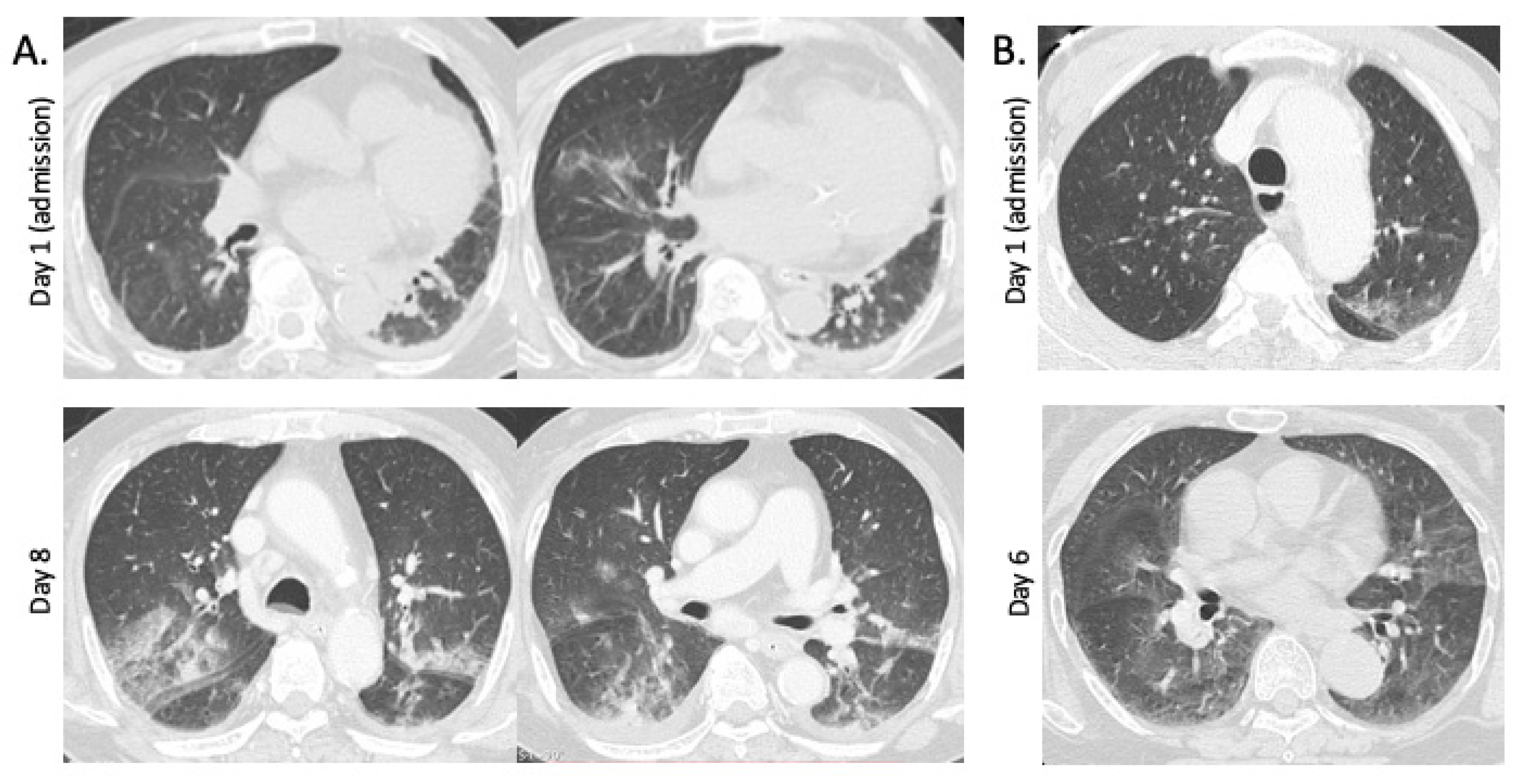
3.1. Chest CT Findings in Fatal and Non-Fatal Hospitalized Patients with SFTS
3.2. Patient Characteristics and Complications during Hospitalization
3.3. Laboratory Findings
4. Discussion
5. Conclusions
Author Contributions
Funding
Institutional Review Board Statement
Informed Consent Statement
Data Availability Statement
Acknowledgments
Conflicts of Interest
References
- Yu, X.J.; Liang, M.F.; Zhang, S.Y.; Liu, Y.; Li, J.D.; Sun, Y.L.; Zhang, L.; Zhang, Q.F.; Popov, V.L.; Li, C.; et al. Fever with Thrombocytopenia Associated with a Novel Bunyavirus in China. N. Engl. J. Med. 2011, 364, 1523–1532. [Google Scholar] [CrossRef] [PubMed] [Green Version]
- World Health Organization. An, R.&D Blueprint for Action to Prevent Epidemics: Plan of Action. 2016. Available online: https://www.who.int/who-documents-detail/an-r-d-blueprint-for-action-to-prevent-epidemics (accessed on 14 May 2020).
- World Health Organization. An, R.&D Blueprint for Action to Prevent Epidemics: Funding and Coordination Models for Prepared and Response. 2016. Available online: https://www.who.int/blueprint/what/improving-coordination/workstream_5_document_on_financing.pdf?ua=1 (accessed on 14 May 2020).
- Saijo, M.; Shimojima, M.; Yamagishi, T.; Ohishi, K.; Morikawa, S.; Hasegawa, H.A. Severe Fever with Thrombocytopenia Syndrome (SFTS), a New Tick-Borne Virus Infection—The First Case Diagnosed in Japan, 2012. Infect. Agents Surveill. Rep. 2013, 34, 40–41. (In Japanese) [Google Scholar]
- National Institute for Infectious Diseases (NIID). Severe Fever with Thrombocytopenia Syndrome (SFTS) in Japan, 2013. IASR 2014, 35, 31–32. [Google Scholar]
- Kobayashi, Y.; Kato, H.; Yamagishi, T.; Shimada, T.; Matsui, T.; Yoshikawa, T.; Kurosu, T.; Shimojima, M.; Morikawa, S.; Hasegawa, H.; et al. Severe Fever with Thrombocytopenia Syndrome, Japan, 2013–2017. Emerg. Infect. Dis. 2020, 26, 692–699. [Google Scholar] [CrossRef] [Green Version]
- Crump, A.; Tanimoto, T. Severe Fever with Thrombocytopenia Syndrome: Japan under Threat from Life-Threatening Emerging Tick-Borne Disease. JMA J. 2020, 3, 295–302. [Google Scholar] [CrossRef] [PubMed]
- Yamaji, K.; Aonuma, H.; Kanuka, H. Distribution of tick-borne diseases in Japan: Past patterns and implications for the future. J. Infect. Chemother. 2018, 24, 499–504. [Google Scholar] [CrossRef] [Green Version]
- Nakamura, S.; Iwanaga, N.; Hara, S.; Shimada, S.; Kashima, Y.; Hayasaka, D.; Abe, K.; Izumikawa, K.; Yanagihara, K.; Miyazaki, Y.; et al. Viral Load and Inflammatory Cytokine Dynamics Associated with the Prognosis of Severe Fever with Thrombocytopenia Syndrome Virus Infection: An Autopsy Case. J. Infect. Chemother. 2019, 25, 480–484. [Google Scholar] [CrossRef] [PubMed]
- Koga, S.; Takazono, T.; Ando, T.; Hayasaka, D.; Tashiro, M.; Saijo, T.; Kurihara, S.; Sekino, M.; Yamamoto, K.; Imamura, Y.; et al. Severe Fever with Thrombocytopenia Syndrome Virus RNA in Semen, Japan. Emerg. Infect. Dis. 2019, 25, 2127–2128. [Google Scholar] [CrossRef] [PubMed] [Green Version]
- Akagi, K.; Miyazaki, T.; Oshima, K.; Umemura, A.; Shimada, S.; Morita, K.; Senju, H.; Tashiro, M.; Takazono, T.; Saijo, T.; et al. Detection of Viral RNA in Diverse Body Fluids in an SFTS Patient with Encephalopathy, Gastrointestinal Bleeding and Pneumonia: A Case Report and Literature Review. BMC Infect. Dis. 2020, 20, 281. [Google Scholar] [CrossRef]
- Yun, J.H.; Hwang, H.J.; Jung, J.; Kim, M.J.; Chong, Y.P.; Lee, S.O.; Choi, S.H.; Kim, Y.S.; Woo, J.H.; Kim, M.Y.; et al. Comparison of Chest Radiographic Findings between Severe Fever with Thrombocytopenia Syndrome and Scrub Typhus: Single Center Observational Cross-Sectional Study in South Korea. Medicine 2019, 98, e17701. [Google Scholar] [CrossRef] [Green Version]
- Deng, B.; Zhou, B.; Zhang, S.; Zhu, Y.; Han, L.; Geng, Y.; Jin, Z.; Liu, H.; Wang, D.; Zhao, Y.; et al. Clinical Features and Factors Associated with Severity and Fatality among Patients with Severe Fever with Thrombocytopenia Syndrome Bunyavirus Infection in Northeast China. PLoS ONE. 2013, 8, e80802. [Google Scholar] [CrossRef] [Green Version]
- Zhu, Y.; Wu, H.; Gao, J.; Zhou, X.; Zhu, R.; Zhang, C.; Bai, H.; Abdullah, A.S.; Pan, H. Two Confirmed Cases of Severe Fever with Thrombocytopenia Syndrome with Pneumonia: Implication for a Family Cluster in East China. BMC Infect. Dis. 2017, 17, 537. [Google Scholar] [CrossRef] [PubMed] [Green Version]
- Gando, S.; Saitoh, D.; Ogura, H.; Fujishima, S.; Mayumi, T.; Araki, T.; Ikeda, H.; Kotani, J.; Kushimoto, S.; Miki, Y.; et al. A Multicenter, Prospective Validation Study of the Japanese Association for Acute Medicine Disseminated Intravascular Coagulation Scoring System in Patients with Severe Sepsis. Crit. Care. 2013, 17, R111. [Google Scholar] [CrossRef] [PubMed] [Green Version]
- Shimada, S.; Posadas-Herrera, G.; Aoki, K.; Morita, K.; Hayasaka, D. Therapeutic Effect of Post-Exposure Treatment with Antiserum on Severe Fever with Thrombocytopenia Syndrome (SFTS) in a Mouse Model of SFTS Virus Infection. Virology 2015, 482, 19–27. [Google Scholar] [CrossRef] [PubMed] [Green Version]
- Truszkiewicz, K.; Poręba, R.; Gać, P. Radiological Cardiothoracic Ratio in Evidence-Based Medicine. J. Clin. Med. 2021, 10, 2016. [Google Scholar] [CrossRef] [PubMed]
- Wang, Z.J.; Reddy, G.P.; Gotway, M.B.; Yeh, B.M.; Hetts, S.W.; Higgins, C.B. CT and MR Imaging of Pericardial Disease. Radiographics. 2003, 23, S167–S180. [Google Scholar] [CrossRef] [PubMed]
- Muggli, D.; Müller, M.A.; Karlo, C.; Fornaro, J.; Marincek, B.; Frauenfelder, T. A Simple Method to Approximate Liver Size on Cross-Sectional Images Using Living Liver Models. Clin. Radiol. 2009, 64, 682–689. [Google Scholar] [CrossRef] [PubMed]
- Nuffer, Z.; Marini, T.; Rupasov, A.; Kwak, S.; Bhatt, S. The Best Single Measurement for Assessing Splenomegaly in Patients with Cirrhotic Liver Morphology. Acad. Radiol. 2017, 24, 1510–1516. [Google Scholar] [CrossRef] [PubMed]
- National Institute for Infectious Diseases (NIID). Severe Fever with Thrombocytopenia Syndrome (SFTS) in Japan. 2021. Available online: https://www.niid.go.jp/niid/ja/sfts/sfts-idwrs/7415-sfts-nesid.html (accessed on 1 December 2021). (In Japanese).
- Koo, H.J.; Lim, S.; Choe, J.; Choi, S.H.; Sung, H.; Do, K.H. Radiographic and CT Features of Viral Pneumonia. Radiographics 2018, 38, 719–739. [Google Scholar] [CrossRef] [PubMed] [Green Version]

| All (n = 22) | Fatal (n = 6) | Non-Fatal (n = 16) | p-Value | |
|---|---|---|---|---|
| Time from symptoms onset to chest CT, days ± SD | 4.95 ± 4.09 | 5.17 ± 0.98 | 4.94 ± 1.16 | 0.910 |
| Time from admission to chest CT, days ± SD | 0.14 ± 0.35 | 0.00 ± 0.00 | 0.25 ± 0.11 | 0.041 |
| Abnormal chest CT findings | 19 (86.4) | 6 (100) | 13 (81.3) | 0.532 |
| Consolidation | 5 (22.7) | 2 (33.3) | 3 (18.8) | 0.585 |
| Ground-glass opacity | 12 (54.5) | 6 (100) | 6 (37.5) | 0.015 |
| Interstitial septal thickening | 15 (68.1) | 6 (100) | 9 (56.3) | 0.121 |
| Centrilobular nodule | 8 (36.4) | 2 (33.3) | 6 (37.5) | 1.000 |
| Bronchial wall thickening | 8 (36.4) | 2 (33.3) | 6 (37.5) | 1.000 |
| Cardiomegaly | 8 (36.4) | 3 (50.0) | 5 (31.3) | 0.624 |
| Pleural effusion | 5 (22.7) | 3 (50.0) | 2 (12.5) | 0.100 |
| Pericardial effusion | 4 (18.2) | 1 (16.7) | 3 (18.8) | 1.000 |
| Mediastinal lymph node enlargement | 3 (13.6) | 1 (16.7) | 2 (12.5) | 1.000 |
| Hepatomegaly | 6 (27.3) | 1 (16.7) | 5 (31.3) | 0.634 |
| Splenomegaly | 3 (13.6) | 0 (0.0) | 3 (18.8) | 0.532 |
| All (n = 22) | GGO (+) (n = 12) | GGO (−) (n = 10) | p-Value | |
|---|---|---|---|---|
| Consolidation | 5 (22.7) | 3 (25.0) | 2 (20.0) | 1.000 |
| Interstitial septal thickening | 15 (68.2) | 9 (75.0) | 6 (60.0) | 0.652 |
| Centrilobular nodule | 8 (36.4) | 3 (25.0) | 5 (50.0) | 0.378 |
| Bronchial wall thickening | 8 (36.4) | 4 (33.3) | 4 (40.0) | 1.000 |
| Cardiomegaly | 8 (36.4) | 7 (58.3) | 1 (10.0) | 0.031 |
| Pleural effusion | 5 (22.7) | 4 (33.3) | 1 (10.0) | 0.323 |
| Pericardial effusion | 4 (18.2) | 3 (25.0) | 1 (10.0) | 0.594 |
| Mediastinal lymph node enlargement | 3 (13.6) | 3 (25.0) | 0 (0.0) | 0.221 |
| Hepatomegaly | 6 (27.3) | 4 (33.3) | 2 (20.0) | 0.646 |
| Splenomegaly | 3 (13.6) | 3 (25.0) | 0 (0.0) | 0.221 |
| All (n = 22) | GGO (+) (n = 12) | GGO (−) (n = 10) | p-Value | 95% CI | |
|---|---|---|---|---|---|
| Age, ±SD | 71.4 ± 9.9 | 76.2 ± 1.9 | 65.6 ± 3.2 | 0.009 | 2.98–18.15 |
| Male gender | 15 (68.1) | 8 (66.7) | 7 (70.0) | 1.000 | |
| Smoking | 6 (27.2) | 2 (16.7) | 4 (40.0) | 0.348 | |
| Farmers, hunters, living or working in wooded and hilly areas | 19 (86.3) | 9 (41.6) | 10 (100) | 0.221 | |
| Underlying diseases | 17 (77.3) | 11 (75.0) | 6 (60.0) | 0.135 | |
| Chronic lung diseases | 8 (36.4) | 5 (41.6) | 3 (30.0) | 0.675 | |
| Cardiovascular diseases | 12 (54.5) | 7 (58.3) | 5 (50.0) | 1.000 | |
| Chronic kidney diseases | 2 (9.0) | 1 (8.3) | 1 (10.0) | 1.000 | |
| Diabetes mellitus | 4 (18.1) | 2 (16.6) | 2 (20.0) | 1.000 | |
| Cancer | 2 (9.0) | 1 (8.3) | 1 (10.0) | 1.000 | |
| General symptoms | |||||
| Fatigue | 16 (72.7) | 9 (75.0) | 7 (70.0) | 1.000 | |
| Fever | 18 (81.8) | 9 (75.0) | 9 (90.0) | 0.594 | |
| Chill | 2 (9.1) | 0 (0.0) | 2 (20.0) | 0.195 | |
| Headache | 4 (18.1) | 0 (0.0) | 4 (40.0) | 0.029 | |
| Dizziness | 3 (13.6) | 2 (16.7) | 1 (10.0) | 1.000 | |
| Myalgia | 1 (4.5) | 1 (8.3) | 0 (0.0) | 1.000 | |
| Respiratory symptoms | 9 (40.9) | 6 (50.0) | 3 (30.0) | 0.415 | |
| Cough | 1 (4.5) | 0 (0.0) | 1 (10.0) | 0.455 | |
| Sputum | 2 (9.1) | 2 (16.6) | 0 (0.0) | 0.481 | |
| Dyspnea | 7 (31.8) | 5 (41.6) | 2 (20.0) | 0.381 | |
| Gastrointestinal symptoms | 18 (81.8) | 8 (66.7) | 10 (100) | 0.096 | |
| Nausea | 3 (13.6) | 1 (8.3) | 2 (20.0) | 0.571 | |
| Anorexia | 4 (18.1) | 3 (25.0) | 1 (10.0) | 0.594 | |
| Diarrhea | 11 (50.0) | 3 (25.0) | 8 (80.0) | 0.030 | |
| Skin and other symptoms | |||||
| Skin rash | 8 (36.4) | 5 (41.6) | 3 (30.0) | 0.675 | |
| Tick bite | 7 (31.8) | 4 (33.3) | 3 (30.0) | 1.000 | |
| Lymphadenopathy | 10 (45.5) | 6 (50.0) | 4 (40.0) | 0.691 | |
| Complications and outcomes | |||||
| Secondary infection | 5 (22.7) | 4 (33.3) | 1 (10.0) | 0.323 | |
| Fungal infection | 3 (13.6) | 3 (25.0) | 0 (0.0) | 0.221 | |
| Length of hospital stay (days) | 35.2 | 46.7 | 21.4 | 0.329 | −27.41–77.95 |
| ICU admission | 7 (31.8) | 6 (50.0) | 1 (10.0) | 0.074 | |
| Length of ICU stay (days) | 7.1 | 12.3 | 0.8 | 0.061 | −0.65–23.55 |
| All (n = 22) | GGO (+) (n = 12) | GGO (−) (n = 10) | p-Value | 95% CI | |
|---|---|---|---|---|---|
| Vital signs | |||||
| Consciousness disturbance | 14 (63.6) | 11 (91.7) | 3 (30.0) | 0.006 | |
| Body temperature (°C) | 38.3 | 38.4 | 38.2 | 0.721 | −0.69–0.98 |
| Systolic blood pressure(mmHg) | 127.4 | 133.1 | 120.5 | 0.114 | −3.31–28.48 |
| Heart rate (/min) | 84.8 | 93.3 | 74.6 | 0.002 | 7.54–29.92 |
| SpO2 (%) | 96.5 | 96.3 | 96.7 | 0.695 | −2.29–1.56 |
| SpO2/FiO2 ratio | 412.8 | 401.5 | 426.4 | 0.494 | −99.66–49.74 |
| qSOFA score | 0.96 | 1.17 | 0.70 | 0.191 | −0.22–1.15 |
| Rales | 4 (18.1) | 3 (25.0) | 1 (10.0) | 0.594 | |
| Laboratory data | |||||
| WBC (/μL) | 1725.5 | 1733.3 | 1716.0 | 0.967 | −844.85–879.52 |
| Neutrophil (%) | 64.6 | 65.3 | 63.7 | 0.816 | −12.33–15.48 |
| Lymphocyte (%) | 26.7 | 26.6 | 26.8 | 0.961 | −9.38–8.95 |
| Atypical lymphocyte (%) | 2.09 | 1.92 | 2.30 | 0.803 | −3.55–2.78 |
| Platelet (×103/μL) | 52.6 | 50.9 | 54.6 | 0.781 | −30.83–23.5 |
| Hb (g/dL) | 14.0 | 13.9 | 14.3 | 0.636 | −2.24–1.4 |
| AST (IU/L) | 342.4 | 404.6 | 267.8 | 0.311 | −137.6–411.17 |
| ALT (IU/L) | 117.8 | 128.8 | 104.5 | 0.506 | −50.62–99.29 |
| LDH (IU/L) | 830.4 | 927.5 | 713.8 | 0.242 | −156.39–583.79 |
| CK (IU/L) | 2560.6 | 2670.4 | 2439.8 | 0.908 | −3900.54–4361.67 |
| CRP (mg/dL) | 0.86 | 1.34 | 0.27 | 0.038 | 0.070–2.07 |
| PCT (ng/mL) | 0.27 | 0.37 | 0.19 | 0.143 | −0.07–0.44 |
| Alb (g/dL) | 3.24 | 3.21 | 3.28 | 0.704 | −0.46–0.32 |
| Ferritin (ng/mL) | 20,194.8 | 25,839.5 | 12,433.3 | 0.536 | −31,373.2–58,185.7 |
| BUN (mg/dL) | 26.8 | 25.2 | 28.7 | 0.639 | −19.08–11.99 |
| Cre (mg/dL) | 1.49 | 1.70 | 1.25 | 0.649 | −1.57–2.46 |
| eGFR (mL/min/1.73 m2) | 57.3 | 61.3 | 52.5 | 0.404 | −12.68–30.2 |
| Na (mEq/L) | 132.3 | 130.6 | 134.4 | 0.138 | −9.01–1.38 |
| K (mEq/L) | 4.00 | 4.02 | 3.96 | 0.824 | −0.47–0.58 |
| Cl (mEq/L) | 99.6 | 97.4 | 102.3 | 0.084 | −10.48–0.71 |
| APTT (%) | 48.7 | 52.2 | 44.6 | 0.240 | −5.51–20.74 |
| PT-INR | 1.06 | 1.07 | 1.04 | 0.442 | −0.056–0.12 |
| D-dimer (μg/mL) | 14.4 | 18.4 | 9.8 | 0.211 | −5.35–22.55 |
| FDP (μg/mL) | 27.6 | 32.7 | 20.9 | 0.365 | −14.79–38.37 |
| DIC score | 4.27 | 4.92 | 3.50 | 0.108 | −0.34–3.17 |
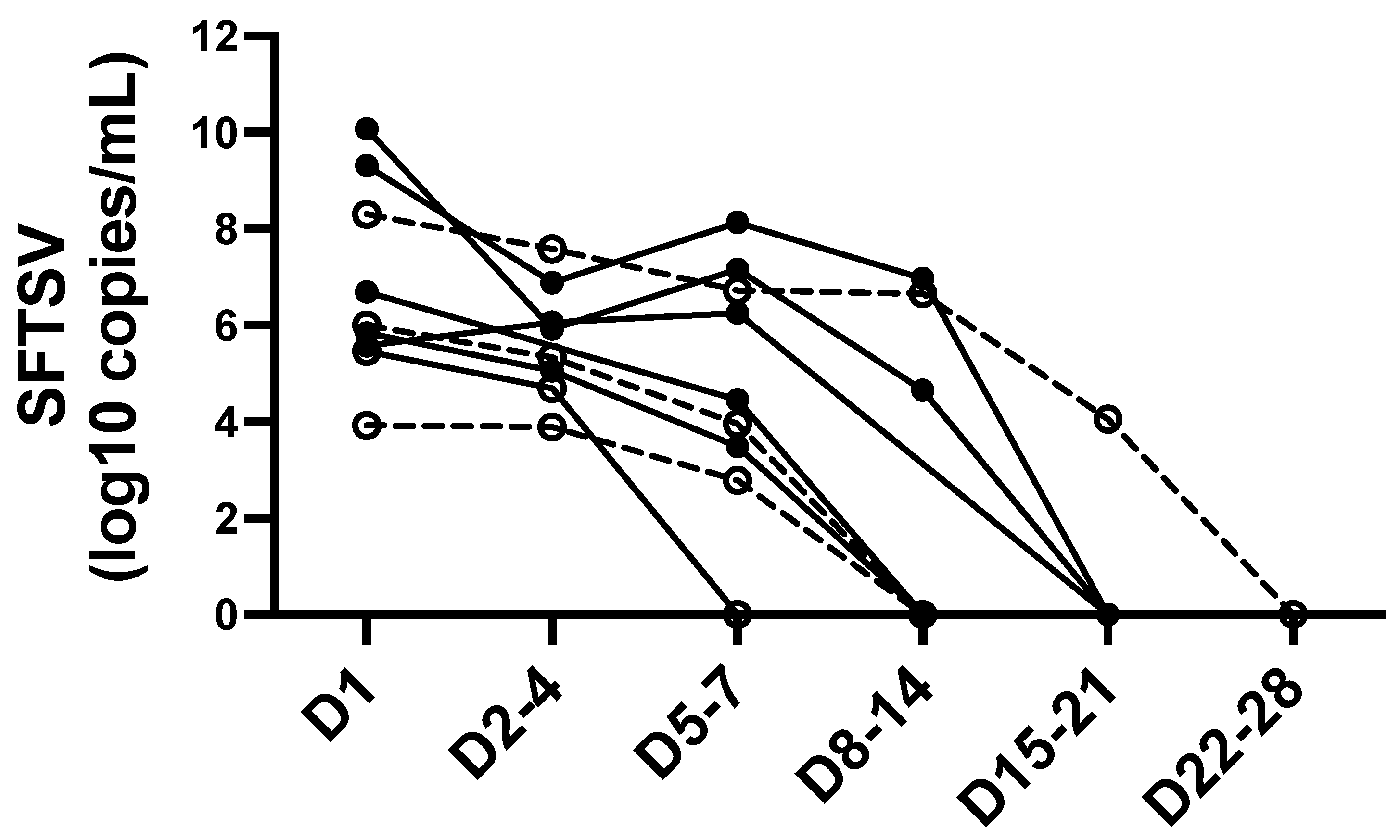
| SFTSV viral load (copies/mL) | 8.76 × 10⁸ | 1.65 × 10⁹ | 1.03 × 10⁸ | 0.390 | −1.63 × 10⁹–4.73 × 10⁹ |
| Proteinuria | 21(95.5) | 12 (100) | 9 (90.0) | 0.455 | |
| Hematuria | 19(86.4) | 10 (83.3) | 9 (90.0) | 1.000 |
Publisher’s Note: MDPI stays neutral with regard to jurisdictional claims in published maps and institutional affiliations. |
© 2022 by the authors. Licensee MDPI, Basel, Switzerland. This article is an open access article distributed under the terms and conditions of the Creative Commons Attribution (CC BY) license (https://creativecommons.org/licenses/by/4.0/).
Share and Cite
Ashizawa, H.; Yamamoto, K.; Ashizawa, N.; Takeda, K.; Iwanaga, N.; Takazono, T.; Sakamoto, N.; Sumiyoshi, M.; Ide, S.; Umemura, A.; et al. Associations between Chest CT Abnormalities and Clinical Features in Patients with the Severe Fever with Thrombocytopenia Syndrome. Viruses 2022, 14, 279. https://doi.org/10.3390/v14020279
Ashizawa H, Yamamoto K, Ashizawa N, Takeda K, Iwanaga N, Takazono T, Sakamoto N, Sumiyoshi M, Ide S, Umemura A, et al. Associations between Chest CT Abnormalities and Clinical Features in Patients with the Severe Fever with Thrombocytopenia Syndrome. Viruses. 2022; 14(2):279. https://doi.org/10.3390/v14020279
Chicago/Turabian StyleAshizawa, Hiroki, Kazuko Yamamoto, Nobuyuki Ashizawa, Kazuaki Takeda, Naoki Iwanaga, Takahiro Takazono, Noriho Sakamoto, Makoto Sumiyoshi, Shotaro Ide, Asuka Umemura, and et al. 2022. "Associations between Chest CT Abnormalities and Clinical Features in Patients with the Severe Fever with Thrombocytopenia Syndrome" Viruses 14, no. 2: 279. https://doi.org/10.3390/v14020279
APA StyleAshizawa, H., Yamamoto, K., Ashizawa, N., Takeda, K., Iwanaga, N., Takazono, T., Sakamoto, N., Sumiyoshi, M., Ide, S., Umemura, A., Yoshida, M., Fukuda, Y., Kobayashi, T., Tashiro, M., Tanaka, T., Katoh, S., Morimoto, K., Ariyoshi, K., Morimoto, S., ... Mukae, H. (2022). Associations between Chest CT Abnormalities and Clinical Features in Patients with the Severe Fever with Thrombocytopenia Syndrome. Viruses, 14(2), 279. https://doi.org/10.3390/v14020279