Article

Rift Valley Fever Virus Non-Structural Protein S Is Associated with Nuclear Translocation of Active Caspase-3 and Inclusion Body Formation

by
Lukas Mathias Michaely
1,
Melanie Rissmann
2,3,4,
Federico Armando
1,
Felicitas von Arnim
2,3,
Markus Keller
2,3,
Martin Eiden
2,3,
Rebecca König
2,3,
Benjamin Gutjahr
2,3,
Wolfgang Baumgärtner
1,†,
Martin H. Groschup
2,3,† and
Reiner Ulrich
5,6,*,†
1
Center of Systems Neuroscience, Department of Pathology, University of Veterinary Medicine Hannover, Foundation, 30559 Hannover, Germany
2
Institute of Novel and Emerging Diseases, Friedrich-Loeffler-Institute, Insel Riems, 17493 Greifswald, Germany
3
Partner Site Hamburg-Lübeck-Borstel-Riems, German Center for Infection Research (DZIF), Insel Riems, 17493 Greifswald, Germany
4
Erasmus Medical Center, Department of Viroscience, 3015 GD Rotterdam, The Netherlands
5
Department of Experimental Animal Facilities and Biorisk Management, Friedrich-Loeffler-Institute, Insel Riems, 17493 Greifswald, Germany
6
Institute of Veterinary Pathology, Faculty of Veterinary Medicine, Leipzig University, 04103 Leipzig, Germany
*
Author to whom correspondence should be addressed.
These authors contributed equally to this work.
Viruses 2022, 14(11), 2487; https://doi.org/10.3390/v14112487
Submission received: 11 September 2022 / Revised: 6 November 2022 / Accepted: 6 November 2022 / Published: 10 November 2022
(This article belongs to the Special Issue Rift Valley Fever Epidemiology, Pathogenesis and Host Response)

Abstract

:
Rift Valley fever phlebovirus (RVFV) causes Rift Valley fever (RVF), an emerging zoonotic disease that causes abortion storms and high mortality rates in young ruminants as well as severe or even lethal complications in a subset of human patients. This study investigates the pathomechanism of intranuclear inclusion body formation in severe RVF in a mouse model. Liver samples from immunocompetent mice infected with virulent RVFV 35/74, and immunodeficient knockout mice that lack interferon type I receptor expression and were infected with attenuated RVFV MP12 were compared to livers from uninfected controls using histopathology and immunohistochemistry for RVFV nucleoprotein, non-structural protein S (NSs) and pro-apoptotic active caspase-3. Histopathology of the livers showed virus-induced, severe hepatic necrosis in both mouse strains. However, immunohistochemistry and immunofluorescence revealed eosinophilic, comma-shaped, intranuclear inclusions and an intranuclear (co-)localization of RVFV NSs and active caspase-3 only in 35/74-infected immunocompetent mice, but not in MP12-infected immunodeficient mice. These results suggest that intranuclear accumulation of RVFV 35/74 NSs is involved in nuclear translocation of active caspase-3, and that nuclear NSs and active caspase-3 are involved in the formation of the light microscopically visible inclusion bodies.

1. Introduction

Rift Valley fever phlebovirus (RVFV, Phenuiviridae) causes Rift Valley fever (RVF), a zoonotic and potentially lethal disease that is currently endemic in Africa and on the Arabian Peninsula [1,2]. Although approximately 50% of infected humans experience no or only mild signs of the disease, the infection can lead to severe or even lethal disease and its complications include renal failure, hemorrhagic fever, hepatitis, encephalitis, ocular complications and possible life-long impairment or even death [2,3,4]. While the disease can pose a threat to public health systems, the impact of Rift Valley fever outbreaks on agricultural economic welfare is of equal or even greater concern [5,6]. Within ruminant livestock, the disease leads to trimester indiscriminate “abortion storms” [7]. Especially among the most susceptible species of sheep, these outbreaks cause high mortality rates among neonatal offspring, thus eliminating complete generations of local ruminant herds [7,8,9]. Therefore, RVF has been recognized as an important emerging disease that warrants further investigation due to limited understanding of its pathogenesis as well as its possible spread towards non-endemic areas [3,10,11].
To address this lack of knowledge and to investigate RVF pathogenesis, a panel of susceptible animal models has been established including ruminants, rodents and monkeys [12,13,14,15,16,17]. It is known that RVF pathogenesis and virulence differs among infected species and even between strains within the same animal model [3]. As described by Kwaśnik et al. (2021), the course of disease can broadly be divided in three categories: (i) cases of severe and acute illness are associated with an uncontrolled viremia marked by high viral loads. (ii) The second pattern exhibits quickly decreasing levels of viremia and mild to no clinical disease. (iii) Third, fever and viremia are displayed during the initial onset of disease, but the main hallmark is the development of delayed signs such as encephalitis or ocular disease, which in humans can manifest weeks to months after the initial infection and the pathogenesis of which remains unclear so far [2,3,10].
On the cellular level, the virus exhibits a high tropism towards mononuclear phagocytes and hepatocytes thus inducing hepatic disease among other signs [2,3]. The virus enters hepatocytes by endocytosis after its viral glycoproteins interact with yet to be unequivocally identified receptors [3,18]. Possible cellular surface receptors include liver/lymph node-specific intracellular adhesion molecules-3 grabbing non-integrin (L-SIGN) cellular C-type lectins that resemble the dendritic cell-specific intercellular adhesion molecule-3-grabbing non-integrin (DC-SIGN)-mediated viral entry into dendritic cells [3,18,19]. Thereafter, the innate immune response may control the viral spread by several mechanisms including a strong interferon (IFN) type I response as seen in many virus infections [2,18,20]. RVFV has developed evasive strategies to counteract this innate immune signaling, mainly due to interactions of the multifunctioning non-structural protein S (NSs) [2,6,18,20]. Another important part of a successful antiviral immune response is represented by quick apoptosis of infected cells via the activation of the Janus kinase and signal transducer and activator of transcription protein (JAK-STAT) pathway [21,22]. This pathway and the follow-on process of cellular apoptosis includes a group of proteases, called caspases, a designation derived as an acronym of their function as cysteinyl-aspartate-specific protease [23]. Caspases are one of the key components during apoptosis with initiating (caspase 8 or caspase 9) as well as effector functions (caspase-3, caspase 6, caspase 7) and have thus been used as an easily accessible marker of apoptosis in several studies [24,25,26,27,28].
While these molecular mechanisms are typically not accessible by light microscopy, an occasional feature of RVF-induced lesions is the occurrence of intranuclear, eosinophilic inclusions [7,22,29,30]. These inclusions have been subject to different investigations, and it has been shown that nuclei of infected cells contain filamentous structures of viral NSs protein [31,32,33,34]. Positive staining results of affected hepatocytes via terminal desoxynucleotidyl transferase dUTP nick end labeling (TUNEL) assay indicate that RVFV replication induces hepatocellular apoptosis [2,29]. However, the role of caspase-3, its interaction with NSs and intranuclear inclusion body formation has yet to be elucidated.
The present study uses archival tissue samples from two mouse strains that have been used in a previously reported infection experiment [35]. C57Bl/6-IFNARtmAgt mice (IFNAR−/−) are a strain of knockout mice that lack the expression of the interferon type A receptor (IFNAR) and therefore are unable to mount a successful interferon response [36,37]. As described above, this immunodeficiency is required to induce severe hepatitis with the attenuated RVFV strain MP12 [36,37]. The second mouse strain of heterozygous Crl:NU(NCr)-Foxn1nu mice (hzNU), infected with RVFV 35/74 was used previously as a control group for homozygous NU mice and carries the NU mutation [35]. Homozygous NU (nude) mice lack thymic development and therefore are severely compromised in their cellular immune response [35,38]. The heterozygous mice used in this study however do not develop an immunodeficient phenotype and can be considered wild-type mice for this study’s purposes [35]. The hypothesis of this study is that a strong expression of NSs induced by the virulent 35/74 strain in immunocompetent mice but not the attenuated MP12 strain in immunodeficient mice leads to intranuclear co-localization of both RVFV NSs and active caspase-3. Therefore, the combined accumulation of NSs and activated caspase-3 within the nucleus is suggested to be the mechanism leading to the formation of eosinophilic, comma-shaped, intranuclear inclusion bodies described in severe cases of RVF. Based on this premise, the aims of the present study are the detection and characterization of intranuclear caspase-3 and NSs accumulations, as well as intranuclear inclusion bodies in liver lesions induced by two RVFV strains with either functional or defective NSs, respectively, in mice.

2. Materials and Methods

2.1. Virus

RVFV strain 35/74 (accession number: JF784386-88) was propagated in BHK-21 cells (Collection of Cell Lines in Veterinary Medicine, Friedrich-Loeffler-Institut, Germany) using minimum essential medium (MEM) supplemented with 2% FBS at a temperature of 37 °C and 5% CO2. RVFV strain MP12 was propagated on Vero-76 cells, following previously published protocols [35,39,40]. The supernatant of both propagations was harvested, at 80% cytopathic effect (CPE) and the TCID50 assay was used to determine the virus titer according to Spearman and Kaerber [41].

2.2. Mouse Experiments and Sampling

All tissues used in the current study were archival specimens from a previously published study concerning the importance of a functional innate and adaptive immune response to overcome RVFV infection, which comprised nine immunodeficient mouse strains as well as four background strains [35]. Formaldehyde-fixed, paraffin-embedded (FFPE) liver samples from five to seven-week-old, female, infected mice that had to be euthanized due to severe signs of disease and exhibited hepatic lesions were selected from the archives along with mock-infected control animals of the same strain and age (Table 1). All of the experiments were approved and authorized by the local authority (Landesamt für Landwirtschaft, Lebensmittelsicherheit und Fischerei Mecklenburg-Vorpommern (permission 7221.3-1-038/17 (19 September 2017)) and performed in accordance with the German regulations and legal requirements.
Immunodeficient C57Bl/6-IFNARtmAgt mice (IFNAR−/−) were bred at the Friedrich-Loeffler-Institute mouse stock, while immunocompetent, heterozygous Crl:NU(NCr)-Foxn1nu mice (hzNU) were commercially obtained from Charles River Laboratories (Sulzfeld, Germany) [38]. Groups of three mice were kept in individually ventilated cages (IsoCage N, Tecniplast, Hohenpeißenberg, Germany). Mice received water and standard mouse food (ssniff Spezialdiäten GmbH, Soest, Germany) ad libitum. Infection of the mice was performed by subcutaneous injection of 0.1 mL virus suspension with a TCID50 of 1.4 × 10³ per animal of RVFV 35/74 or a TCID of 1.43 × 10³ of RVFV MP12 per animal, respectively. Mock-infected control mice received virus-free cell culture medium. Mice were euthanized at a predefined severity score by isoflurane anesthesia and subsequent cardiac exsanguination. hzNU mice represent the background strain for severely immunocompromised NUDE mice that were used in the aforementioned, original study, and do not show any phenotype of the NU mutation [35,38]. Therefore, they can be considered the wild-type for the current study. The inclusion of the immunodeficient IFNAR−/− mice as a second mouse strain is required due to their unique susceptibility towards RVFV MP12 [35,36,37].

2.3. Histology and Immunohistochemistry

Liver samples were fixed in 4% neutral buffered formaldehyde for 21 days and embedded in paraffin wax. Two µm sections were cut and used for immunohistochemistry (IHC) and hematoxylin-eosin (HE) staining. IHC for RVFV proteins and active caspase-3 detection was performed according to previously published protocols [15,39,42]. Briefly, it included an optional antigen retrieval, blocking of nonspecific background signals via the application of serum from the respective host species, followed by overnight incubation of the primary antibodies, application of biotinylated secondary antibodies (biotinylated goat anti rabbit, BA-1000, or biotinylated rabbit anti sheep, BA-6000, Vector Laboratories, Burlingame, CA, USA), application of the avidin–biotin–peroxidase complex method (ABC, Elite PK6100; Vector Laboratories, Burlingame, CA, USA) using 3-amino-9-ethylcarbazol (AEC, Dako, Glostrup, Denmark) as a chromogen and hematoxylin counterstain [15,39,42]. Primary antibodies and protocol details are shown in Table 2. Monoclonal anti-RVFV antibodies were raised against recombinant bacterial proteins and have been subject to a comprehensive examination [34,39,42]. RVFV-infected and non-infected Vero cells were processed as FFPE blocks and used as positive or negative controls, respectively.
Histopathological examination and cell count per mm2 was carried out manually, using a light microscope (Carl Zeiss, Oberkochen, Germany) with a Zeiss Kpl-W10x/18 ocular and a Zeiss 40/0.65 objective. Five representative high-power fields (HPF, 1 HPF equaling 0.159 mm2) were counted per sample. An Olympus BX51 microscope and a DP72 Camera with the manufacturer’s operating software cellSens, version 1.18 (Olympus solution, Tokyo, Japan) was used for digital photography.

2.4. Immunofluorescence

FFPE mouse liver tissue from a RVFV 35/74-infected hzNU mouse and a mock-infected control mouse was used for immunofluorescence to verify the co-localization of IHC expression of RVFV NSs and active caspase-3. Immunofluorescence double staining was carried out according to previously published protocols [43,44]. Briefly, tissue sections were pretreated for antigen retrieval using citrate buffer (pH 6). Following the blocking of nonspecific bindings with goat serum, sections were incubated with primary antibodies (Table 2) targeting RVFV NSs and active caspase-3 at 4 °C overnight. Thereafter, sections were incubated with secondary cyanine 2-conjugated goat anti mouse (Dianova, Hamburg, Germany) and cyanine 3-conjugated goat anti rabbit (Invitrogen, Carlsbad, CA, USA) antibodies, respectively, for 60 min at room temperature. Nuclear counterstain and mounting were performed using 0.01% bisbenzimide (diluted in Aqua bidestillata; bisbenzimide, Sigma-Aldrich Chemie GmbH, Taufkirchen, Germany) for 10 min and mounted using Fluoroshield mounting medium (Sigma-Aldrich, St. Louis, MO, USA) and coverslips. Fluorescence pictures were taken using a fluorescence microscope (BZ-9000E, Keyence Deutschland GmbH, Neu-Isenburg, Germany).

2.5. Statistical Analysis

Statistical workup of the cell count data was performed via the Mann–Whitney U-test using Graphpad Prism (GraphPad Software, Inc., San Diego, CA, USA, version 9.0.0) and SAS 9.4 (SAS Institute, Inc. Cary, NC, USA).

3. Results

3.1. Clinical Signs

After infection, the mice either died or had to be euthanized due to severe signs of disease that included ruffled fur, lack of unprovoked and provoked movement, tachypnea and apathy at 2 or 3 days post infection (dpi).

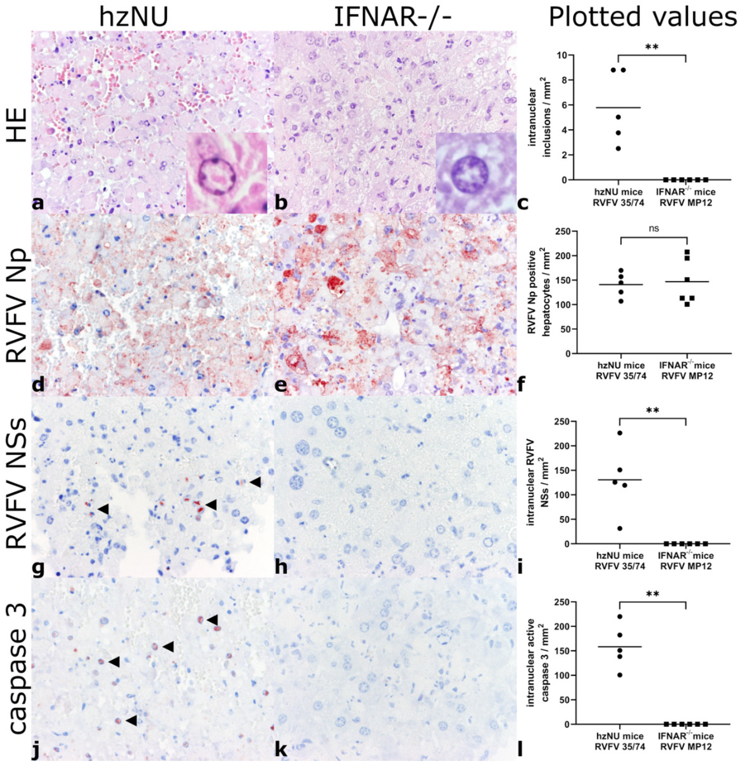
3.2. Histology and Immunohistochemistry

The liver samples of all the RVFV-infected mice exhibited severe diffuse hepatic necrosis, with it affecting over 90% of the analyzed organ tissue (Figure 1). Likewise, numerous condensed, rounded, eosinophilic hepatocytes with pyknotic and karyorrhectic nuclei, suggestive of apoptotic hepatocytes were present, while leukocytic infiltrates were mostly absent. IHC labeling revealed abundant and diffuse extra- and intracellular RVFV nucleoprotein (Np) expression (Figure 1).
Nuclear, comma-shaped, eosinophilic inclusions, mostly associated with complete margination of chromatin, were present in intralesional hepatocytes within RVFV 35/74-infected hzNU mice but were absent in RVFV MP12-infected IFNAR−/− mice. Likewise, in RVFV 35/74-infected hzNU mice, RVFV NSs and active caspase-3 immunoreactivity was present within hepatocytes with a comparable, intranuclear, comma-shaped accumulation of both signals. No intranuclear expression of RVFV NSs or active caspase-3 was detected in RVFV MP12-infected IFNAR−/− mice (Figure 1).
The control mice did not exhibit hepatocellular necrosis, apoptosis or inflammatory lesions. No immunolabeling for RVFV proteins or active caspase-3 was observed (Table 3).

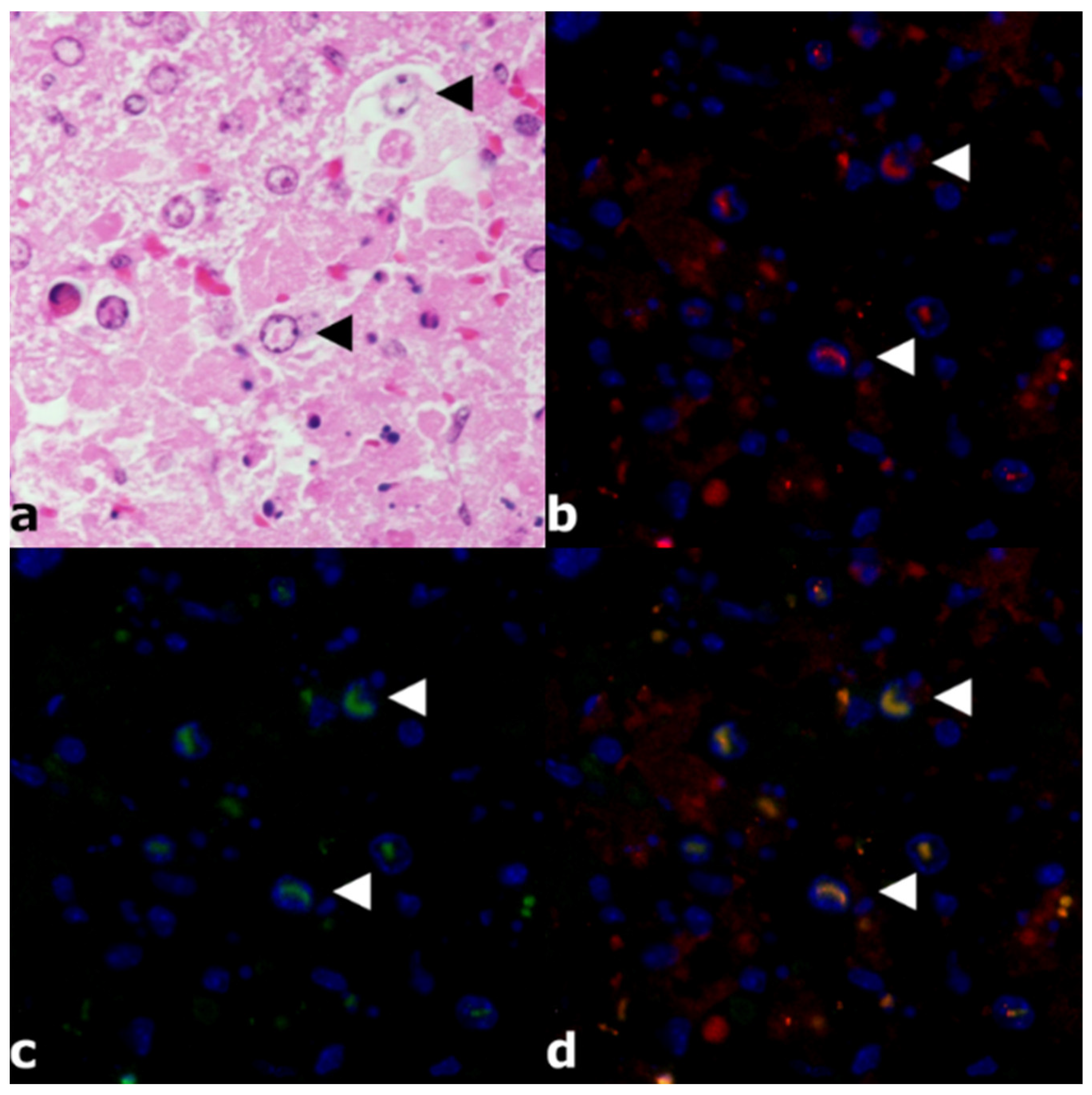
3.3. Immunofluorescence

Liver tissue from a RVFV 37/74-infected hzNU mouse was double labeled for proof of principle analysis of nuclear co-localization of NSs and active caspase-3. Immunofluorescence showed intranuclear co-expression of RVFV NSs and active caspase-3 (Figure 2). HE-stained serial slides from the same FFPE blocks revealed intranuclear, comma-shaped, eosinophilic inclusions with a comparable morphology. (Figure 2). Negative controls from a mock-infected mouse did not show any immunolabeling.

4. Discussion

The initial observation of severe, diffuse, hepatic necrosis with virus strain-dependent presence or absence of inclusion bodies was made during the histopathological analysis of an animal experiment designed for a previously published study [35]. The specimens were re-used for the current manuscript. The strains were selected under the premise to compare two groups in which either attenuated RVFV MP12 or the virulent RVFV 35/74 induces hepatic necrosis of comparable severity [35,40]. The hypothesis is that intranuclear inclusion bodies are formed by nuclear co-localization of NSs protein and active caspase-3 only in RVFV NSs-infected hzNU but not in RVFV MP12-infected IFNAR−/− mice.
Considering that only two conditions, differing in both virus strain and host, are reported here, the available choice of archival samples is a limitation of this study and it should be admitted that a complete experimental design would include RVFV 35/74-infected IFNAR−/− mice and RVFV MP12-infected hzNU mice for unequivocal proof that the observed effect is solely due to the different virus strains. Specimen of RVFV 35/74-infected IFNAR−/− mice are currently unavailable, since they were not needed for the hypothesis of the initial study [35]. However, the lacking results can be anticipated with a high degree of certainty, since it is well known that 35/74 infection leads to severe hepatic necrosis with intranuclear inclusions in other immunocompetent mouse strains [40]. Furthermore, our previous study has shown that MP12 does not induce clinical disease and no hepatic lesions were observed at 14 dpi in the immunocompetent hzNU mice [35]. In all RVFV-infected animals, RVFV Np was expressed diffusely within hepatic lesions, however, intranuclear expression of NSs and pro-apoptotic active caspase-3 was present only in RVFV 35/74-infected mice. In agreement with the results, virulent strains of RVFV are known to induce hepatocellular apoptosis and intranuclear eosinophilic inclusions in mice and lambs [3,7,40,45]. Using a limited number of sheep experimentally infected with RVFV 35/74, comparable intranuclear inclusion bodies, as well as intranuclear NSs and active caspase-3 accumulation, were observed in lambs with severe, coalescing, necrotizing hepatitis, but not in adult sheep with mild to moderate, multifocal, necrotizing hepatitis (unpublished observations).
The NSs protein consists of 265 amino acids, is encoded on the small segment of the viral genome and is one of the main pathogenicity factors of RVFV [18,46,47]. As in the related viruses, Toscana virus, Bhanja virus, and Uukuniemi virus, the protein accumulates within the cytoplasm during viral replication, but differs from other structural and non-structural viral proteins by also forming filamentous structures within the nucleus [33,47,48,49]. It is unclear, whether this trait is unique to RVFV as the localization of NSs in other phlebovirus infections is unknown so far [49]. The intranuclear filaments of RVFV NSs are formed by the C-terminal amino acids of NSs and, as described in the current study, these NSs-filaments are most likely involved in the formation of the eosinophilic, comma-shaped, intranuclear inclusions occasionally observed by light microscopy in RVF hepatitis [22,30,33,47].
NSs plays a significant role in the evasion from host innate immunity by inhibiting IRF-3, NF-κB and AP-1, which are transcription factors inducing IFN-β expression [47,50]. Furthermore, NSs binds to the subunit protein p44 of TFIIH, which is an important transcription factor for RNA polymerase I and II [47,51]. NSs also promotes post-translational degradation of dsRNA-dependent PKR and arrests cell cycle progression by hitherto unknown mechanisms [47,52,53,54]. Additionally, NSs can activate the classical DNA damage-signaling pathway via the ATM kinase, inducing phosphorylation of p53, and activation of Chk.2 and H2A.X [47,55,56,57]. Among 33 NSs-interacting genes investigated in a large chromatin immunoprecipitation study, downregulation of the genes responsible for protein phosphorylation, ubiquitination, immune responses, apoptosis, transcription or development was observed [47,57]. However, this study did not include caspase-3, but only the related caspases 8 and 9, the latter of which was significantly downregulated by NSs activity [57]. Furthermore, NSs facilitates viral translation by sequestration of poly(A)-binding protein 1 and downregulation of protein kinase PKR [52,53,58]. Finally, the involvement of NSs in coagulative disorders has been suggested, which is one of the major hallmarks of human hemorrhagic fever in severe cases of RVF [48,57]. Due to this plethora of adverse effects, NSs has been recognized as a major pathogenicity factor and has been targeted by vaccine developers and researchers [47].
Innate mechanisms such as apoptotic pathways represent a very important first line of defense to contain early virus replication and spread [2,3,20,29]. The effect of RVFV on apoptotic pathways is not fully understood. It is known that intranuclear presence of NSs induces apoptosis by its inhibitory effect on the cell cycle and subsequent activation of p53 [22,59]. On the other hand, there is evidence that NSs inhibits apoptosis [59], and RVFV non-structural protein NSm has also been shown to have an apoptosis-blocking effect [60]. In the current study, the co-localization of NSs and activated caspase-3 in 35/74-infected mice forming characteristic intranuclear, rod-shaped filaments is described. Although caspase-3 is commonly believed to be active within the cytosol during apoptosis [61], there is experimental evidence for nuclear accumulation of activated caspase-3 [62,63,64]. The morphology of the immunoreactive filaments described in the current study is different from the described, coarser granular to diffuse nuclear staining pattern of active caspase-3 in apoptotic cells [62,63,64]. Whether this peculiar co-localization pattern represents physical interaction, maybe delaying apoptosis in favor of increased virus reproduction, needs to be investigated in further experiments. Promising approaches for future investigations include labeling and colocalization of both molecules in infections of different hosts and in vitro models by different RVFV strains using high-resolution microscopy, as well as the investigation of the induction of apoptosis and interferon-stimulated pathways employing overexpression of NSs in in vitro models.
The observation of lacking nuclear NSs and active caspase-3 immunoreactivity in MP12-infected IFNAR−/− mice, despite severe diffuse hepatocellular death, points to a pathomechanistic difference in this experimental condition. Notably, the attenuation of RVFV MP12 renders it non-pathogenic after subcutaneous infection in interferon-competent hosts [35,37]. Therefore, the use of IFNAR−/− knockout mice that do not express the interferon type A receptor and that are unable to mount a successful interferon type 1 response is required in order to induce lesions with this virus strain [35,36]. One of the attenuating mutations of RVFV MP12 is a mutation in the NSs protein [13,16,37,65]. This pathomechanistically incompletely characterized defect in RVFV MP12 NSs offers an explanation to the different observations in RVFV 35/74-infected hzNU mice and RVFV MP12 infected IFNAR−/− mice [13,16,37]. In agreement with the current results, intralesional RVFV glycoprotein N, glycoprotein C, nucleoprotein, and non-structural protein m (Nsm) positive hepatocytes, but lack of intranuclear NSs, is described in the livers of MP12-infected alpacas, although intranuclear NSs was present in parallelly processed MP12-infected Vero cell cultures [15]. Therefore, although NSs is present in RVFV MP12, the mutations apparently disable the intranuclear accumulation of immunohistologically detectable levels of NSs in vivo. The lack of active caspase-3 labeling suggests that a different pathway of apoptosis or another currently unknown mechanism of programmed cell death or necrosis induces hepatocellular cell death in this condition. Whether the lack of active caspase-3 in the RVFV MP12-infected IFNAR−/− mice is a sequela due to the mutated MP12 NSs or due to the phenotype of the IFNAR−/− mice is currently unknown [66].
In summary, this study shows that intranuclear accumulation of NSs and active caspase-3 is associated with intrahepatocytic, intranuclear, eosinophilic, comma-shaped inclusion body formation and severe liver disease in immunocompetent mice infected with the virulent 35/74 strain of RVFV. The mechanism behind this previously unpublished observation may yield further insights into RVF pathogenesis on the cellular level and represents a promising target for further investigations.

Author Contributions

Conceptualization, L.M.M., M.R., W.B., M.H.G. and R.U.; methodology, L.M.M., M.R., F.A., F.v.A., M.K., M.E., R.K., B.G. and R.U.; validation, M.K., M.E., W.B., M.H.G. and R.U.; formal analysis, L.M.M., M.R., F.A., F.v.A., M.K., M.E., R.K., B.G. and R.U.; investigation, L.M.M., M.R., F.A., F.v.A., M.K., M.E., R.K., B.G. and R.U.; resources, W.B., M.H.G. and R.U.; data curation, R.U.; writing—original draft preparation, L.M.M.; writing—review and editing, L.M.M., M.R., F.A. and R.U.; visualization, L.M.M., F.A. and R.U.; supervision, W.B., M.H.G. and R.U.; project administration, R.U.; funding acquisition, W.B. and M.H.G. All authors have read and agreed to the published version of the manuscript.

Funding

This research was performed as part of the Zoonoses Anticipation and Preparedness Initiative (ZAPI project; IMI Grant Agreement no. 115760), with the assistance and financial support of IMI and the European Commission, and in-kind contributions from EFPIA partners. LMM and WB are members of the Center Systems Neuroscience of the University of Veterinary Medicine Hannover, and LMM received financial support for travel expenses.

Institutional Review Board Statement

The study was conducted according to the German animal welfare law and approved by the Institutional Animal welfare officer and the Ethics Committee of the respective authority (“Landesamt für Landwirtschaft, Lebensmittelsicherheit und Fischerei Mecklenburg-Vorpommern”, permission LALLF 7221.3-1-038/17).

Data Availability Statement

The data presented in this study are not publicly available but are available upon reasonable request.

Acknowledgments

The authors thank Elfi Quente, Silvia Schuparis and the animal caretakers of the Friedrich-Loeffler-Institute for their excellent technical assistance.

Conflicts of Interest

The authors declare no conflict of interest.

References

  1. Hartman, A. Rift Valley Fever. Clin. Lab. Med. 2017, 37, 285–301. [Google Scholar] [CrossRef]
  2. Ikegami, T.; Makino, S. The Pathogenesis of Rift Valley Fever. Viruses 2011, 3, 493–519. [Google Scholar] [CrossRef] [PubMed] [Green Version]
  3. Kwaśnik, M.; Rożek, W.; Rola, J. Rift Valley Fever—A Growing Threat to Humans and Animals. J. Vet. Res. 2021, 65, 7–14. [Google Scholar] [CrossRef]
  4. Al-Hazmi, M.; Ayoola, E.A.; Abdurahman, M.; Banzal, S.; Ashraf, J.; El-Bushra, A.; Hazmi, A.; Abdullah, M.; Abbo, H.; Elamin, A.; et al. Epidemic Rift Valley Fever in Saudi Arabia: A Clinical Study of Severe Illness in Humans. Clin. Infect. Dis. 2003, 36, 245–252. [Google Scholar] [CrossRef] [PubMed] [Green Version]
  5. Linthicum, K.J.; Britch, S.C.; Anyamba, A. Rift Valley Fever: An Emerging Mosquito-Borne Disease. Annu. Rev. Entomol. 2016, 61, 395–415. [Google Scholar] [CrossRef]
  6. Gaudreault, N.N.; Indran, S.V.; Balaraman, V.; Wilson, W.C.; Richt, J.A. Molecular Aspects of Rift Valley Fever Virus and the Emergence of Reassortants. Virus Genes 2019, 55, 1–11. [Google Scholar] [CrossRef]
  7. Odendaal, L.; Clift, S.J.; Fosgate, G.T.; Davis, A.S. Ovine Fetal and Placental Lesions and Cellular Tropism in Natural Rift Valley Fever Virus Infections. Vet. Pathol. 2020, 57, 791–806. [Google Scholar] [CrossRef]
  8. Odendaal, L.; Clift, S.J.; Fosgate, G.T.; Davis, A.S. Lesions and Cellular Tropism of Natural Rift Valley Fever Virus Infection in Adult Sheep. Vet. Pathol. 2019, 56, 61–77. [Google Scholar] [CrossRef] [Green Version]
  9. Oymans, J.; Wichgers Schreur, P.J.; van Keulen, L.; Kant, J.; Kortekaas, J. Rift Valley Fever Virus Targets the Maternal-Foetal Interface in Ovine and Human Placentas. PLoS Negl. Trop. Dis. 2020, 14, e0007898. [Google Scholar] [CrossRef]
  10. Bird, B.H.; McElroy, A.K. Rift Valley Fever Virus: Unanswered Questions. Antiviral Res. 2016, 132, 274–280. [Google Scholar] [CrossRef]
  11. Nielsen, S.S.; Alvarez, J.; Bicout, D.J.; Calistri, P.; Depner, K.; Drewe, J.A.; Garin-Bastuji, B.; Rojas, J.L.G.; Schmidt, C.G.; Michel, V.; et al. Rift Valley Fever—Epidemiological Update and Risk of Introduction into Europe. EFSA J. 2020, 18, e06041. [Google Scholar] [CrossRef] [PubMed] [Green Version]
  12. Hossain, M.M.; Wilson, W.C.; Faburay, B.; Richt, J.; McVey, D.S.; Rowland, R.R. Multiplex Detection of Igg and Igm to Rift Valley Fever Virus Nucleoprotein, Nonstructural Proteins, and Glycoprotein in Ovine and Bovine. Vector Borne Zoonotic Dis. 2016, 16, 550–557. [Google Scholar] [CrossRef] [PubMed]
  13. Lorenzo, G.; Lopez-Gil, E.; Warimwe, G.M.; Brun, A. Understanding Rift Valley Fever: Contributions of Animal Models to Disease Characterization and Control. Mol. Immunol. 2015, 66, 78–88. [Google Scholar] [CrossRef]
  14. Poueme, R.; Stoek, F.; Nloga, N.; Awah-Ndukum, J.; Rissmann, M.; Schulz, A.; Wade, A.; Kouamo, J.; Moctar, M.; Eisenbarth, A.; et al. Seroprevalence and Associated Risk Factors of Rift Valley Fever in Domestic Small Ruminants in the North Region of Cameroon. Vet. Med. Int. 2019, 2019, 8149897. [Google Scholar] [CrossRef] [Green Version]
  15. Rissmann, M.; Ulrich, R.; Schroder, C.; Hammerschmidt, B.; Hanke, D.; Mroz, C.; Groschup, M.H.; Eiden, M. Vaccination of Alpacas against Rift Valley Fever Virus: Safety, Immunogenicity and Pathogenicity of Mp-12 Vaccine. Vaccine 2017, 35, 655–662. [Google Scholar] [CrossRef]
  16. Ross, T.M.; Bhardwaj, N.; Bissel, S.J.; Hartman, A.L.; Smith, D.R. Animal Models of Rift Valley Fever Virus Infection. Virus Res. 2012, 163, 417–423. [Google Scholar] [CrossRef]
  17. Wichgers Schreur, P.J.; Oreshkova, N.; van Keulen, L.; Kant, J.; van de Water, S.; Soos, P.; Dehon, Y.; Kollar, A.; Penzes, Z.; Kortekaas, J. Safety and Efficacy of Four-Segmented Rift Valley Fever Virus in Young Sheep, Goats and Cattle. NPJ Vaccines 2020, 5, 65. [Google Scholar] [CrossRef]
  18. Wright, D.; Kortekaas, J.; Bowden, T.A.; Warimwe, G.M. Rift Valley Fever: Biology and Epidemiology. J. Gen. Virol. 2019, 100, 1187–1199. [Google Scholar] [CrossRef]
  19. Lozach, P.-Y.; Kühbacher, A.; Meier, R.; Mancini, R.; Bitto, D.; Bouloy, M.; Helenius, A. Dc-Sign as a Receptor for Phleboviruses. Cell Host Microbe 2011, 10, 75–88. [Google Scholar] [CrossRef] [Green Version]
  20. Lathan, R.; Simon-Chazottes, D.; Jouvion, G.; Godon, O.; Malissen, M.; Flamand, M.; Bruhns, P.; Panthier, J.J. Innate Immune Basis for Rift Valley Fever Susceptibility in Mouse Models. Sci. Rep. 2017, 7, 7096. [Google Scholar] [CrossRef]
  21. Thomson, B.J. Viruses and Apoptosis. Int. J. Exp. Pathol. 2001, 82, 65–76. [Google Scholar] [CrossRef] [PubMed]
  22. Odendaal, L.; Davis, A.S.; Venter, E.H. Insights into the Pathogenesis of Viral Haemorrhagic Fever Based on Virus Tropism and Tissue Lesions of Natural Rift Valley Fever. Viruses 2021, 13, 709. [Google Scholar] [CrossRef] [PubMed]
  23. Shalini, S.; Dorstyn, L.; Dawar, S.; Kumar, S. Old, New and Emerging Functions of Caspases. Cell Death Differ. 2015, 22, 526–539. [Google Scholar] [CrossRef] [PubMed] [Green Version]
  24. Gerhauser, I.; Alldinger, S.; Ulrich, R.; Baumgärtner, W. Spatio-Temporal Expression of Immediate Early Genes in the Central Nervous System of Sjl/J Mice. Int. J. Dev. Neurosci. 2005, 23, 637–649. [Google Scholar] [CrossRef]
  25. Gerhauser, I.; Baumgärtner, W.; Herden, C. Unusual Type of Reactive Astrocytes in the Feline Central Nervous System. Dtsch. Tierarztl. Wochenschr. 2007, 114, 124–128. [Google Scholar]
  26. Mirzayans, R.; Murray, D. Do Tunel and Other Apoptosis Assays Detect Cell Death in Preclinical Studies? Int. J. Mol. Sci. 2020, 21, 9090. [Google Scholar] [CrossRef]
  27. Porter, A.G.; Jänicke, R.U. Emerging Roles of Caspase-3 in Apoptosis. Cell Death Differ. 1999, 6, 99–104. [Google Scholar] [CrossRef]
  28. Snigdha, S.; Smith, E.D.; Prieto, G.A.; Cotman, C.W. Caspase-3 Activation as a Bifurcation Point between Plasticity and Cell Death. Neurosci. Bull. 2012, 28, 14–24. [Google Scholar] [CrossRef]
  29. Smith, D.R.; Steele, K.E.; Shamblin, J.; Honko, A.; Johnson, J.; Reed, C.; Kennedy, M.; Chapman, J.L.; Hensley, L.E. The Pathogenesis of Rift Valley Fever Virus in the Mouse Model. Virology 2010, 407, 256–267. [Google Scholar] [CrossRef] [Green Version]
  30. Ulrich, R. Rift Valley Fever: An Ancient Plague on Its Way out of Africa? Vet. Pathol. 2019, 56, 178–179. [Google Scholar] [CrossRef] [Green Version]
  31. Struthers, J.K.; Swanepoel, R. Identification of a Major Non-Structural Protein in the Nuclei of Rift Valley Fever Virus-Infected Cells. J. Gen. Virol. 1982, 60, 381–384. [Google Scholar] [CrossRef]
  32. Swanepoel, R.; Blackburn, N.K. Demonstration of Nuclear Immunofluorescence in Rift Valley Fever Infected Cells. J. Gen. Virol. 1977, 34, 557–561. [Google Scholar] [CrossRef]
  33. Yadani, F.Z.; Kohl, A.; Préhaud, C.; Billecocq, A.; Bouloy, M. The Carboxy-Terminal Acidic Domain of Rift Valley Fever Virus Nss Protein Is Essential for the Formation of Filamentous Structures but Not for the Nuclear Localization of the Protein. J. Virol. 1999, 73, 5018–5025. [Google Scholar] [CrossRef] [PubMed] [Green Version]
  34. Mroz, C.; Schmidt, K.M.; Reiche, S.; Groschup, M.H.; Eiden, M. Development of Monoclonal Antibodies to Rift Valley Fever Virus and Their Application in Antigen Detection and Indirect Immunofluorescence. J. Immunol. Methods 2018, 460, 36–44. [Google Scholar] [CrossRef]
  35. Michaely, L.M.; Rissmann, M.; Keller, M.; König, R.; von Arnim, F.; Eiden, M.; Rohn, K.; Baumgärtner, W.; Groschup, M.; Ulrich, R. Nsg-Mice Reveal the Importance of a Functional Innate and Adaptive Immune Response to Overcome Rvfv Infection. Viruses 2022, 14, 350. [Google Scholar] [CrossRef] [PubMed]
  36. Bouloy, M.; Janzen, C.; Vialat, P.; Khun, H.; Pavlovic, J.; Huerre, M.; Haller, O. Genetic Evidence for an Interferon-Antagonistic Function of Rift Valley Fever Virus Nonstructural Protein Nss. J. Virol. 2001, 75, 1371–1377. [Google Scholar] [CrossRef] [PubMed] [Green Version]
  37. Lang, Y.; Henningson, J.; Jasperson, D.; Li, Y.; Lee, J.; Ma, J.; Li, Y.; Cao, N.; Liu, H.; Wilson, W.; et al. Mouse Model for the Rift Valley Fever Virus Mp12 Strain Infection. Vet. Microbiol. 2016, 195, 70–77. [Google Scholar] [CrossRef] [Green Version]
  38. Hirasawa, T.; Yamashita, H.; Makino, S. Genetic Typing of the Mouse and Rat Nude Mutations by Pcr and Restriction Enzyme Analysis. Exp. Anim. 1998, 47, 63–67. [Google Scholar] [CrossRef] [Green Version]
  39. Gregor, K.M.; Michaely, L.M.; Gutjahr, B.; Rissmann, M.; Keller, M.; Dornbusch, S.; Naccache, F.; Schön, K.; Jansen, S.; Heitmann, A.; et al. Rift Valley Fever Virus Detection in Susceptible Hosts with Special Emphasis in Insects. Sci. Rep. 2021, 11, 9822. [Google Scholar] [CrossRef]
  40. Gutjahr, B.; Keller, M.; Rissmann, M.; von Arnim, F.; Jäckel, S.; Reiche, S.; Ulrich, R.; Groschup, M.H.; Eiden, M. Two Monoclonal Antibodies against Glycoprotein Gn Protect Mice from Rift Valley Fever Challenge by Cooperative Effects. PLoS Negl. Trop. Dis. 2020, 14, e0008143. [Google Scholar] [CrossRef] [Green Version]
  41. Crabtree, M.B.; Kent Crockett, R.J.; Bird, B.H.; Nichol, S.T.; Erickson, B.R.; Biggerstaff, B.J.; Horiuchi, K.; Miller, B.R. Infection and Transmission of Rift Valley Fever Viruses Lacking the Nss and/or Nsm Genes in Mosquitoes: Potential Role for Nsm in Mosquito Infection. PLoS Negl. Trop. Dis. 2012, 6, e1639. [Google Scholar] [CrossRef] [PubMed]
  42. Jackel, S.; Eiden, M.; Dauber, M.; Balkema-Buschmann, A.; Brun, A.; Groschup, M.H. Generation and Application of Monoclonal Antibodies against Rift Valley Fever Virus Nucleocapsid Protein Np and Glycoproteins Gn and Gc. Arch. Virol. 2014, 159, 535–546. [Google Scholar] [CrossRef]
  43. Armando, F.; Gambini, M.; Corradi, A.; Becker, K.; Marek, K.; Pfankuche, V.M.; Mergani, A.E.; Brogden, G.; de Buhr, N.; von Köckritz-Blickwede, M.; et al. Mesenchymal to Epithelial Transition Driven by Canine Distemper Virus Infection of Canine Histiocytic Sarcoma Cells Contributes to a Reduced Cell Motility In Vitro. J. Cell. Mol. Med. 2020, 24, 9332–9348. [Google Scholar] [CrossRef] [PubMed]
  44. Krüger, N.; Rocha, C.; Runft, S.; Krüger, J.; Färber, I.; Armando, F.; Leitzen, E.; Brogden, G.; Gerold, G.; Pöhlmann, S.; et al. The Upper Respiratory Tract of Felids Is Highly Susceptible to SARS-CoV-2 Infection. Int. J. Mol. Sci. 2021, 22, 10636. [Google Scholar] [CrossRef] [PubMed]
  45. Odendaal, L.; Davis, A.S.; Fosgate, G.T.; Clift, S.J. Lesions and Cellular Tropism of Natural Rift Valley Fever Virus Infection in Young Lambs. Vet. Pathol. 2020, 57, 66–81. [Google Scholar] [CrossRef]
  46. Borrego, B.; Moreno, S.; de la Losa, N.; Weber, F.; Brun, A. The Change P82l in the Rift Valley Fever Virus Nss Protein Confers Attenuation in Mice. Viruses 2021, 13, 542. [Google Scholar] [CrossRef]
  47. Lihoradova, O.; Ikegami, T. Countermeasure Development for Rift Valley Fever: Deletion, Modification or Targeting of Major Virulence Factor Nss. Future Virol. 2014, 9, 27–39. [Google Scholar] [CrossRef] [PubMed] [Green Version]
  48. Léger, P.; Nachman, E.; Richter, K.; Tamietti, C.; Koch, J.; Burk, R.; Kummer, S.; Xin, Q.; Stanifer, M.; Bouloy, M.; et al. Nss Amyloid Formation Is Associated with the Virulence of Rift Valley Fever Virus in Mice. Nat. Commun. 2020, 11, 3281. [Google Scholar] [CrossRef] [PubMed]
  49. Ly, H.J.; Ikegami, T. Rift Valley Fever Virus Nss Protein Functions and the Similarity to Other Bunyavirus Nss Proteins. Virol. J. 2016, 13, 118. [Google Scholar] [CrossRef] [PubMed] [Green Version]
  50. Billecocq, A.; Spiegel, M.; Vialat, P.; Kohl, A.; Weber, F.; Bouloy, M.; Haller, O. Nss Protein of Rift Valley Fever Virus Blocks Interferon Production by Inhibiting Host Gene Transcription. J. Virol. 2004, 78, 9798–9806. [Google Scholar] [CrossRef] [PubMed] [Green Version]
  51. Le May, N.; Dubaele, S.; Proietti De Santis, L.; Billecocq, A.; Bouloy, M.; Egly, J.M. Tfiih Transcription Factor, a Target for the Rift Valley Hemorrhagic Fever Virus. Cell 2004, 116, 541–550. [Google Scholar] [CrossRef]
  52. Habjan, M.; Pichlmair, A.; Elliott, R.M.; Overby, A.K.; Glatter, T.; Gstaiger, M.; Superti-Furga, G.; Unger, H.; Weber, F. Nss Protein of Rift Valley Fever Virus Induces the Specific Degradation of the Double-Stranded Rna-Dependent Protein Kinase. J. Virol. 2009, 83, 4365–4375. [Google Scholar] [CrossRef] [PubMed] [Green Version]
  53. Ikegami, T.; Narayanan, K.; Won, S.; Kamitani, W.; Peters, C.J.; Makino, S. Rift Valley Fever Virus Nss Protein Promotes Post-Transcriptional Downregulation of Protein Kinase Pkr and Inhibits Eif2alpha Phosphorylation. PLoS Pathog. 2009, 5, e1000287. [Google Scholar] [CrossRef] [PubMed] [Green Version]
  54. Kalveram, B.; Lihoradova, O.; Indran, S.V.; Lokugamage, N.; Head, J.A.; Ikegami, T. Rift Valley Fever Virus Nss Inhibits Host Transcription Independently of the Degradation of Dsrna-Dependent Protein Kinase Pkr. Virology 2013, 435, 415–424. [Google Scholar] [CrossRef] [Green Version]
  55. Austin, D.; Baer, A.; Lundberg, L.; Shafagati, N.; Schoonmaker, A.; Narayanan, A.; Popova, T.; Panthier, J.J.; Kashanchi, F.; Bailey, C.; et al. P53 Activation Following Rift Valley Fever Virus Infection Contributes to Cell Death and Viral Production. PLoS ONE 2012, 7, e36327. [Google Scholar] [CrossRef]
  56. Mansuroglu, Z.; Josse, T.; Gilleron, J.; Billecocq, A.; Leger, P.; Bouloy, M.; Bonnefoy, E. Nonstructural Nss Protein of Rift Valley Fever Virus Interacts with Pericentromeric DNA Sequences of the Host Cell, Inducing Chromosome Cohesion and Segregation Defects. J. Virol. 2010, 84, 928–939. [Google Scholar] [CrossRef] [PubMed] [Green Version]
  57. Benferhat, R.; Josse, T.; Albaud, B.; Gentien, D.; Mansuroglu, Z.; Marcato, V.; Soues, S.; Le Bonniec, B.; Bouloy, M.; Bonnefoy, E. Large-Scale Chromatin Immunoprecipitation with Promoter Sequence Microarray Analysis of the Interaction of the Nss Protein of Rift Valley Fever Virus with Regulatory DNA Regions of the Host Genome. J. Virol. 2012, 86, 11333–11344. [Google Scholar] [CrossRef] [PubMed] [Green Version]
  58. Copeland, A.M.; Altamura, L.A.; Van Deusen, N.M.; Schmaljohn, C.S. Nuclear Relocalization of Polyadenylate Binding Protein During Rift Valley Fever Virus Infection Involves Expression of the Nss Gene. J. Virol. 2013, 87, 11659–11669. [Google Scholar] [CrossRef] [Green Version]
  59. Baer, A.; Austin, D.; Narayanan, A.; Popova, T.; Kainulainen, M.; Bailey, C.; Kashanchi, F.; Weber, F.; Kehn-Hall, K. Induction of DNA Damage Signaling Upon Rift Valley Fever Virus Infection Results in Cell Cycle Arrest and Increased Viral Replication. J. Biol. Chem. 2012, 287, 7399–7410. [Google Scholar] [CrossRef] [PubMed] [Green Version]
  60. Won, S.; Ikegami, T.; Peters, C.J.; Makino, S. Nsm Protein of Rift Valley Fever Virus Suppresses Virus-Induced Apoptosis. J. Virol. 2007, 81, 13335–13345. [Google Scholar] [CrossRef] [PubMed] [Green Version]
  61. Nagata, S. Apoptotic DNA Fragmentation. Exp. Cell Res. 2000, 256, 12–18. [Google Scholar] [CrossRef] [PubMed]
  62. Taimen, P.; Kallajoki, M. NuMA and Nuclear Lamins Behave Differently in Fas-Mediated Apoptosis. J. Cell Sci. 2003, 116, 571–583. [Google Scholar] [CrossRef] [Green Version]
  63. Sinha, A.; Schalk, S.; Lager, K.M.; Wang, C.; Opriessnig, T. Singular PCV2a or PCV2b Infection Results in Apoptosis of Hepatocytes in Clinically Affected Gnotobiotic Pigs. Res. Vet. Sci. 2012, 92, 151–156. [Google Scholar] [CrossRef] [Green Version]
  64. Zuzarte-Luis, V.; Berciano, M.T.; Lafarga, M.; Hurle, J.M. Caspase Redundancy and Release of Mitochondrial Apoptotic Factors Characterize Interdigital Apoptosis. Apoptosis 2006, 11, 701–715. [Google Scholar] [CrossRef] [PubMed]
  65. Kortekaas, J.; Oreshkova, N.; van Keulen, L.; Kant, J.; Bosch, B.J.; Bouloy, M.; Moulin, V.; Goovaerts, D.; Moormann, R.J. Comparative Efficacy of Two Next-Generation Rift Valley Fever Vaccines. Vaccine 2014, 32, 4901–4908. [Google Scholar] [CrossRef] [Green Version]
  66. Chawla-Sarkar, M.; Lindner, D.J.; Liu, Y.-F.; Williams, B.R.; Sen, G.C.; Silvermann, R.H.; Borden, E.C. Apoptosis and Interferons: Role of Interferon-Stimulated Genes as Mediators of Apoptosis. Apoptosis 2003, 8, 237–249. [Google Scholar] [CrossRef]
Figure 1. Histology and immunohistochemistry: HE-stained slides exhibit diffuse hepatic necrosis and apoptosis in RVFV 35/74-infected hzNU mice (a) and RVFV MP12-infected IFNAR−/− mice (b). Insets: high magnification of intranuclear inclusion (a) and control nucleus from uninfected placebo mouse (b). Intranuclear inclusions are present in hzNU mice only (c). IHC labelling of RVFV Np shows diffuse expression throughout the samples (df). Intranuclear RVFV NSs accumulation in RVFV 35/74 infected hzNU mice (g), but not in RVFV MP12-infected IFNAR−/− mice (h). Statistical analysis reveals increased intranuclear NSs in RVFV 35/74 only (i). Intranuclear active caspase-3 accumulation in RVFV 35/74-infected hzNU mice (j), but not in RVFV MP12-infected IFNAR−/− mice (k). Statistical analysis reveals increased intranuclear active caspase-3 in RVFV 35/74 only (l). Statistical significance of the cell counts was determined by the Mann–Whitney U test: **: p < 0.01; ns: not significant. Abbreviations: hzNU: heterozygous Crl:NU(NCr)-Foxn1nu mice; IFNAR−/−: C57Bl/6-IFNARtmAgt mice; HE: hematoxylin-eosin stain; IHC: immunohistochemistry; RVFV: Rift Valley fever virus; Np: nucleoprotein; NSs: non-structural protein S.
Viruses 14 02487 g001
Figure 2. Immunofluorescence in liver tissue from a RVFV 35/74-infected hzNU mouse. (a) HE staining reveals hepatic necrosis with apoptotic hepatocytes and intranuclear, eosinophilic inclusions (arrowheads). (b) Immunofluorescence labels active caspase-3 (red) that accumulates in hepatocellular nuclei (arrowheads). (c) Immunofluorescence labels RVFV NSs (green) that accumulates in hepatocellular nuclei (arrowheads). (d) Merged image displays yellow intranuclear co-localization of RVFV NSs and active caspase-3 that matches the longitudinal, comma-shaped appearance of intranuclear inclusions seen in the HE-stained slides (arrowheads). Abbreviations: hzNU: heterozygous Crl:NU(NCr)-Foxn1nu mice; HE: hematoxylin-eosin staining; RVFV: Rift Valley fever virus; NSs: nonstructural protein S.
Viruses 14 02487 g002
Table 1. Animal overview, applied treatment, age of animals, clinical signs and time point of tissue sampling.
NMouse StrainInfected withAgeClinical SignsSampling
6IFNAR−/−1.43 × 103 TCID50 RVFV MP125–7 weeksRuffled fur, lack of movement, tachypnea, apathy3 dpi
2IFNAR−/−Placebo medium5–7 weeksNone14 dpi
5hzNU1.4 × 103 TCID50 RVFV 35/745–7 weeksRuffled fur, lack of movement, tachypnea, apathy2–3 dpi
2hzNUPlacebo medium5–7 weeksNone14 dpi
Abbreviations: N: Number of animals per group; RVFV: Rift Valley fever phlebovirus; IFNAR−/−: C57Bl/6-IFNARtmAgt mice; hzNU: heterozygous Crl:NU(NCr)-Foxn1nu mice; dpi: days post infection.
Table 2. Primary antibodies used for immunohistochemistry and immunofluorescence.
Target AntigenAntibodySpecies/ClonalityPretreatmentConcentrationSource
NPS24NPSheep, pcNone1:2000FLI [34,39,40]
NSsNSs5F12Mouse, mcHcb1:100FLI [34,39,40]
Active (cleaved) Caspase-3Active®® Caspase-3 AntibodyRabbit, pcNone1:200R&D systems
Abbreviations: RVFV: Rift Valley fever phlebovirus; NP: RVFV nucleoprotein; mc: monoclonal; Hcb: heated citrate buffer (pH 6); FLI: Friedrich-Loeffler-Institute; pc: polyclonal; NSs: RVFV non-structural protein S.
Table 3. Summarized histological and immunohistological findings.
Mouse StrainInfected withHepatocellular NecrosisRVFV NPIntranuclear InclusionsIntranuclear NSsIntranuclear
Active Caspase-3
IFNAR−/−1.43 × 103 TCID50 RVFV MP12Severe132.08 [100.63–207.55]000
hzNU1.4 × 103 TCID50 RVFV 35/74Severe144.65 [106.92–169.81]5.03 [2.52–8.81]125.79 [31.45–226.42]150.94 [100.63–220.13]
IFNAR−/−Placebo mediumNone0000
hzNUPlacebo mediumNone0000
All values are median cell counts per mm2 with minimum/maximum values given in brackets. Abbreviations: IFNAR−/−: C57Bl/6-IFNARtmAgt mice; hzNU: heterozygous Crl:NU(NCr)-Foxn1nu mice; RVFV: Rift Valley fever phlebovirus; NP: RVFV Nucleoprotein; Gc: RVFV Glycoprotein c; NSs: intranuclear RVFV non-structural protein S.
Publisher’s Note: MDPI stays neutral with regard to jurisdictional claims in published maps and institutional affiliations.

