
Clinical Update of Severe Fever with Thrombocytopenia Syndrome


Abstract
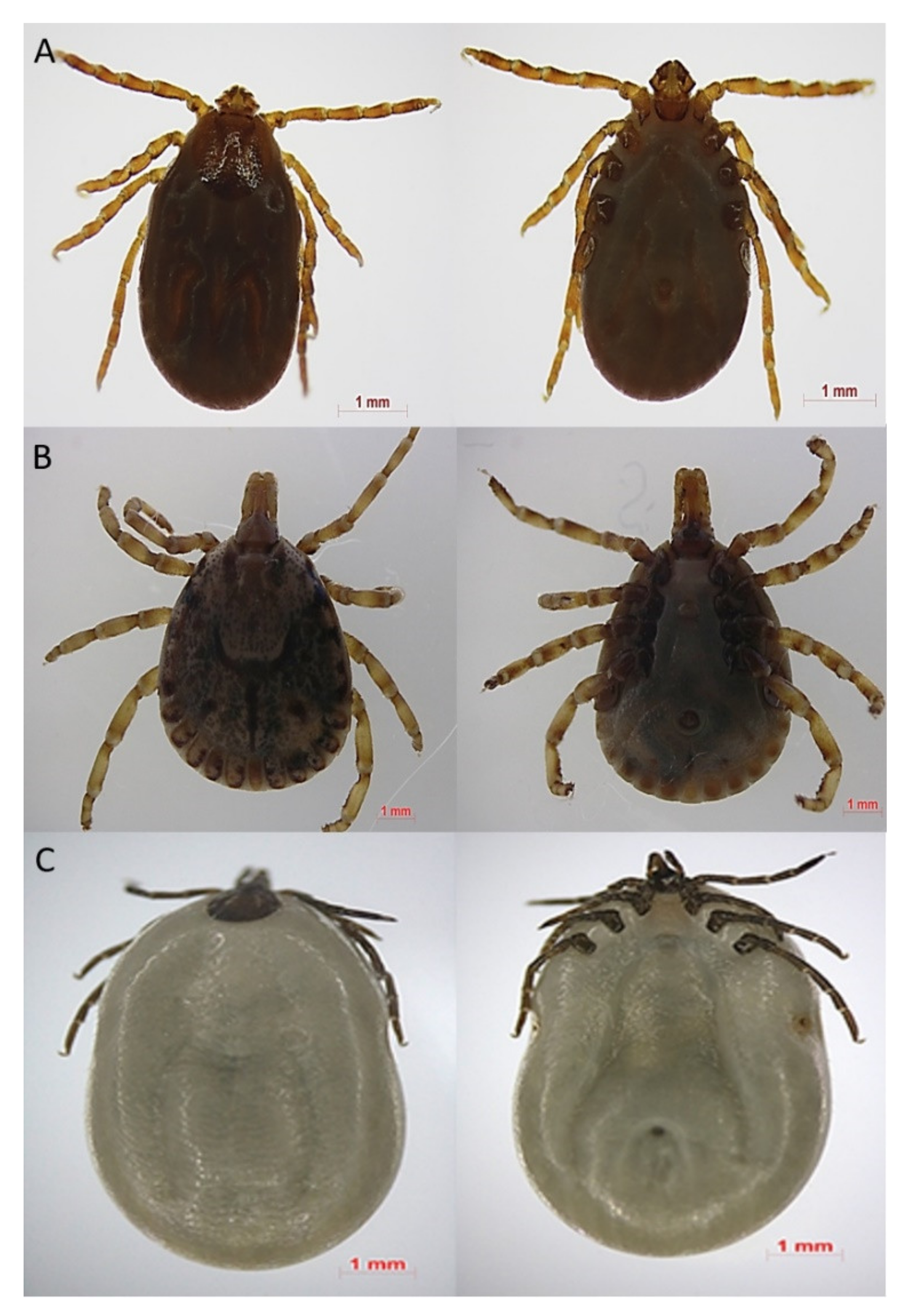
1. Introduction
2. SFTSV Epidemiology and Transmission
3. Pathogenesis
4. Clinical Manifestations
5. SFTS Diagnosis
6. SFTS Treatment
6.1. Antiviral Agents
6.1.1. Ribavirin
6.1.2. Favipiravir
6.2. Steroids
6.3. Intravenous Immunoglobulin
6.4. Plasma Exchange or Convalescent Plasma Therapy
6.5. Monoclonal Antibodies
7. Prevention
8. Conclusions
Author Contributions
Funding
Institutional Review Board Statement
Informed Consent Statement
Data Availability Statement
Conflicts of Interest
References
- Zhang, Y.Z.; Zhou, D.J.; Qin, X.C.; Tian, J.H.; Xiong, Y.; Wang, J.B.; Chen, X.P.; Gao, D.Y.; He, Y.W.; Jin, D.; et al. The ecology, genetic diversity, and phylogeny of Huaiyangshan virus in China. J. Virol. 2012, 86, 2864–2868. [Google Scholar] [CrossRef]
- Yu, X.J.; Liang, M.F.; Zhang, S.Y.; Liu, Y.; Li, J.D.; Sun, Y.L.; Zhang, L.; Zhang, Q.F.; Popov, V.L.; Li, C.; et al. Fever with thrombocytopenia associated with a novel bunyavirus in China. New Engl. J. Med. 2011, 364, 1523–1532. [Google Scholar] [CrossRef] [PubMed]
- Xu, B.; Liu, L.; Huang, X.; Ma, H.; Zhang, Y.; Du, Y.; Wang, P.; Tang, X.; Wang, H.; Kang, K.; et al. Metagenomic analysis of fever, thrombocytopenia and leukopenia syndrome (FTLS) in Henan Province, China: Discovery of a new bunyavirus. PLoS Pathog. 2011, 7, e1002369. [Google Scholar] [CrossRef] [PubMed]
- Walter, C.T.; Barr, J.N. Recent advances in the molecular and cellular biology of bunyaviruses. J. Gen. Virol. 2011, 92, 2467–2484. [Google Scholar] [CrossRef]
- Fu, Y.; Li, S.; Zhang, Z.; Man, S.; Li, X.; Zhang, W.; Zhang, C.; Cheng, X. Phylogeographic analysis of severe fever with thrombocytopenia syndrome virus from Zhoushan Islands, China: Implication for transmission across the ocean. Sci. Rep. 2016, 6, 19563. [Google Scholar] [CrossRef] [PubMed]
- Niu, G.; Li, J.; Liang, M.; Jiang, X.; Jiang, M.; Yin, H.; Wang, Z.; Li, C.; Zhang, Q.; Jin, C.; et al. Severe fever with thrombocytopenia syndrome virus among domesticated animals, China. Emerg. Infect. Dis. 2013, 19, 756–763. [Google Scholar] [CrossRef] [PubMed]
- Yoo, J.R.; Lee, K.H.; Heo, S.T. Surveillance results for family members of patients with severe fever with thrombocytopenia syndrome. Zoonoses Public Health 2018, 65, 903–907. [Google Scholar] [CrossRef]
- Yoo, J.R.; Heo, S.T.; Park, D.; Kim, H.; Fukuma, A.; Fukushi, S.; Shimojima, M.; Lee, K.H. Family cluster analysis of severe fever with thrombocytopenia syndrome virus infection in Korea. Am. J. Trop. Med. Hyg. 2016, 95, 1351–1357. [Google Scholar] [CrossRef] [PubMed]
- Kim, W.Y.; Choi, W.; Park, S.W.; Wang, E.B.; Lee, W.J.; Jee, Y.; Lim, K.S.; Lee, H.J.; Kim, S.M.; Lee, S.O.; et al. Nosocomial transmission of severe fever with thrombocytopenia syndrome in Korea. Clin. Infect. Dis. 2015, 60, 1681–1683. [Google Scholar] [CrossRef]
- Kim, K.H.; Yi, J.; Kim, G.; Choi, S.J.; Jun, K.I.; Kim, N.H.; Choe, P.G.; Kim, N.J.; Lee, J.K.; Oh, M.D. Severe fever with thrombocytopenia syndrome, South Korea, 2012. Emerg. Infect. Dis. 2013, 19, 1892–1894. [Google Scholar] [CrossRef]
- Takahashi, T.; Maeda, K.; Suzuki, T.; Ishido, A.; Shigeoka, T.; Tominaga, T.; Kamei, T.; Honda, M.; Ninomiya, D.; Sakai, T.; et al. The first identification and retrospective study of severe fever with thrombocytopenia syndrome in Japan. J. Infect. Dis. 2014, 209, 816–827. [Google Scholar] [CrossRef]
- Sun, J.; Lu, L.; Wu, H.; Yang, J.; Ren, J.; Liu, Q. The changing epidemiological characteristics of severe fever with thrombocytopenia syndrome in China, 2011–2016. Sci. Rep. 2017, 7, 9236. [Google Scholar] [CrossRef] [PubMed]
- Choi, S.J.; Park, S.W.; Bae, I.G.; Kim, S.H.; Ryu, S.Y.; Kim, H.A.; Jang, H.C.; Hur, J.; Jun, J.B.; Jung, Y.; et al. Severe fever with thrombocytopenia syndrome in South Korea, 2013–2015. PLoS Negl. Trop. Dis. 2016, 10, e0005264. [Google Scholar] [CrossRef] [PubMed]
- Mita, T. Epidemiology of Severe Fever with Thrombocytopenia Syndrome in Japan. Juntendo Med. J. 2019, 65, 130–135. [Google Scholar] [CrossRef]
- Korea Disease Control and Prevention Agency. Ticks and Rodents borne Infectious Diseases Guideline 2020. Available online: http://www.kdca.go.kr/board/board.es?mid=a20507020000&bid=0019&act=view&list_no=365644 (accessed on 23 June 2021).
- Tran, X.C.; Yun, Y.; Van An, L.; Kim, S.H.; Thao, N.T.P.; Man, P.K.C.; Yoo, J.R.; Heo, S.T.; Cho, N.H.; Lee, K.H. Endemic severe fever with thrombocytopenia syndrome, Vietnam. Emerg. Infect. Dis. 2019, 25, 1029–1031. [Google Scholar] [CrossRef]
- Peng, S.H.; Yang, S.L.; Tang, S.E.; Wang, T.C.; Hsu, T.C.; Su, C.L.; Chen, M.Y.; Shimojima, M.; Yoshikawa, T.; Shu, P.Y. Human case of severe fever with thrombocytopenia syndrome virus infection, Taiwan, 2019. Emerg. Infect. Dis. 2020, 26, 1612–1614. [Google Scholar] [CrossRef] [PubMed]
- Zohaib, A.; Zhang, J.; Saqib, M.; Athar, M.A.; Hussain, M.H.; Chen, J.; Sial, A.U.; Tayyab, M.H.; Batool, M.; Khan, S.; et al. Serologic evidence of severe fever with thrombocytopenia syndrome virus and related viruses in Pakistan. Emerg. Infect. Dis. 2020, 26, 1513–1516. [Google Scholar] [CrossRef]
- Lin, T.L.; Ou, S.C.; Maeda, K.; Shimoda, H.; Chan, J.P.; Tu, W.C.; Hsu, W.L.; Chou, C.C. The first discovery of severe fever with thrombocytopenia syndrome virus in Taiwan. Emerg. Microbes Infect. 2020, 9, 148–151. [Google Scholar] [CrossRef]
- McMullan, L.K.; Folk, S.M.; Kelly, A.J.; MacNeil, A.; Goldsmith, C.S.; Metcalfe, M.G.; Batten, B.C.; Albariño, C.G.; Zaki, S.R.; Rollin, P.E.; et al. A new phlebovirus associated with severe febrile illness in Missouri. New Engl. J. Med. 2012, 367, 834–841. [Google Scholar] [CrossRef]
- Staples, J.E.; Pastula, D.M.; Panella, A.J.; Rabe, I.B.; Kosoy, O.I.; Walker, W.L.; Velez, J.O.; Lambert, A.J.; Fischer, M. Investigation of heartland virus disease throughout the United States, 2013–2017. Open Forum Infect. Dis. 2020, 7, ofaa125. [Google Scholar] [CrossRef]
- Yun, S.M.; Park, S.J.; Park, S.W.; Choi, W.; Jeong, H.W.; Choi, Y.K.; Lee, W.J. Molecular genomic characterization of tick- and human-derived severe fever with thrombocytopenia syndrome virus isolates from South Korea. PLoS Negl. Trop. Dis. 2017, 11, e0005893. [Google Scholar] [CrossRef]
- Yun, S.M.; Park, S.J.; Kim, Y.I.; Park, S.W.; Yu, M.A.; Kwon, H.I.; Kim, E.H.; Yu, K.M.; Jeong, H.W.; Ryou, J.; et al. Genetic and pathogenic diversity of severe fever with thrombocytopenia syndrome virus (SFTSV) in South Korea. JCI Insight 2020, 5, e129531. [Google Scholar] [CrossRef]
- Li, P.; Tong, Z.D.; Li, K.F.; Tang, A.; Dai, Y.X.; Yan, J.B. Seroprevalence of severe fever with thrombocytopenia syndrome virus in China: A systematic review and meta-analysis. PLoS ONE 2017, 12, e0175592. [Google Scholar] [CrossRef]
- Kimura, T.; Fukuma, A.; Shimojima, M.; Yamashita, Y.; Mizota, F.; Yamashita, M.; Otsuka, Y.; Kan, M.; Fukushi, S.; Tani, H.; et al. Seroprevalence of severe fever with thrombocytopenia syndrome (SFTS) virus antibodies in humans and animals in Ehime prefecture, Japan, an endemic region of SFTS. J. Infect. Chemother. 2018, 24, 802–806. [Google Scholar] [CrossRef]
- Gokuden, M.; Fukushi, S.; Saijo, M.; Nakadouzono, F.; Iwamoto, Y.; Yamamoto, M.; Hozumi, N.; Nakayama, K.; Ishitani, K.; Nishi, N.; et al. Low seroprevalence of severe fever with thrombocytopenia syndrome virus antibodies in individuals living in an endemic area in Japan. Jpn. J. Infect. Dis. 2018, 71, 225–228. [Google Scholar] [CrossRef] [PubMed]
- Han, M.A.; Kim, C.M.; Kim, D.M.; Yun, N.R.; Park, S.W.; Han, M.G.; Lee, W.J. Seroprevalence of severe fever with thrombocytopenia syndrome virus antibodies in rural areas, South Korea. Emerg. Infect. Dis. 2018, 24, 872–874. [Google Scholar] [CrossRef] [PubMed]
- Li, J.; Li, S.; Yang, L.; Cao, P.; Lu, J. Severe fever with thrombocytopenia syndrome virus: A highly lethal bunyavirus. Crit. Rev. Microbiol. 2021, 47, 112–125. [Google Scholar] [CrossRef] [PubMed]
- Yoo, J.R.; Heo, S.T.; Song, S.W.; Bae, S.G.; Lee, S.; Choi, S.; Lee, C.; Jeong, S.; Kim, M.; Sa, W.; et al. Severe fever with thrombocytopenia syndrome virus in ticks and sfts incidence in humans, South Korea. Emerg. Infect. Dis. 2020, 26, 2292–2294. [Google Scholar] [CrossRef] [PubMed]
- Hoogstraal, H.; Roberts, F.H.; Kohls, G.M.; Tipton, V.J. Review of Haemaphysalis (kaiseriana) Longicornis neumann (resurrected) of Australia, New Zealand, New Caledonia, Fiji, Japan, Korea, and Northeastern China and USSR, and its parthenogenetic and bisexual populations (Ixodoidea, Ixodidae). J. Parasitol. 1968, 54, 1197–1213. [Google Scholar] [CrossRef]
- Kim, U.J.; Kim, D.M.; Kim, S.E.; Kang, S.J.; Jang, H.C.; Park, K.H.; Jung, S.I. Case report: Detection of the identical virus in a patient presenting with severe fever with thrombocytopenia syndrome encephalopathy and the tick that bit her. BMC Infect. Dis. 2018, 18, 181. [Google Scholar] [CrossRef] [PubMed]
- Zhuang, L.; Sun, Y.; Cui, X.M.; Tang, F.; Hu, J.G.; Wang, L.Y.; Cui, N.; Yang, Z.D.; Huang, D.D.; Zhang, X.A.; et al. Transmission of severe fever with thrombocytopenia syndrome virus by haemaphysalis longicornis ticks, China. Emerg. Infect. Dis. 2018, 24, 868–871. [Google Scholar] [CrossRef]
- Ding, S.; Yin, H.; Xu, X.; Liu, G.; Jiang, S.; Wang, W.; Han, X.; Liu, J.; Niu, G.; Zhang, X.; et al. A cross-sectional survey of severe fever with thrombocytopenia syndrome virus infection of domestic animals in Laizhou City, Shandong Province, China. Jpn. J. Infect. Dis. 2014, 67, 1–4. [Google Scholar] [CrossRef]
- Liu, L.; Guan, X.H.; Xing, X.S.; Shen, X.F.; Xu, J.Q.; Yue, J.L.; Huo, X.X.; Sha, S.; Wu, H.X.; Huang, J.; et al. Epidemiologic analysis on severe fever with thrombocytopenia syndrome in Hubei province, 2010. Zhonghua Liu Xing Bing Xue Za Zhi 2012, 33, 168–172. [Google Scholar] [PubMed]
- Wang, T.; Li, X.L.; Liu, M.; Song, X.J.; Zhang, H.; Wang, Y.B.; Tian, B.P.; Xing, X.S.; Li, S.Y. Epidemiological characteristics and environmental risk factors of severe fever with thrombocytopenia syndrome in Hubei Province, China, from 2011 to 2016. Front. Microbiol. 2017, 8, 387. [Google Scholar] [CrossRef] [PubMed]
- Chen, C.; Li, P.; Li, K.F.; Wang, H.L.; Dai, Y.X.; Cheng, X.; Yan, J.B. Animals as amplification hosts in the spread of severe fever with thrombocytopenia syndrome virus: A systematic review and meta-analysis. Int. J. Infect. Dis. 2019, 79, 77–84. [Google Scholar] [CrossRef]
- Kang, J.G.; Cho, Y.K.; Jo, Y.S.; Chae, J.B.; Joo, Y.H.; Park, K.W.; Chae, J.S. Severe fever with thrombocytopenia syndrome virus in dogs, South Korea. Emerg. Infect. Dis. 2019, 25, 376–378. [Google Scholar] [CrossRef] [PubMed]
- Kida, K.; Matsuoka, Y.; Shimoda, T.; Matsuoka, H.; Yamada, H.; Saito, T.; Imataki, O.; Kadowaki, N.; Noguchi, K.; Maeda, K.; et al. A case of cat-to-human transmission of severe fever with thrombocytopenia syndrome virus. Jpn. J. Infect. Dis. 2019, 72, 356–358. [Google Scholar] [CrossRef] [PubMed]
- Chung, J.K.; Kim, C.M.; Kim, D.M.; Yun, N.R.; Park, J.W.; Seo, J.; Kim, Y.S. Severe fever with thrombocytopenia syndrome associated with manual de-ticking of domestic dogs. Vector Borne Zoonotic Dis. 2020, 20, 285–294. [Google Scholar] [CrossRef]
- Kobayashi, Y.; Kato, H.; Yamagishi, T.; Shimada, T.; Matsui, T.; Yoshikawa, T.; Kurosu, T.; Shimojima, M.; Morikawa, S.; Hasegawa, H.; et al. Severe fever with thrombocytopenia syndrome, Japan, 2013–2017. Emerg. Infect. Dis. 2020, 26, 692–699. [Google Scholar] [CrossRef]
- Yu, K.M.; Jeong, H.W.; Park, S.J.; Kim, Y.I.; Yu, M.A.; Kwon, H.I.; Kim, E.H.; Kim, S.M.; Lee, S.H.; Kim, S.G.; et al. Shedding and transmission modes of severe fever with thrombocytopenia syndrome phlebovirus in a ferret model. Open Forum Infect. Dis. 2019, 6. [Google Scholar] [CrossRef]
- Chen, Y.; Jia, B.; Huang, R.; Yan, X.; Xiong, Y.; Yong, L.; Chao, W. Occupational severe fever with thrombocytopenia syndrome following needle-stick injury. Infect. Control. Hosp. Epidemiol. 2017, 38, 760–762. [Google Scholar] [CrossRef]
- Zhu, Y.; Wu, H.; Gao, J.; Zhou, X.; Zhu, R.; Zhang, C.; Bai, H.; Abdullah, A.S.; Pan, H. Two confirmed cases of severe fever with thrombocytopenia syndrome with pneumonia: Implication for a family cluster in East China. BMC Infect. Dis. 2017, 17, 537. [Google Scholar] [CrossRef] [PubMed]
- Koga, S.; Takazono, T.; Ando, T.; Hayasaka, D.; Tashiro, M.; Saijo, T.; Kurihara, S.; Sekino, M.; Yamamoto, K.; Imamura, Y.; et al. Severe fever with thrombocytopenia syndrome virus RNA in Semen, Japan. Emerg. Infect. Dis. 2019, 25, 2127–2128. [Google Scholar] [CrossRef] [PubMed]
- Moon, J.; Lee, H.; Jeon, J.H.; Kwon, Y.; Kim, H.; Wang, E.B.; Seo, C.W.; Sung, S.A.; Kim, S.H.; Seok, H.; et al. Aerosol transmission of severe fever with thrombocytopenia syndrome virus during resuscitation. Infect. Control. Hosp. Epidemiol. 2018, 40, 238–241. [Google Scholar] [CrossRef]
- Sun, Y.; Jin, C.; Zhan, F.; Wang, X.; Liang, M.; Zhang, Q.; Ding, S.; Guan, X.; Huo, X.; Li, C.; et al. Host cytokine storm is associated with disease severity of severe fever with thrombocytopenia syndrome. J. Infect. Dis. 2012, 206, 1085–1094. [Google Scholar] [CrossRef] [PubMed]
- Hu, L.F.; Wu, T.; Wang, B.; Wei, Y.Y.; Kong, Q.X.; Ye, Y.; Yin, H.F.; Li, J.B. The regulation of seventeen inflammatory mediators are associated with patient outcomes in severe fever with thrombocytopenia syndrome. Sci. Rep. 2018, 8, 159. [Google Scholar] [CrossRef]
- Lu, Q.B.; Zhang, S.Y.; Cui, N.; Hu, J.G.; Fan, Y.D.; Guo, C.T.; Qin, S.L.; Yang, Z.D.; Wang, L.Y.; Wang, H.Y.; et al. Common adverse events associated with ribavirin therapy for severe fever with thrombocytopenia syndrome. Antivir. Res. 2015, 119, 19–22. [Google Scholar] [CrossRef]
- Deng, B.; Zhang, S.; Geng, Y.; Zhang, Y.; Wang, Y.; Yao, W.; Wen, Y.; Cui, W.; Zhou, Y.; Gu, Q.; et al. Cytokine and chemokine levels in patients with severe fever with thrombocytopenia syndrome virus. PLoS ONE 2012, 7, e41365. [Google Scholar] [CrossRef]
- Maghazachi, A.A. Role of chemokines in the biology of natural killer cells. Curr. Top. Microbiol. Immunol. 2010, 341, 37–58. [Google Scholar]
- Sun, L.; Hu, Y.; Niyonsaba, A.; Tong, Q.; Lu, L.; Li, H.; Jie, S. Detection and evaluation of immunofunction of patients with severe fever with thrombocytopenia syndrome. Clin. Exp. Med. 2014, 14, 389–395. [Google Scholar] [CrossRef]
- Marty, A.M.; Jahrling, P.B.; Geisbert, T.W. Viral hemorrhagic fevers. Clin. Lab. Med. 2006, 26, 345–386. [Google Scholar] [CrossRef]
- Qu, B.; Qi, X.; Wu, X.; Liang, M.; Li, C.; Cardona, C.J.; Xu, W.; Tang, F.; Li, Z.; Wu, B.; et al. Suppression of the interferon and NF-κB responses by severe fever with thrombocytopenia syndrome virus. J. Virol. 2012, 86, 8388–8401. [Google Scholar] [CrossRef]
- Choi, Y.; Park, S.J.; Sun, Y.; Yoo, J.S.; Pudupakam, R.S.; Foo, S.S.; Shin, W.J.; Chen, S.B.; Tsichlis, P.N.; Lee, W.J.; et al. Severe fever with thrombocytopenia syndrome phlebovirus non-structural protein activates TPL2 signalling pathway for viral immunopathogenesis. Nat. Microbiol. 2019, 4, 429–437. [Google Scholar] [CrossRef] [PubMed]
- Zoja, C.; Garcia, P.B.; Remuzzi, G. The role of chemokines in progressive renal disease. Front. BioSci. 2009, 14, 1815–1822. [Google Scholar] [CrossRef] [PubMed]
- Wasmuth, H.E.; Tacke, F.; Trautwein, C. Chemokines in liver inflammation and fibrosis. Semin. Liver Dis. 2010, 30, 215–225. [Google Scholar] [CrossRef] [PubMed]
- Petreaca, M.L.; Yao, M.; Liu, Y.; Defea, K.; Martins-Green, M. Transactivation of vascular endothelial growth factor receptor-2 by interleukin-8 (IL-8/CXCL8) is required for IL-8/CXCL8-induced endothelial permeability. Mol. Biol. Cell 2007, 18, 5014–5023. [Google Scholar] [CrossRef] [PubMed]
- Seynhaeve, A.L.; Vermeulen, C.E.; Eggermont, A.M.; ten Hagen, T.L. Cytokines and vascular permeability: An in vitro study on human endothelial cells in relation to tumor necrosis factor-alpha-primed peripheral blood mononuclear cells. Cell Biochem. Biophys. 2006, 44, 157–169. [Google Scholar] [CrossRef]
- Jin, C.; Liang, M.; Ning, J.; Gu, W.; Jiang, H.; Wu, W.; Zhang, F.; Li, C.; Zhang, Q.; Zhu, H.; et al. Pathogenesis of emerging severe fever with thrombocytopenia syndrome virus in C57/BL6 mouse model. Proc. Natl. Acad. Sci. USA 2012, 109, 10053–10058. [Google Scholar] [CrossRef]
- Liu, Y.; Wu, B.; Paessler, S.; Walker, D.H.; Tesh, R.B.; Yu, X.J. The pathogenesis of severe fever with thrombocytopenia syndrome virus infection in alpha/beta interferon knockout mice: Insights into the pathologic mechanisms of a new viral hemorrhagic fever. J. Virol. 2014, 88, 1781–1786. [Google Scholar] [CrossRef]
- Suzuki, T.; Sato, Y.; Sano, K.; Arashiro, T.; Katano, H.; Nakajima, N.; Shimojima, M.; Kataoka, M.; Takahashi, K.; Wada, Y.; et al. Severe fever with thrombocytopenia syndrome virus targets B cells in lethal human infections. J. Clin. Invest. 2020, 130, 799–812. [Google Scholar] [CrossRef]
- Kang, C.K.; Choi, S.J.; Koh, J.; Jeon, Y.K.; Kim, K.H.; Chung, J.; Choe, P.G.; Kim, N.J.; Park, W.B.; Oh, M.D. (18)F-FDG PET and histopathologic findings in a patient with severe fever with thrombocytopenia syndrome. Ticks Tick Borne Dis. 2018, 9, 972–975. [Google Scholar] [CrossRef]
- Matsuno, K.; Orba, Y.; Maede-White, K.; Scott, D.; Feldmann, F.; Liang, M.; Ebihara, H. Animal models of emerging Tick-Borne Phleboviruses: Determining target cells in a lethal model of SFTSV infection. Front. Microbiol. 2017, 8, 104. [Google Scholar] [CrossRef]
- Liu, Q.; He, B.; Huang, S.Y.; Wei, F.; Zhu, X.Q. Severe fever with thrombocytopenia syndrome, an emerging tick-borne zoonosis. Lancet Infect. Dis. 2014, 14, 763–772. [Google Scholar] [CrossRef]
- Kim, M.C.; Chong, Y.P.; Lee, S.O.; Choi, S.H.; Kim, Y.S.; Woo, J.H.; Kim, S.H. Differentiation of severe fever with thrombocytopenia syndrome from scrub typhus. Clin. Infect. Dis. 2018, 66, 1621–1624. [Google Scholar] [CrossRef]
- Kim, U.J.; Oh, T.H.; Kim, B.; Kim, S.E.; Kang, S.J.; Park, K.H.; Jung, S.I.; Jang, H.C. Hyperferritinemia as a diagnostic marker for severe fever with thrombocytopenia syndrome. Dis. Markers 2017, 2017, 6727184. [Google Scholar] [CrossRef]
- Yun, J.H.; Hwang, H.J.; Jung, J.; Kim, M.J.; Chong, Y.P.; Lee, S.O.; Choi, S.H.; Kim, Y.S.; Woo, J.H.; Kim, M.Y.; et al. Comparison of chest radiographic findings between severe fever with thrombocytopenia syndrome and scrub typhus: Single center observational cross-sectional study in South Korea. Medicine 2019, 98, e17701. [Google Scholar] [CrossRef]
- Miyamoto, S.; Ito, T.; Terada, S.; Eguchi, T.; Furubeppu, H.; Kawamura, H.; Yasuda, T.; Kakihana, Y. Fulminant myocarditis associated with severe fever with thrombocytopenia syndrome: A case report. BMC Infect. Dis. 2019, 19, 266. [Google Scholar] [CrossRef] [PubMed]
- Park, S.Y.; Kwon, J.S.; Kim, J.Y.; Kim, S.M.; Jang, Y.R.; Kim, M.C.; Cho, O.H.; Kim, T.; Chong, Y.P.; Lee, S.O.; et al. Severe fever with thrombocytopenia syndrome-associated encephalopathy/encephalitis. Clin. Microbiol. Infect. 2018, 24, 432.e1–432.e4. [Google Scholar] [CrossRef] [PubMed]
- Ding, F.; Zhang, W.; Wang, L.; Hu, W.; Soares Magalhaes, R.J.; Sun, H.; Zhou, H.; Sha, S.; Li, S.; Liu, Q.; et al. Epidemiologic features of severe fever with thrombocytopenia syndrome in China, 2011–2012. Clin. Infect. Dis. 2013, 56, 1682–1683. [Google Scholar] [CrossRef] [PubMed]
- Kato, H.; Yamagishi, T.; Shimada, T.; Matsui, T.; Shimojima, M.; Saijo, M.; Oishi, K. Epidemiological and clinical features of severe fever with thrombocytopenia syndrome in Japan, 2013–2014. PLoS ONE 2016, 11, e0165207. [Google Scholar] [CrossRef] [PubMed]
- Li, H.; Lu, Q.B.; Xing, B.; Zhang, S.F.; Liu, K.; Du, J.; Li, X.K.; Cui, N.; Yang, Z.D.; Wang, L.Y.; et al. Epidemiological and clinical features of laboratory-diagnosed severe fever with thrombocytopenia syndrome in China, 2011–2017: A prospective observational study. Lancet Infect. Dis. 2018, 18, 1127–1137. [Google Scholar] [CrossRef]
- Wang, L.; Wan, G.; Shen, Y.; Zhao, Z.; Lin, L.; Zhang, W.; Song, R.; Tian, D.; Wen, J.; Zhao, Y.; et al. A nomogram to predict mortality in patients with severe fever with thrombocytopenia syndrome at the early stage-A multicenter study in China. PLoS Negl. Trop. Dis. 2019, 13, e0007829. [Google Scholar] [CrossRef] [PubMed]
- Zhang, Y.Z.; He, Y.W.; Dai, Y.A.; Xiong, Y.; Zheng, H.; Zhou, D.J.; Li, J.; Sun, Q.; Luo, X.L.; Cheng, Y.L.; et al. Hemorrhagic fever caused by a novel Bunyavirus in China: Pathogenesis and correlates of fatal outcome. Clin. Infect. Dis. 2012, 54, 527–533. [Google Scholar] [CrossRef] [PubMed]
- Hwang, J.; Kang, J.G.; Oh, S.S.; Chae, J.B.; Cho, Y.K.; Cho, Y.S.; Lee, H.; Chae, J.S. Molecular detection of severe fever with thrombocytopenia syndrome virus (SFTSV) in feral cats from Seoul, Korea. Ticks Tick Borne Dis. 2017, 8, 9–12. [Google Scholar] [CrossRef]
- Yoshikawa, T.; Fukushi, S.; Tani, H.; Fukuma, A.; Taniguchi, S.; Toda, S.; Shimazu, Y.; Yano, K.; Morimitsu, T.; Ando, K.; et al. Sensitive and specific PCR systems for detection of both Chinese and Japanese severe fever with thrombocytopenia syndrome virus strains and prediction of patient survival based on viral load. J. Clin. Microbiol. 2014, 52, 3325–3333. [Google Scholar] [CrossRef]
- Yang, Z.D.; Hu, J.G.; Lu, Q.B.; Guo, C.T.; Cui, N.; Peng, W.; Wang, L.Y.; Qin, S.L.; Wang, H.Y.; Zhang, P.H.; et al. The prospective evaluation of viral loads in patients with severe fever with thrombocytopenia syndrome. J. Clin. Virol. 2016, 78, 123–128. [Google Scholar] [CrossRef] [PubMed]
- Park, S.J.; Kim, Y.I.; Park, A.; Kwon, H.I.; Kim, E.H.; Si, Y.J.; Song, M.S.; Lee, C.H.; Jung, K.; Shin, W.J.; et al. Ferret animal model of severe fever with thrombocytopenia syndrome phlebovirus for human lethal infection and pathogenesis. Nat. Microbiol. 2019, 4, 438–446. [Google Scholar] [CrossRef]
- Jung, S.I.; Kim, Y.E.; Yun, N.R.; Kim, C.M.; Kim, D.M.; Han, M.A.; Kim, U.J.; Kim, S.E.; Kim, J.; Ryu, S.Y.; et al. Effects of steroid therapy in patients with severe fever with Thrombocytopenia syndrome: A multicenter clinical cohort study. PLoS Negl. Trop. Dis. 2021, 15, e0009128. [Google Scholar] [CrossRef]
- Lee, H.; Choi, W.Y.; Kim, C.M.; Yun, N.R.; Kim, D.M.; Pyun, S.H.; Yu, B.J.; Lee, Y.M. A case of SFTS coinfected with E. coli bacteremia. BMC Infect. Dis. 2021, 21, 25. [Google Scholar] [CrossRef]
- Bae, S.; Hwang, H.J.; Kim, M.Y.; Kim, M.J.; Chong, Y.P.; Lee, S.O.; Choi, S.H.; Kim, Y.S.; Woo, J.H.; Kim, S.H. Invasive pulmonary aspergillosis in patients with severe fever with thrombocytopenia syndrome. Clin. Infect. Dis. 2020, 70, 1491–1494. [Google Scholar] [CrossRef]
- Sun, Y.; Liang, M.; Qu, J.; Jin, C.; Zhang, Q.; Li, J.; Jiang, X.; Wang, Q.; Lu, J.; Gu, W.; et al. Early diagnosis of novel SFTS bunyavirus infection by quantitative real-time RT-PCR assay. J. Clin. Virol. 2012, 53, 48–53. [Google Scholar] [CrossRef]
- Huang, X.Y.; Hu, X.N.; Ma, H.; Du, Y.H.; Ma, H.X.; Kang, K.; You, A.G.; Wang, H.F.; Zhang, L.; Chen, H.M.; et al. Detection of new bunyavirus RNA by reverse transcription-loop-mediated isothermal amplification. J. Clin. Microbiol. 2014, 52, 531–535. [Google Scholar] [CrossRef]
- Baek, Y.H.; Cheon, H.S.; Park, S.J.; Lloren, K.K.S.; Ahn, S.J.; Jeong, J.H.; Choi, W.S.; Yu, M.A.; Kwon, H.I.; Kwon, J.J.; et al. Simple, rapid and sensitive portable molecular diagnosis of SFTS virus using reverse transcriptional loop-mediated isothermal amplification (RT-LAMP). J. Microbiol. Biotechnol. 2018, 28, 1928–1936. [Google Scholar] [CrossRef] [PubMed]
- Ra, S.H.; Kim, M.J.; Kim, M.C.; Park, S.Y.; Park, S.Y.; Chong, Y.P.; Lee, S.O.; Choi, S.H.; Kim, Y.S.; Lee, K.H.; et al. Kinetics of serological response in patients with severe fever with thrombocytopenia syndrome. Viruses 2020, 13, 6. [Google Scholar] [CrossRef] [PubMed]
- Li, Z.; Qi, X.; Zhou, M.; Bao, C.; Hu, J.; Wu, B.; Wang, S.; Tan, Z.; Fu, J.; Shan, J.; et al. A two-tube multiplex real-time RT-PCR assay for the detection of four hemorrhagic fever viruses: Severe fever with thrombocytopenia syndrome virus, Hantaan virus, Seoul virus, and dengue virus. Arch. Virol. 2013, 158, 1857–1863. [Google Scholar] [CrossRef] [PubMed]
- Ergönül, O.; Celikbaş, A.; Dokuzoguz, B.; Eren, S.; Baykam, N.; Esener, H. Characteristics of patients with Crimean-Congo hemorrhagic fever in a recent outbreak in Turkey and impact of oral ribavirin therapy. Clin. Infect. Dis. 2004, 39, 284–287. [Google Scholar] [CrossRef] [PubMed]
- Huggins, J.W.; Hsiang, C.M.; Cosgriff, T.M.; Guang, M.Y.; Smith, J.I.; Wu, Z.O.; LeDuc, J.W.; Zheng, Z.M.; Meegan, J.M.; Wang, Q.N.; et al. Prospective, double-blind, concurrent, placebo-controlled clinical trial of intravenous ribavirin therapy of hemorrhagic fever with renal syndrome. J. Infect. Dis. 1991, 164, 1119–1127. [Google Scholar] [CrossRef]
- Lee, M.J.; Kim, K.H.; Yi, J.; Choi, S.J.; Choe, P.G.; Park, W.B.; Kim, N.J.; Oh, M.D. In vitro antiviral activity of ribavirin against severe fever with thrombocytopenia syndrome virus. Korean J. Intern. Med. 2017, 32, 731–737. [Google Scholar] [CrossRef] [PubMed]
- Liu, W.; Lu, Q.B.; Cui, N.; Li, H.; Wang, L.Y.; Liu, K.; Yang, Z.D.; Wang, B.J.; Wang, H.Y.; Zhang, Y.Y.; et al. Case-fatality ratio and effectiveness of ribavirin therapy among hospitalized patients in china who had severe fever with thrombocytopenia syndrome. Clin. Infect. Dis. 2013, 57, 1292–1299. [Google Scholar] [CrossRef]
- Cui, N.; Bao, X.L.; Yang, Z.D.; Lu, Q.B.; Hu, C.Y.; Wang, L.Y.; Wang, B.J.; Wang, H.Y.; Liu, K.; Yuan, C.; et al. Clinical progression and predictors of death in patients with severe fever with thrombocytopenia syndrome in China. J. Clin. Virol. 2014, 59, 12–17. [Google Scholar] [CrossRef] [PubMed]
- Shimojima, M.; Fukushi, S.; Tani, H.; Yoshikawa, T.; Fukuma, A.; Taniguchi, S.; Suda, Y.; Maeda, K.; Takahashi, T.; Morikawa, S.; et al. Effects of ribavirin on severe fever with thrombocytopenia syndrome virus in vitro. Jpn. J. Infect. Dis. 2014, 67, 423–427. [Google Scholar] [CrossRef]
- Oh, W.S.; Heo, S.T.; Kim, S.H.; Choi, W.J.; Han, M.G.; Kim, J.Y. Plasma exchange and ribavirin for rapidly progressive severe fever with thrombocytopenia syndrome. Int. J. Infect. Dis. 2014, 18, 84–86. [Google Scholar] [CrossRef] [PubMed]
- Park, I.; Kim, H.I.; Kwon, K.T. Two treatment cases of severe fever and thrombocytopenia syndrome with oral ribavirin and plasma exchange. Infect. Chemother. 2017, 49, 72–77. [Google Scholar] [CrossRef] [PubMed]
- Shimojima, M.; Fukushi, S.; Tani, H.; Taniguchi, S.; Fukuma, A.; Saijo, M. Combination effects of ribavirin and interferons on severe fever with thrombocytopenia syndrome virus infection. Virol. J. 2015, 12, 181. [Google Scholar] [CrossRef] [PubMed]
- Furuta, Y.; Takahashi, K.; Shiraki, K.; Sakamoto, K.; Smee, D.F.; Barnard, D.L.; Gowen, B.B.; Julander, J.G.; Morrey, J.D. T-705 (favipiravir) and related compounds: Novel broad-spectrum inhibitors of RNA viral infections. Antivir. Res. 2009, 82, 95–102. [Google Scholar] [CrossRef] [PubMed]
- Takayama-Ito, M.; Saijo, M. Antiviral drugs against severe fever with thrombocytopenia syndrome virus infection. Front. Microbiol. 2020, 11, 150. [Google Scholar] [CrossRef]
- Tani, H.; Fukuma, A.; Fukushi, S.; Taniguchi, S.; Yoshikawa, T.; Iwata-Yoshikawa, N.; Sato, Y.; Suzuki, T.; Nagata, N.; Hasegawa, H.; et al. Efficacy of T-705 (Favipiravir) in the treatment of infections with lethal severe fever with thrombocytopenia syndrome virus. mSphere 2016, 1, e00061-15. [Google Scholar] [CrossRef] [PubMed]
- Baba, M.; Toyama, M.; Sakakibara, N.; Okamoto, M.; Arima, N.; Saijo, M. Establishment of an antiviral assay system and identification of severe fever with thrombocytopenia syndrome virus inhibitors. Antivir. Chem. Chemother. 2017, 25, 83–89. [Google Scholar] [CrossRef][Green Version]
- Tani, H.; Komeno, T.; Fukuma, A.; Fukushi, S.; Taniguchi, S.; Shimojima, M.; Uda, A.; Morikawa, S.; Nakajima, N.; Furuta, Y.; et al. Therapeutic effects of favipiravir against severe fever with thrombocytopenia syndrome virus infection in a lethal mouse model: Dose-efficacy studies upon oral administration. PLoS ONE 2018, 13, e0206416. [Google Scholar] [CrossRef]
- Gowen, B.B.; Westover, J.B.; Miao, J.; van Wettere, A.J.; Rigas, J.D.; Hickerson, B.T.; Jung, K.H.; Li, R.; Conrad, B.L.; Nielson, S.; et al. Modeling severe fever with thrombocytopenia syndrome virus infection in golden Syrian hamsters: Importance of STAT2 in preventing disease and effective treatment with favipiravir. J. Virol. 2017, 91, e01942-16. [Google Scholar] [CrossRef]
- Suemori, K.; Saijo, M.; Yamanaka, A.; Himeji, D.; Kawamura, M.; Haku, T.; Hidaka, M.; Kamikokuryo, C.; Kakihana, Y.; Azuma, T.; et al. A multicenter non-randomized, uncontrolled single arm trial for evaluation of the efficacy and the safety of the treatment with favipiravir for patients with severe fever with thrombocytopenia syndrome. PLoS Negl. Trop. Dis. 2021, 15, e0009103. [Google Scholar] [CrossRef]
- Kim, U.J.; Kim, D.M.; Ahn, J.H.; Kang, S.J.; Jang, H.C.; Park, K.H.; Jung, S.I. Successful treatment of rapidly progressing severe fever with thrombocytopenia syndrome with neurological complications using intravenous immunoglobulin and corticosteroid. Antivir. Ther. 2016, 21, 637–640. [Google Scholar] [CrossRef]
- Nakamura, S.; Azuma, M.; Maruhashi, T.; Sogabe, K.; Sumitani, R.; Uemura, M.; Iwasa, M.; Fujii, S.; Miki, H.; Kagawa, K.; et al. Steroid pulse therapy in patients with encephalopathy associated with severe fever with thrombocytopenia syndrome. J. Infect. Chemother. 2018, 24, 389–392. [Google Scholar] [CrossRef]
- Hiraki, T.; Yoshimitsu, M.; Suzuki, T.; Goto, Y.; Higashi, M.; Yokoyama, S.; Tabuchi, T.; Futatsuki, T.; Nakamura, K.; Hasegawa, H.; et al. Two autopsy cases of severe fever with thrombocytopenia syndrome (SFTS) in Japan: A pathognomonic histological feature and unique complication of SFTS. Pathol. Int. 2014, 64, 569–575. [Google Scholar] [CrossRef]
- Chen, X.; Yu, Z.; Qian, Y.; Dong, D.; Hao, Y.; Liu, N.; Gu, Q. Clinical features of fatal severe fever with thrombocytopenia syndrome that is complicated by invasive pulmonary aspergillosis. J. Infect. Chemother. 2018, 24, 422–427. [Google Scholar] [CrossRef]
- Sakaguchi, K.; Koga, Y.; Yagi, T.; Nakahara, T.; Todani, M.; Fujita, M.; Tsuruta, R. Severe fever with thrombocytopenia syndrome complicated with pseudomembranous aspergillus tracheobronchitis in a patient without apparent risk factors for invasive aspergillosis. Intern. Med. 2019, 58, 3589–3592. [Google Scholar] [CrossRef]
- Denic, S.; Janbeih, J.; Nair, S.; Conca, W.; Tariq, W.U.; Al-Salam, S. Acute thrombocytopenia, leucopenia, and multiorgan dysfunction: The first case of SFTS bunyavirus outside China? Case Rep. Infect. Dis. 2011, 2011, 204056. [Google Scholar] [CrossRef] [PubMed]
- Ding, Y.P.; Liang, M.F.; Ye, J.B.; Liu, Q.H.; Xiong, C.H.; Long, B.; Lin, W.B.; Cui, N.; Zou, Z.Q.; Song, Y.L.; et al. Prognostic value of clinical and immunological markers in acute phase of SFTS virus infection. Clin. Microbiol. Infect. 2014, 20, O870–O878. [Google Scholar] [CrossRef] [PubMed]
- Yoo, J.R.; Kim, S.H.; Kim, Y.R.; Lee, K.H.; Oh, W.S.; Heo, S.T. Application of therapeutic plasma exchange in patients having severe fever with thrombocytopenia syndrome. Korean J. Intern. Med. 2019, 34, 902–909. [Google Scholar] [CrossRef] [PubMed]
- Oh, W.S.; Yoo, J.R.; Kwon, K.T.; Kim, H.I.; Lee, S.J.; Jun, J.B.; Ryu, S.Y.; Kim, H.A.; Hur, J.; Wi, Y.M.; et al. Effect of early plasma exchange on survival in patients with severe fever with thrombocytopenia syndrome: A multicenter study. Yonsei Med. J. 2017, 58, 867–871. [Google Scholar] [CrossRef]
- Choi, S.; Kim, M.C.; Kwon, J.S.; Kim, J.Y.; Lee, K.H.; Kim, S.H. Case report: Use of plasma exchange followed by convalescent plasma therapy in a critically ill patient with severe fever and thrombocytopenia syndrome-associated encephalopathy: Cytokine/chemokine concentrations, viral loads, and antibody responses. Am. J. Trop. Med. Hyg. 2018, 99, 1466–1468. [Google Scholar] [CrossRef] [PubMed]
- Guo, X.; Zhang, L.; Zhang, W.; Chi, Y.; Zeng, X.; Li, X.; Qi, X.; Jin, Q.; Zhang, X.; Huang, M.; et al. Human antibody neutralizes severe Fever with thrombocytopenia syndrome virus, an emerging hemorrhagic Fever virus. Clin. Vaccine Immunol. 2013, 20, 1426–1432. [Google Scholar] [CrossRef] [PubMed]
- Kim, K.H.; Kim, J.; Ko, M.; Chun, J.Y.; Kim, H.; Kim, S.; Min, J.Y.; Park, W.B.; Oh, M.D.; Chung, J. An anti-Gn glycoprotein antibody from a convalescent patient potently inhibits the infection of severe fever with thrombocytopenia syndrome virus. PLoS Pathog. 2019, 15, e1007375. [Google Scholar] [CrossRef] [PubMed]
- Reece, L.M.; Beasley, D.W.; Milligan, G.N.; Sarathy, V.V.; Barrett, A.D. Current status of severe fever with thrombocytopenia syndrome vaccine development. Curr. Opin. Virol. 2018, 29, 72–78. [Google Scholar] [CrossRef]
- Robles, N.J.C.; Han, H.J.; Park, S.J.; Choi, Y.K. Epidemiology of severe fever and thrombocytopenia syndrome virus infection and the need for therapeutics for the prevention. Clin. Exp. Vaccine Res. 2018, 7, 43–50. [Google Scholar] [CrossRef] [PubMed]
- Kwak, J.E.; Kim, Y.I.; Park, S.J.; Yu, M.A.; Kwon, H.I.; Eo, S.; Kim, T.S.; Seok, J.; Choi, W.S.; Jeong, J.H.; et al. Development of a SFTSV DNA vaccine that confers complete protection against lethal infection in ferrets. Nat. Commun. 2019, 10, 3836. [Google Scholar] [CrossRef] [PubMed]
- Yu, K.M.; Park, S.J.; Yu, M.A.; Kim, Y.I.; Choi, Y.; Jung, J.U.; Brennan, B.; Choi, Y.K. Cross-genotype protection of live-attenuated vaccine candidate for severe fever with thrombocytopenia syndrome virus in a ferret model. Proc. Natl. Acad. Sci. USA 2019, 116, 26900–26908. [Google Scholar] [CrossRef]
- Park, E.S.; Shimojima, M.; Nagata, N.; Ami, Y.; Yoshikawa, T.; Iwata-Yoshikawa, N.; Fukushi, S.; Watanabe, S.; Kurosu, T.; Kataoka, M.; et al. Severe fever with thrombocytopenia syndrome phlebovirus causes lethal viral hemorrhagic fever in cats. Sci. Rep. 2019, 9, 11990. [Google Scholar] [CrossRef]
- Ryu, B.H.; Kim, J.Y.; Kim, T.; Kim, M.C.; Kim, M.J.; Chong, Y.P.; Lee, S.O.; Choi, S.H.; Kim, Y.S.; Woo, J.H.; et al. Extensive severe fever with thrombocytopenia syndrome virus contamination in surrounding environment in patient rooms. Clin. Microbiol. Infect. 2018, 24, 911.e1–911.e4. [Google Scholar] [CrossRef]


| Variables | Univariate | Multivariate | ||||
|---|---|---|---|---|---|---|
| HR | 95% CI | p-Value | aHR† | 95% CI | p-Value | |
| Prior antibiotic treatment | 1.55 | (0.76–3.16) | 0.234 | 1.90 | (0.75–4.81) | 0.174 |
| Ribavirin | 1.61 | (0.75–3.45) | 0.217 | 1.06 | (0.34–3.25) | 0.923 |
| Steroids | 4.57 | (1.96–10.66) | <0.001 | 3.31 | (1·26–8.73) | 0.016 |
| IVIG | 1.61 | (0.74–3.51) | 0.235 | 0.74 | (0.32–1.72) | 0.482 |
| Plasmapheresis | 2.19 | (1.03–4.68) | 0.043 | 1.40 | (0.41–4.78) | 0.593 |
Publisher’s Note: MDPI stays neutral with regard to jurisdictional claims in published maps and institutional affiliations. |
© 2021 by the authors. Licensee MDPI, Basel, Switzerland. This article is an open access article distributed under the terms and conditions of the Creative Commons Attribution (CC BY) license (https://creativecommons.org/licenses/by/4.0/).
Share and Cite
Seo, J.-W.; Kim, D.; Yun, N.; Kim, D.-M. Clinical Update of Severe Fever with Thrombocytopenia Syndrome. Viruses 2021, 13, 1213. https://doi.org/10.3390/v13071213
Seo J-W, Kim D, Yun N, Kim D-M. Clinical Update of Severe Fever with Thrombocytopenia Syndrome. Viruses. 2021; 13(7):1213. https://doi.org/10.3390/v13071213
Chicago/Turabian StyleSeo, Jun-Won, Dayoung Kim, Nara Yun, and Dong-Min Kim. 2021. "Clinical Update of Severe Fever with Thrombocytopenia Syndrome" Viruses 13, no. 7: 1213. https://doi.org/10.3390/v13071213
APA StyleSeo, J.-W., Kim, D., Yun, N., & Kim, D.-M. (2021). Clinical Update of Severe Fever with Thrombocytopenia Syndrome. Viruses, 13(7), 1213. https://doi.org/10.3390/v13071213